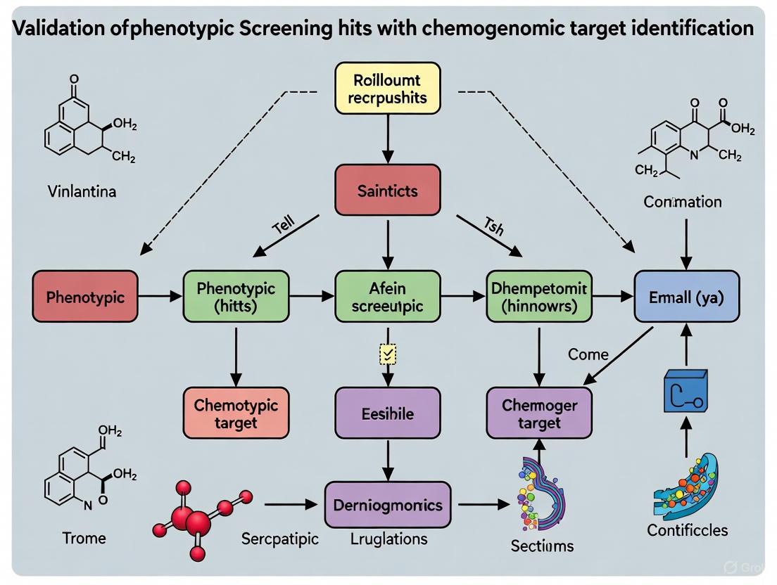
Validating Phenotypic Screening Hits: A Chemogenomic Framework for Target Identification and Deconvolution
This article provides a comprehensive guide for researchers and drug development professionals on integrating chemogenomics with phenotypic screening to validate hits and identify mechanisms of action.
Validating Phenotypic Screening Hits: A Chemogenomic Framework for Target Identification and Deconvolution
Abstract
This article provides a comprehensive guide for researchers and drug development professionals on integrating chemogenomics with phenotypic screening to validate hits and identify mechanisms of action. It covers the foundational principles of phenotypic drug discovery (PDD), detailing how it expands druggable target space and enables the discovery of first-in-class therapies. The content explores practical methodologies, including the use of annotated chemical libraries, affinity-based pull-down techniques, and label-free target identification strategies. It further addresses common troubleshooting and optimization challenges, such as mitigating the limitations of genetic and small-molecule screens and leveraging AI for data integration. Finally, the article presents robust validation frameworks and comparative analyses of computational and experimental tools, offering a complete roadmap for translating phenotypic observations into validated, druggable targets.
The Resurgence of Phenotypic Screening and the Chemogenomics Advantage
Why Phenotypic Screening is a Powerhouse for First-in-Class Drugs
In the evolving landscape of pharmaceutical research, phenotypic drug discovery (PDD) has re-emerged as a profoundly effective strategy for identifying first-in-class therapeutics. Between 1999 and 2008, phenotypic screening was responsible for the discovery of over half of the first-in-class small-molecule drugs approved by the FDA [1]. This approach, which identifies bioactive compounds based on their observable effects on disease phenotypes without requiring prior knowledge of a specific molecular target, contrasts with target-based drug discovery (TDD) that focuses on modulating predefined molecular targets [2]. The renewed appreciation for PDD stems from its ability to capture complex biological interactions within realistic disease models, thereby uncovering novel mechanisms of action (MoA) that would likely remain undiscovered through hypothesis-driven target-based approaches [3] [1]. This guide objectively examines the performance of phenotypic screening against target-based approaches, supported by experimental data and methodological frameworks essential for modern drug development.
Phenotypic vs. Target-Based Screening: A Comparative Analysis
The distinction between phenotypic and target-based screening strategies represents a fundamental dichotomy in drug discovery philosophy. Phenotypic screening evaluates compounds based on their ability to elicit a desired therapeutic effect in complex biological systems, including cells, tissues, or whole organisms [2]. This target-agnostic approach embraces biological complexity and has consistently identified novel therapeutic mechanisms. In contrast, target-based screening employs reductionist principles, focusing on compounds that selectively interact with a predefined molecular target, typically a protein with established disease relevance [3] [2].
Table 1: Strategic Comparison Between Phenotypic and Target-Based Screening Approaches
| Parameter | Phenotypic Screening | Target-Based Screening |
|---|---|---|
| Discovery Bias | Unbiased, allows novel target identification [2] | Hypothesis-driven, limited to known pathways [2] |
| Mechanism of Action | Often unknown at discovery, requires deconvolution [2] | Defined from the outset [2] |
| Biological Context | Captures complex systems-level interactions [3] [2] | Reductionist, focused on single targets [2] |
| Success Profile | Higher rate of first-in-class drug discovery [1] | More effective for follower drugs with optimized properties [4] |
| Technical Requirements | High-content imaging, functional genomics, AI analysis [2] | Structural biology, computational modeling, enzyme assays [2] |
| Target Validation | Required after compound identification [2] | Completed before screening begins [2] |
The disproportionate success of phenotypic screening in generating first-in-class therapeutics is particularly evident in complex disease areas with polygenic origins, such as cancer, neurodegenerative disorders, and rare diseases [2] [1]. Phenotypic approaches have expanded the "druggable target space" to include unexpected cellular processes—including pre-mRNA splicing, target protein folding, trafficking, and degradation—and revealed entirely new classes of drug targets [1].
Experimental Evidence: Key Success Stories and Data
The efficacy of phenotypic screening is demonstrated through multiple first-in-class therapies discovered through this approach. Notable examples include ivacaftor and lumicaftor for cystic fibrosis, risdiplam and branaplam for spinal muscular atrophy (SMA), and the immunomodulatory drugs thalidomide, lenalidomide, and pomalidomide [3] [1].
Table 2: Clinically Successful Drugs Discovered Through Phenotypic Screening
| Drug | Disease Indication | Key Experimental Model | Mechanism of Action |
|---|---|---|---|
| Ivacaftor/Lumicaftor [1] | Cystic Fibrosis | Cell lines expressing disease-associated CFTR variants [1] | CFTR channel potentiators and correctors [1] |
| Risdiplam/Branaplam [1] | Spinal Muscular Atrophy | SMN2 splicing modulation assays [1] | SMN2 pre-mRNA splicing modification [1] |
| Lenalidomide/Pomalidomide [3] [1] | Multiple Myeloma | TNF-α production inhibition assays [3] | Cereblon-mediated degradation of transcription factors IKZF1/3 [3] |
| Daclatasvir [1] | Hepatitis C | HCV replicon phenotypic screen [1] | Modulation of HCV NS5A protein [1] |
| SEP-363856 [1] | Schizophrenia | Phenotypic screen in disease models | Novel mechanism targeting trace amine-associated receptor 1 [1] |
For glioblastoma multiforme (GBM), researchers developed a sophisticated phenotypic screening approach that combined tumor genomic profiling with molecular docking to create rationally enriched chemical libraries [5]. This methodology involved:
- Target Selection: Identification of 755 genes with somatic mutations overexpressed in GBM patient samples from The Cancer Genome Atlas [5]
- Network Analysis: Mapping these genes onto a protein-protein interaction network to construct a GBM-specific subnetwork [5]
- Virtual Screening: Docking approximately 9,000 in-house compounds to 316 druggable binding sites on proteins in the GBM subnetwork [5]
- Phenotypic Validation: Screening selected compounds against patient-derived GBM spheroids, leading to the identification of compound IPR-2025 [5]
This compound demonstrated potent anti-GBM activity with single-digit micromolar IC50 values, significantly outperforming standard-of-care temozolomide, while showing no toxicity to normal cell lines [5]. The success of this integrated approach highlights how modern PDD can overcome traditional limitations through strategic combination with target-informed library design.
Diagram 1: Phenotypic Screening Workflow. This flowchart outlines the key steps in phenotypic drug discovery, from initial screening to target identification.
Methodological Framework: Validating Phenotypic Hits Through Chemogenomics
A critical challenge in phenotypic screening remains target deconvolution—identifying the molecular mechanism responsible for the observed therapeutic effect [2]. Modern chemogenomic approaches have revolutionized this process through computational and experimental methods that systematically link compound structures to biological targets.
Computational Target Prediction Methods
Recent advances in bioinformatics have produced sophisticated in silico target prediction platforms that accelerate mechanism of action elucidation. A comprehensive 2025 benchmark study systematically evaluated seven target prediction methods using an FDA-approved drug dataset [6]:
Table 3: Comparison of Computational Target Prediction Methods
| Method | Type | Algorithm | Database | Performance Notes |
|---|---|---|---|---|
| MolTarPred [6] | Ligand-centric | 2D similarity | ChEMBL 20 | Most effective in benchmark study [6] |
| RF-QSAR [6] | Target-centric | Random forest | ChEMBL 20&21 | Web server implementation [6] |
| TargetNet [6] | Target-centric | Naïve Bayes | BindingDB | Multiple fingerprint types [6] |
| ChEMBL [6] | Target-centric | Random forest | ChEMBL 24 | Morgan fingerprints [6] |
| CMTNN [6] | Target-centric | Neural network | ChEMBL 34 | Stand-alone code [6] |
| PPB2 [6] | Ligand-centric | Nearest neighbor/Neural network | ChEMBL 22 | Multiple algorithms [6] |
| SuperPred [6] | Ligand-centric | 2D/fragment/3D similarity | ChEMBL & BindingDB | ECFP4 fingerprints [6] |
These computational methods employ either target-centric approaches (building predictive models for specific targets) or ligand-centric strategies (identifying similar compounds with known targets) [6]. The benchmark analysis revealed that MolTarPred demonstrated particular effectiveness, especially when using Morgan fingerprints with Tanimoto scoring metrics [6].
Experimental Target Identification Protocols
Complementing computational approaches, experimental methods for target identification have seen significant advances:
Cellular Thermal Shift Assay (CETSA): This label-free method detects changes in protein thermal stability upon compound binding in live cells [4]. The technique measures the melting curve of proteins in compound-treated versus control cells, identifying stabilized targets that shift their denaturation profiles.
Thermal Proteome Profiling (TPP): A proteome-wide extension of CETSA, TPP uses multiplexed quantitative mass spectrometry to monitor thermal stability shifts across thousands of proteins simultaneously [5] [4]. This approach was successfully applied to identify multiple targets engaged by the anti-glioblastoma compound IPR-2025 [5].
Transcriptomics Analysis: RNA sequencing of compound-treated versus untreated cells can reveal pathway-level effects that inform mechanism of action [5]. This approach provides complementary data to direct binding assays by capturing downstream consequences of target engagement.
Diagram 2: Target Deconvolution Strategies. This diagram illustrates the integrated computational and experimental approaches for identifying the molecular targets of phenotypic screening hits.
Essential Research Tools for Phenotypic Screening
Successful implementation of phenotypic screening campaigns requires specialized research tools and reagents designed to capture relevant biology while enabling high-throughput operation.
Table 4: Essential Research Reagents and Platforms for Phenotypic Screening
| Research Tool | Function | Application Notes |
|---|---|---|
| High-Content Imaging Systems [2] | Automated microscopy and image analysis for multiparametric phenotypic assessment | Enables quantification of complex morphological changes in cells [2] |
| 3D Spheroid/Organoid Cultures [2] [5] | Physiologically relevant disease models that better mimic tissue architecture | Patient-derived GBM spheroids used in glioblastoma screening [5] |
| iPSC-Derived Cell Models [2] | Patient-specific cell types for disease modeling and compound screening | Particularly valuable for neurological disorders [2] |
| Transcreener HTS Assays [7] | Biochemical assays for enzyme activity detection (kinases, ATPases, etc.) | Flexible platform for multiple target classes using FP, FI, or TR-FRET detection [7] |
| Chemical Libraries with Diverse Annotation [2] [5] | Collections of compounds for screening; non-annotated libraries preferred for novel target discovery | Rationally designed libraries tailored to disease genomics enhance hit rates [5] |
| Zebrafish Embryo Models [8] | Whole-organism screening with high genetic similarity to humans | Used for neuroactive drug screening and toxicology studies [2] |
Advanced research platforms like Recursion OS and Insilico Medicine's Pharma.AI exemplify the integration of these tools with artificial intelligence for enhanced phenotypic discovery. The Recursion OS platform leverages approximately 65 petabytes of proprietary data and includes models like Phenom-2 (a 1.9 billion-parameter vision transformer) and MolPhenix for predicting molecule-phenotype relationships [9]. Similarly, Insilico's PandaOmics module leverages 1.9 trillion data points from over 10 million biological samples and 40 million documents for target identification and prioritization [9].
Phenotypic screening remains a powerhouse for first-in-class drug discovery because it embraces biological complexity, reveals unexpected mechanisms of action, and identifies novel therapeutic targets that would elude hypothesis-driven approaches. The historical success of this approach—from early observations of penicillin's effects to modern high-throughput campaigns—underscores its enduring value in the pharmaceutical development landscape [2] [1].
The future of phenotypic discovery lies in strategic integration with complementary technologies: advanced disease models (3D organoids, patient-derived cells), sophisticated target deconvolution methods (computational prediction, thermal proteome profiling), and artificial intelligence platforms that can extract meaningful patterns from high-dimensional phenotypic data [2] [9] [1]. By combining the unbiased nature of phenotypic screening with modern tools for mechanistic elucidation, drug discovery researchers can systematically address the challenges of complex diseases and deliver the transformative medicines that patients urgently need.
Chemogenomics represents a pivotal paradigm in modern drug discovery, systematically exploring the interaction between small molecules and biological targets. This approach establishes comprehensive ligand-target structure-activity relationship matrices to accelerate the identification and validation of therapeutic targets. Within the context of phenotypic screening, chemogenomics provides a powerful framework for deconvoluting the mechanisms of action of bioactive compounds. This guide examines the core principles, methodologies, and practical applications of chemogenomics, with a focused analysis of experimental platforms and reagent solutions that enable researchers to bridge chemical and biological spaces effectively.
Chemogenomics aims at the systematic identification of small molecules that interact with the products of the genome and modulate their biological function [10]. This field operates on the fundamental premise of establishing and expanding a comprehensive ligand-target Structure-Activity Relationship (SAR) matrix, representing a key scientific challenge for the 21st century following the elucidation of the human genome [10]. The chemogenomic approach utilizes small molecules as tools to establish relationships between targets and phenotypic outcomes, operating through two primary directional strategies: reverse chemogenomics (investigating biological activity starting from enzyme inhibitors) and forward chemogenomics (identifying relevant targets of pharmacologically active small molecules) [11].
The expansion of the physically available and bioactive chemical space represents a central objective of chemogenomics [10]. Effective systematic expansion appears possible when conserved molecular recognition principles serve as the founding hypothesis for compound design. These principles include approaches focusing on target families, privileged scaffolds, protein secondary structure mimetics, co-factor mimetics, and diversity-oriented synthesis (DOS) and biology-oriented synthesis (BIOS) libraries [10]. This systematic framework enables researchers to navigate the complex landscape of chemical-biological interactions with greater precision and efficiency.
Chemogenomics in Phenotypic Screening Hit Validation
Phenotypic drug discovery represents a powerful approach for identifying compounds that produce desired therapeutic effects without pre-supposing specific molecular targets, particularly valuable for infectious diseases where few well-validated targets exist [12]. A significant advantage of phenotypic screening is that active compounds modulate mechanisms or pathways essential for pathogen survival while possessing necessary properties for cellular permeation, metabolic stability, and target access without significant efflux [12]. However, a major limitation remains the lack of knowledge regarding the molecular target and binding mode of hits, which could enable structure-guided optimization approaches.
Target identification for phenotypic screening hits presents substantial challenges, as experimental determination can be complex, time-consuming, expensive, and not always successful [12]. Computational target prediction platforms have emerged as valuable tools to generate testable hypotheses, utilizing both ligand and protein-structure information to produce ranked sets of predicted molecular targets [12]. These platforms address the critical need for efficient mechanism deconvolution in phenotypic discovery programs.
Table 1: Challenges in Phenotypic Screening and Chemogenomic Solutions
| Challenge | Impact on Drug Discovery | Chemogenomic Approach |
|---|---|---|
| Unknown molecular target | Difficult to optimize compounds rationally | Computational target prediction and chemogenomic library screening |
| Unknown binding mode | Limited structure-guided optimization | 3D binding pose prediction and binding site analysis |
| Potential scaffold liabilities | Late-stage failure due to poor pharmacokinetics/toxicology | Early liability screening and scaffold hopping |
| Target-related unattractiveness | Wasted resources on less therapeutic targets | Early target identification for prioritization |
| Multi-target interactions | Unpredictable efficacy or toxicity | Selective compound profiling and polypharmacology assessment |
The premise of computational target identification rests on molecular recognition principles: structurally similar compounds interacting through similar pharmacophores will be recognized by similar protein binding sites [12]. If a phenotypic hit molecule shares similarity with a compound bound to a specific protein site in structural databases, this information can identify proteins with similar binding sites in the pathogen proteome, enabling target hypothesis generation [12].
Experimental Platforms and Methodologies
Computational Target Prediction Workflow
An advanced target prediction platform for phenotypic actives against Mycobacterium tuberculosis exemplifies the integrated computational approach [12]. The methodology employs a fragment-based strategy to address limited chemical space coverage in structural databases, drawing analogy to fragment-based drug discovery principles that increase efficiency in chemical space sampling [12].
Preparative Steps:
- PDB Fragment Space Creation: All small molecule ligands in the Protein Data Bank (PDB) are fragmented to generate molecular fragments capturing diverse pharmacophoric patterns. For each fragment, the binding cavity is defined and fragment-protein interactions analyzed [12].
- Target Space Generation: A comprehensive M. tuberculosis target space is assembled, including existing PDB structures (2,055 structures) and high-quality modeled protein structures (3,667 structures) generated using Rosetta homology modeling [12].
Platform Workflow:
- Fragmentation of Phenotypic Active: The active hit compound is fragmented in silico to generate molecular fragments [12].
- Fragment Similarity Search: Fragments from the phenotypic hit are compared against the PDB fragment database to identify identical or similar fragments with known binding environments [12].
- Cavity Comparison: Identified PDB fragments define cavities that are compared against the M. tuberculosis target space to find similar binding sites [12].
- Docking and Binding Mode Analysis: The complete phenotypic hit is docked into putative targets, and binding modes are analyzed for consistency with observed structure-activity relationships [12].
The following diagram illustrates the core logical workflow of this computational approach:
Chemogenomic Set Assembly and Validation
An alternative empirical approach utilizes curated chemogenomic compound sets - libraries of highly annotated biologically active compounds screened for phenotypic outcomes in disease-relevant models [13]. While chemical probes represent the highest quality tools for such purposes, molecules in a chemogenomic set may exhibit less stringent individual potency and selectivity properties but are assembled to provide broader selectivity profiles with non-overlapping off-target activity that enables mechanistic deconvolution [13].
The compilation of an NR1 nuclear receptor family chemogenomic set demonstrates rigorous assembly criteria [13]:
Selection Criteria:
- Compound-Bioactivity Annotations: Sourced from public repositories (PubChem, ChEMBL, IUPHAR/BPS, BindingDB, Probes&Drugs) compiled in curated datasets [13]
- Potency Requirements: Cellular potency ≤10 µM (preferably ≤1 µM) based on community-agreed criteria [13]
- Selectivity Standards: Up to five off-targets at final concentration [13]
- Chemical Diversity: Analyzed using Tanimoto similarity of Morgan fingerprints and Murcko molecular frameworks [13]
- Mode of Action Diversity: Inclusion of agonists, antagonists, and inverse agonists [13]
- Commercial Availability: Ensuring broad accessibility to the research community [13]
Validation Workflow:
- Identity and Purity Verification: NMR, LC-UV, LC-ELSD, and LC-MS confirmation (≥95% purity) [13]
- Viability Assessment: Primary cell viability assay in multiple cell lines (HEK293T, U-2 OS, MRC-9 fibroblasts) using confluence measurement to determine growth rate [13]
- Multiplex Toxicity Profiling: High-content microscopy-based multiplex assay evaluating apoptosis, cytoskeleton alterations, membrane permeabilization, and mitochondrial mass using orthogonal stains [13]
- Off-Target Liability Screening: Differential scanning fluorimetry (DSF) screening against representative kinases and bromodomains (BRD4, TRIM24, BRPF1, AURKA, CDK2, MAPK1, GSK3B, CSNK1D, ABL1, FGFR3) [13]
- In-Family Selectivity Profiling: Uniform hybrid reporter gene assays on main targets and all NRs in respective subfamilies [13]
Table 2: Experimental Validation Methods for Chemogenomic Sets
| Validation Method | Key Parameters Measured | Exclusion Criteria |
|---|---|---|
| Cell Viability Assay | Growth rate (GR), confluency over time | GR ≤ 0.5, atypical cellular phenotypes |
| Multiplex Toxicity Assay | Apoptosis, cytoskeleton, membrane integrity, mitochondrial mass | Phenotypic effects, precipitation, non-specific toxicity |
| Differential Scanning Fluorimetry | Protein melting temperature (ΔTm) | ΔTm > 1.8°C (≥ 2 × SD) on liability targets |
| Reporter Gene Assays | In-family selectivity, potency confirmation | Lack of intended activity, poor potency |
| Compound Solubility | Kinetic solubility in assay conditions | Insufficient solubility for testing |
Comparative Analysis of Research Platforms
Cheminformatics Platforms for Chemogenomics
The implementation of chemogenomic approaches requires robust cheminformatics platforms capable of handling diverse chemical data and supporting target prediction workflows. The following table compares key platforms used in chemogenomic research:
Table 3: Cheminformatics Platform Comparison for Chemogenomics Applications
| Platform | License Model | Key Strengths | Target Prediction Capabilities | Integration Options |
|---|---|---|---|---|
| RDKit | Open-source (BSD) | Comprehensive functionality, high performance, active community | Ligand-based similarity searching, molecular descriptor calculation, fingerprint generation | Python, KNIME, PostgreSQL cartridge, Java, C++ |
| ChemAxon Suite | Commercial | Enterprise-level chemical data management, user-friendly interfaces | Chemical database management, substructure and similarity search | Java-based APIs, Pipeline Pilot, KNIME |
| CDK (Chemistry Development Kit) | Open-source | Cross-platform compatibility, extensive descriptor calculation | Molecular descriptor calculation, fingerprint generation, SAR analysis | Java-based applications, various programming languages |
| Open Babel | Open-source | Format conversion, structure manipulation | Chemical file format conversion, basic molecular manipulation | Command-line utilities, programming interfaces |
RDKit deserves particular emphasis as it has become a de facto standard in the field due to its comprehensive functionality, high performance, and active community [14]. While RDKit itself is a library rather than a standalone application, it provides robust capabilities for molecular descriptor calculation, fingerprint generation for similarity searching, and substructure search - all critical for chemogenomic applications [14]. RDKit supports multiple fingerprint types (Morgan fingerprints similar to ECFP, RDKit Fingerprint, Topological Torsion, Atom Pair, and MACCS keys) and similarity metrics (Tanimoto, Dice, Cosine, etc.) essential for ligand-based virtual screening [14]. Its integration with the PostgreSQL database system via the RDKit cartridge enables efficient chemical database management and searching at scale [15].
Specialized Chemogenomic Sets
The development of specialized chemogenomic sets for specific protein families represents another strategic approach. The following table compares two recently developed chemogenomic sets for nuclear receptor families:
Table 4: Comparative Analysis of Nuclear Receptor Chemogenomic Sets
| Parameter | NR1 Family Set [13] | NR4A Family Set [16] |
|---|---|---|
| Family Coverage | 19 NRs across 7 subfamilies | 3 receptors (Nur77/NR4A1, Nurr1/NR4A2, NOR1/NR4A3) |
| Compound Count | 69 comprehensively annotated modulators | 8 validated direct modulators |
| Activity Types | Agonists, antagonists, inverse agonists | Agonists and inverse agonists |
| Selection Criteria | Potency (≤10 µM), selectivity (≤5 off-targets), commercial availability | Direct binding validation, orthogonal cellular activity, commercial availability |
| Validation Methods | Viability assays, multiplex toxicity, DSF liability screening, reporter gene assays | ITC, DSF, reporter gene assays, solubility, multiplex toxicity |
| Proven Applications | Autophagy, neuroinflammation, cancer cell death | Endoplasmic reticulum stress, adipocyte differentiation |
The NR1 family chemogenomic set demonstrates the comprehensive approach to set assembly, with 69 compounds rigorously selected and validated to cover all 19 members of the NR1 family [13]. This set was optimized for complementary activity/selectivity profiles and chemical diversity to ensure orthogonality in phenotypic screening applications [13]. Proof-of-concept applications revealed roles of NR1 members in autophagy, neuroinflammation, and cancer cell death, confirming the set's suitability for target identification and validation [13].
In contrast, the NR4A family set represents a more focused approach, with 8 validated direct modulators addressing a smaller receptor subgroup [16]. The comparative profiling of NR4A modulators revealed a lack of on-target binding and modulation for several putative ligands, highlighting the critical importance of experimental validation in tool compound selection [16]. This smaller set nonetheless enabled the linking of orphan targets with phenotypic effects in endoplasmic reticulum stress and adipocyte differentiation [16].
The Scientist's Toolkit: Essential Research Reagents and Platforms
Successful implementation of chemogenomics approaches requires access to specialized reagents, platforms, and databases. The following table details key solutions for researchers in this field:
Table 5: Essential Research Reagent Solutions for Chemogenomics
| Resource Category | Specific Solutions | Function in Chemogenomics |
|---|---|---|
| Cheminformatics Platforms | RDKit, ChemAxon Suite, CDK, Open Babel | Chemical structure handling, descriptor calculation, similarity searching, database management [14] [15] |
| Chemical Databases | ChEMBL, PubChem, BindingDB, IUPHAR/BPS | Source of compound-bioactivity annotations for target identification [13] |
| Structural Databases | Protein Data Bank (PDB) | Source of protein-ligand complex structures for binding site analysis [12] |
| Target Prediction Tools | Fragment-based platforms, reverse docking approaches | Generation of target hypotheses for phenotypic hits [12] |
| Validation Assays | Reporter gene assays, DSF, ITC, multiplex toxicity assays | Experimental confirmation of compound-target interactions and selectivity [13] |
| Curated Chemogenomic Sets | NR1 family set, NR4A set, kinase chemogenomic sets | Annotated compound libraries for phenotypic screening and target deconvolution [16] [13] |
Chemogenomics provides a systematic framework for bridging chemical and biological spaces, enabling efficient target identification and validation in phenotypic drug discovery. The integration of computational prediction platforms with empirically validated chemogenomic sets offers complementary strategies for deconvoluting the mechanisms of action of bioactive compounds. Computational approaches like the fragment-based target prediction platform leverage structural information to generate testable target hypotheses, while carefully curated chemogenomic sets enable empirical target validation through selective modulation. As these methodologies continue to mature and integrate, they promise to accelerate the transformation of phenotypic screening hits into validated therapeutic targets, ultimately enhancing the efficiency of drug discovery pipelines across diverse therapeutic areas.
The concept of the druggable genome, first defined twenty years ago as the subset of the human genome encoding proteins capable of binding drug-like molecules, has fundamentally transformed target selection in pharmaceutical research [17]. Early estimates suggested approximately 4,500 genes constituted this space, but technological advances have continuously expanded this frontier [18] [17]. Today, researchers are moving beyond simple ligandability assessments to multi-parameter evaluations that encompass disease modification, tissue expression, functional sites, and safety profiles [17]. This evolution reflects a critical transition from asking "can this protein bind a drug?" to the more complex question: "can this target yield a successful drug?" [17].
Within this expanded framework, phenotypic screening has emerged as a powerful strategy for identifying novel biological insights and first-in-class therapies without requiring prior knowledge of specific molecular pathways [19]. However, a significant challenge persists in bridging the gap between phenotypic hits and target identification. This guide examines how integrative approaches, particularly Mendelian randomization (MR) and chemogenomic libraries, are validating phenotypic screening hits and expanding the druggable genome, with direct comparisons of their performance against conventional methods.
Case Study 1: Mendelian Randomization in Oncology Target Discovery
A 2025 study systematically applied druggable genome-wide Mendelian randomization to identify novel therapeutic targets for lung squamous cell carcinoma (LUSC), a non-small cell lung cancer subtype with poor prognosis and limited treatment options [18]. The research employed a multi-tiered validation approach using expression quantitative trait loci (eQTL) and protein QTL (pQTL) data from two independent datasets (ieub4953 and finngen) [18].
Table 1: LUSC-Related Genes Identified via Mendelian Randomization
| Gene Symbol | Identification Method | Effect on LUSC Risk | Associated Risk Factors |
|---|---|---|---|
| DNMT1 | cis-eQTL | Protective | Smoking (p=0.035) |
| ACSS2 | cis-eQTL | Risk factor | Smoking, Pulmonary fibrosis |
| YBX1 | cis-eQTL | Risk factor | Smoking, Phthisis, Alcohol |
| SELENOS | cis-eQTL | Risk factor | Pulmonary fibrosis |
| PPARA | cis-eQTL | Protective | Smoking, Pulmonary fibrosis |
| MST1 | cis-pQTL | Protective | Alcohol abuse |
| CPA4 | cis-pQTL | Protective | Phthisis (p=0.031) |
| MPO | cis-pQTL | Risk factor | Not specified |
Experimental Protocol and Validation
The methodology followed a rigorous multi-step process to ensure causal inference:
- Druggable Gene Selection: Researchers compiled 5,859 unique druggable genes from DGIdb v4.2.0 and Finan et al. databases [18].
- Instrumental Variable Selection: Genetic variants significantly associated with gene expression (±1 Mb window) were extracted as instrumental variables, with a genome-wide significance threshold of P < 5 × 10⁻⁸ and minimum F-statistic of 10 to ensure strength [18].
- Mendelian Randomization Analysis: Causal effects were estimated using Wald ratio (single IV) or inverse-variance weighted (IVW) method (multiple IVs) [18].
- Sensitivity Analyses: Bayesian co-localization, summary-data-based MR (SMR) analysis, and HEIDI tests were conducted to verify pleiotropic associations between gene expression and LUSC risk [18].
- Clinical Correlation: Researchers assessed prognosis, immune infiltration, and single-cell expression patterns for validated targets [18].
This approach successfully identified eight LUSC-related genes with causal associations, demonstrating how MR can prioritize targets for further investigation. The DNMT1, ACSS2, YBX1, SELENOS, and PPARA genes were identified through blood cis-eQTL analysis, while MST1, CPA4, and MPO emerged from cis-pQTL analysis [18].
Performance Assessment
The MR approach demonstrated several advantages for target identification. By using genetic variants as instrumental variables, the method avoids confounding factors and reverse causality inherent in observational studies, providing stronger evidence for causal target-disease relationships [18]. The methodology also enabled systematic interrogation of thousands of druggable genes simultaneously, significantly expanding the potential target space beyond conventionally investigated candidates.
However, the study also revealed limitations. Bayesian co-localization analysis showed negative results (PPH3 + PPH4 < 0.8) for all identified genes, suggesting insufficient evidence for shared causal variants between gene expression and LUSC risk [18]. This highlights a key consideration for MR-based approaches—while they can identify statistically significant associations, complementary methods may be needed to fully establish biological mechanisms.
Case Study 2: Integrating Single-Cell MR in Ophthalmology
Study Design and Findings
A 2025 investigation into primary open-angle glaucoma (POAG) exemplified how integrating single-cell technologies with MR can reveal cell-type-specific therapeutic targets and repurposable drugs [20]. This research employed druggable genome-wide and single-cell MR using POAG genome-wide association study data, blood, and single-cell eQTL datasets [20].
Table 2: POAG Therapeutic Targets Identified via Integrated MR Approach
| Gene Symbol | Cell Type Specificity | Effect on POAG Risk | Odds Ratio (95% CI) | Potential Repurposed Drugs |
|---|---|---|---|---|
| YWHAG | Not specified | Risk factor | 1.207 (1.131-1.288) | Not identified |
| GFPT1 | CD4+KLRB1- T cells | Protective (paradoxical risk in specific T cells) | 0.874 (0.840-0.910) | Trimipramine, Desipramine, Cyclosporin |
Experimental Workflow
The study implemented a comprehensive roadmap for target identification and validation:
- Druggable Genome Annotation: 4,463 druggable genes were sourced from Finan et al. and intersected with 19,127 blood eQTL genes [20].
- Single-Cell cis-eQTL Analysis: Immune cell-specific eQTLs were derived from the OneK1K database, comprising scRNA-seq data from 1.27 million peripheral blood mononuclear cells across 982 donors [20].
- Causal Inference: MR analysis was performed with Steiger filtering to ensure correct causal direction [20].
- Drug Repurposing Prediction: Molecular docking using DSigDB/CB-Dock2 confirmed strong binding of existing drugs to identified targets (Vina score < -5) [20].
- Safety Assessment: Phenome-wide association studies (PheWAS) were conducted to assess potential off-target effects [20].
Performance Assessment
The integration of single-cell resolution provided a critical advancement over bulk tissue analyses. Researchers discovered a cell-type-specific paradoxical effect where high GFPT1 expression in CD4+KLRB1-T cells increased POAG risk (OR = 1.448), contrary to its protective role at the bulk tissue level [20]. This finding highlights how cellular context dramatically influences target validation and underscores the limitation of conventional approaches that overlook microenvironment heterogeneity.
The molecular docking component successfully identified three FDA-approved drugs with strong binding affinity to GFPT1, while PheWAS analysis indicated no significant off-target effects, accelerating the path to clinical translation [20]. This end-to-end pipeline—from genetic discovery to repurposing candidates—demonstrates how modern MR approaches can de-risk early drug development.
Comparative Analysis: MR Versus Conventional Phenotypic Screening
Methodological Comparison
Traditional phenotypic screening has contributed significantly to drug discovery, enabling identification of novel therapeutic mechanisms without molecular target preconceptions [19]. However, both small molecule and genetic screening approaches face inherent limitations in subsequent target identification and validation.
Table 3: Performance Comparison of Target Identification Methods
| Parameter | Mendelian Randomization | Small Molecule Screening | Genetic Screening |
|---|---|---|---|
| Target Identification Capability | Direct causal inference | Indirect, requires deconvolution | Direct for genetic targets |
| Throughput | High (genome-wide) | Moderate to high | High with CRISPR |
| Clinical Translation Success | Higher (genetically validated targets) | Variable | Lower (genetic-pharmacologic disconnect) |
| Cell Type Resolution | Achievable with sc-eQTL integration | Limited without specialized assays | Achievable with scRNA-seq |
| Limitations | Limited by GWAS sample size and diversity | Limited to ~1,000-2,000 of 20,000+ genes [19] | Fundamental differences between genetic and small molecule effects [19] |
Limitations and Mitigation Strategies
Conventional phenotypic screening faces several constraints. Small molecule libraries interrogate only a small fraction (approximately 1,000-2,000 targets) of the human genome's 20,000+ genes, creating significant coverage gaps in the druggable genome [19]. Furthermore, chemical tool compounds used for target validation often suffer from poor selectivity, creating uncertainty in associating phenotypes with specific molecular targets [19].
Genetic screening approaches, while enabling systematic perturbation of gene function, face a different set of challenges. There are fundamental differences between genetic and small molecule perturbations, including temporal resolution (permanent gene knockout versus transient pharmacological inhibition), compensation mechanisms, and the inability of genetic approaches to mimic allosteric modulation or protein degradation [19].
Mendelian randomization addresses several of these limitations by leveraging natural genetic variation as a surrogate for lifelong drug target modulation, providing human physiological context that is absent from in vitro models [18] [20]. The methodology also benefits from very large sample sizes available through biobanks, enabling robust statistical power that exceeds many conventional screening approaches.
The Scientist's Toolkit: Essential Research Reagents and Workflows
Key Research Reagent Solutions
Successful expansion of the druggable genome requires specialized reagents and datasets:
Table 4: Essential Research Reagents and Resources for Druggable Genome Studies
| Resource Type | Specific Examples | Function and Application |
|---|---|---|
| Druggable Genome Databases | Finan et al. (4,463 genes), DGIdb v4.2.0 | Define the initial target universe for screening [18] [20] |
| QTL Datasets | eQTLGen Consortium (blood cis-eQTL), OneK1K (sc-eQTL), pQTL datasets | Provide genetic instruments for MR studies [18] [20] |
| GWAS Resources | FinnGen, UK Biobank, ieuge | Supply outcome data for causal inference [18] [20] |
| Analytical Tools | TwoSampleMR R package, SMR software, COLOC for Bayesian colocalization | Enable statistical analysis and causal inference [20] |
| Validation Resources | PDBe-KB (protein structures), ChEMBL (bioactive molecules), canSAR | Facilitate structural and chemical validation of targets [17] |
Integrated Workflow Visualization
The following diagram illustrates the comprehensive workflow for expanding the druggable genome through integrated genetic and functional approaches:
Integrated Workflow for Expanding Druggable Genome
The integration of Mendelian randomization with phenotypic screening frameworks represents a powerful strategy for expanding the druggable genome and validating novel therapeutic targets. The case studies in LUSC and POAG demonstrate how genetically validated targets provide de-risked starting points for drug development, with higher likelihood of clinical translation success [18] [20]. The addition of single-cell resolution addresses critical limitations of conventional phenotypic screening by revealing cell-type-specific effects and paradoxical signaling that would otherwise remain obscured [20].
Future expansion of the druggable genome will increasingly rely on knowledge graphs that integrate data from gene-level to protein residue-level, enabling artificial intelligence approaches to navigate the complexity of biological systems and identify high-quality targets [17]. As these technologies mature, the scientific community can anticipate continued growth in the number of therapeutic targets, particularly for diseases with high unmet need where conventional target identification approaches have proven insufficient.
The combination of human genetic evidence from MR with functional validation from phenotypic screening creates a virtuous cycle for drug discovery—where genetic findings inspire phenotypic assays, and phenotypic observations motivate genetic investigations—ultimately accelerating the development of novel therapies for complex diseases.
The decline in pharmaceutical research and development productivity has spurred a resurgence of interest in phenotypic drug discovery (PDD). Unlike target-based approaches, PDD identifies compounds based on their ability to modulate disease-relevant phenotypes without prior knowledge of specific molecular targets, making it particularly valuable for complex diseases and first-in-class medicine development [21]. However, a significant challenge emerges during hit validation: understanding the mechanism of action (MOA) of phenotypically active compounds in the context of widespread polypharmacology—the phenomenon where single compounds interact with multiple biological targets [22] [23].
This guide examines the integration of phenotypic screening with chemogenomic target identification technologies, comparing experimental approaches and computational frameworks that enable researchers to navigate the complex polypharmacology of hit compounds while accelerating the development of novel therapeutics.
The Polypharmacology Landscape in Drug Discovery
Polypharmacology represents a paradigm shift from the traditional "one drug–one target" model toward understanding drugs' complex interactions with multiple biological targets. Research indicates that most drug molecules interact with six known molecular targets on average, even after optimization [23]. This multi-target activity presents both challenges and opportunities:
Therapeutic Advantages: Polypharmacology can enhance therapeutic efficacy for complex, multifactorial diseases, particularly in central nervous system (CNS) disorders and oncology, where modulating multiple pathways simultaneously may yield superior clinical outcomes [24] [22].
Validation Challenges: Promiscuous binding complicates target deconvolution and MOA determination, potentially introducing off-target effects that contribute to adverse drug reactions [25] [26].
The polypharmacology index (PPindex) has been developed as a quantitative metric to compare target specificity across compound libraries, with steeper slopes (larger absolute values) indicating more target-specific libraries [23].
Table 1: Polypharmacology Index Comparison Across Selected Compound Libraries
| Library Name | PPindex (All Targets) | PPindex (Without 0/1 Target Bins) | Relative Specificity |
|---|---|---|---|
| DrugBank | 0.9594 | 0.4721 | Most specific |
| LSP-MoA | 0.9751 | 0.3154 | Intermediate |
| MIPE 4.0 | 0.7102 | 0.3847 | Intermediate |
| Microsource Spectrum | 0.4325 | 0.2586 | Most polypharmacologic |
Phenotypic Screening: A Target-Agnostic Approach
Phenotypic screening assesses compound effects in physiologically relevant systems without requiring predefined molecular targets, potentially increasing translational success rates [23] [21]. This approach is particularly valuable for:
CNS Drug Discovery: The intricate interplay of neurotransmitter systems makes target-agnostic approaches particularly suitable for neuropsychiatric disorders [24].
Complex Disease Pathologies: Diseases involving multiple genetic factors and compensatory pathways may be better addressed through phenotypic approaches [21].
First-in-Class Therapeutics: Phenotypic screening has demonstrated a superior track record in discovering first-in-class medicines compared to target-based approaches [21].
However, the primary challenge remains target deconvolution—identifying the molecular mechanisms responsible for observed phenotypic effects [26] [21]. This process becomes increasingly complex when considering the polypharmacology of hit compounds, where multiple simultaneous interactions may contribute to the overall phenotypic response.
Chemogenomic Approaches for Target Identification
Chemogenomics systematically studies the interactions between chemical compounds and biological targets, providing powerful tools for target deconvolution in phenotypic screening.
Knowledge-Based Chemogenomic Platforms
Comprehensive knowledgebases enable researchers to leverage existing compound-target interaction data for polypharmacology prediction:
Drug Abuse Knowledgebase (DA-KB): This specialized resource centralizes chemogenomics data related to drug abuse and CNS disorders, incorporating genes, proteins, chemical compounds, and bioassays to facilitate polypharmacology analysis [25].
Computational Analysis of Novel Drug Opportunities (CANDO): This platform employs fragment-based multitarget docking with dynamics to construct compound-proteome interaction matrices, which are then analyzed to determine similarity of drug behavior based on proteomic interaction signatures [22].
TargetHunter Platform: Provides computational algorithms for polypharmacological target identification and tool compounds for validation, particularly for GPCRs implicated in complex disorders [25].
Experimental Target Deconvolution Methods
Advanced experimental techniques enable direct identification of compound-target interactions:
Limited Proteolysis (LiP): A novel, label-free proteomics approach that detects structural changes in proteins upon compound binding, allowing for comprehensive identification of drug targets and off-targets without requiring chemical modification of the compound [26].
Compressed Phenotypic Screening: An innovative pooling approach where multiple perturbations are combined into unique pools, significantly reducing sample requirements and costs while maintaining the ability to deconvolve individual compound effects through computational regression analysis [27].
High-Content Imaging with Morphological Profiling: Using multiplexed fluorescent dyes (e.g., Cell Painting assay) to capture complex morphological features, enabling classification of compounds based on phenotypic fingerprints that can be linked to mechanisms of action [27].
Comparative Analysis of Experimental Approaches
Table 2: Comparison of Target Identification and Validation Methodologies
| Method | Key Features | Throughput | Information Gained | Key Limitations |
|---|---|---|---|---|
| Limited Proteolysis (LiP) | Label-free, detects protein structural changes | Medium | Direct binding information, proteome-wide coverage | Requires specialized expertise in proteomics |
| Compressed Phenotypic Screening | Pooled compounds, computational deconvolution | High | Cost-efficient morphological profiling | Limited by pool size and deconvolution accuracy |
| Computational CANDO Platform | In silico docking, proteome-wide interaction prediction | Very High | Putative interaction signatures for repurposing | Dependent on quality of structural and chemical data |
| High-Content Morphological Profiling | Multiplexed imaging, phenotypic fingerprinting | Medium | Functional classification based on phenotype | Indirect target inference requires validation |
Integrated Workflow for Hit Validation
Successful validation of phenotypic screening hits requires an integrated approach that combines complementary technologies:
Diagram 1: Hit Validation Workflow
Critical Considerations for Hit Triage and Validation
Effective hit validation requires addressing several key challenges:
Biological Knowledge Integration: Successful hit triage leverages three types of biological knowledge: known mechanisms, disease biology, and safety considerations, while structure-based triage alone may be counterproductive [28].
Polypharmacology Assessment: Early evaluation of compound promiscuity using tools like PPindex helps prioritize compounds with desirable multi-target profiles while minimizing off-target liabilities [23] [29].
Chain of Translatability: Establishing a clear connection between the phenotypic assay, disease relevance, and clinical translation is essential for prioritizing hits with genuine therapeutic potential [21].
The Scientist's Toolkit: Essential Research Reagents and Platforms
Table 3: Key Research Reagent Solutions for Phenotypic Screening and Target Identification
| Tool/Platform | Primary Function | Application in Validation |
|---|---|---|
| Cell Painting Assay | Multiplexed morphological profiling | Phenotypic classification and mechanism of action prediction [27] |
| Chemogenomic Libraries | Collections of target-annotated compounds | Target deconvolution in phenotypic screens [23] |
| DA-KB Knowledgebase | Domain-specific chemogenomics database | Polypharmacology analysis for CNS targets [25] |
| CANDO Platform | Computational proteome docking | Predicting drug-target interactions and repurposing opportunities [22] |
| LiP-MS Platform | Limited proteolysis mass spectrometry | Direct identification of drug-target interactions [26] |
Case Studies and Applications
CNS Drug Discovery: Ulotaront
A compelling example of successful phenotypic polypharmacology drug discovery comes from CNS research, where the SmartCube platform was used to identify ulotaront, a first-in-class antipsychotic currently in Phase III clinical trials [24]. This approach:
- Used in vivo phenotypic profiling without target preconceptions
- Identified a compound with a novel mechanism of action not involving dopamine receptor antagonism
- Demonstrated placebo-like tolerability despite complex polypharmacology
- Highlighted the power of behavioral phenotypic drug discovery for CNS applications
Oncology: Imatinib Polypharmacology
Imatinib, initially developed as a selective BCR-ABL inhibitor for chronic myeloid leukemia, exemplifies the importance of understanding polypharmacology:
- Originally discovered through high-throughput screening [22]
- Later found to inhibit multiple kinase targets (PDGF-R, c-Kit, c-fms) [22]
- Demonstrates that therapeutic efficacy may derive from multi-target effects [22]
- Drug resistance often emerges through mutations affecting binding, prompting development of next-generation inhibitors [22]
The integration of phenotypic screening with chemogenomic target identification represents a powerful strategy for addressing the challenges of polypharmacology in drug discovery. Key advancements driving this field include:
Improved Computational Prediction: Machine learning and network-based approaches are enhancing our ability to predict polypharmacological profiles and identify promising multi-target therapeutics [29].
Advanced Proteomics Technologies: Innovations like LiP-MS are providing more comprehensive and direct methods for target deconvolution [26].
High-Content Compression Methods: Pooled screening approaches are increasing the throughput and efficiency of phenotypic discovery campaigns [27].
Specialized Knowledgebases: Domain-specific resources like DA-KB are enabling more focused investigation of complex disease mechanisms [25].
As the field advances, the most successful drug discovery pipelines will likely embrace a holistic approach that acknowledges the inherent polypharmacology of most effective drugs while developing sophisticated tools to understand, predict, and optimize these complex interaction profiles for improved therapeutic outcomes.
A Practical Toolkit: From Phenotypic Hit to Target Hypothesis
Designing and Curating a Chemogenomic Library for Phenotypic Screening
The drug discovery paradigm has significantly shifted from a reductionist 'one target—one drug' vision to a more complex systems pharmacology perspective that acknowledges a single drug often interacts with several targets [30]. This evolution, driven by the need to address complex diseases like cancers and neurological disorders, has catalyzed the revival of phenotypic drug discovery (PDD) strategies. Phenotypic screening does not rely on a priori knowledge of specific drug targets, presenting a major challenge: deconvoluting the mechanism of action and identifying the therapeutic targets responsible for the observed phenotype [30]. Chemogenomic libraries represent a powerful solution to this challenge. A chemogenomic library is a collection of well-defined pharmacological agents where a hit in a phenotypic screen suggests that the annotated target or targets of the probe molecules are involved in the phenotypic perturbation [31]. Effectively, these libraries integrate small-molecule chemogenomics with genetic approaches, expediting the conversion of phenotypic screening projects into target-based drug discovery approaches [31].
The core value of a chemogenomic library lies in its annotation—the rich information linking compounds to their known protein targets, biological pathways, and even disease associations. This annotation transforms a simple collection of compounds into a sophisticated hypothesis-testing tool. Furthermore, the emergence of advanced cell-based phenotypic screening technologies, including induced pluripotent stem (iPS) cell technologies, gene-editing tools like CRISPR-Cas, and high-content imaging assays such as "Cell Painting," has increased the resolution and throughput of phenotypic readouts, making the need for well-curated libraries even more critical [30]. This guide will objectively compare the key strategies, experimental protocols, and performance data involved in designing and applying chemogenomic libraries for phenotypic screening.
Core Design Strategies and Comparative Analysis
Designing a chemogenomic library is a balancing act between comprehensive coverage of biological targets and practical considerations of library size, cost, and screening efficiency. Different strategies prioritize these factors differently, leading to distinct library designs. The following table summarizes the quantitative aspects of several design strategies as evidenced by recent research.
Table 1: Comparison of Chemogenomic Library Design Strategies and Performance
| Design Strategy | Reported Library Size | Target / Pathway Coverage | Key Design Criteria | Reported Applications / Outcomes |
|---|---|---|---|---|
| Systems Pharmacology Network Integration [30] | ~5,000 compounds | A large and diverse panel of drug targets involved in diverse biological effects and diseases. | Integration of drug-target-pathway-disease relationships & morphological profiles; scaffold diversity for broad coverage. | Target identification and mechanism deconvolution for phenotypic assays; integration with Cell Painting morphological profiles. |
| Precision Oncology-Focused Design [32] | A minimal library of 1,211 compounds (virtual); a physical library of 789 compounds (pilot). | 1,386 anticancer proteins; 1,320 targets covered by the physical library. | Library size, cellular activity, chemical diversity & availability, and target selectivity; adjusted for cancer. | Pilot screening on glioblastoma patient cells identified highly heterogeneous, patient-specific phenotypic vulnerabilities. |
| Machine Learning-Driven Feature Extraction [33] | 1,862 drugs (in underlying dataset). | 1,554 human target proteins (enzymes, GPCRs, ion channels, nuclear receptors). | Use of L1-regularized classifiers to identify informative chemogenomic features (chemical substructure-protein domain pairs). | Extraction of biologically meaningful substructure-domain associations; maintained drug-target interaction prediction performance. |
Beyond the general strategies, specific analytical procedures have been developed for particular therapeutic areas. For precision oncology, this involves designing compound collections adjusted for library size, cellular activity, chemical diversity and availability, and target selectivity to cover a wide range of protein targets and biological pathways implicated in various cancers [32]. The resulting libraries can be characterized by their compound and target spaces, providing a quantitative assessment of their coverage before any physical screening takes place.
Experimental Protocols for Library Construction and Validation
Protocol 1: Building a Systems Pharmacology Network for Library Curation
This protocol outlines the methodology for constructing a comprehensive data network to inform the selection of compounds for a chemogenomic library, as described in the development of a 5,000-compound library [30].
1. Data Acquisition and Integration:
- Bioactivity Data: Source standardized bioactivity data (e.g., IC50, Ki, EC50) and target annotations from public databases like ChEMBL.
- Pathway and Disease Context: Integrate pathway information from the Kyoto Encyclopedia of Genes and Genomes (KEGG) and disease associations from the Human Disease Ontology (DO) to provide biological and clinical context to the drug-target relationships.
- Morphological Profiling Data: Incorporate high-content imaging data from public benchmarks like the Broad Bioimage Benchmark Collection (BBBC022 - Cell Painting assay). This links chemical structures to a rich layer of phenotypic information.
2. Data Processing and Network Construction:
- Molecule Standardization: Process chemical structures to ensure consistency. Software like
ScaffoldHuntercan be used to decompose molecules into hierarchical scaffolds and fragments, enabling analysis of chemical diversity and privilege structures [30]. - Graph Database Implementation: Integrate the heterogeneous data sources (molecules, proteins, pathways, diseases, morphological features) into a high-performance NoSQL graph database, such as Neo4j. In this model, nodes represent entities (e.g., a molecule, a target protein), and edges represent the relationships between them (e.g., "Molecule A inhibits Target B") [30].
3. Library Curation and Filtering:
- Compound Selection: Filter the universe of available compounds based on the richness of their bioactivity data and the quality of their target annotations.
- Scaffold-Based Diversity: Apply scaffold-based filtering to ensure the final library encompasses a diverse chemical space that represents a broad swath of the druggable genome, avoiding over-representation of similar chemotypes [30].
Network-Based Library Construction Workflow
Protocol 2: A Machine Learning Approach for Identifying Chemogenomic Features
This protocol details a classifier-based method for extracting the fundamental associations between drug chemical substructures and protein domains that govern drug-target interactions [33]. This approach can inform library design by highlighting the most informative features.
1. Data Preparation:
- Drug-Target Interactions: Obtain a gold-standard set of known drug-target interactions from a database like DrugBank. This serves as positive examples for model training.
- Compound Representation: Encode the chemical structures of all drugs into a binary fingerprint vector (e.g., 881-dimensional using PubChem substructures), where each element indicates the presence or absence of a specific chemical substructure.
- Protein Representation: Encode the target proteins into a binary vector representing the presence or absence of protein domains from a database like PFAM.
2. Feature Vector Construction for Drug-Target Pairs:
- Represent each drug-target pair by the tensor product (also known as the Kronecker product) of the drug fingerprint vector and the protein domain vector.
- This operation generates a very high-dimensional feature vector where each feature corresponds to a specific pair of a chemical substructure and a protein domain [33].
3. Model Training and Feature Extraction:
- Apply L1-Regularized Classifiers: Train a binary classifier, such as L1-regularized logistic regression (L1LOG) or L1-regularized support vector machine (L1SVM), to predict drug-target interactions from the tensor product feature vectors.
- Extract Informative Features: The L1-regularization has the property of driving the weights of many features to zero. The non-zero weights in the resulting model correspond to the specific substructure-domain pairs that are most informative and predictive of the interaction [33]. These features form a biologically meaningful, minimal set.
Machine Learning Feature Identification Process
Successful construction and application of a chemogenomic library rely on a suite of publicly available data resources, software tools, and physical reagents.
Table 2: Essential Toolkit for Chemogenomic Library Research and Screening
| Tool / Resource Name | Type | Primary Function in Chemogenomics |
|---|---|---|
| ChEMBL [30] | Public Database | Provides curated bioactivity data (e.g., IC50, Ki) and target annotations for small molecules, forming a foundational data source for library annotation. |
| Cell Painting [30] | Experimental Assay | A high-content, image-based morphological profiling assay that generates a rich phenotypic signature for compounds, used for mechanistic deconvolution. |
| Neo4j [30] | Software / Database | A graph database platform used to integrate heterogeneous data (drug, target, pathway, phenotype) into a unified systems pharmacology network. |
| ScaffoldHunter [30] | Software | Analyzes and visualizes the molecular scaffold hierarchy of compound libraries, enabling diversity analysis and chemoinformatic curation. |
| PubChem Substructure Fingerprints [33] | Chemical Descriptor | A standardized set of 881 chemical substructures used to numerically represent a molecule for machine learning and chemogenomic analysis. |
| PFAM Database [33] | Public Database | A comprehensive collection of protein families and domains, used to functionally annotate and numerically represent target proteins. |
| C3L Explorer [32] | Web Platform / Data | A publicly available data exploration and visualization platform for a specific precision oncology-focused chemogenomic library and its screening results. |
The strategic design and curation of a chemogenomic library are pivotal for bridging the gap between phenotypic observation and target identification. As demonstrated, approaches range from extensive, systems-level networks encompassing thousands of compounds to more focused, disease-specific libraries and in silico models that distill the fundamental principles of drug-target interactions. The choice of strategy depends on the specific research goals, whether for broad mechanistic deconvolution or identifying patient-specific vulnerabilities in precision oncology. The continued development and application of these libraries, supported by robust public data resources and advanced computational methods, firmly position chemogenomics as a cornerstone of modern phenotypic drug discovery.
Affinity-based pull-down methods represent a cornerstone biochemical approach for identifying the molecular targets of small molecules discovered through phenotypic screening [34] [35]. When unbiased phenotypic screening reveals compounds that produce desirable biological effects, the critical subsequent challenge lies in identifying their specific protein targets—a process essential for understanding mechanisms of action, optimizing lead compounds, and predicting potential off-target effects [34] [36]. Among the experimental strategies available, affinity-based pull-down methods stand out for their direct approach to capturing and identifying protein binding partners [35]. These techniques function by chemically modifying the small molecule of interest with an affinity tag, creating a bait molecule that can selectively isolate target proteins from complex biological mixtures such as cell lysates [34] [35]. The two predominant strategies—on-bead affinity matrices and biotin tagging—offer complementary advantages and limitations that researchers must carefully consider when validating phenotypic hits through chemogenomic target identification research [34].
Core Principles and Comparative Analysis
Fundamental Mechanisms
On-bead affinity matrix approach involves covalently attaching a small molecule to a solid support (e.g., agarose beads) through a linker, creating an immobilized affinity matrix [34] [35]. This matrix is then incubated with a cell lysate containing potential target proteins. After washing away non-specifically bound proteins, specifically bound targets are eluted and identified through mass spectrometry analysis [34]. The linker, often polyethylene glycol (PEG), is crucial as it positions the small molecule away from the bead surface, potentially improving accessibility to protein binding partners [34].
Biotin-tagged approach utilizes the strong non-covalent interaction between biotin and streptavidin (Kd ≈ 10-15 M) [34]. In this method, the small molecule is conjugated to a biotin tag through a chemical linkage, creating a mobile bait probe [34] [35]. This biotinylated molecule is incubated with a cell lysate or living cells to allow formation of compound-protein complexes, which are then captured using streptavidin-coated beads [34]. The bound proteins are typically eluted under denaturing conditions (e.g., SDS buffer at 95-100°C) and identified via SDS-PAGE and mass spectrometry [34].
Performance Comparison and Experimental Data
Table 1: Comparative Analysis of On-Bead Matrix vs. Biotin-Tagged Pull-Down Methods
| Parameter | On-Bead Affinity Matrix | Biotin-Tagged Approach |
|---|---|---|
| Tagging System | Covalent attachment to solid support (e.g., agarose beads) | Biotin tag conjugated to small molecule |
| Complexity of Probe Synthesis | Moderate to high | Moderate |
| Representative Successful Applications | KL001 (CRY), Aminopurvalanol (CDK1), BRD0476 (USP9X) [35] | Withaferin (vimentin), stauprimide (NME2), PNRI-299 (Ref-1/AP-1) [34] [35] |
| Cellular Permeability | Limited to cell lysate applications | Possible in live cells but permeability may be reduced by biotin tag [34] |
| Elution Conditions | Native conditions possible (e.g., excess free ligand) | Often requires denaturing conditions (SDS, heat) [34] |
| Key Advantages | Preserves protein function for downstream assays; reusable matrix | Strong binding affinity; versatile detection methods |
| Major Limitations | Potential steric hindrance from beads; requires optimization of attachment site | Harsh elution conditions may denature proteins; biotin tag may affect cellular permeability and bioactivity [34] |
| Compatibility with Intact Cellular Context | No (lysate-based only) | Yes (with potential limitations due to tag effects) [34] |
Table 2: Experimental Data from Selected Studies Using Each Method
| Compound | Method | Identified Target | Key Experimental Findings | Reference |
|---|---|---|---|---|
| KL001 | On-bead matrix | Cryptochrome (CRY) | Identified circadian clock protein; validated through competitive binding and functional assays | [35] |
| Aminopurvalanol | On-bead matrix | CDK1 | Confirmed known cyclin-dependent kinase target; demonstrated method specificity | [35] |
| PNRI-299 | Biotin-tagged | Activator Protein 1 (AP-1)/Ref-1 | Identified redox factor 1 as molecular target; explained compound's mechanism in transcription regulation | [34] [35] |
| Withaferin | Biotin-tagged | Vimentin | Discovered interaction with type III intermediate filament protein; validated through imaging and co-localization | [35] |
Experimental Protocols
Protocol for On-Bead Affinity Matrix Pull-Down
1. Probe Preparation:
- Covalently attach small molecule to agarose beads using a heterobifunctional crosslinker (e.g., PEG-based spacer) at a specific site that doesn't interfere with bioactivity [34] [37].
- Prepare control beads without conjugated small molecule or with an inactive analog.
2. Sample Preparation:
- Lyse cells in appropriate buffer (e.g., 50 mM Tris-HCl, pH 7.5, 150 mM NaCl, 1% IGEPAL CA-630, protease inhibitors) [38].
- Clarify lysate by centrifugation at 16,000 × g for 15 minutes at 4°C.
3. Binding Reaction:
- Incubate cell lysate (typically 0.5-1 mg total protein) with small molecule-conjugated beads (25-50 μL bead volume) for 2-4 hours at 4°C with gentle rotation [37].
- In parallel, incubate control lysate with control beads.
4. Wash Steps:
- Pellet beads by gentle centrifugation (500 × g for 1 minute).
- Wash 3-5 times with 10-20 bead volumes of wash buffer (e.g., 50 mM Tris-HCl, pH 7.5, 300 mM NaCl) to remove non-specifically bound proteins [39] [37].
- Optimize stringency by adjusting salt concentration or adding mild detergents.
5. Elution:
- Elute specifically bound proteins using either:
- Competitive elution with excess free small molecule (2-4 hours incubation)
- Low pH buffer (e.g., 100 mM glycine, pH 2.5-3.0)
- SDS-PAGE sample buffer (for direct analysis by electrophoresis) [37]
6. Analysis:
- Separate eluted proteins by SDS-PAGE and visualize with Coomassie or silver staining.
- Identify specifically bound proteins (present in experimental but absent in control eluates) by excising bands and analyzing via LC-MS/MS [34] [40].
- Validate putative targets through orthogonal methods (e.g., Western blotting, functional assays).
On-Bead Affinity Matrix Workflow: This diagram illustrates the sequential process of immobilizing a small molecule to beads, incubating with cell lysate, and identifying bound target proteins.
Protocol for Biotin-Tagged Pull-Down
1. Probe Preparation:
- Synthesize biotin-conjugated small molecule using a chemical linker at a position known not to affect biological activity [34].
- Confirm conjugation through analytical methods (HPLC, mass spectrometry).
2. Binding Reaction:
- Incubate biotinylated small molecule (typically 1-10 μM) with cell lysate or intact cells for 1-2 hours at 4°C [34].
- For live cell studies, optimize concentration and incubation time to maintain cell viability.
3. Capture:
- Add streptavidin-coated beads (25-50 μL) and incubate for 1 hour at 4°C with gentle rotation.
- Include controls: no compound, unconjugated biotin, or excess free compound competition.
4. Wash Steps:
- Pellet beads by gentle centrifugation (500 × g for 1 minute).
- Wash 3-5 times with wash buffer (e.g., 50 mM Tris-HCl, pH 7.5, 150 mM NaCl, 0.1% Triton X-100) [34].
5. Elution:
- Elute bound proteins by boiling beads in 1× SDS-PAGE sample buffer for 5-10 minutes [34].
- Alternative elution methods include competition with excess free ligand or biotin.
6. Analysis:
- Analyze eluted proteins by SDS-PAGE and mass spectrometry as described for on-bead method.
- For Western blot analysis of specific candidates, split eluate for parallel analysis.
Biotin-Tagged Pull-Down Workflow: This diagram shows the process of creating a biotinylated small molecule, forming complexes with target proteins, and capturing them with streptavidin beads for analysis.
The Scientist's Toolkit: Essential Research Reagents
Table 3: Key Research Reagent Solutions for Affinity Pull-Down Experiments
| Reagent/Category | Specific Examples | Function and Application Notes |
|---|---|---|
| Solid Supports | Agarose beads, Magnetic beads | Provide solid matrix for immobilization; magnetic beads enable easier handling and high-throughput applications [37] |
| Affinity Tags | Biotin, GST, His-tag | Enable specific capture of bait molecule or bait-target complexes; biotin offers strongest non-covalent interaction [34] [37] |
| Binding Matrices | Streptavidin beads, Glutathione Sepharose, Ni-NTA resin | Capture tagged molecules; choice depends on tag used [34] [39] [37] |
| Linkers/Crosslinkers | PEG spacers, Photoactivatable linkers (diazirines, benzophenones) | Connect small molecule to tag or solid support; optimize length to minimize steric hindrance [34] |
| Lysis Buffers | IGEPAL CA-630, Triton X-100, CHAPS | Extract proteins while maintaining native interactions; detergent choice affects complex stability [38] [37] |
| Protease Inhibitors | Complete Mini tablets (Roche), PMSF | Prevent protein degradation during isolation process [38] |
| Elution Reagents | Reduced glutathione (for GST), Imidazole (for His-tag), SDS sample buffer | Release captured proteins; specific to affinity system or denaturing for general elution [39] [37] |
| Detection Methods | Coomassie/silver staining, Western blotting, LC-MS/MS | Identify and validate captured proteins; MS is essential for unknown target identification [34] [40] |
Strategic Implementation in Target Validation
Method Selection Guidelines
Choosing between on-bead matrix and biotin-tagged approaches requires careful consideration of several factors. The on-bead affinity matrix method is particularly advantageous when working with small molecules where conjugation can be strategically designed to minimize interference with binding activity, or when the resulting protein complexes need to be studied under native conditions for functional assays [34]. This method has proven successful for compounds like KL001 and BRD0476, where the targets (cryptochrome and USP9X, respectively) were successfully identified and validated [35].
The biotin-tagged approach offers greater flexibility for live-cell applications and is ideal when the small molecule can tolerate conjugation without significant loss of potency [34]. However, researchers must be cautious about potential reduced cellular permeability due to the biotin tag and the need for harsh elution conditions that may denature proteins and preclude subsequent functional analysis [34]. The successful identification of vimentin as the target for withaferin demonstrates the power of this approach when optimized appropriately [35].
Technical Considerations and Optimization
Minimizing Non-Specific Binding: Non-specific binding remains a significant challenge in both approaches. Effective strategies include:
- Using appropriate control beads (empty beads or beads with inactive analogs)
- Optimizing wash stringency by adjusting salt concentration (150-500 mM NaCl) and detergent type/concentration
- Including competitor molecules (e.g., unlabeled biotin for biotin-based systems) during washes [34] [37]
Validation of Specific Interactions: Putative targets identified through pull-down experiments require rigorous validation:
- Employ orthogonal techniques such as Cellular Thermal Shift Assay (CETSA), Drug Affinity Responsive Target Stability (DARTS), or Surface Plasmon Resonance (SPR)
- Demonstrate dose-dependent competition with free compound
- Confirm functional consequences of binding through enzymatic or cellular assays [35]
- Use genetic approaches (knockdown/knockout) to validate functional relevance
Troubleshooting Common Issues:
- Low target yield may require optimization of bait concentration, incubation time, or lysis conditions
- High background binding can be addressed by increasing wash stringency or including specific competitors
- Failure to detect known interactions may indicate improper probe orientation or steric hindrance, necessitating alternative conjugation strategies [34] [37]
Both on-bead affinity matrices and biotin-tagged approaches provide powerful, complementary tools for identifying protein targets of small molecules discovered through phenotypic screening. The selection between these methods depends on multiple factors including the chemical nature of the small molecule, required experimental conditions (lysate vs. live cells), and downstream applications. As drug discovery continues to leverage phenotypic screening for identifying novel therapeutic candidates, these affinity-based pull-down methods remain essential for bridging the critical gap between observed phenotypic effects and specific molecular targets, ultimately accelerating the development of targeted therapies with improved efficacy and safety profiles.
Phenotypic screening has demonstrated its advantage in the discovery of first-in-class therapeutics by identifying active compounds based on measurable biological responses in the absence of prior knowledge of their molecular targets [3]. However, a significant bottleneck in this unbiased approach is target deconvolution—the process of identifying the precise molecular targets responsible for the observed phenotypic effect [41] [3]. This identification is critical for understanding the mechanism of action (MoA), optimizing lead compounds, and predicting potential side effects.
Label-free target identification strategies have emerged as powerful tools to address this challenge. Unlike affinity-based methods that require chemical modification of the bioactive compound—a process that can alter its biological activity or be impossible for complex natural products—label-free methods utilize the small molecules in their native state [42] [34]. These techniques detect the biophysical and thermodynamic consequences of drug-target engagement, primarily by measuring the ligand-induced stabilization of proteins against denaturation by heat, chemical denaturants, or proteolysis [43] [44]. Among the most prominent of these methods are the Cellular Thermal Shift Assay (CETSA), Drug Affinity Responsive Target Stability (DARTS), and the Stability of Proteins from Rates of Oxidation (SPROX). This guide provides a comparative analysis of these three key technologies, offering experimental data and protocols to inform their application in validating hits from phenotypic screens.
Technology Comparison at a Glance
The table below summarizes the core principles, advantages, and limitations of DARTS, CETSA, and SPROX, providing a high-level overview to guide method selection.
Table 1: Comparative Overview of DARTS, CETSA, and SPROX
| Feature | DARTS | CETSA | SPROX |
|---|---|---|---|
| Fundamental Principle | Ligand binding reduces protein's susceptibility to proteolysis [41] [44]. | Ligand binding increases protein's thermal stability, reducing heat-induced denaturation [43] [44]. | Ligand binding increases protein's resistance to chemical denaturation, measured via methionine oxidation rates [43] [44]. |
| Typical System | Cell lysates / Purified proteins [43] | Intact cells, cell lysates, tissues [43] [45] | Cell lysates [43] |
| Key Readout | Protease resistance on SDS-PAGE or via MS | Soluble protein post-heating (WB/MS) | Methionine oxidation level (MS) |
| Throughput | Low to Medium [43] | Medium (WB) to High (MS/HTS) [43] | Medium to High [43] |
| Key Advantages | - Low cost- No specialized equipment- Works with diverse compound classes [41] [44] | - Works in physiologically relevant live-cell contexts- Can study membrane proteins & cellular engagement [43] [45] | - Can analyze high molecular weight proteins & weak binders [43]- Provides potential binding site information [44] |
| Primary Limitations | - Protease selection & concentration are critical [46]- Challenging for low-abundance targets [43] | - Limited to soluble proteins in HTS formats [43]- Antibody-dependent for WB format [46] | - Limited to methionine-containing peptides [43]- Requires significant MS expertise [43] |
Detailed Methodologies and Experimental Protocols
Drug Affinity Responsive Target Stability (DARTS)
The DARTS protocol exploits the concept that a small molecule binding to its target protein often stabilizes the protein's native conformation, making it less vulnerable to degradation by non-specific proteases [41] [44].
Table 2: Key Reagents for DARTS Experimentation
| Reagent / Solution | Function / Purpose |
|---|---|
| Cell Lysate | Source of native proteins and potential drug targets. |
| Pronase | A mixture of proteases; commonly used for its broad specificity in DARTS. |
| SDS-PAGE Gel | To separate proteins by molecular weight for downstream analysis. |
| Western Blot Materials | For specific detection of a hypothesized target protein. |
| Mass Spectrometry | For unbiased identification of potential target proteins. |
Basic DARTS Workflow:
- Preparation: Incubate cell lysates with the drug of interest or a vehicle control.
- Digestion: Subject the lysates to limited proteolysis using a broad-spectrum protease (e.g., pronase) for a set time. The protease type and concentration must be optimized empirically [46].
- Termination: Stop the proteolysis reaction.
- Analysis: Analyze the samples by SDS-PAGE. Protein bands that show enhanced resistance to proteolysis in the drug-treated sample are identified as potential binding partners. Detection can be achieved via Coomassie/silver staining (for abundant proteins) or Western blot (for hypothesis-driven validation). For target discovery, the samples are analyzed by liquid chromatography with tandem mass spectrometry (LC-MS/MS) [41] [44].
Cellular Thermal Shift Assay (CETSA)
CETSA is based on the principle of ligand-induced thermal stabilization. When a drug binds to its target protein, it often increases the protein's melting temperature (Tm), meaning it remains folded and soluble at higher temperatures than the unbound protein [43].
Core CETSA Protocol:
- Treatment: Treat intact cells or cell lysates with the compound or a control.
- Heating: Aliquot the sample and heat each to a gradient of temperatures (e.g., from 37°C to 65°C).
- Lysis & Separation: Lyse the heated cells (if using intact cells) and separate the soluble protein from the denatured, aggregated protein by high-speed centrifugation or filtration.
- Quantification: Quantify the remaining soluble target protein. This is typically done via Western blot using a protein-specific antibody. For proteome-wide applications, the soluble fraction is analyzed by quantitative mass spectrometry (MS-CETSA or Thermal Proteome Profiling, TPP) [43] [41].
A key variant is the Isothermal Dose-Response CETSA (ITDR-CETSA), where a fixed temperature (near the protein's Tm) is used while varying the compound concentration. This allows for the determination of binding affinity (EC50), providing a quantitative measure of target engagement in cells [43].
Table 3: Key Reagents for CETSA Experimentation
| Reagent / Solution | Function / Purpose |
|---|---|
| Live Cells or Lysate | Provides the physiological context for target engagement. |
| Thermocycler / Heat Blocks | For precise temperature control during the melt curve. |
| Lysis Buffer | To release soluble proteins from cells post-heating. |
| Protein-Specific Antibodies | For detection in the Western blot-based format. |
| TMT or iTRAQ Reagents | For multiplexed quantitative mass spectrometry (TPP). |
Stability of Proteins from Rates of Oxidation (SPROX)
SPROX utilizes chemical denaturation rather than heat or proteases. It measures the rate of methionine oxidation by hydrogen peroxide, which is faster in denatured (unfolded) proteins compared to natively folded proteins. Ligand binding stabilizes the folded state, shifting the denaturation curve [43] [44].
Standard SPROX Workflow:
- Denaturation: Incubate drug-treated and control lysates with a gradient of a chemical denaturant (e.g., guanidinium chloride).
- Oxidation: Introduce a fixed concentration of hydrogen peroxide to oxidize exposed methionine residues.
- Quenching & Digestion: Quench the oxidation reaction and digest the proteins with trypsin.
- MS Analysis: Analyze the peptides by LC-MS/MS. The methionine-containing peptides from the target protein will show a shifted denaturation curve (increased resistance to denaturation) in the drug-treated sample compared to the control. This provides both target identity and information on the thermodynamic parameters of binding [43] [44].
Integrated Applications in Phenotypic Hit Validation
The true power of these label-free methods is realized when they are integrated into a cohesive workflow for validating hits from phenotypic screens. The following diagram illustrates a strategic pipeline for target deconvolution.
A typical integrated workflow proceeds as follows:
- Initial Screening and Hypothesis Generation: A phenotypic screen identifies a hit compound. Bioinformatics analysis of transcriptomic or proteomic data from treated cells can generate initial hypotheses about the potential pathways or protein targets involved.
- The Validation Cycle: The label-free methods are applied in a tiered manner:
- DARTS can serve as a rapid, low-cost initial screen in cell lysates to test for obvious stabilization of specific proteins [44].
- CETSA, particularly in its MS-based TPP format, is then used in intact cells to provide unbiased, proteome-wide discovery of targets and confirm engagement in a physiologically relevant environment. ITDR-CETSA can further quantify cellular binding affinity [43] [45].
- SPROX can be employed to provide complementary data, especially for weak binders or to gain insights into binding thermodynamics and domain-level interactions [43].
- Mechanistic Confirmation: The identified targets are finally validated using orthogonal methods such as genetic knockdown/knockout, functional cellular assays, or biophysical techniques like Surface Plasmon Resonance (SPR), to firmly establish the causal link between target engagement and the phenotypic effect [46].
DARTS, CETSA, and SPROX are indispensable tools in the modern drug discovery arsenal, each offering unique strengths for the critical task of target deconvolution. The choice of method depends on the specific research question, available resources, and the stage of the validation pipeline. While DARTS offers a simple and accessible entry point, CETSA excels in physiological relevance and proteome-wide application, and SPROX provides detailed thermodynamic insights. By understanding their comparative performance and implementing them within an integrated workflow, researchers can efficiently bridge the gap between phenotypic observation and mechanistic understanding, ultimately accelerating the development of novel therapeutics.
Functional genomics provides powerful tools for deciphering gene function and validating hits from phenotypic screens. Chemical-genetic methods, which systematically profile the effects of genetic perturbations on drug sensitivity, have become indispensable for identifying the mechanisms of action of small molecules with therapeutic potential [47]. The core principle is that sensitivity to a small molecule is influenced by the expression level of its molecular target(s) [47]. For example, reduced expression of a drug's target often leads to hypersensitivity, while increased expression can confer resistance [47]. With the advent of high-throughput technologies, two primary gene perturbation methods have emerged: RNA interference (RNAi) and Clustered Regularly Interspaced Short Palindromic Repeats (CRISPR). This guide objectively compares their performance when integrated with small molecule studies, providing a framework for selecting the optimal strategy for chemogenomic target identification.
Understanding the fundamental mechanisms of RNAi and CRISPR is crucial for appreciating their applications and limitations in functional genomics screens.
RNA Interference (RNAi): The Knockdown Pioneer
RNAi silences gene expression at the mRNA level. The process can be triggered by exogenous double-stranded RNAs (dsRNAs) or endogenous microRNAs (miRNAs) [48].
- Mechanism: dsRNA introduced into the cell is cleaved by the endonuclease Dicer into small fragments like small interfering RNAs (siRNAs) or miRNAs. These associate with the RNA-induced silencing complex (RISC). The antisense strand guides RISC to complementary mRNA, leading to mRNA cleavage or translational blockade by the RISC protein Argonaute [48].
- Outcome: This process results in gene knockdown, a reduction—but not complete elimination—of gene expression at the translational level [48].
CRISPR-Cas9: The Genome Editing Powerhouse
CRISPR-Cas9 enables precise genome editing at the DNA level. The system requires two components: a guide RNA (gRNA) and a CRISPR-associated endonuclease protein (Cas9) [48].
- Mechanism: The gRNA, like a GPS, directs the Cas9 nuclease to a specific target DNA sequence. Cas9 then creates a double-strand break (DSB) in the DNA [48].
- Outcome: The cell repairs the DSB via error-prone non-homologous end joining (NHEJ), often resulting in insertions or deletions (indels) that disrupt the gene, leading to a permanent gene knockout [48]. Variations like CRISPR interference (CRISPRi) use a catalytically dead Cas9 (dCas9) fused to repressor domains to block transcription without cutting DNA, achieving reversible knockdown [49].
The diagram below illustrates the core mechanisms of each technology.
Diagram 1: Core mechanisms of RNAi (knockdown) and CRISPR-Cas9 (knockout).
Comparative Analysis: CRISPR vs. RNAi in Functional Genomics
The table below provides a direct, data-driven comparison of RNAi and CRISPR technologies across key parameters relevant to target identification and validation.
Table 1: Performance comparison of RNAi and CRISPR-Cas9 in gene silencing applications.
| Feature | RNAi | CRISPR-Cas9 |
|---|---|---|
| Mechanism of Action | Post-transcriptional; targets mRNA for degradation or translational inhibition [48] | Genomic; creates double-strand breaks in DNA leading to frameshift mutations [48] |
| Genetic Outcome | Gene knockdown (transient, reversible, partial reduction) [48] | Gene knockout (typically permanent, complete disruption) [48] |
| Specificity & Off-Target Effects | High off-target risk due to seed-sequence effects and interferon response [48] | Higher specificity; off-target effects reduced with optimized gRNA design [48] |
| Phenotype Penetrance | Partial, allowing study of essential genes [48] | Complete, which can be lethal for essential genes [48] |
| Screening Applications | Identification of sensitizers and resistance mechanisms [47] | Identification of essential genes and synthetic lethal interactions [50] [49] |
| Experimental Timeline | Faster onset of phenotype (hours to days) | Slower onset, requires time for protein turnover |
| Key Advantage | Studies dose-dependent gene effects; reversible [48] | High confidence in genotype-phenotype links due to DNA-level modification [48] |
| Key Limitation | Incomplete knockdown and high off-target rates confound results [48] | Knocking out essential genes can be lethal, limiting scope [48] |
Application in Chemogenomics: Experimental Workflows
In chemogenomic target identification, both technologies are used in pooled screens to find genes that modulate a cell's response to a small molecule. A typical workflow involves treating a genetically perturbed cell population with the compound and identifying gRNAs or shRNAs that become enriched or depleted.
Pooled Screening Workflow
The following diagram outlines the generalized, high-throughput workflow for both CRISPR and RNAi screening, highlighting their parallel paths.
Diagram 2: Generalized workflow for pooled CRISPR or RNAi screening under small molecule treatment.
Detailed Experimental Protocols
Protocol 1: CRISPR Knockout Screen for Synergistic Lethality [50]
This protocol is ideal for identifying genes whose knockout synergizes with a drug to kill cells.
- Library Design: Use a genome-wide sgRNA library (e.g., the Brunello library) [50].
- Viral Production: Co-transfect HEK293T cells with the sgRNA library plasmid, psPAX2, and pMD2.G using a transfection reagent to produce lentivirus. Harvest the viral supernatant after 60 hours [50].
- Cell Transduction: Transduce the target cell line (e.g., U251 glioblastoma cells) with the lentiviral library at a low multiplicity of infection (MOI ~0.3) to ensure most cells receive a single sgRNA. Select transduced cells with puromycin for 2 days [50].
- Drug Selection: Treat the population of transduced cells with the small molecule at a predetermined inhibitory concentration (e.g., IC~10~) for a prolonged period (e.g., 18 days). Maintain an untreated control population in parallel [50].
- Genomic DNA Extraction and Sequencing: Harvest cells from both treated and untreated groups at the start (T~0~) and end (T~Final~) of the experiment. Extract genomic DNA and perform a two-step PCR to amplify the integrated sgRNA cassettes and attach sequencing adapters [50].
- Data Analysis: Sequence the PCR products and map reads to the sgRNA library. Use algorithms like MAGeCK to identify sgRNAs that are significantly depleted in the drug-treated group compared to the control, indicating a synergistic lethal interaction [50].
Protocol 2: RNAi Screen for Modifiers of Drug Sensitivity [47]
This protocol is suited for identifying genes whose partial knockdown sensitizes or desensitizes cells to a compound.
- Library Design: Use a genome-wide shRNA library.
- Viral Production: Produce lentiviral particles containing the shRNA library, similar to the CRISPR protocol.
- Cell Transduction: Transduce the target cell line with the shRNA library at a low MOI and select with puromycin.
- Drug Treatment: Split the transduced cell population into two groups: one treated with the small molecule and an untreated control. The drug concentration is often chosen to achieve a moderate effect (e.g., IC~20-30~) to allow for the detection of both sensitizing and protective genetic perturbations [47].
- Harvest and Sequencing: After several population doublings under selection, harvest cells from both conditions. Amplify and sequence the integrated shRNA barcodes.
- Data Analysis: Compare shRNA abundance between treated and untreated samples. shRNAs that are depleted in the treated sample identify genes whose knockdown sensitizes cells (potential combination targets). shRNAs that are enriched identify genes whose knockdown confers resistance (potential resistance mechanisms) [47] [49].
The Scientist's Toolkit: Essential Research Reagents
Successful execution of functional genomics screens relies on a core set of reagents and tools. The following table details these essential components.
Table 2: Key research reagents and solutions for functional genomics screens.
| Reagent / Solution | Function | Examples & Notes |
|---|---|---|
| Genome-Wide Library | Collection of sgRNAs or shRNAs targeting every gene in the genome for systematic perturbation. | Brunello (CRISPR) [50]; GeCKOv2 (CRISPR) [50]; Commercially available shRNA libraries (RNAi). |
| Lentiviral Packaging System | Produces replication-incompetent viral particles to efficiently deliver genetic material into target cells. | psPAX2 (packaging plasmid), pMD2.G (envelope plasmid) [50]. |
| Cell Lines | The cellular model for the screen; should be highly transducible and relevant to the disease/biology. | HEK293T for virus production [50]; Disease-relevant lines (e.g., U251, MCF-7) for screening [50]. |
| Selection Antibiotic | Selects for cells that have successfully integrated the viral vector, ensuring a pure population. | Puromycin is most common [50]. |
| Next-Generation Sequencing (NGS) Platform | Quantifies the abundance of each guide RNA in a pooled population before and after selection. | Illumina HiSeq X10 [50]. |
| Bioinformatics Software | Statistically analyzes NGS data to identify significantly enriched or depleted guides/genes. | MAGeCK [50]; Model-based Analysis of Genome-wide CRISPR/Cas9 Knockout. |
The choice between RNAi and CRISPR is not one of absolute superiority but of strategic alignment with research goals. CRISPR knockout is generally preferred for identifying essential genes and synthetic lethal interactions with small molecules due to its high specificity and complete penetrance, leading to high-confidence hits [48] [49]. RNAi knockdown, despite its higher off-target risk, remains valuable for studying the effects of partial gene suppression, mimicking pharmacological inhibition, and investigating essential genes where complete knockout is lethal [48]. For a rigorous validation of phenotypic screening hits, a tandem approach is often the most powerful: using a primary CRISPR screen to generate a high-confidence shortlist of candidate targets, followed by RNAi-mediated knockdown to confirm the dose-dependent effects of target inhibition in secondary validation. This combined strategy leverages the respective strengths of both toolkits to deconvolute the mechanism of action of small molecules with greater efficiency and confidence.
The complexity of biological systems necessitates a comprehensive approach to understanding cellular functions and interactions. Single-omics studies, while valuable, often fail to capture the intricate interplay between various molecular layers that drive phenotypic outcomes in response to chemical perturbations [51] [52]. Integrating multi-omics data encompassing transcriptomics, proteomics, and morphological profiling is emerging as a transformative strategy for validating phenotypic screening hits, offering a holistic perspective on disease mechanisms and therapeutic opportunities [51] [53]. This integrated approach is particularly vital for pinpointing and validating drug targets that address unmet medical needs, as it enables researchers to cross-validate findings across complementary molecular layers and elucidate precise mechanisms of action [52].
The transition from a phenotypic hit to a validated chemical probe represents one of the most significant challenges in modern drug discovery [54]. Phenotypic screening allows identification of biologically active compounds without prior knowledge of specific molecular targets, but this advantage becomes a liability during target deconvolution, where identifying the cellular target responsible for the observed phenotype has been described as "finding the needle in the haystack" [54]. This review examines how the strategic integration of transcriptomic, proteomic, and morphological profiling data creates a powerful framework for overcoming this challenge, accelerating the development of robust chemical probes from phenotypic screening campaigns.
Experimental Approaches and Methodologies
Transcriptomic Profiling Technologies
Transcriptomic analysis investigates gene transcription and transcriptional regulation at the overall cellular level, specifically exploring the dynamic changes in gene expression from DNA to RNA [51]. RNA sequencing (RNA-seq) has become the preferred method for understanding global gene regulation due to its high throughput and sensitivity [55]. In a typical workflow for validating phenotypic screening hits, RNA is extracted from compound-treated and control cells, followed by library preparation, sequencing, and differential expression analysis.
The standard analytical pipeline includes quality control of raw sequencing data, alignment to reference genomes, quantification of gene expression, and identification of differentially expressed genes (DEGs) using tools such as DESeq2 [56]. Researchers typically apply thresholds such as |log2FoldChange| > 1 and p-value < 0.05 to identify statistically significant DEGs [56] [57]. Functional annotation through Gene Ontology (GO) and pathway analysis using databases like the Kyoto Encyclopedia of Genes and Genomes (KEGG) helps interpret the biological significance of the observed transcriptional changes [56].
Proteomic Profiling Technologies
Proteomics provides a direct window into the functional effectors within biological systems, capturing changes that may not be apparent at the transcript level due to post-transcriptional regulation, protein turnover, and post-translational modifications [55] [56]. Mass spectrometry-based approaches, particularly those using isobaric tags (e.g., TMT, iTRAQ), have become the gold standard for quantitative proteomics in phenotypic screening validation [56] [57].
Standard proteomic workflows involve protein extraction and digestion, peptide labeling, liquid chromatography separation, and tandem mass spectrometry (LC-MS/MS) analysis [56]. The resulting RAW files are processed through database search engines such as Sequest HT within Proteome Discoverer for protein identification and quantification [56]. Differentially expressed proteins (DEPs) are typically identified using thresholds such as |log2FoldChange| > 1.2 and p-value < 0.05 [56] [57]. The correlation between transcriptomic and proteomic data is often surprisingly low (approximately 0.40 in mammals), highlighting the critical need to measure both layers for comprehensive biological insight [51].
Morphological Profiling Technologies
Morphological profiling, particularly through the Cell Painting assay, represents a powerful phenotypic approach that captures a wide range of morphological features across various cellular compartments in response to chemical perturbations [58] [53]. This unbiased method uses fluorescent dyes to characterize eight cellular components or organelles across five imaging channels, generating high-dimensional data that comprehensively capture compound-induced phenotypic changes [53].
The standard Cell Painting protocol uses six fluorescent dyes to mark specific cellular components: actin filaments (phalloidin), plasma membrane (wheat germ agglutinin), nucleoli (syto 14), endoplasmic reticulum (concanavalin A), mitochondria (dye not specified), and DNA (hoechst) [53]. High-throughput automated imaging captures morphological changes, followed by computational extraction of morphological features using either handcrafted feature engineering or deep learning approaches [53]. These profiles enable clustering of compounds with similar mechanisms of action (MOA) and prediction of bioactivity similarity, providing a phenotypic bridge between chemical structure and molecular omics data [58] [53].
Table 1: Key Publicly Available Morphological Profiling Datasets for Method Comparison
| Dataset | Description | Perturbations | Application in Target ID |
|---|---|---|---|
| JUMP-CP | Largest public reference dataset from 12 centers | ~116,000 chemical & ~15,000 genetic | MOA prediction, batch effect handling [53] |
| BBBC021 | Most common benchmark dataset | 113 compounds at 8 concentrations | Method performance evaluation [53] |
| CPJUMP1 | Paired chemical and genetic perturbations | Targets same genes in U2OS & A549 | Gene-compound relationship investigation [53] |
| RxRx | Genetic, small-molecule & viral perturbations | Multiple modalities | Phenotypic similarity assessment [53] |
Integrated Analysis Workflows
Integrative analysis of multi-omics data requires specialized computational approaches that can handle the heterogeneity of different data types. Strategies range from correlation-based analyses that identify concordant and discordant features between transcriptomic and proteomic datasets, to more advanced network-based integration and multivariate statistical methods [55] [51]. The application of machine learning approaches, network-based analyses, and advanced factorization methods (e.g., MOFA+) provide deeper insights than traditional techniques [52].
A common integrative workflow begins with identifying overlapping and unique differentially expressed genes and proteins, typically visualized through Venn diagrams [56]. Nine-square grid analyses then categorize relationships between transcript and protein changes, highlighting patterns such as post-transcriptional regulation [56]. Combined enrichment analyses reveal biological processes and pathways significantly altered across multiple molecular layers, providing stronger evidence for pathway engagement than single-omics approaches [56] [57].
Figure 1: Integrated multi-omics workflow for target identification and validation.
Comparative Performance of Omics Integration Strategies
Case Study: Epilepsy Research
A compelling example of transcriptome-proteome integration comes from a study comparing human brain tissue from patients with and without epilepsy [56] [57]. This research identified 1,604 differentially expressed genes (584 upregulated, 1,020 downregulated) and 694 differentially expressed proteins (331 upregulated, 363 downregulated) in epileptic lesions [56] [57]. Integrated analysis revealed that these molecular changes were mainly enriched in biological processes such as D-aspartate transport, transmembrane transport, cell junctions, vesicle transport, and metabolic processes [56] [57].
The study demonstrated how multi-omics integration can prioritize candidate targets more effectively than single-approach analyses. While transcriptomics alone provided a large candidate list, the combined approach highlighted three key proteins—TPPP3, PCSK1, and DPYSL3—that showed significant alterations at both transcript and protein levels in epilepsy patients [57]. These findings were subsequently validated using RT-qPCR, western blot, and immunohistochemical staining, confirming the value of this integrated approach for identifying high-confidence therapeutic targets [56] [57].
Table 2: Transcriptomic and Proteomic Analysis in Epilepsy Brain Tissue
| Analysis Type | Differentially Expressed Molecules | Key Enriched Biological Processes | Identified Key Targets |
|---|---|---|---|
| Transcriptomics | 1,604 DEGs (584↑, 1,020↓) | Transmembrane transport, Cell junctions | N/A |
| Proteomics | 694 DEPs (331↑, 363↓) | Vesicle transport, Metabolic processes | N/A |
| Integrated Analysis | Concordant DEGs/DEPs | D-aspartate transport, Metabolic processes | TPPP3, PCSK1, DPYSL3 |
Performance Metrics in Morphological Profiling
In morphological profiling, specific metrics have been developed to evaluate the performance of integration strategies for mechanism of action prediction [53]. The Not-Same-Compound (NSC) matching accuracy measures a model's ability to correctly classify profiles of excluded compounds based on training data, typically using a 1-Nearest-Neighbor classifier [53]. The more stringent Not-Same-Compound-and-Batch (NSCB) metric excludes both the compound and its experimental batch during training, providing a robust measure of generalizability across experimental conditions [53]. The difference between NSC and NSCB (Drop) quantifies batch effects, with smaller values indicating more robust integration methods [53].
Advanced deep learning approaches are increasingly applied to morphological profiling data, enabling direct prediction of compound properties and mechanisms of action from raw images without handcrafted feature engineering [53]. These methods show particular promise for identifying relationships between chemical structure, morphological impact, and molecular targets, effectively bridging phenotypic and target-based screening paradigms [53].
Cross-Technology Comparison
Each profiling technology offers distinct advantages and limitations for target validation. Transcriptomics provides comprehensive coverage of gene expression changes with high sensitivity but may not reflect functional protein levels. Proteomics directly measures effector molecules but with lower coverage and dynamic range than transcriptomic methods. Morphological profiling captures integrated phenotypic responses but may not directly reveal molecular targets.
The most powerful insights emerge from integrating these complementary approaches. For example, compounds with similar morphological profiles often share mechanisms of action, providing a phenotypic bridge to connect transcriptomic and proteomic changes [53]. Similarly, concordant changes across transcriptomic and proteomic layers provide higher confidence in target engagement than either approach alone [56] [57].
Table 3: Comparison of Omics Technologies for Target Validation
| Technology | Key Strengths | Limitations | Coverage | Target Resolution |
|---|---|---|---|---|
| Transcriptomics | High sensitivity, Comprehensive gene coverage | Poor correlation with protein levels (~0.4) | Genome-wide | Indirect |
| Proteomics | Direct effector measurement, PTM information | Lower coverage, Complex sample prep | ~Thousands of proteins | Direct |
| Morphological Profiling | Functional phenotypic readout, Unbiased | Does not directly identify molecular targets | Cellular features | Phenotypic |
The Scientist's Toolkit: Essential Research Reagents and Solutions
Successful integration of omics technologies requires carefully selected reagents and computational tools. The following table summarizes key solutions essential for implementing the described methodologies.
Table 4: Essential Research Reagents and Solutions for Multi-Omics Integration
| Category | Specific Reagents/Tools | Function/Application | Key Features |
|---|---|---|---|
| Transcriptomics | TRIzol RNA extraction kit, DESeq2, Illumina sequencing platforms | RNA isolation, differential expression analysis, sequencing | High RNA quality, statistical robustness, high throughput [56] |
| Proteomics | TMT/iTRAQ labeling kits, LC-MS/MS systems, Proteome Discoverer | Protein quantification and identification, data analysis | Multiplexing capability, quantification accuracy [56] |
| Morphological Profiling | Cell Painting dye set, High-content imagers, CellProfiler | Cellular staining, image acquisition, feature extraction | Comprehensive coverage, high throughput [53] |
| Data Integration | MOFA+, CCA methods, Python/R packages | Multi-omics data integration | Handling data heterogeneity, pattern recognition [52] |
| Validation | CRISPR/RNAi libraries, Western blot reagents, qPCR kits | Functional validation of candidate targets | Target specificity, orthogonal confirmation [56] [52] |
Integrated Data Analysis and Interpretation Strategies
Correlation Analysis Frameworks
A critical step in omics integration involves analyzing correlations between transcriptomic and proteomic data. The nine-square grid approach provides a visual framework for categorizing these relationships, highlighting patterns such as concordant up/downregulation, discordant changes suggesting post-transcriptional regulation, and changes unique to one molecular layer [56]. This analysis helps prioritize candidates based on consistent evidence across multiple data types.
In the epilepsy case study, the combined transcriptomic and proteomic analysis showed that differentially expressed genes and proteins were mainly enriched in specific biological processes including D-aspartate transport, transmembrane transport, cell junctions, and vesicle transport [56] [57]. This integrated enrichment analysis provides stronger evidence for pathway engagement than single-omics approaches alone.
Advanced Integration Approaches
Recent advances in computational methods have enabled more sophisticated integration strategies. Machine learning approaches can identify complex, non-linear relationships between different omics layers that might be missed by traditional correlation analyses [52]. Network-based integration methods map multiple omics data types onto biological networks, revealing how changes at different molecular levels converge on specific pathways and processes [55] [52].
Factor analysis methods such as MOFA+ (Multi-Omics Factor Analysis) can simultaneously identify latent factors that explain variation across multiple omics datasets, effectively extracting the biological signal shared across different molecular layers while filtering out technical noise [52]. These approaches are particularly valuable for identifying master regulators of phenotypic responses to chemical perturbations.
Figure 2: Multi-omics data integration methods for target identification.
The integration of transcriptomics, proteomics, and morphological profiling represents a powerful paradigm shift in target validation following phenotypic screening. By combining these complementary approaches, researchers can overcome the limitations of individual methods, resulting in more confident target identification and reduced attrition in downstream development [52]. The case studies and methodologies presented demonstrate how integrated omics approaches can bridge the gap between phenotypic observations and molecular mechanisms, accelerating the transformation of screening hits into validated chemical probes [54].
As multi-omics technologies continue to advance, several emerging trends promise to further enhance their utility for target validation. Single-cell multi-omics approaches are overcoming the limitations of bulk tissue analysis by enabling correlated measurements of transcriptomic and proteomic changes within individual cells, revealing cell-type-specific responses to chemical perturbations [51]. Spatial omics technologies add another dimension by preserving tissue architecture, allowing researchers to relate molecular changes to specific tissue compartments and cellular neighborhoods [51]. Finally, continued improvements in AI and machine learning are enabling more sophisticated integration of diverse data types, potentially revealing novel biological insights that would remain hidden when analyzing each data type in isolation [53] [52].
These technological advances, combined with the growing availability of public reference datasets and standardized analytical workflows, are making integrated multi-omics approaches increasingly accessible to the drug discovery community. As these methods continue to mature, they promise to transform target validation from a major bottleneck in phenotypic screening to a streamlined, systematic process that reliably produces high-quality chemical probes for exploring biological systems and developing novel therapeutics.
Navigating Pitfalls and Enhancing Success in Target Deconvolution
Addressing Key Limitations of Small Molecule and Genetic Screening
Phenotypic screening, which employs either small molecule libraries or genetic perturbation tools, represents an empirical strategy for interrogating incompletely understood biological systems. This approach has led to novel biological insights, revealed previously unknown therapeutic targets, and provided starting points for the development of first-in-class therapies [59] [19]. Notable successes include pharmacological chaperones like lumacaftor for cystic fibrosis and gene-specific alternative splicing correctors like risdiplam for spinal muscular atrophy [19]. Similarly, functional genomics studies have contributed fundamental concepts like synthetic lethality, exemplified by the development of PARP inhibitors for BRCA-mutant cancers [19].
Despite these achievements, both small molecule and genetic screening approaches face significant limitations that can hinder their effectiveness and translational potential. A comprehensive understanding of these constraints is essential for phenotypic screening practitioners to optimize their use and interpret results appropriately [60]. This guide provides an objective comparison of these methodologies, their key limitations with supporting experimental data, and strategies to leverage their complementary strengths through chemogenomic validation approaches.
Comparative Analysis of Screening Limitations
Table 1: Key Limitations of Small Molecule and Genetic Screening Approaches
| Limitation Category | Small Molecule Screening | Genetic Screening |
|---|---|---|
| Target Space Coverage | Covers only 1,000-2,000 of ~20,000 protein-coding genes (~5-10%) [19] | Theoretical genome-wide coverage but limited by model system and technical constraints |
| Physiological Relevance | Pharmacological inhibition may not mimic genetic loss-of-function; transient vs. permanent effects [19] | Genetic perturbations may not reflect pharmacological inhibition; compensation mechanisms [19] |
| Technical Artifacts | Compound toxicity, chemical reactivity, assay interference [19] | Off-target effects (RNAi), incomplete knockout (CRISPR), genetic compensation [19] |
| Model System Limitations | Limited translation between cell lines and primary cells [61] | Differences between engineered models and primary patient samples [61] |
| Throughput Considerations | Lower throughput for complex phenotypic assays [19] | Higher throughput for genetic perturbations but complex assays remain challenging [19] |
| Hit Validation Complexity | Target deconvolution required but often challenging [19] | Genetic hits require pharmacological validation for druggability [19] |
Table 2: Experimental Evidence Highlighting Model System Limitations from Leukemia Screening
| Screening Model | Similarity to Patient Samples | Hit Rate Variance | Key Findings |
|---|---|---|---|
| Primary Patient AML Cells | Gold standard reference | ~0.99% with diversity collections [61] | Highest clinical relevance but limited availability |
| Engineered Human Leukemia Models | High similarity to patient samples [61] | Similar to primary samples [61] | Recapitulate growth factor dependency and molecular circuitry |
| Established Leukemia Cell Lines | Striking differences from patient samples [61] | Higher hit rates (~1.84% with targeted libraries) [61] | Abnormal karyotypes, selected for in vitro growth |
Limitations of Small Molecule Screening
Restricted Target Coverage and Library Biases
The most fundamental limitation of small molecule screening lies in the restricted biological space that compound libraries can interrogate. Even the most comprehensive chemogenomics libraries cover only approximately 1,000-2,000 targets out of the 20,000+ protein-coding genes in the human genome, representing just 5-10% of the potential target space [19]. This constrained coverage aligns with studies of chemically addressed proteins and creates significant gaps in biological understanding. The bias in library composition toward certain protein families (e.g., kinases, GPCRs) means entire target classes remain underexplored, potentially missing crucial biological mechanisms and therapeutic opportunities.
Library design significantly influences screening outcomes. Biologically active collections and diversity-oriented synthesis libraries each offer distinct advantages and limitations in phenotypic screening [19]. The former provides compounds with known bioactivity but may limit novel discoveries, while the latter offers structural novelty but may yield lower hit rates. Understanding these trade-offs is essential for appropriate library selection based on screening objectives.
Technical Artifacts and Hit Validation Challenges
Small molecule screens are susceptible to various technical artifacts that can complicate result interpretation. Compounds may exhibit assay interference through fluorescence, absorbance, or luminescence properties, particularly in high-throughput screening formats [19]. Additional complications include chemical reactivity, promiscuity, aggregation, and cytotoxicity unrelated to the intended phenotypic outcome. These factors contribute to high false-positive rates and necessitate rigorous hit validation.
Perhaps the most significant challenge in small molecule phenotypic screening is target deconvolution—identifying the specific molecular target(s) responsible for the observed phenotype [19]. This process remains resource-intensive and often fails, creating a major bottleneck in the drug discovery pipeline. Various approaches exist for target identification, including chemoproteomics, affinity purification, and photoaffinity labeling, but each has limitations in applicability, efficiency, and success rates [19].
Limitations of Genetic Screening
Discrepancies Between Genetic and Pharmacological Effects
Genetic screening approaches, including RNA interference (RNAi) and CRISPR-Cas9, enable systematic perturbation of gene function but face limitations in predicting pharmacological outcomes. Fundamental differences exist between genetic knockout and pharmacological inhibition, including temporal aspects (acute vs. chronic perturbation), compensation mechanisms, and pleiotropic effects [19]. These discrepancies can lead to situations where genetic ablation of a target does not recapitulate the effects of its pharmacological inhibition, or vice versa.
For example, research has demonstrated that some putative cancer dependencies identified through RNAi screening, such as MELK in breast cancer, could be mutated using CRISPR without apparent fitness defects [62]. This highlights the potential for false-positive findings and emphasizes the importance of using complementary approaches for validation. The phenomenon of genetic compensation, where related genes upregulate to compensate for the loss of a targeted gene, further complicates the interpretation of genetic screening results [19].
Technical Limitations and Model System Concerns
While CRISPR-based screens theoretically offer genome-wide coverage, practical limitations restrict their effectiveness. Incomplete gene knockout, variations in editing efficiency, and differences in guide RNA potency can create false negatives [19]. Each genetic screening technology also presents method-specific artifacts—RNAi is susceptible to off-target effects through seed sequence matches, while CRISPR can generate off-target edits at sites with sequence similarity to the intended target.
The choice of model system significantly impacts genetic screening outcomes, as demonstrated by comparative studies in leukemia. Engineered human leukemia models showed greater similarity to primary patient samples than established cell lines in drug response profiles [61]. This underscores the importance of model system selection, as screens conducted in cell lines with highly abnormal karyotypes and adapted to in vitro growth may identify vulnerabilities not present in more physiologically relevant systems.
Integrated Chemogenomic Validation Strategies
Experimental Design for Complementary Screening
Leveraging the complementary strengths of small molecule and genetic screening requires integrated experimental designs. One powerful approach involves conducting parallel screens using both methodologies in the same biological system, then prioritizing hits that show concordance between approaches. For instance, genes identified as essential in genetic screens can be prioritized as targets for small molecule screening, while compounds identified in phenotypic screens can be used to validate genetic hits.
A key consideration is the selection of appropriate model systems that balance physiological relevance with practical screening requirements. Research in leukemia demonstrates that engineered human models show higher similarity to primary patient samples than traditional cell lines, suggesting their utility as intermediate systems [61]. When working with complex phenotypes, implementing more physiologically relevant assays—such as co-culture systems, three-dimensional models, or primary patient-derived cells—can improve translational potential despite potentially lower throughput.
Target Identification and Validation Workflows
Integrated target identification workflows combine multiple orthogonal approaches to overcome the limitations of individual methods. Chemoproteomic strategies using covalent probes or photoaffinity labels can facilitate target identification for small molecule hits [19] [62]. Complementary genetic approaches, such as resistance generation or CRISPR-based modifier screens, can provide additional evidence for target engagement and pathway involvement.
For genetic screening hits, pharmacological validation remains essential to establish druggability. This may involve testing existing tool compounds against the target, developing new chemical probes, or employing emerging modalities such as proteolysis-targeting chimeras (PROTACs) [19]. Multi-omics approaches, including transcriptomics, proteomics, and metabolomics, can provide systems-level validation of both genetic and small molecule screening hits within relevant biological pathways.
Research Reagent Solutions for Screening and Validation
Table 3: Essential Research Reagents for Screening and Target Identification
| Reagent Category | Specific Examples | Key Function | Considerations |
|---|---|---|---|
| Compound Libraries | APExBIO inhibitors, structurally diverse collections [61] | Phenotypic screening, target identification | Coverage bias, chemical diversity, drug-like properties |
| Genetic Perturbation Tools | CRISPR guide RNA libraries, RNAi collections [62] | Systematic gene function analysis | On-target efficiency, off-target effects, delivery method |
| Cell Model Systems | Primary patient cells, engineered human models, cell lines [61] | Biological context for screening | Physiological relevance, scalability, genetic stability |
| Target Identification Reagents | Covalent probes, photoaffinity labels, affinity matrices [19] [62] | Small molecule target deconvolution | Efficiency, specificity, applicability to different compound classes |
| Validation Tools | Tool compounds, PROTACs, resistance generation systems [19] | Hit confirmation and mechanistic studies | Specificity, potency, pharmacological properties |
| Multi-omics Platforms | RNA-seq, proteomics, metabolomics kits [63] [61] | Systems-level validation | Comprehensiveness, integration capabilities, data quality |
The limitations of both small molecule and genetic screening approaches underscore the importance of employing integrated, complementary strategies in phenotypic drug discovery. Recognizing that each methodology illuminates different aspects of biology enables researchers to design more effective screening campaigns and interpretation frameworks. The convergence of advanced screening technologies with artificial intelligence, multi-omics profiling, and improved model systems promises to address many current limitations.
Future directions in the field include the development of more comprehensive compound libraries covering under-explored target space, improved genetic tools with reduced off-target effects, and more physiologically relevant model systems that better recapitulate human disease [19] [63]. Additionally, continued advancement in computational methods for data integration and analysis will enhance the extraction of meaningful biological insights from complex screening datasets. By acknowledging and strategically addressing the limitations of both small molecule and genetic screening, researchers can maximize the potential of phenotypic approaches to deliver novel therapeutic strategies for challenging diseases.
In modern drug discovery, phenotypic screening has a proven track record for delivering novel biology and first-in-class therapies. However, this approach presents a unique challenge: while it can identify compounds that produce a desired therapeutic effect, the specific biological targets and mechanisms of action (MoA) often remain unknown [64] [28]. This fundamental difference from target-based screening necessitates a sophisticated and multi-faceted strategy for hit triage and validation, a critical stage on the road to clinical candidates. This process is further complicated because phenotypic screening hits act through a variety of mostly unknown mechanisms within a large and poorly understood biological space [28]. This guide objectively compares the predominant strategies and tools used to prioritize these promising candidates, framing the discussion within the broader thesis that successful validation of phenotypic screening hits is powerfully enabled by chemogenomic target identification research.
Foundational Concepts: Hit Triage vs. Hit Validation
Before comparing strategies, it is essential to define the key phases in the journey from a primary screen to a validated lead.
- Hit Identification (Hit ID): The initial process of identifying molecules with desirable biological activity from a large compound library through a high-throughput screen [65].
- Hit Triage: The multi-step process of prioritizing primary screening hits for further investigation. This involves confirming activity in dose-response, filtering out assay artifacts and pan-assay interference compounds (PAINS), and conducting an initial medicinal chemistry review [65]. The core challenge is a low signal-to-noise ratio, making effective triage a prerequisite for successful campaigns [66].
- Hit Validation: The subsequent phase where prioritized hit series are confirmed through orthogonal assays that provide greater physiological relevance or use different readout technologies (e.g., biophysical methods). This stage also includes initial assessment of structure-activity relationships (SAR) and key absorption, distribution, metabolism, and excretion (ADME) properties [65].
The workflow below illustrates the progression from a primary screen to validated hits ready for the hit-to-lead phase.
Comparative Analysis of Hit Triage and Validation Strategies
A successful hit triage and validation strategy is enabled by three types of biological knowledge: known mechanisms, disease biology, and safety. Conversely, a purely structure-based hit triage can be counterproductive [64] [28]. The table below compares the two dominant screening paradigms and their implications for downstream triage.
Table 1: Comparison of Phenotypic vs. Target-Based Screening Paradigms
| Aspect | Phenotypic Screening | Target-Based Screening |
|---|---|---|
| Starting Point | Observable cellular or organismal phenotype | Known molecular target (e.g., protein, gene) |
| Hit Triage Complexity | High (MoA is unknown) [28] | Straightforward (MoA is presumed) [28] |
| Target Identification | Required post-screening; major challenge | Not required; target is known a priori |
| Strength | Novel biology, first-in-class therapies [64] | Rational design, easier optimization |
| Key Triage Cues | Disease-relevant biology, safety profiles [28] | On-target potency, selectivity |
The Role of Chemogenomics in Deconvoluting Mechanism of Action
Chemogenomics bridges the gap between phenotypic screening and target-based understanding. It uses large-scale genomic and chemical data to infer a compound's mechanism of action [67]. The core approach involves generating a "chemogenomic profile" for a hit compound—a combined set of measurements of the response of each individual gene or protein to that compound—and comparing it to reference profiles of compounds with known targets or genetic perturbations [67].
Two primary experimental chemogenomic approaches are used for target identification:
- Fitness-Based Profiling: Measures the fitness of a library of gene-deletion or gene-knockdown strains (e.g., yeast deletion collection) in the presence of the hit compound. Strains that show heightened sensitivity or resistance to the compound implicate those genes in the compound's MoA or in buffering its effects [67].
- Transcriptional Profiling: Measures genome-wide RNA expression changes in response to treatment with the hit compound. The resulting signature is compared to a database of expression profiles from genetic perturbations or treatments with well-characterized compounds to infer the target or pathway affected [67].
In-silico Target Prediction: A Performance Comparison of Chemogenomic Models
Computational, or in-silico, target prediction has emerged as a powerful tool to narrow down potential targets for experimental testing, thereby reducing time and cost [68] [69]. These methods are generally classified into ligand-based, structure-based, and the more recent chemogenomic models that integrate information from both the chemical and biological spaces.
A 2023 study developed an ensemble chemogenomic model that integrates multi-scale information of chemical structures and protein sequences, providing robust performance data for comparison [69]. The model was trained on 153,281 compound-target interactions from public databases and validated against external datasets.
Table 2: Performance Metrics of Ensemble Chemogenomic Model for Target Prediction [69]
| Validation Method | Top-1 Hit Rate | Top-5 Hit Rate | Top-10 Hit Rate | Enrichment Factor (Top-10) |
|---|---|---|---|---|
| 10-Fold Cross-Validation | 26.78% | 46.22% | 57.96% | ~50-fold |
| External Validation (Natural Products) | Not Specified | Not Specified | >45% | Not Specified |
The high enrichment factors demonstrate that this approach can significantly prioritize potential targets for experimental validation. The study concluded that the ensemble chemogenomic model showed equivalent or better predictive ability compared to other state-of-the-art methods [69].
Experimental Protocol for In-silico Target Prediction
For researchers aiming to implement such a strategy, the core methodology can be summarized as follows [69]:
- Data Collection: Curate a comprehensive dataset of known compound-target interactions from public databases like ChEMBL [65] and BindingDB . Bioactivity data (e.g., Ki, IC50) is used to define positive (strong binder) and negative (weak or non-binder) pairs.
- Descriptor Calculation:
- Compound Representation: Calculate multiple types of molecular descriptors (e.g., 2D physicochemical descriptors, extended connectivity fingerprints) from the chemical structure.
- Protein Representation: Calculate protein descriptors from amino acid sequence information (e.g., physicochemical properties, sequence composition).
- Model Training: Construct a machine learning model (e.g., ensemble classifier) using the combined compound-protein descriptor pairs as input features and the binary interaction (yes/no) as the output.
- Target Prediction: For a novel hit compound, create feature vectors by pairing it with all human targets in the database. Input these pairs into the trained model and rank the targets based on the model's predicted interaction scores. The top-k ranked targets are the highest-priority candidates for experimental testing.
The Scientist's Toolkit: Essential Research Reagents and Software
Successful hit triage and validation relies on a suite of experimental and computational tools. The following table details key solutions used in the field.
Table 3: Essential Research Toolkit for Hit Triage and Validation
| Tool / Reagent | Type | Primary Function in Hit Triage/Validation |
|---|---|---|
| Barcoded Yeast Deletion Library | Biological Reagent | Enables genome-wide, competitive fitness-based chemogenomic profiling for MoA deconvolution [67]. |
| RDKit | Open-Source Software | A cheminformatics toolkit for manipulating structures, calculating molecular descriptors, and supporting machine learning workflows for virtual screening and property prediction [70]. |
| AutoDock Vina | Open-Source Software | A molecular docking program used for structure-based virtual screening to predict how a small molecule binds to a protein target and estimate binding affinity [70]. |
| DataWarrior | Open-Source Software | An interactive program for data visualization and analysis with "chemical intelligence," used to explore SAR, filter compounds, and predict properties [70]. |
| Orthogonal Assay Systems | Experimental Protocol | Secondary assays using different readout technologies (e.g., biophysical, functional) to confirm on-target activity and rule out assay-specific artifacts [65]. |
Integrated Workflow: Combining Strategies for Success
No single strategy is sufficient for robust hit validation. The most successful campaigns integrate multiple approaches to build confidence in the selected candidates. The following diagram outlines a comprehensive workflow that leverages the strengths of both experimental and computational chemogenomic approaches.
This integrated workflow emphasizes that in-silico predictions generate a ranked list of target hypotheses, which are then integrated with evidence from experimental profiling. The convergence of evidence from these complementary approaches provides the strongest basis for selecting targets for costly orthogonal validation experiments.
Hit triage and validation in phenotypic screening remains a complex but manageable challenge. A data-driven approach that leverages biological knowledge and integrates multiple strategies is key to success. As the comparative data shows, modern chemogenomic models for in-silico target prediction achieve high enrichment rates, making them invaluable for prioritizing experimental work. When these computational approaches are combined with experimental chemogenomic profiling and rigorous orthogonal validation, researchers can effectively deconvolute the mechanism of action of phenotypic hits, derisking the subsequent journey toward clinical candidates and novel therapeutics.
Overcoming Challenges in Data Heterogeneity and Assay Variability
In modern drug discovery, phenotypic screening has re-emerged as a powerful strategy for identifying first-in-class therapeutics with novel mechanisms of action [1]. However, this approach presents significant challenges in data heterogeneity and assay variability that can compromise the validation of screening hits and the identification of genuine molecular targets. Genomic data variability from laboratory reports impacts both clinical decisions and population-level analyses, though the extent of this variability and its impact on data utility remain poorly characterized [71]. This guide examines these challenges within the context of chemogenomic target identification and provides standardized methodologies for validating phenotypic screening outcomes.
Data Heterogeneity in Phenotypic Screening
Data heterogeneity stems from multiple sources throughout the phenotypic screening workflow. In molecular diagnostics, variability manifests through differing sequencing technologies, inconsistent reporting of limitations, and non-standardized variant interpretation [71]. A recent analysis of genomic test reports revealed that only 89% identified the sequencing technology applied, 83% described test limitations, and 84% described limits of detection, with none describing the limit of blank for detecting false positives [71]. Furthermore, RNA transcript identifiers were missing for 43% of variants analyzed by next-generation sequencing, and 38% of variants with allele frequencies ≥30% lacked indication of potential germline origin [71].
| Source of Heterogeneity | Impact on Data Integrity | Validation Approach |
|---|---|---|
| Variability in genomic assay methodology across labs [71] | Challenges in data collation and reliable use in centralized databases [71] | Implementation of standardized reporting frameworks with required data elements |
| Differences in limits of detection reporting [71] | Inconsistent identification of true positives and false negatives | Establishment of uniform standards for sensitivity/specificity reporting |
| Non-standardized variant interpretation [71] | Potential misclassification of germline vs. somatic variants | Development of consensus guidelines for variant annotation |
| Inconsistent reporting of test limitations [71] | Overestimation or underestimation of clinical significance | Mandatory disclosure of all assay limitations and confidence metrics |
Experimental Protocols for Validation Studies
Proper validation study design is crucial for generating accurate bias parameters that can be transported across studies. Three primary sampling approaches for internal validation studies yield different valid parameters [72].
Protocol 1: Sampling by Imperfect Measure
This design samples participants based on their classified status (e.g., 100 self-reported vaccinated and 100 self-reported unvaccinated individuals). This approach validly estimates predictive values but produces biased sensitivity and specificity estimates due to altered exposure prevalence in the validation sample [72]. The sampling changes the marginal exposure prevalence (e.g., from 30% in the study population to 43% in the validation sample), making estimates of sensitivity and specificity invalid for transport to other populations [72].
Protocol 2: Sampling by Gold Standard
This approach samples participants based on their true status (e.g., 100 with verified vaccination and 100 without). This design allows for valid calculation of sensitivity and specificity but invalidates predictive values due to the intentional sampling that alters prevalence [72]. While this method generates transportable sensitivity and specificity estimates, it is often infeasible as researchers rarely have gold standard measures for entire study populations [72].
Protocol 3: Random Sampling
This method takes a random sample of the study population independent of classification or true status. This approach enables valid estimation of all parameters (sensitivity, specificity, PPV, NPV) but offers no control over sample size distribution, potentially resulting in imprecise estimates for rare classifications [72].
Standardizing Assay Performance Metrics
Addressing assay variability requires implementing robust statistical frameworks for comparing performance across platforms and laboratories. The Analysis of Means for Variances (ANOMV) method tests whether group standard deviations differ significantly from the square root of the average group variance [73]. To enhance robustness against non-normal data, permutation simulations compute decision limits, though this can produce slightly different results each time [73]. Researchers can ensure reproducibility by setting a random seed during analysis [73].
Table 2: Assay Validation Parameters and Acceptance Criteria
| Performance Parameter | Calculation Method | Acceptance Criteria |
|---|---|---|
| Sensitivity (Se) | True Positives / (True Positives + False Negatives) [72] | ≥ 90% for definitive assays |
| Specificity (Sp) | True Negatives / (True Negatives + False Positives) [72] | ≥ 95% for definitive assays |
| Positive Predictive Value (PPV) | True Positives / (True Positives + False Positives) [72] | Dependent on disease prevalence |
| Negative Predictive Value (NPV) | True Negatives / (True Negatives + False Negatives) [72] | Dependent on disease prevalence |
| Limit of Detection | Lowest concentration reliably distinguished from blank [71] | Appropriate for intended use context |
| Inter-assay Coefficient of Variation | (Standard Deviation / Mean) × 100% | ≤ 20% for high-throughput screens |
Visualization of Phenotypic Screening Validation Workflow
The integration of chemogenomic libraries with phenotypic screening requires a systematic approach to address variability at each stage.
The Scientist's Toolkit: Research Reagent Solutions
Table 3: Essential Research Reagents for Phenotypic Screening Validation
| Reagent/Category | Function in Validation | Implementation Considerations |
|---|---|---|
| Chemogenomic Library | Collection of compounds with known targets or mechanisms for hypothesis generation [1] | Coverage of diverse target classes, structural diversity, and well-annotated activities |
| CRISPR/Cas9 Screening Tools | Functional genomics validation of putative targets through genetic perturbation | Genome-wide and focused libraries with high efficiency and minimal off-target effects |
| Pathway-Specific Reporters | Cell-based assays monitoring activation of specific signaling pathways | Selection based on relevance to disease biology and compatibility with screening formats |
| Polypharmacology Profiling Panels | Assessment of compound activity across multiple targets to identify unintended activities [1] | Broad target coverage with validated assay conditions and appropriate controls |
| Genetic Reference Materials | Standardized genomic materials for assay calibration and cross-laboratory comparison [71] | Well-characterized variants with established allele frequencies and clinical significance |
| Variant Annotation Databases | Resources for consistent interpretation of genomic findings [71] | Regular updates, transparent curation criteria, and clinical evidence levels |
Data Presentation Standards for Enhanced Reproducibility
Effective communication of complex datasets requires appropriate visualization strategies that maintain scientific rigor while ensuring accessibility. Tables should be used when presenting precise values or summarizing large datasets, while figures excel at showing trends, patterns, and relationships [74]. For continuous data, scatterplots, box plots, and histograms better represent distributions than bar or line graphs, which can obscure important distribution characteristics [74].
All visual elements must adhere to accessibility standards, including sufficient color contrast (minimum 4.5:1 for small text, 3:1 for large text) to ensure legibility for individuals with low vision or color blindness [75] [76]. Quantitative displays should show the full data distribution where possible, as summary statistics alone may suggest conclusions that differ from what the full dataset reveals [74].
Overcoming data heterogeneity and assay variability requires systematic validation frameworks that address both technical and biological sources of variation. By implementing standardized experimental protocols, robust statistical methods, and transparent reporting practices, researchers can enhance the reliability of phenotypic screening outcomes and accelerate the identification of novel therapeutic targets. The integration of chemogenomic approaches with rigorous validation strategies represents a powerful paradigm for advancing drug discovery while navigating the complexities of biological systems.
The Role of AI and Machine Learning in Data Integration and Pattern Recognition
In modern drug discovery, phenotypic screening serves as a powerful approach for identifying biologically active compounds without requiring prior knowledge of specific molecular targets. However, a significant challenge emerges during the validation of phenotypic screening hits, where researchers must determine the precise cellular targets and mechanisms of action for these compounds. Artificial Intelligence (AI) and Machine Learning (ML) are fundamentally transforming this validation landscape by enabling sophisticated data integration and pattern recognition capabilities that were previously impossible. These technologies can process and synthesize vast, heterogeneous datasets—from chemical structures and genomic information to high-content imaging and proteomic data—to generate testable hypotheses about compound mechanisms. This article explores the current AI/ML landscape in data integration, provides performance comparisons of different computational approaches, and details experimental protocols for validating phenotypic screening hits within the context of chemogenomic target identification research.
The Evolving AI/ML Landscape in Data Integration
AI and ML are revolutionizing data analytics strategies across the pharmaceutical industry, moving beyond traditional descriptive reporting toward predictive and prescriptive intelligence [77]. This transformation is particularly impactful for integrating the complex, multi-modal data generated during phenotypic screening campaigns.
From Historical Analysis to Predictive Intelligence
Traditional analytics in drug discovery has primarily focused on retrospective analysis—determining what happened during a screening campaign and why it happened. AI and ML are shifting this paradigm toward predictive forecasting and prescriptive recommendations [77]. Machine learning algorithms can now process large volumes of streaming data to forecast cellular responses, compound efficacy, or potential toxicity issues before they become problematic in later development stages. Prescriptive analytics takes this further by recommending specific experimental follow-ups, such as which target identification approaches might be most fruitful for a given hit series [77].
Real-Time Data Integration and Decision Making
One of the most significant developments in AI-driven data integration is the capability for real-time analytics. In the context of phenotypic screening, this enables researchers to respond to data as it's generated, rather than waiting for complete datasets [77]. AI systems can continuously integrate incoming data from multiple sources—high-content imaging, transcriptomics, proteomics—and dynamically adjust hypotheses about potential mechanisms of action. This dramatically reduces the decision-making lag between obtaining initial screening results and designing validation experiments [77].
Automated Machine Learning (AutoML) for Broader Accessibility
AutoML platforms are making sophisticated AI capabilities accessible to researchers without deep computational backgrounds [77]. These platforms can automatically construct, train, and optimize models with minimal human intervention, allowing domain experts (e.g., cell biologists, pharmacologists) to apply machine learning to their target identification challenges directly. This democratization of AI tools accelerates the validation process for phenotypic hits by reducing dependencies on specialized data science teams [77].
Performance Comparison of AI/ML Approaches for Target Identification
Various AI/ML approaches have been developed and applied to the challenge of target identification for phenotypic screening hits. The table below summarizes the performance characteristics of major computational strategies based on empirical validations.
Table 1: Performance Comparison of AI/ML Approaches for Target Identification
| Method | Key Principles | Reported Success Rate | Data Requirements | Key Advantages | Key Limitations |
|---|---|---|---|---|---|
| Structure-Based Deep Learning (AtomNet) | Convolutional neural network analyzing 3D protein-ligand complexes [78] | 91% success across 22 internal projects; 7.6% average hit rate in academic collaborations [78] | Protein structures (X-ray, cryo-EM, or homology models) [78] | Successful for targets without known binders or high-quality structures [78] | Computationally intensive; requires substantial processing resources [78] |
| Fragment-Based Target Prediction | Combines ligand similarity and protein structure comparison through molecular fragmentation [12] | 60% target prediction rate when similarity to known ligands exists [12] | Known ligand-protein complexes for reference; protein structures for binding site comparison [12] | Generates 3D binding poses for visualization; enables scaffold hopping [12] | Limited by coverage of known ligand space in structural databases [12] |
| Ligand-Based Similarity Searching | Identifies similar compounds with known targets using chemical similarity metrics [12] | Varies widely based on chemical similarity and target coverage [12] | Databases of compounds with known target annotations [12] | Fast computation; simple implementation [12] | Limited to well-studied target classes; cannot find novel target relationships [12] |
| Reverse Docking Approaches | Docks a query compound into multiple potential target structures [12] | Historically modest success in prospective discovery [12] | Library of protein structures for screening [12] | Comprehensive target space exploration [12] | Computationally demanding; limited by available protein structures [12] |
Empirical Performance in Large-Scale Studies
Recent large-scale empirical evaluations demonstrate the growing maturity of AI/ML approaches for target identification. In one of the most comprehensive studies reported to date, a deep learning-based system (AtomNet) was evaluated across 318 individual target identification projects spanning all major therapeutic areas and protein classes [78]. The system successfully identified novel hits across diverse projects, achieving an average dose-response hit rate of 6.7% for internal projects and 7.6% for academic collaborations—significantly higher than typical HTS hit rates which often range from 0.001% to 0.15% [78]. Importantly, this success extended to challenging target classes, including protein-protein interactions and allosteric sites [78].
Performance Considerations for Different Target Classes
The performance of AI/ML approaches varies significantly across different target classes and data availability scenarios. Structure-based methods typically show superior performance for targets with high-quality structural information, while ligand-based approaches remain valuable for well-studied target families with extensive chemical libraries available [12]. For novel targets without known binders or high-resolution structures, hybrid approaches that combine multiple data types and prediction strategies generally outperform any single method [78].
Experimental Protocols for AI-Enhanced Target Identification
This section details specific experimental methodologies and workflows for applying AI/ML approaches to validate phenotypic screening hits through chemogenomic target identification.
Fragment-Based Target Prediction Workflow
The fragment-based target prediction platform represents a sophisticated methodology that combines ligand and structure-based approaches [12]. The workflow proceeds through several well-defined stages:
Table 2: Key Steps in Fragment-Based Target Prediction
| Step | Process Description | Key Outputs |
|---|---|---|
| 1. Preparative Phase | Fragment all small molecule ligands in PDB; create database of fragments and their binding environments [12] | Database of PDB fragment space; M. tuberculosis target space including experimental structures and homology models [12] |
| 2. Input Preparation | Fragment the phenotypically active compound of interest [12] | Set of molecular fragments representing the active compound [12] |
| 3. Fragment Matching | Identify identical or similar fragments in the PDB fragment database [12] | Matching fragments with associated protein binding sites and interaction patterns [12] |
| 4. Binding Site Comparison | Identify similar binding sites in the target organism proteome [12] | Ranked list of potential targets with similar sub-pockets [12] |
| 5. Binding Pose Generation | Dock the complete phenotypic hit into identified binding sites [12] | 3D structures of predicted targets with active molecule bound [12] |
AI Target Prediction Workflow: This diagram illustrates the fragment-based approach for predicting targets of phenotypic screening hits, combining ligand and protein structure information.
Structure-Based Deep Learning Screening Protocol
For structure-based approaches using deep learning, a rigorous protocol ensures comprehensive coverage and minimizes bias:
Virtual Screening Setup: Score compounds from synthesis-on-demand chemical spaces (e.g., 16-billion compound library) using convolutional neural networks that analyze 3D protein-ligand complexes [78].
Compound Filtering: Remove molecules prone to assay interference or those too similar to known binders of the target or its homologs to ensure novelty [78].
Neural Network Scoring: The AtomNet model analyzes 3D coordinates of each generated protein-ligand co-complex, producing ranked lists of ligands by predicted binding probability [78].
Diversity Selection: Cluster top-ranked molecules and algorithmically select highest-scoring exemplars from each cluster without manual cherry-picking to ensure chemical diversity [78].
Experimental Validation: Synthesize selected compounds (e.g., through Enamine) with quality control (LC-MS >90% purity, NMR validation) followed by physical testing at reputable CROs with counter-screens for assay interference [78].
AI Model Training and Validation Protocol
For AI models used in target identification, rigorous training and validation protocols are essential:
Data Curation: Collect diverse datasets including known active/inactive compounds, structural information, and assay results from public and proprietary sources [78].
Feature Engineering: Develop molecular descriptors, structural fingerprints, and interaction features that represent relevant chemical and biological properties [12].
Model Training: Implement appropriate validation strategies including time-split validation to prevent data leakage and ensure generalizability to new chemical entities [78].
Performance Benchmarking: Evaluate models using multiple metrics including area under the curve (AUC), enrichment factors, and prospective success rates across diverse target classes [78].
The Scientist's Toolkit: Essential Research Reagents and Solutions
Successful implementation of AI-enhanced target identification requires specific research reagents and computational resources. The table below details key components of the experimental toolkit.
Table 3: Essential Research Reagents and Computational Resources for AI-Enhanced Target Identification
| Category | Specific Resource | Function/Application |
|---|---|---|
| Chemical Libraries | Synthesis-on-demand libraries (e.g., Enamine) [78] | Provide access to vast chemical space (billions of compounds) beyond physical screening collections |
| Structural Databases | Protein Data Bank (PDB) [12] | Source of experimental protein-ligand complexes for structure-based approaches |
| Target Annotation Databases | CHEMBL, BindingDB [12] | Provide compound-target relationships for ligand-based approaches and model training |
| Homology Modeling Resources | Rosetta, MODELLER [12] | Generate structural models for targets without experimental structures |
| Computational Infrastructure | High-performance computing clusters (40,000+ CPUs, 3,500+ GPUs) [78] | Enable large-scale virtual screening campaigns against billions of compounds |
| AI/ML Frameworks | PyTorch, TensorFlow, Hugging Face Transformers [79] | Provide flexible environments for developing and deploying custom AI models |
| Experimental Validation Assays | Biochemical assays, cellular thermal shift assays (CETSA), proteomics [78] | Confirm computational predictions through physical experimental validation |
Integration with Traditional Chemogenomic Approaches
AI and ML approaches do not operate in isolation but rather enhance and integrate with traditional chemogenomic methodologies for comprehensive target identification.
Complementary to Experimental Chemogenomics
Computational target prediction serves as a powerful hypothesis generation tool that can prioritize targets for experimental validation using chemogenomic approaches [12]. The predictions can guide more focused experimental designs, such as:
- Chemical Proteomics: Designing pull-down experiments with appropriate bait compounds and control samples [19]
- Genome-Wide CRISPR Screens: Informing library design and prioritization of gene families based on computational predictions [19]
- Transcriptomic Profiling: Guiding interpretation of gene expression changes following compound treatment [19]
Addressing Limitations of Traditional Screening
AI approaches help mitigate several limitations inherent in both small molecule and genetic screening approaches. For small molecule screening, AI can expand coverage beyond the limited target space (approximately 1,000-2,000 targets) addressed by best-in-class chemogenomic libraries [19]. For genetic screening, AI can help bridge the fundamental differences between genetic and small molecule perturbations by accounting for temporal, spatial, and structural factors in compound action [19].
AI and machine learning have evolved from supplemental tools to essential components of the target identification workflow for phenotypic screening hits. The empirical evidence across hundreds of targets demonstrates that computational approaches can substantially replace HTS as the primary screening method while maintaining or even improving hit rates [78]. The integration of AI-driven data integration and pattern recognition with traditional chemogenomic approaches creates a powerful synergistic framework for accelerating the validation of phenotypic screening hits. As these technologies continue to advance—with improvements in model accuracy, computational efficiency, and accessibility—they promise to further transform the landscape of early drug discovery by enabling more rapid and comprehensive identification of therapeutic targets and mechanisms of action.
Establishing Confidence: Comparative Analysis and Validation Frameworks
Systematic Comparison of In Silico Target Prediction Methods
The shift from traditional phenotypic screening to target-based approaches has revolutionized modern drug discovery, making the accurate identification of a small molecule's protein targets paramount [80]. This process, known as target prediction, is crucial for understanding a compound's mechanism of action (MoA), anticipating off-target effects responsible for adverse reactions, and uncovering hidden polypharmacology for drug repurposing opportunities [80] [81]. Insufficient efficacy and unforeseen off-target effects account for a significant proportion of clinical phase II failures, highlighting the critical need for reliable early-stage target identification [81].
In silico target prediction methods have emerged as powerful, cost-effective tools to address this challenge, leveraging the vast amounts of bioactivity data deposited in public chemogenomic databases [81]. However, the reliability and consistency of these methods vary considerably, posing a significant challenge for researchers seeking to integrate them into their workflows [80]. This guide provides an objective, data-driven comparison of state-of-the-art in silico target prediction methods, framing the analysis within the context of validating hits from phenotypic screens. It is designed to equip researchers, scientists, and drug development professionals with the knowledge to select and apply the most appropriate computational tools for their chemogenomic target identification research.
Methodologies and Algorithmic Foundations
Computational target prediction methods can be broadly classified into three categories based on their underlying approach and the data they utilize.
Ligand-Based Methods
Ligand-based methods operate on the principle that structurally similar molecules are likely to have similar biological activities and target profiles [81]. These methods are typically implemented using machine learning (ML), where independent binary classifiers are trained on ligand descriptors associated with specific targets. While effective for well-characterized targets with ample ligand data, a key limitation is their inability to generalize to targets with few or structurally diverse known ligands, as the mapping functions cannot be reliably established [81].
Structure-Based Methods
Structure-based methods, such as molecular docking, rely on the three-dimensional (3D) crystal structure information of proteins [81]. They predict interactions by docking a query compound into the binding sites of a panel of targets or by mapping to pharmacophores derived from ligand-target complexes. A significant drawback is their limited applicability to proteins without solved 3D structures. Furthermore, uncertainties in the relationship between bioactivities and the physicochemical properties used for scoring, coupled with insufficient accuracy of scoring functions, can limit their predictive performance [81].
Chemogenomic Methods
Chemogenomic methods represent an advanced approach that integrates information from both the chemical (ligand) and biological (target) spaces [81]. These models use descriptors representing compound-target pairs—combining molecular descriptors (e.g., chemical fingerprints) with protein descriptors (e.g., sequence information, gene ontology terms)—as input to predict the probability of an interaction. This approach mitigates key weaknesses of pure ligand-based methods by sharing information across targets with similar sequences, thereby increasing the effective number of ligands for poorly characterized targets and more fully exploring the interaction landscape [81].
Systematic Performance Comparison
A precise evaluation of seven stand-alone and web-server target prediction methods—MolTarPred, PPB2, RF-QSAR, TargetNet, ChEMBL, CMTNN, and SuperPred—was conducted using a shared benchmark dataset of FDA-approved drugs, providing a direct and fair performance assessment [80].
Key Performance Metrics
The performance of target prediction tools is typically evaluated using metrics that reflect their ability to correctly identify true targets while minimizing false positives. The most critical metrics are:
- Precision (Positive Predictive Value): The accuracy of the predictions, calculated as the number of true positive predictions divided by the total number of positive predictions made (True Positives / (True Positives + False Positives)) [82]. A high precision indicates a low rate of false alarms.
- Recall (Sensitivity): The ability of the method to find all true targets, calculated as the number of true positive predictions divided by the total number of actual true targets (True Positives / (True Positives + False Negatives)) [82]. A high recall indicates that the method misses few true targets.
- Enrichment: The fold-increase in the likelihood of finding a true target within the top-k predictions compared to random chance. For example, a state-of-the-art ensemble chemogenomic model demonstrated a 230-fold enrichment for true targets in the top-1 prediction and a 50-fold enrichment in the top-10 predictions [81].
Quantitative Performance Data
Table 1: Overall Performance of Target Prediction Methods on a Shared Benchmark
| Method | Type | Key Algorithmic Features | Reported Performance (on Benchmark) |
|---|---|---|---|
| MolTarPred | Not Specified | Morgan fingerprints with Tanimoto score | Most effective method in systematic comparison [80] |
| Ensemble Chemogenomic Model | Chemogenomic | XGBoost; combines multiple protein & molecular descriptors | 26.78% top-1 recall; 57.96% top-10 recall (~230-fold & ~50-fold enrichment) [81] |
| TargetFinder | Plant miRNA | FASTA program with penalty scoring for mismatches/bulges | 89% precision, 97% recall in Arabidopsis [82] |
| psRNATarget | Plant miRNA | Smith-Waterman algorithm & RNAup for accessibility | High precision in intersection with other tools [82] |
Table 2: Performance on External and Specialized Datasets
| Method / Context | Dataset | Performance Notes |
|---|---|---|
| Multiple Tools for Plants | Non-Arabidopsis Species | Maximum 70% recall after optimization (corresponding precision: 65%); indicates diversity of interaction features beyond model organisms [82] |
| Ensemble Chemogenomic Model | Natural Products | >45% of known targets enriched in the top-10 predictions [81] |
| Combination Strategy | Plant miRNAs | Union of TargetFinder & psRNATarget for high coverage; Intersection of psRNATarget & Tapirhybrid for high precision [82] |
Impact of Model Optimization
- High-Confidence Filtering: Applying high-confidence filters can significantly reduce the number of false positives but at the cost of reduced recall. This trade-off makes such filtering less ideal for applications like drug repurposing, where the goal is to generate as many plausible hypotheses as possible [80].
- Descriptor and Score Selection: The choice of molecular representation and similarity metric directly impacts performance. For instance, within the MolTarPred method, the use of Morgan fingerprints with Tanimoto scores was found to outperform the use of MACCS fingerprints with Dice scores [80].
- Data Diversity: The performance of tools trained on specific datasets (e.g., Arabidopsis for plant miRNAs) can drop significantly when applied to other species (e.g., other plants), indicating the impact of training data diversity on model generalizability [82].
Experimental Protocols for Method Evaluation
To ensure the validity and reliability of method comparisons, rigorous experimental design is essential. The following protocols are adapted from established validation practices in computational and clinical chemistry.
Benchmarking and Cross-Validation Protocol
This protocol outlines the steps for a robust internal evaluation of a target prediction method's performance.
- Dataset Curation: Collect a large set of known compound-target interactions with reliable bioactivity data (e.g., Ki ≤ 100 nM for positive set, Ki > 100 nM for negative set) from databases like ChEMBL and BindingDB [81].
- Data Partitioning: Employ a stratified tenfold cross-validation strategy. The dataset is randomly split into ten folds, ensuring each fold maintains a similar distribution of active and inactive interactions. The model is trained on nine folds and tested on the held-out tenth fold. This process is repeated ten times, with each fold serving as the test set once [81].
- Performance Calculation: Calculate performance metrics (Precision, Recall, Enrichment) for each test fold and report the average across all ten folds. This provides a stable estimate of model performance.
External Validation Protocol
External validation assesses how a model generalizes to completely new data, which is critical for judging its practical utility.
- Independent Test Set: Compile an external dataset that was not used in any part of the model training or benchmark optimization. This can include data on new compound classes (e.g., natural products) or interactions from different species [81] [82].
- Blinded Prediction: Use the pre-trained model to predict targets for the compounds in the external set without any retraining or parameter adjustment specific to this set.
- Performance Assessment: Calculate the same performance metrics as in the internal benchmark against the known truths in the external set. A significant drop in performance from internal to external validation suggests potential overfitting and limited generalizability [82].
Comparison of Methods Experiment Protocol
This protocol, inspired by clinical laboratory validation standards, provides a framework for a fair head-to-head comparison of multiple prediction methods [83].
- Shared Benchmark Dataset: All methods must be evaluated on the same benchmark dataset of known interactions, as seen in the comparison of the seven major tools [80]. This eliminates performance variability arising from different test data.
- Coverage of Chemical and Target Space: The benchmark specimens (interactions) should be selected to cover the entire working range of the methods, including diverse chemical structures, target families, and activity strengths [83].
- Data Analysis and Graphing:
- Graphical Inspection: Visually inspect results using a difference plot (e.g., predicted score difference vs. known activity) to identify discrepant results and systematic errors [83].
- Statistical Comparison: Use appropriate statistics to quantify performance differences. For results covering a wide activity range, linear regression statistics can help estimate the nature (constant or proportional) of systematic errors between method predictions and ground truth [83].
Workflow and Signaling Pathways
The following diagram illustrates a generalized, high-level workflow for validating phenotypic screening hits using an ensemble of in silico target prediction methods, culminating in the generation of testable mechanistic hypotheses.
Figure 1: A workflow for validating phenotypic hits using in-silico target prediction.
The architecture of a modern, ensemble chemogenomic model integrates multiple descriptors from both compounds and proteins to predict interactions, as visualized in the diagram below.
Figure 2: Architecture of an ensemble chemogenomic prediction model.
The Scientist's Toolkit
Table 3: Essential Research Reagents and Computational Resources for Target Prediction
| Item Name | Type (Software/Data/Server) | Function in Target Prediction Research |
|---|---|---|
| ChEMBL | Database | A manually curated database of bioactive molecules with drug-like properties, providing bioactivity data (e.g., binding constants) for training and validating prediction models [81]. |
| BindingDB | Database | A public, web-accessible database of measured binding affinities, focusing primarily on the interactions of proteins considered to be drug-targets with small, drug-like molecules [81]. |
| MolTarPred | Web Server / Stand-alone Code | A target prediction method identified as the most effective in a recent systematic comparison, supporting the use of Morgan fingerprints [80]. |
| psRNATarget | Web Server | A plant small RNA target analysis server using a Smith-Waterman algorithm and target site accessibility calculation; useful for high-precision predictions when combined with other tools [82]. |
| TargetFinder | Web Server / Algorithm | A tool for plant miRNA target prediction that uses a FASTA program and a penalty scoring scheme for mismatches, bulges, or gaps [82]. |
| UniProt | Database | Provides comprehensive, high-quality protein sequence and functional information, including Gene Ontology (GO) terms, which can be used to generate protein descriptors for chemogenomic models [81]. |
| Morgan Fingerprints | Computational Representation | A type of circular fingerprint that encodes the local environment around each atom in a molecule; proven to be effective for molecular similarity comparisons in target prediction [80]. |
The systematic comparison of in silico target prediction methods reveals a diverse landscape where no single tool is universally superior. The choice of method must be guided by the specific research context. MolTarPred has demonstrated top performance in a direct comparison, while advanced ensemble chemogenomic models offer robust performance with high enrichment factors, making them particularly valuable for drug repurposing where recall is critical [80] [81].
Key considerations for researchers include the trade-off between precision and recall, the profound impact of the training data on a tool's applicability domain, and the demonstrated value of using method combinations to balance coverage and confidence. As the field evolves, the incorporation of diverse biological data and the development of more adaptive algorithms promise to further enhance our ability to illuminate the mechanisms of bioactive compounds, thereby accelerating drug discovery and development.
In modern drug discovery, phenotypic screening has experienced a significant resurgence as a powerful approach for identifying novel therapeutic compounds with complex mechanisms of action. However, a critical challenge remains: successfully translating observed phenotypic effects into clearly defined molecular targets and ultimately into effective clinical therapies. The high attrition rates in clinical trials, where an estimated 52% of phase II failures are due to insufficient efficacy often caused by poor targeting, underscore the necessity of robust validation strategies [69].
This guide establishes a framework for a multi-modal validation cascade, a structured series of experimental approaches designed to progressively build confidence in target identification from initial phenotypic hits. By integrating cellular assays with chemogenomic analysis and in vivo models, researchers can create a compelling chain of evidence that bridges the gap between observational biology and mechanistic understanding. The following sections provide a detailed comparison of methodologies, experimental protocols, and reagent solutions essential for implementing this cascade, with performance data to guide strategic selection.
Core Components of the Validation Cascade
A robust validation cascade is built upon three foundational pillars, each providing a distinct layer of evidence.
Phenotypic Screening and Initial Hit Characterization
The cascade begins with functional analysis in biologically relevant systems. This involves using value-adding in vitro assays to measure the biological activity of a potential target, characterize compound pharmacology, and assess the effects of modulating its function [84]. The key advantage of starting with a phenotypic approach is its ability to demonstrate drug efficacy within a cellular environment, where the target operates in its normal biological context rather than as a purified component in a biochemical screen [85]. This contextual relevance provides higher physiological confidence from the outset, though it comes with the challenge of subsequent target deconvolution.
Chemogenomic Target Identification and Deconvolution
Chemogenomic approaches represent the core bridge in the validation cascade, systematically linking compound activity to potential biological targets. Modern chemogenomic methods integrate chemical structure information with protein data to predict compound-target interactions [69]. These models leverage both ligand and target spaces to extrapolate bioactivities, overcoming limitations of traditional machine learning methods that consider only ligand information. By combining a compound with multiple protein targets and evaluating these pairs through established models, researchers can generate probability scores for interactions and rank potential targets for further validation [69]. Advanced ensemble models utilizing multi-scale descriptors have demonstrated remarkable predictive capability, with one study reporting that 57.96% of known targets were identified in the top-10 predictions—approximately a 50-fold enrichment over random expectation [69].
In Vivo Validation and Pathophysiological Relevance
The final component establishes pathophysiological relevance through in vivo models that recapitulate key aspects of human disease. This stage provides the ultimate test of whether target modulation translates to meaningful therapeutic effects in a whole-organism context. As noted by Dr. Kilian V. M. Huber of the University of Oxford, "The only real validation is if a drug turns out to be safe and efficacious in a patient" [85]. While in vivo models cannot fully predict human responses, they remain indispensable for assessing complex physiological interactions, bioavailability, and potential toxicity profiles before advancing to clinical trials.
The following workflow diagram illustrates the integration of these components into a cohesive validation strategy:
Experimental Methodologies and Performance Benchmarking
This section provides detailed protocols and performance data for key methodologies in the validation cascade, enabling direct comparison of their capabilities and appropriate application.
Target Deconvolution Techniques
Target deconvolution begins with a compound demonstrating efficacy in a phenotypic screen and works retrospectively to identify its molecular target [85]. Several experimental approaches enable this identification:
Table 1: Comparison of Target Deconvolution Techniques
| Technique | Principle | Throughput | Key Advantage | Key Limitation |
|---|---|---|---|---|
| Affinity Chromatography [85] | Immobilized compound pulls down interacting proteins from cell lysates | Medium | Direct physical interaction evidence | Compound modification may alter binding |
| Expression Cloning [85] | cDNA library screening with compound detection | Low | Can identify novel targets without prior knowledge | Technically challenging, low throughput |
| Protein Microarray [85] | Incubation of compound with immobilized protein libraries | High | Parallel screening of thousands of proteins | Limited to soluble, correctly folded proteins |
| Biochemical Suppression [85] | Genetic modifications to test compound sensitivity | Medium | Functional validation in cellular context | Limited to genetically tractable systems |
Affinity Chromatography Protocol:
- Compound Modification: Design and synthesize a derivative of the hit compound containing a linker moiety (e.g., PEG spacer) while preserving biological activity.
- Matrix Immobilization: Covalently couple the modified compound to a solid support matrix (e.g., agarose beads).
- Cell Lysate Preparation: Lyse disease-relevant cells or tissues using non-denaturing conditions to preserve native protein structures.
- Affinity Purification: Incubate the immobilized compound matrix with cell lysate, followed by extensive washing to remove non-specifically bound proteins.
- Target Elution: Elute specifically bound proteins using high salt, competitive binding with unmodified compound, or mild denaturing conditions.
- Protein Identification: Analyze eluted proteins by mass spectrometry (LC-MS/MS) for identification.
Chemogenomic Modeling Approaches
Chemogenomic models represent a powerful computational approach that integrates compound and target information to predict interactions. The performance of these models depends heavily on the descriptors used to represent chemical and biological spaces:
Table 2: Performance Comparison of Chemogenomic Model Types
| Model Descriptors | Target Prediction Accuracy (Top-1) | Target Prediction Accuracy (Top-10) | Key Application | Validation Method |
|---|---|---|---|---|
| Multi-scale Ensemble [69] | 26.78% | 57.96% | Broad target identification | Stratified 10-fold CV |
| Ligand-Based Only [69] | ~15% (estimated) | ~35% (estimated) | Targets with abundant ligand data | Similarity searching |
| Structure-Based [69] | Limited by 3D structure availability | Varies significantly | Targets with known 3D structures | Molecular docking |
Ensemble Chemogenomic Model Protocol:
- Data Curation: Collect compound-target interactions from public databases (e.g., ChEMBL, BindingDB) with standardized bioactivity measurements (e.g., Ki ≤ 100 nM for positive interactions) [69].
- Molecular Representation: Calculate multiple descriptor types for each compound:
- Protein Representation: Generate multi-level protein descriptors:
- Physicochemical properties (e.g., amino acid composition, sequence motifs)
- Protein sequence-derived features (e.g., autocorrelation descriptors, transition descriptors)
- Gene Ontology terms for biological process, molecular function, and cellular component [69]
- Model Training: Train multiple base classifiers (e.g., Random Forest, SVM, Neural Networks) using different descriptor combinations.
- Model Ensemble: Integrate predictions from base classifiers using stacking or weighted voting to produce final interaction scores.
- Validation: Perform stratified 10-fold cross-validation and external validation with temporal or structural splits to assess generalization.
Functional Validation Methods
Functional validation provides critical evidence that observed phenotypes result from modulation of the proposed target:
Table 3: Functional Validation Methods Comparison
| Method | Experimental Readout | Time Requirement | Evidence Level | Key Consideration |
|---|---|---|---|---|
| siRNA/siRNA Knockdown [85] | Phenotypic recapitulation of drug effect | 2-6 days | High | Partial vs. complete inhibition |
| CRISPR-Cas9 Knockout | Complete abolition of gene function | 2-4 weeks | Very high | Developmental compensation possible |
| Antibody Blockade | Specific protein function inhibition | 1-2 days | Medium-High | Epitope accessibility and specificity |
| Tool Compound Use [84] | Pharmacology comparison with hit compound | 1-3 days | Medium | Compound selectivity profile critical |
siRNA Target Validation Protocol:
- siRNA Design: Design 3-5 siRNA duplexes targeting different regions of the candidate gene mRNA using established design rules.
- Control Selection: Include appropriate controls (non-targeting siRNA, transfection controls, and known positive controls).
- Transfection Optimization: Optimize transfection conditions (reagent concentration, cell density, time) using a fluorescently-labeled control siRNA.
- Gene Knockdown: Transfect candidate siRNAs into disease-relevant cell models and incubate for 48-72 hours.
- Efficiency Validation: Measure target protein knockdown by Western blot or qPCR to confirm ≥70% reduction.
- Phenotypic Assessment: Evaluate whether siRNA-mediated knockdown recapitulates the phenotypic effect observed with the original compound.
- Rescue Experiment: Express an siRNA-resistant version of the target gene to confirm phenotype reversal (gold standard validation).
The Scientist's Toolkit: Essential Research Reagents and Solutions
Successful implementation of the validation cascade requires specific research tools and reagents. The following table details essential solutions for key experimental approaches:
Table 4: Essential Research Reagent Solutions for Validation Cascades
| Reagent Category | Specific Examples | Primary Function | Key Considerations |
|---|---|---|---|
| Cell-Based Models [84] | 3D cultures, iPSCs, co-culture systems | Provide physiologically relevant context for phenotypic screening | Match model complexity to biological question |
| Affinity Purification Tools [85] | NHS-activated sepharose, streptavidin beads | Immobilization of compound baits for target pulldown | Minimal compound modification to preserve binding |
| Gene Modulation Reagents [85] | siRNA libraries, CRISPR-Cas9 systems | Targeted gene knockdown/knockout for functional validation | Off-target effects control essential |
| Protein Analysis Tools | Luminex assays, qPCR platforms | Biomarker identification and validation at protein and transcript levels | Multiplexing capability increases efficiency |
| Chemogenomic Databases [69] | ChEMBL, DrugBank, TTD | Source of compound-target interaction data for model building | Data quality and standardization critical |
| Animal Models | Disease-specific transgenic models | In vivo validation of target-pathology relationship | Species-specific differences in biology |
Integrated Workflow for Cascade Validation
The power of the multi-modal validation cascade emerges from the strategic integration of complementary approaches. The following diagram illustrates how these methodologies interconnect to build compelling evidence for target identification:
Building a robust multi-modal validation cascade from cellular assays to in vivo models requires strategic integration of complementary approaches. The experimental data and protocols presented in this guide demonstrate that no single method provides sufficient evidence for confident target identification. Rather, the convergence of evidence from orthogonal approaches—phenotypic screening, chemogenomic prediction, and functional validation—creates a compelling case for therapeutic target engagement.
Successful implementation hinges on understanding the strengths and limitations of each methodological approach and strategically sequencing them to build progressive evidence. The performance benchmarks provided enable informed decision-making about resource allocation throughout the validation process. By adopting this comprehensive cascade approach, researchers can significantly de-risk the target identification and validation process, potentially reducing the high attrition rates that have long plagued drug discovery and development.
This guide compares the performance of a novel macrofilaricidal lead compound, identified through a multivariate phenotypic screening strategy, against established screening methodologies and therapeutic alternatives. The presented experimental data demonstrate that this approach achieves a remarkable >50% hit rate for compounds with submicromolar activity against adult filarial worms, substantially outperforming traditional target-based screening and model organism approaches [86]. The case study situates these findings within the broader thesis that integrating phenotypic screening with chemogenomic libraries creates a powerful framework for both lead compound and novel target identification in parasitology.
Human filarial diseases, such as onchocerciasis and lymphatic filariasis, affect billions worldwide and require new macrofilaricidal drugs due to the limitations of current treatments, which primarily clear microfilariae but fail to eliminate adult worms [86]. The discovery of direct-acting macrofilaricides has been historically hampered by screening constraints imposed by the parasite's complex life cycle, particularly the difficulty of conducting high-throughput screens against adult parasites [86].
This case study objectively compares a novel phenotypic screening strategy that leverages abundantly accessible microfilariae (mf) as a primary screen to prioritize compounds for subsequent testing on adult worms. We present quantitative data comparing this approach against alternative methods and provide the experimental protocols necessary for replication.
Performance Comparison of Screening Strategies
Comparative Efficacy of Screening Approaches
Table 1: Performance comparison of different screening methodologies for identifying macrofilaricidal leads.
| Screening Method | Hit Rate | Throughput | Cost | Key Limitations |
|---|---|---|---|---|
| Multivariate Phenotypic (Featured) | >50% (sub-µM activity) [86] | Moderate (adult worms) to High (mf) [86] | Moderate | Requires specialized phenotypic assays |
| Conventional Adult Screening | Not specified in results | Low (adult parasite availability) [86] | High | Limited by adult parasite biomass [86] |
| C. elegans Model Screening | Lower than mf primary screen [86] | High | Low | Poor predictive power for filarial activity [86] |
| Virtual Screening (Protein Structures) | Lower than phenotypic approach [86] | Very High | Very Low | Limited by target identification and validation [86] |
Compound Activity Profiling
Table 2: Efficacy data of selected hit compounds from the multivariate screen against B. malayi life stages.
| Compound Class/Example | EC50 vs. Microfilariae | EC50 vs. Adult Worms | Key Phenotypic Effects on Adults |
|---|---|---|---|
| NSC 319726 | <100 nM [86] | Not specified | Strong effects on viability [86] |
| Unspecified lead | <500 nM [86] | Submicromolar [86] | Effects on neuromuscular control, fecundity, metabolism [86] |
| Stage-discriminatory compounds (n=5) | Low potency or slow-acting [86] | High potency [86] | Strong adult effects with minimal mf impact [86] |
Experimental Protocols
Primary Bivariate Microfilariae Screen
Objective: Identify compounds affecting motility and viability of B. malayi microfilariae (mf) [86].
Workflow:
Detailed Methodology:
- Parasite Preparation: Isolate B. malayi mf from rodent hosts and purify using column filtration to remove debris and improve assay signal-to-noise ratio [86].
- Assay Setup: Seed healthy mf into assay plates. Include heat-killed mf as positive controls for viability assessment [86].
- Compound Treatment: Apply compounds from a diverse chemogenomic library (e.g., Tocriscreen 2.0 library containing 1,280 bioactive compounds targeting GPCRs, kinases, ion channels, and nuclear receptors) at 100 µM concentration [86].
- Motility Assessment (12 hpt): Record 10-frame videos per well to minimize parasite congregation. Analyze motility using normalized worm area calculations to control for density variations [86].
- Viability Assessment (36 hpt): Use standardized viability metrics. Achieve Z'-factors >0.7 for motility and >0.35 for viability, indicating robust assay performance [86].
- Hit Selection: Apply Z-score >1 threshold to identify primary hits, expected to yield approximately 2.7% hit rate (35 compounds from 1,280) [86].
Secondary Multivariate Adult Worm Screen
Objective: Characterize hit compounds against adult B. malayi worms across multiple fitness traits [86].
Workflow:
Detailed Methodology:
- Parasite Source: Obtain adult B. malayi worms from infected organisms, acknowledging the biomass limitations that constrain throughput at this stage [86].
- Multiplexed Phenotyping: Assess each compound's effects on four key fitness traits in parallel:
- Neuromuscular Control: Quantify motility and coordination phenotypes.
- Fecundity: Measure egg production and embryonic development.
- Metabolism: Assess metabolic activity using appropriate assays.
- Viability: Determine adult worm survival under compound exposure.
- Dose-Response Profiling: Generate eight-point dose-response curves for confirmed hits to calculate EC50 values for different phenotypic endpoints [86].
- Stage-Specific Activity Assessment: Compare potency against adults versus microfilariae to identify compounds with preferential macrofilaricidal activity [86].
The Scientist's Toolkit: Research Reagent Solutions
Table 3: Essential research reagents and platforms for replicating the multivariate phenotypic screening approach.
| Reagent/Platform | Function | Specific Example/Properties |
|---|---|---|
| Chemogenomic Compound Libraries | Target-informed chemical interrogation | Tocriscreen 2.0 library (1,280 compounds targeting GPCRs, kinases, ion channels, nuclear receptors) [86] |
| High-Content Imaging Systems | Multiplexed phenotypic data acquisition | Cell Painting assay with 5-channel imaging (nuclei, ER, mitochondria, F-actin, Golgi/ membranes) [27] |
| Automated Morphological Analysis | Quantitative feature extraction from images | Pipelines generating 886+ morphological features for multivariate analysis [27] |
| Brugia malayi Life Cycle | Parasite material for screening | Abundant microfilariae for primary screening; adult worms for secondary confirmation [86] |
| Computational Deconvolution Tools | Analysis of pooled screening data | Regression-based frameworks for inferring single perturbation effects from pooled screens [27] |
Discussion
The data presented demonstrate that the multivariate phenotypic screening strategy outperforms conventional target-based approaches and model organism screening for identifying novel macrofilaricidal leads [86]. The high hit rate (>50% for submicromolar compounds) achieved through this method underscores the value of using disease-relevant phenotypes rather than presupposed molecular targets for first-in-class drug discovery [1].
The integration of chemogenomic libraries adds particular value by linking bioactive compounds to potential molecular targets, creating a path for both drug repurposing and novel target validation [86]. This approach has proven effective across multiple therapeutic areas, successfully identifying compounds with unexpected mechanisms of action that would likely have been missed in conventional reductionist screens [1].
This case study supports the broader thesis that phenotypic screening, when combined with chemogenomic libraries and multivariate assessment, provides a powerful framework for deconvoluting novel therapeutic leads. The methodology described offers a template for researchers seeking to identify new chemical matter for intractable parasitic diseases while simultaneously generating hypotheses about vulnerable biological pathways in these pathogens.
In modern drug discovery, deconvoluting the mechanism of action of phenotypic screening hits is a significant challenge. A core part of this process is the precise identification of the macromolecular targets through which small molecules exert their therapeutic effects. Researchers have at their disposal two primary paradigms: established experimental methods and powerful in silico computational approaches. The former provides direct biological evidence but can be labor-intensive and low-throughput, while the latter offers speed and scalability but requires rigorous validation. This guide provides an objective comparison of these methodologies, focusing on their performance in validating phenotypic screening hits within chemogenomic research. By benchmarking their accuracy, throughput, and resource requirements, we aim to equip scientists with the data needed to design integrated and efficient target identification workflows.
Quantitative Comparison of Method Performance
The table below summarizes the key performance metrics for a selection of prominent computational target prediction methods, as systematically benchmarked on a shared dataset of FDA-approved drugs [6].
Table 1: Benchmarking Computational Target Prediction Methods [6]
| Method Name | Type | Core Algorithm | Key Database Source | Reported Performance Notes |
|---|---|---|---|---|
| MolTarPred [6] | Ligand-centric | 2D Similarity | ChEMBL 20 [6] | Most effective method in benchmark; optimized with Morgan fingerprints [6]. |
| DeepTarget [87] | AI / Integrative | Deep Learning | Drug viability & omics data [87] | Outperformed RoseTTAFold All-Atom & Chai-1 in 7/8 tests; predicts pathway-level effects [87]. |
| CMTNN [6] | Target-centric | Multitask Neural Network | ChEMBL 34 [6] | Evaluated in benchmark; uses modern ONNX runtime [6]. |
| PPB2 [6] | Ligand-centric | Nearest Neighbor/Naïve Bayes/Deep Neural Network | ChEMBL 22 [6] | Performance assessed in comparative study [6]. |
| RF-QSAR [6] | Target-centric | Random Forest | ChEMBL 20 & 21 [6] | Web server method included in benchmark [6]. |
The performance of computational methods is intrinsically linked to the experimental data used to build and validate them. The table below compares the fundamental characteristics of experimental and computational approaches.
Table 2: Comparison of Experimental and Computational Approaches
| Feature | Experimental Approaches | Computational Approaches |
|---|---|---|
| Core Principle | Direct physical measurement of binding or functional effect (e.g., binding affinity, gene expression) [6]. | Prediction based on similarity (ligand-centric) or model-based estimation (target-centric) [6]. |
| Typical Throughput | Low to medium; can be labor-intensive and complex despite high-throughput advances [6]. | Very high; capable of screening millions of compounds virtually in days [88]. |
| Primary Strength | High biological context and direct evidence of interaction. | Unparalleled speed and scalability for hypothesis generation. |
| Primary Limitation | Resource-intensive, requires physical compounds and assays. | Reliant on the quality and comprehensiveness of existing training data [6]. |
| Data Integration Role | Generates ground-truth data for validation and model training. | Used to guide experiments, enrich interpretation, and generate detailed models [89]. |
Detailed Experimental Protocols
To ensure reproducible and valid results, both computational and experimental workflows must be rigorously designed.
Protocol 1: High-Confidence Benchmarking of Computational Tools
This protocol is adapted from systematic comparisons of target prediction methods [6].
- Database Curation: Source bioactivity data from a structured, versioned database like ChEMBL (e.g., version 34). Filter records for high-confidence interactions, for example, using a minimum confidence score of 7, which indicates a direct protein target assignment. Retain only unique ligand-target pairs with standard values (IC50, Ki, EC50) below a threshold (e.g., 10,000 nM) [6].
- Benchmark Dataset Preparation: Create a test set of known drugs (e.g., FDA-approved) that are excluded from the main database to prevent bias. Randomly select a subset (e.g., 100 molecules) for validation [6].
- Method Execution & Analysis: Run multiple target prediction methods (both stand-alone codes and web servers) on the benchmark dataset. Compare their performance based on metrics like recall and precision. Explore optimization strategies, such as using different molecular fingerprints (e.g., Morgan vs. MACCS) and similarity metrics [6].
Protocol 2: Integrating Computation with Experiment for Validation
This protocol outlines strategies for combining both worlds, moving beyond simple independent comparison [89].
- Guided Simulation (Restrained) Approach: During molecular dynamics (MD) or Monte Carlo (MC) simulations, incorporate experimental data as external energy restraints. This guides the conformational sampling of the biomolecule toward states that are compatible with the experimental observations. This requires software like GROMACS or CHARMM that can implement such restraints [89].
- Search and Select (Reweighting) Approach: First, use computational methods (MD, MC, or random conformation generation) to create a large ensemble of possible molecular conformations. Subsequently, use the experimental data to filter and select the subset of conformers that best match the data, using algorithms based on maximum entropy or maximum parsimony (e.g., with programs like ENSEMBLE or BME) [89].
- Experimental Validation: Confirm the top computational predictions using established experimental techniques such as binding affinity assays (e.g., for direct binding validation) or gene expression analyses (e.g., to confirm functional downstream effects) [6].
Methodologies and Workflow Visualization
Workflow for Benchmarking Target Identification Methods
This diagram illustrates the logical flow for a rigorous benchmark study, from data preparation to performance assessment.
Strategies for Integrative Target Validation
This diagram outlines the core computational strategies for integrating experimental data to enrich the interpretation of phenotypic hits and propose mechanistic models [89].
Successful target identification relies on a suite of key databases, software, and experimental tools.
Table 3: Essential Reagents and Resources for Target ID
| Resource Name | Type | Primary Function in Target ID | Key Feature/Context |
|---|---|---|---|
| ChEMBL [6] [88] | Database | Source of curated bioactivity data for model training and benchmarking. | Extensively annotated with experimentally validated drug-target interactions and confidence scores [6]. |
| AlphaFold [6] [88] | Computational Tool | Provides high-quality protein structure predictions for targets lacking experimental structures. | Expands target coverage for structure-based methods like docking [6]. |
| Molecular Dynamics Software (e.g., GROMACS, CHARMM) [89] | Computational Tool | Models dynamic behavior of ligand-target complexes and incorporates experimental restraints. | Reveals interaction stability and conformational changes guided by data [89]. |
| DeepTarget [87] | Computational Tool | AI-based prediction of primary and secondary drug targets, including mutation-specific effects. | Integrates multi-omics and viability data; mirrors cellular context [87]. |
| Binding Affinity Assays (e.g., SPR, ITC) | Experimental Reagent | Directly measures the binding strength between a small molecule and a purified target protein. | Provides ground-truth validation for computational predictions [6]. |
| CRISPR-Cas9 [88] | Experimental Reagent | Validates molecular targets by creating gene knockouts and observing phenotypic consequences. | Used for experimental target validation in concert with computational predictions [88]. |
The benchmark data and methodologies presented reveal a clear trajectory for the field of target identification: the future lies in strategic integration, not in the isolation of computational or experimental approaches. Computational tools like MolTarPred and DeepTarget demonstrate strong and increasingly accurate predictive power, making them ideal for generating high-probability hypotheses from phenotypic screening data at high speed [6] [87]. However, their reliability is ultimately grounded in the high-confidence experimental data found in resources like ChEMBL [6].
The most powerful workflows will use these computational predictions to prioritize targets for downstream experimental validation, creating a closed loop where experimental results further refine the computational models [89] [88]. Furthermore, as the line between traditional and AI-driven methods blurs, the adoption of explainable AI (XAI) will be critical for building trust and providing interpretable mechanistic insights to researchers [88]. Therefore, the most effective strategy for validating phenotypic hits is to leverage the scalability of computational methods to navigate the vast chemical and target space, while relying on focused experimental protocols to provide the definitive biological confirmation required for successful drug development.
Conclusion
The integration of phenotypic screening with chemogenomic target identification represents a powerful, systems-level approach to modern drug discovery. This synergy successfully addresses the historical challenge of target deconvolution, enabling the systematic translation of complex biological observations into well-defined, druggable targets and novel mechanisms of action. As explored through the foundational, methodological, troubleshooting, and validation intents, the future of this field lies in the continued refinement of multi-omics integration, the application of sophisticated AI and machine learning models, and the development of even more physiologically relevant screening systems. By adopting these integrated strategies, researchers can accelerate the discovery of first-in-class therapies for complex diseases, confidently navigating from phenotypic hit to clinically viable target.
References
- 1. Phenotypic Drug Discovery: Recent successes, lessons ... - PMC [https://pmc.ncbi.nlm.nih.gov/articles/PMC9708951/]
- 2. in Phenotypic Discovery: An... | Technology Networks Screening Drug [https://www.technologynetworks.com/drug-discovery/articles/phenotypic-screening-a-powerful-tool-for-drug-discovery-398572]
- 3. and targeted Phenotypic discovery in immune therapeutics... drug [https://pubs.rsc.org/en/content/articlehtml/2025/ra/d5ra03914b]
- 4. Label-free target identification in drug discovery via phenotypic ... [https://pubmed.ncbi.nlm.nih.gov/30933868/]
- 5. Phenotypic Screening of Chemical Libraries Enriched by ... [https://pmc.ncbi.nlm.nih.gov/articles/PMC7919753/]
- 6. A precise comparison of molecular target prediction methods [https://pubs.rsc.org/en/content/articlehtml/2025/dd/d5dd00199d]
- 7. High Throughput Screening Assays for Drug Discovery [https://bellbrooklabs.com/high-throughput-screening-assays-drug-discovery/?srsltid=AfmBOoqvd27qrSBWdqkJofFU2oLG3GiJYiDfH1VpRN9NSYmQ2geq5kOu]
- 8. The Application of Phenotypic High-Throughput Screening ... [https://pmc.ncbi.nlm.nih.gov/articles/PMC2836266/]
- 9. Beyond Legacy Tools: Defining Modern AI Drug Discovery ... [https://www.biopharmatrend.com/business-intelligence/what-is-ai-drug-discovery/]
- 10. strategies to expand the bioactive Chemogenomic chemical space [https://pubmed.ncbi.nlm.nih.gov/19835567/]
- 11. TARGET VALIDATION IN CHEMOGENOMICS [https://www.sciencedirect.com/science/article/abs/pii/B9780123693938500031]
- 12. A platform for target prediction of phenotypic screening hit ... [https://pmc.ncbi.nlm.nih.gov/articles/PMC6983931/]
- 13. Chemogenomics for NR1 nuclear hormone receptors [https://www.nature.com/articles/s41467-024-49493-6]
- 14. A Comparative Analysis of Cheminformatics Platforms [https://intuitionlabs.ai/articles/cheminformatics-platforms-comparison]
- 15. A Curated List of Cheminformatics Software and Libraries [https://neovarsity.org/blogs/curated-list-cheminformatics-software-libraries]
- 16. Comparative Profiling and Chemogenomics Application of ... [https://pmc.ncbi.nlm.nih.gov/articles/PMC12516683/]
- 17. The druggable genome: Twenty years later [https://www.frontiersin.org/journals/bioinformatics/articles/10.3389/fbinf.2022.958378/full]
- 18. Druggable genome-wide Mendelian randomization ... [https://www.nature.com/articles/s41598-025-15977-8]
- 19. The limitations of small molecule and genetic screening in ... [https://www.sciencedirect.com/science/article/pii/S2451945625003459?dgcid=rss_sd_all]
- 20. Druggable Genome Mendelian Randomization and GWAS ... [https://pmc.ncbi.nlm.nih.gov/articles/PMC12584030/]
- 21. Opportunities and challenges in phenotypic drug discovery [https://www.nature.com/articles/nrd.2017.111]
- 22. Exploring Polypharmacology in Drug Discovery and ... [https://pmc.ncbi.nlm.nih.gov/articles/PMC6376875/]
- 23. How polypharmacologic is each chemogenomics library? [https://pmc.ncbi.nlm.nih.gov/articles/PMC7052528/]
- 24. Phenotypic Polypharmacology Drug Discovery for CNS ... [https://researchwith.montclair.edu/en/publications/phenotypic-polypharmacology-drug-discovery-for-cns-applications/]
- 25. Chemogenomics knowledgebased polypharmacology ... [https://www.frontiersin.org/journals/pharmacology/articles/10.3389/fphar.2014.00003/full]
- 26. Bringing Precision to Drug Target Identification [https://biognosys.com/bringing-precision-to-drug-target-identification/]
- 27. Scalable, compressed phenotypic screening using pooled ... [https://www.nature.com/articles/s41587-024-02403-z]
- 28. Hit Triage and Validation in Phenotypic Screening [https://pubmed.ncbi.nlm.nih.gov/32888500/]
- 29. Polypharmacological Drug-target Inference for ... [https://pubmed.ncbi.nlm.nih.gov/29797496/]
- 30. Development of a chemogenomics library for phenotypic ... [https://pmc.ncbi.nlm.nih.gov/articles/PMC8611952/]
- 31. ChemoGenomic Annotated Library for Phenotypic Screening [https://www.chemdiv.com/catalog/sets/chemogenomic-annotated-library-for-phenotypic-screening/]
- 32. Chemogenomic library design strategies for precision ... [https://www.sciencedirect.com/science/article/pii/S2589004223012865]
- 33. Identification of chemogenomic features from drug–target ... [https://pmc.ncbi.nlm.nih.gov/articles/PMC3436839/]
- 34. Target identification of small molecules: an overview of the ... [https://bmcbiotechnol.biomedcentral.com/articles/10.1186/s12896-023-00815-4]
- 35. Target identification of small molecules - PubMed Central - NIH [https://pmc.ncbi.nlm.nih.gov/articles/PMC10566111/]
- 36. Target protein identification in live cells and organisms with ... [https://elifesciences.org/articles/102667]
- 37. Pull-down assays [https://www.sigmaaldrich.com/US/en/technical-documents/protocol/protein-biology/enzyme-activity-assays/pull-down-assays?srsltid=AfmBOopjXMxxH0SNCbwTXqtKzUUV291ioDukux3P-RQa4Lj8Ku7jcTJk]
- 38. Accurate Protein Complex Retrieval by Affinity Enrichment ... [https://pmc.ncbi.nlm.nih.gov/articles/PMC4288248/]
- 39. Using Affinity Pulldown Assays to Study Protein ... [https://pmc.ncbi.nlm.nih.gov/articles/PMC11195431/]
- 40. Chemical pulldown combined with mass spectrometry to ... [https://www.sciencedirect.com/science/article/pii/S2666166722008826]
- 41. Label-free target identification in drug discovery via ... [https://www.sciencedirect.com/science/article/abs/pii/S1367593118301716]
- 42. Chemical proteomics accelerates the target discovery of ... [https://www.sciencedirect.com/science/article/abs/pii/S0006295224006099]
- 43. Applications of the Cellular Thermal Shift Assay to Drug ... - PMC [https://pmc.ncbi.nlm.nih.gov/articles/PMC12072176/]
- 44. Review Label-free target protein characterization for small ... [https://www.sciencedirect.com/science/article/abs/pii/S0731708522005283]
- 45. Top 5 Drug Discovery Trends 2025 Driving Breakthroughs [https://www.pelagobio.com/cetsa-drug-discovery-resources/blog/drug-discovery-trends-2025/]
- 46. Discovery of Anti-tumor Targets Based on Photo-affinity ... [https://www.sciencedirect.com/science/article/abs/pii/S022352342501013X]
- 47. CRISPR approaches to small molecule target identification [https://pmc.ncbi.nlm.nih.gov/articles/PMC5834945/]
- 48. RNAi vs. CRISPR: Guide to Selecting the Best Gene ... [https://www.synthego.com/blog/rnai-vs-crispr-guide/]
- 49. CRISPR-based genetic interaction maps inform therapeutic ... [https://tcr.amegroups.org/article/view/18387/html]
- 50. A CRISPR knockout negative screen reveals synergy ... [https://pmc.ncbi.nlm.nih.gov/articles/PMC7434905/]
- 51. Advances in Integrated Multi-omics Analysis for Drug-Target ... [https://pmc.ncbi.nlm.nih.gov/articles/PMC11201992/]
- 52. Integrating Multi-Omics Data for Effective Target ... [https://www.nygen.io/resources/blog/multi-omics-data-in-drug-discovery]
- 53. Morphological profiling for drug discovery in the era of ... [https://pmc.ncbi.nlm.nih.gov/articles/PMC11182685/]
- 54. From Phenotypic Hit to Chemical Probe - PubMed Central - NIH [https://pmc.ncbi.nlm.nih.gov/articles/PMC7730769/]
- 55. Integrating transcriptome and proteome profiling [https://pubmed.ncbi.nlm.nih.gov/27343053/]
- 56. A transcriptomic and proteomic analysis and comparison of ... [https://www.nature.com/articles/s41598-025-00986-4]
- 57. A transcriptomic and proteomic analysis and comparison of ... [https://pubmed.ncbi.nlm.nih.gov/40350490/]
- 58. Morphological profiling data resource enables prediction of ... [https://www.sciencedirect.com/science/article/pii/S2589004225007060]
- 59. The limitations of small molecule and genetic screening in ... [https://pubmed.ncbi.nlm.nih.gov/41232535/]
- 60. "Limitations of phenotypic screening: A review by ... [https://www.linkedin.com/posts/davide-gianni-15a3a61_drugdiscovery-phenotypicscreening-functionalgenomics-activity-7394474755808043008-PJyj]
- 61. Comparative small molecule screening of primary human ... [https://www.nature.com/articles/s41375-024-02400-w]
- 62. Target Identification Strategies [https://www.discoveryontarget.com/17/Target-Identification-Validation]
- 63. 2025 Trends: Multiomics [https://www.genengnews.com/topics/omics/2025-trends-multiomics/]
- 64. Review Hit Triage and Validation in Phenotypic Screening [https://www.sciencedirect.com/science/article/pii/S2451945620303317]
- 65. How To Optimize Your Hit Identification Strategy [https://www.evotec.com/sciencepool/how-to-optimize-your-hit-identification-strategy-evotec]
- 66. Data-driven approaches used for compound library design ... [https://pmc.ncbi.nlm.nih.gov/articles/PMC6018726/]
- 67. Chemogenomics - an overview [https://www.sciencedirect.com/topics/medicine-and-dentistry/chemogenomics]
- 68. Chemogenomic Approaches for Revealing Drug Target ... [https://pmc.ncbi.nlm.nih.gov/articles/PMC8844939/]
- 69. In-silico target prediction by ensemble chemogenomic model ... [https://jcheminf.biomedcentral.com/articles/10.1186/s13321-023-00720-0]
- 70. Top 10 Open-Source Software Tools in the Pharmaceutical ... [https://intuitionlabs.ai/articles/top-10-opensource-software-pharma-industry]
- 71. Genomic Data Heterogeneity across Molecular Diagnostic ... [https://pubmed.ncbi.nlm.nih.gov/37517825/]
- 72. Common misconceptions about validation studies - PMC [https://pmc.ncbi.nlm.nih.gov/articles/PMC7750925/]
- 73. Heterogeneity of Variance Tests [https://www.jmp.com/support/help/en/19.0/jmp/heterogeneity-of-variance-tests.shtml]
- 74. Scientific Data Presentation: a Picture Is Worth a Thousand ... [https://www.aje.com/arc/scientific-data-presentation-picture-worth-thousand-words]
- 75. Text has enhanced contrast | ACT Rule | WAI [https://www.w3.org/WAI/standards-guidelines/act/rules/09o5cg/]
- 76. Elements must meet minimum color contrast ratio thresholds [https://dequeuniversity.com/rules/axe/4.8/color-contrast]
- 77. AI & ML Integration: Transforming Data Analytics Strategies ... [https://medium.com/@jagansaravana27/ai-ml-integration-transforming-data-analytics-strategies-in-2025-a1f48cd74c34]
- 78. AI is a viable alternative to high throughput screening [https://www.nature.com/articles/s41598-024-54655-z]
- 79. 5 Key Performance Benchmarks for AI Development in 2025 [https://dev.to/lofcz/5-key-performance-benchmarks-for-ai-development-in-2025-2mco]
- 80. A precise comparison of molecular target prediction methods [https://pubs.rsc.org/en/content/articlelanding/2025/dd/d5dd00199d]
- 81. In-silico target prediction by ensemble chemogenomic model ... [https://pmc.ncbi.nlm.nih.gov/articles/PMC10123967/]
- 82. A comparison of performance of plant miRNA target prediction ... [https://pmc.ncbi.nlm.nih.gov/articles/PMC4035075/]
- 83. The Comparison of Methods Experiment [https://www.westgard.com/lessons/basic-method-validation/lesson23.html]
- 84. - Sygnature Discovery Target Validation [https://www.sygnaturediscovery.com/drug-discovery/integrated-drug-discovery/target-validation/]
- 85. & Target in Drug... | Technology Networks Identification Validation [https://www.technologynetworks.com/drug-discovery/articles/target-identification-validation-in-drug-discovery-312290]
- 86. Multivariate chemogenomic screening prioritizes new ... [https://www.nature.com/articles/s42003-023-04435-8]
- 87. New Computational Tool Shows Strong Accuracy in ... [https://ascopost.com/news/november-2025/new-computational-tool-shows-strong-accuracy-in-predicting-cancer-drug-targets]
- 88. opportunities for innovation in drug discovery - PubMed Central [https://pmc.ncbi.nlm.nih.gov/articles/PMC12641420/]
- 89. Combining Experimental Data and Computational Methods for ... [https://pmc.ncbi.nlm.nih.gov/articles/PMC7594097/]