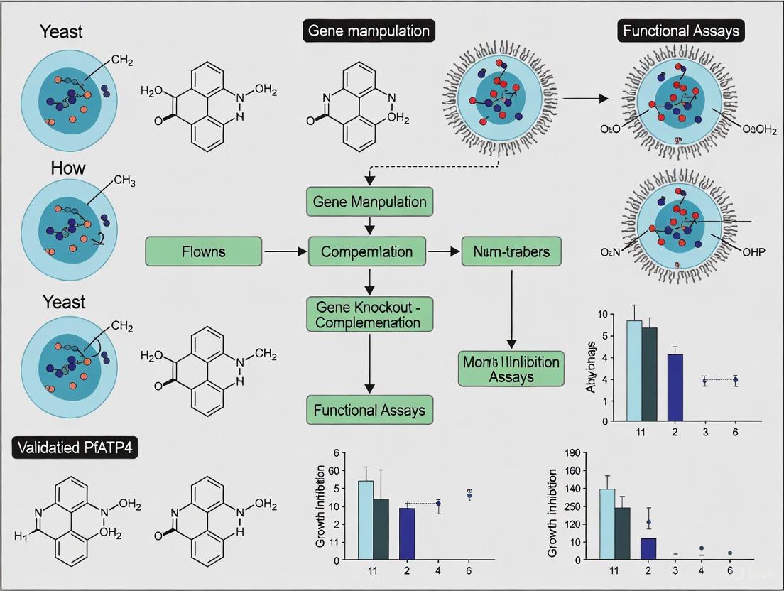
Validating PfATP4 as a High-Value Antimalarial Target: A Roadmap for Yeast Model Systems
The Plasmodium falciparum cation transporter PfATP4 is a leading antimalarial drug target, with inhibition causing rapid parasite death.
Validating PfATP4 as a High-Value Antimalarial Target: A Roadmap for Yeast Model Systems
Abstract
The Plasmodium falciparum cation transporter PfATP4 is a leading antimalarial drug target, with inhibition causing rapid parasite death. However, validating its function and screening for inhibitors has been hampered by an inability to express functional protein in heterologous systems like yeast, a challenge underscored by recent discoveries of its essential binding partner, PfABP. This article provides a comprehensive methodological framework for researchers aiming to establish a yeast model for PfATP4. We cover the foundational biology of PfATP4 and its regulators, strategic design for heterologous expression, troubleshooting of persistent expression hurdles, and a suite of functional assays for validating pump activity and inhibitor sensitivity. By synthesizing the latest structural insights and known resistance mutations, this guide aims to accelerate the development of a much-needed platform for next-generation antimalarial discovery.
Deconstructing PfATP4: Essential Biology and a Newly Discovered Regulator
The Critical Role of PfATP4 in Parasite Sodium Homeostasis and Survival
The Plasmodium falciparum Cation ATPase 4 (PfATP4) has emerged as one of the most promising antimalarial drug targets in recent decades, representing a critical vulnerability in the parasite's physiological machinery. This P-type ATPase transporter is localized to the parasite plasma membrane where it functions primarily as a sodium efflux pump, maintaining the low intracellular sodium concentration essential for parasite survival [1] [2]. The parasite inhabits a challenging ionic environment within the host erythrocyte, where sodium concentrations approximate those of blood plasma (~130-135 mM) due to parasite-induced new permeability pathways in the host cell membrane [1] [3]. Despite this high extracellular sodium concentration, the parasite maintains its cytosolic sodium at approximately 10 mM, establishing a substantial inward sodium electrochemical gradient that serves as an energy source for nutrient uptake [1]. PfATP4 is postulated to function as a Na+/H+-ATPase, extruding sodium ions from the parasite in exchange for protons, thereby simultaneously regulating intracellular sodium concentration and imposing an "acid load" on the parasite [2] [4]. The critical nature of this homeostatic function is evidenced by the fact that PfATP4 inhibition triggers rapid sodium dysregulation, parasite swelling, and ultimately parasite death [2] [5].
The validation of PfATP4 as a drug target represents a case study in modern antimalarial discovery. Unlike traditional target-based approaches, PfATP4 emerged from phenotypic screening campaigns followed by resistance mutation mapping [1] [6]. This protein has demonstrated remarkable "druggability," with multiple structurally distinct chemical classes - including spiroindolones, pyrazoleamides, aminopyrazoles, and dihydroisoquinolones - converging on PfATP4 as their primary target [1] [6] [4]. The clinical potential of targeting PfATP4 was demonstrated by cipargamin (KAE609), a spiroindolone that progressed through Phase II clinical trials with favorable results [1] [4]. However, the emergence of resistance mutations in PfATP4 has highlighted the need for deeper understanding of its structure and function to design next-generation inhibitors [3] [7].
Physiological Function of PfATP4 in Sodium Homeostasis
Ionic Challenges of the Intraerythrocytic Environment
The Plasmodium falciparum parasite faces extraordinary ionic challenges during its intraerythrocytic lifecycle. Upon invading a human red blood cell, the merozoite transitions from the high-sodium environment of blood plasma to the low-sodium, high-potassium environment of the host erythrocyte cytosol [1]. However, this relatively stable environment is short-lived. Approximately 12-18 hours after invasion (at the ring stage), the parasite induces "New Permeability Pathways" (NPPs) in the host erythrocyte membrane that dramatically increase permeability to various solutes, including sodium and potassium [1] [3]. The resulting sodium influx overwhelms the host erythrocyte's ouabain-sensitive Na+/K+-ATPase, leading to a progressive increase in erythrocyte cytosolic sodium concentration that eventually equilibrates with the extracellular plasma [1]. The intraerythrocytic parasite is further enclosed by the parasitophorous vacuole membrane, which contains high-conductance broad-selectivity channels thought to render it freely permeable to ions at the metabolically active trophozoite stage [1] [3]. Consequently, the parasite is bathed in a high-sodium environment at the parasitophorous vacuole space, creating a substantial challenge for maintaining ionic homeostasis.
PfATP4-Mediated Sodium Regulation
To survive in this hostile ionic environment, the parasite maintains a low cytosolic sodium concentration (~10 mM) through the active extrusion of sodium ions via PfATP4 [1] [2]. The sodium electrochemical gradient maintained by PfATP4 is substantial, combining the concentration gradient with the parasite's inwardly negative membrane potential (approximately -95 mV) [1]. This gradient serves not only to protect the parasite from sodium toxicity but also provides energy for secondary transport processes. A key example is the Na+-coupled phosphate transporter PfPiT, which relies on the sodium gradient established by PfATP4 to import essential inorganic phosphate for nucleotide, nucleic acid, and phospholipid synthesis [8]. This relationship underscores the integrated nature of ion regulation and nutrient acquisition in the parasite.
Direct evidence for PfATP4's role in sodium extrusion comes from experiments demonstrating orthovanadate-sensitive Na+ efflux from pre-loaded parasites against a steep concentration gradient [1]. Additionally, parasites with resistance-conferring mutations in PfATP4 show altered sodium regulation when exposed to PfATP4 inhibitors [2]. The essential nature of PfATP4-mediated sodium homeostasis is further highlighted by the observation that PfATP4 inhibitors trigger a rapid increase in intraparasitic sodium concentration and pH, leading to parasite swelling and eventual lysis [2] [5]. This distinctive biochemical signature has become a hallmark of PfATP4-targeting compounds and provides a reliable method for classifying compounds with this mechanism of action.
Table 1: Key Characteristics of PfATP4 Function
| Characteristic | Description | Experimental Evidence |
|---|---|---|
| Cellular Localization | Parasite plasma membrane | Immunofluorescence and epitope tagging [1] |
| Ion Specificity | Na+ extrusion, potentially in exchange for H+ | Na+ efflux assays, intracellular pH measurements [1] [2] |
| Intracellular [Na+] | ~10 mM in parasite cytosol vs. ~130 mM in host erythrocyte | Fluorescent indicator measurements [1] |
| Inhibitor Sensitivity | Orthovanadate-sensitive; targeted by multiple chemotypes | ATPase activity assays, resistance mutation mapping [1] [6] |
| Essential Function | Critical for parasite survival | Transposon mutagenesis, inhibitor studies [8] [2] |
Yeast Models in PfATP4 Research: Methodologies and Applications
Yeast as a Heterologous System for Malaria Research
The yeast Saccharomyces cerevisiae has emerged as a powerful heterologous system for studying Plasmodium falciparum proteins, including PfATP4 and other essential parasite transporters. Yeast offers several advantages for malaria research: well-established genetic tools, rapid growth, and the ability to functionally express membrane proteins that often prove challenging in other expression systems [8]. Additionally, yeast shares fundamental cellular processes with Plasmodium while lacking the complex ethical and technical challenges associated with continuous parasite culture. The utility of yeast models extends beyond basic functional characterization to include high-throughput drug screening against specific parasite targets, enabling rapid identification of novel inhibitors without immediate need for parasite culture [8].
A key strength of yeast-based systems is the ability to create conditionally dependent strains where parasite proteins replace essential yeast proteins, creating assays where yeast survival directly reports on the function of the parasite protein of interest. This approach has been successfully applied to multiple Plasmodium transporters, including the equilibrative nucleoside transporter PfENT1 and the sodium-coupled phosphate transporter PfPiT [8]. In these engineered strains, chemical inhibition of the parasite transporter results in measurable growth defects, providing a straightforward readout for inhibitor identification and characterization.
Experimental Workflow for Yeast-Based PfPiT Studies
The application of yeast models to study sodium-coupled transport in Plasmodium is exemplified by recent work on PfPiT, which depends on the sodium gradient established by PfATP4. The experimental workflow involves:
Strain Engineering: The parent yeast strain EY918, which lacks four of the five endogenous phosphate transporters and relies solely on Pho84 for phosphate uptake, serves as the starting point [8]. Through targeted gene replacement, the PHO84 coding sequence is replaced with a yeast codon-optimized version of PfPiT, creating a strain where phosphate uptake and consequently viability are entirely dependent on PfPiT function [8].
Functional Characterization: The PfPiT-dependent strain is subjected to radioactive phosphate uptake assays to determine kinetic parameters (Km = 56 ± 7 μM in 1 mM NaCl, decreasing to 24 ± 3 μM in 25 mM NaCl) and ion dependence, confirming its function as a sodium-coupled phosphate transporter [8].
Growth Assay Development: Conditions are established under which yeast growth is strictly dependent on phosphate uptake mediated by PfPiT, creating a 22-hour growth assay suitable for high-throughput screening [8].
Compound Screening: Libraries of compounds are screened for inhibition of PfPiT-dependent yeast growth, followed by confirmation of direct transport inhibition using radioactive uptake assays [8].
This workflow has already proven successful in identifying specific PfPiT inhibitors from small compound collections, demonstrating the utility of yeast-based systems for antimalarial discovery [8].
Yeast-Based Screening Workflow for Sodium-Coupled Transporters
The Scientist's Toolkit: Essential Research Reagents
Table 2: Key Research Reagents for PfATP4 and Associated Transport Studies
| Reagent/Resource | Function/Application | Example Use Case |
|---|---|---|
| S. cerevisiae EY918 strain | Parent strain with four endogenous phosphate transporters knocked out | Base for engineering PfPiT-dependent strain [8] |
| Yeast codon-optimized PfPiT | Heterologous expression of parasite transporter in yeast | Creation of phosphate uptake-dependent strain [8] |
| Radioactive [³²P]phosphate | Direct measurement of phosphate transport kinetics | Determination of Km and ion dependence [8] |
| Synthetic complete media with variable phosphate | Controlled phosphate availability for growth assays | PfPiT-dependent growth condition optimization [8] |
| CRISPR-Cas9 for P. falciparum | Endogenous tagging and gene editing in parasites | C-terminal 3×FLAG tagging of PfATP4 for native purification [3] |
| PfATP4 inhibitors (Cipargamin, PA21A092) | Positive controls for PfATP4 disruption | Validation of Na+ homeostasis assays [3] [2] |
| Na+-depleted culture media | Manipulation of extracellular sodium environment | Testing Na+ dependence of physiological processes [2] |
Structural Insights into PfATP4 Function and Inhibition
CryoEM Structure Reveals Novel Features
For years, attempts to determine the high-resolution structure of PfATP4 using heterologous expression systems proved unsuccessful, hindering structure-based drug design [3] [7]. This barrier was recently overcome through innovative approaches that enabled the endogenous purification of PfATP4 directly from CRISPR-engineered P. falciparum parasites cultured in human red blood cells [3]. The resulting 3.7 Å resolution cryoEM structure represents a landmark achievement that provides unprecedented insights into PfATP4 organization and function.
The structure confirms that PfATP4 contains the five canonical domains characteristic of P2-type ATPases: the transmembrane domain (TMD), nucleotide-binding (N) domain, phosphorylation (P) domain, actuator (A) domain, and extracellular loop (ECL) domain [3]. The TMD consists of 10 helices arranged in three clusters (TM1-2, TM3-4, TM5-10), with the ion-binding site located between TM4, TM5, TM6, and TM8 [3]. Although the resolution was insufficient to directly visualize bound Na+ ions, the arrangement of coordinating sidechains closely matches that of cation-bound SERCA (sarco/endoplasmic reticulum Ca2+-ATPase), suggesting the structure represents a Na+-bound state [3]. The ATP-binding site between the N and P domains shows generally conserved architecture with other P2-type ATPases, though notable differences in sidechain arrangements were observed that may have implications for inhibitor specificity [3].
Discovery of PfABP: A Novel Regulatory Protein
Perhaps the most surprising revelation from the endogenous PfATP4 structure was the discovery of a previously unknown interacting protein, designated PfATP4-Binding Protein (PfABP) [3] [7]. This apicomplexan-specific protein forms a conserved interaction with TM9 of PfATP4 and was found to be the third most abundant protein in tryptic digest mass spectrometry of purified PfATP4 samples [3]. Functional studies demonstrated that loss of PfABP leads to rapid degradation of PfATP4 and parasite death, indicating its essential role in stabilizing the transporter [3] [7]. This discovery opens entirely new avenues for antimalarial development, as PfABP represents a potential drug target that may be less prone to resistance mutations than PfATP4 itself [7].
PfATP4 Function in Parasite Sodium Homeostasis
Mapping Resistance Mutations Informs Drug Design
The endogenous PfATP4 structure has provided a crucial framework for understanding the structural basis of drug resistance. When resistance-conferring mutations identified in both laboratory-selected strains and clinical isolates are mapped onto the structure, striking patterns emerge [3]. Mutations conferring resistance to the spiroindolone cipargamin (such as G358S/A) primarily cluster around the proposed sodium-binding site within the TMD [3]. Structural analysis suggests that the G358S mutation may block cipargamin binding by introducing a serine sidechain into the inhibitor binding pocket [3]. Similarly, the A211V mutation that arose under pyrazoleamide (PA21A092) pressure is located within TM2 adjacent to the ion-binding site [3]. Interestingly, parasites with the A211V mutation show increased susceptibility to cipargamin, suggesting complex interactions between different inhibitor classes and their binding sites [3] [6].
Table 3: Clinically Relevant PfATP4 Mutations and Their Impact
| Mutation | Compound Selector | Structural Location | Proposed Resistance Mechanism |
|---|---|---|---|
| G358S/A | Cipargamin (Spiroindolone) | TM3, near Na+ binding site | Steric hindrance of inhibitor binding [3] |
| A211V | PA21A092 (Pyrazoleamide) | TM2, adjacent to ion-binding site | Altered binding pocket conformation [3] [6] |
| I203L | GNF-Pf4492 (Aminopyrazole) | TM2 | Modified inhibitor access pathway [6] |
| A187V | GNF-Pf4492 (Aminopyrazole) | TM1/TM2 interface | Allosteric effects on binding site [6] |
| S374R | MB14 | Not specified in results | Reduced inhibitor affinity [2] |
PfATP4 Inhibitors: Mechanisms and Experimental Assessment
Diverse Chemotypes Converge on PfATP4
PfATP4 represents a remarkable example of target convergence, where multiple structurally distinct chemical classes have been identified that disrupt its function [1] [6] [4]. These include:
Spiroindolones (e.g., cipargamin/KAE609): This class progressed to Phase II clinical trials and demonstrated faster parasite clearance than standard artemisinin treatments [1] [4]. Spiroindolones trigger rapid Na+ accumulation and parasite swelling [2].
Pyrazoleamides (e.g., PA21A092): These compounds show potent antimalarial activity and disrupt Na+ homeostasis similarly to spiroindolones, though resistance mutations map to distinct but adjacent regions of PfATP4 [3] [6].
Aminopyrazoles (e.g., GNF-Pf4492): Identified through phenotypic screening, this class selects for resistance mutations in PfATP4 (A187V, I203L, A211T) and produces the characteristic Na+ dysregulation phenotype [6].
Dihydroisoquinolones (e.g., (+)-SJ733): This class shares phenotypic effects with other PfATP4 inhibitors and shows cross-resistance patterns indicating a shared target [1].
Additionally, screening of the Medicines for Malaria Venture's 'Malaria Box' and 'Pathogen Box' revealed that 7% and 9% of antimalarial compounds in these collections, respectively, exhibit the biochemical signature of PfATP4 inhibition [2]. This remarkable convergence underscores both the essential nature of PfATP4 function and its unique "druggability."
Mechanism of Action: From Ionic Disruption to Parasite Death
The pathway from PfATP4 inhibition to parasite death involves a cascade of biochemical and cellular events:
Direct Inhibition: Compounds bind to PfATP4, blocking its Na+ extrusion capability [2] [5].
Sodium Accumulation: Intracellular Na+ concentration rises rapidly due to continued influx through various pathways [1] [2].
pH Perturbation: As PfATP4 may function as a Na+/H+ exchanger, inhibition leads to cytosolic alkalinization [2].
Osmotic Imbalance: Elevated Na+ concentration draws water into the parasite, causing swelling [2] [5].
Parasite Lysis: Severe swelling culminates in membrane rupture and parasite death [2] [5].
This mechanism is distinct from traditional antimalarials that target metabolic pathways or hemozoin formation, offering potential against multidrug-resistant parasites [5].
Beyond Trophozoite Death: Disruption of Parasite Egress
Recent research has revealed that PfATP4 inhibitors exert additional effects beyond killing intraerythrocytic trophozoites. Several PfATP4-associated compounds from the Malaria Box and Pathogen Box disrupt the schizont-to-ring transition by blocking merozoite egress from infected erythrocytes [2]. Detailed investigation demonstrates that these compounds prevent egress rather than invasion, and appear to work by inhibiting the activation of protein kinase G (PfPKG), an essential regulator of the egress cascade [2]. This effect is attenuated in Na+-depleted media and in parasites with resistance-conferring PfATP4 mutations, establishing a direct link between PfATP4 function and egress regulation [2]. This expanded understanding of PfATP4's role throughout the parasite lifecycle enhances its attractiveness as a drug target.
The critical role of PfATP4 in parasite sodium homeostasis and survival is now firmly established through multiple lines of evidence: physiological studies, genetic validation, structural biology, and inhibitor phenotyping. The convergence of diverse chemical classes on this target underscores its fundamental importance to parasite biology. Recent advances - particularly the endogenous PfATP4 structure and discovery of PfABP - have opened new avenues for drug development that may overcome existing resistance challenges.
The integration of yeast-based screening models with parasite studies creates a powerful pipeline for identifying and optimizing novel inhibitors, not only of PfATP4 itself but also of dependent transport systems like PfPiT. As drug resistance continues to undermine current antimalarial therapies, targeting PfATP4 and associated proteins offers a promising strategy for next-generation treatments that exploit essential ion regulatory pathways in the malaria parasite.
The pursuit of validating PfATP4, a sodium efflux pump in Plasmodium falciparum, as a premier antimalarial drug target has been significantly hampered by a persistent and critical roadblock: the repeated failure to achieve its functional expression in heterologous systems. This review objectively compares the unsuccessful attempts at heterologous expression against the successful application of endogenous parasite studies and surrogate yeast models. We summarize quantitative biochemical data gathered from native sources, detail the experimental protocols that ultimately proved successful, and situate these findings within the broader context of using yeast-based research to validate antimalarial targets. The analysis reveals that the inherent molecular complexity of PfATP4, including its recently discovered essential binding partner, fundamentally limited the utility of heterologous systems and directed the field toward more physiologically relevant expression platforms.
The Heterologous Expression Bottleneck: A Persistent Challenge
The functional characterization of any membrane protein, including its biochemical properties, transport kinetics, and inhibitor sensitivity, typically relies on the ability to express and purify the protein in a heterologous system. For PfATP4, this approach was deemed essential for structural studies and high-throughput drug screening.
However, multiple independent laboratories have consistently reported an inability to express functional PfATP4 in standard heterologous systems such as Xenopus laevis oocytes and presumably yeast or bacterial cultures [9]. One study explicitly notes that while an initial report described some success in oocytes, subsequent efforts to replicate this work were unsuccessful, stating that "at least one other laboratory has been unable to reproduce this finding or to achieve functional expression of the transporter" [9]. This reproducibility crisis significantly stalled the mechanistic study of PfATP4 and the direct biochemical validation of proposed inhibitors.
The recent discovery of a previously unknown protein partner, PfATP4-Binding Protein (PfABP), has provided a compelling explanation for these historical failures [3] [7] [10]. This essential modulator was found to co-purify with PfATP4 directly from parasite-infected human red blood cells. Crucially, experiments showed that the loss of PfABP led to the rapid degradation of PfATP4 and parasite death, indicating its critical role in stabilizing the pump [7] [10]. Its absence in heterologous expression systems likely explains the instability and lack of function of recombinant PfATP4.
Table 1: Documented Challenges in Heterologous Expression of PfATP4
| Heterologous System | Reported Outcome | Key Evidence |
|---|---|---|
| Xenopus laevis Oocytes | Non-functional expression / Unreproducible | Initial report of Ca²⁺-dependent ATPase activity could not be replicated by other labs [9]. |
| Yeast/Bacterial Systems | Implied failure | Lack of published success and noted "inability to achieve functional expression in a heterologous system" [11]. |
Successful Endogenous Characterization: A Path to Validation
Faced with the heterologous expression barrier, researchers pivoted to studying PfATP4 directly from its native environment—the Plasmodium falciparum parasite. This approach yielded the first high-resolution structural data and definitive biochemical characterization.
Protocol 1: Endogenous Protein Purification and Cryo-EM from Parasites
- Parasite Culture: CRISPR-Cas9 was used to insert a 3×FLAG epitope tag at the C-terminus of the PfATP4 gene in Dd2 P. falciparum parasites [3].
- Membrane Preparation: PfATP4 was affinity-purified directly from parasites cultured in hundreds of liters of human red blood cells [10].
- Functional Assay: The purified protein was confirmed to exhibit Na⁺-dependent ATPase activity that was inhibited by known PfATP4 inhibitors like cipargamin, validating its functionality [3].
- Structure Determination: A 3.7 Å resolution cryo-electron microscopy (cryoEM) structure was determined from the endogenously purified protein, revealing the overall architecture and the unexpected density for PfABP [3].
Protocol 2: Membrane ATPase Assay in Native Parasite Membranes
- Membrane Preparation: Membranes were isolated from intact asexual blood-stage P. falciparum parasites [9].
- ATPase Activity Measurement: ATP hydrolysis was measured by quantifying inorganic phosphate (Pi) production over time. Na⁺-dependent activity was isolated by comparing Pi production in high (152 mM) vs. low (2 mM) Na⁺ conditions, with choline as a substitute cation [9].
- Inhibitor Sensitivity: The "PfATP4-associated ATPase activity" was defined as the fraction of Na⁺-dependent ATPase activity that was sensitive to inhibition by 500 nM cipargamin. This assay confirmed PfATP4 as the direct target for multiple chemical classes [9].
Quantitative Biochemical Profile from Native Studies
The endogenous characterization of PfATP4 provided the first direct kinetic and biochemical data, which are summarized in the table below.
Table 2: Experimentally Determined Biochemical Properties of Native PfATP4
| Biochemical Parameter | Experimental Value | Experimental Context |
|---|---|---|
| Apparent Kₘ for ATP | 0.2 mM | Measured in parasite membrane preparations [9]. |
| Apparent Kₘ for Na⁺ | 16-17 mM | Measured in parasite membrane preparations [9]. |
| Inhibitor Potency (Cipargamin) | IC₅₀ in the nanomolar range | Inhibited Na⁺-dependent ATPase activity in WT parasite membranes; potency reduced in PfATP4-mutant parasites [9]. |
| Transport Coupling | Na⁺ efflux / H⁺ influx (proposed) | Inhibition leads to increased cytosolic [Na⁺] and increased cytosolic pH [4]. |
Figure 1: A comparative workflow diagram illustrating the failed heterologous expression path versus the successful endogenous study path for PfATP4. The discovery of PfABP provided the key explanation for the initial failures.
The Yeast Model Alternative: A Surrogate Success Story
While heterologous expression of PfATP4 itself failed, the use of yeast (Saccharomyces cerevisiae) as a surrogate model for antimalarial target validation has proven highly successful for other essential Plasmodium membrane transporters. The case of the phosphate transporter PfPiT serves as an exemplary contrast.
Experimental Protocol for Yeast-Based Drug Screening
Protocol: Developing a Yeast Growth Assay for PfPiT Inhibitor Screening
- Strain Engineering: The parent yeast strain EY918, which has four of its five endogenous phosphate transporters knocked out and relies solely on Pho84 for viability, was used [8].
- Gene Replacement: A yeast codon-optimized gene for PfPiT was synthesized. Genome editing was used to replace the PHO84 coding sequence with the PfPiT gene, creating a strain where PfPiT was the sole phosphate transporter [8].
- Functional Validation: A radioactive [³²P]phosphate uptake assay confirmed the transporter's function and ion dependence, showing a Km for phosphate of 56 ± 7 μM in 1 mM NaCl [8].
- Growth Inhibition Screen: A robust 22-hour growth assay was developed under conditions where yeast growth was strictly dependent on phosphate uptake via PfPiT. This assay was used to screen compounds for PfPiT-specific inhibition, successfully identifying two hits from a small library [8].
Comparative Analysis: PfATP4 Failure vs. PfPiT Success in Yeast
Table 3: Contrasting Outcomes for Two Malaria Targets in Heterologous Models
| Aspect | PfATP4 in Heterologous Systems | PfPiT in Engineered Yeast |
|---|---|---|
| Expression Outcome | Failure / Non-functional [9] [11] | Successful functional expression [8] |
| Key Enabling Factor | Requires unknown partner PfABP [3] | Functions as a single subunit transporter |
| Ion Dependence | Inferred from native studies (Na⁺) [9] | Directly measured in yeast (Na⁺-coupled) [8] |
| Drug Screening Utility | Not feasible in heterologous systems | Feasible (e.g., 22-h growth assay) [8] |
| Target Validation | Relied on native parasites & genetics [6] [11] | Achieved via surrogate yeast physiology [8] |
The Scientist's Toolkit: Essential Research Reagents
The following table details key reagents and materials derived from the successful endogenous and yeast-based studies, providing a resource for researchers in the field.
Table 4: Key Research Reagent Solutions for PfATP4 and Surrogate Studies
| Reagent / Material | Function in Research | Source/Example |
|---|---|---|
| CRISPR-Cas9 Engineered Dd2 Parasites | Allows endogenous tagging and purification of PfATP4 from its native context for structural and biochemical studies. | PfATP4-3×FLAG tagged P. falciparum [3]. |
| EY918 Yeast Strain | Parent strain for engineering surrogate models; lacks four phosphate transporters, allowing dependency on a single introduced transporter. | S. cerevisiae strain with pho86Δ, pho87Δ, pho89Δ, pho90Δ, leu2Δ [8]. |
| PfPiT-Expressing Yeast Strain | Validated surrogate system for high-throughput screening of inhibitors against a essential malaria transporter. | EY918 with PHO84 replaced by yeast-codon-optimized PfPiT [8]. |
| Cipargamin | A leading clinical PfATP4 inhibitor; used as a positive control in membrane ATPase assays and resistance studies. | Synthetic spiroindolone; Na⁺-ATPase inhibitor [9]. |
| Radioactive [³²P]Phosphate | Tracer for direct measurement of phosphate uptake kinetics in engineered yeast strains. | Used to determine Km of PfPiT in yeast [8]. |
Figure 2: A schematic illustrating the molecular rationale for the failure of heterologous PfATP4 expression. The absence of its essential stabilizing partner, PfABP, in the heterologous host leads to the degradation and non-function of the pump.
The historical failure to express PfATP4 in heterologous systems was not a mere technical setback but an instructive lesson in the complexity of malaria parasite biology. It underscored that some Plasmodium proteins do not function as solitary units but within essential, pathogen-specific complexes. The discovery of PfABP from endogenous studies vindicated this "failure," turning it into a discovery that opens a new avenue for antimalarial strategies targeting the PfATP4-PfABP interaction [3] [7].
While the yeast model was not a successful platform for PfATP4 itself, its demonstrated success in functionally expressing and enabling drug screening for other essential targets like PfPiT confirms its immense value in the antimalarial toolkit [8]. The path forward for validating complex antimalarial targets will likely involve a synergistic approach: using endogenous parasite systems for definitive structural and functional studies, while continuing to leverage engineered yeast for targets amenable to surrogate expression. This combined strategy maximizes the strengths of each platform to accelerate the development of novel therapies against a formidable global disease.
The validation of PfATP4, a sodium efflux pump located on the plasma membrane of the Plasmodium falciparum parasite, as a promising antimalarial target, represents a critical frontier in the battle against malaria [12] [3]. This P-type ATPase is essential for parasite survival, maintaining low intracellular sodium concentrations in the face of high sodium levels in the host bloodstream [7] [13]. The strategic inhibition of PfATP4 by novel drug candidates such as Cipargamin and PA21A092 causes rapid parasite death, underscoring its therapeutic potential [12] [14]. However, the parasite's notorious ability to develop resistance through mutations in PfATP4 has necessitated a deeper, structural understanding of this target [3] [15].
Research models, particularly yeast-based systems, have historically been instrumental in deconstructing the biology of complex pathogenic proteins. Yeast offers a tractable eukaryotic chassis for expressing and characterizing parasite proteins, allowing for controlled genetic manipulation and high-throughput functional assays [16]. The broader thesis of using yeast model research to validate PfATP4 gains profound significance in this context. It provides a platform not only to study the pump's inherent function and drug inhibition but also to dissect the impact of resistance mutations. The recent, groundbreaking discovery of an essential binding partner, PfATP4-Binding Protein (PfABP), introduces a new variable into this equation [12] [3]. This paradigm shift suggests that future validation efforts in yeast must now account for this stabilizing partner, potentially requiring the co-expression of both proteins to accurately recapitulate the native, functional complex found within the malaria parasite.
Structural & Functional Comparison: PfATP4 With and Without PfABP
The recent elucidation of the high-resolution 3.7 Å cryoEM structure of PfATP4, purified endogenously from CRISPR-engineered parasites, has fundamentally altered our perception of this drug target [12] [3]. The most striking revelation was the identification of a previously unknown protein, PfABP, firmly bound to TM9 of PfATP4's transmembrane domain (TMD) [12] [3]. PfABP is a conserved, apicomplexan-specific protein of unknown function, now recognized as an essential modulator [14]. The table below provides a quantitative comparison of PfATP4's key characteristics in the presence and absence of its binding partner.
Table 1: Comparative Analysis of PfATP4 Properties With and Without PfABP
| Feature | PfATP4 with PfABP (Stabilized State) | PfATP4 without PfABP (Degraded State) |
|---|---|---|
| Protein Stability | Stabilized structure [7] [13] | Rapid degradation of the pump [7] [13] |
| Parasite Viability | Essential for parasite survival [3] [13] | Leads to parasite death [3] [13] |
| Structural State | Na+-bound state conformation; Conserved TMD with 10 helices [12] | Not applicable (protein degraded) |
| Drug Target Profile | Primary target for Cipargamin & PA21A092; Resistance mutations present [12] | Not a viable target (parasite dead) |
| Regulatory Role | Likely modulatory interaction, fine-tuning activity [3] | No regulatory function |
This comparative data underscores PfABP's non-redundant role as a stabilizing factor. Experimentally, the loss of PfABP leads directly to the rapid degradation of the PfATP4 sodium pump and, consequently, the death of the parasite, highlighting that the PfATP4-PfABP complex is the biologically relevant unit for drug targeting [7] [13]. Furthermore, because PfABP appears to be more conserved and less prone to mutations than PfATP4 itself, targeting the interaction interface offers a promising strategy to circumvent existing resistance mechanisms [7] [15].
Experimental Protocols for Key PfABP-PfATP4 Interaction Studies
Protocol 1: Endogenous Purification and CryoEM Structure Determination
The discovery of PfABP was contingent on a methodology that preserved the native state of PfATP4 within the parasite [7] [13].
- CRISPR-Cas9 Engineering: The C-terminus of the PfATP4 gene in Dd2 P. falciparum parasites was engineered to include a 3×FLAG epitope tag using CRISPR-Cas9 [12] [3].
- Endogenous Purification: The tagged PfATP4 protein was affinity-purified directly from parasites cultured in human red blood cells. This endogenous approach was critical for co-purifying native binding partners [12] [3].
- Functional Validation: The purified protein's activity was confirmed through Na+-dependent ATPase assays, which were inhibited by known PfATP4 inhibitors like PA21A092 and Cipargamin [12] [3].
- CryoEM and Modeling: A 3.7 Å resolution structure was determined using single-particle cryo-electron microscopy [12] [3].
- Identification of PfABP: An unassigned helix interacting with TM9 of PfATP4 was identified. Sequence-independent modeling and a search using findMySequence identified this helix as the C-terminus of the protein PF3D7_1315500, which was named PfABP. Its presence was further confirmed by its status as the third most abundant protein in tryptic digest mass spectrometry of the purified sample [3].
Protocol 2: Functional Validation of PfABP via Knockdown
The essential function of PfABP was established through loss-of-function experiments.
- Genetic Disruption: PfABP expression was suppressed or knocked down in P. falciparum parasites [7] [13].
- Phenotypic Assessment: Parasite viability was monitored following the loss of PfABP. The experiments showed rapid parasite death, establishing PfABP as essential for survival [13] [17].
- Biochemical Analysis: The stability of the PfATP4 protein was assessed following PfABP knockdown. Immunoblotting or similar techniques confirmed that PfATP4 levels collapsed, indicating that PfABP is required for the pump's stability [7] [13].
Visualizing the Discovery and Impact of PfABP
The following diagrams outline the key experimental workflow that led to the discovery of PfABP and the logical relationship between PfABP loss and its fatal consequence for the parasite.
Diagram 1: The experimental workflow for the endogenous structural determination of PfATP4, which was crucial for the discovery of its binding partner, PfABP.
Diagram 2: The causal pathway from the experimental knockdown of PfABP to the death of the parasite, demonstrating PfABP's essential role.
The Scientist's Toolkit: Research Reagent Solutions
The critical experiments characterizing the PfATP4-PfABP interaction rely on a specific set of reagents and tools. The table below details these key resources, providing a guide for researchers aiming to replicate or build upon these findings.
Table 2: Essential Research Reagents for Studying PfATP4 and PfABP
| Research Reagent | Function and Application in PfATP4/PfABP Research |
|---|---|
| CRISPR-Cas9 System | Used for endogenous epitope tagging (e.g., 3×FLAG) of PfATP4 in P. falciparum parasites, enabling subsequent purification [12] [3]. |
| 3×FLAG Epitope Tag | An affinity tag inserted at the C-terminus of PfATP4 to facilitate immunoaffinity purification of the native protein complex from parasites [12] [3]. |
| Cryo-Electron Microscopy | The primary technique used to determine the 3.7 Å resolution structure of endogenously purified PfATP4, revealing the bound PfABP [7] [3]. |
| PfATP4 Inhibitors (Cipargamin, PA21A092) | Established chemical tools to functionally validate purified PfATP4 activity in Na+-dependent ATPase assays and study inhibition mechanisms [12] [14]. |
| ModelAngelo & findMySequence | Computational tools used for sequence-independent modeling of the unidentified PfABP helix and for identifying its amino acid sequence within the P. falciparum proteome [3]. |
| Human Red Blood Cells & Culture Medium | The essential native environment for culturing P. falciparum parasites, required for the endogenous purification of correctly assembled and partnered PfATP4 [7] [13]. |
The discovery of PfABP marks a true paradigm shift in the pursuit of PfATP4 as an antimalarial target. It moves the focus from a single target protein to an essential, two-component complex. This new understanding has profound implications for the broader thesis of using models like yeast for target validation. Future work must now incorporate PfABP to create a more physiologically relevant system for studying PfATP4 function, inhibitor binding, and the mechanistic basis of resistance. The spatial organization of known resistance mutations, such as G358S, can now be analyzed in the context of this complete structure, potentially revealing new drug-binding pockets that are less susceptible to resistance [12] [3]. Ultimately, targeting the PfATP4-PfABP interaction interface presents a novel, and potentially more durable, therapeutic avenue. By designing compounds that disrupt this essential partnership, the scientific community can exploit a critical vulnerability in the malaria parasite, offering fresh hope in the global fight against this resilient disease [7] [15].
Cataloging Clinically Relevant Resistance Mutations (e.g., G358S, A211V)
Plasmodium falciparum ATP4 (PfATP4) has emerged as a leading antimalarial target due to its essential function in maintaining sodium homeostasis in the malaria parasite. This P-type ATPase functions as a Na+ efflux pump located on the parasite plasma membrane, exporting Na+ from the parasite cytosol while importing H+ equivalents [1] [18]. The critical nature of this ion regulation is highlighted by the fact that parasites maintain a low cytosolic [Na+] (~10 mM) despite being surrounded by an environment with ~135 mM Na+ in the host erythrocyte cytosol and parasitophorous vacuole [3] [12]. The validation of PfATP4 as a drug target comes from its identification as the target of structurally diverse antimalarial compounds including spiroindolones (e.g., cipargamin), pyrazoleamides (e.g., PA21A092), and dihydroisoquinolones (e.g., (+)-SJ733) [3] [1] [18]. Inhibition of PfATP4 induces rapid parasite death through disruption of intracellular Na+ homeostasis, causing a rise in cytosolic [Na+] that leads to osmotic swelling and parasite clearance [18] [19]. The clinical relevance of this target is further strengthened by the emergence of resistance mutations in PfATP4 under drug pressure both in laboratory settings and in clinical trials [3] [18] [19].
Clinically Relevant PfATP4 Mutations and Their Functional Impact
Comprehensive Mutation Catalog
Research has identified numerous mutations in PfATP4 associated with resistance to various chemotypes. The table below summarizes key clinically relevant mutations, their resistance profiles, and functional consequences.
Table 1: Clinically Relevant PfATP4 Resistance Mutations
| Mutation | Location/ Domain | Resistance Profile | Functional Consequences | Clinical/Experimental Evidence |
|---|---|---|---|---|
| G358S | TM3, near Na+ binding site | Confers high-level resistance to cipargamin and (+)-SJ733 [3] [18] [19] | Reduces PfATP4 affinity for Na+; increases resting cytosolic [Na+]; blocks drug binding [3] [18] | Emerged in 22/25 recrudescent cases in cipargamin Phase 2b trial [18] [19] |
| A211V | TM2, adjacent to ion-binding site | Decreased sensitivity to pyrazoleamides (PA21A092); increased susceptibility to spiroindolones (cipargamin) [3] [20] | Alters drug binding pocket; may cause conformational changes affecting compound access [3] [20] | Selected under continuous pyrazoleamide pressure in vitro; confirmed via CRISPR-Cas9 [3] [20] |
| G358A | TM3, near Na+ binding site | Resistance to cipargamin and (+)-SJ733 [3] | Similar mechanism to G358S; steric hindrance of drug binding [3] | Found in recrudescent parasites from clinical trials [3] |
| A211T | TM2 | Resistance to pyrazoleamide GNF-Pf4492; increased cipargamin sensitivity [20] | Alters ion transport function; hypersensitizes to spiroindolones [20] | Selected in vitro under GNF-Pf4492 pressure for 70 days [20] |
Structural Basis of Resistance
Recent structural insights from a 3.7 Å cryoEM structure of PfATP4 have illuminated the molecular mechanisms underlying these resistance mutations [3] [12]. The G358S mutation is located on transmembrane helix 3 (TM3), immediately adjacent to the proposed Na+ coordination site within the transmembrane domain. The introduction of a serine sidechain at this position potentially blocks cipargamin binding by steric hindrance within the proposed drug binding pocket [3]. Similarly, the A211V mutation resides within TM2 near both the ion-binding site and the proposed cipargamin binding site, suggesting that the valine substitution may alter the conformation of the drug binding pocket in a way that interferes with pyrazoleamide binding while potentially creating a more favorable interaction surface for spiroindolones [3] [20]. This structural information provides a framework for understanding how distinct mutations confer differential resistance patterns across chemotypes.
Experimental Methodologies for Characterizing PfATP4 Mutations
Resistance Selection Protocols
In Vitro Evolution Experiments
To generate PfATP4-mutant parasites resistant to cipargamin, researchers have implemented sequential drug pressure protocols [18] [19]. Parasite cultures (typically Dd2 or 3D7 strains) are exposed to incrementally increasing concentrations of cipargamin over approximately four months, starting with sub-nanomolar concentrations (e.g., 0.5 nM) and gradually escalating to micromolar levels (up to 5 μM) as parasites adapt [18] [19]. Cultures are maintained with regular medium changes and drug replenishment, with parasite survival monitored via thin blood smears or SYBR Green fluorescence assays. Resistant populations are cloned by limiting dilution, and their genomes are sequenced to identify resistance-conferring mutations in pfatp4 [18].
CRISPR-Cas9 Gene Editing
For functional validation of specific mutations, CRISPR-Cas9 has been employed to introduce point mutations into endogenous pfatp4 [3] [20]. The protocol involves:
- Design of repair templates: Single-stranded oligonucleotides containing the desired mutation (e.g., A211V) with ~35 bp homology arms
- sgRNA construction: Guide RNAs targeting sequences near the mutation site
- Transfection: Delivery of Cas9-sgRNA ribonucleoprotein complexes and repair templates to infected erythrocytes via electroporation
- Selection: Drug selection (e.g., with WR99210 if using hDHFR selectable marker) and clone isolation
- Genotype confirmation: Sanger sequencing of the modified pfatp4 locus [3] [20]
Phenotypic Characterization Assays
Drug Sensitivity Profiling
The half-maximal inhibitory concentration (IC50) of PfATP4 inhibitors against mutant parasites is determined using standardized growth inhibition assays [20] [18]. Synchronized ring-stage parasites are cultured in 96-well plates at 1% parasitemia and 3% hematocrit with serial dilutions of compounds (typically spanning 0.1 nM to 1 μM). After 48 hours of exposure, parasite growth is quantified via 3H-hypoxanthine incorporation or SYBR Green I fluorescence using flow cytometry. Dose-response curves are fitted using non-linear regression to calculate IC50 values [20].
Intracellular Sodium Measurement
Resting cytosolic [Na+] in mutant parasites is measured using the Na+ indicator dye Sodium Green Tetraacetate [18] [19]. Synchronized trophozoite-stage parasites are isolated by saponin lysis of erythrocytes, loaded with 10 μM Sodium Green AM ester for 60 minutes at 37°C, washed, and resuspended in Na+-free medium. Fluorescence is measured using a fluorescence microplate reader (excitation 490 nm, emission 535 nm). Calibration is performed using the ionophores gramicidin and monensin in buffers with known Na+ concentrations to convert fluorescence values to [Na+] [18].
Figure 1: Workflow for Drug Sensitivity Profiling of PfATP4 Mutations
Resistance Mechanisms and Signaling Pathways
Molecular Mechanisms of PfATP4 Inhibition and Resistance
The molecular mechanisms of PfATP4 inhibition and subsequent resistance development involve complex interactions at the drug binding site and alterations in ion transport function. The recently solved cryoEM structure of PfATP4 reveals that the protein contains five canonical P-type ATPase domains: transmembrane domain (TMD), nucleotide-binding (N) domain, phosphorylation (P) domain, actuator (A) domain, and extracellular loop (ECL) domain [3] [12]. The ion-binding site is located between TM4, TM5, TM6, and TM8, similar to other P2-type ATPases like SERCA [3]. PfATP4 inhibitors such as cipargamin and pyrazoleamides bind within the transmembrane domain, interfering with Na+ binding and transport. Resistance mutations primarily cluster around the Na+ binding site, with G358S on TM3 and A211V on TM2 both positioned to directly or allosterically influence drug binding [3].
The G358S mutation confers resistance through a dual mechanism: it directly impedes drug binding through steric hindrance while simultaneously reducing the pump's affinity for Na+ [18] [19]. This results in parasites with elevated resting cytosolic [Na+] that are less susceptible to further Na+ dysregulation by PfATP4 inhibitors. Interestingly, the A211V mutation exhibits a paradoxical resistance profile, conferring resistance to pyrazoleamides while hypersensitizing parasites to spiroindolones [3] [20]. This suggests that rather than generally compromising drug binding, this mutation induces specific conformational changes that differentially affect how distinct chemotypes interact with the binding pocket.
Figure 2: Mechanism of PfATP4 Inhibition and Resistance Development
Metabolic Adaptation Pathways
Beyond direct target mutations, parasites exhibit metabolic adaptations to PfATP4 inhibition. Transcriptomic and metabolomic analyses of A211V mutant parasites under sublethal drug pressure reveal significant alterations in phospholipid signaling pathways [20]. Specifically, parasites treated with PA21A092 show increased enzymatic activities associated with phosphatidylcholine, phosphatidylserine, and phosphatidylinositol synthesis, suggesting activation of protein kinases via phospholipid-dependent signaling as a compensatory mechanism to ionic perturbation [20]. This metabolic adaptation represents a separate resistance mechanism that could be targeted to prevent emergence of resistance.
The Scientist's Toolkit: Essential Research Reagents
Table 2: Key Research Reagents for PfATP4 Mutation Studies
| Reagent/Cell Line | Specific Example | Research Application | Key Features/Properties |
|---|---|---|---|
| Engineered Parasite Lines | Dd2A211V (CRISPR-edited) [20] | Resistance mechanism studies; cross-resistance profiling | Confirmed A211V mutation; hypersensitive to cipargamin; resistant to pyrazoleamides |
| PfATP4 Inhibitors | Cipargamin (KAE609) [18] [19] | Positive control for assays; resistance selection | Spiroindolone; clinical candidate; IC50 ~0.4-1.1 nM against wild-type |
| PfATP4 Inhibitors | PA21A092 [3] [20] | Pyrazoleamide representative; cross-resistance studies | Pyrazoleamide; selects for A211V/T mutations |
| Fluorescent Indicators | Sodium Green Tetraacetate [18] [19] | Cytosolic Na+ measurements | Cell-permeant AM ester; fluorescence increases with Na+ binding |
| Antimalarial Controls | Artemether-lumefantrine [18] [19] | Off-target resistance assessment; combination studies | Standard care ACT; no cross-resistance with PfATP4 mutations |
| Expression Systems | TgATP4 in T. gondii [18] [19] | Heterologous functional characterization | ATP4 homolog; functional conservation with PfATP4 |
Discussion and Clinical Implications
The cataloging of clinically relevant PfATP4 mutations reveals critical patterns with direct implications for antimalarial development. The emergence of the G358S mutation in clinical trials demonstrates the real-world potential for resistance development to even the most promising PfATP4 inhibitors [18] [19]. The differential resistance patterns observed with mutations like A211V – conferring resistance to one chemotype while hypersensitizing to another – suggest opportunities for strategic combination therapies that could suppress resistance emergence [3] [20]. Furthermore, the discovery of an apicomplexan-specific PfATP4-binding protein (PfABP) in the recent cryoEM structure presents a novel avenue for inhibitor design that might circumvent existing resistance mechanisms [3] [12].
The structural biology advances represented by the 3.7 Å PfATP4 cryoEM structure now enable structure-guided drug design to develop next-generation inhibitors less susceptible to existing resistance mechanisms [3] [12]. Additionally, the metabolic adaptations observed in resistant parasites highlight the potential for targeting compensatory pathways to preserve the efficacy of PfATP4-directed compounds. As resistance to current artemisinin-based combinations spreads in Africa [21], the validation of PfATP4 mutations and their functional characterization provides critical insights for developing new antimalarial strategies with improved resistance profiles.
Building the Yeast Model: From Vector Design to Functional Expression
Strategic Selection of Yeast Expression Systems and Promoters
The rising spread of artemisinin-resistant Plasmodium falciparum parasites underscores a pressing need for novel antimalarial drugs with new mechanisms of action [8]. Target-based screening in heterologous expression systems offers a powerful strategy for early drug discovery. The yeast Saccharomyces cerevisiae has emerged as a preferred chassis for functional expression and validation of Plasmodium membrane proteins and essential enzymes, bridging the gap between target identification and compound screening in parasites [8] [22] [23].
This guide objectively compares yeast expression systems and promoter strategies, focusing on their application in validating the antimalarial target PfATP4 and other targets like PfPiT. We present comparative performance data and detailed methodologies to inform selection for malaria drug discovery projects.
Comparison of Yeast Expression Systems for Malaria Target Validation
The table below summarizes three distinct yeast-based expression platforms developed for the functional study and inhibition screening of different Plasmodium targets.
Table 1: Comparison of Yeast-Based Platforms for Malaria Target Validation
| Malaria Target | Target Function & Rationale | Yeast Genetic Engineering | Key Assay Readout | Performance & Key Findings |
|---|---|---|---|---|
| PfPiT [8] [24] | Sodium-coupled inorganic phosphate (Pi) transporter; essential for parasite proliferation. | Replacement of endogenous yeast Pi transporter (PHO84) with a yeast-codon-optimized PfPiT gene. |
|
|
| PfCHA [22] | Ca²⁺/H⁺ antiporter; fundamental for Ca²⁺ signalling in parasite life cycle. | Expression of PfCHA in a yeast strain lacking the vacuolar Ca²⁺/H⁺ exchanger (vcx1Δ). |
|
|
| PvDHS [23] | Deoxyhypusine synthase; essential for post-translational modification of eIF5A. | Replacement of the endogenous yeast DYS1 gene with genes for P. vivax DHS (PvDHS) or human DHS (HsDHS). |
|
|
The following workflow generalizes the process of developing and using such a yeast-based screening platform:
Promoter Engineering Strategies for Controlled Gene Expression
Precise control of target gene expression is critical. Synthetic biology provides tools to engineer promoters with specific strengths and regulatory properties.
Table 2: Comparison of Yeast Promoter Engineering Strategies
| Engineering Strategy | Key Methodology | Induction/Strength Profile | Best Use Cases |
|---|---|---|---|
| Hybrid Promoters [25] | Fusion of Upstream Activating Sequences (UAS) from one promoter to the core promoter of another. | Dynamic range up to 50-fold for galactose-inducible systems. | Tuning expression levels of toxic proteins or pathway enzymes. |
| Minimal Leaky Promoters [26] | Direct fusion of bacterial operators upstream of TATA-box, plus >1-kbp insulator sequences. | >10³-fold induction with minimal leakiness. | High-throughput screening where background growth is undesirable. |
| Constitutive Promoters [25] | Use of strong, unregulated native promoters (e.g., pGPD, pTEF1). | Constant high-level expression. | Complementation assays or expressing selection markers. |
The diagram below illustrates the design principle for constructing a high-performance, minimal-leak synthetic promoter.
Experimental Protocols for Key Validation Assays
This protocol outlines the key steps for a growth inhibition screen against a phosphate transporter target.
Strain and Media Preparation:
- Use the engineered S. cerevisiae strain where PfPiT is the sole phosphate transporter.
- Culture yeast in synthetic complete media with 25 mM phosphate (SC+25Pi) prior to the assay.
- For the assay, use 2x synthetic complete media with 24 mM phosphate (2xSC+24Pi).
Growth Inhibition Assay:
- Dilute overnight cultures to a standardized optical density (OD600).
- Dispense cells into a 96-well plate containing the test compounds.
- Incubate the plate with continuous shaking for 22 hours at 30°C.
- Monitor growth by measuring OD600 at regular intervals.
Counter-Screen and Validation:
- Counter-screen all compounds against the parental yeast strain (expressing the native transporter Pho84) to identify off-target effects.
- Confirm hits using a radioactive [³²P]phosphate uptake assay to directly measure inhibition of phosphate transport.
This protocol measures the function of ion transporters in real-time.
Strain Transformation and Culture:
- Co-transform yeast with the plasmid expressing the malaria target (e.g., PfCHA) and a plasmid expressing the calcium-sensitive protein apoaequorin.
- Grow transformed cells in selective media with glucose to exponential phase.
Gene Expression and Apoaequorin Reconstitution:
- Wash cells and transfer to media with galactose for 4 hours to induce target gene expression.
- Harvest cells and resuspend in test medium.
- Incubate cells with coelenterazine (the apoaequorin cofactor) for 30 minutes in the dark to form functional aequorin.
Cation Challenge and Luminescence Measurement:
- Transfer the cell suspension to a luminometer.
- Inject a pulse of the cation of interest (e.g., Ca²⁺, Mn²⁺, Mg²⁺).
- Monitor the resulting burst of luminescence, which is inversely proportional to the transporter's ability to clear the cation from the cytoplasm.
The Scientist's Toolkit: Key Research Reagent Solutions
Table 3: Essential Research Reagents for Yeast-Based Malaria Research
| Reagent / Tool | Function in Experimental Workflow | Specific Examples from Research |
|---|---|---|
| CRISPR-Cas9 System | For precise endogenous tagging or gene knockout in yeast and Plasmodium. | Endogenous C-terminal 3xFLAG tagging of PfATP4 in P. falciparum [3]. |
| Codon-Optimized Genes | Enhances functional expression of Plasmodium genes in the heterologous yeast host. | A S. cerevisiae codon-optimized version of the PfPiT gene was synthesized [8]. |
| Reporter Assays | Provides a quantitative readout for target activity or cellular stress. | Apoaequorin for cytosolic Ca²⁺ [22]; GFP for promoter activity [26]; Western blot for eIF5A hypusination [23]. |
| Specialized Media | Creates selective pressure or defined conditions for functional assays. | Phosphate-limited media for PfPiT screens [8]; Galactose media for inducible expression [22]. |
Yeast expression systems provide a versatile and powerful platform for validating novel antimalarial targets like PfATP4 and PfPiT. The strategic selection of a chassis depends on the target type: essential enzymes like PvDHS require clean gene replacement backgrounds [23], while ion transporters like PfPiT and PfCHA necessitate the knockout of specific endogenous transporters and specialized functional assays [8] [22].
The choice of promoter is equally critical. For initial functional expression and complementation, strong constitutive promoters are effective. For high-throughput compound screening where background growth can lead to false negatives, modern synthetic promoters with high induction ratios and minimal leakiness are superior [26]. By matching the engineered yeast system and expression strategy to the biological and chemical questions at hand, researchers can de-risk early antimalarial drug discovery and accelerate the identification of novel inhibitory compounds.
The ongoing battle against malaria, a disease causing hundreds of thousands of deaths annually, is increasingly threatened by the spread of parasite resistance to first-line treatments like artemisinin-based combination therapies (ACTs) [8]. This urgent need has accelerated research into novel antimalarial drug targets, particularly essential parasite membrane transport proteins. Among these, the Plasmodium falciparum sodium efflux pump PfATP4 has emerged as a promising target for multiple new compound classes [4]. However, a significant challenge in validating and targeting PfATP4 has been the historical difficulty of expressing this protein in heterologous systems [3] [12]. This guide explores and compares a groundbreaking solution to this problem: the co-expression of PfATP4 with its newly discovered binding partner, PfABP. We frame this innovative co-expression strategy within the established context of yeast-based antimalarial target validation, a approach that has proven successful for other essential parasite transporters such as PfPiT [8].
PfATP4 as a High-Value Antimalarial Target
PfATP4 is a P-type ATPase cation pump located on the plasma membrane of the Plasmodium falciparum parasite. Its primary physiological role is to maintain low intracellular sodium concentration ([Na+] ~10 mM) by actively extruding Na+ against a steep gradient, a process crucial for parasite survival within host red blood cells where sodium levels are much higher (~135 mM) [3] [10]. Chemically distinct compound classes, including spiroindolones (e.g., Cipargamin) and pyrazoleamides, have converged upon PfATP4 as their target [4]. Inhibition of PfATP4 leads to a rapid disruption of cytosolic Na+ homeostasis, resulting in a cascade of lethal events for the parasite: cellular swelling, changes in membrane lipid composition, and induction of premature schizogony [11]. The clinical potential of this target is underscored by Cipargamin's successful progression through Phase II clinical trials, demonstrating potent and rapid clearance of parasites [4] [11].
The Power of Yeast-Based Validation
Yeast (Saccharomyces cerevisiae) expression systems provide a powerful platform for validating essential parasite genes as drug targets. The approach is based on a simple yet robust principle: if an essential parasite protein can functionally replace its non-essential yeast homolog, then yeast growth becomes dependent on the parasite protein's function. This system can then be used to screen for compounds that specifically inhibit the parasite protein, thereby halting yeast growth.
A proven precedent for this strategy exists with PfPiT, the parasite's single sodium-coupled inorganic phosphate transporter. Researchers created a yeast strain where all five endogenous phosphate transporters were knocked out and replaced with PfPiT as the sole phosphate transporter [8]. In this system, yeast growth became strictly dependent on PfPiT-mediated phosphate uptake. This engineered strain enabled the development of a 22-hour growth assay to screen for PfPiT inhibitors, successfully identifying two specific inhibitors from a small compound library [8]. This successful application provides a strong methodological foundation for applying similar strategies to other targets, including PfATP4.
Table: Successful Yeast-Based Assay for a Malaria Transporter
| Aspect | Description for PfPiT Assay [8] |
|---|---|
| Target | PfPiT (Plasmodium phosphate transporter) |
| Yeast Engineering | Knockout of 5 endogenous phosphate transporters; expression of PfPiT as sole transporter |
| Validated Function | Sodium-coupled phosphate uptake (Km = 24-56 µM) |
| Assay Readout | 22-hour growth assay |
| Screening Outcome | 2 specific inhibitors identified from 21 compounds |
| Key Advantage | Simple, inexpensive, high-throughput capable |
A Paradigm Shift: The Discovery of PfABP
Overcoming the PfATP4 Expression Barrier
A major obstacle in PfATP4 research had been the consistent failure to express functional PfATP4 in heterologous systems, which prevented high-resolution structural studies and detailed functional characterization [3] [12]. A groundbreaking advancement came in late 2024 and 2025, when researchers determined the first cryoEM structure of PfATP4 at 3.7 Å resolution by purifying the protein directly from CRISPR-engineered P. falciparum parasites cultured in human red blood cells [3] [10] [12].
This structural breakthrough led to a critical discovery: an additional helix interacting with transmembrane helix 9 (TM9) of PfATP4 that did not belong to the pump itself. This helix was identified as the C-terminus of a previously uncharacterized protein, PF3D7_1315500, now named PfATP4-Binding Protein (PfABP) [3] [12]. This discovery was only possible because the complex was isolated from its native environment, highlighting a significant limitation of previous heterologous expression attempts.
The Essential Role of PfABP
PfABP forms a conserved, apicomplexan-specific interaction with PfATP4 and appears to play a crucial modulatory role. Key findings establish its importance:
- Stabilization: Loss of PfABP leads to the rapid degradation of PfATP4, indicating PfABP is essential for maintaining PfATP4 stability [10].
- Essentiality: PfABP is itself essential for parasite survival [3] [10].
- Conservation: PfABP is highly conserved across malaria parasites but is absent in humans, making the PfATP4-PfABP interface a unique therapeutic opportunity with potentially reduced risk of off-target effects in humans [10].
- Regulation: The interaction suggests PfABP likely modulates PfATP4's ATPase activity and sodium transport function, similar to how regulatory subunits control human ion pumps [10].
The discovery of PfABP fundamentally changes the perspective on PfATP4, indicating it does not function in isolation but as part of a protein complex. This presents a new vulnerability that can be exploited for drug development.
Co-expression Strategy: PfATP4 with PfABP
Conceptual Workflow and Rationale
The historical difficulties in expressing PfATP4 alone, combined with the new knowledge of its essential partnership with PfABP, strongly suggest that a co-expression strategy is necessary for functional studies. Attempting to express PfATP4 in heterologous systems like yeast without its binding partner likely fails because the parasite pump requires PfABP for proper folding, stability, or regulation. Co-expression aims to replicate the native biological context found in the malaria parasite, providing the necessary cellular environment for PfATP4 to function as it does in vivo.
Comparative Analysis: PfPiT vs. PfATP4 Co-expression
The successful yeast model for PfPiT provides a benchmark against which a potential PfATP4-PfABP co-expression system can be evaluated. The table below contrasts the two approaches, highlighting both the established methodology and the novel requirements for the PfATP4-PfABP complex.
Table: Comparison of Yeast-Based Engineering Strategies for Two Malaria Transporters
| Engineering & Functional Aspect | PfPiT Strategy (Validated) [8] | PfATP4-PfABP Strategy (Proposed) |
|---|---|---|
| Target Identity | Phosphate transporter (PfPiT) | Sodium pump (PfATP4) with binding partner (PfABP) |
| Yeast Host Engineering | Knockout of 5 endogenous phosphate transporters (Δpho84, etc.) | Likely knockout of ENA ATPases or other cation transporters |
| Parasite Gene Expression | Single gene (PfPiT) integrated as sole phosphate transporter | Two genes (PfATP4 + PfABP) requiring coordinated expression |
| Molecular Function | Sodium-coupled phosphate uptake | Sodium efflux, ATP-dependent |
| Critical Cofactors/Partners | Sodium ions | Sodium ions, ATP, PfABP binding partner |
| Validated Inhibitors | Yes (2 identified from small screen) | Multiple (Cipargamin, PA21A092) but resistance exists |
| Key Challenge | Creating phosphate auxotrophy | Achieving stable PfATP4 expression via PfABP co-expression |
Experimental Design & Protocols
Proposed Protocol for Yeast Co-expression
Building upon the validated PfPiT protocol [8] and incorporating insights from the native PfATP4 purification [3] [12], the following detailed protocol is proposed for establishing a yeast co-expression model.
Step 1: Yeast Strain Engineering
- Parent Strain: Select an appropriate background strain (e.g., EY918, used for PfPiT, which has auxotrophic markers and defined transporter knockouts) [8].
- Gene Knockout: Use CRISPR-Cas9 to delete non-essential yeast cation exporters (e.g., ENA ATPases) to create a sodium-sensitive background, making yeast growth dependent on functional PfATP4.
- Plasmid Construction: Clone a yeast codon-optimized version of the PfATP4 gene and the PfABP gene into expression vectors with strong, inducible yeast promoters (e.g., GAL1/GAL10). A polycistronic vector or two compatible plasmids with different selection markers are viable options.
- Transformation: Co-transform the PfATP4 and PfABP constructs into the engineered yeast strain and select on appropriate dropout media.
Step 2: Functional Validation Assays
- Growth Assay: Test transformants on media with high sodium (~135 mM, mimicking blood plasma [3]) and low sodium conditions. Growth in high sodium should be dependent on the expression of both PfATP4 and PfABP.
- Biochemical Assay: Measure Na+-dependent ATPase activity in yeast membranes, comparing strains expressing PfATP4 alone versus PfATP4+PfABP. The assay should demonstrate ATP hydrolysis that is inhibited by known PfATP4 inhibitors like Cipargamin [3] [12].
- Localization:
Step 3: High-Throughput Screening (HTS) Ready Assay
- Optimization: Determine the optimal pH, incubation time (a 22-hour assay was successful for PfPiT [8]), and cell density for a robust growth-based HTS assay.
- Validation: Test known PfATP4 inhibitors (Cipargamin, PA21A092) to confirm they inhibit the growth of the co-expression strain but not control strains.
- Screening: Implement the assay in a 384-well format to screen large compound libraries for novel inhibitors that disrupt PfATP4 function or the PfATP4-PfABP interaction.
The Scientist's Toolkit: Essential Research Reagents
The following table details key reagents and their critical functions, derived from both the established PfPiT protocol and the novel PfATP4-PfABP research, that are essential for implementing this co-expression strategy.
Table: Essential Research Reagents for PfATP4-PfABP Co-expression Studies
| Research Reagent | Function & Application | Experimental Example from Literature |
|---|---|---|
| CRISPR-Cas9 System | Engineering yeast host strain by knocking out endogenous cation transporters. | Used for C-terminal tagging of PfATP4 in parasites [3] [12]. |
| S. cerevisiae Strain EY918 | Parental yeast strain with auxotrophic markers; background for creating transporter knockouts. | Used as parent strain for PfPiT expression [8]. |
| Codon-Optimized PfATP4/PfABP Genes | Ensures high expression levels of parasite proteins in the yeast heterologous system. | A yeast codon-optimized PfPiT gene was successfully synthesized and expressed [8]. |
| Synthetic Complete Media (SC) | Defined media for culturing engineered yeast strains; allows control of sodium/phosphate levels. | SC+25Pi media used for PfPiT yeast culture and assays [8]. |
| Cipargamin (KAE609) | Known PfATP4 inhibitor; critical control compound for validating the functional output of the assay. | Inhibits Na+-dependent ATPase activity of native PfATP4 [3] [4] [12]. |
| Epitope Tags (3×FLAG, HA) | For protein detection, localization (immunofluorescence), and interaction studies (co-IP). | 3×FLAG tag used for endogenous purification of PfATP4 [3] [12]. |
| Antibodies for Detection | To verify protein expression and co-localization of PfATP4 and PfABP in yeast. | Mass spectrometry confirmed PfABP identity in native complex [3]. |
The discovery of PfABP as an essential stabilizing partner for PfATP4 represents a paradigm shift in antimalarial drug discovery. It provides a clear explanation for past failures in heterologous expression and outlines a clear path forward: the co-expression of this protein complex. Implementing this strategy within a yeast model, following the blueprint successfully established for PfPiT, offers a promising and cost-effective platform to validate this crucial target. This system would not only enable the screening of direct PfATP4 inhibitors but also open the door to discovering first-in-class compounds that disrupt the PfATP4-PfABP interaction. By targeting this apicomplexan-specific partnership, researchers can aim to develop novel antimalarials that are less susceptible to existing resistance mechanisms and offer a higher therapeutic index, potentially overcoming one of the most significant challenges in modern malaria control.
The validation of PfATP4, a sodium efflux pump located on the Plasmodium falciparum plasma membrane, as a promising antimalarial target represents a significant advance in parasitology [1]. However, its functional study in heterologous systems, such as yeast, presents substantial challenges due to the complex native membrane environment essential for its stability and activity [3]. Recent structural studies reveal that PfATP4's native state within the parasite involves a specific lipid milieu and a critical, previously unknown binding partner, the PfATP4-Binding Protein (PfABP) [3] [10] [7]. This guide objectively compares different experimental approaches for studying PfATP4, emphasizing the importance of mimicking its native membrane and chaperone interactions to generate physiologically relevant data for drug discovery.
Comparative Analysis of PfATP4 Expression Systems
The table below compares the key characteristics of heterologous expression systems with the native parasite system for PfATP4 study.
Table 1: Comparison of Systems for Studying PfATP4 Function and Inhibition
| Experimental System | Key Features & Components | Advantages | Limitations | Key Findings Enabled |
|---|---|---|---|---|
| Heterologous Expression (e.g., Yeast, Xenopus oocytes) | Requires only PfATP4 gene; lacks native P. falciparum lipids and PfABP [3] [1]. | Potentially higher protein yield; scalable for initial screening [27]. | Historically unsuccessful for PfATP4 structural studies; lacks regulatory subunits; may not reflect true conformation or drug sensitivity [3] [7]. | Initial inference of Ca2+-dependent ATPase activity (later disputed) [1]. |
| Native Parasite Expression (Endogenous Purification) | Full complement of native lipids; presence of the essential modulator PfABP [3] [10]. | Reveals physiologically accurate structure and authentic protein-protein interactions [3] [7]. | Technically challenging; requires culture of parasites in human RBCs; lower protein yield [10]. | Discovery of PfABP; high-resolution structure (3.7 Å); mapping of resistance mutations; identification of true Na+-bound state [3]. |
The Critical Role of PfABP as a Native Chaperone
A pivotal finding from endogenous studies is the discovery of PfATP4-Binding Protein (PfABP), an apicomplexan-specific protein that forms a conserved, modulatory interaction with PfATP4 [3]. This interaction was entirely missed in previous heterologous expression attempts.
- Stabilizing Function: PfABP acts as a essential chaperone, binding tightly to TM9 of PfATP4's transmembrane domain. Loss of PfABP leads to the rapid degradation of PfATP4 and parasite death, confirming its crucial role in maintaining the pump's stability [10] [7].
- Modulatory Role: PfABP is hypothesized to regulate PfATP4's activity, functioning similarly to regulatory subunits found in human ion pumps. This presents a novel, unexplored avenue for drug design [10].
- Target Advantage: As PfABP is conserved across malaria parasites but absent in humans, targeting the PfATP4-PfABP interface offers potential for high selectivity and a lower risk of side effects in future therapeutics [7].
Detailed Experimental Protocols
Protocol A: Endogenous PfATP4 Purification and Structural Analysis fromP. falciparum
This protocol, derived from recent cryo-EM studies, is critical for capturing the native state of PfATP4 [3].
- Parasite Culture and Engineering: Culture P. falciparum Dd2 strain parasites in human red blood cells. Use CRISPR-Cas9 to insert a 3×FLAG epitope tag at the C-terminus of the endogenous pfatp4 gene.
- Membrane Protein Extraction: Harvest infected red blood cells. Solubilize parasite membranes using a suitable detergent to extract PfATP4 in its native complex.
- Affinity Purification: Use anti-FLAG affinity resin to isolate the PfATP4 complex from the solubilized membrane extract.
- Functional Validation: Confirm biological activity of the purified protein via a Na+-dependent ATPase activity assay. Validate target engagement by demonstrating inhibition of this activity with known PfATP4 inhibitors like Cipargamin and PA21A092 [3].
- Cryo-EM Structure Determination:
- Prepare the purified sample on cryo-EM grids and vitrify.
- Collect single-particle cryo-EM data.
- Perform 3D reconstruction to achieve a high-resolution structure (3.7 Å).
- Build an atomic model, identifying and modeling unassigned densities, such as the helix corresponding to PfABP [3].
Protocol B: Yeast Two-Hybrid (Y2H) Assay for PfATP4-PfABP Interaction Mapping
The Y2H system is ideal for validating and characterizing the binary interaction between PfATP4 and PfABP in a eukaryotic in vivo environment [27].
- Strain and Plasmid Preparation:
- Use AH109 or Y187 yeast reporter strains.
- Clone the gene for PfATP4 (as "Bait") into a vector fused to the Gal4 DNA-Binding Domain (BD).
- Clone the gene for PfABP (as "Prey") into a vector fused to the Gal4 Activation Domain (AD) [27].
- Co-transformation and Selection:
- Co-transform both plasmids into the yeast reporter strain.
- Plate the transformed yeast on synthetic dropout (SD) media lacking leucine and tryptophan (-Leu/-Trp) to select for cells containing both plasmids [27].
- Interaction Testing:
- Pick grown colonies and streak on higher-stringency media, such as SD media lacking leucine, tryptophan, and histidine (-Leu/-Trp/-His), to test for activation of the HIS3 reporter gene.
- For a more stringent selection, include SD media lacking adenine (-Ade) to test for activation of the ADE2 reporter [27].
- β-galactosidase Filter Assay:
- Transfer yeast colonies onto a filter paper.
- Snap-freeze the filter in liquid nitrogen to permeabilize cells.
- Thaw the filter on a second filter paper soaked in Z-buffer/X-β-Gal solution.
- Incubate at room temperature and monitor for the development of blue color, indicating activation of the LacZ reporter gene due to protein interaction [27].
Experimental Workflow and Signaling Pathway
The following diagram illustrates the logical workflow for validating PfATP4 and its interaction with PfABP using a combination of endogenous studies and yeast model systems.
The Scientist's Toolkit: Essential Research Reagents
This table details key reagents required for the experimental protocols discussed in this guide.
Table 2: Key Research Reagents for PfATP4 and PfABP Studies
| Reagent / Resource | Function / Application | Examples / Specifications |
|---|---|---|
| PfATP4 Inhibitors | Tool compounds for functional validation and resistance studies. | Cipargamin (KAE609), (+)-SJ733, PA21A092 [3] [19] [28]. |
| CRISPR-Cas9 System | For endogenous tagging of pfatp4 in P. falciparum. | Enables insertion of 3×FLAG tag for native purification [3]. |
| Anti-FLAG Affinity Resin | Immunoaffinity purification of endogenously tagged PfATP4 complex. | Critical for isolating native PfATP4-PfABP complex from parasites [3]. |
| Yeast Two-Hybrid System | To confirm and map binary protein-protein interactions. | AH109 or Y187 yeast strains; Gal4-based BD and AD vectors [27]. |
| Defined Media for Y2H | Selection for yeast transformants and interaction reporters. | SD/-Leu/-Trp (double dropout), SD/-Leu/-Trp/-His/-Ade (quadruple dropout) [27]. |
| X-β-Gal | Chromogenic substrate for detecting β-galactosidase reporter activity in Y2H. | Used in filter assay to visualize positive protein interactions [27]. |
| Cryo-EM Equipment | High-resolution structural determination of native protein complexes. | Enables visualization of PfATP4-PfABP interaction at near-atomic resolution (3.7 Å) [3] [7]. |
Successfully modeling PfATP4 in heterologous systems like yeast is contingent upon faithfully replicating its native parasitic environment. The discovery of PfABP has redefined PfATP4 not as a solitary pump, but as a complex regulated by an essential chaperone [3] [7]. Therefore, future research using yeast models must incorporate the co-expression of PfABP and consider the composition of the local lipid membrane to achieve proper folding, stability, and function of PfATP4. Embracing these considerations of native membrane and chaperone requirements is paramount for generating reliable data that can accelerate the rational design of next-generation antimalarials targeting this critical pathway.
Designing Control Experiments with Known P-type ATPases
The validation of Plasmodium falciparum ATPase 4 (PfATP4) as a promising antimalarial target represents a critical frontier in malaria drug discovery. With rising drug resistance undermining current therapies, PfATP4 has emerged as a leading target due to its essential role in maintaining sodium homeostasis in the malaria parasite [3] [13]. Research using yeast (Saccharomyces cerevisiae) as a model system provides a powerful platform for studying this complex membrane protein, enabling high-throughput compound screening and functional characterization [24]. Within this context, the strategic selection and use of known P-type ATPases as experimental controls is fundamental to generating biologically meaningful and reproducible data.
Well-designed control experiments establish experimental baselines, verify assay performance, distinguish specific from non-specific effects, and provide critical reference points for interpreting results obtained with PfATP4. This guide systematically compares the performance characteristics of relevant P-type ATPases and provides detailed methodologies for their implementation in yeast-based research, offering a standardized framework for advancing PfATP4-targeted antimalarial development.
Comparative Analysis of P-type ATPases for Control Experiments
Table 1: Characteristics of P-type ATPases for Control Experimental Design
| P-type ATPase | Origin | Primary Ion Substrate | Cellular Function | Known Inhibitors | Utility in Control Experiments |
|---|---|---|---|---|---|
| PfATP4 | P. falciparum | Na+ | Sodium efflux, pH regulation [3] [7] | Cipargamin, PA21A092 [3] | Primary target under investigation. |
| SERCA | Rabbit/Sarco-endoplasmic reticulum | Ca2+ | Calcium transport into SR/ER [3] | Thapsigargin | Structural & mechanistic control; shares conformational homology [3]. |
| Na+/K+ ATPase (NKA) | Porcine/Kidney | Na+, K+ | Sodium efflux, potassium influx [3] | Ouabain | Functional homology control for sodium transport. |
| ScPMA1 | S. cerevisiae | H+ | Cytoplasmic pH regulation | Eosin | Yeast endogenous activity control; validates assay system. |
Table 2: Quantitative Functional Parameters in Heterologous Systems
| ATPase | Expression System | Specific Activity (μmol Pi/min/mg) | Km for ATP (μM) | Ion Dependence | Inhibitor Sensitivity (IC50) |
|---|---|---|---|---|---|
| PfATP4 | P. falciparum (Endogenous) [3] | Na+-dependent activity confirmed [3] | - | Na+ | Cipargamin: ~nM range [3] |
| SERCA | Heterologous (e.g., COS) | 1.5 - 3.5 | 1 - 10 | Ca2+ | Thapsigargin: ~10 nM |
| NKA | Heterologous (e.g., HeLa) | 1.0 - 2.5 | 0.1 - 0.5 | Na+, K+ | Ouabain: ~100 nM |
| PfPiT | S. cerevisiae [24] | - | - | Phosphate (Km: 24-56 μM) [24] | Compound-specific [24] |
Experimental Protocols for Control-Based Assays
Yeast-Based Growth Assay for Functional Validation
This protocol leverages yeast strains where endogenous genes have been replaced with target transporter genes to create a growth-dependent assay, as successfully demonstrated for the phosphate transporter PfPiT [24].
- Strain Generation: Create a yeast strain where the essential phosphate transporter function is made dependent on PfATP4 or a control ATPase (e.g., SERCA). This can be achieved by deleting endogenous yeast transporters (e.g.,
pho84Δ) and complementing with a plasmid expressing the target gene [24]. - Culture Conditions: Grow the engineered yeast strains in synthetic complete media where the availability of a specific ion (e.g., Na+ for PfATP4) or nutrient is controlled and becomes the limiting factor for growth.
- Compound Treatment: Serially dilute test compounds (e.g., Cipargamin, Thapsigargin, Ouabain) in DMSO and add to the growth medium. A negative control (DMSO only) and a positive control (a known lethal inhibitor) must be included in each assay plate.
- Growth Measurement: Incubate cultures at 30°C with shaking for 22-48 hours. Monitor growth by measuring optical density at 600 nm (OD600) at regular intervals [24].
- Data Analysis: Calculate percent growth inhibition relative to the DMSO control. The positive control should show >90% inhibition, validating the assay's performance. A control ATPase like SERCA helps determine if growth inhibition is specific to PfATP4 or a general effect on P-type ATPases.
Radioactive Phosphate Uptake Assay
This direct functional assay measures the ATPase activity or transport function by quantifying the uptake of radioactive phosphate.
- Sample Preparation: Harvest yeast cells expressing PfATP4 or control ATPases during mid-logarithmic growth phase. Wash and resuspend in uptake buffer (e.g., with 1-25 mM NaCl for PfATP4) at a standardized cell density [24].
- Uptake Reaction: Initiate the reaction by adding buffer containing γ-labeled 32P-ATP or 32P-inorganic phosphate (32Pi). For sodium-dependent activity verification, parallel reactions should be run in buffer where NaCl is replaced by an osmotically equivalent amount of Choline-Cl.
- Inhibition Assay: Pre-incubate cells with compounds for 15-30 minutes before adding the radioactive substrate.
- Termination and Measurement: Stop the reaction at specific time points (e.g., 1, 2, 5, 10 minutes) by rapid filtration onto nitrocellulose membranes and immediate washing with ice-cold buffer. Measure the radioactivity retained on the filter using a scintillation counter [24].
- Control Normalization: Express results as a percentage of uptake in the DMSO control. The Na+-free condition serves as the baseline for non-specific uptake. The use of control ATPases like NKA helps distinguish target-specific inhibition from general interference with phosphate metabolism.
Solvent-Induced Proteome Profiling (SPP) for Target Engagement
This proteomics-based approach detects drug-target interactions by measuring ligand-induced shifts in protein thermal stability, and can be adapted for yeast models [29].
- Lysate Preparation: Lysate Preparation: Prepare lysates from yeast cultures expressing PfATP4 or from live yeast cells treated with the drug of interest. The live-cell treatment can identify interactions that require a native cellular environment [29].
- Solvent Denaturation: Aliquot the lysate and treat with a range of concentrations of a denaturing solvent (e.g., dimethyl sulfoxide, urea).
- Compound Incubation: Incubate paired lysate aliquots with either the test compound (e.g., Cipargamin) or a vehicle control.
- Protein Digestion and Quantification: Digest proteins with trypsin and analyze the resulting peptides by liquid chromatography coupled to tandem mass spectrometry (LC-MS/MS) using a high-performance instrument like the Orbitrap Astral MS for maximal proteome coverage [29].
- Data Analysis: Generate solvent denaturation curves for all detected proteins. A significant rightward shift in the denaturation curve of a specific protein (e.g., PfATP4) in the drug-treated sample indicates stabilization and direct binding. Control ATPases spiked into the lysate can serve as internal standards for the technique. A "one-pot" mixed-drug SPP, including a known stabilizer of a control ATPase, can be used as a positive control for the experimental workflow [29].
Visualization of Experimental Workflows and Signaling Relationships
The following diagrams illustrate the core experimental workflows and biological context for designing control experiments with P-type ATPases.
Workflow for Yeast-Based Control Assays
Diagram 1: Yeast-Based Control Assay Workflow. This flowchart outlines the key steps in a functional growth or uptake assay, highlighting the parallel integration of essential control experiments at the treatment stage to ensure valid and interpretable results.
PfATP4 Cellular Context and Control Relevance
Diagram 2: PfATP4 Function and Inhibition. This diagram shows the functional role of PfATP4 in the parasite, its newly discovered regulatory binding partner PfABP [3] [7], and the consequence of its inhibition, providing the biological rationale for its validation as a drug target.
The Scientist's Toolkit: Essential Research Reagent Solutions
Table 3: Key Reagents for P-type ATPase Control Experiments
| Reagent / Solution | Function / Utility | Example in Context |
|---|---|---|
| Engineered Yeast Strains | Provides a tractable, eukaryotic host for functional expression and screening of Plasmodium and control ATPases. | Yeast strain with endogenous Pho84 replaced by PfPiT for phosphate uptake assays [24]. |
| Defined Culture Media | Controls ion/nutrient availability to create growth dependency on the target transporter, enabling functional assays. | Synthetic complete media with defined NaCl and phosphate concentrations [24]. |
| Reference Inhibitors | Serve as positive controls to validate assay performance and mechanism of action. | Cipargamin for PfATP4, Thapsigargin for SERCA, Ouabain for NKA [3]. |
| Radiolabeled Substrates (e.g., 32P-ATP, 32Pi) | Enables direct, quantitative measurement of ATPase or transport activity in uptake assays. | Used to measure Na+-dependent phosphate uptake kinetics of PfPiT in yeast [24]. |
| Solvent Denaturation Buffers | For Solvent-induced Proteome Profiling (SPP); generates protein stability curves to detect direct ligand binding. | Used to detect stabilization of PfATP4 and other targets by antimalarial compounds [29]. |
| CRISPR/Cas9 Tools | For endogenous tagging and genetic manipulation in parasites and yeast, ensuring native expression. | Used to insert a 3×FLAG tag at the C-terminus of PfATP4 in P. falciparum for purification [3]. |
| High-Sensitivity Mass Spectrometry | Provides unparalleled proteome coverage for interaction and stability profiling studies like SPP. | Orbitrap Astral mass spectrometer used for SPP to identify drug-stabilized proteins [29]. |
Solving Expression and Toxicity Challenges in a Heterologous Host
Diagnosing and Resolving Protein Misfolding and Aggregation
Protein misfolding and aggregation represent a fundamental biological challenge with profound implications across human health, from neurodegenerative disorders to infectious diseases. These processes occur when proteins deviate from their native three-dimensional structure, adopting abnormal conformations that promote assembly into toxic oligomers and insoluble fibrils [30] [31]. In neurodegenerative diseases like Alzheimer's and Parkinson's, misfolded proteins including amyloid-β, tau, and α-synuclein accumulate as pathological hallmarks, disrupting cellular homeostasis through multiple mechanisms including organelle dysfunction, impaired protein degradation, and induction of chronic stress responses [32] [30] [31]. The detection and characterization of these aberrant protein species is crucial for both understanding disease mechanisms and developing targeted therapeutic interventions.
Beyond neurodegeneration, the principles of protein misfolding are highly relevant to antimicrobial drug discovery, particularly in targeting essential parasite enzymes. The malaria parasite Plasmodium falciparum poses a formidable threat to global health, with emerging resistance to first-line artemisinin-based combination therapies driving the urgent need for novel therapeutic targets [33] [24]. Among these, the parasite's cation efflux pump PfATP4 has emerged as a promising candidate, maintaining sodium homeostasis critical for parasite survival [3] [34]. This review examines contemporary methodologies for diagnosing and resolving protein misfolding and aggregation, with specific application to validating PfATP4 as an antimalarial target through yeast model systems, providing researchers with comparative experimental approaches and technical frameworks for target validation.
Current Methodologies for Detecting Protein Aggregation
Scientists employ diverse technical approaches to monitor protein aggregation in vitro and in living cells, each offering distinct advantages and limitations. The choice of methodology depends on the specific research question, required sensitivity, and context of analysis.
Table 1: Comparison of Primary Protein Aggregation Detection Methods
| Method | Principle | Applications | Key Advantages | Key Limitations |
|---|---|---|---|---|
| Thioflavin T (ThT) Binding | Fluorescence enhancement upon binding β-sheet-rich structures [32] | Detecting amyloid fibrils and fibrillar oligomers [32] | Simple, widely adopted, works in real-time [32] | Limited sensitivity for prefibrillar oligomers without β-sheet structure [32] |
| Congo Red Binding | Absorbance shift and birefringence under polarized light [32] | Identifying amyloid fibrils in tissues and solutions [32] | Histopathological validation, characteristic spectral shift [32] | Less marked response with protofibrils versus mature fibrils [32] |
| ANS Fluorescence | Fluorescence increase and blue shift in hydrophobic environments [32] | Assessing surface hydrophobicity of prefibrillar aggregates [32] | Detects early oligomers, correlates with toxicity potential [32] | Does not specifically indicate amyloid structure [32] |
| Electron Microscopy | High-resolution imaging of aggregate morphology [35] | Visualizing fibril structure and organization | Nanoscale resolution, direct structural information [35] | Requires specialized equipment, sample fixation may introduce artifacts [35] |
| Immunological Methods | Antibody recognition of specific aggregate conformations [35] | Detecting disease-specific aggregates in tissues and cells | High specificity, can distinguish conformational variants [35] | May miss heterogeneous or novel aggregate species [35] |
Advanced biophysical techniques including Fourier transform infrared spectroscopy (FTIR) and circular dichroism (CD) spectroscopy provide additional insights into secondary structural changes during aggregation, particularly the transition from random coil to β-sheet conformation [35]. For atomic-resolution structural analysis, methods such as X-ray diffraction, solid-state nuclear magnetic resonance (NMR), and cryo-electron microscopy (cryo-EM) have proven invaluable [3] [35]. The recent 3.7 Å cryo-EM structure of PfATP4 purified from engineered P. falciparum parasites exemplifies the power of these approaches, revealing not only detailed architectural information but also a previously unknown binding partner, PfABP [3].
Research Reagent Solutions for Aggregation Studies
Table 2: Essential Research Reagents for Protein Misfolding and Aggregation Studies
| Reagent Category | Specific Examples | Research Applications | Key Functions |
|---|---|---|---|
| Molecular Probes | Thioflavin T, Congo Red, ANS [32] | In vitro aggregation kinetics, inhibitor screening | Reporting on β-sheet content, hydrophobicity, and fibril formation through fluorescence or absorbance changes [32] |
| Antibodies | Conformation-specific antibodies [35] | Dot-blot assays, immunohistochemistry, immunoprecipitation | Selective recognition of oligomeric versus fibrillar species, post-translationally modified aggregates [35] |
| Chaperone Proteins | Hsp70, Hsp90, GroEL [31] | Refolding assays, proteostasis maintenance studies | Assist proper protein folding, prevent aggregation, refold misfolded proteins [31] |
| Proteostasis Modulators | Proteasome inhibitors (MG132), autophagy inducers (rapamycin) [30] | Investigating protein clearance pathways | Modulating cellular degradation machinery to study aggregate clearance [30] |
| Yeast Expression Systems | Saccharomyces cerevisiae with engineered transporters [24] | Heterologous expression of pathogen targets, drug screening | Providing a simplified eukaryotic system for studying membrane proteins and conducting genetic screens [24] |
Experimental Protocols for Key Assays
Thioflavin T Binding Assay for Fibril Detection
Purpose: To monitor amyloid fibril formation in vitro through fluorescence spectroscopy. Reagents: Thioflavin T dye, protein of interest (purified), appropriate aggregation buffer. Procedure:
- Prepare Thioflavin T stock solution (1-10 mM in buffer or water) and filter through 0.2 μm filter.
- Incubate protein sample under aggregation-promoting conditions (e.g., with shaking, at elevated temperature).
- At designated time points, remove aliquots and mix with ThT solution to achieve final ThT concentration of 10-20 μM.
- Measure fluorescence with excitation at 440-450 nm and emission at 480-485 nm.
- Plot fluorescence intensity versus time to obtain aggregation kinetics. Technical Notes: Maintain consistent pH and temperature during measurements. Include protein-only and dye-only controls. Be aware that some aggregation inhibitors may spectrally interfere with ThT fluorescence [32].
Yeast-Based Growth Assay for Transporter Inhibition
Purpose: To identify inhibitors of essential parasite transporters using yeast as a surrogate system. Reagents: Engineered yeast strain (e.g., expressing PfPiT or PfATP4 as sole phosphate or cation transporter), compound library, appropriate selective media. Procedure:
- Culture engineered yeast strain overnight in permissive media to mid-log phase.
- Wash cells and resuspend in selective media where growth depends on transporter function.
- Dispense cells into 96-well plates containing test compounds at various concentrations.
- Monitor growth by optical density (OD600) over 22-48 hours.
- Calculate percentage growth inhibition relative to untreated controls.
- Confirm hits in secondary assays (e.g., radioactive uptake for transporters). Technical Notes: Include parental strain controls to exclude compounds affecting general yeast growth. Optimize inoculum size and media composition for robust signal-to-noise ratio [24].
Intracellular Sodium and pH Measurement in Parasites
Purpose: To assess PfATP4 inhibitor activity through changes in parasite ion homeostasis. Reagents: Synchronized parasite cultures, sodium- or pH-sensitive fluorescent dyes (e.g., SBFI, BCECF), PfATP4 inhibitors. Procedure:
- Isbrate parasites from infected red blood cells using saponin permeabilization.
- Load parasites with fluorescent dye according to manufacturer protocols.
- Treat with PfATP4 inhibitors at various concentrations and time points.
- Measure fluorescence using appropriate filters (e.g., ratio metric measurements for pH dyes).
- Correlate fluorescence changes with intracellular Na+ or H+ concentrations using calibration curves. Technical Notes: Include known PfATP4 inhibitors (e.g., cipargamin) as positive controls. Monitor parasite morphology for swelling indicative of osmotic imbalance [34] [33].
PfATP4 as a Case Study in Target Validation
Structural and Functional Insights into PfATP4
Recent structural biology advances have transformed our understanding of PfATP4, a P-type ATPase that maintains sodium homeostasis in Plasmodium falciparum by extruding Na+ in exchange for H+ across the parasite plasma membrane [3] [34]. The 3.7 Å cryo-EM structure of endogenously purified PfATP4 reveals canonical P-type ATPase domains including the transmembrane domain (TMD), nucleotide-binding (N) domain, phosphorylation (P) domain, actuator (A) domain, and an extracellular loop (ECL) domain [3]. Notably, this structure revealed a previously unknown apicomplexan-specific binding partner, PfABP, which forms a conserved interaction with TM9 of PfATP4 and may represent a novel modulatory component [3].
The ion-binding site within the TMD is located between TM4, TM5, TM6, and TM8, with coordinating side chains conserved relative to other P2-type ATPases like SERCA [3]. Structural analysis suggests the determined conformation represents a Na+-bound state, consistent with the pump's function in maintaining low intracellular Na+ concentrations (~10 mM) against the high Na+ environment of the bloodstream (~135 mM) [3]. This detailed structural information provides a framework for understanding resistance mutations and designing next-generation inhibitors.
Signaling Pathways in PfATP4 Inhibition
The molecular events following PfATP4 inhibition involve interconnected signaling pathways that ultimately lead to parasite death. The following diagram illustrates the key processes:
Yeast Models for Validating PfATP4-Targeting Compounds
Yeast-based systems provide powerful platforms for studying membrane transporter function and inhibition. For phosphate transport, researchers have successfully engineered Saccharomyces cerevisiae strains where PfPiT (the Plasmodium falciparum sodium phosphate uptake transporter) serves as the sole phosphate transporter, enabling compound screening against this essential malaria target [24]. Similar approaches can be adapted for PfATP4, leveraging yeast genetics to create sensitive assay systems.
The experimental workflow for yeast-based transporter inhibition studies involves multiple validation steps, as illustrated below:
This approach enabled the identification of α-azacyclic acetamide-based PfATP4 inhibitors, which demonstrated fast-killing activity against blood-stage parasites and shared the characteristic disruption of Na+ and pH homeostasis seen with other PfATP4 inhibitors [33]. Resistance selection studies with these compounds identified mutations in PfATP4, confirming the target and highlighting the value of yeast systems for initial compound validation.
The interdisciplinary methodologies for diagnosing and resolving protein misfolding and aggregation provide powerful frameworks for antimicrobial drug discovery. In the context of antimalarial development, combining structural biology insights with functional assays in model systems like yeast enables comprehensive target validation and inhibitor identification. The case of PfATP4 illustrates how understanding protein structure and function at molecular levels, coupled with medium-throughput screening approaches, can yield promising therapeutic candidates with novel mechanisms of action.
For researchers pursuing similar strategies, integrating multiple complementary techniques—from biophysical aggregation monitoring to heterologous expression systems—provides the most robust path toward validating novel targets. As structural biology techniques continue to advance, particularly in cryo-EM and computational prediction, our ability to visualize and understand complex membrane proteins like PfATP4 will further accelerate drug discovery efforts against malaria and other infectious diseases.
The validation of novel antimalarial drug targets requires robust experimental systems to assess essential gene function and characterize resistance mechanisms. Plasmodium falciparum ATP4 (PfATP4), a sodium efflux pump critical for parasite sodium homeostasis, represents one of the most promising antimalarial targets in recent decades [2] [28]. This guide objectively compares the experimental approaches for studying PfATP4-induced toxicity, focusing specifically on inducible systems and genetic suppressors within yeast models, which provide a powerful platform for target validation and inhibitor screening.
PfATP4 as a Validated Antimalarial Target
PfATP4 is a type II P-type ATPase located on the parasite plasma membrane that functions as a Na+ efflux pump, maintaining low intracellular Na+ concentration in exchange for H+ [2] [19]. Inhibition of PfATP4 by diverse chemical scaffolds—including spiroindolones, pyrazoleamides, and dihydroisoquinolones—disrupts Na+ homeostasis, leading to increased cytosolic Na+ concentration, parasite swelling, and eventual death [2] [28] [19].
Table 1: Clinically Advanced PfATP4 Inhibitors
| Compound | Chemical Class | Clinical Status | Key Resistance Mutations | Reference |
|---|---|---|---|---|
| Cipargamin (KAE609) | Spiroindolone | Phase II trials | G358S, A211V, G223S | [36] [19] |
| (+)-SJ733 | Dihydroisoquinolone | Phase I trials | L350H, P996T | [36] [19] |
| PA21A092 | Pyrazoleamide | Preclinical | A211V | [12] [36] |
The essential nature of PfATP4 for asexual blood-stage parasite proliferation has been confirmed through random transposon mutagenesis, making it a validated target for antimalarial development [8]. Furthermore, the recent determination of the endogenous PfATP4 structure at 3.7 Å resolution via cryo-electron microscopy has revealed a previously unknown apicomplexan-specific binding partner, PfABP, opening new avenues for inhibitor design [12] [3].
Yeast Models for Studying PfATP4 Function
Yeast (Saccharomyces cerevisiae) provides an ideal heterologous system for studying essential parasite genes and conducting mode-of-action investigations. The core principle involves engineering yeast strains where target parasite genes complement essential yeast functions, allowing for functional characterization and genetic screening.
Yeast-Based Phosphate Transporter Assay Development
A highly relevant example of this approach is the development of a yeast-based assay to identify inhibitors of the Plasmodium falciparum phosphate transporter PfPiT [8]. Researchers created a yeast strain (EY918) where four of the five endogenous phosphate transporters were knocked out, with viability maintained solely by PHO84. They then replaced PHO84 with a yeast codon-optimized version of PfPiT, resulting in a strain whose growth became strictly dependent on phosphate uptake mediated by PfPiT [8].
Table 2: Key Research Reagent Solutions for Yeast-Based Assays
| Research Reagent | Function in Experiment | Key Features/Benefits | Reference |
|---|---|---|---|
| S. cerevisiae strain EY918 | Parental strain with four knocked-out phosphate transporters | Maintains viability via single remaining transporter (PHO84) | [8] |
| Yeast codon-optimized PfPiT | Heterologously expressed parasite transporter | Allows functional complementation in engineered yeast | [8] |
| SC+25Pi/2xSC+24Pi media | Defined growth media with specific phosphate concentration | Enables control of phosphate availability for growth assays | [8] |
| Radioactive [³²P]phosphate | Tracer for uptake assays | Enables quantitative measurement of transporter activity | [8] |
This platform enabled researchers to characterize PfPiT kinetics (Km = 56 ± 7 μM in 1 mM NaCl) and establish a 22-hour growth assay to screen for PfPiT inhibitors [8]. The same foundational principles can be directly applied to developing yeast models for studying PfATP4-induced toxicity and identifying genetic suppressors.
Workflow for Yeast-Based Target Validation
The following diagram illustrates the general experimental workflow for validating antimalarial targets in yeast models, specifically applied to phosphate transporters as described by [8]:
Inducible Systems for Studying Essential Genes
Studying essential genes in haploid organisms like malaria parasites requires specialized genetic tools that allow conditional gene expression or function. Merodiploid systems represent a powerful approach for mutational analysis of essential genes without causing lethal phenotypes.
Merodiploid System for PfATP4 Mutational Analysis
Rachuri et al. (2025) developed a genetic system for studying PfATP4 involving merodiploid states where the endogenous PfATP4 gene is conditionally expressed, and a second mutated allele is introduced to assess its functional impact [37]. This system enabled:
- Site-Directed Mutagenesis: Introduction of specific mutations into the PfATP4 gene to assess their impact on transporter function and inhibitor sensitivity
- Functional Complementation: Assessment of whether mutant alleles can rescue parasites when endogenous PfATP4 expression is suppressed
- Resistance Mechanism Characterization: Determination of how specific mutations confer resistance while potentially imposing fitness costs
- Structure-Function Analysis: Validation of residues involved in Na+ coordination and the phosphorylation cycle of PfATP4 [37]
This merodiploid approach confirmed the phenotypic consequences of resistance-associated mutations and provided a structural basis for understanding the fitness costs associated with certain PfATP4 mutations [37].
Yeast Systems for Mode-of-Action Studies
Beyond direct target validation, yeast models have proven invaluable for identifying mechanisms of action of antimalarial compounds. As demonstrated in studies of imidazolopiperazine antimalarials, yeast in vitro evolution can identify resistance mechanisms through prolonged culture under drug pressure [38]. Sequencing resistant yeast clones reveals mutations in genes involved in key biological processes—in the case of IZPs, genes involved in endoplasmic reticulum-based lipid homeostasis and autophagy [38].
Genetic Suppressors of PfATP4 Inhibition
Genetic suppressors—second-site mutations that ameliorate the toxic effects of primary mutations or inhibitor treatment—provide powerful tools for understanding gene function and resistance mechanisms.
Clinically Relevant PfATP4 Resistance Mutations
The G358S mutation in PfATP4 has emerged as a clinically relevant resistance mechanism, identified in recrudescent parasites from cipargamin Phase 2a clinical trials [19]. This mutation:
- Confers high-level resistance to cipargamin and (+)-SJ733, allowing parasites to withstand micromolar drug concentrations
- Decreases PfATP4's affinity for Na+ and increases resting cytosolic [Na+] in parasites
- Does not appear to impose significant fitness costs or defects in parasite growth/transmission [19]
Table 3: Characterized Genetic Suppressors of PfATP4 Inhibition
| Mutation | Compound Selected | Resistance Level | Physiological Impact | Reference |
|---|---|---|---|---|
| G358S | Cipargamin | High-level (clinical) | Decreased Na+ affinity, higher resting [Na+]cyt | [19] |
| G223S | Multiple inhibitors | Modest (2-3 fold) | Associated with field isolates | [36] |
| A211V | Pyrazoleamides | Variable | Increased susceptibility to cipargamin | [12] [36] |
| L350H | (+)-SJ733 | Moderate | Fitness cost observed | [19] |
Field Polymorphisms in PfATP4
Surveillance of PfATP4 polymorphisms in field isolates reveals natural genetic variation that may affect drug susceptibility. In Ugandan parasite isolates, common mutations include:
- Q1081K/R (69% of isolates)
- G1128R (68% of isolates)
- G223S (25% of isolates)
- D1116G/N/Y (16% of isolates) [36]
The G223S mutation was significantly associated with decreased susceptibility to all three PfATP4 inhibitors tested (KAE609, PA92, and SJ733), while D1116G/N/Y mutations were associated with decreased susceptibility specifically to SJ733 [36].
Experimental Protocols for Genetic Suppressor Studies
In Vitro Evolution for Resistance Selection
The following diagram outlines the workflow for selecting and characterizing genetic suppressors through in vitro evolution, as applied to PfATP4 inhibitors:
Detailed Protocol:
- Parasite Culture: Maintain synchronized cultures of P. falciparum parasites (e.g., 3D7 or Dd2 strains) in complete RPMI medium at 4% hematocrit [2]
- Drug Pressure Application: Initiate selection with a low inhibitor concentration (e.g., 2.5 nM cipargamin) and gradually increase concentration over 3-4 months as parasites adapt [19]
- Clone Isolation: Limit dilution cloning to isolate resistant parasite populations
- Genomic DNA Extraction: Extract DNA from resistant clones and parental lines for whole-genome sequencing
- Variant Analysis: Identify single nucleotide polymorphisms (SNPs) and copy number variations, focusing on non-synonymous mutations in pfatp4
- Phenotypic Characterization:
- Determine IC50 values against PfATP4 inhibitors and unrelated antimalarials
- Measure cytosolic Na+ concentrations using fluorescent indicators
- Assess parasite volume changes via flow cytometry
- Evaluate fitness costs through growth competition assays [19]
CRISPR-Cas9 Genome Editing for Mutational Validation
Protocol:
- Guide RNA Design: Design sgRNAs targeting specific PfATP4 loci for introducing resistance mutations
- Donor Template Construction: Create homologous repair templates containing desired mutations and selection markers
- Parasite Transfection: Transfect ring-stage parasites with CRISPR-Cas9 plasmids and donor templates
- Selection and Cloning: Apply drug selection (e.g., WR99210 for hDHFR selection) and isolate cloned lines
- Genotype Confirmation: Verify mutations via Sanger sequencing and quantitative PCR [12]
Yeast models and genetic suppressor approaches provide powerful complementary tools for validating PfATP4 as an antimalarial target and understanding resistance mechanisms. Inducible systems enable detailed structure-function analysis of essential genes, while genetic suppressors identified through in vitro evolution reveal key resistance mechanisms and potential fitness costs. The integration of these approaches—from heterologous expression in yeast to validation in parasite systems—creates a robust framework for target validation and resistance management in antimalarial drug development.
Optimizing Membrane Extraction and Purification Protocols
The purification of membrane proteins represents one of the most significant technical challenges in modern biologics research, particularly in the context of antimalarial drug discovery. Membrane proteins account for 20-30% of all coding regions in sequenced genomes yet remain structurally characterized for only a small fraction due to difficulties in extraction, solubilization, and purification [39]. Within antimalarial research, this challenge is particularly acute for promising drug targets like PfATP4, a sodium efflux pump in Plasmodium falciparum that has emerged as a leading target for novel antimalarial compounds [3] [7].
The optimization of membrane extraction and purification protocols takes on critical importance in the context of validating PfATP4 as an antimalarial target using yeast model systems. Yeast-based expression platforms provide valuable tools for high-throughput screening of potential inhibitors, as demonstrated by similar approaches used for PfPiT, the Plasmodium falciparum sodium-coupled phosphate transporter [8]. However, the functional and structural validation of these targets ultimately requires direct analysis of the native proteins purified from their natural environments. Recent breakthroughs in purifying PfATP4 directly from parasite-infected human red blood cells have revealed previously unknown biology, including the discovery of PfABP (PfATP4-Binding Protein), which forms a conserved, modulatory interaction with PfATP4 [3]. This discovery, which would likely have been missed in heterologous expression systems, underscores the vital importance of optimized purification strategies that maintain native protein interactions and functionality.
Comparative Analysis of Membrane Protein Purification Techniques
Fundamental Classification of Membrane Proteins
Membrane proteins are broadly classified based on their level of interaction with membrane lipid bilayers. Peripheral membrane proteins associate non-covalently with the membrane surface and can generally be purified using milder techniques that do not disrupt the phospholipid bilayer. In contrast, integral membrane proteins associate through strong hydrophobic interactions that require bilayer disruption using detergents for extraction and solubilization [39]. This distinction fundamentally influences the selection of appropriate purification strategies, with integral membrane proteins like PfATP4 presenting greater technical challenges throughout the purification pipeline.
Core Purification Chromatography Techniques
The following table summarizes the principal chromatography techniques employed in membrane protein purification, with particular emphasis on their application to antimalarial drug target research:
Table 1: Comparison of Core Chromatography Techniques for Membrane Protein Purification
| Technique | Separation Principle | Best Application Stage | Standard Conditions | Elution Method | Advantages | Limitations |
|---|---|---|---|---|---|---|
| Affinity Chromatography (AC) | Reversible interaction between protein and immobilized specific ligand | Capture or intermediate step | Conditions favoring specific binding to ligand | Change pH, ionic strength, or use competitive ligand | High selectivity and capacity; sample concentration | Requires suitable ligand availability |
| Ion Exchange Chromatography (IEX) | Differences in surface charge | Intermediate purification | Buffer pH 0.5-1 unit away from protein pI | Increasing salt concentration or changing pH | High resolution and loading capacity | Highly dependent on buffer pH and composition |
| Hydrophobic Interaction Chromatography (HIC) | Differences in hydrophobicity | Capture or intermediate step | High ionic strength buffer (e.g., 1.5 M ammonium sulfate) | Decreasing salt concentration | Excellent "next step" after IEX or ammonium sulfate precipitation | Potential protein precipitation at high salt concentrations |
| Size Exclusion Chromatography (SEC) | Differences in molecular size and shape | Final polishing | Isocratic conditions; variable buffer composition | Single buffer, no gradient | Buffer exchange capability; gentle separation | Limited sample volume; dilution of sample |
| Reversed Phase Chromatography (RPC) | Hydrophobicity in presence of organic solvents | Final polishing of peptides/analytical work | Aqueous buffer with organic modifiers | Increasing organic solvent concentration | High resolution for analytical separations | Often denatures proteins; limited recovery of native structure |
Method development for each technique follows a systematic approach. For IEX, selection begins with choosing the optimal ion exchanger using small-scale columns, followed by scouting for the optimal pH to maximize capacity and resolution, typically beginning 0.5 to 1 pH unit away from the isoelectric point of the target protein. Gradient elution is then optimized, usually starting with a 10-20 column volume linear gradient, before transitioning to step elution for larger-scale applications [40]. Similarly, HIC optimization requires careful study of binding conditions, often beginning with high salt concentrations (e.g., 1-1.5 M ammonium sulfate) and evaluating potential protein precipitation issues [40].
Specialized Considerations for Malaria Research Applications
Challenges in Purifying Plasmodium Membrane Proteins
The purification of membrane proteins from Plasmodium species presents unique challenges beyond those encountered with typical mammalian or bacterial membrane proteins. These include the necessity of working with parasite proteins expressed in their native host environment—human red blood cells—which introduces substantial complexity in sample preparation and contaminant management. Recent research has demonstrated that heterologous expression of PfATP4 in systems like yeast or bacteria consistently fails, necessitating direct purification from parasite-infected erythrocytes [7]. This requirement significantly complicates the initial extraction and solubilization stages but is essential for preserving native structure and function.
The importance of this native approach was dramatically illustrated by the recent discovery of PfABP, a previously unknown binding partner of PfATP4, through endogenous purification from parasite-infected red blood cells [3]. This apicomplexan-specific binding partner forms a conserved interaction with PfATP4 and appears to play a crucial modulatory role. Loss of PfABP leads to rapid degradation of the PfATP4 sodium pump and parasite death, suggesting it may represent a novel drug target with potential advantages over direct PfATP4 inhibition [3] [7]. This breakthrough finding would almost certainly have been missed using standard heterologous expression systems.
Integration with Yeast Model Systems
Yeast-based models provide valuable platforms for initial target validation and inhibitor screening despite the limitations for structural studies. The development of Saccharomyces cerevisiae strains where essential parasite transporters like PfPiT serve as the sole phosphate transporter enables robust growth-based assays for inhibitor identification [8]. These systems allow for high-throughput screening of compound libraries under conditions where yeast growth is directly dependent on phosphate uptake mediated by the parasite transporter.
The experimental workflow for such yeast-based assays typically involves:
- Creation of engineered yeast strains where the target malaria protein is the sole functional transporter
- Optimization of growth conditions dependent on the transporter function
- Implementation of radioactive uptake assays to characterize kinetic parameters
- Development of growth inhibition assays for high-throughput compound screening
- Validation of hits through secondary assays measuring direct transport inhibition [8]
This approach successfully identified inhibitors of PfPiT through a small-scale compound screen, demonstrating the utility of yeast models for initial target validation and drug discovery campaigns [8].
Experimental Protocols for Membrane Protein Purification and Analysis
Protocol for Endogenous Purification of PfATP4 from Parasite-Infected Erythrocytes
The successful purification of functional, natively structured PfATP4 requires a meticulous approach that maintains the protein's integrity throughout the process:
- CRISPR Engineering: Insert a 3×FLAG epitope tag at the C-terminus of PfATP4 in Dd2 P. falciparum parasites using CRISPR-Cas9 genome editing to enable affinity purification [3].
- Cell Culture and Harvest: Culture engineered parasites in human red blood cells using standard malaria culture techniques. Harvest parasites at the trophozoite stage when PfATP4 expression is optimal.
- Membrane Extraction: Isolate parasite membranes using hypotonic lysis and differential centrifugation. Solubilize membrane proteins using carefully optimized detergents that maintain PfATP4-PfABP interactions while effectively extracting the complex from lipid bilayers.
- Affinity Purification: Perform anti-FLAG affinity chromatography under conditions that preserve protein complexes. Include protease and phosphatase inhibitors throughout to maintain protein integrity.
- Validation of Functionality: Confirm Na+-dependent ATPase activity in purified samples and verify inhibition by established PfATP4 inhibitors such as PA21A092 and Cipargamin [3].
- Structural Studies: For cryoEM analysis, further purify the protein complex using size exclusion chromatography to ensure homogeneity. Flash-freeze in liquid nitrogen for single-particle analysis [3].
Yeast-Based Functional Assay Protocol for Transporter Inhibition
For initial screening and validation of potential inhibitors targeting malaria membrane transporters:
- Strain Development: Create a Saccharomyces cerevisiae strain (e.g., EY918 background) where the malaria transporter (e.g., PfPiT) serves as the sole phosphate transporter by replacing endogenous transporters (e.g., PHO84) with a yeast codon-optimized version of the parasite gene [8].
- Growth Assay Optimization: Establish conditions under which yeast growth is strictly dependent on phosphate uptake mediated by the parasite transporter. For PfPiT, this includes characterizing sodium dependence as it functions as a Na+-coupled cotransporter [8].
- Kinetic Characterization: Determine kinetic parameters (Km, Vmax) using radioactive [³²P]phosphate uptake assays under varying conditions of pH and ion concentrations.
- High-Throughput Screening: Implement a robust growth inhibition assay (e.g., 22-hour duration) suitable for screening compound libraries. Include appropriate controls to distinguish specific transporter inhibition from general toxicity.
- Hit Validation: Confirm specific inhibition of transporter function through direct radioactive uptake assays with candidate compounds.
Diagram 1: Membrane Protein Workflow Comparison. This flowchart compares parallel pathways for heterologous expression in yeast versus endogenous purification from native sources.
Data Presentation and Analytical Methods
Quantitative Analysis of Purification Success Metrics
The evaluation of purification success requires multiple orthogonal methods to assess purity, functionality, and structural integrity:
Table 2: Analytical Methods for Evaluating Membrane Protein Purification Quality
| Analytical Method | Key Metrics | Optimal Results | Application in Malaria Research |
|---|---|---|---|
| SDS-PAGE & Western Blot | Band intensity, specificity, purity | Single dominant band at expected molecular weight | Verification of PfATP4 purification; detection of PfABP co-purification [3] |
| Mass Spectrometry | Protein identification, post-translational modifications, interacting partners | High sequence coverage; identification of known and novel interactors | Discovery of PfABP through tryptic digest mass spectrometry [3] |
| Enzymatic Activity Assay | Specific activity, kinetic parameters, inhibitor sensitivity | Maintenance of native activity; appropriate kinetic profile | Measurement of Na+-dependent ATPase activity of PfATP4 [3] |
| Size Exclusion Chromatography with MALS | Oligomeric state, homogeneity, molecular weight | Single symmetric peak; expected molecular weight | Assessment of PfATP4-PfABP complex stability and stoichiometry |
| Negative Stain EM | Particle distribution, homogeneity, structural integrity | Uniform particles with expected morphology | Initial assessment of sample quality before cryoEM [3] |
| Single Particle CryoEM | Resolution, map quality, model completeness | High-resolution reconstruction (e.g., <4Å) | Determination of 3.7Å PfATP4 structure [3] |
Validation of Target Engagement and Inhibitor Efficacy
For antimalarial target validation, demonstrating specific engagement between potential therapeutic compounds and their molecular targets is essential:
Table 3: Methods for Validating Target Engagement in Antimalarial Research
| Method | Principle | Throughput | Key Applications | Examples from Literature |
|---|---|---|---|---|
| Solvent Proteome Profiling (SPP) | Detects ligand-induced protein stability shifts | Medium to High | Target identification without compound modification | Identification of PfATP4 as target of cipargamin [29] |
| Cellular Thermal Shift Assay (CETSA) | Measures thermal stability changes upon ligand binding | Medium | Target engagement in cell lysates or intact cells | Validation of PfATP4 inhibitor binding |
| Surface Plasmon Resonance (SPR) | Direct measurement of binding kinetics and affinity | Low to Medium | Quantitative analysis of compound-target interactions | Characterization of PfATP4 inhibitor Kd values |
| Yeast-Based Growth Assays | Growth inhibition dependent on target function | High | High-throughput compound screening | Identification of PfPiT inhibitors [8] |
| Radioactive Transport Assays | Direct measurement of transporter function | Low to Medium | Mechanistic studies of inhibitor action | Confirmation of PfPiT inhibitor specificity [8] |
The Scientist's Toolkit: Essential Reagents and Materials
Successful membrane protein purification and analysis requires carefully selected reagents and specialized materials:
Table 4: Essential Research Reagents for Membrane Protein Purification in Antimalarial Research
| Category | Specific Reagents/Materials | Function/Purpose | Application Examples |
|---|---|---|---|
| Detergents | n-Dodecyl-β-D-maltopyranoside (DDM), Lauryl Maltose Neopentyl Glycol (LMNG) | Membrane protein solubilization while maintaining native structure | Extraction of PfATP4 from parasite membranes [39] |
| Chromatography Resins | Anti-FLAG M2 Affinity Gel, Ni-NTA Superflow (for His-tagged proteins) | Affinity purification of tagged membrane proteins | Purification of FLAG-tagged PfATP4 [3] |
| Protease Inhibitors | Complete EDTA-free Protease Inhibitor Cocktail, PMSF | Prevention of protein degradation during purification | Maintenance of PfATP4 integrity during extraction |
| Lipids/Amphipols | Soybean Polar Lipid Extract, Styrene Maleic Acid (SMA) copolymer | Membrane mimetics for stabilizing extracted proteins | Stabilization of PfATP4 after detergent extraction |
| Buffers & Salts | HEPES, Tris, NaCl, Glycerol, Ammonium Sulfate | Maintenance of optimal pH, ionic strength, and stability | Preservation of PfATP4-PfABP interaction [3] |
| Enzyme Assay Components | ATP, NADH, Phosphoenolpyruvate, Lactate Dehydrogenase, Pyruvate Kinase | Coupled enzyme system for measuring ATPase activity | Functional validation of purified PfATP4 [3] |
| CryoEM Reagents | Graphene oxide grids, Uranyl formate | Sample support and negative stain for EM | Structural analysis of PfATP4 [3] |
Visualization of Key Signaling Pathways and Experimental Workflows
Diagram 2: PfATP4-PfABP Functional Relationship. This diagram illustrates the functional interaction between PfATP4 and its binding partner PfABP, and the consequences of inhibition or complex disruption.
The optimization of membrane extraction and purification protocols requires a balanced consideration of multiple factors, including protein source, detergent selection, purification strategy, and analytical validation. For antimalarial targets like PfATP4, the choice between heterologous expression in yeast models and endogenous purification from native sources represents a strategic trade-off between practical convenience and biological completeness. Yeast systems provide unparalleled opportunities for high-throughput screening and initial target validation [8], while endogenous purification from parasite-infected erythrocytes reveals native complex architecture and novel biology, as demonstrated by the discovery of PfABP [3] [7].
The most productive approach integrates both methodologies: using yeast-based systems for initial compound screening and mechanistic studies, followed by endogenous purification for structural characterization and validation of compound binding to native targets. This integrated strategy leverages the strengths of each system while mitigating their individual limitations. As techniques for membrane protein manipulation continue to advance, particularly in cryoEM and mass spectrometry-based approaches like Solvent Proteome Profiling [29], the opportunities for optimizing antimalarial drug discovery through improved membrane protein purification will continue to expand.
The future of membrane protein research in antimalarial development will likely see increased emphasis on native purification approaches that preserve physiological interactions, combined with increasingly sophisticated yeast models that more accurately replicate the parasite's cellular environment. These advances promise to accelerate the identification and validation of next-generation antimalarial targets beyond PfATP4, addressing the critical need for novel therapies to combat drug-resistant malaria.
Verifying Native Folding and Complex Formation with PfABP
The validation of PfATP4 as a druggable antimalarial target has been significantly advanced by two parallel research approaches: the study of isolated soluble domains and the structural analysis of the native, full-length protein complex. Research utilizing yeast models has been instrumental in characterizing the catalytic properties of PfATP4. However, a pivotal 2025 structural study revealed that the native conformation and essential function of PfATP4 are critically dependent on its complex formation with a previously unknown binding partner, PfATP4-Binding Protein (PfABP). This guide provides a direct comparison of the experimental data and methodologies arising from these approaches, offering researchers a framework for target validation in antimalarial drug discovery.
Experimental Approaches and Key Findings at a Glance
The following table summarizes the core findings from the two primary experimental strategies used to investigate PfATP4.
| Experimental Attribute | Study of Soluble Catalytic Domain (N/P Domains) | Study of Endogenous Full-Length PfATP4 Complex |
|---|---|---|
| Protein Source | Heterologous expression in E. coli [41] | Endogenous purification from CRISPR-engineered P. falciparum parasites [12] [3] |
| Key Finding | Mg²⁺-dependent ATPase activity; identification of key catalytic residues (e.g., Lys619, Lys652) [41] | Discovery of the essential native complex with PfABP; high-resolution (3.7 Å) cryoEM structure [12] [3] |
| Sodium (Na⁺) Dependence | Insensitive to Na⁺ presence or absence [41] | Exhibits Na⁺-dependent ATPase activity [12] |
| Response to Known PfATP4 Inhibitors | Insensitive to chemotypes like MMV007275; inhibited only by vanadate [41] | Activity is inhibited by established PfATP4 inhibitors (Cipargamin, PA21A092) [12] |
| Implication for Drug Discovery | Defines catalytic core but lacks context for inhibitor binding observed in whole parasites. | Reveals native physiological structure, resistance mutation locations, and a novel target (PfABP) [10] [7]. |
Detailed Experimental Protocols and Data
Functional Analysis of the Soluble Catalytic Domain
This approach focused on a truncated construct of PfATP4 (Met426-Gly913) containing the nucleotide-binding (N) and phosphorylation (P) domains [41].
- Expression & Purification: The cytosolic domain construct was expressed in E. coli and purified via a C-terminal His₁₀-tag using affinity chromatography, yielding up to 2 mg of protein from a 500 mL culture [41].
- Activity Assay: ATPase activity was measured in vitro via a colorimetric phosphate (Pi) release assay. The kinetic parameters were determined by fitting data to the Michaelis-Menten equation [41].
- Cation Dependence: Activity was tested under varying concentrations of Mg²⁺, Na⁺, K⁺, and Ca²⁺, and at different pH levels [41].
- Inhibitor Screening: The protein was treated with five Malaria Box compounds and vanadate to assess inhibition [41].
- Site-Directed Mutagenesis: Key residues (e.g., Lys619, Lys652, Asp451) were mutated to methionine or asparagine to evaluate their role in ATP hydrolysis [41].
Quantitative Results from Soluble Domain Studies [41]
| Parameter Tested | Experimental Condition | Result |
|---|---|---|
| Kinetic Parameters | Wildtype (150 mM Na⁺) | Km = 0.17 ± 0.04 mM; Vmax = 13.6 ± 0.9 nmol Pi min⁻¹ mg⁻¹ |
| Wildtype (0 mM Na⁺) | Km = 0.10 ± 0.02 mM; Vmax = 15.4 ± 0.7 nmol Pi min⁻¹ mg⁻¹ | |
| Key Mutations | K619M | Vmax decreased to ~25% of wildtype |
| K652M | Vmax decreased to ~25% of wildtype | |
| D451N | Vmax largely unchanged | |
| Cation Dependence | Mg²⁺ | Strong dependence, peak activity at 2.5 mM |
| Na⁺, K⁺, Ca²⁺ | No significant effect on activity | |
| Inhibitor Testing | MMV compounds (e.g., MMV007275) | No inhibition |
| Vanadate | ~60% activity remaining at 9-fold excess over ATP |
Structural and Functional Analysis of the Endogenous PfATP4-PfABP Complex
This approach prioritized studying the protein directly purified from its native cellular environment [12] [3].
- Protein Sourcing and Purification: C-terminally 3xFLAG-tagged PfATP4 was expressed in its native context using CRISPR-Cas9-engineered P. falciparum parasites cultured in human red blood cells. The complex was affinity-purified from parasite membranes for structural and functional studies [12] [7].
- CryoEM Structure Determination: Single-particle cryo-electron microscopy was used to determine a 3.7 Å resolution structure, enabling de novo modeling of the protein [12] [3].
- Functional Validation: The purified complex demonstrated Na⁺-dependent ATPase activity that was inhibited by known PfATP4 inhibitors, confirming its functional integrity [12].
- PfABP Essentiality Assessment: Experiments showed that loss of PfABP led to the rapid degradation of PfATP4 and parasite death, establishing the functional necessity of the complex [10] [7].
Key Structural Insights from Endogenous PfATP4 [12] [3]
| Structural Feature | Description | Implication |
|---|---|---|
| Overall Architecture | 10 transmembrane helices; canonical P-type ATPase domains (A, N, P, ECL, TMD) resolved. | Confirms PfATP4 as a type 2 cation pump and provides a accurate structural model [12]. |
| Ion-Binding Site | Located between TM4, TM5, TM6, and TM8; sidechains conserved and positioned similarly to ion-bound SERCA. | Structure is consistent with a Na⁺-bound state, explaining its Na⁺ dependence [12]. |
| PfABP Interaction | A single helix from PfABP (PF3D7_1315500) interacts with TM9 of PfATP4. | Reveals a specific, stabilizing protein-protein interaction that is essential for function [12] [3]. |
| Resistance Mutations | Mutations like G358S (Cipargamin resistance) map to the proposed Na⁺/drug-binding pocket in the TMD. | Provides a structural framework for understanding drug resistance mechanisms [12] [3]. |
The Scientist's Toolkit: Essential Research Reagents
The following reagents and systems are critical for research in this field, as evidenced by the reviewed studies.
| Reagent / System | Function in Research | Example Use Case |
|---|---|---|
| CRISPR-Cas9 Engineered Parasites | For endogenous tagging and purification of native protein complexes from P. falciparum. | Introducing a 3xFLAG tag at the C-terminus of PfATP4 for affinity purification [12] [3]. |
| Yeast Expression Systems | Heterologous expression and functional characterization of Plasmodium membrane proteins. | Functional screening for inhibitors of other parasite transporters (e.g., PfPiT) [8]. |
| C-terminal His-Tag | Affinity purification of recombinant proteins expressed in heterologous systems (e.g., E. coli). | Purification of the soluble catalytic domain of PfATP4 [41]. |
| Cryo-Electron Microscopy | High-resolution structure determination of large, endogenous protein complexes. | Solving the 3.7 Å structure of the native PfATP4-PfABP complex [12] [7]. |
| Phosphate Release Assay | Colorimetric measurement of ATPase enzyme activity. | Determining kinetic parameters (Km, Vmax) of the soluble PfATP4 domain [41]. |
Visualizing the Workflow and Protein Interaction
The following diagrams outline the core experimental workflow and the critical protein interaction discovered in the native complex.
Diagram 1: Workflow for Native Complex Analysis
Diagram 2: PfATP4-PfABP Functional Interaction
The integration of data from heterologous expression systems and the analysis of proteins in their native context is powerful yet can yield divergent results. The yeast model and studies of isolated soluble domains are invaluable for defining fundamental biochemistry and for initial inhibitor screening against specific targets like PfATP4 [41] [8]. However, the discovery of PfABP demonstrates that full target validation requires studying proteins within their native biological environment. Targeting the PfATP4-PfABP interface presents a novel, and potentially more durable, therapeutic strategy with reduced risk of resistance, as it may be less prone to mutation than the pump itself [10] [7] [42]. For drug development professionals, this underscores the necessity of complementing functional data from model systems with structural and functional insights from endogenously purified complexes.
Assaying Function: From Sodium Transport to Drug Screening
Implementing Sodium-Sensitive Fluorescent Assays for Pump Activity
Sodium-sensitive fluorescent assays provide a critical methodology for studying ion pump activity, enabling researchers to visualize and quantify intracellular sodium (Na⁺) dynamics in real-time. These assays are particularly valuable for investigating the function of P-type ATPase pumps like PfATP4, a sodium efflux pump in Plasmodium falciparum that has emerged as a leading antimalarial drug target [3] [7]. Unlike electrophysiological techniques such as patch clamping, which is invasive and low-throughput, fluorescence imaging offers a facile means to monitor Na⁺ transients, making it suitable for drug discovery campaigns targeting ion regulators [43] [44].
The fundamental principle underlying these assays involves using chemical dyes or protein-based sensors that change their fluorescent properties upon binding Na⁺ ions. When the concentration of intracellular Na⁺ increases due to pump inhibition or channel activation, these indicators exhibit increased fluorescence intensity or spectral shifts, providing a quantifiable signal of pump activity [45] [46]. This approach has become indispensable for validating PfATP4 function in malaria research, especially as drug resistance to current antimalarials continues to spread globally [3] [7].
Comparison of Sodium-Sensitive Fluorescent Indicators
Performance Characteristics of Common Indicators
Selecting an appropriate sodium-sensitive fluorescent indicator is crucial for experimental success, as probes vary significantly in their properties and applications. The table below summarizes key performance metrics for commonly used sodium indicators:
Table 1: Performance Characteristics of Sodium-Sensitive Fluorescent Indicators
| Indicator | Excitation/Emission (nm) | Dissociation Constant (Kd) | Loading Conditions | Key Advantages | Primary Limitations |
|---|---|---|---|---|---|
| SBFI | 340-380/505 [43] [46] | ~11-29 mM (varies with K⁺) [46] | 5 hours at 37°C [43] | Ratiometric measurement; reduces artifacts from cell morphology, dye loading, and photobleaching [45] | Low brightness/quantum yield; requires UV excitation; long loading time; compartmentalization issues [43] [45] [46] |
| Sodium Green | 488/515-516 [45] [46] | ~6-21 mM (varies with K⁺) [46] | Not specified | 41-fold Na⁺/K⁺ selectivity; higher quantum yield than SBFI; compatible with 488nm laser [46] | Non-ratiometric; dye-protein interactions may dampen response [46] |
| CoroNa Green | 492/516 [43] [46] | ~80 mM [46] | 1 hour at 37°C [43] | Small size improves cellular loading; broad Na⁺ response range [46] | Lower affinity; may not detect small Na⁺ changes [46] |
| ANG-2/ING-2 | 525/545 [43] or 492/516 [45] | Higher affinity for Na⁺ [45] | 1 hour at room temperature [43] | High sensitivity; compatible with HTS; large signal amplitude [45] [47] [48] | Non-ratiometric (single-wavelength) [45] |
| SoNa 520 | ~488/520 [45] | Not specified | 1 hour at 37°C [45] | Highest sensitivity; visible light excitation; large fluorescence response [45] | Newer probe with less extensive validation [45] |
Selection Guidelines for Specific Applications
Choosing the optimal sodium indicator depends on experimental requirements. SBFI remains valuable for ratiometric measurements when photobleaching or uneven dye loading is a concern, despite its technical challenges [45] [46]. For confocal microscopy or flow cytometry, Sodium Green and CoroNa Green offer advantages with their 488 nm excitation compatibility [46]. In high-throughput screening environments for drug discovery, ANG-2/ING-2 provides superior performance with larger signal amplitudes and an expanded screening window [47] [48]. The newer SoNa 520 shows promise for maximum sensitivity but requires further validation [45].
For PfATP4 studies specifically, the indicator's affinity must align with expected intracellular sodium concentrations. PfATP4 maintains the parasite's low intracellular Na⁺ (~10 mM) against high extracellular Na⁺ (~135 mM) in infected red blood cells [3]. Inhibiting PfATP4 causes intracellular Na⁺ to rise, requiring indicators with appropriate Kd values within the physiological detection range.
Experimental Protocols for PfATP4 Activity Assessment
Cell Culture and Dye Loading
Parasite Culture Preparation: For PfATP4 studies, culture Plasmodium falciparum parasites in human red blood cells using standard malaria culture conditions [3]. Alternatively, for heterologous expression systems, engineer yeast or other host cells to express PfATP4, potentially with a C-terminal epitope tag for purification [3].
Dye Loading Protocol:
- Indicator Preparation: Reconstitute cell-permeant AM ester dyes (SBFI-AM, CoroNa Green AM, ANG-2/ING-2 AM) in anhydrous DMSO. Prepare working concentrations in Hanks' Balanced Salt Solution (HBSS) or appropriate buffer with 0.02% Pluronic F-127 to improve dye solubility and loading [43] [47].
- Loading Conditions: Incubate cells with dye solution under optimized conditions: 5 hours at 37°C for SBFI, 1 hour at 37°C for CoroNa Green, or 1 hour at room temperature for ANG-2 [43].
- Enhancing Cytoplasmic Retention: For challenging cell types like CHO cells, include probenecid (0.5-1.25 mM) to inhibit organic anion transporters that may export the dye [47].
- Post-Loading Handling: Gently rinse cells with buffer to remove extracellular dye. For no-wash protocols, add TRS masking solution to quench extracellular fluorescence [47] [48].
Figure 1: Experimental Workflow for Sodium Assays
Fluorescence Measurement and Calibration
Measurement Setup:
- For ratiometric indicators like SBFI, use dual-excitation (340/380 nm) with emission at 505 nm [43] [46].
- For single-wavelength indicators (Sodium Green, CoroNa Green, ANG-2), use appropriate excitation/emission filters (~492/516 nm for CoroNa Green, ~525/545 nm for ANG-2) [43] [45].
- Acquire images using fluorescence microscopy with 20× or higher objectives, or use plate readers for higher throughput [43].
In Situ Calibration:
- Ionophore Application: Apply the pore-forming antibiotic gramicidin (10 µM) with monensin to equilibrate intra- and extracellular Na⁺ concentrations [43] [46].
- Na⁺ Gradients: Expose cells to calibration solutions with known Na⁺ concentrations (0-120 mM) while blocking active transport with ouabain, a Na⁺/K⁺ ATPase inhibitor [43].
- Kd Determination: Generate a standard curve of fluorescence versus Na⁺ concentration to calculate the apparent Kd of the indicator under experimental conditions [43] [46].
PfATP4-Specific Functional Assays
Inhibitor Screening:
- Baseline Recording: Measure baseline fluorescence for 1-2 minutes to establish resting intracellular Na⁺.
- Compound Application: Add PfATP4 inhibitors (e.g., Cipargamin, PA21A092) and monitor fluorescence changes [3].
- Control Measurements: Include vehicle controls and known inhibitors for comparison.
- Validation: Confirm PfATP4-specific effects using parasites with resistance mutations (e.g., G358S, A211V) that reduce drug sensitivity [3].
Ion Specificity Controls:
- Perform parallel experiments in Na⁺-free solution (Na⁺ replaced by choline) to confirm Na⁺ specificity [43].
- Test K⁺ interference by measuring indicator response in solutions with varying Na⁺/K⁺ ratios [46].
Advanced Applications and High-Throughput Implementation
HTS-Compatible Sodium Assays
For drug discovery programs targeting PfATP4, high-throughput screening approaches are essential. The Brilliant Sodium Assay system utilizes ING-2/ANG-2 as the core fluorescent indicator in a format compatible with 384-well and 1536-well plates [47] [48]. This platform enables screening of compound libraries against PfATP4 and other sodium channels/transporters with minimal artifacts.
Key advantages of HTS fluorescent sodium assays include:
- Real-time kinetic measurements of intracellular Na⁺ fluctuations
- Reduced false positives compared to membrane potential assays [48]
- Compatibility with automated patch clamp validation for hit confirmation [48]
- Adaptability to various instruments (FLIPR, FDSS, plate readers) [48]
Table 2: Research Reagent Solutions for Sodium Fluorescent Assays
| Reagent/Chemical | Function in Assay | Application Notes |
|---|---|---|
| Pluronic F-127 | Non-ionic surfactant to improve dye solubility and loading [43] [47] | Essential for hydrophobic AM esters; use at 0.02% final concentration [43] |
| Probenecid | Organic anion transport inhibitor to improve dye retention [47] | Critical for CHO cells and other cells with active anion transport; use at 0.5-1.25 mM [47] |
| Gramicidin | Pore-forming ionophore for calibration [43] [46] | Equilibrates intra- and extracellular Na⁺; use with monensin at 10 µM [43] |
| Ouabain | Na⁺/K⁺ ATPase inhibitor [43] | Increases intracellular Na⁺ by blocking active efflux; useful for assay validation [43] |
| TRS Solution | Extracellular fluorescence quencher [47] [48] | Enables no-wash protocols; masks background from extracellular dye [47] |
Alternative Thallium Flux Assays
When Na⁺ indicator sensitivity is insufficient, thallium (Tl⁺) flux assays provide a robust alternative. Thallium ions pass through Na⁺ channels and activate Tl⁺-sensitive fluorescent indicators (Thallos, Thallos Gold) with high signal-to-noise ratios [47]. This approach is particularly valuable for:
- High-throughput screening of Na⁺ channel modulators
- Channels with low Na⁺ permeability
- Multiplexing with GFP-based reporters using red-shifted Thallos Gold (Ex/Em = 530/550 nm) [47]
Figure 2: Sodium Pump Assay Validation Pathway
Technical Considerations and Troubleshooting
Common Technical Challenges and Solutions
Indicator Compartmentalization:
- Problem: Dyes accumulate in organelles rather than cytoplasm, giving erroneous signals.
- Solutions: Reduce loading temperature; shorten incubation time; use probenecid; try different indicator formulations [47] [46].
Poor Signal-to-Noise Ratio:
- Problem: Fluorescence changes are small relative to background.
- Solutions: Use TRS to quench extracellular fluorescence; optimize dye concentration; employ ratiometric dyes; try higher sensitivity indicators (ANG-2, SoNa 520) [45] [47] [48].
Dye Toxicity and Photobleaching:
- Problem: Cell viability decreases during extended experiments or signals fade rapidly.
- Solutions: Minimize light exposure; use antioxidants in buffer; try different dye chemistries; reduce illumination intensity [44].
Interference from Other Ions:
- Problem: Indicators respond to K⁺ or other cations besides Na⁺.
- Solutions: Include proper controls; calibrate in physiological K⁺ concentrations; use highly selective indicators; validate with ion substitution experiments [46].
PfATP4-Specific Methodological Considerations
Recent structural insights into PfATP4 reveal important considerations for assay design. The discovery of PfABP, an apicomplexan-specific binding partner that stabilizes PfATP4, suggests that assays should preserve this protein-protein interaction for physiologically relevant results [3] [7]. Additionally, mapping resistance mutations (e.g., G358S, A211V) near the ion-binding site informs the use of engineered parasites for counter-screening and specificity testing [3].
For yeast models expressing PfATP4, ensure proper membrane targeting and functionality through:
- Co-expression of PfABP to maintain pump stability [3]
- Verification of Na⁺-dependent ATPase activity in purified membranes [3]
- Testing inhibitor sensitivity against known PfATP4-targeting compounds [3]
Sodium-sensitive fluorescent assays provide powerful tools for investigating PfATP4 pump activity and validating this crucial antimalarial target. The continuing development of more sensitive, selective, and user-friendly fluorescent indicators has significantly enhanced our ability to screen for novel PfATP4 inhibitors and study resistance mechanisms. By implementing optimized protocols with appropriate controls and calibration methods, researchers can generate high-quality data to advance antimalarial drug discovery programs. The recent structural insights into PfATP4 and its binding partner PfABP further refine these approaches, enabling more targeted and physiologically relevant assessment of pump function in both native parasite and heterologous yeast expression systems.
Quantifying ATPase Activity and Its Sodium Dependence
The validation of novel antimalarial targets demands robust and quantitative biochemical approaches. This guide focuses on the experimental frameworks used to quantify the sodium-dependent ATPase activity of PfATP4, a plasmodial sodium efflux pump and a leading antimalarial target. The emergence of resistance to artemisinin-based combination therapies underscores the urgent need for new drugs, making the mechanistic understanding of targets like PfATP4 paramount [8]. The challenge of heterologously expressing this parasite protein has led researchers to employ diverse strategies, from studying the endogenous protein in its native context to developing innovative surrogate systems in yeast. This guide objectively compares the data, methodologies, and key reagents from these approaches, providing a foundation for advancing PfATP4-targeted drug discovery.
Comparative Analysis of Key Studies on Sodium-Dependent ATPases
The following table summarizes core quantitative data and experimental contexts from pivotal studies on PfATP4 and related ATPase models.
Table 1: Comparative Quantitative Data on ATPase Activity and Sodium Dependence
| Study System | Key Measured Activity | Sodium Dependence (Apparent KNa+) | ATP Dependence (Apparent K0.5) | Primary Assay Method |
|---|---|---|---|---|
| Endogenous PfATP4 (from P. falciparum) | Na+-dependent ATPase activity; inhibited by Cipargamin and PA21A092 [3] | Implied by Na+ dependence and ion-binding site conservation [3] | Not explicitly quantified | ATPase activity assay of affinity-purified protein [3] |
| Yeast-based PfPiT Assay | Phosphate uptake Km: 56 ± 7 µM (1 mM NaCl); 24 ± 3 µM (25 mM NaCl) [8] | Directly demonstrated via reduced phosphate Km with increased Na+ [8] | Not applicable (Pi transporter, not ATPase) | Radioactive [32P]phosphate uptake and growth assays [8] |
| Rat Na,K-ATPase α-isoforms (in HeLa cells) | Ouabain-inhibitable ATPase activity [49] | α1: 1.15 mM; α2: 1.05 mM; α3: 3.08 mM [49] | α1: 0.43 mM; α2: 0.54 mM; α3: 0.21 mM [49] | ATPase activity assay in transfected cell membranes [49] |
Detailed Experimental Protocols for Key Assays
Protocol 1: ATPase Activity Assay for PfATP4 Validated with Inhibitors
This protocol is adapted from the study that determined the cryoEM structure of PfATP4 by measuring the activity of the endogenously purified pump [3].
- Protein Source: PfATP4 is affinity-purified from a CRISPR-Cas9-engineered P. falciparum Dd2 strain cultured in human red blood cells, featuring a C-terminal 3×FLAG tag [3].
- Assay Principle: The enzymatic hydrolysis of ATP by PfATP4 is measured. The activity is defined as Na+-dependent because it is contingent on the presence of sodium ions, which the pump transports.
- Key Controls & Validation: The obtained Na+-dependent ATPase activity is shown to be specifically inhibited by known PfATP4-targeting compounds, such as Cipargamin and PA21A092. This pharmacological validation is crucial to confirm that the measured activity originates from PfATP4 and not other ATPases [3].
- Application in Target Validation: This direct biochemical assay confirms that PfATP4 is a functional sodium pump and provides a platform for evaluating the mechanism of action of novel inhibitors by showing they directly inhibit its ATPase function.
Protocol 2: Yeast-Based Growth and Uptake Assay for Sodium-Coupled Phosphate Transporter (PfPiT)
This protocol details a surrogate system for studying a related sodium-dependent parasite transporter, PfPiT, which is also essential and a potential drug target [8].
- Strain Engineering: A Saccharomyces cerevisiae strain (EY918) is engineered where the gene for PfPiT replaces the native phosphate transporter gene PHO84. This creates a yeast strain whose growth and phosphate uptake are entirely dependent on the function of PfPiT [8].
- Assay Principle: Phosphate uptake is directly measured using radioactive [32P]phosphate. The sodium dependence is quantified by performing the assay under different sodium concentrations (e.g., 1 mM vs. 25 mM NaCl) and calculating the Michaelis constant (Km) for phosphate [8].
- Growth Assay for Inhibitor Screening: A 22-hour growth assay is established under conditions where yeast proliferation is strictly dependent on PfPiT-mediated phosphate uptake. Growth inhibition in the presence of a compound indicates potential inhibition of PfPiT, which is then confirmed with the radioactive uptake assay [8].
- Application in Target Validation: This system provides a high-throughput compatible platform to identify specific inhibitors of essential parasite nutrient transporters, leveraging yeast genetics and physiology.
Table 2: Essential Research Reagent Solutions for ATPase and Transport Studies
| Research Reagent | Function in Experiment | Specific Example |
|---|---|---|
| CRISPR-Cas9 Engineered Parasites | Allows endogenous tagging and purification of the native protein complex from the pathogen. | P. falciparum Dd2 with C-terminal 3×FLAG tag on PfATP4 [3] |
| Heterologous Expression System | Provides a tractable platform to study the function of specific parasite proteins in isolation. | S. cerevisiae with PfPiT as the sole phosphate transporter [8] |
| Site-Directed Mutagenesis | Creates ouabain-resistant forms of ATPase isoforms to study their function in heterologous cells amidst background activity. | Rat α2* (L111R, N122D) and α3* (Q108R, N119D) isoforms [49] |
| Specific Pharmacologic Inhibitors | Validates the identity of the measured activity and probes mechanistic function. | Cipargamin and PA21A092 for PfATP4 [3] |
Conceptual Workflows and Pathway Diagrams
The following diagrams illustrate the core experimental and biological concepts described in this guide.
PfATP4 Validation Workflow
PfPiT Sodium Coupling
The quantitative assessment of ATPase activity and its sodium dependence is a cornerstone of ion transporter research and target validation. The studies compared herein reveal a spectrum of approaches, each with distinct strengths. The direct analysis of endogenously purified PfATP4 offers the highest biological fidelity, revealing native complexes and providing a definitive benchmark for its biochemical function [3]. In contrast, engineered yeast models provide a powerful, high-throughput platform for inhibitor screening and functional characterization of essential transporters like PfPiT, despite being a surrogate system [8]. Together, these complementary methodologies provide the scientific community with a robust toolkit to advance the development of next-generation antimalarials targeting the parasite's critical ion and nutrient homeostasis pathways.
Profiling Inhibitor Sensitivity Against Wild-type and Mutant PfATP4
The P-type ATPase PfATP4 is a sodium efflux pump in Plasmodium falciparum essential for maintaining the parasite's intracellular sodium homeostasis and is a leading target for novel antimalarial drugs [3] [19]. Its inhibition by chemically diverse compounds triggers a rapid increase in cytosolic sodium, leading to parasite swelling and death [36] [34]. However, the clinical advancement of PfATP4 inhibitors is challenged by the emergence of resistance-conferring mutations in PfATP4 [19]. This guide provides a comparative analysis of the sensitivity of wild-type and mutant PfATP4 to major inhibitors, underpinned by structural insights and experimental data, to inform the development of next-generation antimalarials and validate PfATP4 as a robust target using yeast model systems.
Comparative Inhibitor Sensitivity Profiles
The sensitivity of PfATP4 to inhibitors varies significantly based on the presence of specific mutations. The data below compare the half-maximal inhibitory concentrations (IC₅₀) for key inhibitors against wild-type and mutant PfATP4.
Table 1: Inhibitor Sensitivity Profiles for Wild-type and Mutant PfATP4
| Parasite Genotype | Cipargamin (KAE609) IC₅₀ | PA92 IC₅₀ | SJ733 IC₅₀ | Key Associated Mutations |
|---|---|---|---|---|
| Wild-type (Reference) | 0.4 - 1.1 nM [19] | 5 - 13 nM [36] | 10 - 60 nM [36] | N/A |
| G358S Mutant | Withstands µM concentrations [19] | Data Incomplete | Withstands µM concentrations [19] | G358S |
| G223S Mutant | Increased (0.5 nM median in field isolates) [36] | Increased (9.1 nM median in field isolates) [36] | Increased (65 nM median in field isolates) [36] | G223S |
| D1116G/N/Y Mutant | Minimal change [36] | Minimal change [36] | Increased [36] | D1116G, D1116N, D1116Y |
Table 2: Impact of PfATP4 Mutations on Parasite Biology and Drug Resistance
| Mutation | Impact on Na+ Affinity | Associated Fitness Cost | Clinical/Experimental Relevance |
|---|---|---|---|
| G358S | Reduces affinity for Na+ [19] | No observed growth defect [19] | Emerged in clinical trial recrudescences [19] |
| G223S | Not explicitly defined | Not explicitly defined | Common in Ugandan field isolates; associated with decreased susceptibility to multiple inhibitors [36] |
| A211V | Not explicitly defined | Not explicitly defined | Selected in vitro; confers resistance to pyrazoleamides like PA21A092 [3] |
Structural and Functional Insights into PfATP4
PfATP4 Architecture and Mutations
Recent structural biology breakthroughs have illuminated the molecular basis of drug action and resistance. A 3.7 Å cryoEM structure of PfATP4 revealed its five canonical P-type ATPase domains and the location of the ion-binding site between transmembrane helices TM4, TM5, TM6, and TM8 [3]. This structure provided a framework for mapping resistance mutations, showing that many, such as G358S on TM3, cluster around the sodium-binding site and can sterically hinder inhibitor binding [3] [19].
A pivotal discovery was the identification of a previously unknown binding partner, PfABP (PfATP4-Binding Protein). This apicomplexan-specific protein interacts with TM9 of PfATP4, stabilizes the pump, and is essential for parasite survival. Targeting PfABP presents a new therapeutic avenue potentially less prone to resistance [3] [7].
Mechanism of Inhibition and Resistance
PfATP4 functions as a Na+ efflux pump, and its inhibition causes a cascade of events: a rise in cytosolic [Na+], an increase in cytosolic pH, parasite swelling due to osmotic imbalance, and ultimately, cell death [19] [34]. Resistance mutations like G358S protect the parasite by making the pump less sensitive to inhibitors. This mutation also alters the pump's function, reducing its affinity for Na+ and leading to a higher resting cytosolic [Na+] in the parasite, but without an apparent fitness cost, explaining its emergence in clinical settings [19].
Figure 1: Mechanism of PfATP4 inhibition and mutation-based resistance. The diagram contrasts normal pump function, consequences of inhibitor binding, and the altered function of resistant mutants like G358S.
Experimental Protocols for Profiling Inhibitor Sensitivity
Yeast-Based Growth Assay for Phosphate Transporter
While direct heterologous expression of PfATP4 in yeast has been challenging [3] [11], yeast models have been successfully employed to study other essential Plasmodium transporters, demonstrating the utility of this system for antimalarial target validation [24].
Principle: A Saccharomyces cerevisiae strain is genetically engineered to depend solely on the P. falciparum sodium-coupled inorganic phosphate transporter (PfPiT) for phosphate uptake. Yeast growth under phosphate-limited conditions becomes dependent on PfPiT function [24].
Procedure:
- Strain Engineering: Use genome editing to create a yeast strain where the P. falciparum PfPiT gene is the sole phosphate transporter.
- Compound Screening: Culture the engineered yeast strain in a phosphate-limited growth medium in the presence of the test compound.
- Growth Measurement: Monitor yeast growth (e.g., by optical density at 600 nm) over 22 hours.
- Validation: Confirm hits by performing radioactive [³²P]phosphate uptake assays to measure direct inhibition of phosphate transport [24].
This assay provides a simple, high-throughput method to identify specific inhibitors of essential Plasmodium nutrient transporters.
Parasite-Based Ex Vivo Drug Susceptibility Assay
This method directly measures the sensitivity of P. falciparum field isolates or laboratory strains to PfATP4 inhibitors.
Principle: The assay measures the concentration of a drug required to inhibit the growth of asexual blood-stage parasites by 50% (IC₅₀) ex vivo [36].
Procedure:
- Parasite Culture: Synchronize fresh clinical isolates or laboratory-adapted P. falciparum parasites at the ring stage and culture them in human red blood cells.
- Drug Exposure: Incubate parasites with a serial dilution of the PfATP4 inhibitor (e.g., Cipargamin, PA92, SJ733) for 72 hours.
- Growth Assessment: Quantify parasite growth using methods like [³H]hypoxanthine incorporation or SYBR Green I fluorescence.
- IC₅₀ Calculation: Use non-linear regression to determine the drug concentration that inhibits 50% of parasite growth compared to an untreated control [36].
- Genotype Correlation: Sequence the pfatp4 gene from the same isolates and correlate specific mutations with observed changes in IC₅₀ values [36].
In Vitro Selection and Analysis of Resistant Mutants
This protocol is used to generate and characterize resistant parasites under drug pressure.
Procedure:
- Selection: Expose cultures of a susceptible P. falciparum strain (e.g., Dd2) to incrementally increasing concentrations of a PfATP4 inhibitor over several months [19].
- Cloning: Isolate cloned parasite lines from the resistant population.
- Genotyping: Sequence the pfatp4 gene of resistant clones to identify mutations.
- Phenotypic Characterization:
- IC₅₀ Determination: Assess sensitivity to the selecting drug and other PfATP4 inhibitors to check for cross-resistance.
- Physiological Assays: Measure the resting cytosolic [Na⁺] in mutants and their response to drug-induced Na⁺ influx [19].
- Fitness Cost: Perform growth competition assays between mutant and wild-type parasites in the absence of drug to evaluate any fitness cost associated with the resistance mutation [19].
The Scientist's Toolkit: Research Reagent Solutions
Table 3: Essential Reagents for PfATP4 and Antimalarial Research
| Reagent / Tool | Function and Application | Example Use Case |
|---|---|---|
| CRISPR-Cas9 Engineered Parasites | Enables endogenous tagging and genetic manipulation of pfatp4 in P. falciparum. | C-terminal tagging of PfATP4 with a 3xFLAG epitope for endogenous protein purification [3]. |
| PfATP4 Inhibitors (Cipargamin, SJ733, PA92) | Tool compounds for probing PfATP4 function and selecting resistant mutants. | Used in ex vivo susceptibility assays and in vitro evolution experiments [36] [19]. |
| Yeast Two-Hybrid (Y2H) System | Detects protein-protein interactions by reconstituting a transcription factor in yeast. | Identified interaction between PfMyb2 and PFC0365w; useful for finding partners like PfABP [50] [51]. |
| Specialized Yeast Growth Media | Supports growth of engineered yeast strains and allows for selection based on reporter gene activation. | Used in Y2H screens and yeast-based transporter assays for auxotrophic selection [24] [52]. |
| Cryo-Electron Microscopy (Cryo-EM) | Determines high-resolution 3D structures of proteins in near-native states. | Solved the 3.7 Å structure of PfATP4 purified directly from parasites, revealing PfABP [3] [7]. |
Figure 2: Experimental workflow for PfATP4 research. The diagram outlines parallel paths for target discovery, compound screening, and resistance mechanism analysis, highlighting the integration of yeast and parasite-based models.
Benchmarking Against Data from Native Parasite Systems
This guide objectively compares the performance and experimental outcomes of native parasite systems against heterologous expression models for the functional study and validation of Plasmodium falciparum ATP4 (PfATP4), a leading antimalarial drug target. The following data, synthesized from recent peer-reviewed research, demonstrates that research conducted in the native parasite system yields critical structural and biological insights that have, to date, been unattainable through heterologous models like yeast.
Table 1: Overall System Comparison for PfATP4 Research
| Research Aspect | Native Parasite System | Heterologous Models (e.g., Yeast) |
|---|---|---|
| Protein Expression & Purification | Successful; yields functional protein for analysis [53] [7] | Largely unsuccessful [53] [11] |
| Structural Resolution | High-resolution (3.7 Å) cryoEM structure achieved [53] | Limited to homology modeling based on related proteins (e.g., SERCA) [11] |
| Identification of Novel Biology | Revealed PfABP, a novel and essential regulatory protein [53] [7] | Not applicable |
| Functional Validation | Direct measurement of Na+-dependent ATPase activity and inhibitor response in purified protein [53] | Relies on genetic complementation or indirect assays in the host organism |
| Resistance Mutation Mapping | Precise spatial mapping of mutations within the endogenous protein structure [53] | Inferred from sequence analysis and phenotypic resistance in parasites |
Experimental Data and Performance Benchmarks
Protein Expression and Structural Elucidation
The foundational step of obtaining sufficient functional protein for structural studies has been a major bottleneck. The performance of the two systems differs drastically.
Table 2: Benchmarking of Expression and Structural Analysis
| Experimental Parameter | Native Parasite System | Heterologous Models |
|---|---|---|
| Expression Host | CRISPR-engineered P. falciparum in human red blood cells [53] | Yeast or bacterial systems [53] |
| Protein Source | Endogenously purified PfATP4 [53] | Recombinant expression attempted [53] [11] |
| Key Outcome | Successful determination of a 3.7 Å cryoEM structure [53] | Expression of functional PfATP4 largely unsuccessful [53] [11] |
| Structural Insights | De novo atomic model of 982 residues; identification of all five canonical P-type ATPase domains and an unknown helix [53] | Reliance on dynamic homology modeling using SERCA as a template [11] |
Experimental Protocol: Native System CryoEM [53]
- Genetic Engineering: A 3×FLAG epitope tag was inserted at the C-terminus of the native pfatp4 gene in Dd2 P. falciparum parasites using CRISPR-Cas9.
- Cell Culture & Purification: Parasites were cultured in human red blood cells. The PfATP4 protein was then affinity-purified from the parasite membranes.
- Functional Assay: The purified protein was confirmed to be functional by demonstrating Na+-dependent ATPase activity that was inhibited by known PfATP4 inhibitors (PA21A092 and Cipargamin).
- Imaging & Modeling: The protein structure was determined to 3.7 Å resolution using single-particle cryo-electron microscopy (cryoEM), enabling de novo modeling.
Functional and Mechanistic Insights
Beyond structure, the native system provides direct functional readouts and has uncovered previously unknown biology critical for drug discovery.
Table 3: Benchmarking of Functional and Mechanistic Insights
| Insight Category | Findings from Native Parasite Systems | Implications for Drug Discovery |
|---|---|---|
| Novel Regulatory Protein | Discovery of PfATP4-Binding Protein (PfABP), an apicomplexan-specific modulator that interacts with PfATP4's TM9 helix [53] [7]. | Presents a new, unexplored drug target. PfABP is essential for parasite survival and may be less mutation-prone than PfATP4 itself [7]. |
| Inhibitor Resistance Mapping | Clinically relevant resistance mutations (e.g., G358S, A211V) map to the transmembrane domain, adjacent to the proposed Na+ and drug-binding sites [53]. | Provides a structural framework to understand resistance mechanisms and guide the design of next-generation inhibitors that overcome them. |
| Target Validation | Solvent Proteome Profiling (SPP) in P. falciparum directly identified PfATP4 as the target of Cipargamin, validating its mechanism of action in a native context [29]. | Confirms on-target activity within the complex cellular environment of the parasite, de-risking the target for further drug development. |
Experimental Protocol: Target Deconvolution via Solvent Proteome Profiling (SPP) [29] This protocol identifies drug targets by detecting shifts in protein thermal stability upon ligand binding.
- Treatment: Live P. falciparum parasites or parasite lysates are treated with the drug of interest (e.g., Cipargamin).
- Solvent Denaturation: The samples are subjected to a range of solvent concentrations to induce protein denaturation.
- Proteome Analysis: The denatured proteome is digested with trypsin, and the peptides are analyzed by high-resolution mass spectrometry (e.g., Orbitrap Astral MS).
- Data Analysis: Proteins stabilized by drug binding (like PfATP4) are identified by their increased resistance to solvent denaturation in treated samples compared to controls.
Diagram 1: Solvent Proteome Profiling (SPP) for Target Deconvolution.
The Scientist's Toolkit: Research Reagent Solutions
The following table details key reagents and methodologies critical for advancing PfATP4 research, particularly in native systems.
Table 4: Essential Research Reagents and Methods for PfATP4 Studies
| Reagent / Method | Function / Application | Example in PfATP4 Research |
|---|---|---|
| CRISPR-Cas9 Genetic Engineering | Enables precise endogenous tagging and gene modification in P. falciparum. | C-terminal 3×FLAG tagging of PfATP4 for affinity purification from native parasites [53]. |
| Cryo-Electron Microscopy (CryoEM) | High-resolution structural determination of proteins purified in near-native states. | Solved the 3.7 Å structure of PfATP4, revealing its domain architecture and bound partner, PfABP [53] [7]. |
| Solvent Proteome Profiling (SPP) | Proteome-wide target deconvolution by detecting ligand-induced protein stability shifts. | Identified PfATP4 as the direct target of Cipargamin without requiring compound modification [29]. |
| Phenotypic High-Throughput Screening (HTS) | Identifies novel antimalarial compounds based on parasite-killing activity. | Discovered α-azacyclic acetamide-based inhibitors of PfATP4, confirming target via resistance selection and WGS [33]. |
| Dynamic Homology Modeling | Computational prediction of protein structure and conformational states. | Generated models of PfATP4's catalytic cycle using SERCA as a template, guiding mutational analysis [11]. |
| Whole-Genome Sequencing (WGS) | Identifies mutations conferring drug resistance in parasites, revealing the drug target. | Confirmed PfATP4 as the target of new chemical series by identifying resistance-associated mutations [33]. |
Diagram 2: Comparative Workflow and Outcomes of Research Systems.
Conclusion
Establishing a functional yeast model for PfATP4, while challenging, represents a transformative goal for antimalarial drug discovery. This outline synthesizes a path forward that integrates the critical new understanding of PfATP4's native structure and its essential interaction with PfABP. Successfully overcoming the historical hurdles of heterologous expression would create a powerful and scalable platform. Such a system would be invaluable for high-throughput screening of novel inhibitors, mechanistic studies of resistance mutations like G358S, and structure-function analysis. Ultimately, a validated yeast model would de-risk and accelerate the development of durable next-generation antimalarials that target the PfATP4-PfABP complex, potentially overcoming the resistance issues that plague current clinical candidates.
References
- 1. The malaria parasite cation ATPase PfATP4 and its role in ... [https://pmc.ncbi.nlm.nih.gov/articles/PMC4559606/]
- 2. PfATP4 inhibitors in the Medicines for Malaria Venture ... [https://www.frontiersin.org/journals/cellular-and-infection-microbiology/articles/10.3389/fcimb.2022.1060202/full]
- 3. Endogenous structure of antimalarial target PfATP4 reveals ... [https://www.nature.com/articles/s41467-025-64815-y]
- 4. The malaria parasite cation ATPase PfATP4 and its role in ... [https://www.sciencedirect.com/science/article/pii/S2211320715300051]
- 5. What are PfATP4 inhibitors and how do they work? [https://synapse.patsnap.com/article/what-are-pfatp4-inhibitors-and-how-do-they-work]
- 6. Mutations in the P-Type Cation-Transporter ATPase 4 ... [https://pmc.ncbi.nlm.nih.gov/articles/PMC4340351/]
- 7. New Insights into Malaria Could Reshape Treatment [https://www.cuimc.columbia.edu/news/new-insights-malaria-could-reshape-treatment]
- 8. Yeast-based assay to identify inhibitors of the malaria ... [https://pmc.ncbi.nlm.nih.gov/articles/PMC11532756/]
- 9. Biochemical characterization and chemical inhibition of ... [https://pmc.ncbi.nlm.nih.gov/articles/PMC6109929/]
- 10. Drexel Researchers Gain New Insights from Malaria ... [https://drexel.edu/news/archive/2025/October/Drexel-Researchers-Gain-New-Insights-From-Malaria-Causing-Parasite]
- 11. Mutational analysis of an antimalarial drug target, PfATP4 [https://pmc.ncbi.nlm.nih.gov/articles/PMC11745376/]
- 12. Endogenous structure of antimalarial target PfATP4 reveals ... [https://pmc.ncbi.nlm.nih.gov/articles/PMC12537908/]
- 13. College of Medicine [https://drexel.edu/medicine/news-events/news-archive/2025/October/Drexel-Researchers-Gain-New-Insights-From-Malaria-Causing-Parasite/]
- 14. Endogenous structure of antimalarial target PfATP4 reveals an ... [https://pubmed.ncbi.nlm.nih.gov/41115914/?utm_source=FeedFetcher&utm_medium=rss&utm_campaign=None&utm_content=03p46Y7JD_6J4y-p_9biDJxV3XsOcVftSLxWLkcpbTk&fc=None&ff=20251110125031&v=2.18.0.post22+67771e2]
- 15. New Discovery Offers Fresh Hope In Global Fight Against ... [https://jknewsmedia.com/new-discovery-offers-fresh-hope-in-global-fight-against-malaria/]
- 16. Computational modeling of the Plasmodiumfalciparum ... [https://pmc.ncbi.nlm.nih.gov/articles/PMC1457034/]
- 17. Identification of antimalarial target binding partner The new ... [https://www.facebook.com/TheScienceMission/posts/identification-of-antimalarial-target-binding-partnerthe-new-research-underlines/1139730041642596/]
- 18. A G358S mutation in the Plasmodium falciparum Na+ ... [https://pmc.ncbi.nlm.nih.gov/articles/PMC9525273/]
- 19. A G358S mutation in the Plasmodium falciparum Na+ ... [https://www.nature.com/articles/s41467-022-33403-9]
- 20. Metabolic responses in blood-stage malaria parasites ... [https://malariajournal.biomedcentral.com/articles/10.1186/s12936-023-04481-x]
- 21. Changes in susceptibility of Plasmodium falciparum to ... [https://www.nature.com/articles/s41467-025-62810-x]
- 22. A yeast expression system for functional and pharmacological ... [https://pmc.ncbi.nlm.nih.gov/articles/PMC3488005/]
- 23. An experimental target-based platform in yeast for screening ... [https://journals.plos.org/plosntds/article?id=10.1371/journal.pntd.0012690]
- 24. Yeast-based assay to identify inhibitors of the malaria ... [https://www.sciencedirect.com/science/article/pii/S2211320724000484]
- 25. Saccharomyces cerevisiae Promoter Engineering before ... [https://pmc.ncbi.nlm.nih.gov/articles/PMC8229000/]
- 26. Designing strong inducible synthetic promoters in yeasts [https://www.nature.com/articles/s41467-024-54865-z]
- 27. Principle and Protocol of Yeast Two Hybrid System [https://www.creativebiomart.net/resource/principle-protocol-principle-and-protocol-of-yeast-two-hybrid-system-445.htm]
- 28. Mutations in the P-Type Cation-Transporter ATPase 4 ... [https://www.mmv.org/newsroom/news-resources-search/mutations-p-type-cation-transporter-atpase-4-pfatp4-mediate]
- 29. Validation of Solvent Proteome Profiling for Antimalarial ... [https://www.sciencedirect.com/science/article/pii/S2211320725000491]
- 30. Exploring Protein Misfolding and Aggregate Pathology in ... [https://www.mdpi.com/2076-3417/15/18/10285]
- 31. Misfolded proteins and neurodegenerative diseases [https://www.bmglabtech.com/en/blog/misfolded-proteins-and-neurodegenerative-diseases/]
- 32. Techniques for Monitoring Protein Misfolding and Aggregation ... [https://pmc.ncbi.nlm.nih.gov/articles/PMC3615250/]
- 33. Identification of α-azacyclic acetamide-based inhibitors of P ... [https://pmc.ncbi.nlm.nih.gov/articles/PMC12476894/]
- 34. PfATP4 inhibitors in the Medicines for Malaria Venture Malaria ... [https://pmc.ncbi.nlm.nih.gov/articles/PMC9747762/]
- 35. The detection methods currently available for protein ... [https://www.sciencedirect.com/science/article/abs/pii/S0891061824000334]
- 36. Associations between Varied Susceptibilities to PfATP4 ... [https://pmc.ncbi.nlm.nih.gov/articles/PMC8448140/]
- 37. Mutational analysis of an antimalarial drug target, Pf ATP4 [https://pubmed.ncbi.nlm.nih.gov/39773028/]
- 38. Pan-active imidazolopiperazine antimalarials target the ... [https://www.nature.com/articles/s41467-020-15440-4]
- 39. Strategies for the purification of membrane proteins [https://pubmed.ncbi.nlm.nih.gov/20978985/]
- 40. Principles and Standard Conditions for Different ... [https://www.sigmaaldrich.com/US/en/technical-documents/technical-article/protein-biology/protein-purification/principles-and-conditions-for-purification-techniques?srsltid=AfmBOoqiL8XmXWDj9-lldPISjLFgYgzHlfWi7QvwGQrb8sqCE41KRtgi]
- 41. A Soluble Expression Construct of the Isolated Catalytic ... [https://pmc.ncbi.nlm.nih.gov/articles/PMC12152296/]
- 42. New Malaria Protein Structure Reveals Drug Targets [https://www.technologynetworks.com/biopharma/news/new-malaria-protein-structure-reveals-drug-targets-405904]
- 43. Comparison of fluorescence probes for intracellular sodium ... [https://pmc.ncbi.nlm.nih.gov/articles/PMC5045488/]
- 44. Fluorescence‐Based HTS Assays for Ion Channel ... [https://pmc.ncbi.nlm.nih.gov/articles/PMC11648840/]
- 45. Performance Comparison of Fluorescent Sodium Ion ... [https://www.aatbio.com/resources/application-notes/performance-comparison-of-fluorescent-sodium-ion-indicators]
- 46. Fluorescent Na+ and K+ Indicators—Section 21.1 [https://www.thermofisher.com/us/en/home/references/molecular-probes-the-handbook/indicators-for-na-k-cl-and-miscellaneous-ions/fluorescent-na-and-k-indicators.html]
- 47. Sodium Channel Assays | Sodium Indicators [https://ionbiosciences.com/channel/sodium-channel-assays/]
- 48. Brilliant Sodium Assays | Sodium Channel Assay [https://ionbiosciences.com/store/brilliant-sodium-assays/]
- 49. Comparison of the substrate dependence properties ... [https://www.sciencedirect.com/science/article/pii/S0021925818553916]
- 50. A densely overlapping gene fragmentation approach ... [https://pubmed.ncbi.nlm.nih.gov/21530591/]
- 51. A protein interaction network of the malaria parasite ... - PubMed [https://pubmed.ncbi.nlm.nih.gov/16267556/]
- 52. An Overview of Yeast Two-Hybrid (Y2H) Screening [https://bitesizebio.com/23607/an-overview-of-yeast-two-hybrid-y2h-screening/]
- 53. Endogenous structure of antimalarial target PfATP reveals an... 4 [https://www.nature.com/articles/s41467-025-64815-y?error=cookies_not_supported&code=4705e354-f64b-4677-b281-79f247bf2aae]