Strategic Kinase Library Design: Mastering the Balance Between Broad Coverage and High Selectivity
This article provides a comprehensive guide for researchers and drug development professionals on designing kinase-focused compound libraries that successfully balance extensive kinome coverage with high selectivity.
Strategic Kinase Library Design: Mastering the Balance Between Broad Coverage and High Selectivity
Abstract
This article provides a comprehensive guide for researchers and drug development professionals on designing kinase-focused compound libraries that successfully balance extensive kinome coverage with high selectivity. We explore the foundational principles of kinase inhibitor binding, review cutting-edge computational and data-driven design methodologies, and present optimization strategies to mitigate promiscuity. By synthesizing insights from cheminformatics analyses, free energy calculations, and broad profiling data, this resource offers a strategic framework for creating compact, efficient libraries that accelerate the discovery of selective chemical probes and therapeutic candidates, ultimately improving the success rate of kinase-targeted drug discovery programs.
The Kinase Inhibitor Landscape: Core Principles of Coverage and Selectivity
The ATP-binding pocket is a critical functional site found in kinase domains and other ATP-utilizing enzymes. This conserved structural feature represents both an opportunity and a challenge in drug discovery, particularly in the development of kinase-targeted therapies. The pocket typically consists of approximately 250 residues folded into a characteristic structure containing six α-helices and five β-strands, creating a binding environment for ATP molecules [1].
Core Structural Motifs: Several key motifs define the ATP-binding site, including the Walker A motif (P-loop) with a primary sequence of GxxGxGKS/T, the Walker B motif with a sequence of hhhhD (where h represents hydrophobic amino acids), and the C motif (LSGGQ) [1] [2]. These motifs work cooperatively to facilitate ATP binding and hydrolysis, with the Walker A motif directly interacting with phosphate groups through a critical lysine residue, while the Walker B motif contains a glutamate residue that can perform nucleophilic attacks on the ATP molecule [1].
The high degree of structural conservation across protein kinases creates a significant challenge for developing selective inhibitors, as compounds designed to target the ATP-binding pocket frequently interact with multiple kinase family members, leading to polypharmacology and potential off-target effects [3] [4] [5].
Quantitative Analysis of Selectivity Challenges
Kinase Inhibitor Selectivity Profiles
Recent large-scale profiling of kinase inhibitors reveals the extent of the selectivity challenge. The following table summarizes findings from a chemical proteomics study analyzing 1,183 kinase inhibitors across multiple cancer cell lines:
Table 1: Kinase Inhibitor Selectivity Landscape [5]
| Parameter | Value | Context |
|---|---|---|
| Kinases targeted | 235 | Number of kinases bound by at least one inhibitor |
| Kinases with submicromolar affinity | 226 | Kinases bound with high affinity |
| Total compound-target interactions | >500,000 | Profiled using Kinobeads technology |
| Nanomolar interactions | 5,341 | High-affinity interactions considered for analysis |
| Compounds with no targets | 67 | Compounds showing no detectable binding |
| Most frequently targeted kinases | GSK3A, MAPK14, GAK, RIPK2, RET | Each targeted by >100 compounds |
| Correlation between binding & enzymatic assays | Pearson's r = 0.385-0.674 | Discrepancy between physical binding and functional inhibition |
Selectivity-Potency Relationship
Analysis of selectivity patterns reveals a significant trend: kinase selectivity and potency are inversely correlated. This relationship has been observed across multiple datasets, including both binding assays and kinase functional data [3]. This inverse correlation presents a fundamental challenge for medicinal chemists seeking to develop highly potent yet selective kinase inhibitors.
Although selective and non-selective compounds generally exhibit similar physicochemical characteristics, researchers have identified specific molecular features that appear more frequently in compounds that bind to multiple kinases [3]. These findings support a scaffold-oriented approach for building compound collections aimed at kinase targets, where core structural elements are optimized to enhance selectivity while maintaining potency [3].
Experimental Protocols for Selectivity Assessment
Kinobeads Competition Binding Profiling
Purpose: To quantitatively assess compound binding to endogenous kinases in native cellular environments [5].
Protocol Details:
- Cell Lysate Preparation: Prepare lysates from five cancer cell lines (K-562, COLO-205, MV-4-11, SK-N-BE(2), and OVCAR-8) to maximize kinase representation.
- Kinobeads Composition: Use seven broad-spectrum immobilized kinase inhibitors on Sepharose beads to capture approximately 300 human protein and lipid kinases.
- Competition Binding: Incubate test compounds at two concentrations (100 nM and 1 µM) with cell lysates and Kinobeads.
- Quantification: Use label-free mass spectrometry to quantify protein binding to Kinobeads in the presence of compounds compared to DMSO controls.
- Data Analysis: Calculate apparent dissociation constants (Kdapp) using a random forest classifier trained on multiple parameters including residual binding, peptide counts, and intensity variations.
Technical Considerations: The assay measures physical interaction with thousands of endogenous proteins in parallel under near-physiological conditions. The two-concentration design enables higher throughput but provides only approximate Kdapp values, particularly for weak interactions [5].
Computational Solvent Mapping for Druggability Assessment
Purpose: To identify druggable hot spots in protein-protein interfaces and ATP-binding pockets, accounting for side-chain flexibility [6].
Protocol Details:
- Initial Mapping: Perform computational solvent mapping using multiple small molecular probes on the protein surface.
- Consensus Site Identification: Cluster favorable probe positions and rank them based on average free energy.
- Side-Chain Selection: Identify potentially important side chains near main hot spots using defined rules.
- Conformer Generation: Generate energetically accessible conformers for selected side chains.
- Remapping: Map all alternative structures and select the conformation with the highest number of probe clusters.
Application: This method has been successfully applied to identify druggable sites at protein-protein interaction interfaces, which typically contain multiple small pockets rather than a single large binding cavity [6]. The approach is particularly valuable for assessing the potential of allosteric binding sites that may offer improved selectivity compared to the conserved ATP-binding pocket.
Frequently Asked Questions (FAQs)
Q1: Why is achieving selectivity for kinase targets so challenging?
A: The primary challenge stems from the high structural conservation of the ATP-binding pocket across the human kinome. Despite identifying 38 residues that make up the ATP pocket, the limited variability among these residues creates difficulty in designing inhibitors that discriminate between closely related kinases [4]. Additionally, the inverse correlation between selectivity and potency means that optimizing for one property often compromises the other [3].
Q2: What experimental approaches best assess kinase inhibitor selectivity?
A: The Kinobeads competition binding assay provides a comprehensive method for profiling compound binding against hundreds of endogenous kinases in native cellular environments [5]. This method offers advantages over recombinant kinase assays because it accounts for physiological conditions, including native protein complexes, post-translational modifications, and cellular cofactors that influence binding. For early-stage assessment, computational solvent mapping can predict druggable sites and their potential for selective targeting [6].
Q3: How can we design selective inhibitors despite ATP-pocket conservation?
A: Successful strategies include:
- Targeting less conserved regions adjacent to the ATP-binding pocket
- Exploiting structural differences in plasticity and conformational adaptability
- Utilizing scaffold-oriented library design to explore diverse chemical space
- Focusing on allosteric sites that show greater family-specific variation [3] [7]
- Targeting transient pockets that form through specific protein movements
Q4: What role do protein dynamics play in achieving selectivity?
A: Conformational adaptability, particularly low-energy side-chain motions within 6 Å of binding hot spots, significantly influences druggability and potential selectivity [6]. Some kinases exhibit unique patterns of flexibility in their ATP-binding pockets that can be exploited through structure-based drug design. The ability of a pocket to expand and adapt to accommodate drug-sized ligands varies among kinases, providing opportunities for selective targeting.
Research Reagent Solutions
Table 2: Essential Research Tools for ATP-Binding Pocket Studies
| Reagent/Tool | Function | Application Context |
|---|---|---|
| Kinobeads | Immobilized kinase inhibitor beads for affinity enrichment | Profiling compound binding to ~300 endogenous kinases from cell lysates [5] |
| Fragment Libraries | Collections of small, structurally diverse compounds | Identifying binding hot spots and assessing druggability [6] |
| PKIS/PKIS2/KCGS | Public kinase inhibitor sets with annotated activities | Benchmarking selectivity profiles and identifying chemical starting points [5] |
| Computational Solvent Mapping | Virtual fragment screening algorithm | Predicting druggable sites and hot spots from protein structure [6] |
| Walker Motif Analysis | Sequence-based identification of ATP-binding residues | Determining key residues for ATP binding and catalysis [1] [2] |
Troubleshooting Common Experimental Issues
Problem 1: Poor Correlation Between Binding and Functional Assays
Symptoms: Compounds showing strong binding in Kinobeads assays but weak activity in enzymatic assays, or vice versa [5].
Solutions:
- Consider ATP concentrations: Differences in ATP levels between assays significantly affect results, as most kinase inhibitors are ATP-competitive.
- Account for cellular context: Recombinant kinase assays lack native protein complexes, post-translational modifications, and cellular cofactors present in binding assays using cell lysates.
- Validate with multiple approaches: Use complementary methods (binding, enzymatic, cellular) to build confidence in structure-activity relationships.
Problem 2: Limited Druggability of Target of Interest
Symptoms: Computational mapping reveals few or weak hot spots, with consensus sites binding fewer than 16 probe clusters [6].
Solutions:
- Explore conformational flexibility: Account for side-chain movements that may reveal cryptic binding pockets.
- Consider allosteric sites: Identify less conserved regulatory pockets that may offer better selectivity potential [7].
- Utilize fragment-based approaches: Screen small fragments that can bind to sub-pockets, then grow or link them to increase potency.
Problem 3: Achieving Cellular Activity Without Excessive Polypharmacology
Symptoms: Compounds show excellent in vitro selectivity but poor cellular activity, or conversely, cellular activity accompanied by unwanted off-target effects.
Solutions:
- Optimize scaffold properties: While selective and non-selective compounds often have similar physicochemical properties, specific features can minimize promiscuity [3].
- Leverage structural data: Use available kinase structures in complex with ATP to identify family-specific variations in the ATP pocket [4] [8].
- Employ targeted library design: Focus on chemotypes with demonstrated selectivity for specific kinase families rather than screening ultra-diverse libraries [3].
The structural conservation of the ATP-binding pocket presents a formidable challenge in kinase drug discovery, yet multiple strategies exist to overcome this limitation. The key lies in leveraging the subtle variations that exist within this conserved framework and exploiting differences in conformational dynamics and allosteric sites. As chemical proteomics approaches continue to generate comprehensive interaction maps for thousands of inhibitors [5], and computational methods improve in predicting druggable sites with side-chain flexibility [6], the rational design of selective kinase inhibitors becomes increasingly feasible. Success in this endeavor requires the integrated application of structural biology, computational chemistry, and comprehensive profiling technologies to balance the competing demands of potency, selectivity, and drug-like properties.
Frequently Asked Questions
FAQ 1: What is the fundamental trade-off between broad kinome coverage and off-target liabilities in library design? Broad-coverage libraries are designed to interact with a wide range of kinases, which increases the probability of finding hits for understudied kinases but also increases the risk of polypharmacology, where a single compound binds to multiple unintended kinase targets [5]. This can lead to off-target liabilities, causing cellular toxicity or misleading phenotypic readouts in experiments [5].
FAQ 2: How can I experimentally determine the true selectivity profile of a hit compound from a broad-coverage screen? Biochemical assays on recombinant kinases provide initial selectivity data, but for a physiologically relevant profile, use chemical proteomics approaches like Kinobeads [5]. This method profiles compound-target interactions in native cell lysates, identifying both on- and off-target binding across hundreds of endogenous kinases and other proteins simultaneously [5].
FAQ 3: Our screening hit is potent but shows activity on several off-target kinases. Should we abandon this chemical series? Not necessarily. A promising but non-selective hit can be a starting point for medicinal chemistry optimization. Use the detailed selectivity data from chemical proteomics to guide structural modifications aimed at retaining potency for the primary target while reducing affinity for off-targets [5]. Profiling data from resources like ProteomicsDB can provide insights into structure-activity relationships [5].
FAQ 4: What are the advantages of using a focused kinase library versus a broad-coverage library for a screening campaign? A focused library, such as one built around protein kinase inhibitor scaffolds, can be a highly efficient way to identify tractable hit compounds for a specific kinase family, as it leverages known structure-activity relationships [9]. A broad-coverage library is superior for exploring entirely new biological space or for phenotypic screening where the molecular target is unknown [10].
FAQ 5: How do I validate that a compound's cellular phenotype is due to inhibition of my intended kinase target and not an off-target effect?
- Use at least two chemically distinct probes for the same target.
- Compare the phenotype with genetic knockdown or knockout of the target.
- Perform cellular target engagement assays, such as cellular thermal shift assays (CETSA) or phosphoproteomics, to confirm that the compound interacts with and modulates the intended kinase and its signaling pathway in cells [5].
Troubleshooting Guides
Problem: Hit compounds from a broad-coverage screen show inconsistent activity between biochemical and cellular assays.
- Potential Cause 1: Differences in ATP concentrations. Biochemical assays often use low, optimized ATP levels, while the cellular ATP concentration is much higher, which can reduce the potency of ATP-competitive inhibitors [9].
- Solution: Validate key hits in a biochemical assay performed at a more physiologically relevant ATP concentration (e.g., 1-5 mM) [9].
- Potential Cause 2: The compound may have poor cell permeability or be effluxed from the cell.
- Solution: Check compound properties (e.g., LogP) and consider using assays to measure cellular accumulation or directly measure target engagement in cells [5].
- Potential Cause 3: The observed cellular phenotype is driven by an off-target effect, not the intended kinase [5].
- Solution: Perform a chemical proteomics profile (e.g., Kinobeads) to identify all cellular targets and use orthogonal chemical probes to validate the phenotype [5].
Problem: A selective inhibitor from a published library produces unexpected phenotypic effects in my cellular model.
- Potential Cause: The compound's selectivity profile may be different in your specific cellular context due to variations in kinase expression levels or the presence of unique protein complexes [5].
- Solution: Re-profile the compound's binding in a lysate from your specific cell line using the Kinobeads approach to identify cell line-specific off-targets [5].
Problem: High hit rate in a primary screen with a broad-coverage library, making prioritization difficult.
- Potential Cause: The library contains many promiscuous kinase inhibitors [5].
- Solution:
- Counter-screen: Use a secondary assay to filter out pan-assay interference compounds (PAINS).
- Selectivity Triaging: Perform a medium-throughput selectivity screen (e.g., at two concentrations using Kinobeads) on the top hits to prioritize compounds with cleaner profiles [5].
- Chemoinformatic Analysis: Cluster hits by chemotype and prioritize scaffolds known to yield selective inhibitors.
- Solution:
Experimental Data & Protocols
Table 1: Profiling the Scope of Kinase Inhibitor Polypharmacology
Data from a chemical proteomics study profiling 1,183 kinase inhibitors reveals the extensive off-target interactions possible with tool compounds [5].
| Profiling Statistic | Value | Implication for Library Design |
|---|---|---|
| Number of kinases targeted (at least one inhibitor) | 235 | Broad-coverage libraries can access a large part of the kinome. |
| Number of kinases with sub-µM affinity for ≥1 compound | 226 | A significant portion of the kinome is "druggable". |
| Number of nanomolar compound-target interactions | 5,341 | Illustrates the pervasive nature of polypharmacology. |
| Number of compounds with no identified targets | 67 | Some compounds may be inactive in a native protein context. |
| Range of targets per compound | 1 to >100 | Library design must account for a wide variance in compound selectivity. |
Table 2: Comparing a Broad vs. Focused Screening Approach
A comparison of two distinct strategies for kinase inhibitor discovery, based on data from profiling different compound sets [9] [10] [5].
| Characteristic | Focused Kinase Library (e.g., PKIS) | Broad/Crowdsourced Library (e.g., KCGS) |
|---|---|---|
| Library Size | 843 - 4,727 compounds [9] | 1,183+ compounds (aggregated from multiple sources) [5] |
| Design Principle | Built around known protein kinase inhibitor scaffolds [9] | Assembled from diverse drug discovery programs to maximize structural diversity [5] |
| Primary Strength | High hit rate for tractable leads; efficient for specific kinase families [9] | Excellent for exploring new biological space; high kinome coverage [10] |
| Key Weakness | May miss novel chemotypes or understudied kinases | Higher proportion of promiscuous compounds requiring extensive triaging [5] |
| Best Use Case | Targeted screen for a specific kinase or well-characterized family | Phenotypic screens or projects aiming to discover probes for understudied kinases [10] |
Detailed Protocol: Kinase Inhibitor Profiling Using Kinobeads
This protocol summarizes the chemical proteomics method used to generate the broad profiling data in Table 1 [5].
1. Principle: A mixture of immobilized, broad-spectrum kinase inhibitors (Kinobeads) is used to affinity-capture hundreds of endogenous kinases and other ATP-binding proteins from native cell lysates. A test compound competes with the beads for binding to its protein targets. Quantification by mass spectrometry reveals the compound's interaction profile.
2. Reagents and Materials:
- Kinobeads: A composite of 7 broad-spectrum kinase inhibitors covalently coupled to Sepharose beads [5].
- Cell Lysates: A mix of lysates from 5 cancer cell lines (e.g., K-562, COLO-205, MV-4-11, SK-N-BE(2), OVCAR-8) to maximize kinome coverage.
- Compounds: Compounds of interest, dissolved in DMSO.
- Lysis Buffer: 50 mM HEPES pH 7.5, 0.01% Brij-35, 10 mM MgCl2, 1 mM EGTA, plus protease and phosphatase inhibitors.
3. Procedure:
- Step 1: Competition Binding. Incubate 2.5 mg of total protein lysate with the test compound (typically at 100 nM and 1 µM) or DMSO control for 1 hour.
- Step 2: Affinity Pulldown. Add 17 µL of settled Kinobeads to each sample and incubate with shaking.
- Step 3: Washing and Elution. Wash beads extensively to remove non-specifically bound proteins. Elute bound proteins.
- Step 4: Protein Digestion. Digest eluted proteins with trypsin.
- Step 5: LC-MS/MS Analysis. Analyze peptides by liquid chromatography coupled to tandem mass spectrometry.
- Step 6: Data Analysis. Use MaxQuant/Andromeda for protein identification and quantification. Calculate the percentage of protein bound relative to the DMSO control. Apparent dissociation constants ((K_{d}^{app})) can be approximated from the two concentration points.
Research Reagent Solutions
Table 3: Essential Research Reagents for Kinase Inhibitor Profiling
| Reagent / Material | Function in Experiment | Key Characteristics |
|---|---|---|
| Kinobeads [5] | Affinity capture of a wide range of kinases and nucleotide-binding proteins from native cell lysates. | Composite of 7 immobilized inhibitors; captures ~300 kinases. |
| Published Kinase Inhibitor Set (PKIS/PKIS2) [9] [5] | A focused library of well-characterized kinase tool compounds for screening and probe discovery. | Pre-curated set from pharma companies; high structural diversity. |
| ADP Detection Kits (e.g., Adapta TR-FRET) [9] | Homogeneous, high-throughput method to measure kinase activity by quantifying ADP formation. | Fluorescence-based; suitable for 384-well plates; used for primary screening. |
| Kinase Chemogenomic Set (KCGS) [5] | A collection of highly selective and potent kinase inhibitors designed for target validation. | Comprises 187 compounds vetted for selectivity in biochemical panels. |
Experimental Workflow Visualizations
Diagram 1: Integrated workflow for identifying and validating kinase inhibitors, combining broad profiling with targeted validation to balance coverage and selectivity.
Diagram 2: TR-FRET-based kinase assay workflow for high-throughput screening, used to measure compound potency and selectivity during hit validation.
Gatekeeper Residues, Selectivity Handles, and Subpockets
Frequently Asked Questions (FAQs)
Q1: What is a "gatekeeper" residue in kinase biology, and why is it critical for inhibitor design?
The gatekeeper residue is a single amino acid located in the hinge region of the kinase domain, situated between the N-lobe and C-lobe, distal to the active site [11]. It derives its name from its function: it controls access to a hydrophobic pocket immediately behind the ATP-binding cleft [12]. The size and chemical nature of this residue's side chain are primary determinants of a kinase's susceptibility to small-molecule inhibitors.
- Selectivity Mechanism: Kinases with small gatekeeper residues (e.g., glycine, alanine, serine) possess a larger accessible hydrophobic pocket. This allows inhibitors with a bulky aromatic "bump" (Bumped Kinase Inhibitors, or BKIs) to bind with high affinity, as the "bump" fits into the expanded pocket [13]. Conversely, most human kinases have bulky gatekeeper residues (e.g., threonine, phenylalanine), which sterically block BKI access, thereby providing a basis for selective inhibition of non-human or mutant kinases [13].
- Clinical Relevance: In cancer therapy, a major mechanism of acquired drug resistance involves mutations of the gatekeeper residue from a smaller to a bulkier amino acid (e.g., threonine to isoleucine). This bulky side chain physically impedes drug entry into the hydrophobic pocket, reducing inhibitor efficacy [12].
Q2: Beyond the gatekeeper, what other structural features act as "selectivity handles"?
While the gatekeeper is a well-established selectivity handle, the high conservation of the ATP-binding site means that achieving kinome-wide selectivity requires targeting other distinguishing features. Recent structural bioinformatic analyses have defined the "inhibitor-accessible geometric space," which comprises several subpockets beyond the gatekeeper [14].
Key selectivity handles include:
- The Hydrophobic Spine (R-spine and C-spine): A network of hydrophobic residues connecting the N- and C-lobes that is crucial for kinase activation. Its conformation can be influenced by the gatekeeper and presents opportunities for allosteric inhibition [12].
- The DFG Motif: The conformation of this motif (DFG-"in" for active states, DFG-"out" for inactive states) defines the accessibility of an allosteric pocket, a key feature for Type II inhibitors [14] [12].
- The αC-Helix and its surrounding regions: This includes the αCbot (bottom), αCtop (top), β1, and αD regions. Each presents a unique topological landscape that can be targeted [14].
- The Ribose Binding Pocket: Differences in the depth and composition of this pocket between parasite and human kinases can be exploited to enhance selectivity, for example, by forming specific hydrogen bonds with residues like a conserved glutamic acid in apicomplexan CDPKs [13].
Q3: What experimental strategies can validate engagement with a specific subpocket?
Engaging a specific subpocket is key to a compound's mechanism of action and selectivity. Validation requires a combination of biochemical and biophysical techniques.
- Binding Studies: Use Surface Plasmon Resonance (SPR) or Isothermal Titration Calorimetry (ITC) to directly study the binding kinetics and thermodynamics between your compound and the kinase [15].
- Kinetic Analysis: Determine the mechanism of action by assessing whether the inhibitor is competitive, non-competitive, or uncompetitive with ATP and/or substrate. Changes in inhibitor potency at different ATP concentrations indicate competition for the ATP-binding site, often involving the gatekeeper pocket [15].
- Structural Studies: The most definitive method is to visualize the compound-kinase complex using X-ray crystallography or cryo-electron microscopy. This reveals atomic-level interactions, confirming if and how the compound binds to the intended subpocket, such as the gatekeeper pocket or the allosteric site behind the DFG motif [15].
Troubleshooting Guides
Problem 1: Overcoming Poor Selectarity in a Lead Compound
Observation: Your lead compound potently inhibits the target kinase but also shows significant activity against several off-target kinases, indicating poor selectivity.
| Potential Cause | Troubleshooting Strategy | Experimental Techniques to Employ |
|---|---|---|
| Targeting overly conserved regions | Refine the compound to engage unique "selectivity handles" or subpockets specific to your target kinase. | 1. Kinase Profiling: Screen against a large panel of kinases to identify the off-target profile [15].2. Structural Analysis: Solve the co-crystal structure of your lead compound with both the target and an off-target kinase to identify structural differences you can exploit [14]. |
| Binding to the common ATP-binding motif | Explore opportunities to design compounds that extend into less conserved adjacent pockets, such as the allosteric region or the αCtop area [14]. | 1. Molecular Modeling: Use computational tools to model compound interactions and design analogs that exploit unique subpockets [14].2. SAR Expansion: Synthesize analogs with varied substituents to probe different areas of the binding site and build a structure-activity relationship (SAR) [13]. |
Problem 2: Compound Inefficacy Against Gatekeeper Mutations
Observation: Your inhibitor loses potency against a clinically relevant gatekeeper mutation (e.g., V564I in FGFR2, T790M in EGFR).
| Potential Cause | Troubleshooting Strategy | Experimental Techniques to Employ |
|---|---|---|
| Steric hindrance | The bulkier gatekeeper side chain physically blocks your compound from binding. | 1. "Size-Reduction" Strategy: Design smaller compounds that can bypass the bulkier gatekeeper, though this may reduce potency and selectivity [12].2. Switch Inhibition Mode: Develop allosteric inhibitors that bind outside the ATP pocket and are unaffected by gatekeeper mutations [16].3. Substrate-Based Inhibition: Develop inhibitors that target the substrate-binding site, which is structurally distinct from the ATP pocket and less susceptible to gatekeeper mutations [16]. |
| Induced conformational changes | The mutation may destabilize the autoinhibited state, shifting the kinase's conformational equilibrium toward the active state, for which your compound may have lower affinity [12]. | 1. Conformational Studies: Use techniques like Hydrogen-Deuterium Exchange Mass Spectrometry (HX-MS) or NMR spectroscopy to understand how the mutation alters kinase dynamics and conformational stability [12]. |
Key Experimental Data
Table 1: Impact of Gatekeeper Mutations on Kinase Activity and Dynamics
The following table summarizes quantitative and observational data on the functional consequences of gatekeeper mutations from key studies.
| Kinase (Organism) | Gatekeeper Mutation | Observed Phenotype & Functional Impact | Experimental Techniques Used |
|---|---|---|---|
| ERK2 [11] | Q103G, Q103A | Autoactivation: 10 to 35-fold increase in basal specific activity due to enhanced autophosphorylation of activation lip residues. | Kinase activity assays, Western blotting, LC-MS, Phosphatase treatment |
| FGFR2 [12] | V564I, V564E | Gain-of-Function: ~3 to 4-fold faster trans-autophosphorylation rate; destabilization of the autoinhibited state. | Native gel electrophoresis, Immunoblotting, NMR spectroscopy (CPMG), MD simulations |
| TgCDPK1 (T. gondii) [13] | G128S, G128T | Altered Inhibitor Sensitivity: Shift in sensitivity profile to Bumped Kinase Inhibitors (BKIs) due to reduced size of the hydrophobic pocket. | Enzyme activity assays (Kinase-Glo), IC50 determination against 333 BKIs |
| Subpocket / Region | Location (Relative to ATP site) | Structural & Functional Role | Exploitation for Selectivity |
|---|---|---|---|
| Gatekeeper Pocket [13] | Adjacent to the hinge region | A hydrophobic pocket whose size is controlled by the gatekeeper residue. | BKIs with bulky aromatic groups selectively inhibit kinases with small gatekeepers (e.g., Gly, Ala). |
| Allosteric Pocket [14] | Between the αC-helix (N-lobe) and αE-helix (C-lobe) | Accessible when the DFG motif is in the "OUT" conformation (Type II inhibitors). | Targeting this pocket can achieve high selectivity, as its morphology is less conserved than the ATP site. |
| αCtop / αCbot Regions [14] | Above/Below the αC-helix | The αCbot contains hydrophobic residues from the R-spine. The αCtop is more solvent-exposed. | These regions offer diverse topological features that can be targeted by Type I, II, and III inhibitors for selectivity. |
| Ribose Pocket [13] | Near the ribose moiety of ATP | Varies in depth and residue composition between kinase families. | Forming specific hydrogen bonds (e.g., with a conserved Glu in apicomplexan CDPKs) can enhance selectivity over human kinases. |
Detailed Experimental Protocols
Protocol 1: TR-FRET-Based Kinase Assay for Inhibitor Screening
This protocol is adapted from a study screening for IP6K2 inhibitors using a kinase-focused compound library [9]. The Adapta TR-FRET assay measures ADP formation as a universal readout of kinase activity.
Key Materials:
- Recombinant Kinase: Purified catalytic domain of your kinase of interest (>90% purity by SDS-PAGE) [9].
- Adapta Universal Kinase Assay Kit: Contains Eu-anti-ADP antibody and Alexa Fluor 647-labeled ADP tracer.
- Substrate & Cofactor: Specific kinase substrate (e.g., InsP6 for IP6K2) and ATP.
- Assay Buffer: 50 mM HEPES pH 7.5, 0.01% Brij-35, 10 mM MgCl2, 1 mM EGTA.
- Compound Library: e.g., a focused kinase inhibitor set like the GSK Published Kinase Inhibitor Set (PKIS).
- Equipment: 384-well plates, liquid dispenser (e.g., Multidrop Combi), plate reader capable of TR-FRET (e.g., PerkinElmer EnVision).
Workflow:
Procedure:
- Compound Dispensing: Using a pintool or acoustic dispenser, transfer 50 nL of compound from a stock solution (e.g., 1 mM in DMSO) into a 384-well assay plate. Include DMSO-only wells for controls.
- Kinase Addition & Pre-incubation: Dispense 2.5 µL of 2X kinase solution (e.g., 800 nM for IP6K2) to all wells. Incubate the plate for 20 minutes at room temperature to allow compound-target binding.
- Reaction Initiation: Add 2.5 µL of a 2X solution containing both ATP and substrate (e.g., final concentrations of 10 µM each) to start the enzymatic reaction. Incubate for 30 minutes at room temperature.
- Reaction Stop & Detection: Add 2.5 µL of the detection solution, which contains EDTA (to stop the reaction), the Eu-labeled anti-ADP antibody, and the Alexa Fluor 647 ADP tracer. Incubate for 30 minutes to allow for competitive binding.
- Readout and Analysis: Read the plate on a TR-FRET capable reader. Calculate the HTRF ratio (acceptor emission at 665 nm / donor emission at 615 nm). A decrease in the HTRF ratio indicates higher ADP production and thus kinase activity. Percent inhibition is calculated relative to vehicle (DMSO) and no-enzyme controls [9].
Protocol 2: Determining Inhibitor Potency (IC50) and Specificity
This protocol follows the establishment of a primary screening hit and is critical for lead optimization.
Key Materials:
- Inhibitor Stocks: Serial dilutions of the hit compound (typically 3- or 10-fold, 10 points).
- Kinase Panel: The target kinase and a panel of related or counter-screening kinases.
- Assay Reagents: As in Protocol 1.
Workflow:
Procedure:
- Compound Serial Dilution: Prepare a 3-fold serial dilution of the inhibitor in DMSO across 10 points, covering a range that will bracket the expected IC50 (e.g., from 10 µM to 50 nM).
- Multi-Kinase Profiling: Perform the kinase assay (as described in Protocol 1) for the target kinase and each kinase in your selectivity panel, testing all concentrations of the inhibitor in duplicate or triplicate.
- Data Analysis:
- Calculate the percent inhibition for each concentration relative to controls.
- Plot the percent inhibition against the logarithm of the inhibitor concentration.
- Fit the data to a four-parameter logistic equation (variable slope) to generate a dose-response curve and determine the IC50 value.
- Selectivity Assessment: Calculate a Selectivity Index for each off-target kinase by dividing its IC50 by the IC50 for your target kinase. A high index indicates good selectivity for the target [15].
The Scientist's Toolkit
Table 3: Essential Research Reagents and Materials
| Item | Function & Application in Kinase Research |
|---|---|
| Focused Kinase Compound Libraries (e.g., PKIS, "5K" library) | Pre-curated collections of compounds with known or predicted kinase inhibitor properties. Used for efficient, targeted screening to identify tractable chemical starting points [9]. |
| TR-FRET Kinase Assay Kits (e.g., Adapta, LANCE) | Homogeneous, high-throughput assays that detect ADP formation via fluorescence resonance energy transfer. Ideal for primary screening and IC50 determination due to their robustness and sensitivity [9]. |
| Bumped Kinase Inhibitors (BKIs) | Chemical probes featuring a bulky aromatic substituent designed to selectively inhibit kinases with small gatekeeper residues. Essential tools for studying apicomplexan parasites and for validating gatekeeper-dependent mechanisms [13]. |
| Surface Plasmon Resonance (SPR) | A biophysical technique used to study the real-time kinetics of binding interactions between a kinase and an inhibitor (association rate, kon; dissociation rate, koff; equilibrium binding constant, K_D) [15]. |
| Isotopically Labeled Kinases (e.g., 13C-ILV labeled) | Proteins labeled with stable isotopes for Nuclear Magnetic Resonance (NMR) spectroscopy. Enables the study of kinase conformational dynamics and allostery on microsecond-to-millisecond timescales, as demonstrated in gatekeeper mutation studies [12]. |
Core Concepts & Definitions
Q: What is the "explored kinome" in the context of drug discovery? A: The "explored kinome" refers to the subset of protein kinases (PKs) within the human kinome for which researchers have discovered and developed chemical inhibitors. The entire human kinome consists of approximately 518 to 560 protein kinases [17] [18]. However, as of 2025, only a fraction of these are targeted by FDA-approved drugs, and a larger, yet still incomplete, portion has known potent and selective research inhibitors. The process of expanding this explored space is a central challenge in kinase drug discovery [18].
Q: Why is understanding the scope of the explored kinome important for library design? A: Understanding the boundaries of the explored kinome is crucial for designing targeted kinase libraries. It helps in:
- Identifying Novel Targets: It highlights the vast number of kinases that remain underexplored, guiding new target validation and chemical probe development [18].
- Balancing Selectivity and Coverage: It informs the strategy for selecting compounds that are sufficiently selective to avoid off-target toxicity while covering enough kinome space to be effective, especially for complex diseases involving multiple pathways [19] [18].
- Prioritizing Resources: By knowing which kinases are well-covered, resources can be directed toward designing libraries that fill the gaps in kinome coverage, thereby jumpstarting new drug discovery projects [18].
Data Interpretation & Troubleshooting
Q: The reported number of explored kinases seems to vary between sources. What is the definitive coverage? A: The reported number of explored kinases varies because it is highly dependent on the potency and selectivity thresholds used to define "explored," as well as the source of the data (e.g., commercial panels, internal corporate data, public databases). The table below summarizes key findings from different studies to illustrate this variability.
Table 1: Estimates of Explored Kinome Coverage from Different Studies
| Data Source / Study | Potency Threshold | Number of Kinases with Known Inhibitors | Notes | Citation |
|---|---|---|---|---|
| Eidogen-Sertanty Kinase Knowledgebase (Literature-based) | 10 nM | 164 kinases | Covers kinases with more than 10 known ligands | [18] |
| Eidogen-Sertanty Kinase Knowledgebase (Literature-based) | 100 nM | 235 kinases | Covers kinases with more than 10 known ligands | [18] |
| Janssen Profiling Analysis (DiscoveRx KINOMEscan) | Varies (S65 ≤ 0.05) | ~331 kinases | Coverage extended via broad profiling of 3368 inhibitors; depends on selectivity threshold | [18] |
| FDA-Approved Drugs (2025 Update) | N/A | ~25-30 kinases | 85 approved drugs target about two dozen different kinase enzymes | [20] |
Q: Our team is analyzing kinase profiling data. What is a common pitfall when confirming hits from primary screens? A: A common and non-intuitive pitfall is the relationship between hit confirmation rates and inhibitor selectivity. An analysis of the DiscoveRx KINOMEscan data revealed that for highly selective compounds (defined by a selectivity score S65 ≤ 0.05), the hit confirmation rate in follow-up dose-response (KD determination) experiments was unexpectedly lower than for less selective compounds when the primary displacement (DE) was between 50-75% [18]. This suggests that higher selectivity can paradoxically increase the likelihood of false positives in primary screens under certain conditions. It is crucial to set appropriate primary screening thresholds and not to rely solely on a single selectivity metric.
Q: What methodologies are used to systematically identify chemical transformations that improve kinase selectivity? A: One established method involves data mining large kinase profiling datasets to identify "matched molecular pairs" or "activity cliffs" [19]. The following workflow outlines the protocol:
Experimental Protocol: Mining Kinase Profiling Data for Selectivity Transformations
Principle: Identify pairs of highly similar compounds where a small chemical change causes a significant drop in activity against an "undesired" kinase while maintaining activity against the "target" kinase.
Procedure:
- Data Collection: Compile a kinase profiling dataset containing inhibition constants (e.g., Kd or Ki) for a large number of compounds across multiple kinases. Public datasets like Metz et al. (>150,000 data points) or Kinase SARfari (>430,000 data points) can be used [19].
- Compound Clustering: Cluster all compounds based on molecular similarity (e.g., using ECFP_6 fingerprints and Tanimoto coefficient).
- Pair Identification: Within each cluster, identify pairs of compounds with a Tanimoto similarity above a set threshold (e.g., >0.5 or >0.75).
- Activity Cliff Filtering: Apply an activity filter to the pairs. A standard filter is to retain pairs where one compound is active (pKi > 7 or Ki < 100 nM) against both the target and undesired kinase, while the other compound is active against the target (pKi > 7) but inactive against the undesired kinase (pKi < 5 or Ki > 10,000 nM). This represents a >100-fold change in selectivity [19].
- Analysis & Application: The resulting list of compound pairs and their associated chemical transformations provides a knowledge base for medicinal chemists. These transformations can suggest where analogous changes on a new scaffold might also improve selectivity.
Diagram 1: Workflow for mining selectivity transformations.
Experimental Protocols & Best Practices
Q: What is a standard protocol for assessing kinome-wide activity in cell lysates? A: A widely used method involves using kinome substrate peptide libraries (KsPL) on peptide arrays, such as the PamChip platform. The protocol below details this approach [17].
Experimental Protocol: Kinome Activity Profiling Using Peptide Arrays
Principle: Incubate cell or tissue lysates with a library of kinase substrate peptides immobilized on a microarray. Active kinases in the lysate phosphorylate their specific substrate peptides, and the phosphorylation level is quantified to represent kinome activity.
Procedure:
- Lysate Preparation: Prepare lysates from your experimental and control samples (e.g., treated vs. untreated cells, diseased vs. healthy tissue) using a lysis buffer that preserves kinase activities.
- Peptide Array Incubation: Apply the lysates to the peptide array (e.g., PamChip). The array contains hundreds of peptides derived from known in vivo phosphorylation sites.
- Phosphorylation Reaction: Initiate the kinase reaction by adding ATP and a development mix to the array. The reaction allows active kinases in the lysate to phosphorylate their cognate peptides on the array.
- Detection: Detect the phosphorylated peptides. This is typically done using a fluorescently labeled anti-phosphoantibody. The fluorescence intensity at each peptide spot is proportional to the level of phosphorylation.
- Data Acquisition & Analysis: Scan the array with a fluorescence scanner. Use specialized software (e.g., PIIKA, Kinomics Toolkit) to normalize the data, perform statistical analysis, and identify kinases with significantly altered activities between sample groups [17].
Diagram 2: Peptide array kinome profiling workflow.
Q: What is an alternative method to profile kinome activity without using peptide substrates? A: Kinase inhibitor conjugated beads can be used to enrich active kinases directly from lysates, followed by identification via mass spectrometry (MS). This method is particularly useful for capturing a broad portion of the kinome in a single experiment [17].
Table 2: Essential Research Reagent Solutions for Kinome Analysis
| Reagent / Solution | Function / Application | Example / Note |
|---|---|---|
| Broad Kinase Profiling Panels | Assess inhibitor selectivity and potency across hundreds of kinases in a high-throughput format. | DiscoveRx KINOMEscan, Millipore kinase profiling panels. Used for lead characterization and selectivity screening [18]. |
| Kinome Substrate Peptide Library | A collection of kinase substrate peptides for monitoring global kinome activity in cell lysates. | PamChip arrays (3D peptide array) or in-solution libraries coupled with LC-MS/MS [17]. |
| Pan-Kinase Inhibitor Beads | Simultaneously enrich a large proportion of the kinome from complex cell extracts for downstream analysis. | Beads conjugated with a mixture of kinase inhibitors (e.g., purvalanol B, VI-16832). Enriched kinases are identified by Western blot or MS [17]. |
| Public Kinase Bioactivity Databases | Provide large-scale data for data mining and selectivity analysis. | Kinase SARfari (430,000+ data points), Metz et al. dataset (150,000+ data points) [19]. |
| Analysis Software | Statistical and bioinformatic analysis of kinome profiling data. | PIIKA, Kinomics Toolkit, and KRSA for peptide array data analysis [17]. |
Advanced Concepts & Future Directions
Q: Beyond simple inhibition, are there other pharmacological modes of action for kinase inhibitors? A: Yes. Recent research has revealed that many kinase inhibitors can also act as degraders by triggering the destruction of their target kinases via the cell's native proteolytic systems. A systematic study found that 232 out of 1,570 tested inhibitors lowered the levels of at least one kinase, affecting 66 different kinases in total. This occurs through mechanisms like chaperone deprivation, altered subcellular localization, or induction of protein clustering. This adds a significant, previously overlooked layer to the pharmacology of kinase inhibitors and opens new avenues for drug design [21].
Q: With many kinases still unexplored, what strategies are effective for expanding a kinase-focused compound library? A: Analysis of broad profiling data indicates that a library design based on a maximum number of diverse scaffolds is superior to a design focusing on a limited number of privileged scaffolds. A diverse scaffold approach leads to broader kinome coverage. Furthermore, profiling "tool compounds" or selective probes identified through these broad screens can be used for target validation in phenotypic assays, effectively charting new biological space and informing the development of next-generation libraries [18].
In kinase drug discovery, a primary objective is to design compounds that are both potent against the intended target and selective to minimize off-target interactions. These unintended interactions are a major cause of clinical setbacks, often manifesting as unexpected safety findings or toxicity. For instance, off-target kinase inhibition has been implicated in adverse effects including cardiac dysfunction, thrombocytopenia, and skin toxicity [22]. This technical resource outlines the critical concepts, provides troubleshooting guidance for common experimental challenges, and details methodologies to better understand and mitigate off-target risks during kinase-focused library design and optimization.
Core Concepts and Troubleshooting FAQs
Key Concepts in Kinase Selectivity and Toxicity
- The Off-Target Challenge: Most successful small-molecule drugs interact with an average of six unintended targets at therapeutic doses. Because kinase ATP-binding sites are highly conserved, optimizing for selectivity is particularly challenging but essential to avoid functional and pathological side effects [22].
- Linking Targets to Toxicity: Extensive curation of scientific literature has linked specific kinase off-target interactions to adverse outcomes. For example, off-target inhibition of kinases like SLK, TAK1, FGFR1, and FLT3 has been associated with decreased contractility in cardiomyocytes and cardiotoxicity [22].
- Signaling Crosstalk: Even highly selective inhibitors can cause unintended effects through signaling crosstalk and "retroactivity" within shared pathway components. This means the kinome selectivity profile of a compound is a critical piece of data for interpreting cellular and in vivo results [22].
Troubleshooting Guide: Experimental Pitfalls in Selectivity Profiling
FAQ 1: What is the primary reason for a complete lack of assay window in my binding assay?
- Expert Recommendation: The most common reason is that the instrument was not set up properly. For TR-FRET-based assays, an incorrect choice of emission filters will cause assay failure. It is critical to use the exact filters recommended for your specific instrument model. Always test your microplate reader's TR-FRET setup with your assay reagents before beginning experimental work [23].
FAQ 2: Why are my IC50 values inconsistent with published data when repeating a kinase inhibition assay?
- Expert Recommendation: The primary reason for differences in IC50 values between labs is often differences in the stock solution preparation. Ensure accurate compound weighing, use high-quality solvents, and carefully manage storage conditions to prevent compound degradation [23].
FAQ 3: Why does my compound show potent biochemical inhibition but no cellular activity?
- Expert Recommendation: This discrepancy can arise from several factors:
- The compound may be unable to cross the cell membrane or may be actively pumped out by efflux transporters.
- The biochemical assay uses the active form of the kinase, while the cellular compound may be targeting an inactive form, or an upstream/downstream kinase in the pathway. A binding assay (as opposed to an activity assay) can sometimes be used to study binding to inactive kinase conformations [23].
FAQ 4: How can I effectively measure and compare the selectivity of my lead compounds?
- Expert Recommendation: Beyond simple hit-counting, use robust selectivity metrics. The Window Score (WS) and Ranking Score (RS) are two novel metrics that offer different viewpoints. The WS is based on the difference in activity between the primary target and off-targets, while the RS considers the rank order of all targets based on potency. These are easy to compute and provide complementary information to traditional metrics like the Gini coefficient for prioritizing compounds [24].
FAQ 5: Our kinome coverage seems low despite profiling many compounds. How can we improve it?
- Expert Recommendation: Kinome coverage is highly dependent on the selectivity threshold applied and the diversity of the compound library. A library designed with a maximum number of diverse scaffolds has been shown to be superior for extending kinome coverage compared to a library exploring a limited number of privileged scaffolds. Analyze your coverage at different selectivity thresholds (e.g., S65 and S95) to guide your library design strategy [18].
Experimental Protocols & Data Analysis
Protocol: Kinase Selectivity Profiling Using a TR-FRET Binding Assay
This protocol is adapted from the TR-FRET-based competitive binding assays used for high-throughput kinome selectivity screening [22].
1. Key Materials and Reagents
- LanthaScreen Eu-labeled Kinase Tracer: Europium (Eu)-chelated antibody or tracer that binds the kinase.
- Kinase Protein: Active, full-length or catalytic domain of the kinase of interest.
- Test Compounds: Prepared in DMSO as a serial dilution.
- TR-FRET Buffer: Assay buffer optimized for kinase binding.
- Low-Volume Assay Plates: White, low-volume microplates.
- Compatible Microplate Reader: Equipped with time-resolved fluorescence, Eu excitation (~340 nm), and emission (615 nm and 665 nm) capabilities.
2. Experimental Procedure
- Step 1: Compound Dilution. Prepare a serial dilution of test compounds in DMSO. Further dilute in TR-FRET buffer to a 2X working concentration.
- Step 2: Reaction Setup. In the assay plate, add equal volumes of the 2X compound solution and a 2X kinase/tracer mixture. A typical reaction includes:
- Positive control (DMSO only, maximum binding)
- Negative control (unlabeled competitive ligand at saturating concentration, minimum binding)
- Test compound concentrations in duplicate or triplicate.
- Step 3: Incubation. Cover the plate and incubate at room temperature for 2-5 hours to reach equilibrium.
- Step 4: TR-FRET Measurement. Read the plate on a TR-FRET-compatible microplate reader. Measure the donor (Eu) emission at 615 nm and the acceptor (energy transfer) emission at 665 nm.
3. Data Analysis and Interpretation
- Calculate Ratios: For each well, calculate the emission ratio:
Acceptor Emission (665 nm) / Donor Emission (615 nm). - Normalize Data: Normalize the ratios to the positive and negative controls to determine percent inhibition.
- Generate Dose-Response Curves: Plot the normalized response against the logarithm of compound concentration to determine IC50 values.
- Assess Data Quality: Use the Z'-factor to validate assay robustness. A Z'-factor > 0.5 is considered excellent for screening. The "assay window" is the fold-difference between the top (minimum inhibition) and bottom (maximum inhibition) of the curve [23].
- Selectivity Analysis: Compile IC50 or Ki values across the kinome panel and calculate selectivity metrics like the Window Score or Ranking Score for your compounds [24].
Quantitative Data on Kinase Inhibitor Profiles
Table 1: Common Selectivity Metrics for Kinase Inhibitor Profiling
| Metric Name | Formula / Principle | Interpretation | Key Advantage |
|---|---|---|---|
| Standard Selectivity Score (S(x)) | S(x) = (Number of kinases with activity ≥ x) / (Total kinases tested) [24] | Lower value indicates higher selectivity. Highly dependent on the chosen threshold 'x'. | Simple to calculate and understand. |
| Gini Score | Based on the Lorenz curve from economics; measures inequality in a potency distribution [24] | Ranges from 0 (perfectly promiscuous) to 1 (perfectly selective). | Single, threshold-independent value. |
| Selectivity Entropy | Measures the disorder or uncertainty in the distribution of potencies [24] | Lower entropy indicates a more selective profile. | Incorporates the entire potency distribution. |
| Window Score (WS) | Based on the difference in activity between the primary target and off-targets [24] | A larger window indicates better selectivity for the primary target. | Intuitively relates to the therapeutic window. |
| Ranking Score (RS) | Based on the rank order of all kinase targets by compound potency [24] | A lower score indicates the primary target is the most potently inhibited. | Helps identify the most potent off-targets. |
Table 2: Linking Example Kinase Off-Targets to Clinical Adverse Effects
| Kinase Target (Gene) | Reported Functional/Pathological Effects from Inhibition | Clinical Adverse Effect Implication |
|---|---|---|
| TAK1 | Knockdown resulted in release of cardiac troponins and decreased contractility in iPSC-derived cardiomyocytes [22] | Cardiotoxicity |
| SLK | Knockdown significantly decreased contractility in iPSC-derived cardiomyocytes [22] | Cardiotoxicity |
| Various Tyrosine Kinases | Off-target effects on platelet numbers and function [22] | Thrombocytopenia, decreased clotting |
| JAK2 | Inhibition associated with myeloproliferative disorders [22] | Potential hematological toxicity |
| GSK3 | Involved in multiple cellular processes; literature reports diverse effects [22] | Functional and pathological side effects |
Table 3: Key Research Reagent Solutions for Kinase Off-Target Profiling
| Reagent / Resource | Function in Experiment | Example Use Case |
|---|---|---|
| TR-FRET Kinase Binding Assays | Homogeneous, mix-and-read platform for high-throughput binding affinity (Kd, IC50) measurement [22] | AbbVie's kinome selectivity screen uses this to evaluate hundreds of compounds against 95+ kinases. |
| Pan-Kinase Profiling Panels | Pre-configured sets of kinases for broad selectivity screening (e.g., DiscoveRx KINOMEscan, Millipore panels) [18] | Used to generate comprehensive selectivity data for lead optimization and candidate selection. |
| Z'-LYTE Kinase Activity Assays | A fluorescence-based, coupled-enzyme format for measuring kinase enzymatic inhibition (IC50) [23] | Used for primary screening and confirmation of kinase inhibition potency. |
| Machine Learning Prediction Tools | Computational models to predict compound-kinase activities and prioritize experiments [25] | The IDG-DREAM Challenge showed top models can exceed the accuracy of single-dose assays for predicting Kd. |
| Public Bioactivity Databases (ChEMBL, BindingDB, DTC) | Community resources for obtaining standardized compound-target bioactivity data for model training [25] | Used to build and validate machine learning models for kinome-wide activity prediction. |
Visualizing Workflows and Pathways
Kinase Inhibitor Development and Validation Workflow
Off-Target Toxicity Mechanism Pathway
Kinase Selectivity Data Analysis Logic
Blueprint for Design: Computational and Data-Driven Library Construction
Frequently Asked Questions (FAQs)
FAQ 1: What are the main causes of poor docking poses and how can I correct them? Poor docking poses often result from inadequate handling of ligand or protein flexibility, or inaccuracies in the scoring function [26]. To correct this, ensure your docking protocol includes flexible side chains in the binding site and consider using an ensemble of protein structures to account for receptor flexibility. Post-docking refinement with molecular dynamics (MD) simulations can help stabilize and validate the predicted pose [26].
FAQ 2: How can I improve the selectivity of kinase inhibitors to avoid off-target effects? Designing selective inhibitors requires a focus on specific structural features of the target kinase. Utilize structure-based design to exploit unique residues or sub-pockets in the binding site [27]. Techniques include designing covalent inhibitors that target non-conserved cysteine residues, allosteric inhibitors that bind outside the conserved ATP-binding site, or compounds that stabilize specific kinase conformations (Type I vs. Type II inhibitors) [28] [29].
FAQ 3: My virtual screening hits have good affinity but poor drug-likeness. How can I filter for better properties? Integrate ligand-based filters early in your virtual screening workflow. Apply rules such as Lipinski's Rule of Five and calculate quantitative estimates of drug-likeness (QED) to prioritize compounds [27]. You can also use machine learning models trained on known drug compounds to score and rank your virtual hits based on a multi-parameter optimization that includes potency, selectivity, and ADMET properties [30].
FAQ 4: What strategies are effective for designing kinase-focused compound libraries? A multi-faceted approach is most effective [31]. This can include:
- Ligand-Based Design: Datamining structure-activity relationship (SAR) databases and vendor catalogues for known kinase-privileged scaffolds [31].
- Structure-Based Design: Using molecular docking to select compounds that complement the ATP-binding pocket or known allosteric sites [28].
- Specialized Chemotypes: Deliberately incorporating compounds designed to be covalent inhibitors, macrocyclic inhibitors, or allosteric modulators to enhance selectivity and novelty [31] [28].
FAQ 5: How do I validate my molecular docking protocol before a large-scale virtual screen? Perform a re-docking experiment. Take a crystal structure of a protein-ligand complex, remove the ligand, and then attempt to re-dock it back into the binding site. A successful protocol should be able to reproduce the native binding pose (low root-mean-square deviation, or RMSD, from the crystal structure) and accurately rank the native ligand above decoy molecules [26].
Troubleshooting Guides
Issue 1: Low Hit Rate in Structure-Based Virtual Screening (SBVS)
Problem: After running a large SBVS campaign, very few compounds show confirmed biological activity.
Solution:
- Check Scoring Function Bias: Test multiple scoring functions or use a consensus scoring approach, as different functions have strengths and weaknesses in predicting binding affinities for different protein targets [26].
- Refine the Binding Site Definition: Ensure the binding site grid encompasses all relevant sub-pockets. Using a MD-generated ensemble of protein structures for docking can provide a more realistic representation of the binding site compared to a single, static structure [26] [30].
- Pre-filter Library for Drug-Likeness: Apply physicochemical filters (e.g., molecular weight, logP) to the screening library before docking to remove compounds with poor pharmaceutical properties, ensuring computational resources are focused on more viable hits [30].
Issue 2: Handling Protein Flexibility in Docking
Problem: The receptor is treated as rigid, leading to inaccurate ligand poses that don't account for induced-fit movements.
Solution:
- Use Flexible Residue Docking: If supported by your docking software, designate key binding site residues (e.g., those forming hydrogen bonds or with large side chains) as flexible during the docking calculation [26].
- Employ an Ensemble Docking Approach: Dock your compound library into multiple snapshots or conformations of the target protein derived from MD simulations or multiple crystal structures. This accounts for inherent protein flexibility and increases the chance of finding correct binding modes [26].
Diagram: Troubleshooting Low Hit Rates in SBVS
Issue 3: Generating Selective Inhibitors for a Kinase Target
Problem: Designed compounds inhibit multiple closely related kinases, leading to potential toxicity and off-target effects.
Solution:
- Target Unique Subpockets: Analyze the binding sites of your target kinase and off-target kinases. Design molecules that extend into and interact with unique subpockets or regions that are not conserved across the kinase family [28] [27].
- Explore Allosteric Inhibition: Shift focus from the highly conserved ATP-binding site to less conserved allosteric sites. This often requires specialized library design and screening strategies to identify compounds that bind outside the active site [28] [29].
- Leverage Multi-dimensional Data: Use computational frameworks like CMD-GEN that integrate coarse-grained pharmacophore sampling with chemical structure generation. This helps design molecules that satisfy specific spatial and interaction constraints unique to your target [27].
Diagram: Workflow for Selective Kinase Inhibitor Design
Experimental Protocols
Protocol 1: Standard Structure-Based Virtual Screening Workflow
This protocol outlines the key steps for identifying potential hits using a known protein structure [26] [30].
Target Preparation:
- Obtain the 3D structure of the target protein from PDB.
- Remove water molecules and co-crystallized ligands, except for critical structural waters or ions.
- Add hydrogen atoms and assign correct protonation states to residues (especially His, Asp, Glu) in the binding site.
- Minimize the energy of the protein structure to relieve steric clashes.
Compound Library Preparation:
- Source a compound library in a suitable format (e.g., SDF, MOL2).
- Generate plausible 3D structures for each compound.
- Assign correct bond orders and formal charges.
- Minimize the energy of each compound and generate multiple low-energy conformers for each.
Molecular Docking:
- Define the binding site coordinates, typically centered on a co-crystallized ligand or known active site.
- Select a docking algorithm and scoring function (e.g., AutoDock Vina, GLIDE, GOLD).
- Run the docking simulation, allowing for varying degrees of ligand flexibility.
- Output a ranked list of compounds based on predicted binding affinity (docking score).
Post-Docking Analysis:
- Visually inspect the top-ranked poses to check for sensible binding modes and key intermolecular interactions (H-bonds, hydrophobic contacts, pi-stacking).
- Cluster the results to identify common scaffolds or chemotypes.
- Select a diverse set of top-ranking compounds for in vitro testing.
Protocol 2: Kinase-Focused Library Design and Analysis
This protocol describes a method for creating a targeted library for kinase inhibitor discovery, balancing coverage and selectivity [31] [28].
SAR Data Mining:
- Collect known kinase inhibitors from public (ChEMBL, BindingDB) and commercial databases.
- Identify frequently occurring ("privileged") scaffolds and hinge-binding motifs.
Structure-Based Design:
- Perform docking calculations into the ATP-binding pockets of one or multiple kinase targets to select compounds that form key interactions (e.g., hinge region H-bonds) [28].
- For selective inhibitor design, dock into allosteric binding sites or perform pairwise docking against target and off-target kinases to identify compounds with predicted selectivity [28].
Library Enumeration and Filtering:
- Synthesize or procure compounds matching the designed criteria. Services like Enamine's REAL Database can be used for virtual library generation and analog searching [28].
- Filter the library using criteria like molecular weight (<500 Da), lipophilicity (LogP <5), and presence of unwanted chemical functionalities.
Experimental Validation:
- Screen the designed library against the intended kinase target(s) in biochemical assays.
- For hit confirmation and expansion, use follow-up support such as hit re-supply and straightforward analog synthesis from available building blocks [28].
The Scientist's Toolkit: Research Reagent Solutions
The following table details key resources used in kinase-focused library design and structure-based docking experiments.
| Item Name | Function / Application | Key Features / Examples |
|---|---|---|
| Kinase-Focused Libraries (e.g., Enamine, Asinex) [28] [29] | Pre-designed collections for screening; provides starting points for kinase inhibitor discovery. | Includes hinge binders, allosteric inhibitors, covalent inhibitors; available in pre-plated formats (e.g., 64,960 compounds from Enamine) [28]. |
| Molecular Docking Software (e.g., AutoDock, GOLD, GLIDE) [26] | Predicts ligand conformation and orientation within a protein binding site. | Uses systematic (FRED, Surflex) or stochastic (AutoDock, GOLD) search algorithms; estimates binding affinity [26]. |
| Protein Data Bank (PDB) | Primary repository for 3D structural data of proteins and nucleic acids; source of target structures. | Provides experimental structures (X-ray, NMR, Cryo-EM) for homology modeling and defining binding sites. |
| ZINC Database | Public resource for commercially available compounds for virtual screening. | Contains over 89,000 natural compounds; formats ready for docking (e.g., PDBQT) [30]. |
| REAL Database (Enamine) | Virtual chemical space for hit expansion and analog searching. | Contains over 4.6M compounds for quick follow-up; enables synthesis of novel analogs [28]. |
| Machine Learning Classifiers [30] | Filters virtual screening hits by predicting activity and drug-likeness. | Uses molecular descriptors to distinguish active from inactive compounds; improves hit rates. |
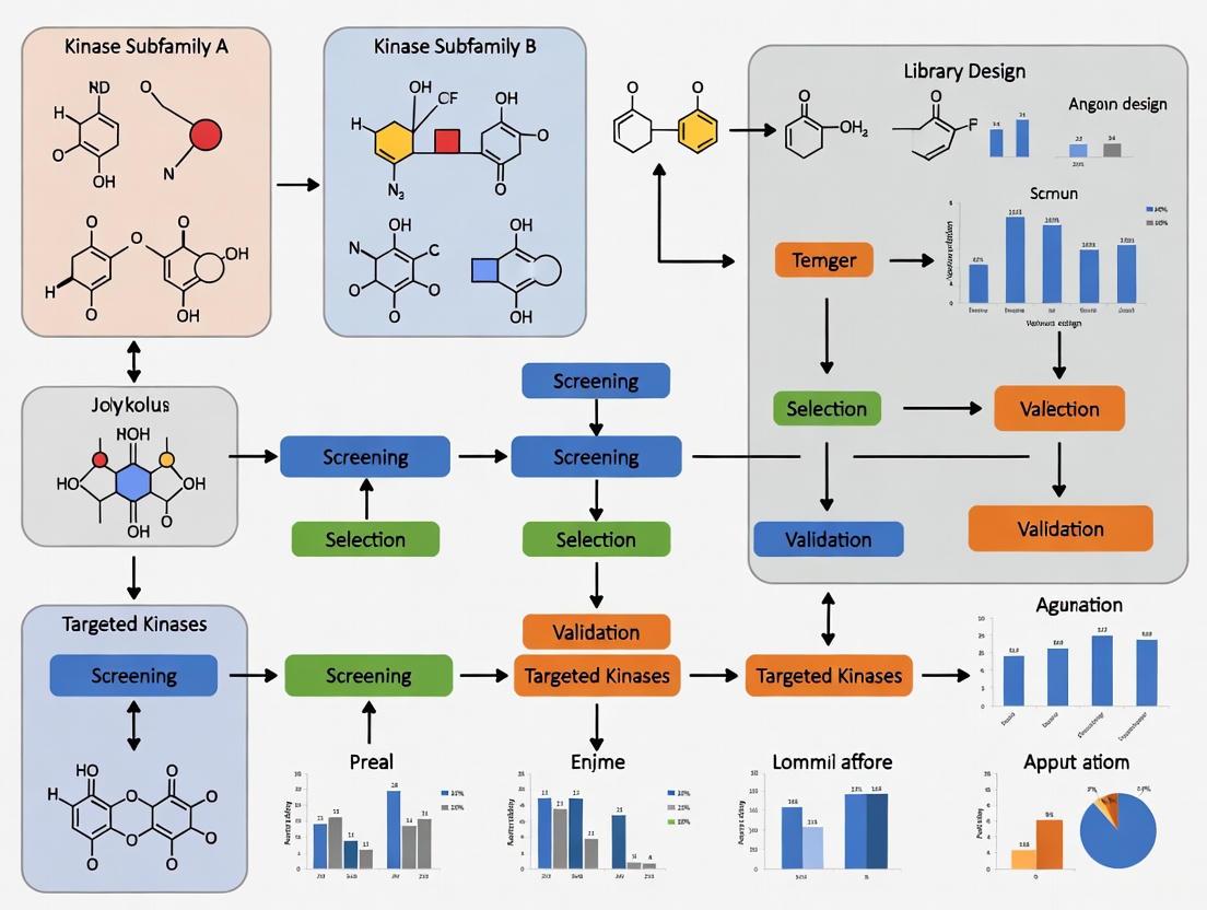
Protein kinases represent a vital drug target class due to their crucial role in key regulatory cell processes and their dysregulation in diseases such as cancer and autoimmune disorders [32]. The central challenge in kinase-focused library design lies in balancing comprehensive coverage of chemical space against practical selectivity for efficient screening. A data-driven curation approach leverages broad profiling data to create focused libraries that maximize biological relevance while maintaining synthetic feasibility and structural diversity.
The KinFragLib framework provides a powerful, data-driven fragment-based drug discovery (FBDD) approach with a subpocket-specific framework for creating potentially feasible kinase inhibitors [32]. However, the vast recombination space of 9,131 fragments presents significant computational and practical challenges for screening. This creates the fundamental selectivity-coverage tradeoff: how to distill a maximally informative yet manageable compound set from extensive profiling data.
Technical Support Center: Troubleshooting Guides and FAQs
Library Design and Curation FAQs
Q: What criteria should I use to filter a kinase-focused fragmentation library? A: An effective filtering pipeline should consider multiple drug-relevant aspects: synthesizability (using commercially available building blocks and synthetic accessibility scores), retrosynthetic pathway availability, favorable molecular properties associated with drug-likeness, and the removal of fragments containing unwanted substructures [32]. The CustomKinFragLib approach reduces libraries from 9,131 to 523 fragments while retaining diverse fragments with drug-like properties and high synthetic tractability.
Q: How can data curation profiles enhance my research data management? A: Data Curation Profiles capture detailed information about specific data forms generated in research, including needs for data curation from the perspective of data producers [33]. These profiles provide the flow of the research process from which data are generated and support exploration of data curation across different research domains in real and practical terms, covering data forms and stages, value, ingest, intellectual property, organization, tools, interoperability, and preservation [34] [33].
Q: What are the benefits of maintaining a well-curated FAQ for a research platform? A: Well-designed FAQ pages remain highly relevant as they help users who prefer self-service resources. Research indicates that 69% of users want to resolve as many issues as possible on their own, and 91% would use a knowledge base that meets their needs [35]. FAQs support asynchronous help and can alleviate challenges users face with site search functions when they use different terminology than the platform creators [35].
Experimental Troubleshooting Guides
Problem: Inefficient phosphorylation in kinase assays
- Cause: Excess salt, phosphate, or ammonium ions may inhibit the kinase activity [36].
- Solution: Purify the DNA prior to phosphorylation. For blunt or 5' recessed ends, heat the substrate/buffer mixture for 10 minutes at 70°C, then rapidly chill on ice before adding ATP and enzyme [36].
Problem: Few or no transformants
- Cause: ATP was not added to the reaction mixture [36].
- Solution: Supplement the reaction with 1mM ATP, as it is required by T4 Polynucleotide Kinase. Alternatively, use 1X T4 DNA Ligase Buffer (contains 1 mM ATP) instead of the 1X T4 PNK Buffer [36].
Problem: Unclear western blot results
- Approach: Optimize your protocol to obtain clearer results. Consult troubleshooting guides created by scientists specifically for improving western blotting, IHC, and IP results [37].
Quantitative Framework for Library Optimization
CustomKinFragLib Filtering Metrics and Outcomes
Table 1: CustomKinFragLib Pipeline Filtering Stages and Results
| Filtering Stage | Criteria Applied | Fragments Retained | Key Benefit |
|---|---|---|---|
| Initial KinFragLib | Data-driven, subpocket-specific framework | 9,131 | Comprehensive coverage of kinase chemical space |
| Synthesizability Filter | Commercially available building blocks | Reduced count | Ensures practical synthetic feasibility |
| Synthetic Accessibility Score | Computational assessment of synthetic complexity | Further reduced | Prioritizes readily synthesizable compounds |
| Retrosynthetic Pathway | Available synthetic routes | Additional filtering | Enhances practical utility for medicinal chemists |
| Drug-like Properties | Molecular properties associated with drug-likeness | Additional filtering | Improves likelihood of favorable ADMET properties |
| Unwanted Substructure | Removal of problematic chemical motifs | 523 | Eliminates promiscuous or toxic fragments |
Key Research Reagent Solutions for Kinase-Focused Screening
Table 2: Essential Research Reagents for Kinase-Focused Library Experiments
| Reagent/Resource | Function/Application | Example Use Cases |
|---|---|---|
| L1000 Assay | Perturbation-based cancer cell line gene expression profiling | Generating gene expression profiles and signatures in cancer cell lines [38] |
| T4 Polynucleotide Kinase (NEB #M0201) | Phosphorylation of DNA ends | Restoring 5' phosphate groups for ligation; radiolabeling DNA/RNA 5' ends [36] |
| T4 DNA Ligase Buffer | Contains 1 mM ATP | Alternative buffer for phosphorylation when ATP supplementation is needed [36] |
| Cultrex Basement Membrane Extract | 3D cell culture substrate | Culture of mouse enteric organoids, human intestinal, gastric, liver, and lung organoids [39] |
| Human Kinase-Focused Fragmentation Library | Fragment-based drug discovery for kinases | Subpocket-specific framework for creating kinase inhibitors through fragment enumeration [32] |
| Fluorogenic Peptide Substrates | Enzyme activity measurements | Enzyme activity assays for various targets including recombinant human ACE-2, BMP-1/PCP [39] |
Experimental Protocols for Library Validation
CustomKinFragLib Library Reduction Protocol
Objective: Reduce a kinase-focused fragmentation library while retaining diverse fragments with drug-like properties and high synthetic tractability.
Materials:
- Initial KinFragLib dataset (9,131 fragments)
- Computational resources for synthetic accessibility scoring
- Database of commercially available building blocks
- Retrosynthetic analysis software
- Drug-likeness and unwanted substructure filters
Methodology:
- Synthesizability Assessment: Filter fragments according to commercially available building blocks to ensure practical synthetic feasibility [32].
- Synthetic Accessibility Scoring: Calculate synthetic accessibility scores for each fragment to prioritize readily synthesizable compounds [32].
- Retrosynthetic Analysis: Filter for fragments with available retrosynthetic pathways to enhance practical utility for medicinal chemists [32].
- Molecular Property Screening: Apply filters for molecular properties often associated with drug-likeness to improve likelihood of favorable ADMET properties [32].
- Unwanted Substructure Removal: Eliminate fragments containing problematic chemical motifs that may cause promiscuous binding or toxicity [32].
- Diversity Assessment: Verify that the final reduced library (523 fragments) maintains chemical and subpocket binding diversity [32].
Kinase Inhibitor Profiling Using L1000 Assay
Objective: Generate perturbation-based cancer cell line gene expression profiles and signatures for kinase inhibitor characterization.
Materials:
- Genomically characterized human cancer cell lines
- L1000 assay platform
- Kinase inhibitors for screening
- Multiplexed compound screening infrastructure
Methodology:
- Cell Culture: Maintain genomically characterized human cancer cell lines under standard conditions [38].
- Compound Treatment: Apply kinase inhibitors at appropriate concentrations and time points using high-throughput multiplexed screening approaches [38].
- Gene Expression Profiling: Utilize the L1000 assay to measure gene expression responses to kinase inhibition [38].
- Signature Generation: Process gene expression data to generate characteristic signatures for each kinase inhibitor [38].
- Vulnerability Analysis: Identify cancer-specific vulnerabilities based on gene expression responses across different cancer cell lines [38].
Visualization of Data Curation Workflows
Kinase-Focused Library Curation Pipeline
Data Curation Profile Development Process
The CustomKinFragLib approach demonstrates that strategic data-driven curation enables a optimal balance between coverage and selectivity in kinase-focused library design. By applying multiple orthogonal filters—synthesizability, synthetic accessibility, retrosynthetic pathways, drug-like properties, and unwanted substructure removal—researchers can distill large fragment libraries (9,131 fragments) into focused sets (523 fragments) that retain chemical and biological diversity while enhancing practical utility. This curation philosophy, supported by robust troubleshooting resources and quantitative frameworks, provides a reproducible template for creating targeted screening libraries across multiple target classes beyond kinases, advancing efficient drug discovery through intelligent library design.
Protein kinases are one of the most important families of drug targets, playing a crucial role in cell signaling processes. A central challenge in kinase inhibitor development is achieving the right balance between coverage (the ability to address a wide range of kinases) and selectivity (specificity for a particular kinase target). Fragment-based drug discovery (FBDD) provides a powerful strategy to tackle this challenge. This approach involves screening small, low molecular weight compounds (fragments) and evolving them into larger, high-affinity ligands. Subpocket-focused fragmentation, as implemented in tools like KinFragLib, offers a data-driven methodology to systematically explore the kinase inhibitor space by decomposing known inhibitors into their core binding elements and enabling their rational recombination. This technical support guide addresses common experimental issues and provides detailed protocols for researchers working in this field [40] [41].
FAQs: Core Concepts and Troubleshooting
1. What is the core premise behind subpocket-focused fragmentation? The method is based on the observation that the kinase ATP-binding site can be divided into distinct, spatially separated subpockets. KinFragLib automatically splits co-crystallized kinase ligands from the KLIFS database into fragments using the BRICS algorithm and assigns each fragment to one of six predefined subpockets based on the ligand's 3D proximity to defined pocket-spanning residues. This allows for a systematic, data-driven deconstruction of the chemical space of known kinase inhibitors [41].
2. What are the defined kinase subpockets, and what are their functional roles? The table below details the six subpockets used in the KinFragLib framework.
Table 1: Kinase Binding Site Subpockets
| Subpocket Name | Functional Role and Characteristics |
|---|---|
| Adenine Pocket (AP) | Binds the adenine moiety of ATP; a highly conserved region. |
| Front Pocket (FP) | Located near the gatekeeper residue; important for selectivity. |
| Solvent-Exposed Pocket (SE) | Accessible to solvent; can accommodate polar groups. |
| Gate Area (GA) | A flexible region that can influence the kinase's DFG motif conformation. |
| Back Pocket 1 (B1) | Adjacent to the adenine pocket; a hydrophobic region. |
| Back Pocket 2 (B2) | Extends from the back pocket 1; can be targeted by Type II inhibitors. |
3. We are getting low fragment coverage in specific subpockets. How can we improve this? Low coverage often stems from a limited chemical starting set. To address this:
- Expand the Input Library: Ensure you are using the most current version of KinFragLib, which is constantly updated with new structures from the KLIFS database. The library is based on over 2,500 kinase DFG-in structures co-crystallized with non-covalent ligands, yielding over 7,000 fragments across the six subpockets [40] [41].
- Analyze Scaffold Diversity: Insights from broad kinase inhibitor profiling suggest that library design based on a maximum number of diverse scaffolds is superior to exploring a limited number of privileged scaffolds. A focused library with a limited scaffold set may not achieve the same kinome coverage as a more diverse library [18].
- Utilize the Custom Filtering Pipeline: Use the accompanying CustomKinFragLib framework to filter the fragment library based on drug-likeness (e.g., Rule of Three, QED), unwanted substructures (PAINS, Brenk filters), and synthesizability. This helps you focus on the most promising chemical matter [41].
4. Our recombined molecules have poor potency or selectivity. What could be going wrong? Poor outcomes from recombination can have several causes:
- Incorrect Fragment Assignment: Verify that the parent fragments were correctly assigned to their subpockets. Review the original crystal structures in KLIFS to confirm the binding mode.
- Ignoring Subpocket Strength: Remember that the strength of binding energy "hot spots" is not uniform. The primary hot spot (often the adenine pocket) contributes the most to binding energy. A fragment must have high ligand efficiency (LE ≥ 0.3 kcal/mol per heavy atom) to be a good starting point for optimization. A weak fragment in a secondary subpocket may not yield a potent molecule when recombined [42].
- Lack of Structural Complementarity: The recombination is structural, not just chemical. Ensure that the recombined fragments are spatially compatible and do not create steric clashes or unnatural torsion angles. Use energy minimization and docking to validate the proposed recombined structures.
5. How do we validate that our recombined molecules are novel and not already known? KinFragLib's recombination of a subset of fragments can generate millions of molecules. The tool provides functionality to check generated molecules against existing databases.
- Cross-Reference with Major Databases: A study showed that recombining only 624 representative fragments generated 6.7 million molecules, over 99% of which were novel compared to ChEMBL [40].
- Leverage Published Lists: The KinFragLib GitHub repository provides a list of publications that have utilized the library, allowing you to see the scope of previously generated ideas [41].
Experimental Protocols & Workflows
Protocol 1: Building a Custom Kinase-Focused Fragment Library
This protocol outlines the steps to create a filtered fragment library using KinFragLib and CustomKinFragLib.
Essential Materials: Table 2: Research Reagent Solutions for KinFragLib
| Reagent / Resource | Function and Description |
|---|---|
| KLIFS Database | The source of structural kinome data, including kinase structures, ligands, and annotated binding site residues. |
| KinFragLib Software | The Python package that performs the subpocket-focused fragmentation and recombination. |
| CustomKinFragLib | An extension of KinFragLib that provides a pipeline for filtering fragments based on multiple criteria. |
| BRICS Algorithm | The method used to break retrosynthetically interesting bonds in the co-crystallized ligands. |
Methodology:
- Environment Setup: Install KinFragLib by creating a Conda environment from the provided
environment.ymlfile and activate it. - Data Acquisition: The library will automatically retrieve the latest kinase-ligand complex data from the KLIFS database (downloads from a specific date, e.g., 06.12.2023, are available for reproducibility).
- Subpocket Assignment and Fragmentation: The tool will process each structure, dividing the binding pocket into the six subpockets (AP, FP, SE, GA, B1, B2) and fragmenting the ligand using BRICS. Fragments are assigned to the subpocket they occupy.
- Library Filtering (CustomKinFragLib): Run the custom filtering pipeline. The default filters include:
- Unwanted Substructures: Removes fragments matching PAINS and Brenk filters.
- Drug-likeness: Applies the Rule of Three (a fragment-specific version of Lipinski's Rule of Five) and Quantitative Estimate of Drug-likeness (QED).
- Synthesizability: Evaluates using the SYBA score and similarity to buyable building blocks.
- Library Output: The final output is a curated collection of fragment pools for each subpocket, ready for analysis or recombination [41].
The following diagram illustrates the key steps of the KinFragLib workflow, from data collection to library generation.
Protocol 2: Recombining Fragments to Generate Novel Inhibitors
This protocol describes how to generate novel kinase inhibitor candidates by recombining fragments from different subpockets.
Methodology:
- Select Subpockets of Interest: Based on your target kinase and desired selectivity profile, choose which subpockets you wish to target. For example, targeting the front pocket (FP) and solvent-exposed region (SE) can enhance selectivity.
- Retrieve Representative Fragments: From your curated KinFragLib library, extract fragments from the selected subpocket pools. You can use the entire set or a subset of representative fragments.
- Perform In-Silico Recombination: Use KinFragLib's recombination function to systematically link fragments from the selected subpockets. The tool generates molecular structures with 3D coordinates.
- Evaluate the Combinatorial Library: Analyze the generated library for size, novelty (e.g., by comparing to ChEMBL), and drug-likeness (e.g., Lipinski's Rule of Five compliance). As reported, this process can generate millions of novel molecules, with a high percentage being rule-of-five compliant [40].
- Structural Validation: It is critical to use molecular docking or other structure-based methods to validate that the recombined molecules can realistically bind to the target kinase conformation without steric clashes.
Troubleshooting Common Experimental Issues
Table 3: Troubleshooting Guide for Subpocket-Focused Experiments
| Problem | Potential Cause | Solution |
|---|---|---|
| Low Fragment Hit Rate | Target lacks strong binding energy "hot spots". | Perform a computational hot spot analysis (e.g., with FTMap) on the target structure prior to screening. Druggable sites contain a strong primary hot spot [42]. |
| Difficulty Achieving Selectivity | Recombined molecule primarily engages conserved regions like the Adenine Pocket. | Focus recombination strategies on fragments that bind to more variable regions, such as the Front Pocket (FP) and Solvent-Exposed (SE) pockets [43]. |
| Poor Ligand Efficiency (LE) | Initial fragment hit has weak affinity, or growth adds heavy atoms without proportional affinity gains. | Start with fragments that have high LE (≥0.3 kcal/mol per heavy atom). Monitor LE closely during fragment optimization and extension [42]. |
| High False Positive Rate in Screening | Fragment aggregation, assay interference, or detection at the limit of the technique. | Implement strict hit validation (e.g., orthogonal biophysical assays, competition experiments) as false positives are common in FBDD [44]. |
Scaffold Hopping and Bioisosteric Replacement for Novelty
Frequently Asked Questions (FAQs)
Q1: What is the fundamental difference between scaffold hopping and bioisosteric replacement?
A1: While both are core strategies in drug design for creating novel compounds, their focus and scope differ. Bioisosteric replacement involves swapping a functional group or a single atom with another that has similar biological or physicochemical properties, primarily aimed at improving pharmacokinetics, metabolic stability, or reducing toxicity [45] [46]. Scaffold hopping, a broader concept, involves replacing the central core framework of a molecule with a chemically different backbone. The goal is to create a novel chemotype that retains biological activity but may occupy new intellectual property (IP) space or have significantly improved drug-like properties [47] [48] [49].
Q2: How does scaffold hopping balance the need for novelty with the similarity property principle?
A2: The similarity property principle states that structurally similar molecules tend to have similar biological activities. Scaffold hopping navigates this by ensuring that while the core scaffold changes, the key 3D spatial arrangement of pharmacophoric features (e.g., hydrogen bond donors/acceptors, aromatic rings, hydrophobic regions) is conserved. This allows the new, structurally distinct molecule to maintain interactions with the biological target [47] [49]. Techniques like 3D pharmacophore modeling and molecular superposition are used to design and validate these hops [47].
Q3: In kinase inhibitor design, what are the strategic goals of applying scaffold hopping?
A3: In the kinome, where ATP-binding sites are highly conserved, scaffold hopping is crucial for:
- Achieving Selectivity: Designing novel cores that can bypass conserved regions and interact uniquely with specific kinase targets to reduce off-target effects.
- Overcoming Resistance: Developing inhibitors that remain effective against mutated kinases, a common resistance mechanism in oncology.
- Exploring New Modalities: Creating chemical space for allosteric, covalent, or bivalent inhibitors by moving away from well-trodden scaffold chemistries [28] [48].
- IP Generation: Creating novel chemical entities that are not covered by existing patents [47] [48].
Q4: What computational tools are available to facilitate scaffold hopping?
A4: Several computational methods enable scaffold hopping, often used in combination:
- Virtual Screening: Docking large compound libraries into a target protein's binding site to identify chemically diverse binders [49].
- Topological Replacement: Tools like SeeSAR's ReCore search for fragments that maintain the 3D geometry of the connection points from the original scaffold [49].
- Feature Trees (FTrees): This method compares molecules based on fuzzy pharmacophore properties and overall topology rather than exact structure, helping find distant chemical relatives [49].
- Shape Similarity Screening: Ligand-based methods that search for compounds with similar molecular shape and pharmacophore feature orientation [49].
- Machine Learning Models: Advanced graph generative and diffusion models can now generate novel scaffolds while constraining key functional motifs [50].
Troubleshooting Guides
Problem 1: Poor Selectivity in Novel Kinase Inhibitors
Issue: After a successful scaffold hop, the new compound shows potent activity against the intended kinase but also has high off-target activity against other kinases in the panel.
| Possible Cause | Diagnostic Steps | Solution |
|---|---|---|
| Incomplete SAR Understanding | Profile the compound against a broad kinome panel. Analyze co-crystal structures or perform docking studies to understand binding interactions. | Perform a medium- or large-step scaffold hop (e.g., ring closure/opening) to alter the overall topology and disrupt off-target interactions while conserving key hinge-binding motifs [47] [48]. |
| Conserved Hinge-Binding Motif | Check if the new scaffold uses the same hinge-binding strategy as the parent and other promiscuous inhibitors. | Explore alternative, less common hinge-binding interactions. Utilize a focused kinase library (e.g., Enamine's 64,960-compound library) designed with novel hinge binders and bioisosteric core replacements to find inspiration [28]. |
| Ligand Over-flexibility | Conduct conformational analysis. A flexible molecule may adapt to multiple binding sites. | Rigidify the scaffold by introducing ring systems or conformational constraints, as seen in the development of Cyproheptadine from Pheniramine, to reduce entropy loss upon binding and improve selectivity [47]. |
Problem 2: Loss of Potency After Scaffold Replacement
Issue: The new scaffold hop resulted in a molecule with the desired novelty and physicochemical properties, but it shows a significant drop in target potency.
| Possible Cause | Diagnostic Steps | Solution |
|---|---|---|
| Disrupted Key Interaction | Use molecular modeling to superimpose the new and old scaffolds in the binding site. Identify lost critical interactions (H-bonds, ionic, etc.). | Employ bioisosteric replacement on the peripheral groups of the new scaffold to reintroduce the lost interaction. For example, a carboxylic acid can be replaced with a tetrazole ring to maintain acidity while altering topology [46]. |
| Incorrect Vector Alignment | Analyze if the substituents attached to the new scaffold point in different directions than the original, misaligning pharmacophores. | Use topological replacement tools (e.g., ReCore) that specifically search for fragments with similar 3D vector orientations for the attachment points [49]. |
| Unfavorable Conformation | Determine the low-energy conformation of the new molecule. It may not be the one that presents the pharmacophore optimally. | Revert to a small-step hop (1° hop), such as a heterocycle replacement (e.g., phenyl to pyridyl), which is more likely to conserve the active conformation and restore activity [47] [48]. |
Problem 3: Inadequate Novelty or IP Space
Issue: The scaffold hop is successful but is considered too structurally similar to existing compounds, offering limited freedom-to-operate.
| Possible Cause | Diagnostic Steps | Solution |
|---|---|---|
| Over-reliance on Small-step Hops | Analyze the maximum common substructure (MCS) between your compound and known inhibitors. A high MCS indicates low novelty. | Pursue topology-based hopping, which can lead to a high degree of structural novelty. Tools like FTrees are designed to find "distant relatives" with similar pharmacophores but different cores [47] [49]. |
| Limited Virtual Screening Library | The chemical library screened was not diverse enough. | Screen larger and more diverse chemical spaces, such as the over 575,000 compounds in the Asinex screening libraries, which include natural product-like and macrocyclic scaffolds with high 3D character [51]. |
| Conservative Design | The design was overly constrained by the parent molecule's structure. | Apply ring opening or closure strategies. A classic example is the hop from the rigid morphine to the more flexible tramadol, which created a novel scaffold with a different activity profile [47]. |
Experimental Protocols & Data Presentation
Key Methodologies for Scaffold Hopping
Table 1: Summary of Core Experimental and Computational Approaches
| Method Category | Key Technique | Underlying Principle | Application in Kinase Library Design |
|---|---|---|---|
| Computational Screening | Virtual Screening with Pharmacophore Constraints [49] | Docking compounds while enforcing key interactions (H-bond donors/acceptors, aromatic) with the target. | Enrich screening hits for novel scaffolds that maintain critical hinge-binding interactions. |
| Shape Similarity Screening [49] | Identifies compounds with similar 3D shape and feature orientation to a known active, regardless of core structure. | Find novel chemotypes that mimic the binding pose of a successful inhibitor. | |
| Structure-Based Design | Topological Replacement [49] | Replaces a core fragment with another that has a similar 3D arrangement of its connection points. | Systematically generate novel core structures that maintain the spatial orientation of key substituents. |
| Bioisosteric Replacement [45] [46] | Swaps an atom/group with another of similar physicochemical properties (e.g., -COOH with tetrazole; phenyl with thiophene). | Optimize ADMET properties and fine-tune potency of a scaffold hop lead, as seen in antihistamine development [47]. | |
| Medicinal Chemistry Synthesis | Heterocycle Replacement (1°-hop) [47] [48] | Swaps or substitutes carbon and heteroatoms in a backbone ring (e.g., carbon for nitrogen in an aromatic ring). | Creates patentably distinct backups (e.g., Vardenafil from Sildenafil) with minimal synthetic effort [47]. |
| Ring Opening or Closure (2°-hop) [47] [48] | Modifies ring systems to alter flexibility and overall topology (e.g., Morphine to Tramadol). | Drastically changes molecular shape to explore new binding modes and improve properties [47]. |
The Scientist's Toolkit: Essential Research Reagents & Materials
Table 2: Key Reagent Solutions for Kinase-Focused Scaffold Hopping
| Research Reagent / Library | Function & Utility | Example Provider / Composition |
|---|---|---|
| Focused Kinase Library | Pre-designed libraries of compounds with scaffolds and motifs known to target the kinome. Provides a starting point for screening novel inhibitors. | Enamine Kinase Library (64,960 compounds) includes hinge binders and allosteric inhibitors derived from bioisosteric core replacements [28]. |
| Diverse Screening Library | Large, structurally diverse compound sets for virtual or HTS screening. Essential for finding truly novel chemotypes through unbiased approaches. | Asinex Screening Libraries (e.g., BioDesign, Elite, Synergy), comprising over 575,000 drug-like compounds with natural product-like frameworks [51]. |
| Validated Inhibitor Sets | Curated collections of known kinase inhibitors at multiple concentrations. Used as tools for target validation and phenotypic fingerprinting. | LSP Optimal Kinase Library (192 compounds at 4 concentrations), designed to have two structurally distinct, selective inhibitors per kinase [52]. |
| Building Blocks & Fragments | Chemical reagents for synthesizing novel compounds. Fragments are used in FBDD to build new scaffolds from minimal binders. | Asinex Building Blocks (22,525 compounds) and Fragments (20,061 compounds), including privileged, natural product-like scaffolds [51]. |
| Computational Software | Enables in silico scaffold hopping through virtual screening, pharmacophore modeling, and topological replacement. | Software suites like SeeSAR (for virtual screening and ReCore) and infiniSee (for FTrees-based chemical space navigation) [49]. |
Workflow and Pathway Visualizations
Scaffold Hopping Workflow for Kinase Inhibitor Design
Scaffold Hop Classification and Example Map
FAQs: Library Design and Selection
Q1: What is the fundamental challenge in designing a kinase-focused library, and how do different library strategies address it?
The core challenge is balancing coverage of the diverse kinome with selectivity against highly conserved ATP-binding pockets. Different libraries address this by focusing on distinct inhibitory mechanisms and leveraging different design principles [53] [14]:
- ATP-pocket focused libraries (e.g., Hinge Binders) aim for broad coverage by targeting the conserved hinge region, a key anchor point for most kinase inhibitors [54].
- Allosteric libraries prioritize selectivity by targeting less-conserved, unique pockets outside the ATP-binding site, such as the myristoyl or PIF pockets [55].
- Data-driven minimal libraries (e.g., LSP-OptimalKinase) use historical bioactivity data to select a small set of compounds that maximally represent the activity and selectivity profiles of a much larger collection [53].
Q2: When should I choose a large, comprehensive kinase library versus a small, optimized informer set?
The choice depends on your campaign's stage and resources [53]:
- Use a large library (e.g., Enamine's 64,960-compound set) for primary, high-throughput screening when you have the capacity to test many compounds and want to maximize the chance of finding novel chemical matter [28].
- Use a small informer set (e.g., the 256-compound LSP-OptimalKinase set) for pilot screens, in complex or expensive assay systems (like phenotypic screens), or when you need to quickly triage multiple targets. These sets capture the performance diversity of larger collections economically [53].
Q3: How are modern computational methods helping to improve kinase library design and screening?
Advanced computational techniques now enable more efficient and selective discovery directly from ultra-large chemical spaces.
- Chemical Space Docking: This method screens billions of make-on-demand compounds by docking representative building blocks and combinatorially assembling top-scoring fragments, avoiding the need to enumerate the entire library. This successfully identified potent ROCK1 kinase inhibitors from a billion-compound space [56].
- Free Energy Calculations: These physics-based simulations (L-RB-FEP+ and PRM-FEP+) accurately predict binding affinity and selectivity. They can optimize for kinome-wide selectivity by simulating the effect of mutating key "selectivity handle" residues (like the gatekeeper) in the target, guiding the selection of compounds with reduced off-target liabilities [57].
Troubleshooting Guides
Problem: High Hit Rate with Promiscuous Binders
Issue: Primary screening yields a high number of hits, but most compounds show poor selectivity and likely target the conserved ATP site non-specifically.
Solutions:
- Employ Allosteric Focused Libraries: Shift screening to a library specifically designed for allosteric kinase inhibitors, such as the 4,800-compound Allosteric Kinase Library from Enamine. These compounds are pre-selected via docking into known allosteric pockets and can offer superior selectivity [55].
- Incorporate Specificity Filters Early: Use computational tools to profile hits against a panel of kinase structures. The Kinase Drug Selectivity (KDS) software, which identifies unique structural features in kinase drug pockets, can help prioritize scaffolds that target unique regions [14].
- Counter-Screen Against Nuisance Compounds: Filter out compounds that match known nuisance or promiscuous chemotypes. Resources like the "nuisance compound" set on Probes&Drugs can help eliminate artifacts [53] [58].
Problem: Low Hit Rate or Lack of Novel Chemotypes
Issue: The screen returns very few active compounds, or the hits are all from well-known, patented chemical series.
Solutions:
- Expand to Ultra-Large Chemical Spaces: Move beyond standard screening collections. Use computational methods like Chemical Space Docking or the REvoLd algorithm to screen trillion-sized make-on-demand libraries (e.g., Enamine REAL Space). This provides access to vast, novel chemistry that is synthetically accessible [59] [56].
- Leverive a Targeted Sublibrary: Focus on a specific, well-designed sublibrary. For example, the 24,000-compound Hinge Binders Library applies validated topological models to identify novel chemotypes capable of forming key hydrogen bonds with the hinge region [54].
- Validate Assay Conditions: Ensure your assay system is optimized for detecting inhibitors. Use a known high-quality chemical probe as a positive control. These are listed in resources like ChemicalProbes.org and the SGC Chemical Probes [58].
Problem: Hit Series Shows Off-Target Toxicity in Cellular Assays
Issue: A promising hit from a biochemical screen shows unexpected toxicity in cells, suggesting off-target inhibition across the kinome.
Solutions:
- Profile with Kinome-Wide Selectivity Panels: Test the compound in a commercial kinome-wide panel (e.g., DiscoverX scanMAX) to identify specific off-targets [57].
- Apply Protein Residue Mutation Free Energy Calculations (PRM-FEP+): Use this advanced computational method to understand the structural basis for off-target binding. By simulating the mutation of a key residue in your target (e.g., the gatekeeper) to match an off-target, you can predict if a compound's binding is sensitive to that residue, guiding selective chemical optimization [57].
- Consult Public Selectivity Data: Before synthesizing new analogs, check existing data for similar structures. Resources like the Kinase Chemogenomic Set and Probe Miner provide selectivity information for many published compounds [58].
Experimental Protocols & Workflows
Protocol 1: Structure-Based Virtual Screening of a Kinase-Targeted Library
This protocol outlines the method used to successfully discover ROCK1 kinase inhibitors from a billion-compound chemical space [56].
Target and Library Preparation:
- Obtain a high-resolution crystal structure of the target kinase (e.g., PDB: 2ETR for ROCK1).
- Define the chemical space using reaction rules and building blocks from a make-on-demand supplier (e.g., Enamine).
Fragment Docking and Selection:
- Dock all available building block fragments into the kinase's binding site using a program like FlexX.
- Apply a pharmacophore constraint to ensure hinge-binding interactions.
- Score poses with a physics-based function like HYDE.
- Manually select the top ~500 fragment poses based on criteria: additional H-bonds, ligand efficiency, favorable linker geometry, and chemical diversity.
Combinatorial Expansion and Product Docking:
- For each selected fragment, combinatorially enumerate all possible full-length products using the supplier's reaction rules.
- Dock the resulting millions of products using the original fragment pose as a template.
Pose Filtering and Hit Selection:
- Filter docked poses by internal strain energy and re-dock with a second program (e.g., FRED) for consensus.
- Cluster the top-scoring compounds and select cluster representatives to ensure chemical diversity.
- Manually inspect and prioritize compounds for purchase and biochemical testing.
Workflow for structure-based virtual screening of ultra-large libraries.
Protocol 2: Optimizing Kinome-Wide Selectivity Using Free Energy Calculations
This protocol is derived from the case study that discovered selective Wee1 inhibitors [57].
Hit Identification with Ligand-Based FEP (L-RB-FEP+):
- Start with a reference compound with known binding affinity to the target kinase (Wee1).
- Generate billions of design ideas using de novo design and enumeration tools.
- Use L-RB-FEP+ to predict the relative binding free energy of these designs versus the reference in the target kinase.
- In parallel, run L-RB-FEP+ for top designs against key off-target kinases (e.g., PLK1) to deselect non-selective compounds early.
- Synthesize and test the most promising, potent, and selective designs.
Selectivity Optimization with Protein Mutation FEP (PRM-FEP+):
- Profile the novel hits in a broad kinome-wide panel (e.g., 403 kinases) to identify off-target liabilities.
- Analyze the results to identify key "selectivity handle" residues (e.g., the gatekeeper residue). Wee1 has a rare Asn gatekeeper.
- For a compound and a problematic off-target, use PRM-FEP+ to calculate the binding free energy change when mutating the on-target's gatekeeper (Asn in Wee1) to match the off-target's gatekeeper (e.g., Thr, Val, Phe).
- A large, unfavorable ΔΔG value for the mutation indicates the compound's binding is highly dependent on the on-target's unique residue, meaning it is inherently selective. Use this to prioritize and optimize compounds.
Workflow for using free energy calculations to achieve kinome-wide selectivity.
The Scientist's Toolkit: Research Reagent Solutions
| Category / Reagent | Function / Application | Example Libraries & Sizes |
|---|---|---|
| Comprehensive Kinase Libraries | Large-scale collections for primary HTS; broad coverage of chemotypes and inhibitory modes. | Enamine Kinase Library (64,960 compounds) [28] |
| Targeted Sublibraries | Focused sets for specific binding modes or pilot screens; higher hit rate for intended goal. | Hinge Binders Library (24,000 compounds) [54], Allosteric Kinase Library (4,800 compounds) [55] |
| Data-Driven Informer Sets | Small, smart subsets representing larger collections; ideal for expensive or complex assays. | LSP-OptimalKinase Set (256 compounds) [53] |
| Ultra-Large Make-on-Demand | Trillion+ sized virtual spaces for novel hit discovery via computational screening. | Enamine REAL Space (Billions of compounds) [59] [56] |
| Publicly Available Probe Sets | Curated, high-quality chemical probes with well-characterized activity and selectivity. | SGC Chemical Probes, ChemicalProbes.org, Bromodomain Toolbox [58] |
| Kinome-Wide Profiling Services | Experimental testing of compound selectivity across hundreds of human kinases. | DiscoverX scanMAX Panel (403 kinases) [57] |
| Computational Selectivity Tools | Software for analyzing and predicting kinase inhibitor selectivity from structural data. | KDS (Kinase Drug Selectivity) Software [14] |
Beyond Promiscuity: Advanced Strategies for Selectivity Optimization
Troubleshooting Guides
Guide 1: Addressing Off-Target Kinase Activity (e.g., PLK1 Inhibition)
Problem: Your Wee1 inhibitor demonstrates potent anti-tumor activity but shows significant off-target inhibition against PLK1, which is associated with dose-limiting toxicities like thrombocytopenia and neutropenia in clinical trials [60].
Solution:
- Root Cause: The ATP-binding pockets of Wee1 and PLK1 share structural similarities, but key residue differences exist, particularly in the selectivity pocket where PLK1 has a bulkier leucine (L130) gatekeeper residue compared to Wee1's asparagine (N376) [60].
- Approach: Utilize structure-based drug design (SBDD) to modify compounds targeting the selectivity pocket. Incorporate larger aliphatic or cyclic substituents that are tolerated by Wee1's smaller gatekeeper but sterically hindered by PLK1's bulkier gatekeeper [60].
- Validation: Perform kinase selectivity profiling across a broad panel. Use colony forming unit–megakaryocyte (CFU-Mk) assays to assess potential thrombocytopenia risk [60].
Table: Selectivity Pocket Modifications and Their Effects
| Compound Type | R1 Substituent | Wee1 Potency | Selectivity (Wee1/PLK1) |
|---|---|---|---|
| AZD1775 (reference) | Allyl | +++ | ~1-fold |
| Linear aliphatic | Extended chain | ++ | >10-fold improvement |
| Bulky aliphatic | Cyclic groups | +++ | >10-fold improvement |
| Aromatic | Phenyl derivatives | + to ++ | >10-fold improvement |
Guide 2: Managing Hematological Toxicity Despite Improved Selectivity
Problem: After achieving improved selectivity over PLK1, your compound still shows myelosuppression in preclinical models.
Solution:
- Root Cause: Hematological toxicity may be an on-target effect of Wee1 inhibition rather than solely caused by PLK1 off-target activity [60].
- Approach:
- Validation: Monitor CDK1 phosphorylation (pY15) as a proximal biomarker of Wee1 inhibition. Correlate levels with both efficacy and toxicity endpoints [60].
Guide 3: Leveraging Computational Methods for Selectivity Optimization
Problem: Traditional medicinal chemistry approaches have failed to yield compounds with sufficient selectivity against kinome-wide off-targets.
Solution:
- Root Cause: Limited understanding of how compound modifications affect binding to diverse kinase off-targets beyond PLK1.
- Approach: Implement free energy perturbation (FEP+) calculations to predict binding affinity for both Wee1 and off-target kinases. Use protein FEP+ to model the impact of single point-mutations on ligand affinity and infer broad kinome selectivity [64].
- Validation: Combine computational predictions with broad kinase panel screening (≥ 100 kinases). For promising candidates, validate in vivo efficacy in relevant xenograft models [64].
Table: Key Experiments for Assessing Selectivity and Toxicity
| Experiment | Methodology | Endpoint Measurements | Interpretation Guide |
|---|---|---|---|
| Kinase Selectivity Profiling | Broad kinase panel screening (e.g., 100-400 kinases) | % Inhibition at 1 μM; IC50 values for key off-targets | >100-fold selectivity over PLK1 and other hematotoxicity-associated kinases desired |
| Cellular Target Engagement | Meso Scale Discovery (MSD) cellular assay for CDK1 phosphorylation (pY15) | IC50 for pY15-CDK1 reduction | Correlate with anti-proliferative activity; indicates on-target engagement |
| In Vitro Thrombocytopenia Assessment | Colony forming unit–megakaryocyte (CFU-Mk) assay | Inhibition of megakaryocyte colony formation | >50% inhibition at therapeutic concentrations indicates high thrombocytopenia risk |
| In Vivo Efficacy | Mouse xenograft models (e.g., A427 model) | Tumor growth inhibition; pY15-CDK1 reduction in tumor tissue | Sustained tumor growth inhibition with intermittent dosing suggests viable therapeutic window |
Frequently Asked Questions (FAQs)
Q1: Is PLK1 inhibition the primary cause of thrombocytopenia observed with Wee1 inhibitors like adavosertib?
Initially, researchers hypothesized that PLK1 inhibition was responsible for thrombocytopenia, as PLK1 inhibitors consistently report this toxicity. However, studies with selective Wee1 inhibitors devoid of PLK1 activity still demonstrated thrombocytopenia in CFU-Mk assays, suggesting this may be an on-target effect of Wee1 inhibition [60].
Q2: What computational approaches are most effective for designing selective Wee1 inhibitors?
The most successful approaches combine multiple computational methods:
- FEP+ calculations predict binding affinity with accuracy within 1.0 kcal/mol of experimental values on average [64].
- Protein FEP+ models the impact of single point-mutations on ligand affinity across kinase families without separate profiling [64].
- Machine learning models (e.g., DeepAutoQSAR) predict ADME properties and CYP inhibition liabilities [64].
- Quantum mechanical calculations assess compound reactivity with CYP enzymes to mitigate drug-drug interaction risks [64].
Q3: Are there successful examples of selective Wee1 inhibitors in clinical development?
Yes, next-generation Wee1 inhibitors with improved selectivity profiles have entered clinical development:
- Azenosertib (ZN-c3) shows optimized pharmacokinetic/pharmacodynamic properties with robust tumor growth inhibition in preclinical models and is in Phase I studies [61].
- SGR-3515 was nominated as a development candidate demonstrating exquisite selectivity, differentiated ADME profile, and superior in vivo efficacy compared to adavosertib [64].
- IMP7068 and Debio 0123 are novel highly selective Wee1 inhibitors that recently entered clinical testing [63].
Q4: What biomarkers predict response to Wee1 inhibition?
The most promising biomarkers include:
- TP53 mutations create synthetic lethality with Wee1 inhibition due to G1 checkpoint deficiency [62].
- CCNE1 amplification (encoding cyclin E1) has shown positive results in clinical trials [63].
- High replication stress signatures indicate increased dependence on G2/M checkpoint control [62].
- CDK1 phosphorylation status serves as a proximal pharmacodynamic marker of target engagement [60] [61].
Experimental Protocols
Protocol 1: Colony Forming Unit–Megakaryocyte (CFU-Mk) Assay for Thrombocytopenia Risk Assessment
Purpose: Evaluate the potential of Wee1 inhibitors to cause thrombocytopenia by assessing their effect on megakaryocyte progenitor cells.
Materials:
- Human CD34+ hematopoietic stem cells or bone marrow mononuclear cells
- MegaCult-C Complete Kit with Cytokine Supplement (or equivalent)
- Test compounds dissolved in appropriate vehicle (e.g., DMSO)
- Collagen-based matrix for megakaryocyte growth
- Double-chamber slides or culture plates
- Acetone for fixation
- Anti-CD41 antibody for immunostaining
Methodology:
- Isolate and purify CD34+ cells from human cord blood or bone marrow (purity >90%).
- Prepare culture medium with optimal cytokine concentrations (thrombopoietin, IL-3, IL-6).
- Mix 5,000-50,000 cells/mL with test compounds at multiple concentrations (typically 0.1-10 μM) and culture in collagen-based matrix.
- Incubate for 10-14 days at 37°C, 5% CO2 in a humidified atmosphere.
- Fix cultures with acetone and stain for CD41 expression to identify megakaryocyte colonies.
- Count colonies (≥10 cells) under microscope; express results as % inhibition relative to vehicle control.
Interpretation: Compounds showing >50% inhibition of CFU-Mk formation at clinically relevant concentrations (unbound Cmax) indicate high thrombocytopenia risk [60].
Protocol 2: Cellular Target Engagement Assay Using CDK1 Phosphorylation
Purpose: Measure compound potency in cellular systems by quantifying inhibition of Wee1-mediated CDK1 phosphorylation.
Materials:
- Cancer cell lines with TP53 mutation (e.g., OVCAR-3, MDA-MB-231)
- Meso Scale Discovery (MSD) phospho-CDK1 (Tyr15) assay kit
- Cell lysis buffer (RIPA with phosphatase/protease inhibitors)
- Test compounds in DMSO
- MSD MULTI-ARRAY or MULTI-SPOT plates
Methodology:
- Seed cells in 96-well plates at 10,000-20,000 cells/well and incubate for 24 hours.
- Treat with compound serial dilutions (typically 0.1 nM-10 μM) for 2-16 hours.
- Lyse cells and transfer lysates to MSD plates pre-coated with capture antibody.
- Incubate with detection antibody and read using MSD SECTOR instrument.
- Normalize pY15-CDK1 levels to total CDK1 or total protein.
- Calculate IC50 values using four-parameter logistic curve fitting.
Interpretation: Compounds should show concentration-dependent reduction of pY15-CDK1. Cellular potency within 10-fold of enzymatic activity suggests good cell permeability [60] [61].
Signaling Pathways and Experimental Workflows
WEE1 Inhibition Mechanism: This diagram illustrates how WEE1 inhibitors disrupt the DNA damage response pathway, forcing cancer cells with DNA damage into mitotic catastrophe.
Selectivity Optimization Workflow: This workflow outlines the integrated computational and experimental approach for addressing selectivity issues in Wee1 inhibitor development.
Research Reagent Solutions
Table: Essential Research Tools for Wee1 Inhibitor Development
| Reagent/Assay | Function/Purpose | Key Applications |
|---|---|---|
| Recombinant Wee1 Kinase | Enzymatic activity assays | Initial compound screening and IC50 determination |
| PLK1 and Kinome Panel | Selectivity profiling | Identification of off-target kinase activities |
| TP53-Mutant Cancer Cells | Cellular efficacy models | Assessment of synthetic lethality (e.g., OVCAR-3, MDA-MB-231) |
| Phospho-CDK1 (Tyr15) Antibody | Target engagement biomarker | MSD, Western blot, and immunofluorescence assays |
| CD34+ Hematopoietic Stem Cells | Myelosuppression assessment | CFU-Mk assays for thrombocytopenia risk prediction |
| MSD Phospho-CDK1 Assay Kit | Cellular potency quantification | High-throughput measurement of target modulation |
| X-ray Crystallography Systems | Structure determination | Co-crystal structures of Wee1-compound complexes for SBDD |
| Mouse Xenograft Models | In vivo efficacy evaluation | PK/PD relationships and therapeutic window determination |
Harnessing Free Energy Calculations (FEP+) for Potency and Selectivity Predictions
Troubleshooting Common FEP+ Challenges
Q1: My FEP+ predictions for a kinase target show poor correlation with experimental data. What are the primary areas I should investigate?
Poor predictive accuracy often stems from three main areas: inadequate sampling of protein flexibility, incorrect ligand protonation/tautomeric states, or issues with the input protein structure. For kinase targets, which often have flexible loops (like the DFG loop) and multiple conformational states, extending the sampling time is frequently necessary. One study found that increasing the prior to REST (pre-REST) sampling time from the default 0.24 ns/λ to 5 ns/λ for regular flexible-loop motions, or 2 × 10 ns/λ for systems with considerable structural changes, significantly improved the accuracy and precision of ∆∆G predictions [65]. Furthermore, ensure that the protonation states of key binding site residues and your ligands have been carefully assigned using tools like the Protein Preparation Wizard, as this fundamentally affects the calculated interaction energies [66] [67].
Q2: How can I apply FEP+ to a kinase project if I don't have a high-resolution crystal structure for my target?
FEP+ has demonstrated robustness when used with high-quality homology models [67]. The molecular dynamics sampling inherent in FEP+ allows the modeled receptor to adapt to the correct conformation for each ligand in the series. Successful applications have been documented for kinases like TYK2, even when the sequence identity of the homology model template was as low as 22% [67]. The critical step is to perform a retrospective benchmark on a set of ligands with known affinities to validate the predictive power of your homology model before proceeding to prospective calculations [68]. This benchmarks the system and confirms the setup is reliable.
Q3: What strategies can I use to improve sampling for kinase systems with significant backbone flexibility or multiple binding poses?
For flexible kinase binding sites, consider these advanced protocols:
- Preliminary MD Simulations: Executing reasonably long (≈100–300 ns) preliminary molecular dynamics (MD) simulations can help identify stable binding modes and correct protein conformations, providing a better starting structure for FEP+ [65].
- Utilize pREST: The protein REST (pREST) methodology allows you to include important flexible protein residues in the ligand binding domain within the "hot region" for enhanced sampling. This is particularly useful for residues known to undergo conformational changes upon ligand binding [65].
- Extended REST Sampling: Extending the REST simulation time from the default 5 ns to 8 ns or more can improve free energy convergence, especially for challenging transformations [65].
Q4: How accurate can I expect my FEP+ predictions to be, and what is the benchmark against experimental data?
When systems are carefully prepared, FEP+ can achieve an accuracy matching the reproducibility of experimental methods. Large-scale validation studies have shown that FEP+ predictions routinely achieve a mean unsigned error of approximately 1.0 kcal/mol relative to experimental binding affinity measurements, which is comparable to the typical error between different experimental assays [69] [66]. The following table summarizes key performance metrics from validation studies:
Table 1: FEP+ Performance Benchmarks Against Experimental Data
| Target Class | Number of Ligands/Transformations | Reported Accuracy (Mean Unsigned Error) | Key Citation |
|---|---|---|---|
| Diverse Protein Classes | 512+ protein-ligand pairs | ~1.0 kcal/mol | [66] |
| Kinases (e.g., TYK2) | Not Specified | ~1.0 kcal/mol | [69] [67] |
| GPCRs (e.g., A2A) | Not Specified | ~1.0 kcal/mol | [69] [67] |
Essential Experimental Protocols
Protocol 1: Structure Preparation and Validation for Kinase FEP+
A rigorous structure preparation protocol is critical for success.
- Protein Preparation: Use the Protein Preparation Wizard to add missing atoms, assign bond orders, and optimize the hydrogen-bonding network. Critically, assign correct tautomerization and protonation states for binding site residues at pH 7.0. For kinases, pay special attention to the catalytic aspartate in the DFG motif and other key residues like the gatekeeper [65] [67].
- Ligand Preparation: Prepare 3D ligand structures using LigPrep, ensuring accurate protonation, tautomeric, and stereochemical states. The use of embedded QikProp or similar tools can help identify unusual pKa values that may require manual adjustment [70].
- Ligand Alignment: For relative FEP+ calculations, a correct common core alignment for the ligand series is paramount. Use core-constrained docking or manual alignment based on a known co-crystal structure to ensure the perturbations are chemically meaningful [69] [70].
Protocol 2: Running a Robust FEP+ Calculation
This protocol outlines the steps for a production-level FEP+ run, incorporating improved sampling parameters.
- Retrospective Benchmarking (Mandatory): Before any prospective predictions, run FEP+ on a congeneric series of ligands with known affinities. This validates your structural model and setup. A correlation of R² > 0.5-0.6 and an average error < 1.2 kcal/mol is a good indicator that the system is well-prepared for prospective design [68].
- Map Generation: Use the FEP Mapper to automatically generate a network of perturbations that connects all ligands in your study. This leverages cycle closure corrections to improve overall accuracy [67].
- Sampling Configuration: Do not rely on default sampling times for flexible kinases. Implement an improved protocol [65]:
- For systems with flexible loops or minor structural changes: Use 5 ns/λ for pre-REST and 8 ns/λ for REST simulations.
- For systems with major backbone movements or large ligand modifications: Use 2 × 10 ns/λ for pre-REST sampling.
- pREST Setup: Based on preliminary MD analysis, identify flexible protein residues in the binding site (e.g., activation loop residues) and include them in the pREST region to enhance their sampling [65].
Protocol 3: Analysis and Troubleshooting of Results
- Analyze Output: Use the FEP+ analysis tools to generate a correlation plot of predicted vs. experimental ∆∆G values and review the structural snapshots for key transformations to ensure the binding modes remain physically reasonable [70].
- Diagnose Errors: Large errors for specific ligands can often be diagnosed by inspecting the time series of the free energy difference for that transformation. A lack of convergence indicates insufficient sampling and may require extending the simulation time or re-evaluating the ligand's binding pose [65] [70].
- Leverage Active Learning: For very large libraries (thousands to millions of compounds), use the Active Learning workflow, which trains a machine learning model on initial FEP+ data to efficiently prioritize compounds for full FEP+ calculation, dramatically accelerating the exploration of chemical space [69].
Workflow Visualization
FEP+ Project Workflow
The Scientist's Toolkit: Key Research Reagent Solutions
Table 2: Essential Computational Tools for FEP+ in Kinase Research
| Tool / Resource | Function in Workflow | Application Context |
|---|---|---|
| Maestro | A comprehensive modeling environment; the primary graphical user interface for setting up, running, and analyzing FEP+ calculations [69]. | Central hub for all structure-based design work. |
| Protein Preparation Wizard | Prepares protein structures for simulation by adding hydrogens, assigning protonation states, optimizing H-bonds, and filling missing loops/side chains [65] [67]. | Critical first step for ensuring a high-quality, physically realistic input structure. |
| LigPrep | Generates accurate 3D ligand structures with correct chiralities, protonation, and tautomeric states [65]. | Essential for preparing ligand libraries for both docking and FEP+ calculations. |
| Desmond | Schrödinger's high-performance molecular dynamics engine; the simulation core that executes the FEP+ calculations on GPUs [71] [67]. | Handles the computationally intensive sampling required for free energy calculations. |
| OPLS4/OPLS5 Force Field | A modern, comprehensive force field that defines the potential energy functions and parameters for proteins, ligands, and solvent [69] [67]. | The physical model governing atomic interactions; fundamental to accuracy. |
| LiveDesign | A collaborative platform that enables real-time project tracking and allows teams to design compounds, run FEP+ predictions, and view results in a shared dashboard [69] [71]. | Facilitates team-based molecular design and decision-making in lead optimization. |
FAQs: PRM-FEP+ in Kinase Library Design
Q1: How can PRM-FEP+ help balance coverage and selectivity in kinase-focused library design? PRM-FEP+ provides accurate predictions of binding affinity changes (ΔΔG) upon mutating residues at protein-protein or protein-ligand interfaces. In kinase library design, this allows you to computationally screen mutations for optimizing interactions with unique selectivity handles in your target kinase, while predicting effects on off-target kinases to ensure broad coverage or desired selectivity profiles. This replaces or prioritizes expensive experimental mutagenesis [72] [73].
Q2: What are the common challenges when simulating charge-changing mutations, and how can they be addressed? Charge-changing mutations (e.g., neutral to charged residue) are technically challenging due to the net change in system charge, which can lead to poor convergence in free energy calculations. A recommended solution is the co-alchemical water approach, where water molecules are alchemically coupled to the mutating residue to neutralize charge differences during the transformation. Additionally, applying a suitability filter based on solvent accessibility (e.g., excluding residues with fractional SASA < 10%) improves results for these difficult cases [72].
Q3: My FEP calculations for a buried residue mutation gave an inaccurate result. What might have gone wrong? Substantial protein rearrangement can be induced if a mutation is performed on a residue that is buried in an environment inhospitable to the new residue. Such conformational changes may not fully sample within a typical FEP simulation timeframe. It is recommended to classify residue burial using fractional Solvent Accessible Surface Area (fSASA). For residues with fSASA < 10%, mutations are considered high-risk and may require extended sampling or be excluded from the study if not critical [72].
Q4: How do I handle protonation state ambiguity for residues like Histidine in FEP+ simulations? The protonation state of Histidine is particularly challenging to determine without crystallographic evidence. Best practice is to exclude mutations where the mutant type side chain is HIS if the correct protonation state is ambiguous. For other residues, using a protein pKa prediction tool, such as constant pH molecular dynamics, to determine the most likely protonation state at physiological pH is advised [69] [72].
Q5: What preparatory steps are crucial for generating reliable starting structures for PRM-FEP+? Two key steps are essential:
- Side-chain Reprediction: Use an implicit solvent method to repredict and optimize the side-chain conformations for the wild-type and mutant residues in the input structure. This eliminates cases where a reasonable side-chain conformation cannot be achieved.
- Structural Validation: For systems without experimental structures, use high-quality homology models and validate their stability with molecular dynamics (MD) simulations. For antibody-antigen complexes, include essential components like surface glycans that can significantly impact binding affinity [72].
Troubleshooting Guides
Table 1: Common PRM-FEP+ Errors and Solutions
| Error / Symptom | Likely Cause | Recommended Solution |
|---|---|---|
| Poor convergence in charge-changing mutations | Net change in system charge during alchemical transformation | Apply the co-alchemical water method to neutralize charge differences [72] |
| Large deviation from experimental ΔΔG for a buried mutation | Insufficient sampling of substantial protein backbone rearrangement | Use fSASA < 10% to filter out high-risk buried residues; use extended sampling [72] |
| Inaccurate prediction for Histidine mutation | Incorrect assignment of protonation state (delta vs. epsilon tautomer) | Exclude ambiguous HIS mutations or perform calculations for both protonation states [72] |
| Unstable simulation in homology model | Structural instability or inaccuracies in the starting model | Run MD simulations to validate model stability before FEP+ [72] |
| Low predictive accuracy across a dataset | High sequence similarity between proteins in training/test sets | Ensure a rigorous benchmark with no data leakage between training and test sets [74] |
Table 2: Key Performance Metrics for PRM-FEP+
| Application | System Type | Typical RMSE (kcal/mol) | Key Challenge Addressed |
|---|---|---|---|
| Protein-Protein Binding Affinity [72] | Charge-changing mutations at interfaces (solvent-exposed) | ~1.2 | Net charge change during simulation |
| Protein-Protein Binding Affinity [72] | Neutral mutations at interfaces | ~0.84 | General prediction accuracy |
| Protein Stability upon Mutation [74] | Single-point mutations in various proteins | Variable (Data-dependent) | High experimental variability in ΔΔG measurements |
Experimental Protocols
Workflow: PRM-FEP+ for Kinase Selectivity Optimization
The following diagram illustrates the core computational workflow for using PRM-FEP+ to engineer selective kinase inhibitors.
Detailed Methodology:
System Setup:
- Structure Preparation: Obtain high-resolution crystal structures of the target kinase and off-targets from the PDB. If a structure is unavailable, build a homology model using a tool like Schrödinger's Prime. Prepare the protein by assigning protonation states and optimizing hydrogen bonds [69] [73].
- Mutation Selection: Based on structural analysis, select residues at the binding interface that are divergent between the target and off-target kinases. These are your "selectivity handles." Design a library of single-point mutations (e.g., mutating a residue in the kinase to alanine or to the corresponding residue in an off-target) [73].
PRM-FEP+ Simulation:
- Alchemical Transformation: Set up a series of parallel MD simulations (lambda windows) that gradually transform the wild-type residue to the mutant residue. For charge-changing mutations, employ the co-alchemical water protocol [72].
- Sampling Parameters: Use a sufficient number of lambda windows (typically 12-24) and ensure adequate sampling time per window (often 5-20 ns, depending on the system) to achieve convergence [69] [72].
- Solvation: Employ explicit solvent models (e.g., TIP3P water) in an orthorhombic box with periodic boundary conditions [69].
Data Analysis:
- Free Energy Analysis: Use the Zwanzig equation or the Multistate Bennett Acceptance Ratio (MBAR) to compute the relative binding free energy (ΔΔG) from the ensemble of simulations [75].
- Quality Control: Monitor the statistical uncertainty (standard error) of the ΔΔG prediction. Simulations with high uncertainty may require extended sampling. Use the overlap of energy distributions between lambda windows as a convergence metric [69] [72].
Workflow: Selectivity Handle Identification
This diagram outlines the strategic process for identifying and exploiting unique residue profiles to achieve kinase selectivity.
The Scientist's Toolkit
Table 3: Key Research Reagent Solutions for PRM-FEP+
| Reagent / Resource | Function in PRM-FEP+ Workflow | Key Considerations |
|---|---|---|
| FEP+ Software [69] | A commercial implementation of FEP for predicting relative binding affinities and protein mutation effects. | Provides a user-friendly GUI, automated setup, and optimized force fields (OPLS4). Essential for robust production calculations. |
| Kinase-Focused Compound Library [73] | A collection of compounds designed to interact with kinase targets. Used for validating predictions. | Contains potential inhibitors/activators; select libraries based on scaffold diversity and known activity data to test designed selectivity. |
| Homology Modeling Tool (e.g., Prime) [69] [72] | Generates 3D structural models when experimental structures are unavailable. | Critical for enabling structure-based calculations on targets without solved crystal structures. Model quality must be validated with MD. |
| Molecular Dynamics Engine (e.g., Desmond) [69] | Performs the underlying molecular dynamics simulations for FEP+ calculations. | Handles complex force fields and explicit solvent models. Requires significant GPU computational resources. |
| Co-alchemical Water Parameters [72] | Specialized parameters that allow water molecules to be alchemically coupled to a mutating residue. | Mitigates convergence issues in charge-changing mutations. A crucial technical component for challenging but therapeutically relevant mutations. |
FAQs: Core Concepts and Experimental Design
Q1: What are the key advantages of allosteric kinase inhibitors over conventional ATP-competitive inhibitors?
Allosteric inhibitors, which bind to sites other than the conserved ATP-binding site, offer several key advantages [43] [76]:
- Enhanced Selectivity: Allosteric sites are less conserved than the ATP-binding pocket, making it easier to develop highly selective inhibitors that minimize off-target effects. Proof of concept includes highly selective inhibitors of Akt that differentiate between its three isoforms [43].
- Ability to Overcome Resistance: Inhibitors targeting the inactive "DFG-out" conformation can be susceptible to resistance mutations. Allosteric inhibitors, by virtue of binding to unique, less conserved regions, may be less prone to such resistance mechanisms [43].
- A "Dimmer Switch" Effect: Unlike orthosteric inhibitors that act as "on/off" switches, allosteric modulators can fine-tune biological activity like a "dimmer switch," offering a more nuanced pharmacological effect [76].
Q2: In covalent inhibitor design, how can I achieve selectivity for my target kinase?
Achieving selectivity for covalent inhibitors involves strategic design focusing on the non-covalent "warhead" and the specific target environment [77]:
- Target a Unique Cysteine Residue: Identify kinases that possess a cysteine residue in the active site region that is not conserved across the wider kinome. For example, Bruton’s tyrosine kinase (BTK) has a cysteine (Cys481) that is shared by only 11 other human kinases, providing a basis for selective targeting [77].
- Engineer the Non-Reactive Portions: The specificity is largely determined by the non-covalent interactions of the inhibitor core. Modifying this structure to fit precisely into the binding pocket of the target kinase can render the inhibitor selective, even if the warhead is promiscuous. Deep generative modeling can be used to design novel inhibitor scaffolds with high specificity [77].
Q3: What are the common causes of off-target toxicity in Antibody-Drug Conjugates (ADCs) and how can they be mitigated?
Off-target toxicity in ADCs is often linked to the linker component and can be mitigated through advanced linker design [78]:
- Cause: Nonspecific Linker Cleavage. Classical cleavable linkers (e.g., dipeptide linkers cleaved by cathepsins) can be activated in normal tissues, releasing the cytotoxic payload outside the tumor [78].
- Mitigation: Develop Tumor-Selective Linkers. Newer linker technologies are designed for highly specific activation within the tumor. Examples include:
- Cathepsin B-Selective Linkers: Replacing the traditional Val-Cit dipeptide with a cyclobutane-1,1-dicarboxamide-citrulline (cBu-Cit) motif makes linker cleavage predominantly dependent on cathepsin B, which is overexpressed in cancer cells [78].
- Novel Enzyme-Cleavable Triggers: Utilizing linkers cleaved by enzymes like β-glucuronidase or sulfatase, which are present at high levels in the tumor microenvironment, can improve specificity [78].
Q4: How can I experimentally measure the selectivity profile of a kinase inhibitor?
Inhibitor selectivity must be assessed using a combination of in vitro and cellular assays [43]:
- In Vitro Profiling: Use broad panels of purified kinase enzymes to measure the compound's inhibitory activity (IC50 or Kd) across a large swath of the kinome. This provides a direct measure of the compound's inherent binding affinity and selectivity.
- Cellular Target Engagement: Techniques like chemical proteomics can confirm which kinases the inhibitor actually engages within a complex cellular environment, accounting for factors like cellular uptake and compartmentalization [43].
- Interpretation: A selective inhibitor targets a well-defined set of kinases. The clinical effectiveness of multi-targeted kinase inhibitors shows that selectivity does not always mean inhibiting only a single kinase, but rather inhibiting a specific, therapeutically relevant set [43].
Troubleshooting Guides
Issue 1: Low Selectivity of a Covalent Kinase Inhibitor
| Possible Cause | Diagnostic Experiments | Proposed Solution |
|---|---|---|
| The warhead targets a commonly conserved cysteine. | Perform a sequence alignment of the catalytic domains of kinome members to check for conservation of the targeted cysteine residue [77]. | Redesign the inhibitor to target a kinase with a unique, non-conserved cysteine residue. |
| The non-covalent scaffold has promiscuous binding. | Conduct broad in vitro kinome profiling (against 50+ kinases) to identify off-target interactions [43]. | Use computational design (e.g., deep generative modeling) to create a novel scaffold with higher specificity for the target kinase's unique shape [77]. |
| The warhead reactivity is too high. | Evaluate the compound's reactivity in a glutathione (GSH) stability assay; high reactivity leads to rapid conjugation and scavenging by GSH. | Optimize the electrophilicity of the warhead (e.g., modulate the Michael acceptor strength in an acrylamide) to balance potency and selectivity. |
Issue 2: Nonspecific Payload Release from an ADC
| Possible Cause | Diagnostic Experiments | Proposed Solution |
|---|---|---|
| An unstable, non-selective chemical trigger. | Incubate the ADC in mouse or human plasma and measure the rate of unconjugated payload appearance over time via LC-MS [78]. | Replace the linker with a more stable, tumor-selective trigger (e.g., switch from a standard Val-Cit to a cBu-Cit dipeptide for cathepsin B-specific cleavage) [78]. |
| Instability of the linker-antibody attachment. | Incubate the ADC in plasma and monitor the drug-to-antibody ratio (DAR) over several days. A decreasing DAR indicates retro-Michael degradation or other attachment instability [78]. | Utilize a more stable attachment chemistry, such as a disulfide rebridging strategy, instead of a standard maleimide conjugation [78]. |
Issue 3: Poor Efficacy of an Allosteric Modulator in Cellular Assays
| Possible Cause | Diagnostic Experiments | Proposed Solution |
|---|---|---|
| Insufficient target occupancy. | Perform a dose-response curve to determine the compound's EC50 (for PAMs) or IC50 (for NAMs) in a cell-based functional assay and compare it to the compound's measured cellular concentration. | Improve the compound's cellular permeability or reduce efflux by modifying its physicochemical properties (e.g., logP, hydrogen bonding). |
| The modulator does not effectively perturb the conformational equilibrium. | Use biophysical techniques (e.g., SPR, X-ray crystallography) to confirm that binding induces the intended conformational change in the target protein [79]. | Re-engineer the compound based on the population-shift model. For a NAM, design it to further stabilize the inactive state; for a PAM, design it to destabilize the inactive state or stabilize the active state [79]. |
| Inhibitor Name | Number of Kinase Targets Inhibited (from a panel of 20 kinases) | Key Off-Target Kinases |
|---|---|---|
| Ibrutinib | 11 | JAK1, JAK2, JAK3, TEC, BLK, others |
| Example Inhibitor A | 1 | None identified |
| Example Inhibitor B | 3 | JAK1, TEC |
| Allosteric Modulator Concentration | Observed Dissociation Constant (Kd) for Target | Dynamic Range Modulation |
|---|---|---|
| 0 μM (No modulator) | 0.097 μM | Baseline |
| 1 μM Inhibitor | 0.45 μM | Affinity decreased ~5-fold |
| 3 μM Inhibitor | 4.6 μM | Affinity decreased ~47-fold |
| 20 μM Activator | 0.12 μM | Affinity increased ~9-fold |
| Linker-Attachment Chemistry | Drug-to-Antibody Ratio (DAR) Remaining After 7 Days in Mouse Plasma | Key Stability Issue |
|---|---|---|
| Classical SMCC Maleimide | 71% | Retro-Michael elimination reaction occurs, leading to deconjugation. |
| Disulfide Rebridging Maleimide | >95% | Prevents retro-Michael reaction by creating a more stable thioether bond. |
Experimental Protocols
Protocol 1: Determining Kinase Inhibitor Selectivity Profile Using an In Vitro Panel
Purpose: To quantitatively measure the affinity and selectivity of a kinase inhibitor across a broad panel of purified kinase enzymes.
Materials:
- Test compound in DMSO
- Multi-well plates containing a panel of 50-100 purified human kinase domains
- Radioactive ATP ([γ-³²P]ATP) or ADP-Glo Kinase Assay Kit
- Specific peptide substrate for each kinase
- Kinase assay buffer
- Microfluidic capillary electrophoresis system or luminescence plate reader
Methodology:
- Dilution Series: Prepare a 10-point, half-log dilution series of the test compound in DMSO.
- Assay Setup: In each well of the assay plate, combine the kinase, substrate, ATP (at Km concentration), and a single concentration of the inhibitor. Include DMSO-only controls for 100% activity.
- Incubation: Incubate the reaction at 30°C for a kinase-specific time (e.g., 60 minutes) to ensure the reaction remains in the linear range.
- Reaction Stop & Detection: Stop the reaction and quantify the amount of phosphorylated product. This can be done by separating product from substrate using capillary electrophoresis (for radioactive assays) or by measuring luminescence (ADP-Glo).
- Data Analysis: For each kinase, plot the percentage of remaining kinase activity against the logarithm of the inhibitor concentration. Fit the data to a sigmoidal dose-response curve to calculate the IC50 value [43].
Protocol 2: Evaluating ADC Linker Stability in Plasma
Purpose: To assess the stability of the linker-cytotoxin bond in plasma, predicting the potential for nonspecific, off-target payload release.
Materials:
- Purified ADC
- Mouse or human plasma
- PBS buffer
- Immunocapture beads (e.g., protein A/G)
- Mass spectrometry-compatible detergent
- Reducing agent (e.g., DTT)
- LC-MS system
Methodology:
- Incubation: Dilute the ADC to a relevant concentration (e.g., 1 mg/mL) in plasma. Incubate at 37°C. Aliquot samples at T=0, 24, 72, and 120 hours.
- Antibody Capture: At each time point, use immunocapture beads to isolate the ADC from the complex plasma matrix.
- Denaturation and Reduction: Denature the captured ADC with a detergent and reduce the inter-chain disulfide bonds with DTT to release light and heavy chains with their attached payloads.
- LC-MS Analysis: Analyze the reduced antibody chains by LC-MS. Deconvolute the mass spectra to determine the mass of the light and heavy chains.
- Data Calculation: The Drug-to-Antibody Ratio (DAR) is calculated by comparing the masses of the conjugated chains to the unconjugated chains. A decrease in DAR over time indicates linker instability and payload loss [78].
The Scientist's Toolkit: Research Reagent Solutions
| Item | Function | Example Application |
|---|---|---|
| Broad Kinase Profiling Service | Provides high-throughput in vitro screening of compound activity against hundreds of human kinases. | Generating an initial selectivity profile for a new inhibitor lead [43]. |
| Covalent Probe Scaffold (e.g., with acrylamide warhead) | A chemical template containing an electrophilic group designed to form a covalent bond with a catalytic cysteine. | Serving as a starting point for the design of targeted covalent inhibitors for kinases like BTK [77]. |
| Cathepsin B-Selective Linker (cBu-Cit) | A peptide linker engineered to be selectively cleaved by cathepsin B over other cathepsins. | Constructing ADCs with reduced off-target payload release in normal tissues [78]. |
| Disulfide Rebridging Maleimide Reagent | A conjugation reagent that creates a stable, homogeneous ADC by preventing the retro-Michael reaction. | Improving the in vivo stability and pharmacokinetics of ADCs [78]. |
| Structure-Switching Biosensor | A engineered receptor (e.g., molecular beacon) whose target affinity is modulated by a conformational change. | A proof-of-concept system for quantitatively studying allosteric inhibition and activation [79]. |
Experimental Workflows and Signaling Pathways
Diagram 1: Allosteric Inhibitor Development Workflow
Diagram 2: Covalent Kinase Inhibitor Mechanism
Diagram 3: ADC Linker Cleavage & Payload Release
FAQs: Understanding PAINS and Chemical Filtering
What are PAINS and why are they a problem in screening campaigns?
Pan-Assay Interference Compounds (PAINS) are chemical compounds that often produce false positive results in high-throughput screens due to their tendency to react nonspecifically with numerous biological targets, rather than affecting one desired target through a specific mechanism [80]. They are problematic because they can waste significant resources, as initial promising activity is not reproducible or progressible during lead optimization [81]. This often leads to flat or uninterpretable structure-activity relationships (SARs) [81].
What are common mechanisms by which PAINS interfere with assays?
PAINS can interfere with assays through multiple mechanisms [81]:
- Chemical Reactivity: Reacting with biological nucleophiles like thiols and amines.
- Metal Chelation: Binding to metals, which can interfere with proteins or assay reagents.
- Redox Activity: Undergoing redox cycling that can generate reactive oxygen species.
- Physicochemical Interference: Forming micelles or aggregates that non-specifically inhibit proteins.
- Spectroscopic Interference: Absorbing light or fluorescing in assays that use optical readouts.
Should a compound be automatically excluded if it contains a PAINS substructure?
No. Automatic exclusion is a dangerous oversimplification [81]. The presence of a PAINS substructure indicates a higher risk of promiscuous behavior but does not guarantee it. Further experimental investigation is always required to confirm whether the observed activity is target-specific or an artifact. Importantly, about 5% of FDA-approved drugs contain PAINS-recognized substructures, but they were discovered through traditional efficacy studies, not target-based screening [81].
How does the context of my assay affect PAINS triage?
Assay conditions critically influence PAINS behavior [81]. Factors such as the assay technology platform (e.g., AlphaScreen, FRET), the presence of detergents (which can disrupt aggregates), and the test concentration can all affect whether a compound will interfere. A compound flagged as a PAINS filter hit may not interfere under your specific assay conditions, and conversely, a compound not flagged by filters may still be an interferent in your assay.
How do PAINS considerations integrate with kinase inhibitor library design?
In kinase library design, the goal is to achieve broad target coverage with selective compounds to confidently link phenotypes to specific kinase modulation [82]. PAINS and other promiscuous compounds undermine this goal by creating ambiguous results. Effective library design uses broad profiling data and cheminformatic tools to select potent, selective inhibitors with minimal off-target overlap, thereby reducing the risk of false conclusions from PAINS-like behavior [18] [82].
Troubleshooting Guides
Issue 1: Frequent Hit Compounds with Flat or Uninterpretable SAR
Problem: Initial screening hits show potent activity but subsequent medicinal chemistry efforts yield flat SAR, making optimization impossible.
Diagnosis and Solution:
| Step | Action | Objective & Details |
|---|---|---|
| 1 | Interrogation | Check the chemical structures of all active compounds against a PAINS substructure filter [81]. |
| 2 | Confirmation | Confirm activity with freshly sourced or resynthesized compound samples to rule out impurities as the source of activity [81]. |
| 3 | Counter-Screening | Test the compound in an orthogonal assay technology that uses a different detection principle to rule out technology-specific interference [81]. |
| 4 | Mechanistic Investigation | Perform follow-up experiments to identify the mechanism of interference, such as conducting assays in the presence of detergents (e.g., Tween-20) to disrupt aggregates, or adding reducing agents (e.g., DTT) to test for redox activity [81]. |
Issue 2: Balancing Selectivity and Kinome Coverage in Library Design
Problem: A kinase-focused library either lacks coverage of key kinases or is populated with overly promiscuous inhibitors, confounding phenotypic screening results.
Diagnosis and Solution:
| Step | Action | Objective & Details |
|---|---|---|
| 1 | Profile Analysis | Utilize broad kinase profiling data (e.g., from services like KINOMEscan or Kinativ) to quantify compound selectivity. Use selectivity scores like S(65) or S(95), which represent the number of kinases a compound hits at 65% or 95% displacement efficiency [18] [82]. |
| 2 | Informatics-Driven Selection | Use cheminformatics tools to select compounds that, as a set, maximize coverage of the target kinome while minimizing off-target overlap. This ensures that multiple probes are available for key kinases, strengthening target validation conclusions [82]. |
| 3 | Structural Diversity | Prioritize libraries built on a maximum number of diverse scaffolds rather than many analogs of a single scaffold. This increases the likelihood of finding progressible chemical matter and reduces the risk of class-wide PAINS behavior [82]. |
| 4 | Potency Thresholding | Apply potency thresholds (e.g., Ki or IC50 < 100 nM or < 10 nM) during selection to ensure the library contains compounds with strong, meaningful activity against their intended targets [18]. |
Experimental Protocols
Protocol 1: A Standard Workflow for Triage of Screening Hits
This protocol provides a step-by-step methodology for identifying and eliminating false positives, including PAINS, from primary screening data.
Objective: To distinguish specific, progressible target modulators from nonspecific assay interferents.
Materials:
- List of screening hits with structures
- PAINS substructure filters (e.g., as implemented in Cheminformatics toolkits)
- Fresh powder samples of hit compounds
- Primary assay reagents
- Orthogonal assay kit (e.g., using a different detection technology)
Procedure:
- Electronic Filtering: Process the list of hit compound structures through a PAINS substructure filter. Flag all compounds that contain known interfering motifs. Note: This is a triage step, not a final verdict [81].
- Dose-Response Confirmation: Retest all hits, including the PAINS-flagged ones, in a dose-response format in the primary assay. This confirms the activity and provides initial potency (IC50) data.
- Orthogonal Assay Validation: Select compounds that confirm activity for testing in a biochemically orthogonal assay (e.g., switch from an affinity-based to an enzyme activity-based assay). Compounds that fail to show activity in the orthogonal assay are likely interferents.
- Clean Compound Verification: For compounds that pass orthogonal testing, including PAINS-flagged ones, obtain fresh, pure samples via repurchase or resynthesis. Confirm that the biological activity is retained with the new sample.
- Specificity Assessment: For the final list of confirmed hits, conduct broader profiling against a panel of related targets (e.g., a kinome screen for a kinase hit) to assess selectivity and identify potential polypharmacology.
The following workflow diagram illustrates the logical relationship and decision points in this triage process:
Hit Triage Workflow
Protocol 2: Assessing Kinase Inhibitor Selectivity using Profiling Data
Objective: To quantitatively analyze broad profiling data for a set of kinase inhibitors to select the most selective compounds for a focused library.
Materials:
- Broad kinase profiling dataset (e.g., from KINOMEscan or Millipore panels)
- Cheminformatics software (e.g., SmallMoleculeSuite or similar [82])
- List of target kinases for the intended library
Procedure:
- Data Compilation: Gather the primary profiling data for your compound set. For KINOMEscan data, this is typically % control or displacement efficiency (DE) values for each compound against each kinase in the panel.
- Define Hit Threshold: Set a threshold for significant binding. A common threshold is DE ≥ 65% or ≥ 95% [18].
- Calculate Selectivity Score: For each compound, calculate a selectivity score (S). A widely used metric is S(x), which is the number of kinases the compound hits at a given 'x' threshold (e.g., S(65)) divided by the total number of kinases tested. A lower S(x) score indicates higher selectivity [18].
- Apply Potency and Selectivity Filters: Filter the compound list based on desired potency (e.g., Kd < 10 nM) and selectivity (e.g., S(65) ≤ 0.05, meaning it hits no more than 5% of the kinome tested) [18].
- Optimize Library Coverage: Use a greedy algorithm or similar informatics approach to select the minimal set of compounds from the filtered list that provides the broadest coverage of your target kinome, prioritizing compounds with minimal off-target overlap [82].
The Scientist's Toolkit: Key Research Reagent Solutions
The following table details essential resources and tools used in the field of chemical filtering and kinase library design.
| Item | Function & Application |
|---|---|
| PAINS Substructure Filters | Electronic filters comprising defined substructural motifs used to flag compounds with a high risk of promiscuous assay interference during the triage of HTS hits [81] [80]. |
| Broad Kinase Profiling Panels (e.g., KINOMEscan) | Commercial assay services that profile compound activity across hundreds of human kinases. They generate the primary data necessary to calculate selectivity scores and assess polypharmacology [18] [82]. |
| Cheminformatics Platforms (e.g., SmallMoleculeSuite) | Data-driven software tools that enable the analysis and design of focused compound libraries based on parameters like binding selectivity, target coverage, and structural diversity [82]. |
| Chemical Probes Portal (chemicalprobes.org) | A public, expert-curated online resource that provides recommendations on high-quality chemical probes that are potent and selective, helping researchers avoid PAINS and poorly characterized tool compounds. |
| ChEMBL Database | A large-scale, open-access bioactivity database containing binding, functional, and ADMET information for a vast array of drug-like molecules. It is a key resource for investigating compound promiscuity and historical bioactivity [82]. |
Benchmarks and Validation: Assessing Library Performance and Quality
Comparative Analysis of Public Kinase Libraries (PKIS, SelleckChem, LINCS, etc.)
Kinase inhibitor libraries are indispensable tools in modern drug discovery and chemical biology, enabling researchers to interrogate the functions of over 500 protein kinases in human biology and disease. Your research is fundamentally shaped by the initial choice of a screening library, which balances broad kinome coverage against high selectivity to minimize off-target effects. This analysis provides a technical framework for selecting and implementing these critical resources, focusing on publicly accessible collections such as the GSK Published Kinase Inhibitor Set (PKIS) and commercially available libraries from suppliers like Selleck Chem and TargetMol [83] [84] [85]. The core challenge in kinase-focused library design lies in navigating the inverse correlation often observed between compound potency and selectivity; analysis of over 20,000 compounds has revealed that highly selective and non-selective compounds often share similar physicochemical properties, yet identifiable features are more frequent in promiscuous binders [3]. This guide is structured to help you troubleshoot specific experimental issues, framed within the broader thesis that effective library design must be purpose-driven, aligning tool compound characteristics with specific research goals—whether for initial phenotypic screening, target validation, or probe development.
Quantitative Library Comparison
The table below provides a consolidated quantitative overview of major kinase inhibitor libraries to facilitate your initial selection process.
Table 1: Key Characteristics of Public and Commercial Kinase Inhibitor Libraries
| Library Name | Provider | Number of Compounds | Primary Screening Context | Notable Features |
|---|---|---|---|---|
| Published Kinase Inhibitor Set (PKIS) | GSK/SGC | 367 | Open-access academic collaborations | Annotated set for orphan kinase probe development; results must be public [83] |
| Kinase Inhibitor Library | Selleck Chem | 2,010 | High-Throughput Screening (HTS) | Includes FDA-approved drugs; mostly ATP-competitive; pre-dissolved in DMSO/water [84] |
| Kinase Inhibitor Library | TargetMol | 2,955 | Chemical genomics & drug screening | Covers ~300 human kinases; 68% comply with Lipinski's Rule of Five [85] |
| LINCS Kinase Inhibitor Library | HMS LINCS | 169 (at 4 concentrations) | Concentration-response profiling | Single compound plated at 4 concentrations (0.08mM - 10mM) for dose studies [86] |
| Highly Selective Inhibitor Library | Selleck Chem | 590 | Target validation & mechanism studies | ≥100-fold selectivity for primary target; covers 123+ targets [87] |
| Protein Kinases Targeted Libraries | Chemspace | 20,000+ (General); 12,000+ (Allosteric) | Virtual & physical screening | Computationally designed using USRCAT similarity; includes allosteric-focused set [88] |
Library Selection Workflow
The following diagram outlines a decision-making workflow to guide your library selection based on primary research objectives.
The Scientist's Toolkit: Essential Research Reagent Solutions
Table 2: Key Reagents and Resources for Kinase Screening Experiments
| Reagent/Resource | Function/Application | Key Characteristics | Example Sources |
|---|---|---|---|
| Pre-plated Inhibitor Libraries | High-throughput phenotypic & target-based screening | Pre-dissolved in DMSO/water; formatted in 96/384-well plates; QC validated [84] [89] | Selleck Chem, TargetMol |
| Annotated Open-Access Sets | Probe development for understudied kinases | Rich annotation & open-data requirement; facilitates collaboration [83] | GSK PKIS, SGC |
| Selective Inhibitor Subsets | Target validation & pathway deconvolution | ≥100-fold selectivity for primary target; minimal off-target effects [87] | Selleck Selective Library |
| Multi-Concentration Sets | Dose-response analysis & IC50 determination | Single compounds plated at serial concentrations saves preparation time [86] | LINCS Library |
| Computational Libraries | In silico screening & novel chemotype discovery | USRCAT similarity search; focuses on shape recognition & novel scaffolds [88] | Chemspace |
| Kinase-Substrate Prediction Tools | Phosphoproteomics data analysis & kinase activity prediction | Python package for kinase prediction & enrichment analysis [90] | The Kinase Library (GitHub) |
Troubleshooting Guides & FAQs
Library Selection & Experimental Design
Question: Our high-content phenotypic screen yielded multiple "hits," but we are struggling with target identification. How can we triage these hits more effectively?
- Answer: This is a common challenge in phenotypic screening. Implement a tiered confirmation strategy:
- Counter-Screen with Selective Library: Prioritize hits against Selleck's Highly Selective Inhibitor Library (590 compounds with ≥100-fold selectivity) [87]. This helps narrow down the potential target space by identifying phenotypes that recapitulate with target-specific inhibition.
- Employ Chemical Proteomics: Use platforms like the Kinase Chemoproteomics Set to characterize the full target landscape of your hit compounds. A recent study profiling 1,000 inhibitors demonstrated how this approach reveals both intended and unexpected targets [91].
- Utilize Public Selectivity Data: Consult publicly available datasets, such as those generated from PKIS screens, to annotate your hits with known kinase inhibition profiles [83] [91].
Question: How do I balance the need for broad kinome coverage with the desire for selectivity when choosing a library for a new project?
- Answer: This dilemma is at the core of kinase library design. The following workflow is recommended:
- Primary Screen with a Focused, Annotated Set: Begin with a library like the PKIS (367 compounds) or a mid-sized commercial library (~2000 compounds). These are large enough to provide coverage but small enough to be well-annotated, helping you avoid the "roads not taken" problem where promising hits are never followed up due to a lack of data [83] [3].
- Focus on Scaffolds, not just Hits: Analyze which chemical scaffolds are represented in your hits. Research indicates that a "scaffold-oriented" approach is more effective for building a knowledge base than pursuing isolated hits [3].
- Progress to Bespoke Libraries: For lead optimization, use computational services, like those from Chemspace, to generate targeted libraries based on your hit scaffolds. Their USRCAT method can find compounds with similar 3D shape but minimal topological similarity, helping you explore novel chemotypes [88].
Technical & Practical Implementation
Question: We observed inconsistent results when repeating an assay with a kinase inhibitor library. What are the key factors to check regarding compound storage and handling?
- Answer: Reproducibility is critical. Inconsistent results often stem from compound degradation and improper handling.
- Storage Conditions: Adhere strictly to vendor recommendations. Most pre-dissolved libraries require storage at -20°C for 12 months or -80°C for 24 months [84] [89]. Use automated plate handlers to minimize freeze-thaw cycles and time out of the freezer.
- DMSO Quality and Concentration: Ensure the DMSO used for reconstitution is of high purity and dry. Even slight absorption of water can affect compound solubility and stability. Maintain consistent DMSO concentrations across all assay plates (typically <1% final concentration in assays) to avoid solvent toxicity.
- Plate Configuration and Tracking: Note that some libraries, like the LINCS set, plate compounds at multiple concentrations across different plate quadrants. Always consult the specific plate map and use barcode tracking to prevent plate handling errors [86].
Question: Our lab is interested in studying a specific, understudied ("orphan") kinase. What public resources can provide a starting point for chemical probe discovery?
- Answer: The GSK Published Kinase Inhibitor Set (PKIS) was explicitly created for this purpose. You can propose a collaborative project to the SGC (Structural Genomics Consortium) to gain access to the PKIS [83]. The key requirement is that all screening data must be released into the public domain, which builds the collective knowledge base for the research community. Recent publications, such as the discovery of inhibitors for understudied kinases like TLK2 and DYRK1A, demonstrate the success of this open-science model [91]. Furthermore, databases like KLIFS and tools like The Kinase Library on GitHub provide essential bioinformatics support for analyzing kinase structures and substrate specificities [88] [90].
Data Analysis & Interpretation
Question: After a kinome-wide screen, how can we meaningfully analyze and prioritize the vast amount of inhibition data generated?
- Answer: Effective data analysis is key to deriving insight.
- Apply Cheminformatic Analysis: Use the annotations provided with libraries like PKIS and commercial sets to analyze selectivity patterns. Look for trends such as the inverse correlation between potency and selectivity that has been observed in large datasets [3].
- Utilize Advanced Bioinformatics Tools: Implement tools like The Kinase Library, a comprehensive Python package, to perform kinase enrichment analysis on your phosphoproteomics data and predict kinase-substrate relationships from your screening results [90].
- Integrate Public Data: Cross-reference your hit lists with resources like the Probe my Pathway (PmP) portal, which allows you to explore the chemical coverage of the human Reactome and place your findings in a broader pathway context [91].
Question: What does "selectivity" truly mean in the context of kinase inhibitors, and how is it quantified?
- Answer: Selectivity refers to an inhibitor's ability to interact principally with a single target over others. It is quantitatively defined by metrics like the Selectivity Score (the number of kinases inhibited beyond a certain threshold, e.g., 90% inhibition at 1µM) or the Gini score. A practical and stringent definition, used by Selleck's Selective Library, requires an inhibitor to have at least a 100-fold higher potency for its primary target compared to any other non-primary target [87]. It is crucial to remember that "selectivity" is context-dependent and should be interpreted within the specific assay conditions and kinome panel used for profiling.
FAQs on Kinase-Focused Library Design
Q1: What are the primary strategies to achieve kinome-wide selectivity in kinase inhibitor design?
Achieving kinome-wide selectivity is a central challenge because kinase drug pockets are highly similar. The primary strategies involve targeting distinguishing local structural features. One approach uses a transformation-invariant binary network protocol that identifies unique "binary units" (structural features from residue pairs in the drug pocket) to distinguish one kinase from all others [14]. Research shows that 66.9% (331) of kinases in the human kinome contain a unique binary unit that can potentially distinguish them from all others, providing a structural basis for highly selective inhibitor design [14]. Another strategy is to target less conserved allosteric sites, such as the J-pocket in kinases like BTK and AURKA, which is structurally diverse and has a lower mutation rate compared to the conserved ATP-binding site [92].
Q2: How can I measure and ensure adequate chemical diversity in a screening library?
An ideal screening library balances chemical diversity with biological relevance and chemical tractability. Key metrics and methods include:
- Chemical Diversity: This is used as a surrogate for exploring unknown biology when prior bioactivity data is lacking. The goal is to cover as much chemical space as possible with a chemically diverse set of compounds [93].
- Biological Relevance: In addition to chemical structure, biological descriptors like the high-throughput screening fingerprint (HTS-FP) can be used. Biodiverse compound subsets, selected based on bioactivity profiles, have been shown to outperform purely chemically diverse libraries in terms of hit rates and the number of unique chemical scaffolds identified [93].
- Chemical Tractability: A compound must be amenable to chemical optimization. Properties like LogP, polar surface area (PSA), and molecular weight (MW) are often used as proxies for ADME (Absorption, Distribution, Metabolism, and Excretion) endpoints. The Quantitative Estimate of Drug-likeness (QED) score has been shown to track well with medicinal chemists' notions of chemical attractiveness [93].
Q3: What experimental and computational methods can be used to profile inhibitor selectivity?
Several methods are available to profile inhibitor selectivity:
- Competitive Activity-Based Protein Profiling (competitive ABPP): This is a powerful chemical proteomic strategy for identifying and screening selective inhibitors. A biological sample is pre-incubated with an inhibitor, then labeled with a broad-spectrum, covalent activity-based probe (ABP). The decrease in labeling of the target enzyme indicates engagement and inhibition by the compound. This method is ideal for determining the specificity of inhibitors across many related enzymes simultaneously [94].
- Structural Bioinformatic Analysis: Tools like the KDS (Kinase Drug Selectivity) software allow for customized visualization and analysis of structural features in the human kinome. This helps in identifying unique, inhibitor-accessible geometric spaces within kinase drug pockets to guide selective drug design [14].
- Generative Deep Learning and Molecular Simulations: A computational framework integrating these techniques can explore "pocket-aware" inhibitors. For example, this involves generating molecules tailored to a specific pocket (like the BTK J-pocket), followed by multi-step screening using molecular docking, druggability evaluation, and molecular dynamics simulations to assess binding stability and affinity [92].
Troubleshooting Common Experimental Issues
Problem: High off-target toxicity in lead kinase inhibitors.
- Potential Cause: The inhibitor targets a common structural motif in the highly conserved ATP-binding pocket shared across many kinases [14].
- Solution:
- Switch Targeting Strategy: Focus on designing inhibitors that bind to less conserved allosteric pockets, such as the J-pocket, or target unique binary units identified through structural bioinformatic analysis [14] [92].
- Utilize Selective Probes: Employ competitive ABPP with broad-spectrum probes (like fluorophosphonate for serine hydrolases) to empirically determine the selectivity profile of your lead compound across the entire kinome in a complex biological sample. This helps identify and mitigate off-target interactions early [94].
Problem: Low hit rate from a phenotypic screen of a diverse compound library.
- Potential Cause: The chemical diversity of the library may not adequately capture the required bioactivity space for the specific phenotype being assayed [93].
- Solution:
- Re-evaluate Library Design: Incorporate bioactivity-based diversity in addition to chemical diversity. If data is available, select compounds using biological descriptors (HTS-FP) or cell-morphology profiles that have been shown to cover a larger fraction of the biological measurement space [93].
- Focus on Tractability: Ensure your library is filtered for drug-like properties (e.g., using QED scores) and that compounds are compatible with the screening technology (e.g., sufficient solubility) to reduce false positives and negatives [93].
Problem: Computational predictions of inhibitor binding affinity do not match experimental results.
- Potential Cause: Static molecular docking may not capture the dynamic conformational changes and entropy-driven contributions to binding, especially for flexible regions like the J-pocket in BTK [92].
- Solution:
- Implement Advanced Simulations: Follow up docking studies with molecular dynamics (MD) simulations to assess the stability of the protein-ligand complex, conformational dynamics, and binding free energies over time [92].
- Use Specialized Algorithms: Apply generative deep learning models that can integrate multidimensional structural data to more accurately capture dynamic conformational changes and predict drug-pocket binding modes [92].
Quantitative Metrics and Data Tables
The table below summarizes key concepts and their quantitative measures for evaluating kinase-focused libraries.
Table 1: Key Metrics for Kinase-Focused Library Evaluation
| Metric Category | Specific Measure | Description and Application | Target Value / Example |
|---|---|---|---|
| Target Coverage | Kinome-Wide Structural Binary Units | Identifies unique structural features (residue pairs) for a specific kinase. A higher number of unique units suggests greater potential for selective targeting [14]. | 331/495 kinases have a unique binary unit [14]. |
| Chemical Diversity | Property-Based Cartesian Space | Partitions compounds in a multi-dimensional space based on physicochemical properties. Diversity is measured by the uniformity of compound distribution across occupied cells [95]. | Select compounds uniformly from occupied cells to maximize represented chemical space [95]. |
| Selectivity Profiling | Competitive ABPP | Quantifies residual enzyme activity after inhibitor incubation. A higher percentage of inhibition indicates greater potency and selectivity for the target [94]. | Percent inhibition of target enzyme vs. off-targets in a proteome-wide assay. |
| Chemical Tractability | QED Score | A quantitative measure of drug-likeness. Higher scores (closer to 1) indicate compounds that are more likely to have favorable ADME properties [93]. | A library should have a high average QED score (e.g., >0.5) [93]. |
Detailed Experimental Protocols
Protocol 1: Profiling Inhibitor Selectivity Using Competitive ABPP
This protocol outlines the steps for using competitive Activity-Based Protein Profiling to assess the selectivity of a serine hydrolase inhibitor [94].
- Sample Preparation: Prepare a complex biological sample, such as a cell lysate or tissue proteome.
- Inhibitor Incubation: Pre-incubate the sample with the inhibitor of interest at various concentrations. Include a DMSO-only control.
- Probe Labeling: Add a broad-spectrum activity-based probe (e.g., a fluorophosphonate (FP) probe with a rhodamine or biotin tag for serine hydrolases) to the sample. The probe will covalently label the active sites of enzymes that were not bound by the inhibitor.
- Analysis:
- Gel-Based Readout: Separate the proteins by SDS-PAGE and visualize labeled proteins using in-gel fluorescence. A decrease in fluorescence intensity for a specific band indicates target engagement by the inhibitor.
- Mass Spectrometry-Based Readout (for comprehensive profiling): Enrich labeled proteins using streptavidin beads (if a biotinylated probe is used), trypsinize, and analyze by LC-MS/MS. Quantify the reduction in peptide abundance for each target enzyme to generate a selectivity profile across the entire proteome.
Protocol 2: Structural Analysis for Selectivity Using Binary Networks
This protocol describes a computational method to identify unique structural features for kinase selectivity [14].
- Data Collection: Integrate available experimental kinase-ligand structures from the PDB with AI-predicted structures (e.g., from AlphaFold2) to create a comprehensive structural kinome.
- Define Geometric Space: Align all kinase structures and define the inhibitor-accessible geometric space within the drug pocket, which includes regions like the ATP, αCbot, αCtop, and allosteric sites.
- Identify Conserved Residues: Identify the 44 structurally conserved residues whose side chains point toward the pocket and can interact with inhibitors.
- Construct Binary Network: Design a network where the basic components are "binary units" consisting of pairs of these conserved residues.
- Iterative Comparison: Perform kinome-wide comparisons to identify binary units that are unique to a specific kinase or shared by only a small number of kinases (<7). These unique units represent potential structural handles for designing selective inhibitors.
The Scientist's Toolkit: Essential Research Reagents & Materials
Table 2: Key Reagents for Selectivity and Diversity Studies
| Item | Function / Application |
|---|---|
| Broad-Spectrum ABP (e.g., FP-rhodamine) | Covalently labels active serine hydrolases (or other enzyme families) in complex proteomes, enabling visualization and quantification of enzyme activity and inhibition [94]. |
| KDS (Kinase Drug Selectivity) Software | Cross-platform software for customized visualization and analysis of binary networks in the human kinome, aiding in the identification of distinguishing structural features for selectivity [14]. |
| DiverseSolutions Software | A tool for generating and analyzing chemical space using property-based descriptors, useful for designing diverse and targeted compound subsets [95]. |
| AlphaFold2 Structural Database | Provides AI-predicted protein structures, filling gaps where experimental structures are unavailable, crucial for comprehensive kinome-wide structural analyses [14]. |
Workflow and Relationship Diagrams
The diagram below illustrates the conceptual workflow for designing a kinase-focused library, integrating goals, strategies, and evaluation metrics.
Library Design Workflow: From Goals to Metrics
The diagram below outlines the key steps in the computational pipeline for discovering pocket-aware inhibitors, as applied to BTK kinase [92].
Computational Pipeline for Inhibitor Discovery
The Role of Broad Kinase Profiling Panels (e.g., DiscoverX) in Experimental Validation
In kinase-focused drug discovery, a fundamental challenge is designing compound libraries that achieve broad kinome coverage while maintaining high selectivity. Broad kinase profiling panels, such as those offered by DiscoverX (KINOMEscan), are indispensable tools for addressing this challenge through experimental validation [18]. They provide the critical empirical data needed to move beyond theoretical chemical space coverage to a practical understanding of a compound's actual interaction profile across hundreds of kinases [18] [82]. This experimental approach allows researchers to directly quantify the trade-off between selectivity and coverage, informing the design of targeted libraries for chemical genetics, lead optimization, and repurposing existing chemical assets [18] [82]. The data generated is key to validating compound selectivity, identifying polypharmacology, and ultimately balancing the exploration of new biological space against the risk of off-target effects [18].
Frequently Asked Questions (FAQs) and Troubleshooting Guide
FAQ Category: Profiling Technology and Experimental Design
Q1: What is the principle behind DiscoverX KINOMEscan profiling? DiscoverX KINOMEscan utilizes an Enzyme Fragment Complementation (EFC) assay technology [96]. This is a homogeneous, cell-free assay system that interrogates biomolecular reactions. In this platform, kinase activity is tied to the complementation of an enzyme fragment, providing a measurable signal for inhibition.
Q2: How many kinases can be covered with broad profiling, and how does this extend the explored kinome? Analysis of broad profiling data has augmented kinome coverage to 331 kinases [18]. This significantly extends the previously explored kinome, which, based on literature and patent data, contained only 164 kinases with more than 10 known ligands with potencies better than 10 nM [18]. Profiling campaigns using panels from providers like DiscoverX and Millipore on thousands of inhibitors are responsible for this expanded coverage [18].
Q3: Should I use a focused or diverse scaffold library for profiling to maximize kinome coverage? The design of your initial library impacts coverage. Evidence suggests that a library design based on a maximum number of diverse scaffolds is superior to one using a limited number of privileged scaffolds for achieving broad kinome coverage [18]. A diverse scaffold approach explores more chemical space, while a focused scaffold library explores depth within a specific chemical series.
Troubleshooting Category: Data Interpretation and Analysis
Q4: Why would a highly selective compound sometimes yield a false positive in a primary screen? An unexpected relationship exists between hit confirmation rates and inhibitor selectivity. Data from large-scale KINOMEscan campaigns has shown that higher selectivity can unexpectedly increase the likelihood of false positives [18]. This underscores the necessity of conducting dose-response confirmation (e.g., KD determination) for primary hits, even those that appear highly selective.
Q5: How do I define a "selective" compound from my profiling data? Selectivity is a spectrum, but common thresholds are used. The S(65) and S(95) scores are standard selectivity metrics provided by DiscoverX [18]. These represent the number of kinases a compound hits with a percent control (a measure of binding) ≥65% or ≥95%, respectively, tested at a standard concentration (e.g., 1 µM), divided by the total number of kinases screened. A lower S-score indicates higher selectivity. Many studies define a highly selective compound as having an S(65) ≤ 0.05 [18].
Q6: My biochemical profiling data and cellular viability data seem to conflict. How can I resolve this? Discrepancies between biochemical and cellular data are common but informative. Cellular factors like permeability, metabolism, and expression levels can cause differences [97]. However, this combination is powerful for identifying new biomarkers and explaining unexpected activities. For example, profiling revealed that the RET inhibitor pralsetinib, but not selpercatinib, also targets TRK kinases, explaining its unique cellular activity in an NTRK3 fusion-positive cell line [97]. Always use cellular profiling to contextualize biochemical results.
Quantitative Data and Panel Comparison Tables
This table shows how the number of kinases covered by a library changes based on the stringency of the selectivity definition.
| Selectivity Threshold (S65) | Number of Potent (≤ 100 nM) Compounds in an 11K Set | Approximate Kinome Coverage (Number of Kinases) |
|---|---|---|
| ≤ 0.05 (Highly Selective) | 117 | 96 Kinases |
| ≤ 0.01 (Extremely Selective) | 28 | 55 Kinases |
Table 2: Comparison of Commercial Kinase Profiling Panels
This table summarizes key features of different kinase panel screening services available to researchers.
| Provider / Service | Panel Size (Number of Kinases) | Key Features & Assay Technology |
|---|---|---|
| DiscoverX KINOMEscan [18] | 456 | EFC assay technology; broad coverage used in large-scale industrial profiling. |
| ICE Bioscience ICEKP [98] | 24 to 416 | Range from focused (CDK, TK) to comprehensive panels; uses HTRF and ADP-Glo detection; offers near-physiological (1 mM ATP) testing. |
| Millipore Panel [18] | Not explicitly stated | Used alongside DiscoverX in large profiling studies to extend kinome coverage. |
This data illustrates the importance of confirming primary screening hits, especially for selective compounds.
| Primary Screen % Control (DE) | Selectivity (S65) | Hit Confirmation Rate (in KD determination) |
|---|---|---|
| ≥ 95% | ≤ 0.05 | 56% |
| ≥ 95% | 0.05 - 0.1 | 67% |
| ≥ 95% | > 0.1 | 75% |
| ≥ 65% and < 95% | ≤ 0.05 | 29% |
Key Experimental Protocols
Protocol 1: Executing a Broad Kinase Profiling Campaign for Library Validation
Objective: To experimentally determine the selectivity and coverage of a kinase inhibitor library using a broad profiling panel.
Compound Library Selection:
Primary Screening:
- Screen all compounds at a single concentration (typically 1 µM) against the broad profiling panel (e.g., DiscoverX's 456-kinase panel) [18].
- The output is a percent control value for each compound-kinase pair, indicating the degree of binding or inhibition.
Data Analysis and Hit Identification:
- Calculate selectivity scores (S(65) and S(95)) for each compound [18].
- Define "hits" based on a percent control threshold (e.g., DE ≥ 95% or ≥ 65%).
Hit Confirmation (KD Determination):
- Subject primary hits to dose-response experiments to determine binding constants (KD or IC50). This step is crucial for validating selective hits, which can have higher false-positive rates [18].
Coverage and Selectivity Mapping:
- Aggregate the confirmed data to generate a kinome-wide interaction map for your library.
- Assess how many distinct kinases are potently (e.g., KD < 100 nM) and selectively (e.g., S(65) ≤ 0.05) covered by your library [18].
Protocol 2: Integrating Biochemical and Cellular Profiling for Target Validation
Objective: To correlate biochemical kinase inhibition with cellular activity to identify new predictive biomarkers and explain mechanism of action.
Methodology: [97]
Biochemical Profiling:
- Profile your inhibitors of interest against a panel of 250+ wild-type kinases in biochemical assays (e.g., mobility shift assays) at 1 µM.
- Determine IC50 values for primary and secondary kinase targets.
Cellular Profiling:
- Screen the same inhibitors against a large panel of cancer cell lines (e.g., 134 lines) in cell viability assays (e.g., 72-hour exposure, ATP-based readout) [97].
- Generate dose-response curves and calculate IC50 values for each inhibitor-cell line pair.
Data Integration and Biomarker Identification:
- Cluster the cellular IC50 profiles to identify inhibitors with similar mechanisms of action [97].
- Correlate sensitivity (low IC50) in specific cell lines with the genomic features of those lines (e.g., mutations, amplifications, fusions).
- Use the biochemical profiling data to rationalize the observed cellular sensitivities. For example, a cell line with an FGFR alteration being sensitive to multiple FGFR inhibitors, irrespective of their specific clinical indication [97].
Experimental Workflow and Signaling Pathway Visualizations
Diagram 1: Kinase Inhibitor Validation Workflow
Diagram 2: Coverage vs. Selectivity in Library Design
The Scientist's Toolkit: Research Reagent Solutions
Table 4: Essential Tools for Kinase Profiling and Library Validation
| Tool / Resource | Function in Validation | Key Characteristics |
|---|---|---|
| Broad Profiling Panels (e.g., DiscoverX) [18] | Generates experimental selectivity data for compounds across hundreds of kinases. | Standardized, high-throughput EFC assay format; provides S-scores and KD data. |
| Focused Kinase Panels (e.g., ICEKP TK, CDK) [98] | In-depth profiling on a specific kinase sub-family. | Tyrosine kinase (TK) or CDK-focused; can use alternative detection methods like HTRF/ADP-Glo. |
| Cellular Viability Panels [97] | Tests the functional consequence of kinase inhibition in a disease-relevant context (e.g., cancer cell lines). | Uses 100+ genetically characterized cell lines; identifies phenotypic sensitivity and new biomarkers. |
| Cheminformatics Platforms (e.g., SmallMoleculeSuite.org) [82] | Analyzes profiling data to design optimized, non-redundant libraries. | Scores compounds based on selectivity, target coverage, and structural diversity; enables data-driven library design. |
Cheminformatics Tools for Library Analysis and Comparison
Troubleshooting Guides and FAQs
This section addresses common challenges researchers encounter when using cheminformatics tools to analyze and design kinase-focused libraries.
FAQ: Library Analysis and Design
Q1: Our kinase screening library is large and costly to profile. How can we reduce its size without significantly compromising kinome coverage?
The relationship between library size and kinome coverage is not linear. Analysis of a 3,368-compound set revealed that kinome coverage gains diminish as library size increases, particularly when using highly selective compounds (S65 ≤ 0.05) as the selection threshold [18]. To optimize your library:
- Profile a strategically selected subset: Begin with a diverse basis set representing major chemical scaffolds present in your full library.
- Apply selectivity filters: Use broad profiling data (e.g., from KINOMEscan) to select compounds with selectivity scores (S65 or S95) below your desired threshold.
- Focus on underrepresented kinases: Identify kinase targets not covered by selective compounds and deliberately include compounds targeting these gaps, even if they are moderately selective.
Q2: When building a focused library, should I prioritize structural diversity or explore a limited number of privileged scaffolds?
The optimal strategy depends on your goal. A comparative analysis of two library design strategies—one maximizing scaffold diversity and another exploring 343 analogs of a single pyrazolylquinazoline scaffold—found that the maximum-scaffold approach provided superior kinome coverage [18]. For a general-purpose library, prioritize structural diversity to access a broader range of chemical space and kinase targets. Reserve deep exploration of a single scaffold for projects focused on optimizing activity against a specific kinase or a closely related kinase subfamily.
Q3: How reliable are selectivity data from broad profiling panels for predicting cellular activity?
Biochemical profiling data are an essential starting point but may not fully predict cellular activity due to factors like cell permeability, differential expression of kinase targets, and cellular feedback mechanisms [18]. To mitigate risk:
- Correlate with cellular profiling: Whenever possible, consult cellular profiling data (e.g., from DiscoverX's KINOMEscan cellular format or Millipore's cellular assays) for your compounds of interest.
- Analyze confirmation rates: Be aware that highly selective compounds can sometimes have higher false-positive rates in primary screens. Always confirm primary screening hits with dose-response experiments (KD determinations) [18].
- Use multiple data sources: Integrate data from multiple sources (biochemical, cellular, phenotypic) to build confidence in a compound's selectivity profile.
Q4: Our phenotypic screen identified a hit, but its nominal target doesn't explain the phenotype. How can we identify the mechanism of action?
This is a common challenge due to polypharmacology. Use a data-driven approach to identify potential off-targets:
- Consult broad profiling data: If the compound has been tested on platforms like KINOMEscan, examine its full selectivity profile to identify all potential kinase targets with significant binding [82].
- Leverage cheminformatics tools: Use the data aggregation and library analysis tools available at www.smallmoleculesuite.org [82] [99]. This platform can help identify compounds with minimal off-target overlap and suggest other compounds that can help deconvolute the phenotype.
- Perform similarity searching: Use the compound's structure to search for structurally similar compounds with known activity profiles. Shared phenotypes among structurally similar compounds can point to the relevant target [82].
Experimental Protocols
Protocol 1: Analyzing Kinase Library Composition and Redundancy
Purpose: To quantify the structural diversity and overlap between different kinase inhibitor libraries.
Methodology:
- Data Collection: Gather compound structures (as SMILES or InChI strings) and annotations from public libraries (e.g., PKIS, LINCS, SelleckChem) and your in-house collection [82].
- Standardize Compounds: Use a cheminformatics toolkit (e.g., RDKit or ChemAxon) to standardize structures, remove duplicates, and generate canonical representations [100].
- Compute Molecular Fingerprints: Calculate molecular fingerprints for all compounds. The Morgan fingerprint (radius 2, equivalent to ECFP4) is a widely used and effective 2D fingerprint [100] [82].
- Assess Structural Similarity: Calculate the pairwise Tanimoto similarity between all fingerprints within and between libraries. Compounds with a Tanimoto similarity ≥ 0.7 are typically considered structural analogs [82].
- Visualize and Quantify: Create a similarity matrix to visualize overlap between libraries. Calculate the frequency and size of structural clusters within each library to quantify diversity [82].
Protocol 2: Assessing Kinome Coverage and Selectivity
Purpose: To determine how many kinases in the kinome are potently and selectively inhibited by a given compound library.
Methodology:
- Acquire Profiling Data: Obtain broad kinase profiling data for your compound set. This can be generated experimentally (e.g., via KINOMEscan) or curated from public sources like ChEMBL and the International Centre for Kinase Profiling [82] [18].
- Define Potency and Selectivity Thresholds: Establish criteria for "active" (e.g., IC50/KD < 100 nM or 10 nM) and "selective" (e.g., S65 ≤ 0.05, meaning the compound hits ≤ 5% of kinases screened at 65% displacement efficiency) [18].
- Map Compound-Target Interactions: For each compound, list all kinase targets that meet the potency and selectivity criteria.
- Calculate Kinome Coverage: Aggregate the data across all compounds in the library to determine the total number of unique kinases covered.
- Identify Gaps and Redundancies: Analyze which kinases are covered by multiple compounds (redundancy) and which are not covered at all (gaps) to guide library refinement [18].
Data Presentation
Table 1: Comparative Analysis of Public Kinase Inhibitor Libraries
This table summarizes the key characteristics of six publicly available kinase inhibitor libraries, highlighting their size, uniqueness, and structural diversity as analyzed by Moret et al. [82].
| Library Name | Abbreviation | Number of Compounds | Number of Unique Compounds | Structural Diversity (Frequency of Tc ≥ 0.7 clusters) |
|---|---|---|---|---|
| SelleckChem Kinase Library | SK | 429 | ~215 (50% overlap with LINCS) | Medium |
| Published Kinase Inhibitor Set (GSK) | PKIS | 362 | 350 | Low (dominated by analog clusters) |
| Dundee Compound Collection | Dundee | 209 | Information Missing | High |
| EMD Kinase Inhibitor Collection | EMD | 266 | Information Missing | Medium |
| HMS-LINCS Small Molecule Collection | LINCS | 495 | ~248 (50% overlap with SK) | High |
| SelleckChem Pfizer Licensed Collection | SP | 94 | Information Missing | Medium |
Table 2: Kinome Coverage Analysis of a 3,368-Compound Library (Based on DiscoveRx KINOMEscan Data)
This table illustrates how the choice of selectivity threshold impacts the theoretical kinome coverage of a screening library, guiding the selection of compounds for a focused subset [18].
| Selectivity Threshold (S65) | Number of Compounds Meeting Threshold | Approximate Number of Kinases Covered |
|---|---|---|
| Highly Selective (≤ 0.01) | 317 | ~70 |
| Selective (≤ 0.05) | 750 | ~150 |
| Moderately Selective (≤ 0.10) | 1,150 | ~200 |
| Promiscuous (> 0.10) | 2,218 | ~250 |
Research Reagent Solutions
The following tools and databases are essential for performing the analyses and experiments described in this guide.
| Resource Name | Type | Function in Research |
|---|---|---|
| RDKit | Open-Source Cheminformatics Toolkit | Provides core functions for handling chemical structures, calculating molecular fingerprints, and performing similarity searches; can be integrated into Python scripts or KNIME workflows [100]. |
| ChEMBL | Bioactive Molecule Database | A manually curated database containing binding, functional, and ADMET information for a vast array of drug-like bioactive compounds; essential for obtaining bioactivity data for analysis [82]. |
| KINOMEscan (DiscoverX) | Broad Kinase Profiling Service | A high-throughput screening platform that measures the interaction of small molecules with a large panel of human kinases, providing critical selectivity data (S-scores) for library analysis and design [18]. |
| Small Molecule Suite | Online Library Analysis Tool | A web-based tool that implements algorithms for analyzing and designing optimized small-molecule libraries based on selectivity, target coverage, and other parameters [82] [99]. |
| PubChem | Chemical Compound Database | A public repository providing information on the biological activities of small molecules, useful for cross-referencing compounds and finding additional bioactivity data [101]. |
| Morgan Fingerprints (ECFP-like) | Computational Molecular Representation | A type of circular fingerprint that encodes the environment of each atom in a molecule; used for chemical similarity searching, clustering, and as features in machine learning models [100] [82]. |
Frequently Asked Questions (FAQs) and Troubleshooting Guides
Library Design and Compound Selection
Q1: What is a more effective strategy for library design: exploring a maximum number of scaffolds or focusing on a limited number of privileged scaffolds?
Analysis of large compound sets demonstrates that a scaffold-oriented approach is superior. A study profiling 3,368 selected kinase inhibitors revealed that library design based on a maximum number of diverse scaffolds provides broader kinome coverage and is more effective than designs relying on a limited number of proprietary privileged scaffolds, even when significant diversity is built around those core structures [18]. This strategy increases the probability of identifying selective compounds for a wider range of kinases.
Q2: Is there a correlation between kinase inhibitor potency and selectivity?
Yes, analysis of kinome-wide selectivity data reveals an inverse correlation between potency and selectivity [3] [18]. Although highly selective and promiscuous compounds often share similar physicochemical properties, specific molecular features are more frequently present in compounds that bind to many kinases [3]. This relationship must be carefully balanced during library design and compound optimization.
Q3: How much kinome coverage can I expect from a high-quality kinase-focused library?
Coverage depends significantly on the potency and selectivity thresholds applied. The Kinase Chemogenomic Set (KCGSv2.0), a collection of 295 potent and selective small-molecule inhibitors, covers 54% of the screenable kinome (262 human kinases), with each inhibitor demonstrating Kd < 100 nM for its target kinase and a selectivity index S10 < 0.04 at 1 µM [102]. Broader profiling of 3,368 inhibitors against 456 kinases significantly extended the explored kinome beyond the approximately 235 kinases for which more than 10 ligands with potencies better than 100 nM were previously known [18].
Table 1: Kinase Inhibitor Library Selectivity and Coverage Profiles
| Library/Profile | Number of Compounds | Kinases Covered | Potency Threshold | Selectivity Metric |
|---|---|---|---|---|
| KCGSv2.0 [102] | 295 | 262 human kinases (54%) | Kd < 100 nM | S10 < 0.04 at 1 µM |
| Janssen 3K Set [18] | 3,368 | 394 human WT kinases | Varies by threshold | S65 ≤ 0.05 |
| Extended Kinome [18] | Profiling data | +55 to +96 kinases | 10 nM / 100 nM | N/A |
Experimental Implementation and Screening
Q4: What are the recommended concentrations for running a cellular screen with a kinase-focused library like KCGSv2.0?
For libraries containing potent kinase inhibitors profiled for selectivity at 1 µM, initial cellular screens should be conducted no higher than 1 µM [102]. If assay throughput permits, conducting the initial screen at 2-3 different concentrations (e.g., 10 nM, 100 nM, and 1 µM) provides valuable concentration-response information at the primary screening stage [102].
Q5: Why might higher selectivity potentially increase the likelihood of false positives in hit confirmation?
Unexpectedly, analysis of confirmation rates from broad profiling campaigns revealed that more selective compounds can sometimes have a higher likelihood of being false positives [18]. This counterintuitive finding underscores the importance of rigorous hit validation procedures, regardless of a compound's apparent selectivity profile. Always include appropriate controls and confirmatory assays to triage initial screening hits.
Q6: What is the optimal storage and handling procedure for kinase inhibitor library plates?
- Storage: Store library plates at -20°C to -80°C until use [102].
- Freeze-Thaw Cycles: Avoid multiple freeze-thaw cycles to maintain compound integrity and ensure best results [102].
- Reconstitution: KCGSv2.0 compounds are provided as 1 µL of 10 mM stock solution in DMSO. For dilution, bring the plate to room temperature, spin down all liquid, and then dilute. A recommended protocol is adding 9 µL of DMSO to create a 1 mM intermediate stock, followed by a 1000-fold dilution in assay media to yield a 1 µM working solution with 0.1% DMSO [102].
Data Interpretation and Validation
Q7: How can I use broad profiling data to jumpstart new kinase projects?
Integrating broad screening data from multiple profiling panels (e.g., DiscoveRx, Millipore) into easily accessible, curated databases allows scientists to immediately access up-to-date structure-activity relationships [18]. This approach has successfully identified starting points for multiple disease area projects, in some cases enabling a fast track to target validation and influencing the project's assay flowchart by leveraging existing chemical assets and their selectivity profiles [18].
Q8: How should data generated from public kinase libraries be shared?
The scientific community encourages public data sharing to advance research. Data can be:
- Published in scientific literature [102].
- Shared directly with the resource provider (e.g., SGC-UNC) [102].
- Deposited in public data repositories such as ChEMBL [102].
All publications should acknowledge the source of the library compounds.
The Scientist's Toolkit: Research Reagent Solutions
Table 2: Essential Resources for Kinase-Focused Screening
| Resource Name | Type | Key Features | Primary Application |
|---|---|---|---|
| KCGSv2.0 [102] | Small Molecule Inhibitor Set | 295 compounds; Kd < 100 nM for target; S10 < 0.04; covers 262 kinases. | Chemical biology, target validation, phenotypic screening. |
| KINOMEscan [18] | Broad Profiling Service | Panel of 456+ kinases; DELFIA format; provides binding constants (Kd). | Lead characterization, selectivity profiling, polypharmacology assessment. |
| ADP-Glo Assay [103] | Biochemical Assay Kit | Luminescence-based; high sensitivity (S/B ratio ~6); low background; automatable. | High-throughput screening (HTS) of compound libraries for target kinase activity. |
| Silencer Select Human Kinase siRNA Library V4 [104] | siRNA Library | Gene-specific knockdown; algorithm-designed with LNA modification for enhanced specificity. | Functional genomics, kinase target identification and validation. |
Experimental Protocols for Key Applications
Protocol 1: Luminescence-Based HTS for Kinase/ATPase Activity
This protocol is adapted from the TRIP13 inhibitor screening campaign [103].
- Assay Principle: The ADP-Glo Kit is a luminescent assay that measures ADP formed from a kinase/ATPase reaction. It is ideal for automated HTS due to its high signal-to-background ratio (~6) and robust performance (z'-factor > 0.85) [103].
- Reaction Setup:
- In a white, low-volume 384-well plate, combine the kinase/ATPase (e.g., TRIP13) with ATP in an appropriate reaction buffer.
- Add compounds from your library (e.g., dissolved in DMSO, final concentration typically 1-10 µM). Include DMSO-only controls.
- Incubate the reaction at room temperature or 30°C for 1-2 hours to allow the enzymatic reaction to proceed.
- ADP Detection:
- Add an equal volume of ADP-Glo Reagent to terminate the kinase reaction and deplete the remaining ATP. Incubate for 40-60 minutes.
- Add a second equal volume of Kinase Detection Reagent to convert ADP to ATP and introduce the luciferase/luciferin reaction. Incubate for 30-60 minutes.
- Signal Measurement and Analysis:
- Measure the luminescent signal using a plate reader.
- Calculate % inhibition relative to controls (e.g., DMSO for 0% inhibition, a known inhibitor or no-enzyme control for 100% inhibition).
- For primary HTS hits, proceed to dose-response studies (IC50 determination) and orthogonal assays like Cellular Thermal Shift Assay (CETSA) to confirm direct target engagement [103].
Protocol 2: Cellular Phenotypic Screening with a Kinase-Focused Library
This protocol outlines the use of a physical compound set for cell-based assays.
- Library Reformating:
- Thaw the KCGSv2.0 plate (or similar) at room temperature and centrifuge briefly to collect liquid [102].
- Create an intermediate dilution plate by adding 9 µL of DMSO per well to the 1 µL of 10 mM stock, yielding 10 µL of 1 mM compound.
- Use this intermediate to make a 1000-fold dilution directly in assay media to achieve the desired screening concentration (e.g., 1 µM) in a cell culture-ready plate.
- Cell Seeding and Treatment:
- Seed cells directly into the assay plate containing the diluted compounds. The final DMSO concentration should be ≤0.1% to minimize solvent toxicity.
- Incubate cells for the desired duration (e.g., 24-72 hours) under standard culture conditions.
- Phenotypic Endpoint Measurement:
- Measure relevant phenotypic endpoints such as cell viability, proliferation, apoptosis, or migration using assays like MTT, CellTiter-Glo, caspase activation, or wound healing.
- Use the blank columns on the plate for controls (e.g., DMSO-only for neutral control, a cytotoxic agent for positive control).
- Data Analysis and Hit Triage:
Visualizing the Strategic Workflow for Library Utilization
The following diagram illustrates a strategic workflow for deploying a kinase-focused library, from screening to target validation, emphasizing the balance between coverage and selectivity.
Conclusion
Achieving the optimal balance between kinome coverage and selectivity is not a singular achievement but a continuous, strategic process integral to modern kinase drug discovery. The integration of foundational principles with advanced computational methodologies—from free energy calculations and subpocket analysis to data-driven library design—provides a powerful toolkit for navigating this complex landscape. The future of kinase library design lies in the intelligent application of these technologies to create smaller, smarter, and more predictive compound collections. This evolution will not only yield more selective chemical probes for target validation but also accelerate the development of safer, more effective kinase-targeted therapeutics with reduced off-target liabilities, ultimately broadening their therapeutic application across diverse disease areas.
References
- 1. ATP-binding motif [https://en.wikipedia.org/wiki/ATP-binding_motif]
- 2. ATP-binding Motif - an overview [https://www.sciencedirect.com/topics/biochemistry-genetics-and-molecular-biology/atp-binding-motif]
- 3. Trends in Kinase Selectivity: Insights for Target Class- ... [https://pubmed.ncbi.nlm.nih.gov/21128601/]
- 4. Sequence and structural analysis of kinase ATP pocket ... [https://pubmed.ncbi.nlm.nih.gov/15474052/]
- 5. Chemical proteomics reveals the target landscape of 1000 ... [https://www.nature.com/articles/s41589-023-01459-3]
- 6. Structural conservation of druggable hot spots in protein ... [https://pmc.ncbi.nlm.nih.gov/articles/PMC3158149/]
- 7. Assessing the structural conservation of protein pockets to ... [https://bmcstructbiol.biomedcentral.com/articles/10.1186/1472-6807-10-9]
- 8. Structural and Enzymatic Insights into the ATP Binding ... [https://pmc.ncbi.nlm.nih.gov/articles/PMC2915725/]
- 9. Use of Protein Kinase Focused Compound Libraries for the ... [https://pmc.ncbi.nlm.nih.gov/articles/PMC6148399/]
- 10. Extending kinome coverage by analysis of kinase inhibitor ... [https://pubmed.ncbi.nlm.nih.gov/25596550/]
- 11. The gatekeeper residue controls autoactivation of ERK2 via a ... [https://pmc.ncbi.nlm.nih.gov/articles/PMC1838713/]
- 12. Gatekeeper mutations activate FGF receptor tyrosine ... [https://pmc.ncbi.nlm.nih.gov/articles/PMC9974468/]
- 13. The Gatekeeper Residue and Beyond [https://pmc.ncbi.nlm.nih.gov/articles/PMC4390295/]
- 14. Strategy toward Kinase-Selective Drug Discovery - PMC [https://pmc.ncbi.nlm.nih.gov/articles/PMC10018734/]
- 15. Step-by-Step Guide to Kinase Inhibitor Development [https://www.reactionbiology.com/step-by-step-guide-to-kinase-inhibitor-development/]
- 16. The prospect of substrate-based kinase inhibitors to improve ... [https://pubs.rsc.org/en/content/articlelanding/2024/sc/d4sc01088d]
- 17. Mapping the Protein Kinome: Current Strategy and Future ... [https://pmc.ncbi.nlm.nih.gov/articles/PMC10047437/]
- 18. Extending kinome coverage by analysis of kinase inhibitor ... [https://www.sciencedirect.com/science/article/abs/pii/S1359644615000203]
- 19. Targeted Kinase Selectivity from Kinase Profiling Data - PMC [https://pmc.ncbi.nlm.nih.gov/articles/PMC4025760/]
- 20. Properties of FDA-approved small molecule protein kinase ... [https://www.sciencedirect.com/science/article/pii/S1043661825001483]
- 21. New Study published in Nature by group of Georg Winter [http://www.oeaw.ac.at/aithyra/news/new-study-published-in-nature-by-lab-of-georg-winter]
- 22. Off-target pharmacological activity at various kinases: Potential functional and pathological side effects - ScienceDirect [https://www.sciencedirect.com/science/article/abs/pii/S1056871923002198]
- 23. Drug Discovery Assays Support—Troubleshooting [https://www.thermofisher.com/us/en/home/technical-resources/technical-reference-library/drug-discovery-development-support-center/drug-discovery-assays-support/drug-discovery-assays-support-troubleshooting.html]
- 24. The use of novel selectivity metrics in kinase research - PMC [https://pmc.ncbi.nlm.nih.gov/articles/PMC5217660/]
- 25. Crowdsourced mapping of unexplored target space ... [https://pmc.ncbi.nlm.nih.gov/articles/PMC8175708/]
- 26. Molecular Docking and Structure-Based Drug Design ... [https://pmc.ncbi.nlm.nih.gov/articles/PMC6332083/]
- 27. A structure-based framework for selective inhibitor design ... [https://www.nature.com/articles/s42003-025-07840-3]
- 28. Kinase Library [https://enamine.net/compound-libraries/targeted-libraries/kinase-library]
- 29. Kinase - Research Areas - Screening Libraries [https://www.asinex.com/kinase]
- 30. Structure based drug design and machine learning ... [https://www.nature.com/articles/s41598-025-17708-5]
- 31. Protocols for the Design of Kinase-focused Compound ... [https://pubmed.ncbi.nlm.nih.gov/29116686/]
- 32. Filtering the Kinase-Focused Fragmentation Library [https://chemrxiv.org/engage/chemrxiv/article-details/683f3b863ba0887c337b6e66]
- 33. Constructing Data Curation Profiles [https://ijdc.net/index.php/ijdc/article/view/117]
- 34. Data Curation Profiles Directory - Purdue e-Pubs [https://docs.lib.purdue.edu/dcp/]
- 35. Ask and you shall receive?: A LibAnswers FAQ usability ... [https://journals.publishing.umich.edu/weaveux/article/id/1023/]
- 36. Troubleshooting Guide for Phosphorylation [https://www.neb.com/en-us/tools-and-resources/troubleshooting-guides/troubleshooting-guide-for-phosphorylation?srsltid=AfmBOook890yBAHaeHznt0NNJCPTm-jX24oUzEK8zRr_Xy4nhBRUfoeD]
- 37. Troubleshooting [https://www.cellsignal.com/learn-and-support/troubleshooting?srsltid=AfmBOorMb6I2ITh2US8GSU5nkl6BGJ3u6GmJpFApUOMvIZQTDaENgYPo]
- 38. Resources, services, and tools [https://www.broadinstitute.org/resources-services-and-tools]
- 39. Protocols & Troubleshooting Guides [https://www.rndsystems.com/resources/protocols-troubleshooting-guides]
- 40. Exploring the Kinase Inhibitor Space Using Subpocket- ... [https://pubmed.ncbi.nlm.nih.gov/33155465/]
- 41. volkamerlab/KinFragLib: Kinase-focused fragment library [https://github.com/volkamerlab/KinFragLib]
- 42. Lessons from hot spot analysis for fragment-based drug ... [https://pmc.ncbi.nlm.nih.gov/articles/PMC4640985/]
- 43. Measuring and interpreting the selectivity of protein kinase ... [https://pmc.ncbi.nlm.nih.gov/articles/PMC2725273/]
- 44. The 'unknown knowns' in fragment screening [https://www.sciencedirect.com/science/article/pii/S0960894X13003442]
- 45. Bioisosteric Replacement and Scaffold Hopping in Lead ... [https://pubmed.ncbi.nlm.nih.gov/27463193/]
- 46. Bioisosteric Replacements [https://www.cambridgemedchemconsulting.com/resources/bioisoteres/]
- 47. Classification of Scaffold Hopping Approaches - PMC [https://pmc.ncbi.nlm.nih.gov/articles/PMC3328312/]
- 48. Molecular medicinal insights into scaffold hopping-based ... [https://www.sciencedirect.com/science/article/abs/pii/S1359644623003616]
- 49. Scaffold Hopping [https://www.biosolveit.de/application-academy/scaffold-hopping/]
- 50. Scaffold Hopping: Methods & Applications [https://www.emergentmind.com/topics/scaffold-hopping-task]
- 51. Screening Libraries [https://www.asinex.com/screening-libraries]
- 52. LSP Optimal Kinase Library | ICCB-Longwood Screening Facility [https://iccb.med.harvard.edu/lsp-optimal-kinase-library]
- 53. The use of informer sets in screening: Perspectives on an ... [https://pmc.ncbi.nlm.nih.gov/articles/PMC8991386/]
- 54. Hinge Binders Library [https://enamine.net/compound-libraries/targeted-libraries/kinase-library/hinge-binders-library]
- 55. Allosteric Kinase Library [https://enamine.net/compound-libraries/targeted-libraries/kinase-library/allosteric-kinase-library]
- 56. Chemical space docking enables large-scale structure ... [https://www.nature.com/articles/s41467-022-33981-8]
- 57. Harnessing free energy calculations for kinome-wide ... [https://www.nature.com/articles/s41467-025-62722-w]
- 58. P&D Compound Sets [https://www.probes-drugs.org/compoundsets]
- 59. Cache: Utilizing ultra-large library screening in Rosetta to ... [https://jcheminf.biomedcentral.com/articles/10.1186/s13321-025-01084-3]
- 60. Selective Wee1 Inhibitors Led to Antitumor Activity In Vitro ... [https://pmc.ncbi.nlm.nih.gov/articles/PMC10184160/]
- 61. Azenosertib Is a Potent and Selective WEE1 Kinase ... [https://pubmed.ncbi.nlm.nih.gov/40231599/]
- 62. WEE1 inhibition in cancer therapy: Mechanisms, synergies ... [https://www.sciencedirect.com/science/article/pii/S1040842825000988]
- 63. Persistent Development of WEE1 Pathway Inhibitors ... [https://www.onclive.com/view/persistent-development-of-wee1-pathway-inhibitors-begins-to-pay-off]
- 64. High precision, computationally-guided discovery of highly ... [https://www.schrodinger.com/life-science/learn/case-studies/high-precision-computationally-guided-discovery-highly-selective-wee1-inhibitors/]
- 65. An Improved Free Energy Perturbation FEP+ Sampling ... [https://www.nature.com/articles/s41598-019-53133-1]
- 66. The maximal and current accuracy of rigorous protein ... [https://pmc.ncbi.nlm.nih.gov/articles/PMC10576784/]
- 67. Relative Binding Free Energy Calculations Applied to Protein ... [https://pmc.ncbi.nlm.nih.gov/articles/PMC5777225/]
- 68. Free Energy Perturbation - Cresset Group [https://cresset-group.com/science/free-energy-perturbation/]
- 69. FEP+ - Schrödinger [https://www.schrodinger.com/platform/products/fep/]
- 70. Free energy calculations for drug design with FEP+ [https://www.schrodinger.com/life-science/learn/education/courses/smdd-fep/]
- 71. FEP+ for Biologics - Life Science - Schrödinger [https://www.schrodinger.com/platform/products/fep-biologics/]
- 72. Relative Binding Affinity Prediction of Charge-Changing Sequence Mutations with FEP in Protein–Protein Interfaces - PMC [https://pmc.ncbi.nlm.nih.gov/articles/PMC6453258/]
- 73. The Design and Application of Target- Focused Compound Libraries ... [https://pmc.ncbi.nlm.nih.gov/articles/PMC3182092/]
- 74. Limitations and challenges in protein stability prediction upon ... [https://pmc.ncbi.nlm.nih.gov/articles/PMC7397395/]
- 75. Free-energy perturbation [https://en.wikipedia.org/wiki/Free-energy_perturbation]
- 76. Allosteric Modulators: An Emerging Concept in Drug ... [https://pmc.ncbi.nlm.nih.gov/articles/PMC4329591/]
- 77. Approach for the Design of Covalent Protein Kinase ... [https://pmc.ncbi.nlm.nih.gov/articles/PMC8778003/]
- 78. Antibody–drug conjugates: Recent advances in linker ... [https://pmc.ncbi.nlm.nih.gov/articles/PMC8727783/]
- 79. The rational design of allosteric inhibitors and activators ... [https://pmc.ncbi.nlm.nih.gov/articles/PMC3523727/]
- 80. Pan-assay interference compounds [https://en.wikipedia.org/wiki/Pan-assay_interference_compounds]
- 81. Seven Year Itch: Pan-Assay Interference Compounds ... [https://pmc.ncbi.nlm.nih.gov/articles/PMC5778390/]
- 82. Cheminformatics tools for analyzing and designing ... [https://pmc.ncbi.nlm.nih.gov/articles/PMC6526536/]
- 83. Seeding collaborations to advance kinase science with the ... [https://pubmed.ncbi.nlm.nih.gov/24283969/]
- 84. Kinase Inhibitor Library | selleckchem.com [https://www.selleckchem.com/screening/kinase-inhibitor-library.html]
- 85. Kinase Inhibitor Library [https://www.targetmol.com/compound-library/kinase_inhibitor_library]
- 86. LINCS 4 - Kinase Inhibitor Library [https://iccb.med.harvard.edu/lincs4-kinase-inhibitor-library]
- 87. Highly Selective Inhibitor Library [https://www.selleckchem.com/screening/selective-library.html]
- 88. Protein Kinases Targeted Libraries [https://chem-space.com/compounds/protein-kinases-targeted-libraries]
- 89. Inhibitor Library | selleckchem.com [https://www.selleckchem.com/screening/inhibitor-library.html]
- 90. The Kinase Library: a Global Atlas of the Human Protein ... [https://github.com/TheKinaseLibrary/kinase-library]
- 91. KCGS and PKIS Publications [https://www.sgc-unc.org/kcgs-and-pkis-publications]
- 92. Exploring pocket-aware inhibitors of BTK kinase by generative ... [https://pubs.rsc.org/en/content/articlehtml/2025/ra/d5ra04840k?page=search]
- 93. Diversity & tractability revisited in collaborative small ... [https://www.sciencedirect.com/science/article/abs/pii/S0968089619309460]
- 94. Strategies for Tuning the Selectivity of Chemical Probes ... [https://pmc.ncbi.nlm.nih.gov/articles/PMC7484133/]
- 95. Novel Algorithms for the Identification of Biologically ... [https://pmc.ncbi.nlm.nih.gov/articles/PMC2753527/]
- 96. Resources [https://www.discoverx.com/resources/tools]
- 97. Comparative kinase and cancer cell panel profiling of ... [https://pmc.ncbi.nlm.nih.gov/articles/PMC9516332/]
- 98. Kinase Panel Screening for Drug Discovery [https://en.ice-biosci.com/index/lists?catname=kinasePanel]
- 99. Cheminformatics Tools for Analyzing and Designing ... [https://www.sciencedirect.com/science/article/pii/S245194561930073X]
- 100. A Comparative Analysis of Cheminformatics Platforms [https://intuitionlabs.ai/articles/cheminformatics-platforms-comparison]
- 101. Cheminformatics: an in-depth guide for beginners [https://neovarsity.org/blogs/cheminformatics-beginners-guide]
- 102. FAQ [https://www.sgc-unc.org/faq]
- 103. Advancing the development of TRIP13 inhibitors - PubMed - NIH [https://pubmed.ncbi.nlm.nih.gov/40228580/]
- 104. Silencer™ Select Human Kinase siRNA Library V4 [https://www.thermofisher.com/store/v3/products/faqs/4397918]