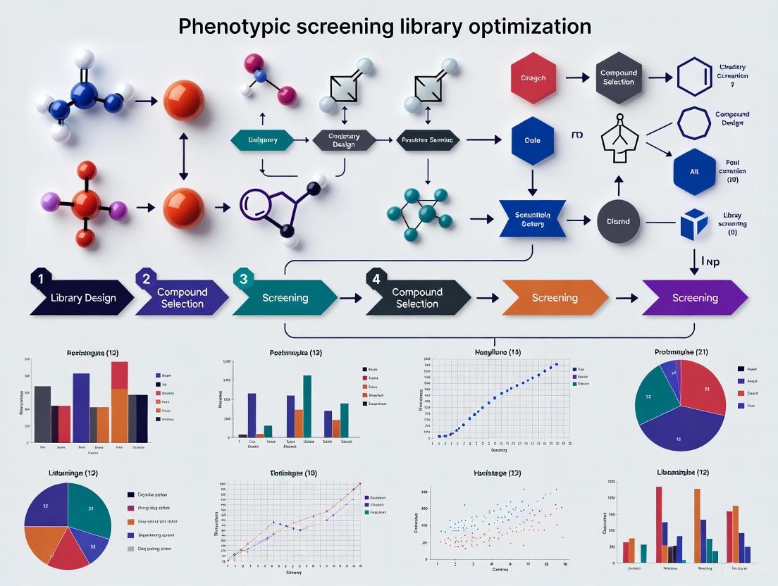
Overcoming Key Hurdles in Phenotypic Screening Library Optimization for Better Drug Discovery
Phenotypic screening has regained prominence for discovering first-in-class medicines but faces significant challenges in library design and optimization that impact efficiency and success rates.
Overcoming Key Hurdles in Phenotypic Screening Library Optimization for Better Drug Discovery
Abstract
Phenotypic screening has regained prominence for discovering first-in-class medicines but faces significant challenges in library design and optimization that impact efficiency and success rates. This article explores the core hurdles and modern solutions, covering foundational principles, advanced methodological applications, practical troubleshooting strategies, and rigorous validation frameworks. It provides researchers and drug development professionals with a comprehensive guide to navigating the complexities of creating optimized screening libraries, from selecting chemically diverse and target-informed compounds to leveraging AI and novel pooling techniques for enhanced predictivity and translatability in complex disease models.
The Resurgence and Rationale of Phenotypic Screening in Modern Drug Discovery
What are the fundamental definitions of Phenotypic and Target-Based Screening in drug discovery?
In modern drug discovery, two principal strategies guide the identification of new therapeutic compounds: phenotypic screening and target-based screening. These approaches differ in their fundamental philosophy and starting point.
- Phenotypic Screening is an empirical strategy that identifies active compounds based on their ability to induce a measurable biological response in cells, tissues, or whole organisms, without prior knowledge of the specific molecular target involved. It is an unbiased method that captures the complexity of biological systems [1] [2] [3].
- Target-Based Screening is a hypothesis-driven approach that begins with a specific, well-characterized molecular target (e.g., a protein, enzyme, or receptor) believed to play a critical role in a disease process. It involves screening compounds for their ability to interact with and modulate that predefined target [1] [2].
The table below summarizes the core strategic differences between these two approaches.
| Feature | Phenotypic Screening | Target-Based Screening |
|---|---|---|
| Starting Point | Observable biological effect or phenotype [1] [3] | Predefined molecular target [2] |
| Knowledge Prerequisite | Does not require prior understanding of disease mechanism [2] | Relies on established knowledge of target and its role in disease [2] |
| Primary Screening Readout | Complex, integrated cellular response (e.g., cell death, differentiation, cytokine secretion) [1] [3] | Specific biochemical interaction (e.g., enzyme inhibition, receptor binding) [2] |
| Throughput | Often lower due to complex assays [3] | Typically high, amenable to HTS [2] [4] |
| Target Deconvolution | Required after hit identification; can be challenging and time-consuming [1] [3] | Not required; target is known from the outset [2] |
| Key Strength | Identifies first-in-class medicines; captures system complexity and polypharmacology [5] [4] | Efficient, rational design; easier to optimize leads; yields best-in-class drugs [2] [3] |
FAQs and Troubleshooting Guides
FAQ: Strategic Considerations
1. When should I choose a phenotypic screening approach over a target-based one? Choose phenotypic screening when investigating diseases with poorly understood molecular mechanisms, when you aim to discover first-in-class drugs with novel mechanisms of action, or when the therapeutic goal involves modulating complex, system-level biological responses, such as in immuno-oncology or neurodegenerative diseases [1] [2] [3]. It is also valuable for uncovering polypharmacology—when a compound acts on multiple targets [6].
2. What are the major limitations of phenotypic screening, and how can I mitigate them? The main limitations are the significant challenge of target deconvolution (identifying the molecular mechanism of action) and generally lower throughput [7] [3].
- Mitigation Strategy: Employ advanced technologies for target identification, such as proteomics (e.g., thermal proteome profiling), CRISPR-based genetic screens, and chemical genomics. Furthermore, using high-content imaging and AI-powered data analysis can extract more mechanistic information from the phenotypic readouts themselves, accelerating deconvolution [7] [1] [4].
3. Can these two strategies be integrated? Yes, and this is a growing trend in modern drug discovery. A hybrid approach is increasingly common, where a target-focused screen is conducted in a cellular context, making it both target-based and phenotypic [3]. For instance, you might screen for compounds that affect the phosphorylation of a specific target protein (target-based readout) using high-content imaging that also captures other cellular morphological changes (phenotypic readout) [3]. This combines the precision of a targeted approach with the contextual richness of a phenotypic one.
Troubleshooting Guide: Common Experimental Issues
Problem: High false-positive rate in a high-throughput phenotypic screen.
- Potential Cause: Assay artifacts or promiscuous, pan-assay interference compounds (PAINS) that generically disrupt assays.
- Solution: Implement rigorous cheminformatics filters to flag and remove PAINS. Use orthogonal, biophysical confirmation methods to validate hits. Ensure your compound library is designed with high chemical diversity and drug-likeness in mind to improve the quality of starting points [4].
Problem: A potent hit from a phenotypic screen has unsuccessful target deconvolution.
- Potential Cause: The compound may act through weak interactions with multiple targets (polypharmacology), making it difficult to pinpoint a single mechanism.
- Solution: Consider that polypharmacology might be integral to the compound's efficacy. Instead of searching for a single target, use system-wide approaches like RNA sequencing and thermal proteome profiling to identify the network of engaged targets and pathways [6].
Problem: A hit compound is effective in a 2D cell culture but loses efficacy in a more complex 3D model.
- Potential Cause: The simplified 2D assay does not recapitulate the tumor microenvironment, including cell-cell interactions, hypoxia, and drug penetration barriers.
- Solution: Move primary screening to more physiologically relevant models from the outset. Use patient-derived spheroids, organoids, or organ-on-chip platforms to better mimic the in vivo disease state and improve translational success [4] [6].
Experimental Protocol: A Phenotypic Screening Case Study
The following workflow and protocol outline a rational approach to phenotypic screening for glioblastoma (GBM), demonstrating how to address the key challenge of library optimization [6].
Diagram: Phenotypic Screening Workflow with Library Optimization.
Detailed Methodology:
Target Selection and Library Enrichment:
- Input Data: Begin with genomic data (e.g., RNA sequencing and mutation data) from patient tumors (e.g., from The Cancer Genome Atlas) [6].
- Differential Expression: Perform analysis to identify genes significantly overexpressed in the disease state compared to normal samples (p < 0.001, FDR < 0.01, log2FC > 1) [6].
- Network Construction: Map the products of these genes onto large-scale human protein-protein interaction networks to construct a disease-specific subnetworK [6].
- Virtual Screening: Identify proteins in this network with druggable binding pockets. Perform molecular docking of an in-house compound library (e.g., ~9000 compounds) against these druggable sites to rank-order compounds based on predicted binding affinity. Select a focused subset of compounds predicted to engage multiple disease-relevant targets for phenotypic screening [6].
Phenotypic Screening Assay:
- Cell Model: Use low-passage, patient-derived glioblastoma cells grown as three-dimensional (3D) spheroids. This model more accurately captures the tumor microenvironment than traditional 2D cell lines [6].
- Screening Protocol:
- Culture GBM cells in ultra-low attachment plates to promote spheroid formation.
- Treat mature spheroids with the enriched compound library at a single-digit micromolar concentration range.
- Incubate for a defined period (e.g., 72-96 hours).
- Measure the primary phenotypic endpoint: inhibition of cell viability using a standard assay like CellTiter-Glo.
- Include standard-of-care drugs (e.g., temozolomide for GBM) as a positive control.
Selectivity and Secondary Phenotyping:
- Counter-Screening: Test active hits against non-transformed primary cell lines (e.g., hematopoietic CD34+ progenitor spheroids or astrocytes) to identify compounds that selectively target diseased cells while sparing normal cells [6].
- Angiogenesis Assay: For oncology, perform a secondary phenotypic assay such as a tube formation assay with brain endothelial cells to assess the compound's ability to inhibit angiogenesis [6].
Target Deconvolution and Mechanism of Action:
- Transcriptomics: Perform RNA sequencing on compound-treated versus untreated cells to observe changes in gene expression and infer affected pathways [6].
- Proteomics: Use mass spectrometry-based thermal proteome profiling (TPP) to identify proteins that show a thermal stability shift upon compound binding, indicating direct target engagement [6].
- Validation: Confirm binding to key targets identified by TPP using cellular thermal shift assays (CETSA) with target-specific antibodies [6].
The Scientist's Toolkit: Key Research Reagent Solutions
The table below lists essential materials and their functions for setting up a phenotypic screening campaign, particularly one based on the protocol above.
| Research Reagent / Tool | Function in the Experiment |
|---|---|
| Patient-Derived Cells & 3D Spheroids | Provides a physiologically relevant disease model that recapitulates key features of the native tumor microenvironment, leading to more translatable results [4] [6]. |
| Diverse & Focused Compound Libraries | A high-quality library is crucial. Diversity libraries explore broad chemical space, while target-focused libraries (e.g., kinase, epigenetic) enrich for activity against specific target families [4]. |
| High-Content Imaging Systems | Enables multiparametric analysis of complex phenotypic outcomes in cell-based assays, such as morphological changes, protein localization, and cell viability [3]. |
| CRISPR Screening Tools | Allows for systematic perturbation of genes to infer gene function and validate potential targets in a phenotypic context [7] [3]. |
| AI/ML Data Analysis Platforms | Machine learning helps "denoise" screening data, prioritize hits, identify frequent hitters, and can even assist in predicting a compound's molecular target from its phenotypic signature [4]. |
| Multi-Omics Platforms (RNA-seq, Proteomics) | Used for target deconvolution. RNA-seq reveals altered pathways, while proteomic methods like thermal proteome profiling identify direct protein targets [1] [6]. |
Troubleshooting Guides & FAQs
This guide addresses common challenges in phenotypic screening library optimization to enable the unbiased discovery of first-in-class therapeutic mechanisms.
Frequently Asked Questions
FAQ 1: Our phenotypic screens generate hits, but we struggle to identify the mechanism of action (MoA). What strategies can improve target deconvolution?
- Challenge: Target deconvolution remains a significant bottleneck, often prolonging discovery timelines and complicating hit validation [1].
- Solution: Implement an integrated approach using modern tools:
- Functional Genomics: Combine your screening hits with CRISPR-based genetic screens to identify genes that modulate the compound's activity or resistance [7].
- Chemical Proteomics: Use techniques like thermal proteome profiling (TPPP) to directly identify protein targets that engage with your hit compound in a cellular context [6] [8].
- AI-Powered Pattern Matching: Leverage artificial intelligence (AI) to integrate phenotypic signatures (e.g., from high-content imaging) with chemical descriptors and omics data to predict potential targets [4] [9].
- Multi-omics Integration: Correlate compound-induced phenotypic changes with transcriptomic (RNA-seq) and proteomic profiles to narrow down the involved pathways [1] [6].
FAQ 2: How can we design a screening library that maximizes the chance of discovering first-in-class mechanisms?
- Challenge: Standard chemogenomic libraries only interrogate a small fraction of the human genome (~1,000-2,000 out of 20,000+ genes), limiting novelty [7].
- Solution: Employ strategic library design:
- Prioritize Diversity: Use libraries optimized for broad structural and chemical diversity to cover wide biological and chemical space [4] [10].
- Incorporate Bioactive Compounds: Enrich libraries with compounds known to be bioactive, including natural products and their analogs, to increase the likelihood of hitting relevant pathways [10].
- Rational Library Enrichment: For complex diseases, create focused libraries by virtually screening compounds against multiple disease-relevant targets identified from genomic and protein-interaction networks [6].
- Quality Control: Rigorously filter libraries to remove pan-assay interference compounds (PAINS), reactive molecules, and compounds with poor drug-like properties [4] [10].
FAQ 3: What are the key considerations for choosing between a pooled versus arrayed CRISPR library for a genetic screen?
- Challenge: Selecting the wrong library format can lead to inconclusive results or unmanageable experimental scale.
- Solution: Base the decision on your screening goals and resources.
- Use Pooled Libraries for Discovery: Ideal for unbiased, genome-wide screens where the phenotype results in enrichment or depletion of cells (e.g., drug resistance or essential gene identification). They are more scalable and do not require automated liquid handling [11].
- Use Arrayed Libraries for Targeted Screens: Best for screening a predefined subset of genes or when assaying complex phenotypes that don't confer a growth advantage (e.g., high-content imaging). Requires automation but allows you to know the exact perturbation in each well from the start [11].
FAQ 4: Our hit compounds are active in simple 2D cell models but fail in more complex, physiologically relevant assays. How can we improve translational relevance early on?
- Challenge: Traditional 2D monolayer assays often fail to capture the complexity of human disease, leading to high attrition rates later in development [6].
- Solution: Invest in more disease-relevant model systems from the outset.
- 3D Models: Utilize three-dimensional cultures, such as patient-derived spheroids and organoids, which better mimic the tumor microenvironment, cell-cell interactions, and drug penetration barriers [4] [6].
- Primary Cells: Whenever possible, use low-passage patient-derived cells instead of immortalized cell lines, as they maintain more authentic biological responses [6].
- Co-culture Systems: Implement assays that include multiple cell types (e.g., immune cells, fibroblasts) to capture complex biological interactions that drive disease [1].
FAQ 5: How can we effectively triage hits to focus on the most promising leads with novel mechanisms?
- Challenge: Phenotypic screens can generate false positives and hits with undesired polypharmacology, wasting resources on invalidated leads [4].
- Solution: Establish a robust, multi-parameter triage cascade.
- Orthogonal Assays: Confirm activity in a functionally independent secondary assay that measures the same phenotype.
- Counter-Screens: Rule out common assay artifacts and undesired mechanisms (e.g., cytotoxicity, interference with the detection system).
- Cheminformatic Filters: Use AI/ML tools to flag frequent hitters, compounds with structural alerts, and undesirable physicochemical properties [4] [9].
- Early ADME/Tox Profiling: Integrate predictive models for absorption, distribution, metabolism, excretion, and toxicity (ADME/Tox) early in the process to deprioritize compounds with poor pharmacokinetic or safety profiles [4] [9].
Experimental Protocols for Key Methodologies
Protocol 1: Phenotypic Screening of an Enriched Compound Library in a 3D Glioblastoma Model
This protocol, adapted from a published study, outlines a rational approach to screen for compounds with selective polypharmacology in a patient-derived glioblastoma (GBM) spheroid model [6].
Library Enrichment & Virtual Screening:
- Input: Use tumor genomic data (e.g., from TCGA) to identify a set of overexpressed and mutated genes in GBM.
- Network Analysis: Map these genes onto a human protein-protein interaction network to create a disease-relevant subnetwork.
- Target Selection: Identify proteins within this subnetwork that contain druggable binding pockets (catalytic sites, protein-protein interfaces).
- Virtual Screening: Dock an in-house compound library (e.g., ~9,000 compounds) against these druggable pockets. Select the top-ranking compounds predicted to bind multiple disease-relevant targets.
Phenotypic Screening in 3D Culture:
- Cell Culture: Plate low-passage patient-derived GBM cells in ultra-low attachment plates to form 3D spheroids.
- Compound Treatment: Treat spheroids with the enriched library compounds. Include standard-of-care (e.g., temozolomide) and DMSO vehicle controls.
- Viability Assay: After 5-7 days, measure cell viability using a 3D-optimized ATP-based assay (e.g., CellTiter-Glo 3D).
- Selectivity Counter-Screen: Test active compounds in parallel against non-transformed primary cell lines (e.g., hematopoietic CD34+ progenitor spheroids or astrocytes) to identify compounds with selective toxicity toward cancer cells.
Secondary Phenotypic Assay (Angiogenesis):
- Tube Formation Assay: Seed brain endothelial cells on a layer of Matrigel. Treat with the selective hit compound and quantify the inhibition of tube network formation after 6-18 hours.
Protocol 2: Executing a Pooled Genome-Wide CRISPR Knockout Screen
This protocol provides a general workflow for conducting a loss-of-function genetic screen to identify genes essential for a specific phenotype [11].
Cell Line Preparation:
- Generate a Cas9-expressing cell line by transducing your target cells with a lentiviral Cas9 construct. Select stable pools using an antibiotic like puromycin.
- Validate Cas9 activity and functionality in the cell line.
sgRNA Library Transduction:
- Produce a high-titer lentiviral stock of a genome-wide pooled sgRNA library (e.g., Brunello library).
- Transduce the Cas9+ cells at a low Multiplicity of Infection (MOI ~0.3-0.4) to ensure most cells receive only one sgRNA. Use ~76 million cells to maintain library representation.
- Apply selection (e.g., blasticidin) to eliminate non-transduced cells.
Phenotypic Selection:
- Split the transduced cell population into treatment and control groups (e.g., drug-treated vs. DMSO control).
- Culture the cells for a sufficient duration (typically 10-14 days) to allow for phenotypic manifestation and enrichment/depletion of specific knockouts.
Genomic DNA (gDNA) Extraction and Sequencing:
- Harvest at least 100-200 million cells from each population. Isolate high-quality gDNA using a maxi-prep method to maintain sgRNA representation.
- Amplify the integrated sgRNA sequences from the gDNA by PCR and prepare next-generation sequencing (NGS) libraries.
Data Analysis:
- Sequence the libraries to a depth of ~10-100 million reads, depending on the screen type (positive/negative).
- Align sequences to the reference sgRNA library and quantify the abundance of each guide in treatment vs. control groups.
- Use specialized algorithms (e.g., MAGeCK) to identify sgRNAs and genes that are significantly enriched or depleted.
Table 1: Key Quantitative Considerations for Screening Library Design
| Parameter | Typical Range or Value | Significance & Rationale |
|---|---|---|
| Chemogenomic Library Coverage | 1,000 - 2,000 / 20,000+ human genes [7] | Highlights the limited fraction of the genome probed by annotated compound sets, underscoring the need for diverse libraries for novel discovery. |
| Recommended Cell Number for Pooled CRISPR Screen | ~76 million cells [11] | Ensures adequate representation of the entire sgRNA library, typically aiming for 200-1000 cells per sgRNA to avoid stochastic dropout. |
| Target Transduction Efficiency (CRISPR) | 30 - 40% [11] | A low MOI is critical to ensure most cells receive a single sgRNA, allowing for clear genotype-to-phenotype linkage. |
| NGS Read Depth (Positive Screen) | ~10 million reads [11] | Sufficient for identifying enriched sgRNAs in a positive selection screen (e.g., for drug resistance). |
| NGS Read Depth (Negative Screen) | Up to ~100 million reads [11] | Deeper sequencing is required to detect subtle depletion signals in negative screens (e.g., for essential genes). |
Table 2: Comparison of Phenotypic Screening Libraries
| Library Type | Key Characteristics | Best Use Cases |
|---|---|---|
| ChemDiversity Library [10] | Emphasizes broad structural diversity; filtered for drug-like properties, PAINS-free. | Unbiased discovery when the goal is to explore entirely novel chemical and biological space. |
| BioDiversity Library [10] | Enriched with known bioactive compounds, drugs, and natural product-like scaffolds. | Increasing the probability of finding a hit by leveraging chemical matter with proven biological activity. |
| Disease-Enriched Library [6] | Virtually screened against a network of disease-specific targets derived from genomic data. | Complex polygenic diseases (e.g., glioblastoma) where selective polypharmacology is desired. |
| CRISPR Knockout Library [11] | Provides complete gene knockouts; genome-wide or focused formats; pooled or arrayed. | Identifying genes essential for a phenotype (synthetic lethality, drug resistance) in an unbiased manner. |
Key Signaling Pathways and Workflows
Integrated Phenotypic Screening Workflow
Multi-Modal Target Deconvolution Strategy
The Scientist's Toolkit: Research Reagent Solutions
Table 3: Essential Research Tools for Phenotypic Screening and Optimization
| Research Tool | Function & Application | Examples / Key Features |
|---|---|---|
| Diverse Compound Libraries [4] [10] | Provide a broad source of chemical matter for unbiased phenotypic screening. | ChemDiversity libraries (structurally diverse), BioDiversity libraries (bioactive-enriched). High drug-likeness, PAINS-free. |
| CRISPR sgRNA Libraries [7] [11] | Enable genome-wide or targeted loss-of-function genetic screens to identify genes involved in a phenotype. | Genome-wide pooled libraries (e.g., Brunello). Arrayed libraries for specific gene sets. Lentiviral delivery for stable integration. |
| 3D Culture Systems [4] [6] | Provide physiologically relevant disease models that better mimic the in vivo microenvironment. | Patient-derived spheroids, organoids. Used for screening and validating compound efficacy and selectivity. |
| High-Content Imaging Systems [1] [4] | Enable multiparametric analysis of complex phenotypic changes in cells (e.g., morphology, signaling). | Used in Cell Painting assays. Generates rich, high-dimensional data for AI/ML analysis. |
| AI/ML Software Platforms [4] [9] | Analyze complex screening data, predict compound properties, prioritize hits, and suggest targets. | Capabilities include virtual screening, ADME/Tox prediction, and image analysis for phenotypic profiling. |
FAQs: Navigating the Throughput-Relevance Trade-off
Q1: Our high-throughput primary screen identified promising hits, but these fail in secondary, more biologically complex assays. How can we improve the translational relevance of our primary screening data?
- Answer: This common challenge often stems from an over-optimization for throughput at the expense of biological context. To bridge this gap:
- Adopt Phased Screening: Implement a tiered screening strategy. Use a high-throughput but simplified assay for primary screening, but immediately follow up on hits with a secondary, lower-throughput assay that incorporates greater biological complexity (e.g., co-culture systems, 3D models, or high-content imaging) [12]. This balances initial speed with necessary validation.
- Leverage Computational Prediction: Use tools like InferLoop, which leverages accessible data (e.g., scATAC-seq) to predict biologically relevant signals, such as cell-type-specific chromatin interactions, that are difficult to capture in a high-throughput setup [13]. This adds a layer of biological insight without additional complex experiments.
- Utilize Advanced Pooling Designs: For genetic screens, employ sophisticated combinatorial pooling methods like DCP-CWGC (Distance- and Balance-aware Constant-Weight Gray Codes). This design allows for the detection of consecutive positives (e.g., overlapping peptides or regulatory elements) and includes error-detection capabilities, making large-scale screens more reliable and biologically informative [14].
Q2: We need to extract multiple phenotypic endpoints from a single screen to capture biological complexity, but this drastically reduces our throughput. What solutions are available?
- Answer: The key is to implement workflows that are multiplexed by design.
- Implement High-Content, Multi-Parameter Assays: Design experiments where multiple relevant parameters are measured simultaneously from the same sample. For example, a protocol using CM-H2DCFDA and TMRM in a high-content microscopy workflow can concurrently quantify intracellular ROS levels, mitochondrial membrane potential, and mitochondrial morphology in individual cells [15]. While image acquisition takes time, the rich, multi-dimensional data extracted per experiment provides a much deeper biological understanding than multiple separate, single-endpoint assays.
- Invest in Automated Data Integration: The bottleneck often shifts from data collection to data analysis. Utilize automated image analysis pipelines and multivariate data analysis tools to efficiently process the complex, multi-parametric data generated, turning it into actionable biological insights [15].
Q3: How can we design a screening library and strategy that is efficient for large-scale discovery while still being sensitive to subtle, cell-type-specific phenotypes?
- Answer: This requires a combination of smart experimental design and modern computational tools.
- Focus on Perturbation Efficiency: Ensure your library design and screening model are optimally matched. For genetic screens, this means using efficient delivery systems (e.g., lentiviral vectors) and well-designed guide RNA or siRNA libraries. For compound screens, this involves careful selection of compound concentration and vehicle controls to minimize false positives/negatives [12].
- Incorporate Cell-Type-Specific Computational Analysis: Use single-cell sequencing technologies (e.g., scATAC-seq) and computational tools like InferLoop to deconvolve cell-type-specific signals from a heterogeneous screening population. This allows you to run a pooled screen but still extract insights relevant to specific cell subtypes, preserving biological relevance [13].
- Employ AI-Driven Pathway Optimization: In fields like synthetic biology, AI models can predict optimal biological pathways (e.g., for metabolite production) before any wet-lab experiment begins. This "in-silico screening" drastically narrows down the experimental space, allowing you to focus high-throughput validation on the most promising, biologically sound targets [16].
Experimental Protocols for Balanced Screening
Protocol: High-Content Multiplexed Analysis of Oxidative Stress and Mitochondrial Function
This protocol allows for the simultaneous quantification of multiple interconnected cellular health parameters in a single, automated workflow, ideal for secondary screening or lower-throughput, high-information-content primary screens [15].
- Key Application: Simultaneously quantifying intracellular reactive oxygen species (ROS) levels, mitochondrial membrane potential (ΔΨm), and mitochondrial morphology in adherent cells.
- Principle: The assay uses two cell-permeable fluorescent reporters: CM-H2DCFDA for ROS and TMRM for ΔΨm and mitochondrial morphology. Automated widefield fluorescence microscopy and subsequent image analysis enable the extraction of intensity- and morphology-based features at a single-cell level.
Workflow:
The following diagram illustrates the key stages of this multiplexed high-content screening protocol.
Detailed Methodology:
- Cell Seeding: Seed adherent cells (e.g., Normal Human Dermal Fibroblasts - NHDF) in a 96-well plate and allow them to adhere overnight [15].
- Reagent Preparation:
- Prepare imaging buffer (e.g., HBSS supplemented with HEPES).
- Prepare stock and working solutions of fluorescent dyes:
- Prepare an inducer of oxidative stress, such as tert-Butyl hydroperoxide (TBHP).
- Microscope Setup: Configure an automated widefield microscope with a 20x air objective, an environmental chamber, and appropriate filter sets for GFP (CM-H2DCFDA) and TRITC (TMRM). Define an acquisition protocol that images multiple non-overlapping fields per well [15].
- Dye Loading and Staining:
- Aspirate the culture medium and wash cells with pre-warmed imaging buffer.
- Load cells with a working solution containing both CM-H2DCFDA (e.g., 1 µM) and TMRM (e.g., 100 nM) in imaging buffer. Incubate for 30-45 minutes at 37°C protected from light [15].
- Replace the dye solution with fresh imaging buffer before microscopy.
- Live-Cell Imaging:
- First Measurement (Basal Conditions): Acquire images for both fluorescent channels to establish baseline ROS levels and mitochondrial parameters [15].
- Second Measurement (Induced Conditions): Add the stress inducer (e.g., TBHP) directly to the wells. After a defined incubation period (e.g., 30 minutes), re-acquire images to measure the induced cellular response [15].
- Image Analysis: Use automated image analysis software to:
- Perform cell segmentation.
- Quantify mean fluorescence intensity for CM-H2DCFDA (ROS) and TMRM (ΔΨm) per cell.
- Extract morphological features (e.g., form factor, aspect ratio) from the TMRM channel to classify mitochondrial morphology [15].
- Data Analysis and QC: Perform multivariate statistical analysis on the extracted feature set to detect differences between cell types or treatments. Implement quality control steps to exclude out-of-focus images or debris [15].
Protocol: Error-Detecting Combinatorial Pooling for Complex Target Identification
This protocol outlines the use of a sophisticated combinatorial pooling strategy to enhance the efficiency and reliability of large-scale genetic or peptide screens, particularly when targeting consecutive or overlapping elements [14].
- Key Application: Efficiently deconvolve positive hits from a large library of samples (e.g., peptides, gRNAs) in a minimal number of pooled tests, with built-in error detection and a focus on detecting consecutive positives.
- Principle: The DCP-CWGC (Distance- and Balance-aware Constant-Weight Gray Codes) method assigns each sample a unique binary "address" that determines its placement in testing pools. The design ensures samples are evenly distributed (balanced), each is tested the same number of times (constant weight), and the pattern allows for the identification of consecutive positives and the detection of experimental errors [14].
Workflow:
The diagram below visualizes the core process of designing and executing a screen using the DCP-CWGC pooling method.
Detailed Methodology:
- Parameter Definition: Determine the screening parameters:
n: The number of samples (items) to be screened.r: The constant Hamming weight (number of '1's in the code), which defines how many pools each sample is placed in.m: The number of pools, which must satisfym ≥ 2r + 1for optimal performance [14].
- Code Generation: Use specialized algorithms to generate the DCP-CWGC.
- Branch-and-Bound Algorithm (BBA): Constructs near-perfectly balanced codes for small to medium
nby traversing an address-joint bipartite graph [14]. - Recursive Combination BBA (rcBBA): Efficiently constructs long codes by recursively combining shorter DCP-CWGCs, ideal for large-scale screens [14].
- Implementation Note: The open-source Python package
codePUBprovides implementations of these algorithms [14].
- Branch-and-Bound Algorithm (BBA): Constructs near-perfectly balanced codes for small to medium
- Pooling Plan and Experiment:
- Create a pooling plan based on the generated code. Each sample's binary address dictates in which of the
mpools it is included (a '1' indicates inclusion). - Perform the pooled assay. For example, combine peptide pools with reporter cells to test for immune activation.
- Create a pooling plan based on the generated code. Each sample's binary address dictates in which of the
- Result Analysis and Hit Deconvolution:
- Identify which pools test positive.
- Error Detection: The DCP-CWGC design expects exactly
r+1positive pools for a single consecutive positive hit. A deviation from this number indicates a potential experimental error (e.g., a false positive or negative) [14]. - Hit Identification: The unique OR-sum of the addresses of consecutive positive items allows for their precise identification, even in the presence of a limited number of errors, by cross-referencing the positive pools [14].
Data Presentation: Comparative Analysis of Screening Approaches
Table 1: Quantitative Comparison of Screening Methodologies
This table summarizes key performance metrics for different screening approaches, highlighting the trade-off between throughput and biological relevance.
| Screening Methodology | Typical Throughput | Key Biological Relevance Features | Key Limitations | Ideal Use Case |
|---|---|---|---|---|
| Biochemical (e.g., ELISA, Enzyme Activity) [12] | Very High (10,000s of data points/day) | Direct measurement of molecular interactions. | Lack of cellular context; may not reflect physiology. | Primary screening for target binding or enzymatic inhibition. |
| Cell-Based (Simple Monolayer) [12] | High (1,000s of data points/day) | Cellular permeability; basic cytotoxicity. | Limited tissue structure; absence of microenvironment. | Primary phenotypic screening (e.g., cell viability, reporter assays). |
| High-Content Analysis (e.g., Multiplexed Imaging) [15] | Medium (100s of wells/day, 10,000s of cells) | Multiplexed readouts (signaling, morphology, subcellular structures) at single-cell resolution. | Throughput limited by image acquisition and analysis time; cost. | Secondary validation & lower-throughput primary screens requiring deep phenotyping. |
| Advanced Pooling (e.g., DCP-CWGC) [14] | Theoretical efficiency gain of log(n) | Capable of detecting complex patterns (e.g., consecutive positives); includes error-detection. | Complex experimental design and deconvolution; not suitable for all assay types. | Large-scale genetic or peptide screens where library size is a major constraint. |
| 3D & Co-culture Models | Low (10s of wells/day) | Physiologically relevant tissue context; cell-cell interactions; improved predictive validity. | Very low throughput; high cost; challenging for automated handling and analysis. | Late-stage secondary validation and mechanistic studies for top hits. |
The Scientist's Toolkit: Essential Research Reagents & Solutions
Table 2: Key Reagents for Balanced Phenotypic Screening
This table details essential reagents and their specific functions in setting up robust and informative screening assays.
| Research Reagent | Function & Role in Screening | Example Application in Protocol |
|---|---|---|
| CM-H2DCFDA [15] | Cell-permeable fluorescent dye that becomes highly fluorescent upon oxidation. Used as a reporter for intracellular Reactive Oxygen Species (ROS) levels. | Multiplexed high-content analysis of oxidative stress and mitochondrial function [15]. |
| TMRM (Tetramethylrhodamine, Methyl Ester) [15] | Cell-permeable, cationic fluorescent dye that accumulates in active mitochondria. Used to measure mitochondrial membrane potential (ΔΨm) and, via high-resolution imaging, mitochondrial morphology. | Multiplexed high-content analysis of oxidative stress and mitochondrial function [15]. |
| DCP-CWGC Code [14] | A specific binary code design used for combinatorial pooling. Its properties (balanced, constant weight, Gray code) enable efficient deconvolution, error detection, and identification of consecutive positives in large-scale screens. | Error-detecting combinatorial pooling for complex target identification (e.g., immunopeptide screening) [14]. |
| Single-Cell ATAC-seq (scATAC-seq) Data [13] | Sequencing data revealing regions of open chromatin in individual cells. Serves as input for computational prediction of regulatory elements. | Used as input for the InferLoop tool to predict cell-type-specific chromatin 3D structure (loops), adding a layer of biological insight without complex Hi-C experiments [13]. |
| HBSS-HEPES Imaging Buffer [15] | A physiological salt solution buffered with HEPES to maintain stable pH outside a CO₂ incubator. Essential for maintaining cell health during live-cell imaging experiments. | Used as the dye loading and imaging medium in the multiplexed oxidative stress protocol [15]. |
Troubleshooting Guides and FAQs
FAQ: Addressing Common Experimental Challenges
Q1: Our phenotypic screen yielded a high hit rate, but many compounds were false positives or promiscuous binders. How can we improve the quality of our initial library?
A1: High false-positive rates often indicate library quality issues. To address this:
- Apply Advanced Cheminformatic Filters: Implement filters beyond basic "drug-likeness" to remove pan-assay interference compounds (PAINS), compounds with poor solubility, and those with reactive functional groups [4].
- Utilize Orthogonal Confirmation: Use biophysical methods like surface plasmon resonance (SPR) early in the workflow to confirm binding and eliminate assay-specific artifacts [17] [4].
- Enhance Library Design: Curate libraries with emphasis on structural novelty, high purity, and validated solution stability to reduce noise and improve discovery efficiency [4].
Q2: Our fragment library screening failed to identify any novel chemotypes for our target. Are we limited by our library's coverage of chemical space?
A2: This is a common limitation. Even diverse experimental fragment libraries (e.g., 1,000-10,000 compounds) represent only a fraction of commercially available fragments (>500,000) and may miss critical chemotypes [17].
- Solution: Integrate virtual screening with your empirical screens. Docking large, commercially available fragment libraries can identify novel, potent chemotypes missing from your physical library, filling "chemotype holes" with little extra resource cost [17].
Q3: How can we balance the need for broad chemical space coverage with the practical constraints of screening capacity?
A3: A hybrid approach is most effective.
- Combine HTS and AI: Use high-throughput screening (HTS) for experimental validation while employing AI and machine learning to "denoise" data, recognize artifacts, and virtually screen vast in-silico chemical spaces to prioritize physical screening efforts [4].
- Use Focused Libraries: For well-validated target families (e.g., kinases, GPCRs), use targeted libraries to increase hit rates while maintaining a core diverse library for broader exploration [18] [4].
Q4: What are the key considerations when moving from a target-based screen to a more complex phenotypic screen?
A4: Phenotypic screening introduces new variables.
- Assay Relevance: Ensure cellular models (e.g., 3D cultures, organoids) are sufficiently physiologically relevant to capture disease complexity [4] [19].
- Target Deconvolution: A major challenge. Integrate high-content imaging, proteomics, and computational techniques early to link phenotypic outcomes to molecular targets [1].
- Compound Logistics: Phenotypic assays often have lower throughput. Employ robotics, assay miniaturization, and integrated workflows to maintain efficiency [4].
Quantitative Data on Library Composition and Performance
The design and composition of a chemical library directly influence screening outcomes. The tables below summarize key performance data and design criteria.
Table 1: Performance Comparison of Screening Methods for AmpC β-lactamase
| Screening Method | Library Size | Hit Rate | Most Potent KI (mM) | Key Findings |
|---|---|---|---|---|
| NMR (TINS) Screening | 1,281 fragments | 3.2% (41 hits) | 0.2 | Discovered novel chemotypes (Avg. Tc* 0.21) [17] |
| Virtual Screening | ~290,000 fragments | Not specified | 0.03 | Filled "chemotype holes" from the empirical library [17] |
| Integrated Approach | Empirical + Virtual | Combined benefits | 0.03 | Captured unexpected and target-tailored chemotypes [17] |
*Tc: Tanimoto coefficient, a measure of structural novelty.
Table 2: Key Design Criteria for Different Library Types
| Library Type | Typical Size | Primary Goal | Key Design/Filtration Criteria | Common Applications |
|---|---|---|---|---|
| Diverse Screening Library | 10,000 - 50,000+ | Maximize exploration of chemical space | Drug-likeness (e.g., Ro5), structural diversity, solubility, purity [18] [4] | Initial HTS, unbiased discovery |
| Focused/Targeted Library | 1,000 - 10,000 | Target specific protein families | Prior knowledge of target class, ligand-based or structure-based design [20] [18] | Kinase, GPCR, epigenetic target screening |
| Fragment Library | 1,000 - 2,000 | Identify weak-binding starting points | Rule of 3 (MW <300, HBD/HBA ≤3, cLogP ≤3), low rotatable bonds [17] [18] | Fragment-Based Drug Discovery (FBDD) |
Experimental Protocols
Protocol 1: Integrated Empirical and Virtual Fragment Screening
This protocol, adapted from a study on AmpC β-lactamase, combines unbiased empirical screening with structure-based virtual screening to maximize chemotype coverage [17].
1. Target Immobilized NMR Screening (TINS) - Primary Empirical Screen
- Objective: Identify fragments binding to the target protein.
- Methodology:
- Immobilize the target protein (e.g., AmpC β-lactamase) on a solid support.
- Screen the empirical fragment library (e.g., 1,281 compounds) using TINS.
- Use a reference protein to subtract non-specific binding.
- Confirm initial hits in a replication experiment.
- Output: A list of confirmed binding fragments.
2. Surface Plasmon Resonance (SPR) - Secondary Confirmatory Assay
- Objective: Validate binding and determine affinity (KD) for NMR hits.
- Methodology:
- Immobilize the target on an SPR chip.
- Inject confirmed NMR hits at varying concentrations.
- Measure binding kinetics (association/dissociation) to determine KD values.
- Output: Affinity data for binding fragments.
3. Enzymological Inhibition Assay (KI Determination)
- Objective: Determine the inhibitory potency of binding fragments.
- Methodology:
- Incubate the target enzyme with a substrate and varying concentrations of the fragment.
- Measure reaction rates (e.g., spectrophotometrically).
- Calculate the inhibition constant (KI) from the dose-response data.
- Output: Functional inhibition data and ligand efficiency (LE).
4. Parallel Virtual Screening of a Large Commercial Library
- Objective: Identify potent chemotypes not present in the empirical library.
- Methodology:
- Select a large library of purchasable fragments with chemotypes unrepresented in the empirical library (e.g., 290,000 compounds).
- Perform molecular docking against the target's active site using an appropriate scoring function.
- Prioritize top-ranking compounds for purchase and testing.
- Output: A list of computationally prioritized fragments.
5. Experimental Validation of Docking Hits
- Objective: Confirm the activity of virtual screening hits.
- Methodology:
- Subject the purchased, docking-prioritized fragments to the same enzymological inhibition assay (Step 3).
- Determine KI values and ligand efficiencies.
- Output: Potent inhibitors discovered via virtual screening.
6. X-ray Crystallography for Structural Insights
- Objective: Understand binding modes and validate docking predictions.
- Methodology:
- Co-crystallize the target protein with bound fragments (from both NMR and docking screens).
- Solve the crystal structure and analyze the protein-fragment interactions.
- Output: Atomic-resolution structures guiding further optimization.
Protocol 2: Phenotypic Screening in a Complex Cell Model
This protocol outlines a generalized workflow for a phenotypic screen using a disease-relevant cellular model [1] [4] [19].
1. Development of a Phenotypically Relevant Assay
- Objective: Establish a robust and translatable cellular model.
- Methodology:
- Select a physiologically relevant cell system (e.g., patient-derived cells, induced pluripotent stem cell (iPSC)-derived tissues, 3D organoids).
- Define a quantifiable, disease-relevant phenotypic endpoint (e.g., neurite outgrowth for neurodegeneration, tumor cell killing in co-culture, cytokine secretion profile).
- Implement a high-content imaging or multiparametric readout system to capture the complex phenotype.
2. Primary Screening and Hit Identification
- Objective: Identify compounds that modulate the desired phenotype.
- Methodology:
- Screen the compound library in the phenotypic assay. Use automation and miniaturization (e.g., 384- or 1536-well plates) for throughput.
- Apply statistical rigor for hit selection (e.g., Z'-factor for assay quality, setting hit thresholds based on standard deviations from the mean).
3. Hit Triage and Counter-Screening
- Objective: Eliminate false positives and prioritize promising leads.
- Methodology:
- Use cheminformatic filters to remove compounds with undesirable properties (PAINS, poor solubility).
- Perform counter-screens against general cytotoxicity to ensure phenotype-specific effects.
- Confirm activity in dose-response experiments.
4. Target Deconvolution
- Objective: Identify the molecular target(s) of the phenotypic hit.
- Methodology:
- Employ computational methods using chemical descriptors and omics data to infer potential targets.
- Use experimental techniques such as chemical proteomics (e.g., affinity purification pull-downs with the hit compound as bait), or genetic approaches (e.g., CRISPR-based screens).
Workflow and Pathway Diagrams
Diagram 1: Screening Strategy Selection Workflow
Diagram 2: Virtual Screening Workflow
The Scientist's Toolkit: Research Reagent Solutions
Table 3: Essential Research Reagents and Libraries for Screening
| Reagent / Resource | Type | Primary Function in Screening |
|---|---|---|
| Diversity Screening Library [4] | Small Molecule Collection | Provides broad coverage of chemical space for initial unbiased screening in HTS or phenotypic campaigns. |
| Focused/Targeted Libraries (e.g., Kinase, GPCR) [18] [4] | Small Molecule Collection | Enriches for compounds active against specific target families, increasing hit rates for those targets. |
| Fragment Library [17] [18] | Small Molecule Collection | Provides low molecular weight starting points for FBDD, enabling efficient coverage of chemical space. |
| FDA-Approved Drug Library [4] | Small Molecule Collection | Used in repurposing screens, offering compounds with known safety profiles for new indications. |
| Target Protein (e.g., AmpC β-lactamase) [17] | Protein Reagent | The biological target for biochemical, biophysical, and structural studies in target-based screening. |
| SPR Instrumentation [17] | Biophysical Instrument | Confirms binding of hits and provides quantitative affinity (KD) and kinetic data. |
| NMR for TINS [17] | Biophysical Instrument | Detects weak binding of fragments in a primary screen using target-immobilized NMR. |
| X-ray Crystallography System [17] | Structural Biology Tool | Determines high-resolution structures of target-hit complexes to guide rational optimization. |
The Problem of False Positives and Assay Artifacts in Primary Screens
Frequently Asked Questions (FAQs)
1. What are the most common types of assay artifacts in primary screens? The most prevalent assay artifacts fall into several key categories [21]:
- Chemical Reactivity: Includes thiol-reactive compounds (TRCs) that covalently modify cysteine residues and redox-active compounds that produce hydrogen peroxide, indirectly modulating target activity.
- Interference with Reporter Enzymes: Compounds that directly inhibit common reporter proteins like firefly or nano luciferase, leading to false positive signals in reporter gene assays.
- Compound Aggregation: Molecules that form colloidal aggregates (SCAMs) at screening concentrations, non-specifically perturbing biomolecules.
- Interference with Optical Detection: Compounds that are intrinsically fluorescent or colored, interfering with fluorescence or absorbance-based readouts.
- Technology-Specific Interference: Signal quenching, inner-filter effects, or disruption of affinity capture components in assays like FRET, TR-FRET, ALPHA, or SPA.
2. How do PAINS filters work, and what are their limitations? Pan-Assay INterference compoundS (PAINS) filters are a set of substructural alerts designed to flag compounds associated with various assay interference mechanisms [21]. However, they have significant limitations: they are often oversensitive, disproportionately flagging compounds as potential false positives while failing to identify a majority of truly interfering compounds. This is because chemical fragments do not act independently from their structural surroundings, and many original PAINS alerts were derived from very few compounds, making them less reliable [21].
3. What computational tools are available to predict assay interference? Researchers can use several modern computational tools that are more reliable than PAINS filters [21]:
- Liability Predictor: A free webtool that uses Quantitative Structure-Interference Relationship (QSIR) models to predict compounds exhibiting thiol reactivity, redox activity, and luciferase inhibitory activity.
- Luciferase Advisor: Predicts luciferase inhibitors in luciferase-based assays.
- SCAM Detective: Predicts colloidal aggregators.
- InterPred: Predicts compounds that exhibit autofluorescence and luminescence interference.
4. Can assay technology itself help reduce false positives? Yes, the choice of detection technology can significantly impact false positive rates. For instance [22]:
- Fluorescence Lifetime Technology (FLT) can offer a superior readout compared to Time-Resolved Fluorescence Resonance Energy Transfer (TR-FRET). FLT measures the characteristic fluorescence decay of a fluorophore, which is less susceptible to common interference issues that affect fluorescence intensity measurements.
- Utilizing readouts in the far-red spectrum for fluorescence-based HTS assays dramatically reduces interference from compound autofluorescence [21].
5. How does a phenotypic screening approach influence false positive rates? Phenotypic screening, which measures functional outcomes in cellular systems, can overcome some limitations of target-based approaches. However, it is not immune to false positives arising from the interference mechanisms listed above [1]. A key challenge is that observed activity may not be due to the intended biological mechanism. Furthermore, target deconvolution for hits from phenotypic screens can be complex and time-consuming [1]. Integrating phenotypic data with multi-omics and AI can help address this by providing a systems-level view and uncovering true mechanisms of action [23].
Troubleshooting Guide: Identifying and Mitigating False Positives
This guide outlines a systematic approach to triage hits from a primary screen.
Step 1: In-silico Triage of Primary Hit List
Action: Filter your hit list using modern computational liability predictors. Methodology:
- Submit your list of hit compounds in SMILES or SDF format to a tool like Liability Predictor (https://liability.mml.unc.edu/) [21].
- The tool will use QSIR models to predict the likelihood of each compound being a thiol-reactive, redox-active, or luciferase-interfering artifact.
- Prioritize compounds with low interference scores for downstream confirmation. Why it works: This provides an initial, rapid assessment of potential chemical liabilities, flagging promiscuous compounds before investing in costly experimental follow-up [21].
Step 2: Experimental Confirmation of Activity
Action: Confirm that the observed activity is real and not an artifact of the primary screening conditions. Methodology:
- Dose-Response: Re-test hits in a concentration-dependent manner (e.g., a 10-point dose-response curve) in the original primary assay. True hits will typically show a saturable, stoichiometric dose-response relationship.
- Orthogonal Assay: Test confirmed hits in a secondary assay that uses a completely different detection technology. For example, if the primary screen was a luciferase-based reporter assay, the orthogonal assay could be a High-Content Imaging (HCI) readout of a relevant downstream protein marker or a mass spectrometry-based assay like RapidFire MS (RF-MS) [22]. Why it works: Orthogonal assays with different readout mechanisms are unlikely to be susceptible to the same interference compounds. A compound active across multiple assay formats is more likely to be a true positive.
Step 3: Investigate Mechanism-Based Activity
Action: Rule out nonspecific mechanisms of action. Methodology:
- Test for Aggregation: Perform assays in the presence and absence of non-ionic detergents (e.g., 0.01% Triton X-100). Inhibition that is reversed by detergent is a strong indicator of colloidal aggregation [21].
- Counter-Screens: Test compounds against unrelated targets or enzymes. A compound that inhibits many unrelated targets is likely a promiscuous, nonspecific inhibitor.
- Cellular Toxicity Assay: For cell-based phenotypic screens, perform a parallel viability assay (e.g., measuring ATP levels). This ensures that the observed phenotype is not simply a consequence of generalized cellular toxicity. Why it works: These experiments help distinguish specific, target-mediated activity from nonspecific effects like aggregation-based inhibition or cytotoxicity.
Quantitative Data on Assay Interference and Mitigation
| Assay Interference Type | External Balanced Accuracy | Number of External Compounds Tested |
|---|---|---|
| Thiol Reactivity | 58-78% | 256 |
| Redox Activity | 58-78% | 256 |
| Luciferase (Firefly) Interference | 58-78% | 256 |
| Luciferase (Nano) Interference | 58-78% | 256 |
| Detection Technology | Principle | Relative Reduction in False Positives (Model System: TYK2 Kinase) |
|---|---|---|
| Time-Resolved Fluorescence Resonance Energy Transfer (TR-FRET) | Measures fluorescence intensity after a time delay to reduce background. | Baseline |
| Fluorescence Lifetime Technology (FLT) | Measures the characteristic decay time of fluorescence, which is largely independent of concentration and fluorescence intensity. | Marked Decrease |
| RapidFire Mass Spectrometry (RF-MS) | Label-free method that directly detects substrate depletion or product formation. | Significant Decrease (considered a gold-standard confirmatory method) |
Experimental Protocols
Objective: To establish a robust, medium-throughput phenotypic assay for identifying inhibitors of CAF activation, a key process in cancer metastasis.
Materials (Research Reagent Solutions):
- Cell Lines: Human primary lung fibroblasts, highly invasive breast cancer cells (MDA-MB-231), human monocytes (THP-1 cells).
- Culture Media: DMEM-F12 and RPMI-1640, supplemented with 10% Fetal Calf Serum (FCS) and 1% penicillin-streptomycin.
- Assay Plates: 96-well plates suitable for In-Cell ELISA (ICE).
- Key Reagents: Primary antibody against α-Smooth Muscle Actin (α-SMA), fluorescently labeled secondary antibody, fixation reagent (e.g., ice-cold methanol), blocking buffer (e.g., 10% donkey serum in PBS).
Methodology:
- Co-culture Setup: Seed human lung fibroblasts alone or in co-culture with MDA-MB-231 breast cancer cells and THP-1 monocytes in a 96-well plate. Include appropriate controls (fibroblasts alone).
- Incubation: Incubate the co-culture for a predetermined period (e.g., 72 hours) at 37°C and 5% CO₂ to allow for CAF activation.
- Cell Fixation and Staining:
- Fix cells with ice-cold methanol.
- Permeabilize and block nonspecific binding sites using a blocking buffer.
- Incubate with anti-α-SMA primary antibody.
- Wash and incubate with a fluorescently conjugated secondary antibody.
- Signal Detection and Analysis: Measure the fluorescence signal using a plate reader. The expression level of α-SMA, a marker of myofibroblast/CAF activation, is the primary readout.
- Validation: A robust assay should show a significant increase (e.g., 2.3-fold) in α-SMA expression in co-culture conditions compared to fibroblasts alone, with a Z'-factor >0.5, indicating its suitability for screening [24].
Protocol 2: Experimental Workflow for Triage of HTS Hits
This workflow diagrams the multi-step process for validating primary screen hits.
Protocol 3: Key Phases of Hit Triage and Validation
This diagram breaks down the critical phases and decision points in the hit validation pipeline.
The Scientist's Toolkit: Essential Research Reagent Solutions
| Item | Function in the Assay |
|---|---|
| Primary Human Lung Fibroblasts | The primary cell type whose activation into a CAF state is being measured. |
| MDA-MB-231 Cell Line | A highly invasive breast cancer cell line used to induce fibroblast activation in co-culture. |
| THP-1 Cell Line | A human monocyte cell line; monocytes/macrophages are key regulators in the CAF activation microenvironment. |
| Anti-α-SMA Antibody | The primary antibody used in the In-Cell ELISA to detect and quantify the levels of α-Smooth Muscle Actin, a key biomarker of CAF activation. |
| Fluorescent Secondary Antibody | Conjugated to a fluorophore; binds to the primary antibody to allow for detection and quantification of α-SMA levels. |
Advanced Strategies for Designing and Enriching Phenotypic Screening Libraries
This technical support center provides troubleshooting guides and FAQs to help researchers navigate common challenges in phenotypic screening library optimization.
Frequently Asked Questions
What is the primary limitation of a diverse screening library? Diverse "chemogenomics" libraries typically interrogate only a small fraction of the human genome—approximately 1,000–2,000 out of 20,000+ genes. This limited coverage can miss critical, novel, or undrugged targets, restricting the scope of your phenotypic discoveries [7].
When should I use a focused versus a diverse library? A focused library is more efficient when structural information on the target or target family is available or when ligands of the target are known. A diverse library is preferable when very little is known about the target and no or few ligands have been identified [25].
How does library design impact target identification (ID) in phenotypic screening? A major challenge is that compounds from phenotypic screens can be highly promiscuous, acting on multiple unexpected targets. This complicates and can even mislead target ID and validation efforts. Strategies like affinity purification and genetic approaches are needed for target deconvolution [7].
What are the key considerations for scaffold selection in library design? The choice of scaffold is critical as it predetermines many properties of the future lead. An ideal scaffold should have favorable ADME properties, present good vector orientation for substituents, enable robust binding interactions, be synthetically amenable, and offer patentability [26].
Troubleshooting Guides
Problem: High Hit Rate with Non-Specific or Promiscuous Compounds
Issue: Initial screen yields many hits, but most compounds show poor selectivity or engage multiple off-targets.
Diagnosis: This is common with libraries built around "privileged" scaffolds or those lacking sufficient chemical diversity, leading to frequent-hitter behavior [7].
Solution:
- Apply PAINS Filters: Use computational filters to identify and remove Pan-Assay Interference Compounds (PAINS) early in the triage process [7].
- Counter-Screening: Implement secondary assays against common off-targets (e.g., kinases, GPCRs) to identify and eliminate promiscuous binders [7].
- Library Refinement: For follow-up libraries, incorporate structural motifs known to avoid promiscuity and enhance selectivity [25].
Problem: Difficulty in Target Identification & Deconvolution
Issue: A compound shows a robust phenotypic response, but its molecular mechanism of action (MOA) remains unknown.
Diagnosis: This is a fundamental limitation of phenotypic screening. Without a known target, further medicinal chemistry optimization and safety profiling are challenging [7].
Solution:
- Affinity Purification Mass Spectrometry: Use biotinylated or photo-affinity probes derived from your hit compound to pull down and identify direct protein targets from cell lysates [7].
- Functional Genomics (CRISPR) Screens: Perform parallel genetic screens to identify genes whose loss-of-function phenocopies or rescues the compound's effect. This can pinpoint pathways and potential targets [7].
- Resistance Mutation Mapping: In microbial or cell-based systems, select for resistance to the compound and sequence the genome to identify mutated genes, which often encode the target or related proteins [7].
Problem: Poor Coverage of Relevant Chemical or Target Space
Issue: The screening library does not yield hits, potentially because it lacks compounds capable of modulating the biology in your specific phenotypic assay.
Diagnosis: The chemical space covered by the library is too narrow, biased towards certain target classes, or lacks the complexity needed for the phenotype [7] [26].
Solution:
- Analyze Library Composition: Computationally map your library's coverage of chemical space and target annotations to identify gaps [26].
- Incorporate Privileged Structures: For specific target families (e.g., kinases, GPCRs), design focused libraries around known privileged scaffolds to increase the likelihood of success [25].
- Explore New Modalities: Consider expanding your library to include compounds suitable for emerging target areas like protein-protein interactions (PPI) or the ubiquitin proteasome pathway, which may require specialized chemotypes [25].
Problem: Inefficient Translation from Hit to Lead
Issue: Confirmed hits have poor drug-like properties (e.g., solubility, metabolic stability), making them difficult to optimize into viable lead compounds.
Diagnosis: The initial library was designed without sufficient consideration of ADMET (Absorption, Distribution, Metabolism, Excretion, Toxicity) properties during the hit identification phase [26].
Solution:
- Early ADMET Profiling: Integrate high-throughput solubility, microsomal stability, and cytotoxicity assays into the primary screening workflow [26].
- Structure-Based Design: If the target is identified, use structural information (e.g., from X-ray crystallography or cryo-EM) to guide optimization of potency and selectivity [1].
- Property-Based Design: Use guidelines like Lipinski's Rule of Five during the library design and hit-to-lead stages to prioritize compounds with better drug-like properties [26].
Experimental Protocols & Data
Quantitative Comparison of Screening Approaches
The table below summarizes key limitations and mitigation strategies for small molecule and genetic screening, two primary tools for phenotypic discovery [7].
| Screening Type | Key Limitation | Quantitative Impact | Mitigation Strategy |
|---|---|---|---|
| Small Molecule Screening | Limited target coverage of chemogenomics libraries | 1,000-2,000 of 20,000+ genes addressed [7] | Use multiple, diverse library types; include covalent and novel chemotypes [7]. |
| Promiscuity & assay interference | PAINS compounds can constitute a significant fraction of initial hits [7] | Early triage with counter-screens and computational filters [7]. | |
| Genetic Screening (e.g., CRISPR) | Fundamental difference from pharmacological perturbation | Genetic knockout is irreversible and complete, unlike transient, partial inhibition by drugs [7] | Use inducible or partial loss-of-function systems (e.g., CRISPRi) for better mimicry [7]. |
| Limited phenotypic robustness in high-throughput | Many validated hits fail in lower-throughput, more complex phenotypic assays [7] | Use high-content, multiparametric readouts; prioritize screens with high biological relevance [7]. |
Protocol: Designing a Focused Library for a Novel Target Family
This methodology outlines the creation of a target-focused library, such as for kinases or other well-characterized families [25].
Principle: When structural information on the target or target family is available, it is more efficient to design or select compounds that can be expected to modulate the target, rather than screening a vast, diverse library [25].
Procedure:
- Target Analysis: Collect all available structural data (X-ray, cryo-EM) of the target family, including apo structures and ligand-bound complexes. Identify conserved binding motifs and key interaction sites.
- Scaffold Identification: Select a core scaffold (e.g., a heterocycle) that can effectively present substituents to the key interaction sites identified in Step 1. The scaffold should be synthetically tractable and have known routes for diversification [26].
- Virtual Library Construction: Using combinatorial chemistry software, generate a virtual library by combining the chosen scaffold with a large set of available reagents at each variable position (R1, R2...). The size of this virtual library can be enormous (e.g., 1 million compounds for 200x50x100 reagents) [26].
- In-Silico Screening:
- Docking: If 3D structures are available, computationally dock virtual compounds into the binding site to score and rank them based on predicted binding affinity.
- Pharmacophore Modeling: Use known active ligands to build a pharmacophore model and filter virtual compounds that match this model.
- QSAR: Use existing bioactivity data to build a Quantitative Structure-Activity Relationship (QSAR) model to predict activity of new virtual compounds.
- ADMET Filtering: Apply computational filters to remove compounds with predicted poor solubility, high metabolic instability, or potential toxicity. Adhere to drug-like property guidelines [26].
- Final Selection and Synthesis: Select a manageable number of compounds (e.g., hundreds to a few thousand) from the top-ranked, filtered virtual list for actual synthesis or acquisition.
The Scientist's Toolkit: Key Research Reagents
| Reagent / Material | Function in Library Design & Screening |
|---|---|
| Chemogenomics Library | A collection of small molecules with known or predicted annotations against a set of biological targets. Used for initial phenotypic screens to provide mechanistic starting points [7]. |
| CRISPR Library | A pooled or arrayed collection of guide RNAs (gRNAs) targeting genes across the genome. Used in functional genomic screens to identify genes involved in a phenotype [7]. |
| Privileged Scaffold | A core molecular structure (e.g., benzimidazole, indole) known to produce ligands for multiple receptor types. Serves as a template for building focused libraries [26]. |
| Photo-affinity Probe | A chemical probe containing a photoreactive group (e.g., diazirine) and an affinity tag (e.g., biotin). Used for target deconvolution by covalently capturing protein targets upon UV irradiation [7]. |
Workflow Visualization
Strategic Library Design Workflow
Phenotypic Screening & Target Deconvolution
Frequently Asked Questions (FAQs)
FAQ 1: What is the core principle behind using multi-omics data for library enrichment? Multi-omics integration combines data from various molecular layers—such as genomics, transcriptomics, proteomics, and metabolomics—to build a comprehensive understanding of disease biology. This integrative approach helps identify key dysregulated pathways and networks in diseases like cancer. By using the tumor's specific genomic profile (e.g., RNA sequence and mutation data), researchers can pinpoint overexpressed proteins and map them onto protein-protein interaction networks to select a collection of biologically relevant targets. A chemical library is then computationally enriched by docking compounds against these selected targets to find molecules that potentially modulate multiple key proteins simultaneously, a strategy known as selective polypharmacology [6].
FAQ 2: What are the primary data sources for obtaining disease-specific genomic and multi-omics data? Large-scale public repositories are essential resources. These include:
- The Cancer Genome Atlas (TCGA): Provides comprehensive molecular profiles of various cancer types, including genomic, epigenomic, transcriptomic, and proteomic data [27].
- NCI Genomic Data Commons (GDC): A unified data repository that standardizes and distributes cancer genomic and clinical data from programs like TCGA. It provides harmonized data aligned to a reference genome, facilitating integrated analysis [28].
- Clinical Proteomic Tumour Analysis Consortium (CPTAC): Focuses on proteogenomic characterization, linking genomic alterations to protein-level changes in cancer [27].
FAQ 3: My multi-omics datasets have complex directional relationships. How can I account for this in analysis? Directional dependencies are a key challenge. Methods like Directional P-value Merging (DPM) have been developed to address this. DPM allows you to define a Constraints Vector (CV) that specifies the expected directional relationship between datasets (e.g., positive correlation between mRNA and protein levels, or negative correlation between promoter DNA methylation and gene expression). This method prioritizes genes with consistent, significant changes across omics layers that align with your biological hypothesis, while penalizing those with conflicting signals, leading to more accurate gene and pathway prioritization [27].
FAQ 4: How do I determine the optimal sampling frequency for different omics layers in a longitudinal study? Not all omics layers change at the same rate. A rational, hierarchical approach is recommended:
- Genome: Generally static; requires a single baseline assessment.
- Epigenome: Dynamic, but relatively stable.
- Transcriptome: Highly sensitive to environment and treatment; may require frequent assessments (e.g., daily or hourly in shift-work studies) [29].
- Proteome: Proteins have longer half-lives; testing frequency can be lower and aligned with specific clinical time points [29].
- Metabolome: Provides a near real-time snapshot of metabolic activity; may also require frequent sampling in certain contexts [29].
Table 1: Recommended Omics Sampling Frequency in Longitudinal Studies
| Omics Layer | Dynamic Nature | Recommended Sampling Frequency | Rationale |
|---|---|---|---|
| Genomics | Static | Once (baseline) | DNA sequence is largely unchanging. |
| Transcriptomics | Highly Dynamic | High Frequency | Gene expression rapidly responds to stimuli, environment, and treatment [29]. |
| Proteomics | Moderately Dynamic | Lower Frequency | Proteins are more stable, with longer half-lives than transcripts [29]. |
| Metabolomics | Highly Dynamic | High Frequency (in specific contexts) | Metabolites provide a real-time view of cellular activity and response [29]. |
FAQ 5: What are common data heterogeneity issues when integrating multi-omics data, and how can they be mitigated? Data heterogeneity arises from:
- Different Data Types: Combining sequence data, expression counts, and intensity values.
- Technical Bias: Variations in platforms, protocols, and batch effects.
- Dimensionality: The vast number of features measured. Mitigation strategies include:
- Data Harmonization: Using pipelines like those from the GDC to re-align genomic data to a common reference genome [28].
- Significance-Based Fusion: Integrating data at the level of P-values and directionality estimates (e.g., fold-change) to overcome scale differences, as done in methods like DPM and ActivePathways [27].
- Advanced Computational Methods: Employing deep learning, graph neural networks (GNNs), and other AI tools to synthesize and interpret complex datasets [30].
Troubleshooting Guides
Issue 1: High Attrition Rate in Phenotypic Screening
Problem: Compounds active in initial phenotypic screens fail to show efficacy in more disease-relevant models or exhibit high toxicity.
Possible Causes and Solutions:
Cause 1: Lack of Biological Relevance in Library Design
- Solution: Implement a target-informed library enrichment strategy.
- Protocol: Begin with the tumor's genomic profile (e.g., from TCGA). Perform differential expression and somatic mutation analysis to identify overexpressed and mutated genes. Map these genes onto a large-scale protein-protein interaction network (e.g., from literature-curated databases) to create a disease-specific subnetwork. Identify proteins within this network that have druggable binding pockets. Finally, computationally dock your compound library against these prioritized targets to select molecules for screening [6].
- Solution: Implement a target-informed library enrichment strategy.
Cause 2: Use of Oversimplified Biological Models
- Solution: Transition to more physiologically relevant screening assays.
- Protocol: Replace traditional 2D monolayer cultures of immortalized cell lines with 3D models. For glioblastoma (GBM) screening, use low-passage, patient-derived GBM spheroids. As a counter-screen for toxicity, employ non-transformed primary cell models, such as 3D spheroids of hematopoietic CD34+ progenitor cells or 2D cultures of astrocytes. This helps identify compounds that selectively inhibit tumor growth without affecting normal cell viability [6].
- Solution: Transition to more physiologically relevant screening assays.
Issue 2: Inconsistent Findings Across Omics Datasets
Problem: Significant genes or pathways identified in one omics dataset (e.g., transcriptomics) are not supported by another (e.g., proteomics).
Possible Causes and Solutions:
Cause 1: Ignoring Directional Biological Relationships
- Solution: Apply directional integration methods in your analysis workflow.
- Protocol: For each omics dataset, generate a matrix of gene-level P-values and a matrix of directional changes (e.g., +1 for up-regulation, -1 for down-regulation). Define a Constraints Vector (CV) that encapsulates the expected relationships (e.g.,
[+1, +1]for concordant mRNA-protein changes). Use the DPM algorithm to merge P-values, which will boost the ranking of genes with consistent changes and penalize those with inconsistent signals. Proceed with pathway enrichment analysis on the merged gene list [27].
- Protocol: For each omics dataset, generate a matrix of gene-level P-values and a matrix of directional changes (e.g., +1 for up-regulation, -1 for down-regulation). Define a Constraints Vector (CV) that encapsulates the expected relationships (e.g.,
- Solution: Apply directional integration methods in your analysis workflow.
Cause 2: Technical Variation and Lack of Standardization
- Solution: Ensure rigorous data preprocessing and harmonization.
- Protocol: When using public data, leverage pre-harmonized data from resources like the GDC, which realigns genomic data to a consistent reference genome (GRCh38) and applies uniform processing pipelines [28]. For in-house data, apply standard quality control checks (e.g., FASTQC for sequence data, normalization for batch effects) before integration.
- Solution: Ensure rigorous data preprocessing and harmonization.
Issue 3: Managing the Scale and Complexity of Multi-Omics Data
Problem: Computational challenges in storing, processing, and analyzing large multi-omics datasets.
Possible Causes and Solutions:
- Cause: Inadequate Computational Infrastructure
- Solution: Utilize cloud computing and specialized data transfer tools.
- Protocol: Store and analyze data on cloud platforms, which are increasingly used for genomic data science due to their scalability and security [31]. For transferring large datasets to and from repositories like the GDC, use the dedicated GDC Data Transfer Tool, which is designed for robust handling of large volumes of files [28]. Ensure your team has or is training for the necessary computational biology skills to manage these workflows.
- Solution: Utilize cloud computing and specialized data transfer tools.
Experimental Workflows and Visualization
Diagram 1: Target-Informed Library Enrichment and Screening Workflow
Diagram 2: Directional Multi-Omics Data Integration (DPM Method)
The Scientist's Toolkit: Research Reagent Solutions
Table 2: Essential Materials for Target-Informed Phenotypic Screening
| Item | Function in the Workflow | Example/Specification |
|---|---|---|
| Patient Genomic Data | Provides the foundational molecular profile of the disease for target identification. | TCGA (The Cancer Genome Atlas) GBM dataset [6]. |
| Protein-Protein Interaction Network | Contextualizes dysregulated genes within a functional biological network. | Combined literature-curated and binary interaction network (e.g., from Rolland et al.) [6]. |
| Compound Library | The source of small molecules for virtual and phenotypic screening. | In-house or commercially available libraries (e.g., ~9000 compounds) [6]. |
| Molecular Docking Software | Computationally predicts how small molecules bind to protein targets for library enrichment. | Software using SVR-KB or other scoring functions [6]. |
| 3D Spheroid Culture | A physiologically relevant model for phenotypic screening that mimics the tumor microenvironment. | Patient-derived glioblastoma multiforme (GBM) spheroids [6]. |
| Primary Normal Cells | Essential for counterscreens to assess compound toxicity and selective polypharmacology. | Hematopoietic CD34+ progenitor cells (3D) or astrocytes (2D) [6]. |
| Pathway Analysis Tool | Interprets the results of multi-omics integration by identifying enriched biological processes. | ActivePathways R package (includes DPM method) [27]. |
Core Concepts and Experimental Workflow
What is the fundamental principle behind high-content phenotypic profiling? High-content phenotypic profiling is a powerful method that uses microscopy images to generate detailed, quantitative profiles of cell morphology in response to genetic or chemical perturbations. Unlike traditional screening that measures single endpoints, it captures hundreds to thousands of morphological features simultaneously, providing a comprehensive view of cellular state at single-cell resolution. The Cell Painting assay, a prominent example, multiplexes multiple fluorescent dyes to label various cellular components, enabling unsupervised and unbiased capture of morphological changes across different cellular compartments [32].
How does Cell Painting specifically enable broad phenotypic profiling? The Cell Painting assay uses a specific combination of six fluorescent stains imaged across five channels to label eight core cellular components: nucleus, nucleoli, endoplasmic reticulum, mitochondria, cytoskeleton (actin and tubulin), Golgi apparatus, plasma membrane, and cytoplasmic RNA [32]. This strategic selection aims to "paint" as much of the cell as possible without prior bias toward specific pathways, making it exceptionally suitable for discovering unanticipated biological effects. Automated image analysis pipelines then extract ~1,500 morphological features (size, shape, texture, intensity, etc.) from each cell to create rich phenotypic fingerprints for each treatment condition [32].
The following diagram illustrates a generalized experimental workflow for a high-content phenotypic profiling project, from experimental design through data analysis:
Troubleshooting Common Experimental Issues
How can I detect and correct for positional effects in multi-well plates? Positional effects are a common technical artifact in high-throughput screening where well location systematically influences measurements. Fluorescence intensity features are particularly susceptible, with approximately 45% showing significant row or column dependencies compared to only 6% of morphological features [33].
- Detection Method: Perform two-way ANOVA on control well data (using well medians) with row and column position as categorical variables. Significant p-values (< 0.0001) indicate positional dependency [33].
- Correction Strategy: Apply median polish algorithm to iteratively calculate and adjust for row and column effects across the entire plate [33].
- Preventive Design: Distribute control wells across all rows and columns to better detect non-uniform positional patterns [33].
What strategies minimize fluorescence bleed-through in multiplexed staining? Fluorescence bleed-through occurs when dye emission spectra overlap, causing signal from one channel to appear in another.
- Filter Optimization: Choose emission filters that maximize separation between fluorophore peak properties [34].
- Dye Selection: Use fluorophores with well-separated excitation/emission spectra [34].
- Sequential Imaging: Acquire images for different channels sequentially rather than simultaneously if equipment permits [32].
- Control Validation: Include single-stain controls to verify channel-specific signal containment.
How do I assess whether my assay quality is sufficient for screening? The Z'-factor is the standard statistical parameter for evaluating assay robustness, incorporating both the signal window and variance around high and low controls [34].
- Calculation: Z' = 1 - [3×(σₚ + σₙ) / |μₚ - μₙ|], where σₚ and σₙ are standard deviations of positive and negative controls, and μₚ and μₙ are their means [34].
- Interpretation:
- Z' > 0.5: Excellent assay suitable for screening
- Z' > 0.4: Adequate for screening
- Z' < 0.4: Requires optimization [34]
Table 1: Troubleshooting Common Experimental Challenges
| Problem | Detection Method | Solution Strategies | Quality Control Metrics |
|---|---|---|---|
| Positional Effects | Two-way ANOVA on control wells; Heat maps of well-averaged measurements [33] | Median polish algorithm; Randomized plate layout; Edge well exclusion [33] | P-value < 0.0001 for row/column factors; Visual inspection of spatial patterns [33] |
| Fluorescence Bleed-Through | Single-stain controls showing signal in non-target channels [34] | Optimize filter sets; Choose dyes with separated spectra; Sequential imaging [34] | >95% signal containment in target channel; Clear separation in control wells [34] |
| Poor Assay Robustness | Z'-factor calculation [34] | Optimize cell density; DMSO tolerance testing; Liquid handler calibration [34] | Z' > 0.4 (acceptable), Z' > 0.5 (excellent) [34] |
| Cell Viability Concerns | Cell count heatmaps; DNA content distribution analysis [33] | Dose range finding; Time-course experiments; Proliferation markers [33] | Dose-dependent response in cell counts; Bimodal DNA distribution in controls [33] |
Experimental Protocols and Methodologies
What is the detailed staining protocol for Cell Painting? The following protocol is adapted from established methodologies [32] [35]:
- Cell Seeding: Seed cells into 384-well plates optimized for imaging (e.g., CELLSTAR Cell Culture Microplates). Optimize density for each cell line (typically 800-1500 cells/well) to prevent overcrowding while maintaining sufficient cells for analysis. Incubate for 24 hours before compound addition [35].
- Compound Treatment: Treat cells with compounds for 48 hours. Include DMSO controls distributed across the plate. For library screening, use a single concentration in duplicate, with hit confirmation in dose-response (triplicate) [35].
- Fixation: Add equal volume of 8% formaldehyde to existing media (final 4%). Incubate 20 minutes at room temperature. Wash twice with PBS [35].
- Permeabilization: Incubate with 0.1% Triton-X-100 for 20 minutes at room temperature. Wash twice with PBS [35].
- Staining: Prepare staining solution in 1% BSA (see Table 2 for specific concentrations). Add 25μL per well, incubate in dark for 30 minutes at room temperature. Wash three times with PBS [35].
- Imaging: Seal plates and image using high-throughput microscope with appropriate filters for each channel. Ensure consistent focus and exposure across plates [32].
Table 2: Core Staining Panel for Cell Painting Assay [32] [35]
| Stain | Cellular Target | Channel | Concentration | Function in Profiling |
|---|---|---|---|---|
| Hoechst 33342 | DNA/Nucleus | DAPI | 4 μg/mL | Nuclear morphology, cell count, cell cycle |
| SYTO 14 | RNA/Nucleoli | Green (FITC/CY3) | 3 μM | Nucleolar morphology, RNA content |
| Phalloidin | F-actin | Red (TxRED) | Manufacturer's recommendation | Cytoskeletal organization, cell shape |
| WGA | Golgi/Plasma Membrane | Red (TxRED) | 1 μg/mL | Golgi complex integrity, membrane morphology |
| Concanavalin A | Endoplasmic Reticulum | Green (FITC) | 20 μg/mL | ER structure, glycosylation patterns |
| MitoTracker | Mitochondria | Far Red (CY5) | 600 nM | Mitochondrial mass, distribution, membrane potential |
What methods are used for phenotypic profiling and data analysis? The analytical workflow transforms raw images into interpretable phenotypic profiles:
- Image Analysis: Use automated software (CellProfiler, etc.) to identify single cells and measure ~1,500 morphological features per cell [32].
- Data Standardization: Apply normalization to address systematic variations (reagent aging, evaporation) [34].
- Quality Control: Implement positional effect correction if needed [33].
- Profile Generation: Aggregate single-cell data to create population-level profiles using distribution-based metrics (not just means) [33].
- Dimensionality Reduction: Apply PCA or t-SNE to visualize phenotypic relationships [35].
- Similarity Assessment: Calculate connectivity scores to quantify similarity between perturbations [36].
The following diagram outlines a systematic approach to troubleshooting data quality issues, connecting specific problems with their solutions:
Research Reagent Solutions
Table 3: Essential Research Reagents for High-Content Phenotypic Profiling
| Reagent Category | Specific Examples | Function in Assay | Technical Considerations |
|---|---|---|---|
| Cell Lines | U2OS, A375, Patient-derived organoids [37] | Provide biological context for perturbations | Authenticate regularly (STR profiling); Monitor mycoplasma status; Optimize seeding density per line [34] |
| Fluorescent Dyes | Hoechst 33342, SYTO 14, Phalloidin conjugates, MitoTracker, WGA, Concanavalin A [35] | Label specific cellular compartments | Validate concentration for each cell type; Check for bleed-through; Protect from light [32] |
| Compound Libraries | LOPAC, Prestwick FDA-approved, Diverse chemical sets [37] [6] | Source of chemical perturbations | Quality control (UPLC-MS); Manage DMSO stocks; Include appropriate controls [34] |
| Microplates | CELLSTAR 384-well, Black-walled plates [35] | Support cell growth and optical clarity | Black polystyrene reduces well-to-well crosstalk; Ensure flatness for consistent focusing [34] |
| Fixation/Permeabilization | Formaldehyde (4%), Triton-X-100 (0.1%) [35] | Preserve cellular structures and enable dye access | Standardize fixation time; Optimize permeabilization for antibody access if needed [32] |
Frequently Asked Questions
Can brightfield images replace fluorescence for bioactivity prediction? Recent evidence suggests that in many cases, deep learning models trained on brightfield images can achieve bioactivity prediction performance comparable to fluorescence-based models. One study demonstrated high prediction performance (average ROC-AUC 0.744) across 140 diverse assays using Cell Painting data, noting that brightfield-only approaches often performed nearly as well as multi-channel fluorescence [38]. This suggests that brightfield images contain substantial biological information relevant to cellular state, though fluorescence typically provides more specific subcellular localization data.
How many compounds are needed to train effective bioactivity prediction models? Surprisingly, models can achieve good predictive performance with relatively small training sets. Research indicates that a few hundred single-concentration activity data points combined with Cell Painting images can reliably predict compound activity across diverse targets [38]. This enables more efficient screening campaigns by prioritizing compounds most likely to be active, potentially reducing the number of compounds needed for full screening.
What statistical metrics best detect differences in cell feature distributions? Research shows that the Wasserstein distance metric is superior for detecting differences between cell feature distributions compared to traditional measures [33]. This metric is particularly sensitive to changes in distribution shape, modality, and subpopulation responses that might be missed by well-averaged measurements like Z-scores or medians [33]. This is crucial for detecting heterogeneous responses within cell populations.
How can I determine if a phenotypic hit is selectively targeting my disease model of interest? Incorporate multiple control cell lines in your screening panel. For example, in esophageal adenocarcinoma research, scientists used six cancer cell lines alongside two tissue-matched non-transformed control lines [37] [35]. Calculate differential activity scores (e.g., Mahalanobis distance threshold, differential Z-score) between disease and control lines to identify selective compounds [35]. Follow-up dose-response validation in both disease and control models confirms selectivity [37].
Phenotypic screening is a powerful approach for identifying clinically relevant treatments and has yielded a disproportionate number of first-in-class medicines. However, its application is constrained by significant limitations of scale, particularly when using high-fidelity models and high-content readouts. High-content assays, such as single-cell RNA sequencing and high-content imaging, are orders of magnitude more expensive than simple functional assays. Furthermore, physiologically representative models derived from clinical specimens can be challenging to generate at sufficient scale. Compression through pooled perturbation screens presents a transformative solution, enabling researchers to substantially reduce sample input, cost, and labor requirements while maintaining the biological relevance essential for discovery.
This technical support guide addresses the key experimental and computational challenges in implementing compressed screens, providing targeted troubleshooting advice to optimize your research outcomes.
FAQs & Troubleshooting Guide
1. What is the fundamental principle behind compressing phenotypic screens?
Compression is achieved by pooling multiple biochemical perturbations together, rather than testing each one individually in separate wells. In a compressed screen, N perturbations are combined into unique pools of size P, with each perturbation appearing in R distinct pools overall. This experimental design reduces the number of required samples, and associated costs and labor, by a factor of P, which is referred to as P-fold compression. The effects of individual perturbations are subsequently deconvoluted using a computational framework, often based on regularized linear regression and permutation testing [39].
2. How do I choose the right pool size and replication level for my screen?
The optimal pool size and replication level depend on your specific library and the expected effect sizes of your perturbations. Benchmarking experiments are critical.
- Challenge: Excessively large pool sizes can make it difficult to detect the signal of individual perturbations, especially those with moderate effects.
- Solution: Conduct a pilot study to test a range of pool sizes and replication levels. One benchmark study using a 316-compound library and a Cell Painting readout systematically tested pool sizes from 3 to 80 drugs per pool, with each drug appearing in 3, 5, or 7 pools. They found that even with large pool sizes, compounds with the largest ground-truth effects were consistently identified as hits [39]. Start with more conservative pool sizes (e.g., 3-10) if your library is expected to contain many bioactive compounds.
- Data to Inform Your Decision: The table below summarizes quantitative findings from a key benchmarking study.
Table 1: Benchmarking Data for Compressed Screening Performance [39]
| Perturbation Library | Phenotypic Readout | Tested Pool Sizes (P) | Tested Replication (R) | Key Finding |
|---|---|---|---|---|
| 316 bioactive compounds | Cell Painting (886 morphological features) | 3 to 80 compounds per pool | 3, 5, or 7 pools per compound | Hits with the largest ground-truth effects were consistently identified across all compressions. |
3. My model system is a primary cell line or tissue; can I use pooled screening approaches?
Yes, recent methodological advances have extended optical pooled screening to more complex and physiologically relevant models.
- Challenge: Traditional optical pooled screens were initially restricted to cancer cell lines due to limitations with in situ sequencing (ISS) efficiency [40].
- Solution: New technologies like PerturbView and Perturb-Multimodal (Perturb-Multi) have been specifically developed for primary cells and tissues.
- PerturbView uses in vitro transcription to amplify barcodes before in situ sequencing, enabling screens in induced pluripotent stem cell-derived neurons, primary immune cells, and tissue sections [41].
- Perturb-Multi enables pooled genetic screening in native mouse tissue with paired single-cell RNA-seq and highly multiplexed imaging readouts, preserving spatial context [42] [43].
- Technical Consideration: Validate that your model system supports efficient detection of perturbation barcodes. For lentiviral delivery, this requires adequate barcode mRNA expression and successful in situ amplification. For difficult-to-transduce cells, optimizing promoters or using fluorescent reporters to select high-expressing subpopulations may be necessary [40].
4. How do I deconvolute the effects of individual perturbations from a pooled screen?
Deconvolution is a computational process that infers the effect of each individual perturbation based on the measured phenotypes of all the pools it was included in.
- Challenge: Accurately assigning a phenotypic signature to each perturbation from a complex set of overlapping pools.
- Solution: Employ a regression-based computational framework. The measured phenotypic readouts (e.g., feature vectors from high-content imaging) from all the pooled samples serve as the input. The model then solves for the individual perturbation effects that best explain the pooled observations. Regularization (e.g., Lasso or Ridge regression) is often applied to prevent overfitting and improve model performance. This approach is inspired by and adapted from methods used to deconvolve guide RNA effects in pooled CRISPR screens [39] [42].
5. What are the main limitations of compressed and optical pooled screening?
While powerful, these methods have specific technical requirements:
- Barcode Detection: The model system must be amenable to efficient in situ sequencing or FISH for perturbation identification. Success depends on barcode expression and the efficiency of the in situ biochemistry [40].
- Phenotype-Barcode Linkage: It is crucial to correctly link the phenotypic image data with the sequenced barcode for each cell. This requires robust image segmentation and registration pipelines [44] [40].
- Assay Compatibility: The phenotypic assay must be compatible with the pooled format and the fixation/permeabilization steps required for barcode detection.
Experimental Protocols for Key Methodologies
Protocol 1: Implementing a Compressed Phenotypic Screen with Biochemical Perturbations
This protocol outlines the steps for a compressed screen using a library of small molecules or protein ligands, based on the methodology established in the referenced benchmark study [39].
- Library and Pool Design: Design your pooling strategy. Decide on the pool size (P) and replication level (R). Use a combinatorial pooling design to ensure each perturbation is uniquely represented across pools.
- Cell Culture and Plating: Plate your cells in multiwell plates, ensuring conditions are optimized for the subsequent phenotypic assay.
- Pooled Perturbation: Treat the cells with the pre-formed pools of perturbations. Include appropriate vehicle control pools.
- Phenotypic Staining and Fixation: At the predetermined assay endpoint, perform the phenotypic readout. For Cell Painting, this involves staining with the six fluorescent dyes (Hoechst 33342, Concanavalin A–AlexaFluor 488, MitoTracker Deep Red, Phalloidin–AlexaFluor 568, Wheat Germ Agglutinin–AlexaFluor 594, and SYTO14), followed by fixation [39].
- High-Content Imaging: Image all plates using a high-content microscope, capturing the five relevant fluorescence channels.
- Image Analysis and Feature Extraction: Use an image analysis pipeline (e.g., with CellProfiler) for illumination correction, quality control, cell segmentation, and morphological feature extraction. This typically yields hundreds of informative morphological features per cell.
- Data Normalization: Perform plate normalization and select highly variable features for downstream analysis.
- Computational Deconvolution: Apply a regularized linear regression model to deconvolve the individual perturbation effects from the pooled data. Use permutation testing to assess the statistical significance of the inferred effects.
Protocol 2: Performing an Optical Pooled CRISPR Screen with In Situ Sequencing
This protocol summarizes the core workflow for linking genetic perturbations to image-based phenotypes via in situ sequencing [45] [40].
- Library Delivery: Transduce your cells with a barcoded lentiviral CRISPR library (e.g., LentiGuide-BC) at a low multiplicity of infection (MOI < 0.3) to ensure most cells receive a single perturbation.
- Phenotypic Assay and Fixation: Conduct any live-cell imaging if required. Then, fix the cells. For immunofluorescence, perform staining and a second fixation.
- In Situ Sequencing (ISS):
- Reverse Transcription: Convert the expressed barcode mRNAs into cDNA within the fixed cells.
- Padlock Probe Hybridization & Ligation: Add padlock probes that hybridize to the cDNA and are ligated to form circular templates.
- Rolling Circle Amplification (RCA): Amplify the circularized probes to create large DNA amplicons ("sequencing spots") localized within each cell.
- Sequencing by Synthesis (SBS): Perform fluorescent in situ sequencing over multiple cycles (e.g., 12 cycles) to determine the barcode sequence of each spot.
- High-Throughput Microscopy: Image the entire sample at low magnification (e.g., 10X) to locate all sequencing spots and cells.
- Image Analysis and Data Integration:
- Base Calling: Analyze the sequencing images to call the base at each cycle for each spot.
- Perturbation Identification: Map the sequence reads to the expected barcodes in your library.
- Cell Segmentation & Phenotyping: Segment the cells from the phenotypic images and extract quantitative features.
- Genotype-Phenotype Linking: Assign each cell's phenotypic data to its identified perturbation, creating a final data matrix for analysis.
Essential Workflow and Pathway Diagrams
Diagram 1: Compressed Screen Workflow
Diagram 2: Optical Pooled Screen Pathway
The Scientist's Toolkit: Research Reagent Solutions
Table 2: Essential Reagents and Materials for Pooled Perturbation Screens
| Item | Function | Example/Notes |
|---|---|---|
| Barcoded Lentiviral Library | Delivers genetic perturbations (e.g., sgRNAs) and associated barcodes to cells. | LentiGuide-BC vector; libraries can be cloned via microarray synthesis for scale [44] [40]. |
| Chemogenomic/Phenotypic Library | A collection of small molecules for biochemical perturbation. | Commercial libraries (e.g., Life Chemicals) or custom sets like a FDA drug repurposing library [39] [10]. |
| Cell Painting Assay Kit | A high-content imaging assay that multiplexes fluorescent dyes to label multiple organelles. | Includes Hoechst 33342 (nuclei), MitoTracker (mitochondria), WGA (Golgi/ membrane), etc. [39] [46]. |
| In Situ Sequencing Reagents | Enzymes and chemicals for padlock-based in situ sequencing of barcodes. | Includes reverse transcriptase, ligase, polymerase for RCA, and fluorescently-labeled nucleotides for SBS [41] [40]. |
| Fixed-cell scRNA-seq Kit | Enables transcriptome-wide profiling of perturbed cells from fixed tissue. | Adapted from platforms like 10x Flex with custom probes for sgRNA detection [42]. |
| RCA-MERFISH Reagents | For highly multiplexed RNA and protein imaging in morphology-preserved tissues. | Includes padlock probes, oligo-conjugated antibodies, and embedding gel [42] [43]. |
This technical support center is designed to assist researchers and scientists in overcoming common computational challenges in phenotypic screening library optimization. A core difficulty in this field is managing high-dimensional, noisy biological data to build robust machine learning (ML) models for bioactivity prediction. The following FAQs, troubleshooting guides, and detailed protocols provide targeted solutions for data denoising, model performance issues, and workflow integration, framed within the context of advanced drug discovery research.
FAQs and Troubleshooting Guides
FAQ 1: What are the most effective methods for denoising high-dimensional biological data before building a prediction model?
Biological data from phenotypic screens are often contaminated by noise from both endogenous biological factors (e.g., cell cycle asynchronicity) and exogenous technical factors (e.g., sample preparation variability) [47]. This noise can obscure true biological signals and lead to irreproducible or inaccurate models.
Solution: Employ data-driven denoising methods that leverage the inherent structure of your data.
Network Filters: This method uses a biological interaction network (e.g., protein-protein interaction) to identify groups of correlated or anti-correlated measurements. These groups are then combined to filter out independent noise [47].
- For assortative signals (where a measurement correlates with its neighbors), use a smoothing filter like the mean or median of neighboring values.
- For disassortative signals (where a measurement anti-correlates with its neighbors), a sharpening filter that adjusts the value to be more distant from its neighbors is more appropriate.
- For heterogeneous networks, first partition the network into structural modules and then apply the most suitable filter to each module for optimal results [47].
Deep Learning-Based Denoising (De-MSI): For specific data types like Mass Spectrometry Imaging (MSI), supervised deep learning can be highly effective. Since obtaining completely noise-free ground truth is challenging, De-MSI constructs a reliable training set by leveraging chemical prior knowledge: it uses isotopic ions (noisier) as input and their corresponding monoisotopic ions (cleaner) as the target to train a deep denoising network [48].
Troubleshooting Guide:
- Problem: Denoising process introduces bias or removes genuine biological signal.
- Check: The parameters of your network filter or the architecture of your deep learning model. Validate the denoised output against known biological pathways or positive controls to ensure critical signals are preserved.
- Problem: Model performance decreases after denoising.
- Check: If you applied the wrong type of network filter (e.g., a smoothing filter on a disassortative signal), as this can further obscure the underlying biological signal [47].
FAQ 2: My bioactivity prediction model is performing poorly. What are the key data-related issues I should investigate?
Poor model performance is frequently traced back to the quality and characteristics of the input data, not the algorithm itself [49].
Solution: Systematically audit your dataset using the following checklist.
- 1. Handle Missing and Corrupt Data: Identify and remove or impute (using mean, median, or mode) missing values. Ensure data has not been mismanaged or combined with incompatible formats [49].
- 2. Check for Data Balance: An imbalanced dataset, where one target class is over-represented, will lead to a biased model. Mitigate this by resampling the data (oversampling the minority class or undersampling the majority class) or by using data augmentation techniques [49].
- 3. Detect and Handle Outliers: Use visualization tools like box plots to identify values that stand out from the dataset. These outliers can be removed to "smooth" the data, but their biological relevance should be considered first [49].
- 4. Apply Feature Scaling: Features on different scales can cause a model to give undue weight to those with larger magnitudes. Apply normalization or standardization to bring all features onto the same scale [49].
- 5. Perform Feature Selection: Not all input features contribute to the output. Using techniques like Univariate Selection, Principal Component Analysis (PCA), or tree-based Feature Importance can improve model performance and reduce training time by eliminating irrelevant features [49].
Troubleshooting Guide:
- Problem: The model performs well on training data but poorly on validation/unseen data (Overfitting).
- Check: This is a classic sign of a low-bias, high-variance model, often caused by an overly complex model learning the noise in the training data. Solutions include simplifying the model, increasing the training data size, or applying regularization techniques [49].
- Problem: The model performs poorly on both training and validation data (Underfitting).
- Check: This indicates a high-bias, low-variance model that has failed to learn the underlying patterns. This can be addressed by using a more powerful model, reducing feature constraints, or training for more epochs [49].
FAQ 3: How can I effectively track and manage the numerous experiments involved in optimizing an ML-driven screening library?
Managing multiple experiments, hyperparameters, and resulting models manually quickly becomes unsustainable and hinders reproducibility and collaboration [50].
Solution: Implement a dedicated experiment tracking tool like MLflow.
- Centralized Tracking: MLflow allows you to log all aspects of an experiment—parameters, metrics, code versions, and output artifacts (like trained models)—in a centralized repository [50].
- Reproducibility: By packaging your code, data, and environment with MLflow Projects, you can ensure that any experiment can be precisely reproduced at a later date [50].
- Model Management: The MLflow Model and Model Registry components help you package, version, and stage models for deployment, facilitating collaboration between data scientists and ML engineers [50].
Troubleshooting Guide:
- Problem: Inability to reproduce a previously successful experiment.
Experimental Protocols
Protocol 1: Denoising Biological Data Using Network Filters
This protocol is adapted from research on denoising large-scale biological data and is applicable to various data types, including transcriptomics and proteomics [47].
1. Key Research Reagent Solutions
| Item | Function |
|---|---|
| Protein-Protein Interaction Network (e.g., from STRING database) | Provides the biological structure (graph G) that defines which molecular measurements are functionally related. |
| Molecular Profiling Data (e.g., RNA-seq expression values) | The noisy measurement data (vector x) that requires denoising. |
| Community Detection Algorithm (e.g., Louvain method) | Partitions the network into modules for applying patchwork filtering on heterogeneous data. |
2. Methodology
- Step 1: Represent Data as a Network. Map your molecular measurements (e.g., genes, proteins) onto a network where nodes represent the molecules and edges represent their functional interactions.
- Step 2: (Optional) Network Partitioning. For large or heterogeneous networks, use a community detection algorithm to decompose the network into distinct modules. This allows for different denoising strategies in different network regions [47].
- Step 3: Apply Network Filter. For each node (i) with measurement (xi), calculate the denoised value ({\hat{x}}{i}) using its immediate neighbors (\nu{i}).
- For assortative relationships, use a smoothing filter:
- For disassortative relationships, use a sharpening filter:
- ( {\hat{x}}{i} = \alpha (x{i} - {\hat{x}}_{i, mean} ) + \bar{\mathbf{x}} ) where (\alpha) is a scaling factor (e.g., 0.8) and (\bar{\mathbf{x}}) is the global mean [47].
- Step 4: Validation. Validate the denoised data by testing its performance on a downstream machine learning task, such as predicting protein expression changes in healthy vs. cancerous tissues, where network filtering has been shown to increase accuracy by up to 43% [47].
The following workflow diagram illustrates the key steps and decision points in this protocol.
Protocol 2: Deep Learning Denoising for Mass Spectrometry Imaging (MSI) Data
This protocol is based on the De-MSI method for denoising MSI data without completely noise-free ground truth [48].
1. Key Research Reagent Solutions
| Item | Function |
|---|---|
| De-MSI Deep Denoising Network (U-Net Architecture) | The core AI model that learns to map noisy isotopic ion images to cleaner monoisotopic ones. |
| DeepION Tool (ISO mode) | Identifies pairs of isotopic ions (Iiso) and their corresponding monoisotopic ions (Imonoiso) from preprocessed MSI data. |
| Preprocessed MSI Data Matrix (M X×Y×H) | The formatted input data, where X and Y are pixel dimensions and H is the number of ion images. |
2. Methodology
- Step 1: Construct Training Dataset Using Chemical Prior Knowledge.
- Use a tool like DeepION to identify pairs of isotopic ions and their monoisotopic counterparts from your preprocessed MSI data. This is possible because isotopic ions are theoretically identical in spatial distribution to their monoisotopes but are typically noisier due to lower intensity [48].
- The paired data {Iiso, Imonoiso} forms your training set, where Iiso is the input and Imonoiso serves as the pseudoground truth.
- Step 2: Train the Deep Denoising Network.
- Employ a U-Net architecture for the denoising network (f(·|\theta)) due to its effectiveness with limited ground truth data.
- Train the network by minimizing the reconstruction loss (Mean Absolute Error) between the network's output (f(I{iso}|\theta)) and the target (I{monoiso}):
- ( L{rec} = \frac{1}{N}\sum{i=1}^{N} | I{i}^{monoiso} - f(I{i}^{iso}|\theta) | ) [48].
- Step 3: Perform Inference on Full Dataset.
- After training, the entire original preprocessed MSI data matrix (M) is fed into the trained network (f(·|\theta)) to generate the final denoised data matrix (M_{denoised}).
- Step 4: Quantitative Evaluation.
- Evaluate the quality of denoised images using metrics like Peak Signal-to-Noise Ratio (PSNR) and Structural Similarity Index (SSIM). De-MSI has achieved a mean PSNR of 18.93 and a mean SSIM of 0.74 on mouse fetus MALDI data [48].
The workflow for the De-MSI method is visualized below.
The following table lists key software tools and resources that support the experimental protocols and overall workflow in AI-driven library design.
| Tool Name | Primary Function | Application in Research |
|---|---|---|
| MLflow [50] | Machine Learning Experiment Tracking | Logs parameters, metrics, and models to manage and reproduce complex optimization runs. |
| Optuna [51] | Hyperparameter Optimization Framework | Automates the search for the best model parameters, supporting reproducible and efficient tuning. |
| Elicit [52] [53] | AI-Powered Literature Review | Helps find and synthesize relevant papers for target identification and methodology design. |
| Scite [52] [53] | AI-Driven Citation Analysis | Analyzes how research articles have been cited, providing insight into citation context (supporting/contrasting). |
| Illustrae [54] | AI Scientific Illustration | Aids in creating accurate, publication-ready figures of biological pathways and molecular models. |
| Network Filtering Algorithm [47] | Biological Data Denoising | Implemented in custom scripts to reduce noise in transcriptomic/proteomic data as per Protocol 1. |
| De-MSI [48] | MSI Data Denoising | A specialized deep learning tool for denoising mass spectrometry imaging data as per Protocol 2. |
Solving Common Pitfalls in Phenotypic Screening Library Implementation
FAQs: Understanding and Addressing False Positives
What are the most common sources of false positives in high-throughput screening (HTS)?
False positives in HTS often arise from compound-mediated assay interference rather than true biological activity. Common mechanisms include:
- Optical Interference: Compound auto-fluorescence or quenching can interfere with fluorescence- or luminescence-based readouts [55].
- Chemical Reactivity: Compounds can react non-specifically with assay components. This includes thiol reactivity, redox reactivity, and inhibition of reporter enzymes like firefly or nanoluciferase [56].
- Compound Aggregation: Molecules can form colloidal aggregates that non-specifically inhibit enzymes [55].
- Pan-Assay Interference Compounds (PAINS): These are chemical compounds with specific substructures that show activity across multiple assay types due to non-specific mechanisms [55].
Why is phenotypic screening particularly prone to challenging false positives?
Unlike target-based screens, phenotypic screening hits act through a variety of mostly unknown mechanisms within a large and poorly understood biological space [57]. This complexity makes it difficult to quickly triage hits based on known target relationships, and structure-based hit triage may be counterproductive [57]. Furthermore, the more complex cellular models used can introduce additional variables.
How can I rapidly identify false-positive hits at the initial screening stage?
Implementing a pipeline for detection is key. For some interference mechanisms, computational models can predict problematic compounds early. For instance, E-GuARD is a novel AI framework that identifies compounds likely to interfere with assays via mechanisms like thiol reactivity or luciferase inhibition [56]. Mass spectrometry (MS)-based HTS is also powerful, as it directly detects analytes and avoids interference common in optical assays [55].
Troubleshooting Guides: Implementing Counter-Screens and Orthogonal Assays
Guide 1: Selecting the Right Counter-Screen
Counter-screens are secondary assays designed to identify and filter out compounds that act through specific, undesired interference mechanisms.
table 1: Common Types of Counter-Screens
| Interference Mechanism | Counter-Screen Strategy | Key Takeaway |
|---|---|---|
| Optical Interference (Fluorescence/Luminescence) | Re-test hits in the same assay format but without the biological target (e.g., cell-free system) [55]. | Confirms that the signal is dependent on the biological system. |
| Reporter Enzyme Inhibition | Test compounds in an assay using the same reporter enzyme but with a different biological context [56]. | Identifies compounds that inhibit the reporter (e.g., luciferase) rather than the target pathway. |
| Cytotoxicity (in non-cytotoxicity assays) | Measure cell viability (e.g., ATP levels) in parallel with the primary assay [6]. | Distinguishes specific activity from general cell death. |
| Chemical Reactivity | Use a general reactivity assay, such as testing for thiol reactivity [56]. | Flags promiscuous, reactive compounds that may have poor drug-like properties. |
Guide 2: Designing an Orthogonal Assay
An orthogonal assay measures the same biological endpoint as the primary screen but uses a fundamentally different detection technology. This is a powerful strategy to confirm true biological activity.
Protocol: Designing an Orthogonal Confirmation Assay
- Define the Phenotype: Clearly define the phenotypic outcome you are measuring (e.g., inhibition of cell viability, reduction of cytokine secretion).
- Choose a Divergent Technology: If your primary screen was luminescence-based, an orthogonal assay could be based on mass spectrometry, high-content imaging, or fluorescence polarization [55].
- Validate the Orthogonal Assay: Ensure the new assay is robust and can recapitulate known positive and negative controls.
- Profile Hits: Test all primary hits in the orthogonal assay. Compounds that are active in both the primary and orthogonal assays have a high probability of being true positives [55].
The following workflow outlines a strategic funnel for triaging and validating hits from a phenotypic screen, incorporating counter-screens and orthogonal assays to prioritize the most promising leads.
Guide 3: Addressing a Novel False-Positive Mechanism in Mass Spectrometry-Based Screens
While mass spectrometry (MS) is less prone to optical interference, novel false-positive mechanisms can emerge.
Problem: A previously unreported mechanism for false-positive hits was identified in a RapidFire MRM-based high-throughput screen, despite MS's general robustness [58].
Mitigation Strategy:
- Develop a Detection Pipeline: Create a specific analytical pipeline to detect compounds acting through this novel mechanism during the initial triage phase [58].
- Implement a Confirmatory Assay: Do not assume MS-based assays are immune to interference. Always plan for a secondary, orthogonal method to confirm the activity of primary hits [58].
The Scientist's Toolkit: Essential Research Reagent Solutions
table 2: Key Resources for False-Positive Mitigation
| Reagent / Tool | Function in Mitigation | Example / Specification |
|---|---|---|
| Diverse Compound Library | Provides a high-quality starting point for screening with reduced inherent bias and interference compounds. | MCE 50K Diversity Library; libraries designed with "drug-likeness" and structural novelty [4]. |
| Focused Chemogenomic Library | Used for counter-screens and understanding mechanism of action. | Libraries of FDA-approved drugs or tool compounds (e.g., kinase, GPCR libraries) [4] [6]. |
| Interference Prediction Tool | Computationally flags compounds with high risk of assay interference before experimental screening. | E-GuARD (QSIR models for thiol reactivity, luciferase inhibition, etc.) [56]. |
| Liability Predictor | An online tool featuring machine learning models to identify interfering compounds [56]. | XGBoost-based quantitative structure-interference (QSIR) models [56]. |
| Orthogonal Detection Reagents | Enables the setup of confirmation assays with a different readout. | Mass spectrometry reagents; antibody-based detection kits for ELISA; fluorescent dyes for imaging [55]. |
Advanced Workflow: Integrating AI and Computational Enrichment
Modern approaches leverage AI and targeted library design to pre-emptively reduce false positives. The E-GuARD framework exemplifies this by using iterative machine learning to enrich screening libraries, actively selecting against compounds prone to interference [56]. The diagram below illustrates this iterative self-distillation process.
Similarly, for phenotypic screening in complex diseases like glioblastoma (GBM), libraries can be rationally enriched by using the tumor's genomic profile to select compounds via virtual docking to multiple disease-relevant targets. This creates a focused library biased towards selective polypharmacology, increasing the likelihood of identifying true, efficacious hits and reducing the background of non-specific false positives [6].
Troubleshooting Guides and FAQs
This section addresses common experimental challenges in phenotypic screening, providing targeted solutions to ensure robust and reproducible results.
Frequently Asked Questions (FAQs)
Q1: What are the primary advantages of assay miniaturization in drug discovery? Assay miniaturization offers multiple key benefits essential for modern drug discovery:
- Reduced reagent consumption and cost: Significantly lowers volumes of often expensive reagents and compounds needed for screening [59].
- Increased throughput: Enables the use of plates with up to 1536 wells, allowing thousands more compounds to be tested in parallel [59].
- Enhanced sensitivity: Concentrating targets in smaller volumes can improve the signal-to-noise ratio and overall assay sensitivity [59].
- Conservation of precious samples: Particularly valuable when working with limited patient-derived materials, such as primary cells or organoids [59] [39].
Q2: How does automation improve High-Throughput Screening (HTS) outcomes? Automation enhances HTS by tackling fundamental sources of human error and variability [60].
- Improved Accuracy & Consistency: Automated liquid handling eliminates manual pipetting errors, ensuring precise compound and reagent transfers across thousands of wells [60].
- Increased Speed & Throughput: Robots can prepare and process vast numbers of assay plates unattended, dramatically accelerating the screening timeline [60].
- Enhanced Data Quality: Automated systems integrate with data acquisition software to minimize manual data handling errors and enable near real-time analysis [60].
Q3: Why are standard QC metrics like Z'-factor sometimes insufficient for phenotypic screens? While Z'-factor effectively measures the robust separation between positive and negative controls based on their means and variances, it is calculated from population averages [61]. In complex phenotypic screens using high-content readouts (e.g., imaging, single-cell RNA-seq), the biological heterogeneity within a sample—the distribution of single-cell responses—is often the critical source of information. A good Z'-factor does not guarantee that the cell-to-cell variability (heterogeneity) is reproducible from plate to plate, which is essential for reliable results in such assays [61].
Q4: My assay has high background signal. What should I check? High background is frequently related to washing efficiency and specific reagent conditions [62].
- Insufficient Washing: Ensure your plate washer is calibrated and that you are following the recommended washing procedure, including complete drainage between steps [62].
- Non-Specific Binding: Optimize the concentration of blocking agents (e.g., BSA, casein) in your buffer [63].
- Reagent Exposure: Protect light-sensitive substrates (e.g., in ELISA) from light before and during use [62].
- Incubation Times: Avoid exceeding recommended incubation times with detection antibodies or substrates [62].
Troubleshooting Common Assay Problems
Table 1: Troubleshooting Common Issues in Phenotypic Screening
| Problem | Possible Causes | Recommended Solutions |
|---|---|---|
| Weak or No Signal | Reagents not at room temperature [62]; Incorrect reagent dilutions [62]; Expired reagents [62]. | Allow all reagents to equilibrate to room temperature (15-20 mins) before starting [62]; Double-check pipetting technique and dilution calculations [62]; Confirm reagent expiration dates [62]. |
| High Background Noise | Inadequate washing [62]; Non-specific binding [63]; Plate sealers reused or not used [62]. | Increase wash cycle number or duration; add a soak step [62]; Titrate antibody concentrations and optimize blocking conditions [63]; Use a fresh, sealed cover for every incubation [62]. |
| Poor Replicate Data | Inconsistent liquid handling [62]; Edge effects (evaporation) [62]; Cell seeding density variation. | Use automated liquid handlers for reproducibility [60]; Always use a plate sealer during incubations and avoid stacking plates [62]; Automate cell dispensing to ensure uniform density across wells. |
| Inconsistent Results Between Runs | Drift in incubation temperature [62]; Reagent batch-to-batch variability [63]; Changes in cell passage number. | Monitor and control incubation temperature precisely [62]; Use large, aliquoted reagent batches where possible [63]; Standardize cell culture protocols and use low-passage cells. |
Experimental Protocols & Methodologies
This section provides detailed workflows for key experiments cited in the troubleshooting guides, enabling researchers to implement these optimized methods directly.
Detailed Protocol: Compressed Phenotypic Screening with Pooled Perturbations
This protocol, adapted from a recent Nature Biotechnology paper, allows for the high-content screening of biochemical perturbation libraries (e.g., compounds, ligands) at a fraction of the cost and sample requirement of conventional methods [39].
1. Principle Perturbations (e.g., drugs) are pooled together in defined combinations, following a compressed sensing experimental design. The effects of individual perturbations are then computationally deconvoluted from the pooled measurements, enabling a P-fold reduction in the number of physical assays required [39].
2. Reagents and Equipment
- Library of perturbations (e.g., 316-compound FDA-approved drug library)
- Model system (e.g., U2OS cells, patient-derived organoids, PBMCs)
- Assay plates (e.g., 384-well or 1536-well for imaging)
- High-content readout system (e.g., microscope for Cell Painting or equipment for scRNA-seq)
- Automated liquid handler (e.g., I.DOT Liquid Handler) [59]
3. Step-by-Step Procedure Step 1: Experimental Design and Pooling Strategy
- Decide on the compression factor (P), which is the number of perturbations pooled into a single well (e.g., P=10 to 80) [39].
- Use a design matrix where each perturbation is present in multiple (R) different pools (e.g., R=3, 5, or 7) to ensure robust deconvolution [39].
- Prepare the perturbation pools according to this design using an automated non-contact dispenser to ensure accuracy [59] [39].
Step 2: Assay Execution
- Seed cells or tissue models (e.g., organoids) into assay plates.
- Treat with the pre-prepared perturbation pools. Include appropriate control pools (e.g., DMSO only).
- Incubate under standard culture conditions for the optimized duration (e.g., 24 hours).
- Process plates for the chosen high-content readout:
Step 3: Image and Data Analysis (for Cell Painting)
- Correct images for illumination irregularities.
- Perform cell segmentation and feature extraction, yielding hundreds of morphological features (e.g., 886) [39].
- Apply plate normalization and select highly variable features for downstream analysis [39].
Step 4: Computational Deconvolution
- For each morphological feature (or principal component), use a regularized linear regression model to deconvolve the effect of each individual perturbation from the pooled measurement data [39].
- Apply permutation testing to assess the statistical significance of the inferred effects [39].
- Calculate an overall effect size metric, such as the Mahalanobis Distance (MD), for each perturbation against the control profile [39].
4. Key QC Metrics
- Coefficient of Variation (CV) for Cell Aggregates: When using 3D models, aim for a CV of size consistency below 8% [64].
- Mahalanobis Distance (MD): Use MD to quantify the magnitude of a compound's effect on the multivariate morphological profile [39].
Workflow: Optimizing a Ligand Binding Assay
This workflow outlines critical steps for establishing a robust and reproducible ligand binding assay (LBA), crucial for target-based screening and validation [63].
1. Assay Design and Reagent Selection
- Choose the appropriate format (e.g., sandwich, competitive) based on the ligand size [63].
- Select high-affinity, high-specificity monoclonal antibodies for the most consistent results [63].
- Use reference standards and prepare a calibration curve with known concentrations of the ligand [63].
2. Optimization of Assay Conditions
- Coating: Optimize the concentration of the capture molecule and the coating conditions (buffer, time, temperature) for uniform plate coating [63].
- Blocking: Systematically test different blocking agents (e.g., BSA, casein) and their concentrations to minimize non-specific binding [63].
- Incubation: Perform time-course experiments to determine the optimal incubation time for the ligand and detection antibody, avoiding equilibrium that can promote non-specific binding [63].
- Signal Detection: If sensitivity is low, employ signal amplification techniques (e.g., enzyme-linked detection with enhanced substrates) [63].
3. Validation and Calibration
- Calibration Curve: Use an appropriate model (e.g., 4-parameter logistic (4PL)) to fit the calibration data [63].
- Quality Control (QC): Include QC samples (low, mid, and high concentration) in each assay run to monitor performance over time [63].
- Minimum Required Dilution (MRD): Determine the MRD for biological samples to minimize matrix effects while maintaining sensitivity [63].
Diagram: LBA Optimization Workflow. This logic flow outlines the iterative process of developing a robust Ligand Binding Assay.
The Scientist's Toolkit: Research Reagent Solutions
Table 2: Essential Reagents and Materials for Advanced Screening
| Item | Function / Application | Key Considerations |
|---|---|---|
| ELISA Plates | Solid support for immobilizing capture antibodies in immunoassays. | Must be specifically designed for high-binding, low non-specific binding. Tissue culture plates are not a substitute [62]. |
| Cell Painting Dye Set | A 6-dye fluorescent kit for profiling cell morphology in high-content imaging. | Includes Hoechst (DNA), ConA (ER), MitoTracker (mito.), Phalloidin (F-actin), WGA (Golgi/ membrane), SYTO14 (nucleoli/RNA) [39]. |
| Polycarbonate Chips | Microfluidic devices for organs-on-chip and miniaturized tissue models. | Preferred over PDMS for applications involving small hydrophobic molecules, as polycarbonate minimizes drug absorption [64]. |
| Gelatin Methacryloyl (GelMA) | A photo-curable bioink for 3D bioprinting tissue constructs. | Provides a tunable, physiologically relevant extracellular matrix (ECM) environment for 3D cell culture and tissue modeling [64]. |
| Reference Standards | Known concentrations of an analyte used for assay calibration. | Essential for generating a reliable calibration curve in LBAs; ensures accuracy and inter-assay comparability [63]. |
| Monoclonal Antibodies | High-specificity binders for target detection in immunoassays. | Provide superior consistency and specificity compared to polyclonal antibodies, reducing batch-to-batch variability [63]. |
Visualizing Complex Screening Workflows and Heterogeneity Analysis
Diagram: Compressed Phenotypic Screening. This workflow shows the key steps for pooling perturbations to enable high-content screening of complex models.
Diagram: Single-Cell Data Analysis Paths. Contrasting traditional well-average analysis with heterogeneity analysis that leverages the full distribution of single-cell data for richer insights [61].
Frequently Asked Questions (FAQs)
Q1: What are the fundamental advantages of 3D models over 2D cultures in phenotypic screening?
3D cell cultures, including spheroids and organoids, offer a more physiologically relevant environment than traditional 2D monolayers. They enable proper cell-cell and cell-extracellular matrix (ECM) interactions, which are critical for maintaining cellular homeostasis, differentiation, and tissue-specific functions [65] [66]. This superior architecture allows for a more precise prediction of pharmacokinetics and pharmacodynamics in drug screening, thereby reducing attrition rates in later stages of drug development [65]. Furthermore, 3D models replicate metabolic gradients and tissue structure in vivo, making them particularly valuable for modeling complex diseases like cancer and for applications in personalized medicine [67] [66].
Q2: My patient-derived organoid yields are low. What are the critical steps to optimize viability?
Successful generation of patient-derived organoids (PDOs) hinges on meticulous sample handling and processing. Key critical steps include [68]:
- Prompt Processing: Transfer tissue samples in cold, antibiotic-supplemented medium immediately after collection. Delays significantly reduce cell viability and organoid formation efficiency.
- Informed Preservation: Choose a preservation method based on your expected processing delay.
- For delays ≤ 6-10 hours: Use short-term refrigerated storage (4°C) in DMEM/F12 medium with antibiotics.
- For delays exceeding 14 hours: Cryopreserve the tissue using a validated freezing medium. Note that a 20-30% variability in live-cell viability can be observed between these two methods [68].
- Strategic Sampling: Be aware of anatomical heterogeneity, especially in colorectal cancer, and sample accordingly to ensure representative disease modeling [68].
Q3: How can I standardize my 3D cultures to improve reproducibility for high-throughput screening?
The complexity and cost of 3D cultures can challenge reproducibility. To enhance standardization [66]:
- Hydrogels and Media: Use consistent, high-quality lots of natural hydrogels (e.g., Corning Matrigel) and pre-formulated culture media to minimize batch-to-batch variability.
- Cell Seeding: Employ automated cell counters instead of manual counting to standardize the initial cell number for each experiment.
- Culture Conditions: Use incubators with gas-permeable plate seals to prevent humidity loss and evaporation during long-term cultures.
- Quality Control: Implement robust quality control measures and validated functional assays to ensure consistent and reproducible research outcomes.
Q4: What are the common pitfalls when adapting 2D cell lines to 3D culture systems?
A major pitfall is the assumption that cells propagated for long periods in 2D monolayers will fully regain their original phenotype upon transition to 3D. Research indicates that cancer cell lines returned to a 3D environment may only show an incomplete restoration of the original cancer phenotype [67]. Therefore, thorough characterization of new and existing cell lines in 3D formats is crucial, as 2D passaging can lead to a loss of ability to respond to external signals appropriately [67].
Troubleshooting Guides
Poor Organoid Formation or Growth
| Symptom | Possible Cause | Solution |
|---|---|---|
| Low cell viability post-thaw | Improper cryopreservation or thawing process. | Use a controlled-rate freezer and pre-warmed thawing media. Confirm viability with trypan blue exclusion [68]. |
| Failure to form organoids | Incorrect ECM composition or cell seeding density. | Optimize Matrigel concentration and cell density. Ensure growth factors (e.g., EGF, Noggin, R-spondin) are fresh and at correct concentrations [68]. |
| Contaminated cultures | Microbial infection from tissue sample or reagents. | Wash tissues thoroughly with antibiotic solution. Use antibiotics in transport and processing media. Perform sterility tests [68]. |
Inconsistent Phenotypic Drug Screening Results
| Symptom | Possible Cause | Solution |
|---|---|---|
| High well-to-well variability | Inconsistent spheroid/organoid size and shape. | Use U-bottom or ultra-low attachment (ULA) microplates to promote uniform aggregation [69]. |
| Poor compound efficacy | Limited drug penetration into 3D core. | Extend treatment duration. Consider smaller spheroid models or compounds with better tissue-penetrating properties [67]. |
| Unreliable readouts | Inadequate analytical techniques for 3D structures. | Implement advanced imaging (e.g., confocal microscopy) and 3D-compatible assays (e.g., ATP content for viability). Leverage AI-driven image analysis [70] [65]. |
Challenges in Imaging and Data Analysis
| Symptom | Possible Cause | Solution |
|---|---|---|
| Poor image quality/light penetration | Light scattering in thick 3D samples. | Use clearing protocols, confocal microscopy, or light-sheet fluorescence microscopy (LSFM) for superior optical sectioning. |
| Difficulty quantifying data | Lack of automated tools for 3D morphology. | Employ machine learning (ML) and artificial intelligence (AI) platforms designed for high-content analysis of 3D models [70] [65]. |
Experimental Protocol: Establishing Patient-Derived Organoids from Colorectal Tissues
This protocol is adapted from a detailed guide for generating organoids from normal crypts, polyps, and tumors [68].
1. Tissue Procurement and Initial Processing (Approximately 2 hours)
- Collect human colorectal tissue samples under sterile conditions following surgical resection or colonoscopy, in accordance with IRB-approved protocols.
- CRITICAL STEP: Immediately place the sample in a 15 mL tube containing 5–10 mL of cold Advanced DMEM/F12 medium supplemented with antibiotics (e.g., penicillin-streptomycin).
- CRITICAL STEP: Process the tissue as quickly as possible. If same-day processing is not feasible, use one of two preservation methods based on the anticipated delay [68]:
Comparison of Tissue Preservation Methods
| Method | Recommended Delay | Procedure | Expected Outcome |
|---|---|---|---|
| Refrigerated Storage | ≤ 6-10 hours | Wash tissue with antibiotic solution and store at 4°C in DMEM/F12 with antibiotics. | Standard viability; suitable for short holds. |
| Cryopreservation | > 14 hours | Wash tissue, cryopreserve in freezing medium (e.g., 10% FBS, 10% DMSO in 50% L-WRN conditioned medium). | Viability may be 20-30% lower; preserves tissue for future use. |
2. Tissue Digestion and Crypt Isolation
- Mince the tissue into small fragments (< 1 mm³) using sterile scalpels.
- Digest the fragments in a solution containing collagenase (e.g., 2 mg/mL) for 30-90 minutes at 37°C with gentle agitation.
- Pellet the fragments and resuspend in a buffer like PBS with EDTA. Shake or pipette vigorously to release individual crypts.
- Filter the suspension through a 70 µm strainer to remove undigested fragments and single cells. Collect the crypts in the flow-through.
3. Embedding in Matrix and Seeding
- Centrifuge the isolated crypts and carefully resuspend the pellet in a cold, appropriate ECM like Corning Matrigel matrix. Avoid air bubbles.
- Plate small droplets (e.g., 20-40 µL) of the Matrigel-cell suspension into pre-warmed cell culture plates.
- Allow the Matrigel to polymerize for 20-30 minutes in a 37°C incubator.
- CRITICAL STEP: After polymerization, gently overlay each well with pre-warmed complete organoid growth medium, supplemented with essential factors like EGF, Noggin, and R-spondin.
4. Culture Maintenance
- Culture the organoids at 37°C with 5% CO₂.
- Change the culture medium every 2-3 days. Organoids should be visible within 3-5 days and are typically ready for passaging or experimentation in 1-3 weeks.
- For passaging, dissociate organoids using a mechanical disruption method or a dissociation reagent (e.g., TrypLE) and re-seed the fragments into fresh Matrigel.
The workflow below summarizes the key stages of this protocol.
The Scientist's Toolkit: Key Research Reagent Solutions
Essential Materials for 3D Cell Culture and Organoid Workflows
| Reagent / Material | Function & Application |
|---|---|
| Corning Matrigel Matrix | A basement membrane extract used as a hydrogel scaffold to support the 3D structure and growth of organoids [69] [68]. |
| Advanced DMEM/F12 | A common base medium for many organoid culture protocols, providing essential nutrients [68]. |
| Growth Factor Cocktails (EGF, Noggin, R-spondin) | Critical supplements in organoid media that mimic the stem cell niche and promote self-renewal and differentiation [68]. |
| Y-27632 (ROCK inhibitor) | Improves cell survival after passaging or thawing, particularly for pluripotent stem cell-derived organoids [71]. |
| Ultra-Low Attachment (ULA) Plates | Surface-treated plates that prevent cell adhesion, forcing cells to aggregate and form spheroids [69]. |
| CRISPR-Cas9 Tools | Enable functional genomics and genetic engineering in organoids to study gene function and model diseases [69] [68]. |
Signaling Pathways in 3D Model Biology
The diagram below illustrates the core signaling pathways that are critical for maintaining intestinal stem cells and are frequently recapitulated in intestinal organoid cultures.
FAQs: Navigating Data Management in Phenotypic Screening
1. Our high-content imaging screens generate terabytes of data. What pipeline architecture can handle this volume while maintaining data integrity?
A robust ETL (Extract, Transform, Load) pipeline architecture is recommended for managing large-scale phenotypic data. This involves three core stages: Extraction from source systems (imaging platforms, databases), Transformation (data cleaning, normalization, and feature extraction), and Loading into a centralized data warehouse [72]. For optimal performance, implement parallel processing where these stages are staggered and run concurrently. For instance, while Monday's extracted data is being transformed, Tuesday's data extraction can begin [72]. Orchestration tools like Apache Airflow or AWS Glue can manage these complex task dependencies, scheduling, and resource management efficiently [72].
2. We are seeing inconsistent phenotypic measurements across different screening batches. How can we identify and correct for this data drift?
Data drift—unexpected changes in data characteristics over time—can significantly impact analytical results [73]. To address this:
- Monitor Key Metrics: Continuously track the statistical properties (e.g., mean, variance, distribution) of your control samples and key phenotypic features across batches [73].
- Implement Alerts: Configure automated alerts to trigger when these metrics deviate beyond predefined thresholds [73].
- Establish a Baseline: Use data from validated initial screens to establish a baseline data profile. New batches can be statistically compared to this baseline to detect significant shifts before they affect downstream analysis [73].
3. What are the most critical metrics to monitor for ensuring our data pipeline's health and reliability?
Focus on these six essential metrics to maintain a reliable pipeline [73]:
| Metric | Description | Why It Matters |
|---|---|---|
| Latency | Time for data to move from source to destination [73]. | High latency indicates bottlenecks, delaying analysis. |
| Throughput | Volume of data processed in a given time [73]. | Measures pipeline capacity and scalability. |
| Error Rate | Number of errors during data processing [73]. | High rates indicate data quality issues or pipeline failures. |
| Uptime | Percentage of time pipeline is operational [73]. | Direct measure of reliability and accessibility. |
| Data Freshness | How up-to-date the data in the destination is [73]. | Ensures analyses and decisions are based on recent information. |
| System Health | CPU, memory, and network usage of underlying systems [73]. | Identifies infrastructure-level bottlenecks or failures. |
4. How can we effectively integrate multi-omics data (transcriptomics, proteomics) with our primary phenotypic screening data?
Integrating heterogeneous data types is a key challenge. A multi-omics approach provides a systems-level view of biological mechanisms [23]. The strategy involves:
- Unified Data Models: Create a common data model or use ontologies to standardize formats and resolve semantic differences across data types (e.g., imaging, transcriptomics, proteomics) [23].
- AI/ML-Powered Integration: Utilize machine learning models, particularly deep learning, to fuse these multimodal datasets. These models can identify complex, non-linear patterns that link phenotypic observations with underlying molecular mechanisms [23].
- FAIR Data Principles: Adhere to Findable, Accessible, Interoperable, and Reusable (FAIR) data standards from the start to reduce integration barriers [23].
Troubleshooting Guides
Issue 1: Pipeline Performance Bottlenecks and High Latency
Symptoms: Data processing jobs take longer than expected, downstream analyses are delayed, system resources are consistently maxed out.
Diagnosis and Resolution:
- Check Extraction Logic: Are you performing full data extracts each time? Switch to incremental extraction methods where only data changed since the last run is pulled. This dramatically reduces the load on both source systems and the pipeline [72].
- Profile Transformation Steps: Identify the slowest steps in your transformation logic. Optimize code (e.g., using vectorized operations in Python), and consider offloading computationally intensive tasks (like feature extraction from images) to distributed processing engines like Apache Spark [72].
- Analyze Load Patterns: Ensure data is being loaded in bulk or batch modes, not row-by-row, to minimize write conflicts and improve efficiency [72].
- Review Resource Allocation: Monitor system health metrics. If CPU or memory is consistently high, it may be necessary to scale up (vertical scaling) or scale out (horizontal scaling) your computing resources [72] [73].
Issue 2: Poor Data Quality and Inconsistent Results
Symptoms: Unexplained variance in assay results, failed model training, inability to replicate findings.
Diagnosis and Resolution:
- Implement Data Validation Checks: Move beyond final quality checks. Build validation rules directly into the pipeline to check for missing values, unexpected ranges, format inconsistencies, and adherence to schema definitions as data flows through [72] [73].
- Standardize and Clean Data: In the transformation stage, apply rigorous data cleaning: remove duplicates, fill missing values using defined strategies (e.g., imputation, flagging), and standardize units and formats (e.g., timestamps, categorical labels) across all data sources [72].
- Isolate, Don't Delete: Instead of automatically discarding records that fail validation, route them to a "quarantine" area for further investigation. This prevents data loss and helps identify systematic issues in source systems [72].
- Track Data Lineage: Use data lineage tools to track the origin of data and all transformations it undergoes. This is crucial for auditing and allows you to trace the root cause of any quality issue back to its source [73].
Issue 3: Scaling Challenges with Increasing Data Volume and Variety
Symptoms: Pipeline runtimes increasing exponentially, new data sources (e.g., new omics layers) are difficult to incorporate.
Diagnosis and Resolution:
- Adopt a Modular Pipeline Design: Build your pipeline with reusable, modular components. This makes it easier to add new data sources or analytical steps without redesigning the entire system [72].
- Leverage Cloud-Native and Serverless Architectures: Utilize cloud platforms that offer auto-scaling capabilities. Serverless architectures (e.g., using AWS Lambda) can automatically handle spikes in data volume without manual intervention, optimizing both performance and cost [74].
- Partition Data: Design your data storage and processing to leverage partitioning by key dimensions like date, assay batch, or customer ID. This allows the pipeline to process smaller, manageable chunks of data in parallel [72].
- Consider ELT over ETL: For some data types, especially raw omics data, consider an ELT (Extract, Load, Transform) approach. Load the raw data first into a powerful cloud data warehouse, and perform transformations there. This leverages the scalability of modern data platforms [75].
The Scientist's Toolkit: Essential Research Reagents & Solutions
The following tools and platforms are critical for constructing and maintaining robust data analysis pipelines in modern phenotypic drug discovery.
| Item | Function |
|---|---|
| High-Content Imaging System | Generates high-dimensional phenotypic profiles from cellular assays (e.g., Cell Painting), providing the primary raw data for analysis [23]. |
| Orchestration Tool (e.g., Apache Airflow, Prefect) | Manages complex workflow dependencies, scheduling, and failure handling in multi-step data pipelines [72]. |
| Data Warehouse (e.g., Amazon Redshift, BigQuery) | Serves as the centralized, scalable repository for cleaned, integrated, and structured data ready for analysis [72]. |
| AI/ML Platform (e.g., PhenAID) | AI-powered platforms that integrate cell morphology data with omics layers to identify phenotypic patterns, predict mechanism of action, and enable virtual screening [23]. |
| Streaming Data Tool (e.g., Apache Kafka) | Enables real-time or near-real-time data ingestion and processing from continuous data sources, crucial for live-cell imaging or sensor data [75]. |
Experimental Protocols for Data Pipeline Validation
Protocol 1: Validating Data Processing Latency and Throughput
Objective: To benchmark and ensure the data pipeline can process a full experimental screening dataset within the required timeframe.
- Data Set Preparation: Prepare a representative dataset of a known size (e.g., 1 TB of imaging files and associated metadata) that reflects the typical composition of a screening campaign.
- Pipeline Instrumentation: Ensure the pipeline is instrumented to log precise timestamps at the start (extraction) and completion (loading) of the process [73].
- Execution and Monitoring: Run the pipeline with the test dataset. Actively monitor the six key health metrics (see FAQ #3), particularly Latency and Throughput [73].
- Analysis: Calculate total processing time and average throughput (data volume/processing time). Compare these against the predefined service-level agreements (SLAs) required for your research timelines. Investigate and optimize any steps that create bottlenecks.
Protocol 2: Establishing a Data Quality Baseline for Phenotypic Features
Objective: To define a "ground truth" profile of a validated control sample (e.g., a compound with a known, strong phenotype) for ongoing data drift detection.
- Control Sample Selection: Select a set of control samples (e.g., reference compounds, negative/positive controls) that will be included in every screening batch.
- Feature Extraction: Run the control samples through the standard image analysis pipeline to extract a set of quantitative morphological features.
- Statistical Profiling: For each feature, calculate baseline statistical properties (mean, median, standard deviation, distribution) across a significant number of replicates (e.g., from 10 independent runs).
- Threshold Definition: Set acceptable control limits (e.g., ±3 standard deviations) for each key feature based on the baseline profile. These thresholds will be used in automated alerts to flag batches where the control data has drifted, indicating potential assay or processing issues [73].
Workflow Visualization
Phenotypic Data Integration Pipeline
Data Quality Monitoring Loop
Frequently Asked Questions (FAQs)
1. What is target deconvolution and why is it a critical step in phenotypic screening? Target deconvolution refers to the process of identifying the specific molecular target(s) of a chemical compound discovered through phenotypic screening [76]. It is an essential step because phenotypic screening identifies hits based on their ability to induce a desired cellular phenotype without prior knowledge of the mechanism of action. Target deconvolution provides the critical link between the observed phenotype and the underlying molecular mechanism, enabling downstream efforts such as compound optimization, mechanistic validation, and assessment of potential off-target effects [76] [77].
2. What are the main classes of experimental approaches for target deconvolution? The primary classes of experimental approaches are affinity-based chemoproteomics, activity-based protein profiling (ABPP), and photoaffinity labeling (PAL) [76]. Additionally, label-free strategies, such as solvent-induced denaturation shift assays (e.g., thermal proteome profiling), have been developed to study compound-protein interactions under native conditions without the need for chemical modification of the compound [76] [77].
3. What common challenges arise during hit triage and how can they be mitigated? A major challenge is the presence of false positives and assay artifacts, which can divert resources away from genuine hits [78] [4]. Mitigation strategies include the use of cheminformatics filters (e.g., to identify and remove pan-assay interference compounds or PAINS), orthogonal biophysical confirmation methods, and involving medicinal chemistry expertise early in the triage process to prioritize compounds with more promising structural and physicochemical properties [78] [4]. Another challenge is the limited coverage of chemical libraries, which only interrogate a fraction of the human proteome [7].
4. How are AI and machine learning transforming target deconvolution? AI and machine learning are improving target deconvolution in several key ways. They can recognize assay-specific artifacts and prioritize more reliable hits from HTS data [4]. Furthermore, by integrating phenotypic signatures with omics data and chemical descriptors, AI can accelerate the identification of a compound's molecular target and mode of action, significantly speeding up this traditionally lengthy process [4] [70] [79]. Knowledge graphs, a form of AI, are also emerging as powerful tools for link prediction and knowledge inference to pinpoint potential targets [79].
Troubleshooting Guides
Issue 1: High Number of False Positives from Screening
Problem: The initial hit list from a phenotypic screen is large and suspected to contain many false positives or promiscuous bioactive compounds [78].
Solution:
- Apply Computational Filters: Use standard filters to identify and remove compounds with undesirable properties, such as PAINS (Pan-assay interference compounds), REOS (Rapid elimination of swill), and compounds with poor solubility or lipophilicity [78] [4].
- Prioritize Scaffolds: Give higher priority to chemical scaffolds that are represented by multiple active compounds in the screen, as this helps validate the hit [78].
- Early Medicinal Chemistry Involvement: Collaborate with medicinal chemists to triage hits based on structural beauty, drug-likeness, and synthetic tractability [78].
Issue 2: The Compound Lacks an Obvious Handle for Affinity-Based Methods
Problem: The hit compound cannot be easily modified with an affinity tag or biotin without disrupting its biological activity or cellular permeability [76] [77].
Solution:
- Use Minimal Tags: Employ a small, minimally disruptive tag (e.g., azide or alkyne) that can be conjugated to a larger affinity handle (e.g., biotin) via "click chemistry" after the compound has been applied to cells or lysates [77].
- Consider Photoaffinity Labeling (PAL): Design a trifunctional probe containing the compound of interest, a photoreactive group (e.g., diazirine), and an enrichment handle. The photoreactive group forms a covalent bond with the target upon light exposure, stabilizing otherwise transient interactions [76].
- Switch to Label-Free Methods: Utilize techniques like thermal proteome profiling or other solvent-induced denaturation shift assays that do not require any chemical modification of the compound [76].
Issue 3: Difficulty Identifying the Primary Target Among Many Potential Binders
Problem: Proteomics-based methods identify a long list of potential binding proteins, making it difficult to distinguish the primary, therapeutically relevant target from incidental or low-affinity binders.
Solution:
- Incorporate Quantitative Proteomics: Use quantitative mass spectrometry (e.g., SILAC, TMT) to compare protein abundance in pull-downs performed with the active compound versus an inactive analog or in the presence of a high-affinity competitor. True targets will show enriched binding that is out-competed [76] [77].
- Employ a Knowledge Graph: Construct or use a pre-existing protein-protein interaction knowledge graph (PPIKG) to narrow down candidate proteins based on their biological relevance to the observed phenotype. This can drastically reduce the number of candidates for validation [79].
- Correlate Binding with Phenotype: Use functional genomics (e.g., CRISPR) to knock down or knock out candidate target genes. If loss of the gene mimics or rescues the compound-induced phenotype, it provides strong evidence for that target's involvement [7].
Experimental Protocols for Key Techniques
Protocol 1: Affinity-Based Pull-Down with Clickable Probes
Methodology: This approach uses a compound modified with a small, click-compatible tag (e.g., an alkyne) to isolate target proteins from a complex biological sample [76] [77].
- Probe Design and Synthesis: Based on structure-activity relationship (SAR) data, synthesize a derivative of the hit compound containing an alkyne tag at a position that does not impair its biological activity.
- Cell Treatment and Lysis: Treat cells with the clickable probe (typically at a concentration near its IC50 or EC50) for a determined period. Include a control with an inactive analog or DMSO. Harvest cells and lyse them under non-denaturing conditions.
- Click Reaction and Biotin Conjugation: Incubate the cell lysates with a biotin-azide conjugate, a Cu(I) catalyst (e.g., TBTA), and a reducing agent (e.g., TCEP) to facilitate the copper-catalyzed azide-alkyne cycloaddition (CuAAC), linking biotin to the probe-bound proteins.
- Affinity Enrichment: Incubate the reaction mixture with streptavidin-coated magnetic beads. Wash the beads extensively with lysis buffer to remove non-specifically bound proteins.
- Target Elution and Identification: Elute bound proteins using SDS-PAGE loading buffer or by on-bead trypsin digestion. Identify the proteins by liquid chromatography-tandem mass spectrometry (LC-MS/MS) and database searching.
Protocol 2: Target Deconvolution using a Knowledge Graph and Molecular Docking
Methodology: This computational approach integrates phenotypic screening data with a knowledge graph to rationally prioritize targets for experimental validation [79].
- Phenotypic Screening: Conduct a phenotype-based high-throughput screen (e.g., a p53-transcriptional-activity luciferase reporter assay) to identify active compounds like UNBS5162.
- Knowledge Graph Construction: Build a protein-protein interaction knowledge graph (PPIKG) centered on the pathway of interest (e.g., p53 signaling). Populate it with data from relevant biological databases covering proteins, interactions, and functions.
- Candidate Target Prediction: Use the knowledge graph's inference capabilities to analyze the pathway and identify node molecules critically related to the phenotype. This can narrow a list of thousands of potential proteins down to a few dozen high-probability candidates [79].
- Virtual Screening via Molecular Docking: Perform molecular docking of the active compound against the three-dimensional structures of the candidate proteins shortlisted by the knowledge graph.
- Experimental Validation: Select the top-ranked candidate(s) from the docking studies (e.g., USP7) for experimental validation using techniques such as cellular target engagement assays, functional studies, or direct binding assays.
Comparative Data Tables
Table 1: Key Target Deconvolution Techniques
| Technique | Principle | Key Requirements | Best For | Key Limitations |
|---|---|---|---|---|
| Affinity Chromatography [76] [77] | Immobilized compound "baits" and isolates binding proteins from a lysate. | High-affinity probe; site for tag attachment without losing activity. | A wide range of target classes; considered a 'workhorse' technology. | Tagging can disrupt activity/function; can miss transient interactions. |
| Activity-Based Protein Profiling (ABPP) [76] [77] | Bifunctional probes covalently bind to active enzymes, labeling them for enrichment. | Target must be an enzyme with a nucleophilic residue (Cys, Ser) in its active site. | Specific enzyme classes (proteases, hydrolases); functional enzyme activity. | Limited to enzymes with reactive nucleophiles; not for all target classes. |
| Photoaffinity Labeling (PAL) [76] | A photoreactive probe binds targets; UV light induces covalent cross-linking. | Compound must be modified with a photoreactive group and a handle. | Integral membrane proteins; transient or low-affinity interactions. | Probe synthesis can be complex; may not work for shallow binding sites. |
| Label-Free Methods (e.g., Thermal Proteome Profiling) [76] | Measures ligand-induced changes in protein thermal stability across the proteome. | No compound modification needed. | Studying interactions under native, physiological conditions. | Can be challenging for low-abundance, very large, or membrane proteins. |
| Knowledge Graph-Based Prediction [79] | Uses AI to infer targets from a network of biological relationships and data. | Availability of comprehensive and high-quality biological databases. | Rapidly narrowing candidate lists; integrating multi-omics data. | Predictions are computational and require experimental validation. |
Table 2: Troubleshooting Common Experimental Challenges
| Problem | Potential Cause | Recommended Solution |
|---|---|---|
| No specific targets identified in pull-down/MS. | Interaction is too weak or transient; probe is inactive. | Use Photoaffinity Labeling (PAL) to capture transient interactions [76]. Verify probe activity in a phenotypic assay prior to use. |
| Long list of potential binders from MS. | Inadequate washing (high background); non-specific binding. | Include a stringent control (e.g., excess free compound) for competition; use quantitative MS to prioritize specific binders [76] [77]. |
| Compound is inactive after tag attachment. | The tag is disrupting key interactions with the target. | Try a different attachment site based on SAR, or use a smaller tag (e.g., alkyne) with post-binding click chemistry [77]. |
| The phenotypic effect cannot be replicated by known targets. | The compound may act through polypharmacology (multiple targets). | Use a multi-pronged deconvolution strategy; consider that the combined effect of several weak interactions may drive the phenotype [1]. |
Experimental Workflow and Pathway Visualization
Target Deconvolution Workflow
p53 Pathway & Regulatory Nodes
The Scientist's Toolkit: Research Reagent Solutions
Table 3: Essential Reagents and Tools for Target Deconvolution
| Item | Function/Description | Example Use Case |
|---|---|---|
| Click Chemistry Kit | Contains reagents (e.g., biotin-azide, Cu(I) catalyst, reducing agent) for conjugating an affinity handle to a clickable probe. | Used in affinity-based pull-downs with alkyne-tagged compounds to biotinylate bound proteins for streptavidin enrichment [77]. |
| Photoaffinity Probe (PAL) | A trifunctional molecule containing the active compound, a photoreactive group (e.g., diazirine), and an enrichment tag (e.g., biotin). | Identifying targets of compounds that bind transiently or with low affinity, such as integral membrane proteins [76]. |
| Streptavidin Magnetic Beads | Solid support for efficient capture and washing of biotinylated protein complexes. | Isolating biotin-tagged target proteins from complex cell lysates in affinity purification and PAL experiments [76] [77]. |
| Stable Cell Line with Reporter Gene | A cell line engineered with a reporter (e.g., luciferase) under the control of a pathway-specific response element. | Conducting high-throughput phenotypic screens for pathway activators/inhibitors (e.g., p53 transcriptional activity) [79]. |
| Protein-Protein Interaction Knowledge Graph (PPIKG) | A computational database that maps known interactions between proteins and other biological entities. | Prioritizing a shortlist of biologically plausible candidate targets from hundreds of potential hits, saving time and resources [79]. |
| Annotated Compound Libraries | Collections of small molecules with detailed information on purity, solubility, and known mechanisms. | Provides high-quality starting points for phenotypic screens and helps in hit triage by avoiding problematic compounds [4]. |
Ensuring Translational Relevance and Benchmarking Library Performance
Frequently Asked Questions (FAQs)
FAQ 1: Why is establishing a ground truth (GT) critical in phenotypic screening? A ground truth is a reference dataset, established using known compounds and validated assays, that serves as a benchmark for your screening method. It is crucial because it allows you to verify that your screening platform can correctly identify and quantify known biological effects before you use it to discover new ones. This process validates your entire workflow—from your model system and readouts to your data analysis—ensuring its robustness and reliability for unbiased discovery [39].
FAQ 2: What are the primary challenges in benchmarking a phenotypic screen? The main challenges include:
- Assay Relevance: Ensuring the cellular model (e.g., 3D spheroids, organoids) and the phenotypic readouts (e.g., high-content imaging, scRNA-seq) accurately reflect the disease biology you are studying [6] [39].
- Library Composition: Selecting a library of known compounds with diverse mechanisms of action (MOAs) that will produce a range of phenotypic responses, from strong to subtle, to thoroughly test your assay's sensitivity [39].
- Data Deconvolution: Developing computational methods to reliably extract the effect of individual perturbations from complex, high-content data, especially when using pooled screening approaches to increase scale [39].
FAQ 3: How can I select appropriate known drugs for my ground truth benchmark? Your benchmark library should be tailored to your specific disease model and the phenotypes you are measuring. A strong GT library often includes:
- FDA-Approved Drugs: A repurposing library of compounds with well-annotated mechanisms provides a foundation of known biological activities [39].
- Compounds with Known Polypharmacology: Include drugs like Metformin or Aspirin, which are known to engage multiple targets, to test your assay's ability to detect complex mechanisms [6].
- Tool Compounds: Select compounds known to induce specific, relevant phenotypic changes in your model system (e.g., a cytoskeletal disruptor for a morphology-based screen) [39].
FAQ 4: My ground truth screen identified a high rate of false positives. What could be the cause? A high false positive rate often stems from assay interference or non-specific compound effects. Common causes and solutions include:
- Compound Aggregation: Some compounds form colloids that non-specifically inhibit proteins [55].
- Chemical Reactivity: Compounds with reactive functional groups can covalently modify proteins [55].
- Optical Interference: Compounds that are fluorescent or quench fluorescence can interfere with light-based detection methods [55].
- Mitigation Strategy: Implement orthogonal, label-free assays (e.g., mass spectrometry-based detection) to confirm hits and use computational filters to flag Pan-Assay Interference Compounds (PAINS) [55].
FAQ 5: How can I validate a compound's polypharmacology after a phenotypic hit? Confirming engagement with multiple intended targets is key to establishing selective polypharmacology. Techniques include:
- Thermal Proteome Profiling (TPP): This mass spectrometry-based method globally assesses which proteins in a cell bind to a compound by measuring their thermal stability, directly confirming target engagement [6].
- Cellular Thermal Shift Assay (CETSA): Using antibodies, you can validate the binding of the compound to specific targets that emerged from TPP or other profiling methods [6].
- RNA Sequencing (RNA-seq): Transcriptomic profiling of treated versus untreated cells can reveal the potential mechanism of action by showing which pathways are up- or down-regulated, providing indirect evidence of multi-target engagement [6].
Troubleshooting Guide
Issue 1: Poor Separation Between Active and Inactive Compounds in Ground Truth Data
Problem: The positive and negative controls in your benchmark screen show minimal difference, making it impossible to distinguish true hits from noise.
Solutions:
- Optimize Assay Conditions: Systematically test different compound concentrations, treatment durations, and cell culture conditions (e.g., 2D vs. 3D) to find the parameters that maximize the dynamic range of the phenotypic response. Pilot experiments should evaluate multiple time points and doses to select the condition with the largest effect size, such as the highest coefficient of variation in a morphological MD score [39].
- Implement Robust QC Metrics: Calculate and monitor standardized quality control metrics for each assay plate. Key metrics include the Z'-factor (for assays with positive and negative controls) and the Strictly Standardized Mean Difference (SSMD), which are more robust for HTS data [55]. A Z'-factor > 0.5 is generally indicative of an excellent assay suitable for screening.
- Refine Phenotypic Readouts: If using high-content imaging, ensure your feature extraction pipeline is capturing biologically relevant morphology changes. Employ feature selection to focus on the most informative cellular attributes, which can improve the separation between active and inactive compounds [39].
Issue 2: Ground Truth Results Fail to Replicate Known Biology
Problem: Known drugs in your benchmark do not produce the expected phenotypic changes, calling into question the biological relevance of your model.
Solutions:
- Validate Model Fidelity: Confirm that your cellular model (e.g., patient-derived organoids, primary cells) expresses the relevant targets and pathways that the known drugs are supposed to modulate. Use genomic or proteomic analysis to verify this [6] [80].
- Verify Compound Activity and Stability: Ensure the compounds are stable under your assay conditions and are used at a concentration known to be effective. Re-test the activity of your benchmark compounds in a simpler, well-established assay (e.g., a target-based biochemical assay) to confirm their potency [55].
- Check for Overcomplicated Assays: Highly complex phenotypic assays can sometimes mask specific, known effects. Try to simplify the readout or break down the phenotype into simpler, more directly measurable components to see if the expected effect emerges [80].
Issue 3: Inconsistent Results Across Replicates and Batches
Problem: The ground truth data shows high variability, making it unreliable for benchmarking.
Solutions:
- Control for Biological Variation: Use cells at a consistent passage number and confluence. For sensitive primary cells or organoids, be aware of phenotypic drift over time and limit the expansion cycles used for screening [39].
- Automate and Standardize Workflows: Implement automated liquid handling to reduce manual variability. Use scheduling software to orchestrate instruments and ensure consistent timing for all assay steps [55].
- Address Edge Effects: Thermal gradients and evaporation in microplates can cause poor performance in edge wells. Mitigate this by pre-incubating assay plates at room temperature after cell seeding to allow for thermal equilibration across the plate [55].
Experimental Protocols
Protocol 1: Benchmarking a Phenotypic Screen Using a Bioactive Compound Library
This protocol outlines the steps to establish a ground truth using a library of known bioactive compounds and a high-content imaging readout, as demonstrated in benchmarking studies for compressed screening [39].
1. Library and Model System Preparation:
- Compound Library: Select a defined library (e.g., 316-compound FDA drug repurposing library). Prepare compound plates at a standardized concentration (e.g., 1 µM in DMSO) [39].
- Cell Model: Culture the chosen cell line (e.g., U2OS) under standard conditions. Seed cells in assay-ready microplates (e.g., 384-well) at an optimized density for confluency after the treatment period.
2. Treatment and Staining:
- Compound Transfer: Use an automated liquid handler to transfer compounds from the library plates to the assay plates containing cells. Include DMSO-only wells as negative controls on every plate.
- Incubation: Incubate cells with compounds for a predetermined time (e.g., 24 hours) based on pilot experiments.
- Cell Staining: Fix and stain cells using a multiplexed fluorescent dye panel like Cell Painting [39]:
- Nuclei: Hoechst 33342
- Endoplasmic Reticulum: Concanavalin A, AlexaFluor 488 Conjugate
- Mitochondria: MitoTracker Deep Red
- F-actin: Phalloidin, AlexaFluor 568 Conjugate
- Golgi Apparatus & Plasma Membrane: Wheat Germ Agglutinin, AlexaFluor 594 Conjugate
- Nucleoli & Cytoplasmic RNA: SYTO 14
3. Image Acquisition and Feature Extraction:
- Imaging: Use a high-content microscope to automatically acquire images in all relevant fluorescence channels across all wells.
- Image Analysis: Apply a computational pipeline for:
- Illumination correction and quality control.
- Cell segmentation based on nuclear and cytoplasmic stains.
- Extraction of morphological features (e.g., intensity, texture, shape, size). This typically yields hundreds of informative morphological features per cell [39].
4. Data Analysis and Ground Truth Establishment:
- Calculate Median Profiles: For each well, compute the median value for each of the extracted morphological features.
- Quantify Phenotypic Effect: Calculate the Mahalanobis Distance (MD) between the median feature vector of each compound-treated well and the median feature vector of the DMSO control wells. The MD is a multivariate measure of effect size [39].
- Phenotypic Clustering: Perform dimensionality reduction (e.g., UMAP, t-SNE) on the well-level data to identify clusters of compounds that induce similar morphological changes. Visually confirm that compounds with shared MOAs cluster together, validating your ground truth.
Protocol 2: Deconvolution of a Compressed Phenotypic Screen
This protocol describes how to deconvolve the effects of individual compounds from a pooled screen, which is a method to establish ground truth with increased efficiency [39].
1. Pooled Library Design:
- Design a pooling scheme where N compounds are combined into unique pools of size P, ensuring each compound appears in R distinct pools. This creates a P-fold compression, reducing the number of samples required [39].
2. Screen Execution:
- Treat cells with the compound pools instead of individual compounds, following the same staining and imaging procedures described in Protocol 1.
3. Computational Deconvolution:
- Regression Model: Use a regularized linear regression model to deconvolve the effect of each individual compound on the morphological features. The model uses the pool composition as the design matrix and the measured phenotypic readouts (e.g., MD scores for each pool) as the response variable [39].
- Hit Identification: Apply permutation testing to the regression coefficients to assess the statistical significance of each compound's effect. Compounds with significant effects are identified as hits.
4. Validation:
- Compare the deconvoluted hits and their effect sizes to those identified in the conventional, full-scale ground truth screen (Protocol 1) to benchmark the accuracy and performance of the compressed approach.
Data Presentation
Table 1: Key Quality Control (QC) Metrics for Ground Truth Assay Validation
Monitor these metrics to ensure the robustness and reproducibility of your phenotypic screening assay [55].
| Metric | Formula / Description | Ideal Value | Interpretation | ||
|---|---|---|---|---|---|
| Z'-Factor | ( 1 - \frac{3(\sigma{p+} + \sigma{p-})}{ | \mu{p+} - \mu{p-} | } ) | > 0.5 | Measures the assay's separation band. An excellent assay has a Z'-factor between 0.5 and 1. |
| Signal-to-Background (S/B) | ( \frac{\mu{p+}}{\mu{p-}} ) | As large as possible | The ratio of the positive control signal to the negative control signal. | ||
| Signal-to-Noise (S/N) | ( \frac{ | \mu{p+} - \mu{p-} | }{\sqrt{\sigma{p+}^2 + \sigma{p-}^2}} ) | > 10 | Measures how well the true signal can be distinguished from background noise. |
| Strictly Standardized Mean Difference (SSMD) | ( \frac{\mu{p+} - \mu{p-}}{\sqrt{\sigma{p+}^2 + \sigma{p-}^2}} ) | > 3 for strong hits | A robust statistical parameter for quantifying the strength of a hit in HTS, less sensitive to outliers than the Z-score. | ||
| Coefficient of Variation (CV) | ( \frac{\sigma}{\mu} \times 100\% ) | < 10-20% | Measures the well-to-well variability of the signal within a control group. |
Table 2: Benchmarking a Compressed vs. Conventional Phenotypic Screen
Example data structure for comparing the performance of a compressed screening method against a conventional, full-scale screen as a ground truth [39].
| Screening Method | Number of Samples | Total Cost (Relative) | Hit Identification Rate | Top Hit Concordance with GT | Notes |
|---|---|---|---|---|---|
| Conventional (GT) | 2,088 wells | 1.0x (Baseline) | 100% (Baseline) | 100% | Serves as the benchmark. Uses 6 replicates per compound. |
| Compressed (P=10) | ~210 pools | ~0.1x | 92% | 98% | 10-fold compression; each compound in 5 pools. |
| Compressed (P=40) | ~52 pools | ~0.025x | 85% | 95% | 40-fold compression; each compound in 5 pools. |
| Compressed (P=80) | ~26 pools | ~0.012x | 75% | 90% | 80-fold compression; each compound in 3 pools. Hit detection declines at very high compression. |
Visualizations
Diagram 1: Ground Truth Establishment Workflow
Diagram 2: Phenotypic Screening & Hit Deconvolution
The Scientist's Toolkit
Table 3: Research Reagent Solutions for Phenotypic Screening
| Item | Function / Application | Example / Specification |
|---|---|---|
| Cell Painting Kit | A multiplexed fluorescent dye set for high-content morphological profiling. Reveals insights into multiple cellular components and organelles [39]. | Probes: Hoechst 33342 (Nuclei), Concanavalin A-AlexaFluor 488 (ER), MitoTracker Deep Red (Mitochondria), Phalloidin-AlexaFluor 568 (F-actin), WGA-AlexaFluor 594 (Golgi/PM), SYTO14 (RNA). |
| Bioactive Compound Library | A curated collection of known drugs and tool compounds used for assay validation and establishing ground truth [39]. | Example: 316-compound FDA drug repurposing library. Compounds should have well-annotated mechanisms of action. |
| CRISPR sgRNA Library | A pooled library of guide RNAs for genome-wide knockout screens to identify genes essential for a phenotype, providing genetic validation [81]. | Example: Brunello library. Requires lentiviral delivery and Cas9-expressing cells. Each gene is targeted by multiple guides to mitigate off-target effects [81]. |
| 3D Cell Culture Matrix | A hydrogel scaffold to support the growth of cells in three-dimensional structures (spheroids, organoids) for more physiologically relevant models [6] [80]. | Example: Matrigel. Used for cultivating patient-derived GBM spheroids or pancreatic cancer organoids [6] [39]. |
| Virtual Screening Software | Computational tools for docking compound libraries to protein targets, enabling the rational design of focused screening libraries based on genomic data [6]. | Application: Docking ~9000 compounds against 316 druggable binding sites on proteins in a GBM subnetwork to enrich for compounds with desired polypharmacology [6]. |
FAQs: Addressing Common Experimental Challenges
FAQ 1: Why is there a low overlap between hits from TPP and transcriptomics data, and how should I interpret this? It is common to observe low overlap between proteins with altered thermal stability identified by TPP and significant changes in gene expression from transcriptomics. This is expected and indicates the technologies provide complementary biological information [82]. TPP captures post-translational changes in protein stability due to factors like ligand binding or protein-complex formation, often independent of protein abundance or mRNA levels. Transcriptomics measures changes in gene expression. Your integrated analysis should treat these as different, complementary data layers. A network-based integration approach (e.g., using the COSMOS framework) can connect deregulated kinases from phosphoproteomics to transcription factors from transcriptomics via proteins with altered thermal stability, even without direct overlap in the hit lists [82].
FAQ 2: My TPP experiment yielded many non-specific hits or failed to detect expected targets. What are key optimization strategies? This often relates to experimental design and data analysis. Key optimization strategies include [83]:
- Prioritize Biological Replicates: Increase the number of independent biological replicates over the number of temperature points. This improves statistical power more effectively.
- Statistical Analysis: For proteins that do not show a complete melting curve within your temperature range, use statistical methods like NPARC (non-parametric analysis of response curves) or GPMelt (a hierarchical Gaussian process model) that do not rely on calculating a melting temperature (Tm). These methods compare entire curve profiles and can identify shifts for proteins with incomplete curves [83].
- Mass Spectrometry Acquisition: To improve sensitivity and quantitative accuracy, consider using Data-Independent Acquisition (DIA) instead of Data-Dependent Acquisition (DDA). DIA provides more consistent quantification across replicates and suffers less from ratio compression, though it requires more complex data processing [83].
FAQ 3: How can I deconvolute the mechanism of action of a phenotypic screening hit using these integrated methods? Integrating TPP with transcriptomics is a powerful strategy for target deconvolution and understanding Mechanism of Action (MoA) [84] [6].
- TPP for Target Engagement: Use TPP on cells treated with your compound of interest. This will identify proteins whose thermal stability shifts upon compound binding, revealing direct and indirect protein targets [6].
- Transcriptomics for Pathway Analysis: Conduct transcriptomics on the same treated cells. This reveals broader functional consequences on cellular pathways and processes [82].
- Network Integration: Use a causal network integration tool like COSMOS. This framework can connect the protein-level perturbations from TPP to the gene expression changes from transcriptomics, helping to reconstruct activated or inhibited signaling pathways and generate testable mechanistic hypotheses about the compound's MoA [82].
FAQ 4: What are the critical controls for a TPP experiment to ensure results are reliable? Robust TPP experiments require several key controls [83]:
- Vehicle Control: Always run a parallel TPP experiment with the vehicle (e.g., DMSO) used to solubilize the compound. The thermal shift for a target protein is calculated by comparing its melting curve in the compound-treated condition to the vehicle control.
- Reference Compounds: When available, include a compound with a known target and established thermal shift profile. This serves as a positive control for your experimental and analytical workflow.
- Genetic Controls: For CRISPR-based genomic screens, always include a non-targeting control guide RNA population. This provides a baseline for comparing the enrichment or depletion of specific guides under selective pressure [85].
Data Presentation: Quantitative Profiles from an Integrated Study
The table below summarizes representative quantitative data from a multi-omics study of ovarian cancer cells treated with the PARP inhibitor Olaparib, illustrating the complementary nature of different omics layers [82].
Table 1: Multi-omics Profiling Data for PARP Inhibition (Olaparib) in Ovarian Cancer Cells
| Omics Technology | Total Features Identified | Significantly Altered Features | Key Deregulated Regulators / Processes |
|---|---|---|---|
| Transcriptomics | 20,493 expressed genes | 44 significantly changed genes | STATs, IRF1 (Interferon signaling); RUNX1, ESR1 (Nuclear receptor signaling) [82] |
| Phosphoproteomics | 11,615 phosphosites | 256 significantly changed phosphosites | ATM-ATR axis, CDKs (DNA damage response, cell cycle) [82] |
| Thermal Proteome Profiling (TPP) | 9,455 proteins | 76 proteins with thermal stability changes | CHEK2, PARP1, RNF146, MX1, Cyclins (e.g., CCNB1) [82] |
Experimental Protocols
Protocol 1: A Workflow for Integrated TPP and Transcriptomics
This protocol outlines the key steps for generating and integrating TPP and transcriptomics data to link phenotype to mechanism.
1. Sample Preparation & Data Generation:
- Cell Treatment: Treat your cellular model (e.g., a patient-derived cancer cell line) with the compound of interest and a vehicle control. Use biological replicates (recommended: n≥3) [83].
- TPP Experiment:
- Harvest cells and aliquot them into 8-10 identical samples.
- Heat each aliquot to a different temperature (e.g., from 37°C to 67°C).
- Centrifuge to separate soluble protein from aggregates.
- Digest the soluble fraction and label with TMT (Tandem Mass Tag) reagents.
- Pool samples and analyze by LC-MS/MS (using DDA or DIA) [83].
- Transcriptomics Experiment:
- Extract total RNA from treated and control cells.
- Prepare sequencing libraries and perform RNA-Seq on an appropriate platform.
2. Data Analysis:
- TPP Data: Process raw MS files. Use a specialized statistical package (e.g., NPARC, GPMelt) to identify proteins with significant thermal shifts between treatment and control conditions [83].
- Transcriptomics Data: Map sequencing reads to a reference genome. Perform differential expression analysis (e.g., using DESeq2) to identify significantly dysregulated genes.
3. Data Integration & Network Modeling:
- Footprinting Analysis: Infer transcription factor (TF) activities from the transcriptomics data and kinase activities from phosphoproteomics data (if available) based on the enrichment of their target genes or phosphosites [82].
- Causal Network Integration: Use the COSMOS framework or a similar tool.
- Inputs: Deregulated TFs, kinases, and TPP hits.
- Process: The algorithm uses a prior knowledge network to find coherent causal paths connecting these inputs (e.g., Perturbed Kinase → TPP Hit → Deregulated TF).
- Output: An active sub-network that provides a mechanistic hypothesis linking the compound's protein engagement to its functional transcriptional outcomes [82].
Protocol 2: CRISPR Knockout Screen for Phenotypic Target Identification
This protocol is adapted from best practices for pooled CRISPR library screens [85].
1. Pre-screen Preparation:
- Select a Phenotype: Choose a screenable phenotype that allows for enrichment or depletion of cells (e.g., drug resistance, survival/growth, FACS-based sorting).
- Generate Cas9-Expressing Cells: Transduce your target cell line with a lentivirus expressing Cas9 and select with an appropriate antibiotic (e.g., puromycin) to create a stable Cas9-expressing line [85].
2. Library Transduction & Screening:
- Virus Production: Produce a high-titer lentiviral stock of your chosen genome-wide sgRNA library (e.g., the Brunello library).
- Determine MOI: Titrate the sgRNA library virus on your Cas9+ cells to achieve a low Multiplicity of Infection (MOI) (recommended: 30-40% transduction efficiency). This ensures most cells receive only one sgRNA, simplifying downstream analysis [85].
- Perform Screen: Transduce Cas9+ cells at the determined MOI. Culture cells for 10-14 days under the selective pressure of your phenotype. Include an unscreened control population [85].
3. Post-screen Analysis:
- Harvest Genomic DNA: Collect genomic DNA from a large number of cells (~100-200 million) from both the screened and control populations.
- NGS Library Prep & Sequencing: Amplify the integrated sgRNA sequences from the gDNA and prepare libraries for Next-Generation Sequencing (NGS). Sequence to a sufficient depth (positive screen: ~10 million reads; negative screen: up to 100 million reads) [85].
- Bioinformatic Analysis: Align sequences to the sgRNA library reference. Compare sgRNA abundance between screened and control populations to identify genes enriched or depleted, which are likely involved in the phenotypic response [85].
Pathway and Workflow Visualization
Integrated TPP and Transcriptomics Workflow
Signaling Pathways from PARP Inhibition Study
The Scientist's Toolkit: Research Reagent Solutions
Table 2: Essential Research Reagents and Materials for Integrated Profiling
| Item / Reagent | Function / Application | Key Considerations |
|---|---|---|
| Tandem Mass Tags (TMT) | Isobaric labeling for multiplexed quantitative proteomics in TPP experiments. | Available in 10-, 16-, and 18-plex formats. Enables pooling of multiple temperature points into a single MS run [83]. |
| COSMOS Framework | A network-based computational framework for multi-omics integration. | Uses causal reasoning to connect transcription factors, kinases, and TPP hits into coherent signaling networks [82]. |
| Genome-wide sgRNA Library | Pooled library for CRISPR knockout screens to identify genes underlying a phenotype. | Libraries like "Brunello" are well-designed for high on-target efficiency. Use at low MOI to ensure single-guide integration [85]. |
| CRISPR/Cas9 System | For stable gene knockout in functional genomic screens. | Requires generation of a stable, robustly expressing Cas9 cell line before sgRNA library transduction [85]. |
| NPARC / GPMelt | Statistical software packages for analyzing TPP data. | Identifies significant thermal shifts without relying on Tm calculation, increasing coverage and reliability [83]. |
FAQs on Patient-Derived Model Selection and Challenges
FAQ: What are the key criteria for selecting the most appropriate patient-derived model for a translational research project?
Selecting the right model requires balancing multiple factors against your project's specific goals and constraints. The ideal model should closely resemble the patient's tumor and accurately predict treatment response. Key selection criteria are summarized in the table below.
Table 1: Key Selection Criteria for Patient-Derived Models
| Criterion | Importance for Translational Potential |
|---|---|
| Establishment Rate | The success rate of growing a patient's tumor tissue in the model. Aggressive cancers often have higher rates. A low rate excludes patients from functional precision medicine. [86] |
| Time to Result | The total time from tissue acquisition to a functional assay result. Must fit within the clinical window for adjuvant treatment (often weeks). [86] |
| Genetic Fidelity | The model's capacity to maintain the genetic profile and heterogeneity of the original parent tumor, minimizing selection bias and genetic drift. [86] |
| Tumor Microenvironment (TME) Capture | The extent to which the model recapitulates non-tumor elements (e.g., immune cells, endothelial cells) and their interactions, which influence treatment response. [86] |
| Cost | The overall cost of the assay must be low enough to allow for wide accessibility and integration into clinical workflows. [86] |
FAQ: What are the common advantages and disadvantages of different patient-derived model types?
Each model class offers a unique set of strengths and weaknesses. Understanding these is crucial for experimental design and data interpretation.
Table 2: Comparison of Common Patient-Derived Model Types
| Model Type | Key Advantages | Key Disadvantages & Challenges |
|---|---|---|
| Patient-Derived Cell Lines (PDC) | • Crucial for drug development and reproducible assays. [86] | • Can diverge genetically from the parent tumor over time. [86]• Low establishment rates for some tumor types. [87] |
| Patient-Derived Spheroids & Organoids (PDS/PDO) | • 3D architecture can better mimic tumor morphology. [87] | • May lack components of the native tumor microenvironment. [87] |
| Patient-Derived Xenografts (PDX) | • Superior biological fidelity; preserves tumor architecture and genetics. [88]• High clinical concordance with patient drug responses (81-100%). [88] | • Time-consuming and expensive to establish.• Uses immunodeficient mice, limiting study of immune responses. [88] |
| Patient-Derived Tissue Slice Cultures (PDTSC) | • Preserves the original tumor's cellular heterogeneity and microenvironment. [86] | • Sh-term viability ex vivo can limit long-term studies. [86] |
FAQ: What are the major challenges in phenotypic screening and how can they be addressed?
Phenotypic Drug Discovery (PDD), which identifies compounds based on effects in disease-relevant models without a pre-specified target, faces several key hurdles.
Table 3: Key Challenges in Phenotypic Screening and Potential Mitigation Strategies
| Challenge | Description | Potential Mitigation Strategies |
|---|---|---|
| Target Deconvolution | Identifying the specific molecular target(s) of a phenotypic hit can be difficult and time-consuming. [89] [8] | Use functional genomics (e.g., CRISPR screens) and multi-omics data integration early in hit validation. [89] [8] |
| Assay Relevance | The disease model must accurately reflect human disease biology to produce translatable results. [89] | Invest in robust, biologically relevant assay development using primary cells or complex co-cultures. [90] |
| Clinical Translation | The high failure rate of preclinical discoveries in human trials. [91] | Implement a "chain of translatability" using models with high clinical predictive power (e.g., PDX) earlier in the pipeline. [89] [92] |
| Data Complexity & Quality | High-content imaging and omics data are complex and noisy, which can obscure true biological signals. [93] [90] | Follow best practices in assay design, include controls, and use AI-powered tools for robust data analysis. [90] |
Troubleshooting Guides for Common Experimental Issues
Issue: Poor Establishment Rates for Patient-Derived Models
Problem: Low success rate in establishing viable cultures or xenografts from patient tumor samples.
Possible Causes and Solutions:
Cause: Sample Quality and Processing.
- Solution: Minimize the time from surgical resection to lab processing. Many protocols recommend keeping this under two hours to maintain tissue viability. [86] Optimize tissue dissociation protocols to avoid excessive damage to cells.
Cause: Non-optimized Growth Conditions.
- Solution: For cell cultures, systematically test different culture media formulations and supplements to identify conditions that support the growth of the specific tumor type. For PDX models, ensure the use of appropriate immunodeficient mouse strains.
Cause: Tumor Type Variability.
- Solution: Acknowledge that establishment rates are directly correlated with tumor grade and aggressiveness. Lower-grade tumors are inherently more challenging to establish ex vivo. [86] Adjust expectations and experimental planning accordingly.
Issue: Low Data Quality in High-Content Phenotypic Screens
Problem: High levels of noise, drift, or inconsistency in data from image-based phenotypic screens, leading to unreliable hit identification.
Possible Causes and Solutions:
Cause: Suboptimal Assay Development.
- Solution: Prioritize robust assay development before scaling up. Optimize key parameters like cell seeding density, incubation conditions, and reagent concentrations to ensure performance and reproducibility. [90]
Cause: Poor Image Acquisition.
- Solution: Tune imaging parameters carefully. Adjust exposure time to avoid over/under-saturation, set the correct autofocus offset, and capture a sufficient number of images per well to be representative of the cell population. [90]
Cause: Technical Variability and Batch Effects.
- Solution: Automate dispensing and imaging steps to reduce human error. Use reagents and cell batches from the same lot to minimize variability. Include positive and negative controls on every plate, randomize sample positions, and use shared "anchor" samples across batches for robust normalization. [90]
Issue: Difficulty in Translating Preclinical PDX Data to Clinical Patients
Problem: Drug responses observed in PDX models fail to predict outcomes in human clinical trials due to biological and technical disparities.
Possible Causes and Solutions:
Cause: Biological Disparities.
- Solution: Recognize that while PDX models have high fidelity, they are not perfect. The use of immunodeficient hosts is a key limitation for immunotherapies. Consider "humanized" PDX models that incorporate a human immune system for relevant studies. [87]
Cause: Computational Limitations.
- Solution: Employ advanced computational frameworks to bridge the translational gap. For example, domain adaptation methods in machine learning can be used to align PDX-derived drug response data with patient genomic profiles, improving clinical prediction. [88]
Issue: Challenges in CRISPR Knockout Screening
Problem: A pooled genome-wide CRISPR screen fails to yield clear, reproducible hits.
Possible Causes and Solutions:
Cause: Low Cell Viability or Poor Transduction Efficiency.
- Solution: For a pooled screen using lentiviral delivery, it is critical to titrate the virus to achieve a low Multiplicity of Infection (MOI), resulting in a transduction efficiency of 30-40%. This ensures most transduced cells receive only a single sgRNA, which is essential for linking genotype to phenotype. [94] Also, confirm that your Cas9-expressing cell line has robust and consistent Cas9 activity.
Cause: Inadequate Screening Scale or Sequencing Depth.
- Solution: Screen with a sufficiently large number of cells to maintain library diversity. A typical recommendation is to use approximately 76 million cells transduced at 40% efficiency for a genome-wide library. [94] Furthermore, ensure adequate sequencing depth during analysis: aim for ~10 million reads for a positive (enrichment) screen and up to ~100 million reads for a more challenging negative (depletion) screen. [94]
Cause: Weak or Complex Phenotype.
- Solution: The phenotypic change you are studying must provide a strong basis for enriching or depleting cells. Optimize the selection conditions (e.g., drug concentration, duration of treatment) to create a clear distinction between populations. For complex phenotypes, consider using a fluorescent reporter and Fluorescence-Activated Cell Sorting (FACS) for more precise enrichment. [94]
Detailed Experimental Protocols
Protocol: Genome-Wide Pooled CRISPR Knockout Screening
This protocol provides a general workflow for conducting a phenotypic screen using a pooled lentiviral sgRNA library to identify genes involved in a specific cellular process. [94]
Workflow Diagram: CRISPR Screening
Key Reagent Solutions:
- Cas9-Expressing Cell Line: A cell line with stable, high-efficiency Cas9 expression is fundamental. Selection is often done with puromycin. [94]
- Pooled sgRNA Lentiviral Library: A library (e.g., Brunello) containing multiple guides per gene to ensure robust results and control for off-target effects. Lentiviral delivery ensures single-copy, stable integration. [94]
- Next-Generation Sequencing (NGS) Platform: Essential for quantifying sgRNA abundance in the final cell populations.
Protocol: Optimizing a High-Content Phenotypic Screening Assay
This protocol outlines best practices for generating high-quality, AI-ready data from image-based phenotypic screens. [90]
Workflow Diagram: Phenotypic Screening
Key Reagent Solutions:
- Biologically Relevant Cell Model: Patient-derived primary cells or iPSC-derived cells are preferred for disease relevance. The model must be compatible with high-throughput formats. [90]
- High-Content Imaging System: An automated microscopy platform capable of multiplexed channel imaging to capture diverse subcellular features. [90]
- AI/Image Analysis Software: Tools like CellProfiler for open-source analysis or commercial platforms like Ardigen's phenAID for advanced, deep learning-based feature extraction and Mode of Action prediction. [90]
The Scientist's Toolkit: Essential Research Reagents & Materials
Table 4: Key Research Reagent Solutions for Translational Research
| Item | Function & Application | Key Considerations |
|---|---|---|
| Patient-Derived Organoids (PDO) | 3D ex vivo cultures that model patient-specific disease biology and drug response for functional precision medicine. [87] | Requires optimized matrix and media; can lack full tumor microenvironment. |
| Patient-Derived Xenograft (PDX) Models | In vivo models that preserve tumor heterogeneity and architecture, used for high-fidelity therapeutic efficacy testing. [87] [88] | Immunodeficient host limits immune studies; time and cost are significant. |
| CRISPR Genome-Wide sgRNA Library | A pooled library of guide RNAs for unbiased, systematic knockout of every gene in the genome to identify genes involved in a phenotype. [94] | Requires a Cas9-expressing cell line and careful titration for single-guide delivery. |
| Organ-on-a-Chip Microfluidic Systems | Microengineered devices that emulate human organ-level physiology and allow for the study of complex interactions and drug effects. [92] | Excellent for modeling organ crosstalk (e.g., in sepsis) but can be technically complex. |
| Domain Adaptation Computational Frameworks (e.g., TRANSPIRE-DRP) | Deep learning models that translate drug response predictions from preclinical models (like PDX) to clinical patients. [88] | Helps overcome the biological dissimilarity between models and humans; requires bioinformatics expertise. |
In modern drug discovery, two principal strategies guide the identification of new therapeutic compounds: phenotypic screening and target-based screening. Phenotypic Drug Discovery (PDD) is an empirical approach that identifies compounds based on their effects on disease phenotypes in physiologically relevant models, without prior knowledge of the specific molecular target [8]. In contrast, Target-Based Drug Discovery (TDD) begins with a specific, hypothesized molecular target and seeks compounds that modulate its activity [2]. The strategic choice between these approaches has significant implications for project success, resource allocation, and the potential for first-in-class medicine discovery.
This technical support document provides a comparative analysis of these methodologies, framed within the context of challenges in phenotypic screening library optimization research. It offers troubleshooting guidance and foundational knowledge to help researchers navigate the technical complexities of both screening paradigms.
Comparative Success Rates and Strategic Applications
Quantitative Comparison of Screening Approaches
Analysis of drug discovery outcomes reveals distinct success patterns for phenotypic and target-based approaches. The table below summarizes key quantitative and qualitative differences.
Table 1: Comparative Analysis of Phenotypic and Target-Based Screening Approaches
| Characteristic | Phenotypic Screening | Target-Based Screening |
|---|---|---|
| Definition | Identifies compounds based on measurable effects in disease-relevant biological systems without a pre-specified target [8]. | Focuses on identifying compounds that interact with a specific, pre-defined molecular target [2]. |
| Historical Success (First-in-Class Drugs) | A majority of first-in-class drugs (1999-2008) were discovered via this approach [8]. | More effective for "follower" drugs that improve upon first-in-class profiles [2]. |
| Key Advantage | Unbiased discovery; captures biological complexity and polypharmacology; expands "druggable" target space [8] [4]. | High efficiency and throughput; rational, mechanism-based design; easier optimization [2]. |
| Primary Challenge | Target deconvolution can be difficult and slow; often more resource-intensive [7] [1]. | Relies on imperfect target validation; high clinical attrition due to lack of efficacy [1] [2]. |
| Ideal Application | Diseases with poorly understood biology; goals of discovering first-in-class drugs or novel mechanisms [8] [4]. | When a target is well-validated with a clear causal link to disease; for optimizing known drug classes [2]. |
Representative Drug Examples
The following table lists notable therapies discovered through each paradigm, illustrating the types of targets and mechanisms uncovered.
Table 2: Exemplary Drugs and Their Discovery Pathways
| Drug/Therapy | Indication | Discovery Approach | Key Insight |
|---|---|---|---|
| Ivacaftor, Tezacaftor, Elexacaftor [8] | Cystic Fibrosis | Phenotypic | Identified CFTR correctors and potentiators without an initial target hypothesis. |
| Risdiplam, Branaplam [8] | Spinal Muscular Atrophy | Phenotypic | Discovered small molecules that modulate SMN2 pre-mRNA splicing. |
| Lenalidomide, Pomalidomide [8] [1] | Multiple Myeloma | Phenotypic (optimized) | Mechanism (Cereblon E3 ligase modulation) elucidated years post-approval. |
| Trastuzumab [2] | HER2+ Breast Cancer | Target-Based | Required prior identification and validation of the HER2 molecular target. |
| Imatinib [8] [2] | Chronic Myelogenous Leukemia | Target-Based | Rationally designed inhibitor of the BCR-ABL fusion protein. |
| HIV Antiretroviral Therapies (e.g., Raltegravir) [2] | HIV/AIDS | Target-Based | Targeted key viral replication enzymes (reverse transcriptase, integrase). |
Troubleshooting Guides and FAQs
This section addresses common technical and strategic challenges encountered during screening campaigns.
Frequently Asked Questions (FAQs)
Q1: When should I prioritize phenotypic screening over a target-based approach? Prioritize phenotypic screening when: (1) the disease biology is complex and poorly understood, (2) no single, well-validated molecular target exists, (3) your goal is to discover a first-in-class medicine with a novel mechanism of action, or (4) you suspect polypharmacology (multi-target activity) is necessary for efficacy [8] [4]. It is particularly valuable in oncology, neurodegeneration, and rare diseases [4].
Q2: What are the major limitations of small-molecule phenotypic screens, and how can I mitigate them? The limitations include:
- Limited Target Coverage: Even the best chemogenomic libraries only interrogate a small fraction ( ~1,000-2,000 out of 20,000+) of the human genome [7].
- Target Deconvolution Difficulty: Identifying the molecular mechanism of action (MoA) of a hit compound is a major bottleneck [7] [1].
- Assay Artifacts and False Positives: Compounds can show activity due to assay interference rather than true biological effects.
- Mitigation: Use orthogonal assays to confirm activity. Implement cheminformatics filters to flag pan-assay interference compounds (PAINS) and other problematic chemotypes [4].
Q3: How can I improve the translational relevance of my phenotypic assays? Move beyond immortalized cell lines in 2D monolayers. Use more physiologically relevant models such as:
- Patient-derived cells [1] [6]
- 3D culture systems like spheroids and organoids [4] [6]
- Co-culture systems that incorporate stromal and immune cells to model the tumor microenvironment [6] These systems better capture the complex biology of human diseases and increase the likelihood of clinical success [4].
Q4: My TR-FRET assay has failed. What is the most common cause? The single most common reason for TR-FRET assay failure is the use of incorrect emission filters. Unlike other fluorescence assays, TR-FRET requires precise filter sets. Always verify that your microplate reader is equipped with the exact filters recommended for your specific TR-FRET assay [95].
Troubleshooting Common Screening Problems
Table 3: Troubleshooting Guide for Common Screening Issues
| Problem | Potential Causes | Solutions & Recommendations |
|---|---|---|
| No Assay Window | Incorrect instrument setup; problematic reagent concentrations; over- or under-developed reactions (for enzymatic assays) [95]. | Verify instrument configuration and filter sets. Test development reagent concentrations. Use controls to validate the entire assay system [95]. |
| High Variability (Poor Z'-factor) | Excessive noise in the data; large standard deviations in controls; cell culture contamination or inconsistency [95]. | Optimize assay conditions to reduce noise. Ensure cell line health and consistent passage number. The Z'-factor considers both the assay window and data variation—aim for >0.5 [95]. |
| Inconsistent EC50/IC50 values | Differences in compound stock solution preparation (concentration, solubility, DMSO content) [95]. | Standardize compound handling and storage protocols. Use controlled, fresh DMSO stocks. Verify compound integrity. |
| Hit Confirmation Failure | The initial hit was a false positive due to assay artifact; compound precipitation or instability in the assay buffer [4]. | Use orthogonal assay technologies to confirm activity. Check compound solubility and stability under assay conditions. |
| Lack of Translation from Biochemical to Cellular Assay | The compound may lack cellular permeability or be subject to efflux; the cellular context may involve compensatory pathways not present in the biochemical assay [95]. | Assess cell permeability early. Consider prodrug strategies. Use a phenotypic cellular assay earlier in the cascade if target engagement is confirmed. |
Experimental Protocols for Key Experiments
Protocol: Phenotypic Screening Using Patient-Derived GBM Spheroids
This protocol is adapted from a study that integrated genomic data to create a focused library for a phenotypic screen against glioblastoma (GBM) [6].
1. Library Design and Target Selection:
- Input Data: Collect tumor genomic data (e.g., RNA-seq, mutations) from sources like The Cancer Genome Atlas (TCGA).
- Target Identification: Perform differential expression analysis to identify overexpressed genes in the disease state. Cross-reference with somatic mutation data.
- Network Analysis: Map these genes onto a human protein-protein interaction network to identify key nodes and pathways.
- Virtual Screening: Dock an in-house or commercial compound library (~9000 compounds used in the study) against the druggable binding sites of the prioritized targets. Select top-ranking compounds for the physical screening library [6].
2. Phenotypic Screening Assay:
- Cell Model: Use low-passage, patient-derived GBM cells cultured as three-dimensional (3D) spheroids. Avoid using immortalized cell lines in 2D monolayers for greater physiological relevance.
- Viability Assay: Seed spheroids in ultra-low attachment plates. Treat with the selected compounds across a range of concentrations (e.g., 1-100 µM). Incubate for a determined period (e.g., 72-96 hours).
- Endpoint Measurement: Quantify cell viability using a robust ATP-based luminescence assay (e.g., CellTiter-Glo 3D).
- Counterscreen: Test active compounds in parallel against non-transformed primary cell lines (e.g., human astrocytes, CD34+ progenitor spheroids) to identify selective compounds [6].
3. Hit Validation and Mechanism of Action (MoA) Studies:
- Dose-Response: Confirm dose-dependent activity of hits and calculate IC~50~ values.
- Secondary Phenotypic Assays: Assess effects on other disease-relevant phenotypes (e.g., endothelial cell tube formation for anti-angiogenesis effect [6]).
- Target Deconvolution:
- RNA Sequencing: Perform transcriptomic profiling (RNA-seq) of compound-treated vs. untreated spheroids to identify significantly altered pathways [6].
- Thermal Proteome Profiling (TPP): Use a mass spectrometry-based TPP platform to identify proteins that show thermal stability shifts upon compound binding, indicating direct target engagement [6].
- Cellular Thermal Shift Assay (CETSA): Validate binding to specific high-confidence targets identified by TPP using antibody-based detection [6].
Protocol: CRISPR/Cas9 Genome-Wide Knockout Screening
Functional genomics screens are a powerful tool for target identification and validation, often used to complement phenotypic small-molecule screens [7] [96].
1. Library and Cell Line Preparation:
- sgRNA Library: Select a genome-wide lentiviral sgRNA library (e.g., a library with >76,000 sgRNAs targeting ~19,000 genes, with 4 guides per gene) [96].
- Cas9-Expressing Cells: Use a cell line that stably and robustly expresses the Cas9 nuclease. The expression level is critical for screening success [96].
2. Screening Execution:
- Viral Transduction: Transduce the Cas9-expressing cells with the pooled sgRNA library at a low MOI (Multiplicity of Infection) to ensure most cells receive only one sgRNA. Use a high cell coverage (e.g., 500-1000x representation per sgRNA) to prevent stochastic drift [96].
- Selection and Expansion: Apply puromycin selection to eliminate non-transduced cells. Expand the population of transduced cells.
- Phenotypic Application: Split the population and apply the selective pressure (e.g., a drug treatment, or a specific growth condition) for the "treatment" group, while maintaining a "control" group without pressure for several cell doublings [96].
3. Analysis and Hit Identification:
- Genomic DNA Extraction and NGS: Harvest cells from both control and treatment groups. Extract genomic DNA and amplify the integrated sgRNA sequences by PCR. Subject the amplicons to next-generation sequencing (NGS).
- sgRNA Abundance Analysis: Bioinformatically count the abundance of each sgRNA in the control vs. treatment groups. sgRNAs that are significantly depleted or enriched in the treatment group identify genes that confer sensitivity or resistance to the condition, respectively [96].
Signaling Pathways and Experimental Workflows
Phenotypic Screening Workflow Integrating Genomic Data
This diagram illustrates a modern, integrated workflow for phenotypic screening that leverages genomic data to enrich the screening library, enhancing the probability of success.
Complementary Roles of Screening Strategies
This diagram outlines the conceptual and operational differences between phenotypic and target-based screening strategies, highlighting their complementary nature in a modern drug discovery pipeline.
The Scientist's Toolkit: Key Research Reagent Solutions
The following table lists essential tools and reagents referenced in the protocols and frequently used in modern phenotypic and functional screening.
Table 4: Essential Research Reagents and Tools for Screening
| Reagent / Tool | Function / Description | Key Considerations |
|---|---|---|
| Patient-Derived Cells & Organoids [4] [6] | Physiologically relevant 3D disease models for phenotypic screening. | Superior to immortalized cell lines for translational predictivity. Requires specialized culture conditions. |
| CRISPR Genome-Wide sgRNA Library [96] | A pooled library of guide RNAs for systematically knocking out every gene in the genome. | Used for functional genomic screens to identify genes essential for a phenotype or drug response. Requires careful maintenance of library representation. |
| Thermal Proteome Profiling (TPP) Platform [6] | A mass spectrometry-based method to identify direct protein targets of a compound by measuring thermal stability shifts. | Powerful for unbiased target deconvolution from phenotypic screens. Technically complex; often requires specialized core facilities. |
| Diverse & Focused Compound Libraries [4] [6] | Collections of small molecules for screening. Diverse libraries explore chemical space; focused libraries target specific gene families. | Library quality is paramount. Pre-filter for drug-likeness and purity. Use target-enriched libraries when a genetic hypothesis exists [6]. |
| High-Content Imaging Systems [4] | Automated microscopy systems that capture multiple phenotypic features (morphology, protein levels, etc.) from cells. | Enables rich, multiparametric readouts in phenotypic screens. Generates large, complex datasets requiring advanced bioinformatics. |
| TR-FRET Assay Kits [95] | Homogeneous assays based on Time-Resolved Fluorescence Resonance Energy Transfer, used for studying biomolecular interactions. | Highly sensitive and suitable for HTS. Requires a microplate reader with very specific emission filters for success [95]. |
Frequently Asked Questions (FAQs)
FAQ 1: What is the primary challenge when optimizing screening libraries for complex diseases like cancer and autoimmune disorders? The primary challenge is navigating disease heterogeneity and complex, dysregulated immune networks. Single-target drugs often show limited efficacy because these conditions involve aberrant activity across multiple cellular components, diverse cytokine networks, and interconnected signaling pathways. Effective library optimization requires strategies that can evaluate multi-target combinations to comprehensively modulate these pathological networks [97].
FAQ 2: How can model-informed approaches improve dosage selection in oncology drug development? Model-informed approaches, such as exposure-response modeling and quantitative systems pharmacology, utilize the totality of nonclinical and clinical data to better understand the relationship between drug exposure, preliminary activity, and adverse reactions. This helps move beyond the traditional "maximum tolerated dose" (MTD) paradigm, which may select unnecessarily high dosages for modern targeted therapies, and instead identifies optimized dosages that maximize the benefit/risk profile [98].
FAQ 3: Why are phenotypic screening strategies valuable for first-in-class drug discovery? Phenotypic screening (PDD) is valuable because it is a target-agnostic approach that focuses on the therapeutic effect on a disease phenotype. This has led to a disproportionate number of first-in-class medicines by revealing unexpected cellular processes, novel mechanisms of action, and new classes of drug targets that would not have been discovered through a pre-specified target-based approach [8].
FAQ 4: What technical hurdles exist for evaluating multi-target drug combinations? The main technical hurdles include the exponential increase in possible combinations due to target diversity, dosing regimens, and mechanisms of action. With limited resources, it is challenging to prioritize the most effective and safest combinations. This requires accurate mapping of key pathogenic nodes within disease networks and identifying predictive biomarkers for treatment response [97].
Troubleshooting Guides
Issue 1: Poor Translation from In Vitro Phenotypic Hit to In Vivo Efficacy
| Potential Cause | Diagnostic Steps | Corrective Action |
|---|---|---|
| Inadequate disease model relevance | Review model's pathophysiological fidelity and clinical predictive value. | Utilize 4D model pools (multiple species, strains, induction strategies) that simulate clinical heterogeneity and various disease subtypes [97]. |
| Ignoring polypharmacology | Analyze the compound's full target signature and functional effects. | Intentionally design or select compounds for multi-target engagement. Use systems biology (gene networks, protein interactions) to identify key nodal targets [8] [97]. |
| Suboptimal dosing regimen | Perform exposure-response analysis and model tumor growth inhibition. | Employ model-informed drug development (MIDD) approaches like exposure-response modeling to simulate efficacy and safety for various dosing regimens [98]. |
Issue 2: High Attrition Due to Toxicity in Lead Optimization
| Potential Cause | Diagnostic Steps | Corrective Action |
|---|---|---|
| Traditional MTD approach | Analyze dose-limiting toxicities and dose-response relationships from early trials. | Shift from MTD to a holistic benefit/risk assessment. Use logistic regression of safety data and exposure-response modeling to select doses balancing efficacy and toxicity [98]. |
| Off-target polypharmacology | Conduct comprehensive profiling against known safety-related targets. | Leverage functional genomics and AI/ML to understand the compound's full mechanism of action and de-risk unintended off-target effects early [8]. |
| Narrow therapeutic index | Characterize the exposure-toxicity relationship and therapeutic window. | Use quantitative systems pharmacology (QSP) models to understand complex interactions and design dosing strategies that minimize adverse reaction risk [98]. |
Summarized Quantitative Data
Table 1: Key Components of the HKEY-AIDMD 3.0 Platform for Multi-Target Evaluation
| Platform Component | Quantitative/Descriptive Scope | Primary Function in Optimization |
|---|---|---|
| Disease Model Library | Nearly 300 autoimmune and allergy-related models [97]. | Provides a broad basis for evaluating drug candidates in biologically relevant systems. |
| 4D Model Pools | Multiple species, strains, and induction strategies for major indications [97]. | Simulates clinical heterogeneity and supports mechanism-based model selection for combination therapy evaluation. |
| Spatiotemporal Omics Database | Integrates single-cell and spatial omics across tissues and immune compartments [97]. | Enables high-resolution identification of differentiating advantages for multi-target combinations. |
| Analysis Methods | Systems biology (gene networks, signaling topology) and machine learning [97]. | Predicts multi-target drug combination effects and validates biomarkers for efficacy and safety. |
Table 2: Model-Informed Approaches for Dosage Optimization in Oncology
| Model-Based Approach | Key Input Data | Utility in Library & Dosage Optimization |
|---|---|---|
| Exposure-Response Modeling | Pharmacokinetics, Adverse Reaction incidence, Efficacy endpoints [98]. | Predicts probability of adverse reactions and efficacy as a function of drug exposure to simulate benefit-risk. |
| Population PK-PD Modeling | Drug exposure metrics, Clinical endpoint measures (safety/efficacy), Covariates [98]. | Links exposure to clinical outcomes; can be coupled with tumor growth models. |
| Quantitative Systems Pharmacology (QSP) | Biological mechanisms, Nonclinical data, Data from drugs in same class [98]. | Evaluates complex interactions to predict therapeutic and adverse effects with limited clinical data. |
| Logistic Regression Analysis | Landmark safety data (e.g., dosage modifications, severe AE incidence) across dosages [98]. | Models the probability of adverse reactions to help select safer dosing regimens. |
Experimental Protocols
Protocol 1: In Vivo Evaluation of Multi-Target Combination Therapies Using 4D Model Pools
Objective: To systematically evaluate the efficacy and safety of multi-target drug combinations in pre-clinical models that reflect clinical heterogeneity.
Methodology:
- Model Selection: From a library of nearly 300 models, select a 4D model pool for a specific indication (e.g., rheumatoid arthritis). This pool should include models across multiple species, genetic strains, and disease induction strategies to represent different disease subtypes and endotypes [97].
- Compound Administration: Animals are randomized into treatment groups:
- Vehicle control
- Single-agent therapy A
- Single-agent therapy B
- Combination of A and B Dosing regimens (dose levels, schedules) should be informed by prior PK/PD modeling [98].
- Efficacy Monitoring: Monitor disease-specific clinical readouts (e.g., arthritis scoring, tumor volume measurement) over time.
- Sample Collection and Analysis: At defined endpoints, collect tissue and blood samples for integrated spatiotemporal omics analysis (e.g., single-cell RNA sequencing, spatial transcriptomics) [97].
- Data Integration and Systems Biology Analysis: Use the omics data to reconstruct gene regulatory networks and protein-protein interaction networks. Apply machine learning methods to identify key nodal targets within the disease network, predict optimal combination outcomes, and discover biomarker signatures for treatment response [97].
Protocol 2: Exposure-Response Analysis for Early Dosage Optimization
Objective: To characterize the relationship between drug exposure, efficacy, and safety to inform the selection of dosing regimens for later-stage trials.
Methodology:
- Data Collection: From early-phase clinical trials, collect rich pharmacokinetic (PK) data (e.g., trough concentration, maximum concentration, area under the curve), preliminary efficacy data (e.g., overall response rate, tumor growth inhibition), and safety data (e.g., incidence of grade 3+ adverse events, dose interruptions/reductions) [98].
- Population PK Modeling: Develop a model to describe the drug's pharmacokinetics and sources of inter-individual variability [98].
- Exposure-Response (E-R) Modeling:
- For Efficacy: Link drug exposure (e.g., trough concentration) to a relevant efficacy endpoint. This may involve coupling a PK model with a tumor growth dynamics model [98].
- For Safety: Link drug exposure to the probability of a key adverse event or a composite landmark safety endpoint (e.g., occurrence of any severe AE) using logistic regression or time-to-event models [98].
- Simulation for Dosage Selection: Use the developed E-R models to simulate virtual patients and predict the probability of efficacy and safety outcomes for different dosing regimens. Visually compare regimens using a Clinical Utility Index plot that balances both objectives to select the optimized dosage(s) for the registrational trial [98].
The Scientist's Toolkit: Research Reagent Solutions
Table 3: Essential Materials for Phenotypic Screening and Library Optimization
| Research Reagent / Tool | Function in Optimization |
|---|---|
| 4D Preclinical Model Pools | Provides a collection of in vivo models (multiple species, strains, inductions) that simulate clinical heterogeneity of a disease, enabling more predictive evaluation of drug combinations [97]. |
| Spatiotemporal Omics Databases | Provides integrated single-cell and spatial omics data across tissues and time; critical for understanding drug mechanism of action, identifying key pathogenic nodes, and discovering biomarkers [97]. |
| Quantitative Systems Pharmacology (QSP) Models | Mechanistic models that incorporate biological pathways and disease processes; used to predict therapeutic and adverse effects of a drug, especially for complex mechanisms like Bispecific T-cell Engagers (BiTEs), before extensive clinical data is available [98]. |
| Population PK/PD Models | Statistical models that correlate or link changes in drug exposure in the population (PK) to changes in pharmacodynamic biomarkers, efficacy, or safety (PD); used to predict outcomes for dosing regimens not directly tested [98]. |
| Machine Learning Algorithms | Analyzes large, complex datasets (e.g., from omics, high-throughput screens) to predict multi-target combination effects, identify patient subpopulations, and optimize lead compounds [97]. |
Experimental Workflow and Signaling Pathways
Diagram 1: Library optimization workflow.
Diagram 2: Multi-target strategy in immune networks.
Conclusion
Optimizing phenotypic screening libraries is a multi-faceted challenge central to unlocking their full potential in discovering novel therapeutics. Success hinges on moving beyond simple compound collections to strategically designed, disease-informed libraries screened in physiologically relevant models. The integration of advanced technologies—including AI-driven library design, high-content imaging, and compressed screening methods—is dramatically enhancing efficiency and predictive power. Future progress will depend on continued interdisciplinary collaboration, the development of even more sophisticated disease models, and the creation of standardized, FAIR data practices. By systematically addressing these optimization challenges, researchers can significantly improve the translation of phenotypic screening hits into clinically effective therapies, ultimately accelerating the delivery of new medicines to patients.
References
- 1. Phenotypic and targeted drug discovery in immune therapeutics [https://pubs.rsc.org/en/content/articlehtml/2025/ra/d5ra03914b]
- 2. Target Identification vs. Phenotypic Screening in Drug ... [https://pharmafeatures.com/strategic-dichotomy-target-identification-vs-phenotypic-screening-in-drug-discovery/]
- 3. Phenotypic Versus Target-Based Screening for Drug ... [https://www.technologynetworks.com/drug-discovery/articles/phenotypic-versus-target-based-screening-for-drug-discovery-300037]
- 4. From Hits to Leads: Improving Early Drug Discovery With ... [https://www.technologynetworks.com/drug-discovery/blog/from-hits-to-leads-improving-early-drug-discovery-with-better-tools-404996]
- 5. Phenotypic vs. target-based drug discovery for first-in-class ... [https://pubmed.ncbi.nlm.nih.gov/23511784/]
- 6. Phenotypic Screening of Chemical Libraries Enriched by ... [https://pmc.ncbi.nlm.nih.gov/articles/PMC7919753/]
- 7. The limitations of small molecule and genetic screening in ... [https://www.sciencedirect.com/science/article/pii/S2451945625003459?dgcid=rss_sd_all]
- 8. Phenotypic Drug Discovery: Recent successes, lessons ... [https://pmc.ncbi.nlm.nih.gov/articles/PMC9708951/]
- 9. Unlocking hit-to-lead efficiency through AI and automation [https://www.ddw-online.com/unlocking-hit-to-lead-efficiency-through-ai-and-automation-36007-202508/]
- 10. Phenotypic Screening Libraries [https://lifechemicals.com/screening-libraries/phenotypic-screening-libraries]
- 11. Everything you need to know about CRISPR library ... [https://www.takarabio.com/learning-centers/gene-function/gene-editing/genome-wide-screening/crispr-library-screening?srsltid=AfmBOorYOqXCRdWL0rt3VDzti94F-TQaQsYLZoZILhY5dwMLQAT2go5c]
- 12. 高通量筛选实验手册 [https://cn.bio-protocol.org/bio101/special_issue_info.aspx?siid=76]
- 13. 李令杰课题组开发出预测细胞类型特异的染色质三维结构的 ... [https://www.shsmu.edu.cn/cbms/info/1081/6081.htm]
- 14. 基于平衡恒重格雷码的误差检测组合池化实验新方法及其在 ... [https://www.ebiotrade.com/newsf/2025-11/20251115090654954.htm]
- 15. 使用高含量显微镜的细胞氧化还原分析 [https://www.jove.com/cn/t/55449/cellular-redox-profiling-using-high-content-microscopy]
- 16. 用于药物开发的合成生物学 [https://www.cas.org/zh-hans/industries/drug-discovery/challenges/synthetic-biology-for-drug-development]
- 17. Increasing Chemical Space Coverage by Combining ... [https://pmc.ncbi.nlm.nih.gov/articles/PMC4215856/]
- 18. Exploring chemical space for “druglike” small molecules in ... [https://www.frontiersin.org/journals/molecular-biosciences/articles/10.3389/fmolb.2025.1553667/full]
- 19. Phenotypic Screening - an overview [https://www.sciencedirect.com/topics/medicine-and-dentistry/phenotypic-screening]
- 20. Coverage and bias in chemical library design [https://www.sciencedirect.com/science/article/abs/pii/S1367593108000604]
- 21. Lies and Liabilities: Computational Assessment of High- ... [https://pmc.ncbi.nlm.nih.gov/articles/PMC11724736/]
- 22. Reducing False Positives through the Application of ... [https://www.sciencedirect.com/science/article/pii/S247255522206717X]
- 23. The Future of Drug Discovery: Integrating Phenotypic Data ... [https://ardigen.com/the-future-of-drug-discovery-integrating-phenotypic-data-with-omics-and-ai/]
- 24. Development of a phenotypic screening assay to measure ... [https://www.frontiersin.org/journals/pharmacology/articles/10.3389/fphar.2025.1526495/full]
- 25. Strategies for small molecule library design [https://pubmed.ncbi.nlm.nih.gov/23947648/]
- 26. Library Design [https://drugdesign.org/chapters/library-design/]
- 27. Directional integration and pathway enrichment analysis ... [https://www.nature.com/articles/s41467-024-49986-4]
- 28. GDC FAQs | NCI Genomic Data Commons [https://gdc.cancer.gov/about-gdc/gdc-faqs]
- 29. Navigating Challenges and Opportunities in Multi-Omics ... [https://pmc.ncbi.nlm.nih.gov/articles/PMC11274472/]
- 30. Multiomics Research: Principles and Challenges in ... [https://www.sciencedirect.com/science/article/pii/S2693125725000019]
- 31. Genomic Data Science Fact Sheet [https://www.genome.gov/about-genomics/fact-sheets/Genomic-Data-Science]
- 32. Cell Painting, a high-content image-based assay for ... [https://pmc.ncbi.nlm.nih.gov/articles/PMC5223290/]
- 33. A statistical framework for high-content phenotypic profiling using cellular feature distributions | Communications Biology [https://www.nature.com/articles/s42003-022-04343-3]
- 34. 10 Tips for Successful High Content Screening (HCS) Assays [https://www.enzo.com/note/10-tips-for-successful-high-content-screening-hcs-assays/]
- 35. High-Content Phenotypic Profiling in Esophageal Adenocarcinoma Identifies Selectively Active Pharmacological Classes of Drugs for Repurposing and Chemical Starting Points for Novel Drug Discovery - PMC [https://pmc.ncbi.nlm.nih.gov/articles/PMC7372582/]
- 36. CONNECTOPEDIA [clue.io] [https://clue.io/connectopedia/cell_painting_overview]
- 37. Multiparametric High-Content Cell Painting Identifies ... [https://pmc.ncbi.nlm.nih.gov/articles/PMC9295120/]
- 38. Cell Painting-based bioactivity prediction boosts high ... [https://www.nature.com/articles/s41467-024-47171-1]
- 39. Scalable, compressed phenotypic screening using pooled ... [https://www.nature.com/articles/s41587-024-02403-z]
- 40. Pooled genetic perturbation screens with image-based phenotypes - PMC [https://pmc.ncbi.nlm.nih.gov/articles/PMC9654597/]
- 41. Multiplexed, image-based pooled screens in primary cells and tissues with PerturbView - PubMed [https://pubmed.ncbi.nlm.nih.gov/39375449/]
- 42. Perturb-Multimodal: a platform for pooled genetic screens with ... [https://pmc.ncbi.nlm.nih.gov/articles/PMC12324982/]
- 43. Perturb-Multimodal: A platform for pooled genetic screens ... [https://www.sciencedirect.com/science/article/pii/S0092867425005720]
- 44. Optical pooled screens in human cells - PMC [https://pmc.ncbi.nlm.nih.gov/articles/PMC6886477/]
- 45. Pooled genetic perturbation screens with image-based phenotypes - PubMed [https://pubmed.ncbi.nlm.nih.gov/35022620/]
- 46. Development of a chemogenomics library for phenotypic ... [https://jcheminf.biomedcentral.com/articles/10.1186/s13321-021-00569-1]
- 47. Denoising large-scale biological data using network filters [https://bmcbioinformatics.biomedcentral.com/articles/10.1186/s12859-021-04075-x]
- 48. De-MSI: A Deep Learning-Based Data Denoising Method ... [https://pmc.ncbi.nlm.nih.gov/articles/PMC12461676/]
- 49. The Lazy Data Scientist's Guide to AI/ML Troubleshooting [https://opendatascience.com/the-lazy-data-scientists-guide-to-ai-ml-troubleshooting/]
- 50. 🚀 MLflow Experiment Tracking: The Ultimate Beginner's ... [https://towardsai.net/p/artificial-intelligence/%F0%9F%9A%80-mlflow-experiment-tracking-the-ultimate-beginners-guide-to-streamlining-ml-workflows]
- 51. FAQ — Optuna 4.7.0.dev documentation [https://optuna.readthedocs.io/en/latest/faq.html]
- 52. AI Tools for Research - Artificial Intelligence (AI) [https://guides.lib.purdue.edu/c.php?g=1371380&p=10592801]
- 53. Artificial Intelligence (Generative) Resources: AI Tools for ... [https://guides.library.georgetown.edu/ai/tools]
- 54. 7 Best AI Scientific Illustration Software Tools for ... [https://illustrae.co/blog/ai-scientific-illustration-software]
- 55. A Researcher's Guide to Optimizing High-throughput ... [https://www.ranomics.com/navigating-the-hurdles-a-researchers-guide-to-optimizing-high-throughput-screens-library-design-and-protein-engineering]
- 56. E-GuARD: expert-guided augmentation for the robust ... [https://jcheminf.biomedcentral.com/articles/10.1186/s13321-025-01014-3]
- 57. Review Hit Triage and Validation in Phenotypic Screening [https://www.sciencedirect.com/science/article/pii/S2451945620303317]
- 58. Overcoming a false-positive mechanism in RapidFire MRM ... [https://www.sciencedirect.com/science/article/pii/S2472555225000450]
- 59. The Ultimate Guide to Assay Miniaturization [https://dispendix.com/blog/the-ultimate-guide-to-assay-miniaturization]
- 60. The Benefits of Automation in High Throughput Screening [https://dispendix.com/blog/benefits-of-automation-in-high-throughput-screening]
- 61. A Metric and Workflow for Quality Control in the Analysis ... [https://pmc.ncbi.nlm.nih.gov/articles/PMC5200891/]
- 62. ELISA Troubleshooting Guide [https://www.thermofisher.com/us/en/home/life-science/protein-biology/protein-biology-learning-center/protein-biology-resource-library/pierce-protein-methods/overview-elisa/elisa-troubleshooting-guide.html]
- 63. Optimizing Ligand Binding Assay Conditions for Accurate ... [https://swordbio.com/blog/optimizing-ligand-binding-assay-conditions-for-accurate-and-reproducible-results/]
- 64. Transforming Drug Discovery with Miniaturized Predictive ... [https://pmc.ncbi.nlm.nih.gov/articles/PMC11944397/]
- 65. Leveraging 3D cell culture and AI technologies for next- ... [https://www.sciencedirect.com/science/article/pii/S3050562325000418]
- 66. Three-Dimensional Culture System: A New Frontier in ... [https://pmc.ncbi.nlm.nih.gov/articles/PMC12292892/]
- 67. 3D Cell Cultures: Next Generation & New Challenges [https://drug-dev.com/cell-culture-market-3d-cell-cultures-next-generation-new-challenges/]
- 68. A Practical Guide to Developing and Troubleshooting Patient ... [https://pmc.ncbi.nlm.nih.gov/articles/PMC12566426/]
- 69. 3D Cell Culture Summit Roadshow 2025 [https://www.corning.com/worldwide/en/products/life-sciences/news-events/events/2025/09/3d-summit-2025.html]
- 70. Recent Successes in AI Phenotypic Drug Discovery and ... [https://ardigen.com/recent-successes-in-phenotypic-drug-discovery-and-the-future-of-ml-ai-methods/]
- 71. The Organoid Culture Handbook [https://www.rndsystems.com/resources/literature/organoid-culture-handbook]
- 72. ETL 管道的工作原理以及如何构建 [https://stripe.com/zh-my/resources/more/etl-pipeline]
- 73. 数据管道监控:指标和最佳实践 [https://www.astera.com/zh-CN/type/blog/data-pipeline-monitoring/]
- 74. 什么是数据摄取? [https://aws.amazon.com/cn/what-is/data-ingestion/]
- 75. 什么是数据管道? [https://www.ibm.com/cn-zh/think/topics/data-pipeline]
- 76. The Deconvolution Revolution: Strategies for Target ... [https://momentum.bio/content/the-deconvolution-revolution-strategies-for-target-identification/]
- 77. Target deconvolution techniques in modern phenotypic ... [https://pmc.ncbi.nlm.nih.gov/articles/PMC3594516/]
- 78. The essential roles of chemistry in high-throughput screening ... [https://pmc.ncbi.nlm.nih.gov/articles/PMC4465542/]
- 79. A novel approach for target deconvolution from phenotype ... [https://www.nature.com/articles/s41598-025-86166-w]
- 80. Phenotypic Screening Assays [https://www.charnwooddiscovery.com/bioscience-drug-discovery-cro/cell-based-assay-services/phenotypic-screening-assays/]
- 81. Everything you need to know about CRISPR library ... [https://www.takarabio.com/learning-centers/gene-function/gene-editing/genome-wide-screening/crispr-library-screening?srsltid=AfmBOopSdH5LGq3Qn4B94DHSKn6MX7m-Uw3s1vNKXnkJGB7BLb-BOvXA]
- 82. Network integration of thermal proteome profiling with multi ... [https://pmc.ncbi.nlm.nih.gov/articles/PMC10987601/]
- 83. Experimental and data analysis advances in thermal ... [https://pmc.ncbi.nlm.nih.gov/articles/PMC10921035/]
- 84. Review Chemoproteomic-enabled phenotypic screening [https://www.sciencedirect.com/science/article/pii/S245194562100012X]
- 85. Everything you need to know about CRISPR library ... [https://www.takarabio.com/learning-centers/gene-function/gene-editing/genome-wide-screening/crispr-library-screening?srsltid=AfmBOoqRxllRrjeFQmfYw_ZRDEHAVj1edwHeRiQpt8_XnmPb7mUjX0u1]
- 86. Opportunities and challenges for patient-derived models of ... [https://www.nature.com/articles/s41698-025-00832-w]
- 87. Role of Patient - Derived of Cancer in Models Oncology Translational [https://pubmed.ncbi.nlm.nih.gov/36612135/]
- 88. TRANSPIRE-DRP: a deep learning framework for translating... [https://translational-medicine.biomedcentral.com/articles/10.1186/s12967-025-07371-9]
- 89. Opportunities and challenges in phenotypic drug discovery [https://www.nature.com/articles/nrd.2017.111]
- 90. From Phenotypic Screening to AI-Powered Insight [https://ardigen.com/from-phenotypic-screening-to-ai-powered-insight-how-to-generate-data-to-get-the-most-from-ardigen-phenaid/]
- 91. Opportunities and challenges in translational science - PMC [https://pmc.ncbi.nlm.nih.gov/articles/PMC8504824/]
- 92. Can patient - derived in vitro models improve clinical in... translation [https://icm-experimental.springeropen.com/articles/10.1186/s40635-025-00814-z]
- 93. Perspectives on phenotypic screening−Screen Design and ... [https://www.sciencedirect.com/science/article/pii/S247255522400008X]
- 94. Everything you need to know about CRISPR library ... [https://www.takarabio.com/learning-centers/gene-function/gene-editing/genome-wide-screening/crispr-library-screening?srsltid=AfmBOorIDO78jg72CkLAqkPGI4pPO531QKqzXoBlB8aqfE5gx3Pf5bSN]
- 95. Drug Discovery Assays Support—Troubleshooting [https://www.thermofisher.com/us/en/home/technical-resources/technical-reference-library/drug-discovery-development-support-center/drug-discovery-assays-support/drug-discovery-assays-support-troubleshooting.html]
- 96. Genome-wide phenotypic screening using CRISPR/Cas9 [https://www.takarabio.com/learning-centers/gene-function/gene-editing/genome-wide-screening?srsltid=AfmBOopMBI7RmvkE7bW6PyWrjSl8ymzyGVhsPhrbsHUllzeie4wOlvX8]
- 97. HKeyBio Launches the HKEY-AIDMD 3.0--a Next- ... [https://www.biospace.com/press-releases/hkeybio-launches-the-hkey-aidmd-3-0-a-next-generation-platform-to-crack-the-toughest-challenge-in-autoimmune-and-allergy-drug-development-multi-target-combination-strategy-optimization]
- 98. FDA-AACR Strategies for Optimizing Dosages for Oncology ... [https://pmc.ncbi.nlm.nih.gov/articles/PMC12536354/]