Optimizing NGS Library Prep for Chemogenomic cDNA: A 2025 Guide for Robust Transcriptomic Profiling in Drug Discovery
This article provides a comprehensive guide for researchers and drug development professionals on optimizing Next-Generation Sequencing (NGS) library preparation specifically for chemogenomic cDNA studies.
Optimizing NGS Library Prep for Chemogenomic cDNA: A 2025 Guide for Robust Transcriptomic Profiling in Drug Discovery
Abstract
This article provides a comprehensive guide for researchers and drug development professionals on optimizing Next-Generation Sequencing (NGS) library preparation specifically for chemogenomic cDNA studies. It covers foundational principles, from nucleic acid extraction to adapter ligation, and details tailored methodological approaches for handling limited, drug-perturbed samples. The content explores critical troubleshooting strategies to mitigate bias and contamination, and offers a framework for the rigorous validation and comparative analysis of library quality. By synthesizing current methodologies and emerging trends, this guide aims to empower scientists to generate high-quality, reproducible transcriptomic data that can reliably inform mechanism-of-action studies and therapeutic development.
The Building Blocks of Chemogenomic NGS: From cDNA to Sequencing-Ready Libraries
Within the context of chemogenomic cDNA research, the quality and success of next-generation sequencing (NGS) experiments are fundamentally dependent on the initial construction of the sequencing library. Proper library preparation minimizes biases, ensures even coverage, and reduces errors, leading to high-quality data essential for discovering novel drug targets and understanding cellular responses to chemical compounds [1]. This application note details the core principles of three critical steps in NGS library preparation—fragmentation, end-repair, and adapter ligation—providing optimized protocols and quantitative data to guide researchers and drug development professionals in generating robust sequencing libraries from cDNA.
Core Step 1: Fragmentation
Principle and Purpose
Fragmentation generates DNA fragments of a uniform, desired length, which is a prerequisite for most short-read sequencing technologies [2]. The optimal insert size is determined by both the sequencing platform's limitations and the specific application [3]. For instance, in cDNA research, fragment size can be tailored for basic gene expression analysis or for more complex investigations into alternative splicing and transcript isoforms [4].
Quantitative Comparison of Fragmentation Methods
The two primary methods for fragmenting DNA are physical and enzymatic. The choice of method impacts sequence bias, required equipment, and hands-on time.
Table 1: Comparison of DNA Fragmentation Methods
| Method | Principle | Optimal Insert Size | Advantages | Disadvantages/Limitations |
|---|---|---|---|---|
| Physical (e.g., Acoustic Shearing) | Uses acoustic energy or sonication to shear DNA [2]. | 100–5000 bp [3]. | Accurate, unbiased results with uniform coverage [2] [1]. | Requires specialized equipment (e.g., Covaris) [2]. |
| Enzymatic | Digests DNA using non-specific endonucleases (e.g., Fragmentase) [3]. | Adjustable via digestion time. | Quick, easy, no special equipment required [2]. | Can introduce sequence bias and a greater number of artifactual indels [3] [5]. |
| Tagmentation | Uses a transposase enzyme to simultaneously fragment and tag DNA with adapters [3] [6]. | Fixed by kit design (e.g., ~450 bp) [5]. | Rapid, reduced sample handling and preparation time [3]. | May exhibit higher sequence bias and offers less flexibility in size modulation [5] [1]. |
Detailed Protocol: Enzymatic Fragmentation of cDNA
Application Note: This protocol is optimized for generating cDNA libraries for transcriptome analysis in chemogenomic studies, where sample input can be limited.
- Reaction Setup: In a nuclease-free PCR tube, combine the following components on ice:
- cDNA (from reverse transcription): 10–100 ng
- 10X Fragmentation Buffer: 5 µL
- Enzymatic Fragmentation Mix: 2 µL
- Nuclease-free water to a final volume of 50 µL.
- Incubation: Place the tube in a thermal cycler and incubate at 20–37°C for 10–20 minutes. The fragmentation time must be optimized empirically to achieve the desired insert size (see Table 1).
- Reaction Termination: Add 5 µL of 0.5 M EDTA to the tube to chelate divalent cations and stop the enzymatic reaction. Mix thoroughly.
- Purification: Purify the fragmented cDNA using magnetic beads or a spin column according to the manufacturer's instructions. Elute in 20 µL of nuclease-free water or an appropriate elution buffer.
- Quality Control: Analyze 1 µL of the purified product using a Bioanalyzer or Tapestation to verify the fragment size distribution.
Core Step 2: End-Repair
Principle and Purpose
Fragmentation produces DNA ends that are often uneven and lack the necessary 5'-phosphate groups for ligation. The end-repair (or "end-polishing") step converts these mixed overhangs into blunt-ended, 5'-phosphorylated fragments, making them compatible with sequencing adapters [3] [1].
Detailed Protocol: End-Repair and A-Tailing
This one-tube protocol combines the end-repair and A-tailing reactions for efficiency.
- Reaction Setup: Combine the following with the purified, fragmented cDNA from the previous step:
- Fragmented cDNA: 20 µL
- 10X End-Repair Buffer: 5 µL
- End-Repair Enzyme Mix (containing T4 DNA Polymerase, T4 Polynucleotide Kinase, and Klenow Fragment): 5 µL
- Nuclease-free water to a final volume of 50 µL.
- Incubation: Incubate the reaction at 20–25°C for 30 minutes.
- A-Tailing Reaction: Without purifying the end-repaired products, add the following directly to the same tube:
- 10X A-Tailing Buffer: 5 µL
- Klenow Fragment (exo–) or Taq Polymerase: 2 µL
- dATP (1 mM): 3 µL.
- Incubation: Incubate at 37°C (for Klenow) or 72°C (for Taq) for 30 minutes. This adds a single 'A' base to the 3' ends of the blunt-ended fragments.
- Purification: Purify the A-tailed DNA using magnetic beads. Elute the final product in 20 µL of nuclease-free water or elution buffer.
Core Step 3: Adapter Ligation
Principle and Purpose
Adapter ligation covalently attaches platform-specific oligonucleotide adapters to the prepared cDNA fragments using a ligase enzyme [2]. These adapters are critical as they:
- Provide complementary overhangs for ligation to the A-tailed fragments.
- Contain sequences for binding to the flow cell during cluster generation.
- Can include barcodes (indexes) for multiplexing samples and unique molecular identifiers (UMIs) for error correction and accurate variant detection [2] [6].
Detailed Protocol: Adapter Ligation
Application Note: The adapter-to-insert ratio is critical for maximizing ligation efficiency and minimizing adapter-dimer formation.
- Reaction Setup: In a clean tube, combine the following:
- A-tailed cDNA: 20 µL
- Universal or Indexed Adapter (15 µM): 2.5 µL
- 10X DNA Ligase Buffer: 5 µL
- T4 DNA Ligase: 2.5 µL
- Nuclease-free water to a final volume of 50 µL.
- A ~10:1 molar ratio of adapter to insert fragment is typically optimal [3].
- Incubation: Incubate at 20–25°C for 30–60 minutes.
- Purification and Size Selection: Purify the ligated product using magnetic beads with a double-sided size selection to remove excess adapters and adapter dimers. This step is crucial as adapter dimers can cluster efficiently and consume sequencing capacity [3] [1].
- Library QC and Quantification: Quantify the final library using a fluorometric method (e.g., Qubit) and qualify it using a Bioanalyzer to confirm the absence of adapter dimers and the correct size profile. For precise clustering on the sequencer, use quantitative PCR (qPCR) or digital PCR (ddPCR) for absolute quantification, as these methods provide superior accuracy compared to spectrophotometry [7].
Performance Data and Troubleshooting
Impact of Library Preparation on Sequencing Performance
Recent comparative studies of commercial library prep kits highlight key performance parameters.
Table 2: Performance of Selected Library Prep Kits in Whole Genome Sequencing [5]
| Kit Name | Technology | Input DNA (PCR-free) | Average Insert Size (by seq. reads) | Key Performance Notes |
|---|---|---|---|---|
| Nextera DNA Flex (Illumina) | Tagmentation | 100 ng | 366 bp | Requires PCR for indexing. Fixed insert size. |
| KAPA HyperPlus (Roche) | Enzymatic | 100 ng | 227 bp | Libraries with longer inserts avoid read overlap, improving genome coverage and SNV/indel detection. |
| NEBNext Ultra II FS (NEB) | Enzymatic | 100 ng | 188 bp | Minimal PCR cycles required. Performance is improved with optimized fragmentation. |
Common Challenges and Solutions
- Challenge: Adapter Dimer Formation. Excess adapters can ligate to each other, creating small fragments that dominate sequencing output [3] [1].
- Solution: Precisely control the adapter-to-insert ratio and perform a rigorous bead-based or gel-based size selection after ligation [3].
- Challenge: PCR Amplification Bias. Excessive PCR cycles during library amplification can distort sequence heterogeneity and reduce library complexity [7] [4].
- Challenge: Insert Size Deviation. Enzymatic fragmentation times that are not optimized can yield insert sizes that deviate from the target, affecting coverage [5].
- Solution: Optimize fragmentation conditions (time, temperature) for each sample type and input amount.
The Scientist's Toolkit: Essential Reagents
Table 3: Key Research Reagent Solutions for NGS Library Prep
| Item | Function | Example Kits/Products |
|---|---|---|
| Enzymatic Fragmentation Mix | Digests double-stranded cDNA/DNA into fragments of desired length. | xGen DNA Library Prep EZ Kit (IDT) [2], KAPA HyperPlus Kit (Roche) [5] |
| Methylated Adapters | Oligonucleotides containing sequencing compatibility sites, indexes for multiplexing, and UMIs. Methylation prevents digestion by certain restriction enzymes. | Illumina TruSeq UDI Adapters [6] |
| T4 DNA Ligase | Covalently links the adapter to the A-tailed DNA fragment. | Found in most commercial ligation-based kits (e.g., IDT, Illumina) [2] [1] |
| Size Selection Beads | Magnetic beads used to purify nucleic acids and select for a specific fragment size range, crucial for removing adapter dimers. | SPRIselect Beads (Beckman Coulter) |
| High-Fidelity DNA Polymerase | Amplifies the adapter-ligated library with minimal bias during optional PCR enrichment. | KAPA HiFi HotStart ReadyMix (Roche) |
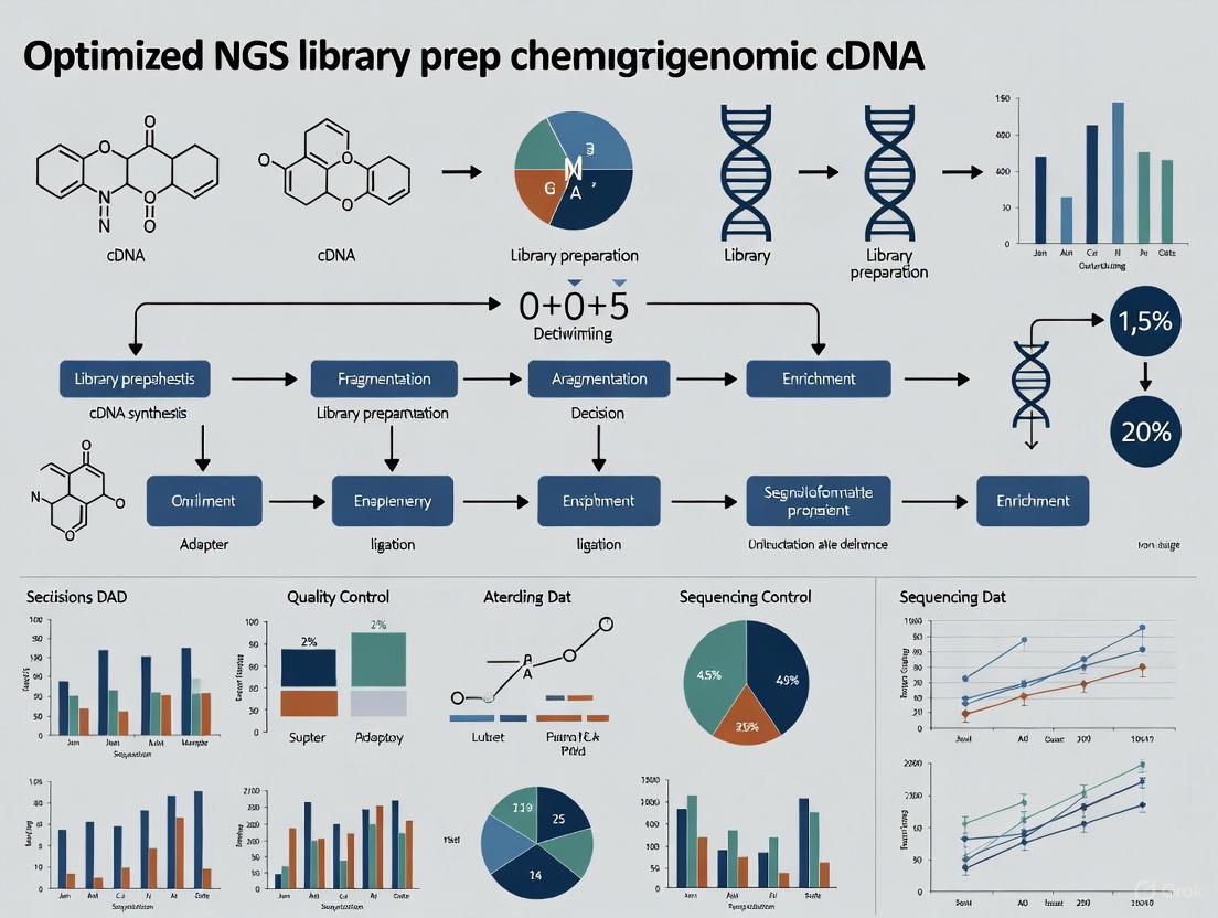
Workflow Visualization
The following diagram illustrates the complete workflow for the core steps of NGS library preparation, from fragmented cDNA to a sequencer-ready library.
Mastering the core principles of fragmentation, end-repair, and adapter ligation is non-negotiable for generating high-quality NGS libraries, especially in the demanding field of chemogenomic cDNA research. The protocols and data presented here provide a robust foundation for constructing libraries that ensure high data quality, minimize biases, and yield accurate, reproducible sequencing results. By carefully selecting fragmentation methods, optimizing reaction conditions, and implementing rigorous quality control, researchers can significantly enhance the reliability of their downstream analyses, thereby accelerating drug discovery and the understanding of chemical-genetic interactions.
Chemogenomics research, which explores the complex interactions between chemical compounds and biological systems, places unique and demanding requirements on next-generation sequencing (NGS) library preparation. The field inherently grapples with two major technical challenges: sample scarcity and complex transcriptomic responses. Researchers often work with limited material, such as rare cell populations treated with compound libraries or patient-derived samples exposed to drug candidates, where starting RNA can be exceptionally scarce [8]. Furthermore, the biological responses to chemical perturbations are multifaceted, involving subtle shifts in diverse RNA species that require highly sensitive and accurate detection methods [9]. This application note details optimized protocols and solutions specifically designed to overcome these challenges, enabling robust and reproducible cDNA library construction for chemogenomic studies.
Quantitative Challenges in Chemogenomic Library Preparation
The success of NGS in chemogenomics is highly dependent on the quantity and quality of input material. The table below summarizes key performance metrics for library preparation methods under conditions of sample scarcity, highlighting the critical thresholds for maintaining data quality.
Table 1: Performance Metrics of Library Prep Methods with Limited Input RNA
| Input RNA Amount | Number of Genes Detected | Detection of Low-Abundance Genes (FPKM 0-5) | Recommended Reverse Transcriptase | Key Limitations |
|---|---|---|---|---|
| 1 ng (bulk sample) | ~18,743 genes | Standard detection | Multiple options | Baseline for comparison |
| 5 pg | ~11,754 genes | Good detection with optimized protocols | Maxima H Minus | ~37% reduction in gene detection |
| 2 pg | Significant reduction | Moderate detection | Maxima H Minus | Mapping rate to marker genes drops to ~50% |
| 0.5 pg | >2,000 genes | Compromised without specialized methods | Maxima H Minus | Requires ultralow input optimization |
Even minor technical variations can significantly impact results. For instance, a pipetting inaccuracy of just 5% can result in a 2 ng variation in template DNA, which becomes critically important when working with scarce samples [10]. Additionally, inefficient library construction is reflected by a low percentage of fragments with correct adapters, leading to decreased sequencing data and increased chimeric fragments [4]. Batch effects arising from variations in reagents, equipment, or operator-related factors can substantially affect gene expression analysis outcomes, with particularly severe impacts on miRNA-seq data [10].
Optimized Protocols for Challenging Chemogenomic Samples
Ultralow Input RNA Sequencing Protocol
Based on systematic optimization studies, the following protocol significantly enhances sensitivity and low-abundance gene detection for scarce chemogenomic samples [8]:
Day 1: Reverse Transcription with Enhanced Efficiency
- Sample Preparation: Dilute extracted RNA in RNase-free water. For inputs below 10 pg, use siliconized (low retention) microcentrifuge tubes to maximize recovery.
- Reverse Transcription Master Mix:
- 2.0 μL 5X RT Buffer
- 1.0 μL dNTPs (10 mM each)
- 0.5 μL RNase Inhibitor (40 U/μL)
- 0.5 μL Maxima H Minus Reverse Transcriptase (200 U/μL)
- 1.0 μL Template-Switching Oligo (rN-modified TSO, 20 μM)
- 1.0 μL Gene-Specific Primer (2 μM)
- Add RNA sample (0.5-10 pg total RNA)
- Adjust to 10.0 μL with nuclease-free water
- Incubation:
- 42°C for 90 minutes
- 70°C for 5 minutes (enzyme inactivation)
- Hold at 4°C
Day 2: cDNA Amplification and Library Construction
- PCR Amplification:
- 10.0 μL RT product
- 15.0 μL 2X PCR Master Mix
- 1.0 μL PCR Primer (25 μM)
- 4.0 μL Nuclease-free water
- Thermocycling Conditions:
- 98°C for 3 minutes
- 18-22 cycles of:
- 98°C for 15 seconds
- 60°C for 30 seconds
- 72°C for 3 minutes
- 72°C for 5 minutes
- Hold at 4°C
- Purification and QC:
- Purify PCR products using magnetic beads (size selection if necessary)
- Quantify using fluorometric methods
- Assess quality via Bioanalyzer/TapeStation
This optimized protocol incorporates rN-modified template-switching oligos (TSO) and m7G-capped RNA templates to significantly improve sequencing sensitivity and low-abundance gene detection capability [8].
Automated Library Preparation for Enhanced Reproducibility
Automation addresses several challenges in chemogenomic library prep, particularly for screening applications involving multiple compounds or time points:
System Setup:
- Utilize liquid handling systems with non-contact dispensing technology
- Implement integrated thermocyclers to minimize sample transfer
- Configure for 96-well or 384-well plates based on throughput needs
Workflow Advantages:
- Reduced hands-on time: Approximately 30 minutes hands-on time for 96 samples with systems like ExpressPlex [10]
- Improved consistency: Inter-user variation coefficient reduced from >15% to <5%
- Miniaturized reactions: Capacity to work with sub-microliter volumes conserves precious samples
- Contamination control: Closed systems minimize environmental exposure [11]
Implementation Considerations:
- Select automation-compatible reagents (e.g., bead-based purification chemistries)
- Validate performance with standard reference materials
- Establish QC checkpoints for critical steps
Ultrasensitive Library Prep Workflow for Scarce Samples
Essential Research Reagent Solutions for Chemogenomics
Successful library preparation for chemogenomics requires carefully selected reagents specifically designed to address the challenges of sample scarcity and complex transcriptomic responses.
Table 2: Essential Research Reagents for Chemogenomic Library Preparation
| Reagent Category | Specific Product Examples | Function in Protocol | Considerations for Chemogenomics |
|---|---|---|---|
| Reverse Transcriptase | Maxima H Minus, SuperScript III | Converts RNA to cDNA; critical for sensitivity | Maxima H Minus shows superior sensitivity for low-abundance genes and minimal end bias [8] |
| Template-Switching Oligos | rN-modified TSO | Facilitates cDNA amplification from minimal input | rN modification significantly improves sequencing sensitivity and low-abundance gene detection [8] |
| Magnetic Beads | Sera-Mag Speedbeads, AMPure XP | Size selection and purification | Core-shell design provides tight size distributions; essential for FFPE and degraded samples [12] |
| Library Prep Kits | NEBNext UltraExpress, Illumina Stranded Total RNA Prep | Streamlined workflow integration | UltraExpress reduces tips by 32% and tubes by 50%; crucial for high-throughput compound screens [12] |
| Automation Systems | ExpressPlex, Callisto Sample Prep System | Standardization and throughput | ExpressPlex enables 96-sample prep in 30 minutes hands-on time; critical for multi-condition studies [10] |
Addressing Technical Complexities in Transcriptomic Responses
Chemogenomic experiments capture complex biological responses to chemical perturbations, requiring special consideration during library preparation:
Minimizing Amplification Bias
- Employ unique molecular identifiers (UMIs) to correct for PCR duplicates and enable accurate quantification [13]
- Utilize high-fidelity polymerases with minimal sequence preference
- Limit PCR cycles (typically 18-22) while maintaining sufficient library complexity
- Implement duplex-specific nucleases to normalize representation in highly abundant transcripts
Handling Diverse RNA Species Chemogenomic responses involve multiple RNA classes beyond mRNA, each requiring specific handling:
Small RNAs (miRNAs, siRNAs):
- Specialized ligation-based methods with pre-annealed adapter constructs [14]
- Biotinylated nucleotide incorporation in RT reaction for efficient cDNA purification
- Reduced gel purification steps to minimize sample loss
Long Non-coding RNAs:
- Ribosomal RNA depletion rather than poly-A selection to capture non-polyadenylated transcripts
- Enhanced fragmentation optimization to address secondary structures
Low-Abundance Transcripts:
- CRISPR-based depletion of abundant transcripts to increase coverage of rare targets [12]
- Hybridization-based capture approaches for focused analysis of specific pathway genes
Transcriptomic Complexity in Chemogenomic Studies
Quality Control and Validation Strategies
Rigorous QC protocols are essential for generating reliable chemogenomics data:
Pre-library Preparation QC:
- RNA Integrity Number (RIN) >8.5 for intact samples, >7.0 for FFPE
- Fluorometric quantification with sensitivity to 1 pg/μL
- Spike-in controls for absolute quantification
Post-library Preparation QC:
- Fragment size distribution analysis (Bioanalyzer/TapeStation)
- Library quantification via qPCR (more accurate than fluorometry for sequencing prediction)
- Adapter dimer detection (<5% threshold)
Post-sequencing QC:
- Sequencing saturation analysis
- Unique mapping rates (>70% for human transcripts)
- Gene detection counts compared to expected values
- Spike-in recovery calculations
For specialized applications like single-cell chemogenomics, additional validation through comparison to bulk RNA-seq or orthogonal methods (qPCR, nanostring) is recommended for a subset of targets.
Chemogenomics presents distinctive challenges for NGS library preparation that demand specialized approaches. The protocols and solutions detailed here address the dual challenges of sample scarcity through ultrasensitive methods and complex transcriptomic responses through optimized reagent systems and specialized handling of diverse RNA species. By implementing these tailored methods—including the use of Maxima H Minus reverse transcriptase, rN-modified template-switching oligos, automated workflows, and rigorous QC protocols—researchers can significantly enhance the quality and reproducibility of their chemogenomic studies. These advanced library preparation techniques enable more accurate characterization of compound-mode-of-action, identification of novel therapeutic targets, and ultimately, more efficient drug discovery pipelines.
The reverse transcription of RNA into complementary DNA (cDNA) is the foundational step in transcriptomic studies, determining the success and quality of all subsequent next-generation sequencing (NGS) data. For researchers in chemogenomics and drug development, where experiments often rely on limited or precious samples derived from compound treatments, optimizing this initial step is paramount for achieving accurate gene expression profiles. Inefficient reverse transcription can introduce significant bias, compromise detection sensitivity, and ultimately lead to misleading biological conclusions. This application note details the critical parameters and optimized protocols for the RNA-to-cDNA conversion, providing a robust framework for constructing high-quality transcriptomic libraries.
The Critical Role of Reverse Transcription
In transcriptomic workflows, RNA is first converted into a more stable DNA copy before sequencing. This cDNA synthesis process directly influences key outcomes:
- Library Complexity: The number of unique RNA molecules successfully captured and converted.
- Gene Detection Sensitivity: The ability to detect low-abundance transcripts, including key drug targets and biomarkers.
- Coverage Uniformity: The evenness of representation across different regions of a transcript.
The fidelity of this process is especially critical in chemogenomic research, where accurately quantifying subtle, compound-induced changes in the transcriptome is essential for understanding mechanisms of action and identifying novel therapeutic targets.
Optimizing Key Parameters for cDNA Synthesis
Primer Design and Selection
The choice of priming strategy is one of the most influential factors in reverse transcription. The table below summarizes the primary options and their optimal use cases.
Table 1: Primer Selection for Reverse Transcription
| Primer Type | Common Uses | Advantages | Limitations |
|---|---|---|---|
| Oligo(dT) | mRNA sequencing, poly-A tailed RNA enrichment [15] | Selects for mature, polyadenylated mRNA; reduces rRNA background. | Inefficient for degraded RNA; biased towards 3' end; unsuitable for non-polyA RNAs. |
| Random Hexamers | Whole transcriptome, degraded RNA [16] | Binds throughout transcript length; can detect non-polyA RNAs. | May not fully reverse transcribe long RNAs due to low binding stability. |
| Random 18mers | Whole transcriptome, long RNA transcripts [16] | Superior detection of long genes and low-abundance transcripts; more stable binding. | Less efficient for very short RNA biotypes (e.g., snRNAs, snoRNAs). |
| Gene-Specific | Targeted expression analysis (qPCR) | Highly specific and sensitive for targeted genes. | Not suitable for global transcriptome profiling. |
A pivotal study investigating primer length found that the commonly used random 6mer does not yield optimal performance. Instead, random 18mer primers demonstrated superior efficiency in overall transcript detection, particularly for long RNA transcripts like protein-coding genes and long non-coding RNAs in complex human tissue samples [16]. The 18mer detected approximately 10% more unique genes than the 6mer, with a significant advantage in detecting lowly expressed genes (FPKM 1-20) [16].
Input RNA Quantity and PCR Amplification
The amount of starting RNA and the subsequent amplification are tightly linked and must be carefully balanced to preserve library diversity and minimize artifacts.
Table 2: Impact of Input RNA and PCR Cycles on Data Quality
| Input RNA | Recommended PCR Cycles | Impact on PCR Duplicates | Effect on Gene Detection |
|---|---|---|---|
| High Input (≥ 125 ng) | Minimal cycles (e.g., 10-12) | Low rate (e.g., < 5%) [17] | High sensitivity; robust detection of low-expression genes. |
| Low Input (15 - 125 ng) | Increased but minimized cycles | High and variable rate (e.g., 34-96%) [17] | Reduced read diversity; fewer genes detected; increased noise. |
| Very Low Input (< 15 ng) | Maximum cycles per protocol | Very high rate; further increased by library conversion [17] | Severe loss of complexity; strong bias towards highly amplified fragments. |
For input amounts above 10 ng but below 125 ng, there is a strong negative correlation between input amount and the proportion of PCR duplicates. A positive correlation exists between the number of PCR cycles and duplicates. Therefore, the highest quality data is obtained using the lowest number of PCR cycles possible for a given input amount [17]. The use of Unique Molecular Identifiers (UMIs) is highly recommended for low-input samples to accurately distinguish biological duplicates from PCR-amplified artifacts during computational analysis [17].
Detailed Experimental Protocol: cDNA Library Construction
The following diagram illustrates the core workflow for constructing a cDNA library, from RNA isolation to ready-to-sequence libraries.
Materials: The Researcher's Toolkit
Table 3: Essential Reagents for cDNA Library Construction
| Reagent / Kit | Function | Considerations for Optimization |
|---|---|---|
| Oligo(dT) Magnetic Beads | Enriches for polyadenylated mRNA from total RNA [15]. | Reduces ribosomal RNA background; critical for mRNA-seq. |
| Reverse Transcriptase | Synthesizes first-strand cDNA using mRNA as a template [15] [18]. | Use high-fidelity, thermostable enzymes for long/structured RNAs. |
| Random Primers (6mer, 18mer) | Initiates reverse transcription at multiple sites along RNA fragments [16]. | 18mers recommended for superior detection of long transcripts [16]. |
| RNase H | Degrades the RNA strand in cDNA:RNA hybrids [15]. | Essential for second-strand synthesis. |
| DNA Polymerase I | Synthesizes the second strand of cDNA [15]. | Creates stable double-stranded cDNA. |
| dNTPs | Building blocks for cDNA synthesis. | Use balanced, high-quality stocks to prevent incorporation errors. |
| Platform-Specific Adapters | Allows cDNA fragments to bind to the sequencing flow cell [19]. | Contains barcodes for sample multiplexing. |
| Library Amplification Mix | PCR master mix containing a high-fidelity polymerase. | Minimize cycles to reduce duplication rates and bias [17]. |
Step-by-Step Procedure
Step 1: Isolation and Quality Control of mRNA
Begin with high-quality total RNA. Isolate mRNA via chromatographic purification using an oligo(dT) matrix to retain poly(A)+ RNA molecules, effectively depleting abundant tRNAs and rRNAs [15]. Assess RNA integrity using an instrument like an Agilent Bioanalyzer to ensure an RNA Integrity Number (RIN) > 8.0 for optimal results.
Step 2: First-Strand cDNA Synthesis
- Combine 1-1000 ng of mRNA with random 18mer primers (or oligo(dT) primers for 3'-end focused assays) and dNTPs [16].
- Denature at 65°C for 5 minutes to remove secondary structures, then immediately place on ice.
- Add reaction buffer, DTT, RNase inhibitor, and a robust reverse transcriptase (e.g., SuperScript II or equivalent) [18].
- Incubate at 42-50°C for 50-60 minutes, followed by enzyme inactivation at 70°C for 15 minutes. This produces an mRNA:cDNA hybrid.
Step 3: Second-Strand cDNA Synthesis
- To the first-strand reaction, add RNase H to nick the mRNA strand, and DNA Polymerase I to synthesize the second DNA strand using the dNTPs provided [15].
- Incubate at 16°C for 1 hour. The low temperature minimizes exonuclease activity and favors polymerase activity.
- Purify the double-stranded cDNA using magnetic beads or column-based purification. The cDNA can now be stored at -20°C.
Step 4: Adapter Ligation and Library Amplification
- Prepare the blunt-ended, double-stranded cDNA for adapter ligation. This may involve end-repair and A-tailing to create compatible ends for ligation.
- Ligate platform-specific adapters to the cDNA fragments. For blunt-end ligations, use high enzyme concentrations and incubate at room temperature for 15-30 minutes. For cohesive-end ligations, incubate at 12-16°C for longer durations [19].
- Perform a limited-cycle PCR (e.g., 10-15 cycles depending on input) to amplify the adapter-ligated library. Minimize PCR cycles to preserve library complexity and reduce duplicate rates [17].
- Purify the final library using magnetic bead-based cleanup to remove primer dimers and fragments that are too short or too long [19].
Step 5: Library Quality Control and Normalization
- Quantify the library accurately using fluorometric methods (e.g., Qubit) and qPCR.
- Assess size distribution with a Bioanalyzer or TapeStation.
- Normalize libraries to an equimolar concentration before pooling to ensure balanced representation in sequencing [19]. Automated systems can significantly improve the consistency of this step.
Troubleshooting Common Issues
- High PCR Duplication Rate: This indicates low library complexity, often due to insufficient input RNA or excessive PCR cycles. Solution: Increase RNA input where possible, and use the minimum number of PCR cycles required. Incorporate UMIs to bioinformatically identify and remove duplicates [17] [4].
- Low Library Yield: Can result from degraded RNA, inefficient enzymatic reactions, or losses during purification. Solution: Check RNA quality, ensure fresh reagents and proper enzyme handling to maintain activity, and use purification methods with high recovery rates [19].
- Adapter Dimer Contamination: Caused by self-ligation of adapters. Solution: Optimize adapter molar ratios during ligation and use bead-based size selection to remove short fragments [19].
The conversion of RNA to cDNA is a critical gateway in the transcriptomic library construction pipeline, whose quality dictates the validity of downstream data and analysis. For drug development professionals, consistent application of optimized protocols—embracing strategic primer selection, careful input RNA quantification, and minimized PCR amplification—is non-negotiable. By adhering to the detailed methodologies and best practices outlined in this application note, researchers can ensure the generation of robust, high-complexity cDNA libraries. This, in turn, provides a reliable foundation for uncovering meaningful biological insights in chemogenomic research and advancing therapeutic discovery.
Next-generation sequencing (NGS) library preparation is a critical first step in any sequencing workflow, profoundly impacting the quality, reliability, and interpretation of generated data. For researchers in chemogenomics and drug development, selecting the appropriate library construction method is paramount for obtaining meaningful biological insights from cDNA experiments. Among the available techniques, ligation-based and tagmentation-based workflows have emerged as two principal approaches, each with distinct advantages, limitations, and optimal application scenarios. This application note provides a detailed comparison of these methodologies, supported by quantitative performance data and step-by-step experimental protocols, to guide researchers in selecting and implementing the optimal strategy for their specific research objectives.
Core Technology Comparison
Fundamental Mechanisms
Ligation-based library preparation involves the physical or enzymatic fragmentation of DNA or cDNA, followed by a series of enzymatic steps to repair ends and ligate specialized adapters to both ends of the fragments using DNA ligase [13]. This traditional approach provides consistent performance across diverse genomic contexts.
Tagmentation-based library preparation utilizes a bead-linked transposome (BLT) system where a transposase enzyme simultaneously fragments DNA and ligates adapters in a single enzymatic step [20] [13]. This innovative approach dramatically reduces hands-on time and workflow complexity by combining multiple steps into one.
Performance Characteristics and Bias Profiles
Each method exhibits distinct performance characteristics and potential biases that researchers must consider:
- Sequence Bias: Tagmentation methods may display sequence pattern preference in the initial 10-15 bases of sequencing reads [20]. However, multiple studies indicate this does not adversely affect library complexity or genome coverage across diverse species and sequence contexts [20].
- Fragment Size Distribution: Ligation-based methods typically produce more variable fragment sizes, while BLT methods generate more homogeneous fragment distributions, independent of DNA input quantity and quality [20].
- Coverage Uniformity: Comparative studies demonstrate that the most crucial factor for even genome coverage is library fragment size, with BLT methods producing superior reproducibility in fragment size homogeneity compared to ligation-based approaches [20].
Quantitative Comparison of Performance Metrics
Table 1: Direct performance comparison of ligation, tagmentation, and PCR-based library prep methods for bacterial genomics [21]
| Performance Metric | Ligation-Based (LIG) | Tagmentation-Based (TAG) | PCR-Based (PCR) |
|---|---|---|---|
| Average Read Length | >5,000 bp | >5,000 bp | <1,100 bp |
| Total Output (Gbp) | 33.62 | 11.72 | 4.79 |
| Mappable Reads | 92.9% | 87.3% | 22.7% |
| Artifactual Tandem Content | 0.9% | 2.2% | 22.5% |
| Output Homogeneity | Most homogeneous | Intermediate | Most variable |
Table 2: Workflow and efficiency comparison between library preparation methods [21] [22] [13]
| Characteristic | Ligation-Based | Tagmentation-Based |
|---|---|---|
| Hands-on Time | ~3-6 hours [22] | ~1-1.5 hours [13] |
| Total Workflow Time | ~6.5 hours [22] | ~3-4 hours [13] |
| Input DNA Requirement | 100-1000 ng [22] | 1-500 ng [13] |
| PCR Requirement | Often required | Optional |
| Multiplexing Capacity | Standard | Standard |
| Cost Considerations | Higher reagent and labor costs | Lower overall cost due to reduced hands-on time |
Detailed Experimental Protocols
Ligation-Based Library Preparation Protocol
Principle: This method utilizes sequential enzymatic reactions to fragment DNA, repair ends, and ligate adapters in a multi-step process [13].
Table 3: Key reagents for ligation-based library prep [13]*
| Reagent | Function |
|---|---|
| Fragmentation Enzyme | Fragments DNA to desired size distribution |
| End Repair Mix | Repairs fragmented ends to create blunt ends |
| A-Tailing Enzyme | Adds single 'A' nucleotide to 3' ends |
| DNA Ligase | Ligates adapters to A-tailed fragments |
| SPRI Beads | Size selection and purification |
| Unique Dual Index Adapters | Enable sample multiplexing |
Step-by-Step Workflow:
DNA Fragmentation:
- Use either mechanical (acoustic shearing) or enzymatic fragmentation methods
- Target fragment size: 200-500bp for standard applications
- Recommended input: 100-1000ng high-quality DNA [22]
End Repair and A-Tailing:
- Incubate fragmented DNA with end repair enzyme mix (30 minutes, 20°C)
- Add A-tailing buffer and enzyme (30 minutes, 37°C)
- Purify using SPRI beads (1.8X ratio)
Adapter Ligation:
- Add unique dual index adapters and DNA ligase
- Incubate (15 minutes, 20°C)
- Purify with SPRI beads (1.8X ratio)
Library Amplification (Optional):
- Add PCR master mix with appropriate cycle number
- Purify final library with SPRI beads (1.0X ratio)
Quality Control:
- Quantify using fluorometric methods (Qubit)
- Assess size distribution (Bioanalyzer/TapeStation)
- Validate library concentration for sequencing
Tagmentation-Based Library Preparation Protocol
Principle: This approach uses bead-linked transposomes to simultaneously fragment DNA and incorporate sequencing adapters in a single reaction [20] [13].
Table 4: Key reagents for tagmentation-based library prep [20] [13]*
| Reagent | Function |
|---|---|
| Bead-Linked Transposomes (BLT) | Simultaneously fragments and tags DNA with adapters |
| Tagmentation Buffer | Optimizes transposase enzyme activity |
| Neutralization Buffer | Stops tagmentation reaction |
| PCR Master Mix | Amplifies library (if required) |
| SPRI Beads | Size selection and purification |
| Unique Dual Index Primers | Enable sample multiplexing |
Step-by-Step Workflow:
Tagmentation Reaction:
- Combine DNA with bead-linked transposomes and tagmentation buffer
- Incubate (5-15 minutes, 55°C)
- Add neutralization buffer to stop reaction
- Recommended input: 1-500ng DNA [13]
Library Amplification (Optional):
- Add PCR master mix with unique dual index primers
- Amplify with limited cycles (typically 12-15 cycles)
- Note: PCR-free workflows are possible with sufficient input
Purification and Size Selection:
- Clean up with SPRI beads (0.6X-0.8X ratio for size selection)
- Elute in appropriate buffer
Quality Control:
- Quantify using fluorometric methods
- Assess fragment size distribution
- Validate library for sequencing
Application-Specific Recommendations
Chemogenomic cDNA Research Considerations
For chemogenomic studies investigating gene expression responses to chemical compounds, several factors warrant special consideration:
- Input Material: Tagmentation methods offer superior performance with limited cDNA samples common in drug screening assays [13].
- Sequence-Specific Bias: Ligation-based approaches may be preferable for detecting transcripts with extreme GC content, as tagmentation can exhibit sequence-specific bias [20].
- Multiplexing Requirements: Both methods support extensive multiplexing, but tagmentation workflows enable more rapid processing of large compound libraries [23].
- Data Reproducibility: For longitudinal studies assessing compound effects over time, ligation-based methods may provide more consistent coverage of transcript ends [21].
Specialized Applications
FFPE and Degraded Samples:
- Modified tagmentation protocols demonstrate excellent performance with degraded RNA from FFPE samples [24]
- Ligation-based methods with specialized repair enzymes can rescue data from heavily damaged samples
Low-Input and Single-Cell Applications:
- Tagmentation methods are preferred for single-cell cDNA libraries due to higher efficiency [25]
- Modified tagmentation protocols enable library preparation from as little as 40ng DNA [25]
Multimodal Sequencing:
- Advanced tagmentation approaches enable concurrent readout of genetic sequence, CpG methylation, and chromatin accessibility from a single library [25]
Method Selection Guide
Table 5: Application-based recommendations for library preparation methods [21] [20] [13]*
| Research Scenario | Recommended Method | Rationale |
|---|---|---|
| Maximum Data Quality | Ligation-based | Superior mappable reads (92.9%) and lowest artifactual content [21] |
| High-Throughput Screening | Tagmentation-based | 65% faster workflow and higher throughput capabilities [23] |
| Limited Input Samples | Tagmentation-based | Effective with 1ng input vs. 100ng for ligation-based [13] |
| Complex Genome Regions | Ligation-based | Reduced sequence-specific bias for challenging regions [20] |
| Cost-Sensitive Projects | Tagmentation-based | Lower reagent costs and reduced hands-on time [23] |
| Multimodal Analysis | Tagmentation-based | Enables concurrent genetic and epigenetic profiling [25] |
The choice between ligation-based and tagmentation-based library preparation methods represents a critical decision point in designing chemogenomic cDNA research studies. Ligation-based methods remain the gold standard for applications demanding the highest data quality and minimal technical artifacts, as evidenced by their superior mappable read rates (92.9%) and low artifactual content [21]. Conversely, tagmentation-based approaches offer compelling advantages in workflow efficiency, requiring significantly less hands-on time (65% reduction) and lower input requirements while maintaining robust performance across most applications [13] [23].
For drug development professionals, the selection framework should prioritize project-specific requirements including input material limitations, throughput needs, data quality thresholds, and budget constraints. As both technologies continue to evolve, tagmentation methods show particular promise for emerging applications in multimodal sequencing and complex sample types, while ligation methods maintain their position for standardized applications requiring maximal data fidelity. By implementing the detailed protocols and considerations outlined in this application note, researchers can make informed decisions that optimize their library preparation strategies for successful chemogenomic investigations.
The Role of Adapters and Barcodes in Multiplexing Drug Treatment Samples
Within chemogenomic cDNA research, where the systematic screening of chemical compounds on biological systems is paramount, Next-Generation Sequencing (NGS) has become an indispensable tool for profiling transcriptomic changes. The efficiency of such studies is often gated by the throughput and cost-effectiveness of the sequencing workflow. Sample multiplexing, the simultaneous sequencing of multiple libraries in a single run, addresses this bottleneck directly [26] [27]. This technique relies on the strategic use of adapters and barcodes (also known as indexes) to enable the precise pooling and subsequent deconvolution of data from dozens of drug treatment samples [27]. By assigning a unique molecular identifier to each sample, researchers can dramatically reduce per-sample costs and minimize technical variability, thereby accelerating the pace of discovery in drug development [26]. These Application Notes detail the principles and provide a robust protocol for implementing adapter and barcode-based multiplexing in chemogenomic studies.
Conceptual Foundations of Multiplexing
The Core Components: Adapters and Barcodes
Multiplexing is fundamentally enabled by attaching short, unique DNA sequences to the cDNA fragments derived from each sample. This process involves two key components:
- Adapters: Short, known oligonucleotide sequences that are ligated to both ends of every cDNA fragment in a library [4]. These adapters are essential for the sequencing process itself, containing complementary sequences that allow the library fragments to bind to the flow cell and be amplified via bridge PCR. They also contain the primer-binding sites necessary for the sequencing reactions [4] [27].
- Barcodes (Indexes): Short, unique DNA sequences embedded within the adapter oligonucleotides [27]. Each sample in a multiplexed pool receives a unique barcode. After a pooled sequencing run is complete, the barcode sequence on each read is used to computationally assign the read back to its sample of origin, a process known as demultiplexing [27].
Advantages of a Multiplexed Workflow
The primary advantage of sample multiplexing is a significant increase in throughput and a reduction in sequencing costs. By pooling multiple samples, the time and reagent expenses for a sequencing run are distributed across all samples in the pool [26] [27]. Furthermore, processing samples in a single multiplexed run, rather than across multiple individual runs, reduces batch effects and technical variability, leading to more robust and reproducible comparative analyses—a critical consideration when assessing the subtle transcriptional impacts of drug treatments [26].
Indexing Strategies: Single vs. Dual Indexing
The configuration of barcodes within the adapters is a critical design choice. The two main strategies are single and dual indexing, with unique dual indexes being the recommended best practice for modern applications [27].
Table 1: Comparison of Single and Dual Indexing Strategies
| Feature | Single Indexing | Dual Indexing (Recommended) |
|---|---|---|
| Barcode Location | A single barcode sequence on one adapter. | Two unique barcode sequences, one on each adapter. |
| Multiplexing Capacity | Lower | Higher |
| Error Detection | Poor; cannot reliably detect index hopping. | Excellent; can identify and filter reads affected by index hopping. |
| Data Fidelity | Lower confidence in sample assignment. | High confidence in sample assignment. |
Index hopping is a phenomenon where barcode sequences are incorrectly assigned during sequencing, potentially leading to cross-contamination of data between samples [27]. Dual indexing provides a robust solution to this problem, as a read must match both expected barcode sequences to be assigned to a sample, thereby preventing misassignment if one index is corrupted [27].
Technical Implementation and Workflow
The integration of adapters and barcodes occurs during the library preparation stage, which transforms cDNA into a sequence-ready library.
Library Preparation Workflow
The following workflow outlines the key steps from fragmented cDNA to a pooled, multiplexed library ready for sequencing:
Key Steps Explained
- End Repair & A-Tailing: The cDNA fragments are blunted and a single 'A' nucleotide is added to the 3' ends. This creates a compatible overhang for ligation with adapters that have a complementary 'T' overhang [4].
- Adapter Ligation: Adapters containing the platform-specific sequences and binding sites for sequencing primers are ligated to the 'A-tailed' cDNA fragments [4]. In many modern kits, these adapters are already partially or fully double-stranded.
- Library Amplification & Indexing: This is the critical step where barcodes are incorporated. A limited-cycle PCR is performed using primers that contain:
- The
P5andP7flow cell binding sequences. - The unique barcode sequences that will identify the sample.
- The sequencing primer binding sites. This step simultaneously amplifies the library to generate sufficient mass for sequencing and adds the complete adapter structure, including the barcodes, to each molecule [27].
- The
- Library QC & Purification: The final library is purified to remove excess primers, adapter dimers, and PCR reagents. Quality control, typically using fluorometry and fragment analysis, is performed to confirm library concentration and size distribution [4].
- Normalization & Pooling: Libraries are quantified and normalized to ensure equimolar representation. They are then combined into a single pool for the sequencing run [27].
Essential Protocol: Multiplexing Drug Treatment Samples
This protocol provides a detailed methodology for generating multiplexed cDNA libraries from drug-treated samples.
Research Reagent Solutions
Table 2: Essential Reagents and Materials for Library Preparation
| Item | Function | Example/Note |
|---|---|---|
| DNA Library Prep Kit | Provides enzymes and buffers for end repair, A-tailing, ligation, and PCR. | Select a kit compatible with your sequencing platform and read length. |
| Unique Dual Indexed Adapters | Pre-synthesized adapter mixes containing unique barcode pairs for each sample. | Commercial sets (e.g., Illumina) are available in various plexities. |
| SPRIselect Beads | Magnetic beads for size selection and purification of the library between steps. | Enables removal of unwanted reagents and selection of optimal fragment sizes. |
| Qubit dsDNA HS Assay | Fluorometric quantification of library concentration. | More accurate for library quantitation than spectrophotometry. |
| Bioanalyzer/TapeStation | Capillary electrophoresis system for assessing library size distribution and quality. | Critical for detecting adapter dimers and verifying insert size. |
Step-by-Step Procedure
- Input Material: Begin with 100 ng of high-quality, double-stranded cDNA derived from drug-treated and control samples. Ensure cDNA is purified and in nuclease-free water.
- End Repair & A-Tailing:
- Combine cDNA with the provided end-prep enzyme mix.
- Incubate at 20°C for 30 minutes, followed by 65°C for 30 minutes.
- Purify the reaction using SPRIselect beads (1.0X ratio) to retain all fragments and elute in nuclease-free water.
- Adapter Ligation:
- To the purified end-repaired cDNA, add ligation buffer, DNA ligase, and a unique dual index adapter for each sample.
- Incubate at 20°C for 15 minutes.
- Purify with SPRIselect beads (0.9X ratio) to remove excess adapters and elute.
- Library Amplification:
- Prepare a PCR mix with the universal P5 and P7 primers and a high-fidelity DNA polymerase.
- Run the following PCR program:
- 98°C for 30 seconds (initial denaturation)
- 8-12 cycles of:
- 98°C for 10 seconds (denaturation)
- 60°C for 30 seconds (annealing)
- 72°C for 30 seconds (extension)
- 72°C for 5 minutes (final extension)
- Final Library Clean-up:
- Purify the amplified library with SPRIselect beads (0.9X ratio).
- Elute in nuclease-free water or TE buffer.
- Quality Control:
- Quantify the final library concentration using the Qubit dsDNA HS Assay.
- Analyze 1 µL of the library on a Bioanalyzer or TapeStation using a High Sensitivity DNA chip to confirm a peak in the expected size range (e.g., 300-500 bp) and the absence of a primer-dimer peak at ~100 bp.
- Pooling for Sequencing:
- Normalize all libraries to the same concentration (e.g., 10 nM) based on Qubit and fragment analyzer data.
- Combine an equal volume of each normalized library into a single tube. This is your multiplexed sequencing pool.
- The final pool can be diluted to the loading concentration required by your specific sequencer.
Data Analysis and Demultiplexing
Upon completion of the sequencing run, the primary data output is a pool of sequence reads from all samples. The process of demultiplexing is the first bioinformatic step, which uses the barcode information to sort the reads back into their respective sample-specific files. This process is typically performed automatically by the sequencer's onboard software or dedicated demultiplexing tools [27]. The output is a set of FASTQ files (or similar), one for each sample, which are then ready for standard downstream processing such as alignment, quantification, and differential expression analysis. The use of unique dual indexes ensures that any reads which have undergone index hopping are identified and either corrected or filtered out, preserving the integrity of the data for critical chemogenomic analyses [27].
Streamlined Protocols for Chemogenomic cDNA: From Low-Input Samples to Target Enrichment
Strategies for Low-Input and Degraded RNA from Treated Cell Cultures
Next-generation sequencing (NGS) has revolutionized biological research by enabling in-depth analysis of transcriptomes, yet analyzing samples with limited material or compromised quality remains a significant challenge [28]. In chemogenomic research, where cell cultures are treated with chemical compounds or drugs, researchers frequently encounter low-input and degraded RNA resulting from treatment-induced cytotoxicity or the necessity of using rare cell populations. These samples are particularly vulnerable to degradation and yield limitations, making conventional RNA sequencing approaches unsuitable [28] [4].
The success of transcriptomic studies in this context heavily depends on selecting appropriate library preparation strategies that can effectively handle minimal inputs while preserving biological complexity [3]. This application note provides a comprehensive framework for generating high-quality sequencing libraries from low-input and degraded RNA derived from treated cell cultures, with specific methodologies optimized for chemogenomic cDNA research.
Key Considerations for Experimental Design
Input Requirements and Sample Quality
Library preparation kits vary significantly in their input requirements, which is a primary consideration when working with limited samples from treated cultures. Input amounts generally fall into three categories: standard input (100-1000 ng), low-input (1-100 ng), and ultra-low-input (below 1 ng) [28] [29]. For degraded samples, which are common in chemogenomic studies involving fixed cells or stressful chemical treatments, higher input amounts may be necessary to compensate for fragmentation [28].
Sample quality assessment is crucial before library preparation. For RNA samples, the RNA Integrity Number (RIN) provides a valuable metric, though specialized kits can handle severely degraded samples with RIN values as low as 2 [30]. In treated cell cultures where extraction yields may be low, verification of sample quantity using sensitive methods such as fluorometry is recommended [31].
Strategic Selection of Library Preparation Technology
The choice of library preparation method significantly impacts data quality, coverage uniformity, and detection sensitivity. Three primary technological approaches have emerged for handling challenging RNA samples:
Template-switching technology: Utilizes the template-switching activity of reverse transcriptase to add universal adapter sequences during cDNA synthesis, enabling efficient library construction from minimal input [32]. This approach is particularly valuable for maintaining sequence representation in ultra-low-input scenarios.
Stranded protocols with specialized chemistry: Employ molecular techniques such as dUTP marking or ligation-based methods to preserve strand orientation information without requiring toxic reagents like actinomycin D [30]. These protocols are essential for accurate transcript annotation and identification of antisense transcription events in chemogenomic studies.
Unique molecular identifiers (UMIs): Incorporate molecular barcodes during reverse transcription to tag individual RNA molecules, enabling bioinformatic correction of amplification biases and PCR duplicates [33]. This technology provides more accurate quantitation, especially important when assessing expression changes in drug-treated samples.
Commercial Kit Comparisons and Selection Guide
Comprehensive Kit Comparison
Table 1: Comparison of Low-Input and Degraded RNA Library Preparation Kits
| Manufacturer | Kit Name | Input Range | Protocol Duration | Automation Compatibility | Key Features |
|---|---|---|---|---|---|
| Takara Bio | SMARTer Universal Low Input RNA Kit | 10-100 ng total RNA or 200 pg-10 ng rRNA-depleted RNA | 2 hours | No | SMART technology with random priming; useful for degraded RNA without polyA-tails [28] |
| Roche | KAPA RNA HyperPrep Kit | 1-100 ng RNA | 4 hours | Yes | Single-tube chemistry; optimized for degraded and low-input samples [28] |
| Watchmaker | Watchmaker RNA Library Prep Kit | 0.25-100 ng total RNA | 3.5 hours | Yes | Novel engineered reverse transcriptase for degraded FFPE samples [28] |
| Illumina | Stranded Total RNA Prep | 1-1000 ng standard quality RNA; 10 ng for FFPE | ~7 hours | Yes | Integrated enzymatic rRNA depletion; works with degraded samples [33] |
| Lexogen | Proprietary Ultra-low Input Technology | 10 pg to 1 ng total RNA | Varies | Yes | Extraction-free capability; works with cell lysates [29] |
| IDT | xGen Broad-Range RNA Library Preparation Kit | 10 ng-1 µg RNA or 100 pg-100 ng mRNA | 4.5 hours | Yes | Adaptase technology eliminates second-strand synthesis [28] |
Table 2: Performance Characteristics Across Input Ranges
| Input Range | Recommended Technology | Expected Gene Detection | Best For |
|---|---|---|---|
| >100 ng | Standard stranded protocols | >80% of transcriptome | High-quality samples from abundant cell cultures |
| 1-100 ng | Modified low-input protocols | 60-80% of transcriptome | Treated cultures with moderate yield |
| 100 pg-1 ng | Template-switching methods | 40-60% of transcriptome | Rare cell populations or limited material |
| 10-100 pg | Ultra-low input specialized kits | 20-40% of transcriptome | Single-cell or subcellular analyses |
Selection Guidelines for Chemogenomic Applications
For chemogenomic studies involving drug-treated cultures, kit selection should be guided by specific experimental parameters:
High-throughput compound screening: Automated-compatible kits such as the KAPA RNA HyperPrep or Watchmaker RNA Library Prep Kit enable processing of multiple samples with minimal hands-on time [28].
Time-course experiments with sequential sampling: Rapid protocol kits like the Takara SMARTer Universal Low Input (2 hours) provide quick turnaround for dynamic transcriptome assessment [28].
Pathway-focused analysis: Targeted RNA sequencing approaches using enrichment panels concentrate sequencing power on genes of interest, providing cost-effective solutions for focused questions [33].
Detailed Experimental Protocols
Protocol A: Ultra-Low Input RNA (10 pg-1 ng) Using Template-Switching Technology
This protocol is adapted from the SMARTer and Lexogen approaches for minute RNA quantities [28] [29].
Workflow Overview:
Step-by-Step Methodology:
RNA Fragmentation and Priming
- Combine 1-10 pg to 1 ng total RNA with 1 µL of 3' SMART CDS Primer II A in nuclease-free water to a total volume of 4.5 µL
- Incubate at 72°C for 3 minutes, then hold at 42°C for 2 minutes
- Brief centrifuge to collect contents
Reverse Transcription with Template Switching
- Prepare RT mix: 2.0 µL 5× First-Strand Buffer, 0.25 µL DTT (100 mM), 1.0 µL SMARTer II A Oligonucleotide, 1.0 µL RNase Inhibitor, 0.25 µL SMARTScribe Reverse Transcriptase
- Add 4.5 µL RT mix to each RNA-primer sample, mix gently by pipetting
- Incubate at 42°C for 90 minutes, then heat-inactivate at 70°C for 10 minutes
cDNA Amplification
- Prepare PCR mix: 25 µL SeqAmp PCR Buffer, 1 µL PCR Primer II A, 1 µL SeqAmp DNA Polymerase, 16.5 µL nuclease-free water
- Add 6.5 µL cDNA from previous step to 43.5 µL PCR mix
- Amplify using cycling conditions: 95°C for 1 min; 15-20 cycles of 95°C for 10 sec, 65°C for 30 sec, 68°C for 3 min; final extension at 72°C for 5 minutes
- Purify using AMPure XP beads at 0.8× ratio
Library Construction and Indexing
- Fragment 100 ng purified cDNA using Covaris shearing (200 bp target)
- Perform end repair and A-tailing using commercial enzyme mix (20°C for 30 min, 65°C for 30 min)
- Ligate Illumina adapters with UMI barcodes (T4 DNA ligase, 20°C for 15 min)
- Clean up using AMPure XP beads (0.8× ratio)
Library Amplification and Final Cleanup
- Amplify with index primers using reduced cycles (8-12 cycles) with high-fidelity polymerase
- Perform double-sided size selection with AMPure XP beads (0.5×/0.8× ratios)
- Quantify using Qubit dsDNA HS Assay and profile with Bioanalyzer High Sensitivity DNA chip
Critical Steps and Troubleshooting:
- For highly degraded samples, increase input amount by 1.5-2× while maintaining the same reaction volumes
- If yields are low after cDNA amplification, increase PCR cycles by 2-3 while monitoring for over-amplification artifacts
- To minimize batch effects, prepare master mixes for all enzymatic steps and process all samples in parallel
Protocol B: Degraded RNA from Treated Cultures (1-100 ng) Using Stranded Chemistry
This protocol utilizes the principles behind KAPA and Illumina stranded kits optimized for compromised samples [28] [33].
Workflow Overview:
Step-by-Step Methodology:
rRNA Depletion
- Dilute 1-100 ng total RNA to 11 µL with nuclease-free water
- Add 1 µL rRNA Removal Solution, mix thoroughly by pipetting
- Incubate at 68°C for 10 minutes, then hold at room temperature for 5 minutes
- Add 1 µL rRNA Removal Beads, incubate at room temperature for 5 minutes
- Place on magnet, transfer supernatant to new tube
RNA Fragmentation and Priming
- Add 2 µL Fragmentation Solution to depleted RNA
- Incubate at 94°C for 4 minutes (adjust time based on desired fragment size)
- Immediately place on ice, add 2 µL Fragmentation Stop Solution
First Strand cDNA Synthesis
- Add 4 µL First Strand Synthesis Act D Mix, 1 µL Random Primers, 2 µL SuperScript II Reverse Transcriptase
- Incubate at 25°C for 10 min, 42°C for 50 min, 70°C for 15 min
- Proceed immediately to second strand synthesis
Second Strand Synthesis with dUTP Incorporation
- Prepare second strand mix: 10 µL Second Strand Marking Master Mix, 4 µL dUTP Solution, 26 µL nuclease-free water
- Add to first strand reaction, incubate at 16°C for 60 minutes
- Purify using AMPure XP beads (0.8× ratio), elute in 25 µL Resuspension Buffer
Adapter Ligation and Library Completion
- Add 2.5 µL End Repair & A-Tailing Control, 12.5 µL Ligation Mix, 2.5 µL RNA Adapter
- Incubate at 20°C for 15 minutes
- Add 5 µL UDG, incubate at 37°C for 30 minutes to digest second strand
- Amplify with index primers: 98°C for 45 sec; 10-12 cycles of 98°C for 15 sec, 60°C for 30 sec, 72°C for 30 sec; 72°C for 1 min
- Perform double-sided size selection with AMPure XP beads
Quality Control Parameters:
- Assess library concentration using Qubit dsDNA HS Assay (target > 5 nM)
- Verify size distribution using Bioanalyzer High Sensitivity DNA chip (peak ~280-320 bp)
- Confirm absence of adapter dimers (< 3% of total signal)
- Check molarity by qPCR using library quantification kit for accurate pooling
The Scientist's Toolkit: Essential Research Reagents
Table 3: Critical Reagents for Low-Input and Degraded RNA Studies
| Reagent Category | Specific Products | Function & Importance | Application Notes |
|---|---|---|---|
| Reverse Transcriptases | SMARTScribe, SuperScript II | cDNA synthesis with high processivity and template-switching capability | Critical for full-length cDNA from degraded templates; engineered enzymes show better performance with inhibitors [28] |
| Library Amplification Kits | KAPA HiFi HotStart ReadyMix, CleanStart HiFi PCR Mastermix | High-fidelity amplification with uniform coverage | Minimize GC bias and maintain sequence representation; essential for accurate variant calling [28] [30] |
| RNA Depletion Kits | Illumina Ribo-Zero Gold, QIAseq FastSelect rRNA | Remove abundant ribosomal RNA | Significantly increases mapping rates; particularly important for bacterial or non-polyA samples [30] [33] |
| Nucleic Acid Purification | AMPure XP Beads, QIAseq Beads | Size selection and cleanup between steps | Bead-based methods preferred for low-input work due to higher recovery rates [28] [30] |
| Quality Control Tools | Agilent Bioanalyzer, TapeStation, Qubit fluorometer | Assess RNA integrity and library quality | Essential for troubleshooting and optimizing input requirements; Bioanalyzer provides critical size distribution data [30] [3] |
| Unique Dual Indexes | Illumina UDI, IDT xGen UDI | Sample multiplexing and cross-contamination reduction | Enable complex experimental designs with multiple treatment conditions and time points [28] [33] |
Data Analysis and Bioinformatics Considerations
Specialized Processing for Challenging Libraries
Sequencing data from low-input and degraded RNA requires specialized bioinformatic processing to extract meaningful biological insights:
Unique Molecular Identifier (UMI) processing: Deduplication based on UMIs provides accurate molecular counting, correcting for amplification biases inherent in low-input protocols [33]. Tools such as UMI-tools or zUMIs should be implemented before alignment to distinguish technical duplicates from biological replicates.
Adapter trimming and quality control: Aggressive adapter trimming is essential for degraded samples with short fragment sizes. Trimming tools should be configured with parameters specific to your library preparation kit, particularly for technologies like Adaptase that add specific sequences [34].
Strand-specific alignment: Ensure alignment software (STAR, HISAT2) is configured for the specific strandedness of your protocol to improve transcript assignment accuracy, particularly important for identifying overlapping transcripts in chemogenomic studies [30].
Quality Metrics for Degraded Samples
Traditional RNA-Seq QC metrics require adaptation for degraded samples:
- Mapping rates >70% may be acceptable for severely degraded samples (RIN < 4)
- 3' bias is expected in degraded samples; normalize using TPM or similar methods that account for transcript length
- Monitor ribosomal RNA content (<5% ideal, though higher percentages may be acceptable with certain depletion strategies)
- Assess duplication rates in context of input amount - higher duplication is expected with lower inputs but should be corrected with UMIs
Successful transcriptomic analysis of low-input and degraded RNA from treated cell cultures requires integrated optimization across sample preparation, library construction, and bioinformatic analysis. Based on the methodologies presented in this application note, the following recommendations emerge for chemogenomic research:
For ultra-low input scenarios (single-cell or limited cell populations), template-switching technologies such as SMARTer protocols provide the most robust performance, enabling library construction from as little as 10 pg total RNA while maintaining strand specificity [28] [29]. For moderately degraded samples from compound-treated cultures, streamlined stranded protocols like the KAPA RNA HyperPrep or Illumina Stranded Total RNA Prep offer the optimal balance of sensitivity, throughput, and data quality [28] [33].
The integration of UMIs is strongly recommended for all low-input applications to control for amplification biases and provide accurate quantitation of expression changes in response to chemical treatments [33]. Additionally, automated library preparation should be considered for studies involving multiple treatment conditions or time points to enhance reproducibility and throughput [28].
By implementing these optimized strategies and protocols, researchers can overcome the technical challenges associated with low-input and degraded RNA, thereby expanding the scope of chemogenomic investigations to include precious samples from complex treatment regimens and rare cell populations.
In the field of chemogenomic cDNA research, the choice between whole transcriptome and targeted RNA-Seq represents a critical strategic decision that directly influences data quality, experimental cost, and biological interpretation. Next-generation sequencing (NGS) library preparation serves as the foundational step that determines the scope, depth, and reliability of transcriptomic data. As the US EPA's ecological high-throughput transcriptomics challenge demonstrated, multiple technical approaches can yield viable results, but their relative strengths must be aligned with specific research objectives [35] [36]. This alignment becomes particularly crucial in drug development pipelines, where decisions progress from initial discovery to targeted validation, requiring different transcriptomic approaches at each phase [37].
The fundamental distinction between these approaches lies in their scope: whole transcriptome sequencing (WTS) aims to capture all RNA species in an unbiased manner, while targeted RNA-Seq focuses sequencing resources on a predefined set of genes of interest. Understanding the technical specifications, performance characteristics, and practical implications of each method enables researchers to optimize their NGS library prep strategy for chemogenomic applications, ultimately enhancing the reliability and actionability of research outcomes in both pharmaceutical development and environmental toxicology.
Technical Comparison: Scope, Applications, and Performance Metrics
Fundamental Methodological Differences
The core distinction between whole transcriptome and targeted RNA-Seq approaches lies in library preparation strategy. Whole transcriptome methods employ random primers during cDNA synthesis, distributing sequencing reads across entire transcripts [38]. This requires effective ribosomal RNA (rRNA) removal prior to library preparation—either through poly(A) selection for mRNA enrichment or rRNA depletion—to prevent sequencing resources from being dominated by abundant ribosomal RNAs [38] [33]. The resulting data provides comprehensive coverage across the transcriptional landscape, enabling detection of novel features and global pattern recognition.
In contrast, targeted RNA-Seq employs either enrichment-based or amplicon-based approaches to focus sequencing on specific transcripts of interest [39]. Enrichment methods use probes to capture targeted regions, while amplicon approaches employ PCR to amplify specific sequences. Both channel sequencing resources toward predefined genes, dramatically increasing coverage depth for those targets while ignoring off-target transcripts. Targeted approaches can be further refined through sentinel gene sets, which represent key portions of the transcriptome for specific applications, as demonstrated by the TempO-Seq platform that won the US EPA challenge by covering 5-11% of the whole transcriptome [35] [36].
Comparative Performance and Applications
Table 1: Technical Comparison of Whole Transcriptome and Targeted RNA-Seq Approaches
| Parameter | Whole Transcriptome Sequencing | Targeted RNA Sequencing | 3' mRNA-Seq |
|---|---|---|---|
| Transcriptome Coverage | Comprehensive; all RNA types (coding, non-coding) [38] | Focused; predefined gene sets [39] | 3' ends of polyadenylated transcripts [38] |
| Primary Applications | Novel isoform discovery, alternative splicing, gene fusions, non-coding RNA analysis [38] | Gene expression validation, pathway-focused studies, clinical biomarker assays [39] [37] | High-throughput gene expression quantification, degraded/FFPE samples [38] |
| Detection Sensitivity | Lower for low-abundance transcripts due to distributed reads [37] | Higher for targeted genes due to concentrated reads [37] [40] | Moderate; limited by 3' UTR annotation quality [38] |
| Differentially Expressed Genes Detected | More comprehensive detection [38] | Limited to predefined panel | Fewer detected, but sufficient for pathway analysis [38] |
| Hands-on Time | ~7 hours turnaround [33] | <9 hours turnaround [33] | Rapid protocol (<3 hours) [38] |
| Compatible Input | 1-1000 ng standard RNA; 10 ng for FFPE [33] | 10 ng standard RNA; 20 ng for FFPE/degraded [39] [33] | Compatible with degraded RNA and FFPE [38] |
| Cost per Sample | Higher [37] | Lower for large studies [37] | Most cost-effective for large-scale studies [38] |
The performance differences between these approaches have direct implications for experimental outcomes. In comparative studies, whole transcriptome sequencing consistently detects more differentially expressed genes due to its comprehensive coverage [38]. However, targeted approaches provide superior sensitivity for low-abundance transcripts within their panel, effectively minimizing the "gene dropout" problem that plagues single-cell whole transcriptome studies [37]. Notably, despite detecting fewer differentially expressed genes, 3' mRNA-Seq and other targeted methods yield highly similar biological conclusions at the pathway and gene set enrichment level [38].
For chemogenomic applications, this sensitivity advantage of targeted approaches proves particularly valuable when analyzing expressed mutations. A 2025 study demonstrated that targeted RNA-Seq uniquely identified clinically relevant variants missed by DNA sequencing alone, while simultaneously verifying that DNA-detected variants were actually expressed [40]. This capability to bridge the "DNA to protein divide" makes targeted RNA-Seq especially valuable for precision oncology and mechanism-of-action studies in drug development.
Decision Framework for Method Selection
Table 2: Strategic Selection Guide for RNA-Seq Approaches
| Research Goal | Recommended Approach | Rationale | Implementation Considerations |
|---|---|---|---|
| Discovery-phase Research | Whole Transcriptome Sequencing | Unbiased detection of novel transcripts, isoforms, and splicing events [38] | Requires higher sequencing depth; more complex bioinformatics analysis |
| Large-scale Screening | 3' mRNA-Seq or Targeted Panels | Cost-effective profiling of many samples; streamlined data analysis [38] [37] | Dependent on well-annotated 3' UTRs; limited transcriptome coverage |
| Low-abundance Transcript Detection | Targeted RNA-Seq | Superior sensitivity for focused gene sets; minimizes dropout rate [37] [40] | Blind to genes outside panel; requires prior knowledge for panel design |
| Challenging Samples (FFPE, degraded) | Targeted RNA-Seq or 3' mRNA-Seq | Robust performance with suboptimal RNA quality [38] [39] | May require specialized protocols; lower RNA input requirements |
| Pathway-focused Validation | Targeted RNA-Seq | Confirms discovery findings; provides quantitative accuracy for specific genes [37] | Custom panel design needed; limited exploratory capability |
| Expression Quantification Only | 3' mRNA-Seq | Simplified analysis; one fragment per transcript enables direct counting [38] | Less information per sample; may miss regulatory events in coding regions |
The strategic selection between these approaches often follows a logical progression throughout the research pipeline. Whole transcriptome sequencing typically serves for initial discovery and atlas-building, as exemplified by initiatives like the Human Cell Atlas [37]. As research questions become more focused, targeted approaches provide the validation and precision required for translational applications. In the drug development continuum, this often means using whole transcriptome methods for target identification and mechanism of action studies, then transitioning to targeted panels for biomarker validation, patient stratification, and clinical trial applications [37].
Experimental Protocols and Methodologies
Whole Transcriptome Library Preparation Protocol
The Illumina Stranded Total RNA Prep provides a representative protocol for whole transcriptome analysis [33]. This workflow begins with RNA quantification and quality assessment, crucial steps that determine subsequent input adjustments. For the library preparation process:
rRNA Depletion: The protocol uses integrated enzymatic RNA depletion to remove both rRNA and globin mRNA in a single, rapid step, compatible with human, mouse, rat, bacterial, and epidemiological samples [33]. This enzymatic depletion offers advantages over bead-based methods for certain sample types.
RNA Fragmentation and cDNA Synthesis: RNA is fragmented, then reverse transcribed into cDNA using random primers. The strand specificity is preserved through incorporation of dUTP during second-strand synthesis [33].
Adapter Ligation: Illumina adapters are ligated to the cDNA fragments, with index sequences incorporated for sample multiplexing. The protocol accommodates up to 384 unique dual indexes, enabling high-throughput sequencing [33].
Library Amplification and QC: The final library is amplified via PCR, followed by quality control using fragment analysis, qPCR, or fluorometry [19]. Libraries are normalized before pooling to ensure equimolar representation.
Recent advancements, such as the Watchmaker Genomics workflow with Polaris Depletion, have demonstrated significant improvements in whole transcriptome library preparation, reducing duplication rates by 15-40% while increasing uniquely mapped reads and detecting 30% more genes compared to standard methods [41]. This enhancement is particularly valuable for chemogenomic studies where accurate quantification of gene expression changes in response to compound treatment is essential.
Targeted RNA-Seq Library Preparation Protocol
Targeted RNA-Seq approaches, such as the Illumina RNA Prep with Enrichment, employ distinct methodologies to focus sequencing resources [39] [33]:
Library Preparation: The process begins with tagmentation-based library prep, which simultaneously fragments cDNA and adds sequencing adapters in a single step, significantly reducing hands-on time to less than 2 hours [33].
Target Enrichment: Hybridization probes designed against target transcripts are added to the library. These can be customized to focus on specific pathways, disease-related genes, or chemogenomic targets of interest. After hybridization, target-bound fragments are captured using streptavidin beads, while non-target fragments are washed away [39].
Library Amplification: Enriched libraries are amplified via PCR to generate sufficient material for sequencing. The amplification step is optimized to maintain representation while minimizing PCR duplicates [39].
Quality Control and Normalization: As with whole transcriptome libraries, targeted libraries undergo rigorous QC assessment using fragment analysis, qPCR, or fluorometry before pooling and sequencing [19]. Accurate normalization is particularly crucial for targeted approaches to prevent overrepresentation of samples.
For amplicon-based targeted approaches, such as the AmpliSeq for Illumina panels, the process involves gene-specific priming rather than hybridization capture, enabling highly efficient amplification of targets of interest from minimal RNA input (as low as 10 ng) [39]. This makes amplicon-based approaches particularly suitable for limited clinical samples like FFPE tissues.
Automation and Quality Control Best Practices
Implementation of robust NGS library preparation benefits significantly from automation and standardized quality control checkpoints:
Adapter Ligation Optimization: Using freshly prepared adapters, maintaining controlled ligation temperature and duration, and ensuring correct molar ratios reduce adapter dimer formation and improve library complexity [19].
Enzyme Handling: Maintaining enzyme stability through cold chain management and avoiding repeated freeze-thaw cycles preserves activity. Automated liquid handling systems like the I.DOT Liquid Handler minimize human error in enzyme dispensing [19].
Library Normalization: Accurate quantification and normalization before pooling ensure equal representation of samples. Automated systems like the G.STATION NGS Workstation provide consistent, bead-based normalization that reduces biased sequencing depth [19].
Quality Control Checkpoints: Implementing QC at multiple stages—post-ligation, post-PCR, and post-normalization—using fragment analysis, qPCR, and fluorometry allows early detection of issues before sequencing [19].
Integration of these best practices throughout the RNA-Seq workflow enhances reproducibility and data quality, particularly important for chemogenomic studies where subtle compound-induced expression changes must be reliably detected.
Visualization of Method Selection and Experimental Workflows
RNA-Seq Method Selection Algorithm
This decision algorithm provides a systematic framework for selecting the most appropriate RNA-Seq method based on research priorities, sample characteristics, and practical constraints. The pathway emphasizes that discovery-oriented research with adequate sample quality favors whole transcriptome approaches, while targeted methods better address needs for sensitivity, cost-effectiveness, and compatibility with challenging samples.
Comparative Experimental Workflow
This comparative workflow visualization highlights the procedural distinctions between the three main RNA-Seq approaches. Whole transcriptome sequencing requires extensive rRNA depletion or poly(A) selection and complex bioinformatics analysis, while targeted methods incorporate specificity earlier in the process through gene-specific probes or primers. The 3' mRNA-Seq approach represents the most streamlined workflow, leveraging oligo(dT) priming to naturally focus on polyadenylated transcripts while minimizing procedural steps.
Essential Research Reagents and Solutions
Table 3: Key Research Reagent Solutions for RNA-Seq Library Preparation
| Reagent Category | Specific Examples | Function in Library Prep | Application Notes |
|---|---|---|---|
| rRNA Depletion Kits | Illumina Stranded Total RNA Prep with enzymatic rRNA depletion [33]; Watchmaker Polaris Depletion [41] | Removes abundant ribosomal RNA to increase informative sequencing reads | Enzymatic depletion more consistent for diverse sample types; essential for non-polyA targets |
| Target Enrichment Panels | Illumina RNA Prep with Enrichment [39]; Afirma Xpression Atlas (593 genes) [40] | Focuses sequencing on genes of interest; increases sensitivity for low-abundance targets | Custom panels enable chemogenomic pathway focus; validated panels ensure reproducibility |
| Library Prep Kits | Illumina Stranded mRNA Prep [33]; Lexogen QuantSeq 3' mRNA-Seq [38] | Converts RNA to sequence-ready libraries with appropriate adapters | Strandedness preserves transcript orientation; unique dual indexes enable sample multiplexing |
| Automation Systems | DISPENDIX G.STATION with I.DOT Liquid Handler [19] | Automates liquid handling for improved reproducibility and throughput | Critical for large-scale chemogenomic screens; reduces human error in nanoliter dispensing |
| Quality Control Tools | Agilent Bioanalyzer/Fragment Analyzer; qPCR quantification [19] [42] | Assesses library quality, size distribution, and quantity | Multiple QC checkpoints prevent failed runs; essential for FFPE and challenging samples |
| Unique Molecular Identifiers (UMIs) | Illumina UMI adapters [33] | Enables digital counting and PCR duplicate removal | Improves quantification accuracy; particularly valuable for low-input samples |
The selection and proper implementation of these reagent systems directly impact data quality. For instance, the Watchmaker Genomics workflow with Polaris Depletion demonstrates how advanced reagent systems can significantly improve performance metrics, reducing duplication rates by 15-40% while increasing gene detection by 30% compared to standard methods [41]. Similarly, automated systems like the DISPENDIX G.STATION standardize library preparation, reducing variability introduced by manual pipetting—particularly important for the nanoliter-scale reactions common in modern library prep protocols [19].
The alignment between research objectives and RNA-Seq methodology selection represents a critical determinant of success in chemogenomic studies. Whole transcriptome sequencing provides the comprehensive, unbiased perspective essential for discovery-phase research, novel biomarker identification, and complete transcriptome characterization. Conversely, targeted RNA-Seq approaches offer superior sensitivity, cost-effectiveness, and practical efficiency for focused hypothesis testing, large-scale screening, and clinical translation.
The evolving landscape of RNA-Seq technologies continues to expand researcher options, with recent advancements demonstrating significant improvements in library preparation efficiency and data quality [41]. Furthermore, as evidenced by the US EPA challenge, sentinel gene approaches can provide biologically relevant results comparable to whole transcriptome methods while dramatically reducing costs [35] [36]. For chemogenomic cDNA research specifically, this methodological flexibility enables more precise alignment between technical capabilities and research phase requirements—from initial compound screening through mechanism elucidation to biomarker validation.
By strategically implementing the appropriate RNA-Seq approach with optimized library preparation protocols, researchers can maximize the return on investment for their transcriptomic studies, ensuring that data quality, biological relevance, and practical constraints remain in balance throughout the investigative process.
Implementing Strand-Specific Protocols to Preserve Transcript Orientation
Within chemogenomic research, next-generation sequencing (NGS) has become an indispensable tool for elucidating complex transcriptional responses to chemical perturbations. A critical yet historically overlooked aspect of transcriptome profiling is the preservation of original transcript orientation, which is lost in conventional, non-strand-specific (NSS) protocols. During standard RNA-seq library preparation, the process of double-stranded cDNA synthesis and adapter ligation discards information pertaining to which genomic strand served as the original template [43]. This loss of strand information presents a significant impediment to accurately quantifying gene expression, particularly for the substantial proportion of the genome featuring overlapping antisense transcription [44] [43].
Strand-specific (SS) protocols have been developed to resolve these ambiguities, enabling researchers to assign sequence reads to their correct genomic strand with high confidence. For drug development professionals investigating intricate regulatory networks, including non-coding antisense RNAs and overlapping transcripts, the adoption of stranded methods provides a more precise and comprehensive view of the transcriptome, ultimately leading to more reliable biomarkers and drug targets [45]. This application note details the implementation, advantages, and key protocols for integrating strand-specificity into chemogenomic NGS workflows.
Core Principles and Advantages of Strand-Specific RNA-Seq
The Problem of Overlapping Genes
In mammalian genomes, a significant number of genes are arranged in an overlapping fashion on opposite DNA strands. It is estimated that in the human genome, approximately 19% (about 11,000 genes) in the Gencode annotation exhibit overlap with a gene on the opposite strand [43]. When using a non-stranded protocol, a sequence read derived from such an overlapping genomic region cannot be bioinformatically assigned to its correct gene of origin (sense or antisense), as the library preparation process has erased this information [46]. Consequently, expression estimation for these genes becomes biased and inaccurate, as reads are often arbitrarily or equally distributed between the overlapping features [44].
Table 1: Impact of Gene Overlap on RNA-Seq Read Assignment
| Metric | Non-Stranded (NSS) Protocol | Strand-Specific (SS) Protocol |
|---|---|---|
| Source of Ambiguous Reads | Overlaps on same strand & opposite strands | Overlaps on same strand only |
| Typical Ambiguous Read Rate | ~6.1% [43] | ~2.9% [43] |
| Expression Estimation | Biased for antisense/overlapping genes [44] | Accurate and unbiased [44] [45] |
| Antisense RNA Detection | Limited and unreliable [46] | Enabled with high confidence [45] |
Key Methodological Approaches
Strand-specific library preparation methods primarily fall into two conceptual classes, both designed to retain the strand-of-origin information throughout the sequencing process [47]:
- Differential Adaptor Ligation: This strategy involves ligating distinct adaptor sequences to the 5' and 3' ends of the RNA transcript in a known orientation. Following reverse transcription and amplification, the resulting cDNA library is flanked by two different adaptors, allowing bioinformatic inference of the original RNA strand for every sequenced read [46] [47].
- Chemical Strand Marking: The leading method in this class is the dUTP second-strand marking method. During the second-strand cDNA synthesis, dTTP is replaced with dUTP. Prior to PCR amplification, the enzyme Uracil-N-Glycosylase (UNG) is used to degrade the dUTP-marked second strand. This ensures that only the first strand (complementary to the original mRNA) is amplified, thereby preserving strand information [43] [45] [47]. This method has been consistently identified as a top-performing protocol due to its high strand specificity, library complexity, and compatibility with standard Illumina paired-end sequencing [47].
Quantitative Comparison of Stranded vs. Non-Stranded Protocols
Direct comparisons between stranded and non-stranded RNA-seq data, derived from the same biological samples, consistently demonstrate the superior quantitative accuracy of stranded protocols.
One study preparing libraries from a gastric cancer cell line (AGS) found that the expression profile determined by the SS protocol showed a significantly higher correlation with quantitative PCR (qPCR) data, which served as an independent standard, than the profile from the NSS protocol [44]. This was especially true for mutually overlapped transcripts, where the NSS protocol's assumption of equal expression led to biased estimates.
Another study using whole blood RNA replicates revealed that a substantial number of genes (1,751) were falsely identified as differentially expressed when comparing stranded to non-stranded libraries from the same sample. This false differential expression was significantly enriched for antisense genes and pseudogenes, highlighting a major source of error in NSS data analysis that can lead to incorrect biological conclusions in chemogenomic screens [43].
Table 2: Performance Comparison of SS and NSS Protocols from Experimental Data
| Performance Metric | Non-Stranded (NSS) Protocol | Strand-Specific (SS) Protocol | Implication for Chemogenomics |
|---|---|---|---|
| Correlation with qPCR Standard | Lower correlation [44] | Higher correlation [44] | More reliable hit identification in drug screens |
| False Differential Expression | High (1,751 genes in a controlled comparison) [43] | Eliminated in same-sample comparison [43] | Reduces false positives/negatives |
| Antisense/Pseudogene Analysis | Inaccurate quantification [43] [45] | Enables reliable detection & quantification [43] [45] | Unveils novel regulatory mechanisms in drug response |
Detailed Strand-Specific Protocol: The dUTP Method
The following section provides a detailed methodology for the dUTP second-strand marking protocol, which can be adapted for automation and is widely used in robust, high-throughput settings [45].
The following diagram illustrates the key steps in the dUTP strand-specific RNA-seq library preparation workflow.
Step-by-Step Protocol
Step 1: RNA Extraction and QC Extract total RNA from chemogenomic samples (e.g., compound-treated cell lines) using a robust method appropriate for your sample type (e.g., TRIzol) [44]. Treat with DNase I to remove genomic DNA contamination. Assess RNA quality and integrity using an instrument like a Bioanalyzer. High-quality RNA (RNA Integrity Number > 8.0) is recommended for optimal library construction.
Step 2: Ribosomal RNA Depletion Use a ribosomal RNA depletion kit, such as RiboZero Gold, which has been shown to be highly effective for stranded protocols [45]. This step is critical for transcriptome analyses in samples where polyA enrichment is not suitable.
Step 3: RNA Fragmentation Fragment the purified RNA to the desired length for sequencing. This is typically done using metal-ion-induced hydrolysis under controlled temperature and time conditions.
Step 4: First-Strand cDNA Synthesis Reverse transcribe the fragmented RNA using random hexamer primers and SuperScript III Reverse Transcriptase (or an equivalent enzyme) in the presence of standard dNTPs (dATP, dCTP, dGTP, dTTP) [44]. This produces the first-strand cDNA, which is complementary to the original RNA template.
Step 5: Purification Purify the first-strand cDNA reaction mixture to remove all residual dNTPs, especially dTTPs. This is a critical step to prevent incorporation of dTTP in the subsequent second-strand synthesis. Carboxylic acid (CA) purification on a magnetic bead-based workstation is effective and amenable to automation [45].
Step 6: Second-Strand cDNA Synthesis Synthesize the second strand using RNAse H, DNA Polymerase I, and a nucleotide mix where dUTP replaces dTTP (containing dATP, dCTP, dGTP, and dUTP) [45] [47]. This creates a double-stranded cDNA molecule where the second strand is labeled with uracil.
Step 7: Adapter Ligation Perform end-repair and A-tailing of the double-stranded cDNA, followed by ligation of Illumina sequencing adapters. Efficient A-tailing helps prevent the formation of chimeric artifacts during ligation [4].
Step 8: UNG Digestion (Key Strand-Specificity Step) Treat the adapter-ligated library with Uracil-N-Glycosylase (UNG). This enzyme specifically degrades the second strand of cDNA that contains uracil, leaving the first strand (which contains thymine) intact [45] [47].
Step 9: Library Amplification Perform a limited-cycle PCR to amplify the remaining single-stranded (first-strand) templates. Because the uracil-marked second strand has been destroyed, only the first strand, which retains the orientation of the original RNA, is amplified.
Step 10: Library QC and Sequencing Purify the final library and perform quality control using a Bioanalyzer and quantitative PCR (qPCR) for accurate quantification [13]. Pool libraries at equimolar concentrations and sequence on an Illumina platform.
The Scientist's Toolkit: Essential Reagents and Materials
Table 3: Key Research Reagent Solutions for Strand-Specific Library Prep
| Item | Function in Protocol | Example Product/Kit |
|---|---|---|
| Ribosomal Depletion Kit | Removes abundant ribosomal RNA to enrich for mRNA and non-coding RNA. | RiboZero Gold [45] |
| Reverse Transcriptase | Synthesizes the first-strand cDNA from the RNA template. | SuperScript III [44] |
| dNTP/dUTP Mix | dUTP is used in place of dTTP during second-strand synthesis to label the strand for later degradation. | Second Strand Synthesis Mix with dUTP |
| Uracil-N-Glycosylase (UNG) | Enzyme that degrades the dUTP-marked second cDNA strand, preserving strand information. | Uracil-N-Glycosylase [45] |
| Illumina-Compatible Adapters | Attached to cDNA fragments to enable bridge amplification and sequencing on Illumina platforms. | Illumina TruSeq UD Indexes [13] |
| Magnetic Beads | Used for automated purification and size selection steps, removing enzymes, nucleotides, and unwanted fragments. | SPRIselect Beads |
| Automated Workstation | Enables high-throughput, reproducible library construction by automating liquid handling and purification. | Magnatrix 1200 Biomagnetic Workstation [45] |
rRNA Depletion Strategies to Enhance Coverage of Informative mRNA Transcripts
In chemogenomic research, where understanding the transcriptomic response of cells to chemical compounds is paramount, the quality of next-generation sequencing (NGS) data is foundational. Ribosomal RNA (rRNA) typically constitutes 80-90% of total RNA in a bacterial cell and up to 90% in eukaryotic cells, which can severely compromise the efficiency of mRNA sequencing by consuming the majority of sequencing reads [48] [49]. Effective rRNA depletion is therefore not merely a preparatory step but a critical determinant in obtaining sufficient coverage of informative mRNA transcripts to uncover biologically significant phenomena, such as novel drug-target interactions and mechanisms of action [4]. This application note details current rRNA depletion methodologies, providing optimized protocols and analytical frameworks to enhance mRNA coverage for robust chemogenomic cDNA research.
Key rRNA Depletion Technologies
The primary strategies for enriching mRNA involve either the targeted removal of abundant ribosomal and globin RNAs or the specific capture of polyadenylated mRNA molecules. The following table summarizes the core technologies and their characteristics.
Table 1: Comparison of Major rRNA Depletion and mRNA Enrichment Strategies
| Strategy | Mechanism | Best For | Key Advantages | Potential Limitations |
|---|---|---|---|---|
| Probe-Based Depletion | DNA or biotinylated RNA probes hybridize to target rRNAs, followed by enzymatic degradation (RNase H) or bead-based pull-down [50] [48]. | Prokaryotic RNA, total RNA-seq from any source, non-polyA transcripts. | High efficiency; compatible with degraded samples (e.g., FFPE) [13] [50]. | Species-specificity of probes can limit application for non-model organisms [49]. |
| mRNA Enrichment | Oligo(dT) beads bind to poly(A) tails of mature mRNAs [48]. | Eukaryotic mRNA, high-quality RNA samples. | Clean background; simple workflow. | Unsuitable for prokaryotes or degraded RNA; biases against non-polyA transcripts. |
| Enzymatic Depletion (Probe-Free) | Enzymatic removal of cDNA derived from abundant rRNA sequences using the input RNA as a universal template [51]. | Total RNA from any species, including non-model and mixed samples. | No probe design needed; universal application; simple, integrated workflow [51]. | Performance may vary with sample type and input amount [51]. |
| Blocking Primer-Based Depletion | Short primers block reverse transcription of rRNA, while mRNA is polyadenylated and selectively amplified [49]. | Non-model bacterial species and microbial co-cultures. | Requires very few oligonucleotides per rRNA species; cost-effective for diverse species [49]. | Requires some rRNA sequence knowledge. |
Detailed Methodologies and Protocols
Protocol A: Probe-Based Depletion with Ribo-Zero Plus
This protocol utilizes the Illumina Ribo-Zero Plus kit, which employs a pool of DNA probes and enzymatic depletion to remove rRNA and globin transcripts [50].
- Principle: DNA probes are hybridized to target rRNA species. Subsequent RNase H treatment specifically degrades the RNA in RNA-DNA hybrids, depleting the unwanted rRNA.
- Compatible Workflows: Illumina Stranded Total RNA Kit and TruSeq Stranded Total RNA workflow [50].
- Input Requirement: 1–1000 ng of total RNA [50].
Procedure:
- DNAse Treatment: Ensure total RNA is free of genomic DNA contamination.
- Hybridization:
- Combine 1–1000 ng of total RNA with the Ribo-Zero Plus probe pool.
- Incubate at 95°C for 2 minutes to denature RNA, then at 68°C for 10 minutes to allow probe hybridization [50].
- RNase H Digestion:
- Add the RNase H enzyme mix to the hybridization reaction.
- Incubate at 37°C for 30 minutes to degrade rRNA hybrids.
- Purification: Clean up the reaction using RNA purification beads or columns to remove probes and degraded RNA.
- Proceed to Library Prep: Use the depleted RNA immediately in your chosen stranded RNA library preparation protocol.
Protocol B: Universal Probe-Free Depletion with Zymo-Seq RiboFree
This protocol is designed for maximum flexibility, depleting rRNA from any organism without predefined probes [51].
- Principle: The kit uses the input RNA itself to drive the enzymatic removal of cDNA derived from highly abundant ribosomal RNA sequences.
- Input Requirement: 10–250 ng of total RNA. Inputs below 10 ng require protocol modifications as per the manufacturer's appendix [51].
Procedure:
- RNA Input Assessment: Use DNA-free total RNA with A260/A280 and A260/A230 ratios ≥ 1.8. For degraded samples (e.g., FFPE), consult the kit appendix for modifications [51].
- Reverse Transcription: Synthesize first-strand cDNA using random hexamers.
- Depletion Reaction:
- Set up the depletion reaction with the provided reagents. For 100 ng input, a 1-hour depletion is sufficient.
- Note: This step simultaneously depletes rRNA and prepares the cDNA for adapter ligation [51].
- Single-Step Adapter Ligation:
- Simultaneously ligate both partial P7 and P5 adapters to the first-strand cDNAs in a single reaction. This reduces hands-on time and protocol steps.
- Library Amplification & Cleanup:
- Amplify the library via PCR using the provided Unique Dual Index (UDI) primers for multiplexing.
- Perform a final bead-based cleanup to size-select and purify the final library.
Protocol C: Customizable Depletion with NEBNext Kits
The NEBNext workflow allows for both standardized and custom probe design, offering flexibility for specific research needs [48].
- Principle: Utilizes targeted DNA probes and RNase H to selectively bind and degrade abundant RNAs.
- Kits Available: Predesigned kits for Human/Mouse/Rat (with or without globin depletion) and Bacteria are available [48].
Procedure:
- Kit Selection: Choose a predesigned kit or use the NEBNext Custom RNA Depletion Design Tool for non-standard species.
- Hybridization and Digestion:
- Hybridize total RNA with the specific probe set.
- Add RNase H Core to digest the RNA-DNA hybrids.
- Bead-Based Cleanup: Use the included beads to remove digested fragments and probes. The NEBNext kit is available with or without integrated purification beads [48].
- Downstream Library Construction: The depleted RNA is ready for input into RNA library prep kits such as the NEBNext Ultra II Directional RNA Library Prep Kit.
Workflow Visualization and Optimization
The following diagram illustrates the key decision points and pathways for selecting an appropriate rRNA depletion strategy.
Optimization via Design of Experiments (DOE)
Statistical Design of Experiments (DOE) is a powerful framework for optimizing key protocol variables. One study efficiently optimized an rRNA depletion protocol by systematically varying three factors: antisense rRNA probe level, total RNA input, and streptavidin bead amount [52]. This approach identified significant interactions between factors and achieved a protocol that removed more rRNA while using fewer reagents and lower cost than the original method [52]. For custom applications, a DOE approach that tests input RNA (e.g., 10-1000 ng), probe concentration, and digestion time can be used to establish optimal conditions for a given sample type [52].
The Researcher's Toolkit
Table 2: Essential Reagents and Kits for rRNA Depletion
| Product / Reagent | Function | Key Features |
|---|---|---|
| Ribo-Zero Plus rRNA Depletion Kit (Illumina) [50] | Depletes cytoplasmic & mitochondrial rRNA, and globin transcripts from human, mouse, rat, and bacterial RNA. | Enzymatic depletion method; bundled with Illumina Stranded Total RNA kit; one-tube depletion for multiple species. |
| NEBNext rRNA Depletion Kits (New England Biolabs) [48] | Depletes rRNA from Human/Mouse/Rat or Bacterial RNA using probes and RNase H. | Available with or without purification beads; compatible with custom probe designs. |
| Zymo-Seq RiboFree Total RNA Library Kit (Zymo Research) [51] | A single kit for probe-free rRNA depletion and library prep from any organism. | Fully integrated depletion and library prep; no probe design needed; simple, automation-friendly workflow. |
| Unique Dual Index (UDI) Adapters [13] [51] | Uniquely labels each sample library to enable multiplexing and accurate demultiplexing. | Essential for pooling samples; prevents index hopping artifacts and enables identification of PCR duplicates. |
| RNA Clean & Concentrator Kits [51] | Purifies RNA input by removing contaminants and performing on-column DNase I digestion. | Critical for ensuring high-quality, DNA-free RNA input, which maximizes depletion efficiency. |
| Magnetic Beads (SPRI) [31] [51] | Purifies and size-selects nucleic acids after key steps like depletion and adapter ligation. | Used for clean-up and size selection to remove enzymes, salts, and unwanted short fragments. |
Selecting and optimizing an rRNA depletion strategy is a critical first step in ensuring the success of chemogenomic NGS studies. As detailed in this application note, the choice between probe-based, probe-free, and mRNA enrichment methods depends heavily on the sample origin, quality, and research objectives. By following the standardized protocols and leveraging the decision framework provided, researchers can significantly enhance the coverage of informative mRNA transcripts. This leads to more sensitive and accurate detection of gene expression changes in response to chemical perturbations, ultimately driving more insightful chemogenomic discoveries.
Next-generation sequencing (NGS) has revolutionized chemogenomic research, enabling the systematic study of how small molecules affect biological systems. A major bottleneck in this process, however, has been the scalability and efficiency of NGS library preparation, particularly for high-throughput compound screening. Traditional manual methods are time-consuming, prone to human error, and exhibit significant variability, which limits the pace of discovery. The integration of automation and microfluidics presents a transformative solution, offering the precision, scalability, and speed required for modern drug development. This application note details protocols and methodologies that leverage these technologies to scale library preparation, specifically within the context of chemogenomic cDNA research for high-throughput compound screening. By implementing these optimized workflows, researchers can achieve superior data quality, reduce reagent costs, and dramatically accelerate the screening timeline.
The Role of Automated NGS Library Prep in Compound Screening
Automated NGS library preparation replaces manual pipetting and sample handling with robotic liquid handling systems. This shift is critical for chemogenomic screens that require processing thousands of compound-treated samples to identify hits based on transcriptional signatures.
- Enhanced Reproducibility and Throughput: Automated systems minimize human error and variability, ensuring that library prep conditions are consistent across thousands of samples [53]. This is paramount for accurately discerning subtle transcriptomic changes induced by bioactive compounds. Systems like the Biomek i7 Hybrid workstation have demonstrated "high degrees of data reproducibility" in automated library prep workflows [53].
- Reduced Costs and Hands-On Time: Automation significantly decreases reagent consumption and frees up valuable personnel time. One study highlighted that an automated liquid handler completed a process in under 30 minutes that traditionally took 8 hours [53]. Furthermore, the use of tagmentation reactions, which combine DNA fragmentation and adapter ligation into a single step, has been a key innovation in reducing library prep costs and hands-on time [4].
- Integration with Microfluidic Workflows: Automated stations are ideal for setting up reactions that will be processed on microfluidic platforms, ensuring precise and accurate dispensing of reagents and cells into microfluidic devices for downstream encapsulation and processing.
Microfluidics for High-Throughput Screening
Microfluidics, particularly droplet-based microfluidics, enables the massive parallelization of reactions in picoliter-to-nanoliter volumes, making it uniquely suited for high-throughput applications.
- Droplet-Based Microfluidics: This technology allows for the encapsulation of single cells or biomolecules into millions of discrete, picoliter-sized droplets, each acting as an isolated bioreactor [54]. This format is ideal for performing thousands of parallel assays on compound-treated cells.
- Key Characteristics and Applications in Screening:
- Ultra-High-Throughput Analysis: Microfluidic devices can generate and process droplets at frequencies up to 20,000 Hz, enabling the screening of vast cellular populations or compound libraries in a short time [54].
- Single-Cell Resolution: By encapsulating single cells, researchers can uncover cell-to-cell heterogeneity in drug response, identifying rare subpopulations of resistant or sensitive cells that bulk sequencing would miss [54].
- Complex Workflow Integration: Microfluidic systems can be designed to integrate multiple steps, including droplet generation, incubation, reagent addition, and sorting, into a seamless workflow [54] [55]. For instance, Fluorescence-Activated Droplet Sorting (FADS) can be used to selectively isolate droplets containing cells with a desired transcriptional signature following compound treatment [56].
Table 1: Comparison of Microfluidic Platforms for High-Throughput Screening
| Platform / Feature | FluidicLab | Dolomite Mitos Dropix | Elveflow-based Systems |
|---|---|---|---|
| Example System | Automatic Microsphere/Droplet Preparation Instrument [57] | Droplet Merging System [57] | LNP Synthesis System [55] |
| Primary Application | Microdroplet/ microsphere generation, LNP synthesis | Droplet manipulation and merging | Lipidic nanoparticle (LNP) synthesis, encapsulation |
| Throughput Capability | High-throughput droplet generation | Controlled droplet interactions | Scalable from 100 µL/min to 30 mL/min [55] |
| Key Advantage for Screening | Integrated solution for droplet-based assays | Enables complex, multi-step reactions in droplets | Precise control over particle size (PDI < 0.2) and high reproducibility [55] |
Application Protocols
Protocol: Automated cDNA Library Prep for a 96-Well Compound Plate
This protocol is designed for use with an automated liquid handling workstation (e.g., Beckman Coulter Biomek series) and the Illumina DNA Prep kit [58], optimized for cDNA derived from compound-treated cells.
Research Reagent Solutions:
- Illumina DNA Prep Kit: Provides all necessary enzymes and buffers for tagmentation, PCR amplification, and cleanup [58].
- Unique Dual Index (UDI) Adapters: Enable multiplexing of up to 96 samples or more for sequencing on a single flow cell.
- SPRIselect Beads: Used for post-tagmentation and post-PCR cleanup to size-select and purify the libraries.
- Nuclease-Free Water: Used for sample and reagent dilution.
Table 2: Key Reagents and Their Functions in Automated Library Prep
| Reagent / Material | Function | Considerations for Automation |
|---|---|---|
| Illumina DNA Prep Tagmentation Mix | Fragments DNA and simultaneously adds adapter sequences via a bead-linked transposome [58]. | Pre-formatted plates reduce pipetting steps. |
| UDI Adapter Plates | Adds unique barcodes to each sample for multiplexing; includes sequences for flow cell binding. | Pre-spotted, low-dead-volume plates are ideal for automation. |
| SPRIselect Beads | Purifies and size-selects DNA fragments after tagmentation and PCR [58]. | Magnetic bead handling must be integrated into the robot's method. |
| PCR Master Mix | Amplifies the adapter-ligated fragments to enrich for successfully constructed libraries. | Use of a robust, low-bias polymerase is critical. |
Workflow:
System Setup: Pre-load the deck of the automated workstation with:
- A 96-well plate containing 50 ng of cDNA per well (in nuclease-free water).
- A source plate containing the Illumina Tagmentation Mix.
- A barcoded UDI adapter plate.
- Reagent reservoirs with SPRIselect Beads, PCR Master Mix, and ethanol.
- A fresh 96-well PCR plate for the final library.
Automated Tagmentation: The robot transfers the Tagmentation Mix to each cDNA sample. The plate is then incubated off-deck at 55°C for 5-15 minutes. The use of bead-linked tagmentation eliminates the need for intermediate purification steps [58] [59].
Neutralize and Adapter Ligation: The robot adds Neutralize Tagment Buffer to stop the reaction. Immediately after, it adds the unique UDI adapters for each well and the Ligation Mix. The plate is incubated at room temperature for 15 minutes.
SPRI Bead Cleanup: The robot performs a double-sided SPRI bead cleanup to remove free adapters and short fragments. The protocol uses a specific bead-to-sample ratio to select for the desired insert size.
PCR Amplification: The robot transfers the purified, adapter-ligated DNA to a new PCR plate and adds the PCR Master Mix. The plate is sealed and cycled off-deck (e.g., 98°C for 30 sec, then 12-15 cycles of 98°C for 10 sec, 60°C for 30 sec, 72°C for 30 sec).
Final SPRI Bead Cleanup: A final bead-based cleanup is performed to remove PCR reagents and primers. The purified libraries are eluted in a resuspension buffer.
Quality Control and Pooling: The robot can be programmed to normalize libraries based on fluorescence quantification (e.g., using a plate reader). Libraries are then pooled into a single tube, ready for sequencing.
Protocol: Droplet-Based Single-Cell cDNA Barcoding for Compound Screening
This protocol uses a microfluidic device (e.g., FluidicLab DG01) to generate single-cell, barcoded cDNA libraries for deep analysis of cell populations after compound perturbation.
Workflow:
Sample and Reagent Preparation:
- Cell Suspension: Prepare a suspension of single, live cells from compound-treated cultures at a density of 500-1,000 cells/µl in a cell-friendly buffer.
- Barcoded Bead Suspension: Load streptavidin beads coated with millions of DNA oligonucleotides containing a PCR handle, a cell barcode (unique to each bead), a unique molecular identifier (UMI), and a poly(dT) sequence for mRNA capture.
- Oil & Surfactant: Load the appropriate continuous phase oil and surfactant into the microfluidic system to stabilize the droplets.
Microfluidic Encapsulation:
- Prime the microfluidic chip (e.g., a PDMS-based chip) with the oil phase.
- Load the cell suspension and barcoded bead suspension into separate inlet channels.
- Run the device to generate monodisperse water-in-oil droplets. The device is tuned such that a high percentage of droplets contain either a single cell and a single barcoded bead, or are empty.
On-chip Lysis and Barcoding:
- The droplets flow through a temperature-controlled section on the chip where a lysis agent (present in the aqueous buffer) ruptures the cells, releasing mRNA.
- The poly(dT) sequences on the barcoded beads capture the mRNA from the lysed cell within the same droplet. The reverse transcription reagents (also pre-loaded in the beads or buffer) initiate the synthesis of barcoded cDNA, where all cDNA from a single cell receives the same cell barcode.
Droplet Collection and Reverse Transcription:
- Emulsions are collected from the outlet channel into a tube.
- The tube is incubated off-chip to complete the reverse transcription reaction.
Droplet Breaking and Library Construction:
- The emulsion is broken, typically using a perfluoro-alcohol, to release the barcoded cDNA.
- The pooled cDNA is then purified and amplified via PCR using primers against the common PCR handle added during reverse transcription.
- The final NGS library is constructed, typically involving tagmentation (as in Protocol 3.1) to add sequencing adapters.
Data Analysis and Integration
Following sequencing, the primary challenge is the bioinformatic processing of the data to extract meaningful biological insights about compound mechanism of action.
- Primary Analysis and Demultiplexing: Use platforms like the Illumina DRAGEN Bio-IT Platform for ultra-rapid base calling, demultiplexing of UDI-based libraries, and alignment. DRAGEN has been shown to reduce secondary analysis time from hours to minutes [59].
- Chemogenomic Data Interpretation:
- Bulk RNA-seq: For libraries prepared as in Protocol 3.1, differential gene expression analysis is performed to identify transcriptional signatures. Gene set enrichment analysis (GSEA) can then link these signatures to known pathways or compound profiles in databases like the Connectivity Map (CMap).
- Single-Cell RNA-seq: For data from Protocol 3.2, tools like Cell Ranger (10x Genomics) or Seurat are used to assign reads to cells based on barcodes, count genes, and perform quality control. Subsequent analysis can identify distinct cell subpopulations and their specific responses to a compound, revealing heterogeneity in drug action.
- Integration with Screening Data: The transcriptional profiles (signatures) become a rich, multi-parametric readout for each compound. They can be used to cluster compounds by mechanism of action, predict off-target effects, and prioritize the most promising hits for further validation.
The convergence of automation, microfluidics, and optimized NGS chemistries creates a powerful pipeline for scaling library preparation in high-throughput compound screening. The protocols outlined herein demonstrate tangible pathways to achieving this scale. Automated liquid handling ensures robust and reproducible processing of bulk samples in 96- or 384-well formats, while droplet microfluidics unlocks the power of single-cell analysis, revealing the complex heterogeneity of cellular responses to therapeutic compounds. By adopting these integrated workflows, research and development teams can de-risk the drug discovery process, generate higher-quality datasets faster, and ultimately accelerate the development of novel therapeutics.
Solving Common Chemogenomic Prep Pitfalls: Bias, Contamination, and QC Failures
Minimizing PCR Amplification Bias and Duplication Artifacts
In the context of chemogenomic cDNA research, where accurately profiling gene expression changes in response to chemical compounds is paramount, the integrity of next-generation sequencing (NGS) data is critical. Polymerase Chain Reaction (PCR) amplification during library preparation introduces two major types of artifacts that can compromise data quality: amplification bias and duplication artifacts. Amplification bias refers to the non-uniform representation of different sequences in the final library, often influenced by base composition [60]. Duplication artifacts arise when multiple sequencing reads originate from a single original molecule due to over-amplification, leading to skewed quantitative measurements [17]. These artifacts can severely impact the detection of true biological signals, especially when studying subtle transcriptomic changes induced by drug treatments. This application note provides detailed protocols and strategies to minimize these artifacts, ensuring more accurate and reliable results for chemogenomic research.
Understanding Amplification Bias
PCR amplification bias systematically distorts the representation of different template sequences in a library. The primary source of this bias is the varying efficiency with which polymerase enzymes amplify sequences of different base compositions [60]. Studies tracing genomic sequences with GC content ranging from 6% to 90% have identified PCR during library preparation as a principal source of bias, with extreme GC content loci being significantly under-represented [60]. This bias manifests severely in standard protocols, where as few as ten PCR cycles can deplete loci with GC content >65% to approximately 1/100th of mid-GC reference loci, while amplicons <12% GC may be diminished to one-tenth of their pre-amplification level [60].
In chemogenomic studies, such bias can lead to inaccurate quantification of transcript abundance, potentially masking or exaggerating the effects of chemical perturbations on gene expression. The impact extends to reduced sensitivity for detecting differentially expressed genes, particularly those with extreme GC content, which may include biologically relevant targets such as the retinoblastoma tumor suppressor gene RB1, known for its GC-rich first exons [60].
Factors Influencing Bias
Several factors contribute to the severity of amplification bias, with thermocycler characteristics and reaction chemistry being particularly influential. Different thermocyclers with varying default ramp rates produce significantly different bias profiles [60]. For instance, a thermocycler with a fast default ramp speed (6°C/s heating, 4.5°C/s cooling) may effectively amplify sequences only within an 11% to 56% GC range, while a slower instrument (2.2°C/s ramp rate) can extend this plateau to 84% GC [60]. This suggests that overly steep thermoprofiles may not allow sufficient time above critical threshold temperatures, causing incomplete denaturation of GC-rich templates.
The choice of polymerase enzyme also critically impacts bias. Standard polymerases often struggle with extreme GC templates, while high-fidelity enzymes with proofreading capabilities demonstrate significantly improved performance across diverse sequence compositions [61]. Additionally, the number of PCR cycles directly correlates with bias accumulation, as errors and uneven amplification compound with each cycle [17] [62].
Table 1: Factors Influencing PCR Amplification Bias and Their Effects
| Factor | Impact on Bias | Mechanism |
|---|---|---|
| Thermocycler Ramp Rate | Slower rates reduce GC bias | Allows more complete denaturation of GC-rich templates [60] |
| Polymerase Type | High-fidelity enzymes with proofreading reduce bias | 3′→5′ exonuclease activity corrects misincorporations [61] |
| Number of PCR Cycles | Fewer cycles reduce bias | Limits exponential amplification of small efficiency differences [17] |
| Reaction Additives | Betaine (1-2M) reduces GC bias | Equalizes template melting temperatures [60] |
| Denaturation Time | Longer times help high-GC templates | Ensures complete strand separation [60] |
Strategies for Bias Minimization
PCR Enzyme and Chemistry Optimization
Selecting appropriate polymerase enzymes is fundamental to minimizing amplification bias. High-fidelity DNA polymerases with proofreading activity (3′→5′ exonuclease domain) demonstrate significantly lower error rates (approximately 1 in 10⁶ to 10⁷ bases) compared to standard Taq polymerase (~1 in 10⁴ bases) [61]. Enzymes such as Q5 Hot Start High-Fidelity DNA Polymerase, Phusion DNA Polymerase, and AccuPrime Taq HiFi are specifically engineered for more uniform amplification across diverse sequence contexts [60] [61]. These enzymes are particularly effective for challenging templates, including those with high GC content or complex secondary structures commonly encountered in cDNA samples.
Reaction chemistry optimization can further reduce bias. The addition of betaine (1-2M final concentration) to PCR reactions helps equalize the melting temperatures of DNA templates with varying GC content, significantly improving the representation of GC-rich sequences [60]. Combining betaine with extended denaturation times (e.g., 80 seconds per cycle versus 10 seconds in standard protocols) can rescue amplification of extremely GC-rich fragments (up to 90% GC), though this may slightly compromise representation of low-GC fragments [60]. Buffer optimization is also critical, as high-fidelity enzymes often require specific buffer compositions to maintain their fidelity and processivity benefits [61].
Thermocycling Parameter Optimization
Thermocycling parameters significantly impact amplification bias and should be carefully optimized. The following protocol has been experimentally validated to reduce bias across diverse template compositions [60]:
Initial Denaturation:
- Standard: 30 seconds at 98°C
- Optimized: 3 minutes at 98°C
Cycling Conditions (10-15 cycles):
- Denaturation: 80 seconds at 98°C (versus 10 seconds in standard protocols)
- Annealing: 30 seconds at appropriate Tm for primers
- Extension: 30 seconds per kb at 72°C
Final Extension:
- 5 minutes at 72°C
Hold:
- 4°C indefinitely
This optimized protocol, with significantly extended denaturation times, helps overcome the limitations of fast-ramping thermocyclers and ensures more complete denaturation of GC-rich templates. When establishing new protocols, it's recommended to validate performance on the specific thermocycler model to be used in experiments, as performance can vary significantly between instruments [60].
Input Material and Cycle Number Management
Careful management of input material and PCR cycle number is crucial for minimizing both bias and duplication artifacts. The amount of input RNA and the number of PCR cycles used for amplification directly impact the rate of PCR duplication, with lower input amounts and higher cycle counts leading to substantially increased duplication rates [17]. For input amounts below 125 ng, 34-96% of reads may be discarded during deduplication, with the percentage increasing as input amount decreases [17].
Recommended Guidelines:
- Use the maximum input RNA possible within kit specifications (typically 100-1000 ng for standard-quality RNA) [17] [13]
- Limit PCR cycles to the minimum necessary for library generation (typically <15 cycles) [61]
- For low-input protocols (<10 ng), consider UMI incorporation and increase cycles judiciously while acknowledging increased duplication rates [17]
Reduced read diversity resulting from excessive cycles and low input not only increases duplication but also leads to fewer genes detected and increased noise in expression counts, fundamentally compromising data quality in chemogenomic experiments [17].
Table 2: Input-Dependent PCR Cycle Recommendations
| Input RNA Amount | Recommended PCR Cycles | Expected Duplication Rate | Data Quality Impact |
|---|---|---|---|
| >250 ng | 8-10 cycles | <10% | Minimal: High complexity, low noise |
| 50-250 ng | 10-12 cycles | 10-25% | Moderate: Good complexity |
| 15-50 ng | 12-14 cycles | 25-50% | Significant: Reduced gene detection |
| <15 ng | 14+ cycles (with UMIs) | 34-96% | Severe: High noise, low complexity [17] |
Understanding and Managing Duplication Artifacts
The Nature and Impact of PCR Duplicates
PCR duplication artifacts occur when multiple sequencing reads originate from the same original molecule due to preferential amplification during PCR. Unlike biological duplicates, which provide independent evidence of transcript presence, PCR duplicates falsely inflate expression estimates for efficiently amplified fragments while under-representing poorly amplified sequences [17] [63]. In RNA-seq experiments, distinguishing true biological duplicates from PCR artifacts based solely on mapping coordinates is problematic, as naturally high expression of certain transcripts produces legitimate reads with identical start and end positions [17].
The impact of duplication artifacts is particularly severe in chemogenomic research applications. False inflation of read counts for efficiently amplified transcripts can lead to incorrect conclusions about gene expression changes in response to chemical treatments. Additionally, reduced library complexity resulting from high duplication rates diminishes statistical power for detecting differentially expressed genes, especially those with modest fold-changes that are nonetheless biologically significant in drug response pathways.
Unique Molecular Identifiers (UMIs)
Unique Molecular Identifiers provide a powerful solution for accurate molecule counting and duplicate identification. UMIs are short random oligonucleotide sequences (typically 5-11 nucleotides) added to each RNA fragment prior to PCR amplification [17] [62]. Each original molecule receives a unique UMI sequence, allowing bioinformatic identification of reads originating from the same molecule despite PCR amplification.
Experimental Considerations for UMI Implementation:
- UMIs should be incorporated during initial cDNA synthesis or early in the library preparation workflow [17] [63]
- UMI design should account for sequencing error rates, with longer UMIs providing greater diversity but increasing cost [62]
- Homotrimeric nucleotide blocks (repeating triplets) for UMI synthesis enable error correction through majority voting, significantly improving accuracy [62]
UMI-based error correction dramatically improves mutation detection accuracy in cDNA studies. Experimental results show that homotrimeric UMI correction can properly identify 98.45-99.64% of common molecular identifiers across sequencing platforms, compared to 68.08-89.95% with standard approaches [62]. This enhanced accuracy is particularly valuable in chemogenomics for detecting rare transcripts and splice variants induced by chemical perturbations.
Comprehensive Protocol for Minimizing Artifacts
Optimized cDNA Library Preparation Protocol
This integrated protocol combines multiple bias-minimization strategies for chemogenomic cDNA research applications:
Step 1: RNA Quality Control and Input Quantification
- Use high-quality RNA (RIN > 8) whenever possible
- Precisely quantify input RNA using fluorometric methods
- Use the maximum recommended input for your library prep kit (typically 100-1000 ng) [13]
Step 2: cDNA Synthesis with UMI Incorporation
- Incorporate UMIs during reverse transcription using UMI-containing primers
- For homotrimeric error-correcting UMIs: Use primers with 6-9 nt barcodes composed of trimer blocks [62]
- Use reverse transcriptases with high processivity and fidelity
Step 3: Library Preparation with Optimized PCR
- Use high-fidelity DNA polymerase with proofreading capability [61]
- Prepare master mix with final concentration of 1M betaine [60]
- Set up 50μL reactions with recommended polymerase buffer system
- Program thermocycler with extended denaturation protocol:
- Initial denaturation: 98°C for 3 minutes
- 10-14 cycles of:
- Denaturation: 98°C for 80 seconds
- Annealing: 60°C for 30 seconds
- Extension: 72°C for 30 seconds (adjust for fragment size)
- Final extension: 72°C for 5 minutes
- Hold at 4°C
Step 4: Library Purification and QC
- Purify libraries using bead-based cleanups (0.8-1.0X ratio)
- Quantify using fluorometric methods
- Assess size distribution using capillary electrophoresis
- Pool libraries at equimolar concentrations for multiplexing
Quality Control and Validation
Rigorous quality control is essential for validating library complexity and identifying residual artifacts:
Pre-sequencing QC Metrics:
- Library concentration: >2nM for most Illumina systems
- Fragment size distribution: appropriate for application (typically 200-500bp for RNA-seq)
- Adapter dimer contamination: <5% of total material
Post-sequencing QC Metrics:
- Sequence duplication rate: <20% for standard inputs (>100ng)
- UMI utilization rate: >90% of reads should contain valid UMIs
- GC content distribution: should reflect expected transcriptome composition
- Gene body coverage: uniform 5' to 3' coverage without strong biases
For chemogenomic applications specifically, spike-in controls (e.g., ERCC RNA Spike-In Mix) can be included to validate quantitative accuracy across the dynamic range of expression [63].
The Scientist's Toolkit
Table 3: Essential Reagents and Tools for Minimizing PCR Artifacts
| Category | Specific Products/Tools | Function and Benefits |
|---|---|---|
| High-Fidelity Enzymes | Q5 Hot Start (NEB), Phusion HF (Thermo), KAPA HiFi (Roche), AccuPrime Taq HiFi | Reduced misincorporation errors (error rates ~10⁻⁶ to 10⁻⁷ vs 10⁻⁴ for standard Taq) and improved amplification of difficult templates [60] [61] |
| Bias-Reducing Additives | Betaine (1-2M), DMSO (1-5%), GC-Rich Enhancers | Equalize melting temperatures of templates with varying GC content, improving coverage uniformity [60] |
| UMI Solutions | IDT UMI Adapters, Homotrimeric UMI Designs, Commercial UMI Kits | Enable accurate molecule counting and distinction of biological duplicates from PCR artifacts [17] [62] |
| Library Prep Kits | Illumina Stranded mRNA Prep, NEBNext Ultra II Directional RNA, xGen RNA Library Prep | Optimized workflows with integrated UMI options and validated bias reduction [13] [64] |
| QC Instruments | Agilent Bioanalyzer/TapeStation, Qubit Fluorometer, qPCR Library Quantification | Accurate quantification and quality assessment to prevent overcycling and ensure library integrity [4] [13] |
| Bioinformatic Tools | UMI-tools, Picard MarkDuplicates, SAMTools, GATK, Homotrimer Correction Scripts | Computational removal of duplicates, error correction, and bias assessment [4] [62] [61] |
Minimizing PCR amplification bias and duplication artifacts is essential for generating high-quality, reliable NGS data in chemogenomic cDNA research. The strategies outlined here—including careful enzyme selection, thermocycling optimization, input management, and UMI implementation—provide a comprehensive approach to addressing these challenges. By adopting these practices, researchers can significantly improve the accuracy of gene expression quantification, enhance detection of subtle transcriptomic changes in response to chemical perturbations, and ultimately generate more meaningful data for drug discovery and development applications. As sequencing technologies continue to evolve, maintaining focus on these fundamental aspects of library preparation will remain critical for extracting biologically valid insights from NGS data.
Preventing and Removing Adapter Dimer Contamination
Adapter dimers are a common and significant artifact in next-generation sequencing (NGS) library preparation, formed when sequencing adapters ligate to each other with no insert DNA in between [65] [66]. These byproducts contain full-length adapter sequences that compete with the target library during sequencing, leading to reduced data quality and wasted sequencing capacity [65] [66]. In chemogenomic cDNA research, where experiments often probe gene expression responses to chemical compounds, adapter dimer contamination can be particularly detrimental. It can obscure the detection of low-abundance transcripts, introduce batch effects, and compromise the integrity of data used for drug discovery decisions [66].
The formation of adapter dimers is primarily a consequence of inefficient ligation during library construction, often exacerbated by low input material or suboptimal reaction cleanup [65] [67]. For cDNA libraries, the risk is heightened because the insert size is similar to that of the adapter dimers themselves, making them difficult to separate [67]. Preventing and removing these artifacts is therefore not merely a routine cleanup step but a critical component of an optimized NGS library preparation protocol, essential for generating reliable, high-quality chemogenomic data.
Quantitative Impact of Adapter Dimers on Sequencing Performance
The presence of adapter dimers has direct and quantifiable consequences on sequencing performance and data output. Their small size allows them to amplify and cluster on the flow cell more efficiently than the intended library fragments [65] [66]. This competition can consume a substantial portion of the sequencing reads, disproportionately reducing the reads available for the target library.
Table 1: Documented Impacts of Adapter Dimers on Sequencing Runs
| Impact Metric | Effect of Adapter Dimers | Consequence for Research |
|---|---|---|
| Read Depletion | Can subtract a significant portion of sequencing reads from desired library fragments [65]. | Reduced sequencing depth for cDNA libraries, potentially missing key transcriptional responses in chemogenomics. |
| Data Quality | Negatively impact data quality; evident as a region of low diversity and base overcall in %base plots [65]. | Compromised base calling accuracy, leading to unreliable gene expression quantification. |
| Run Failure | May cause a sequencing run to stop prematurely [65]. | Complete loss of time, reagents, and precious samples. |
| Recommended Limit | Patterned flow cells: ≤ 0.5%Non-patterned flow cells: ≤ 5% [65]. | Exceeding these thresholds significantly increases the risk of the negative impacts listed above. |
Causes and Prevention of Adapter Dimer Formation
A proactive strategy focused on prevention is the most effective way to mitigate adapter dimer contamination. Understanding the root causes enables researchers to optimize their library preparation protocols accordingly.
Primary Causes of Adapter Dimerization
The formation of adapter dimers can be traced to several technical and practical factors in the lab:
- Insufficient Input Material: Using too little DNA or RNA is a major cause. With limited authentic insert molecules available, the relative concentration of adapters is effectively in excess, promoting adapter-to-adapter ligation [65].
- Poor Quality of Starting Material: Degraded or fragmented nucleic acids (common in FFPE or other challenging samples) result in a lower effective concentration of ligation-competent fragments, again favoring dimer formation [65] [66].
- Inefficient Bead Clean-up: Incomplete removal of excess adapters after the ligation step leaves behind the raw materials for dimer formation in subsequent PCR amplification [65].
- Library Preparation Method: Small RNA sequencing is especially vulnerable because the target insert size (~22 nt) is very close to the size of the adapter dimer product, making physical separation difficult [67].
Strategic Prevention Methods
The following methods, summarized in the table below, are critical for minimizing dimer formation at the source.
Table 2: Strategies for Preventing Adapter Dimer Formation
| Strategy | Principle | Application Note |
|---|---|---|
| Optimize Input Quantity | Use fluorometric quantification to ensure input is within the recommended range for the workflow, reducing the adapter-to-insert ratio [65]. | For low-input chemogenomic samples, use a library prep kit validated for low inputs to maintain a favorable ratio. |
| Use Modified Adapters | Employ adapters with chemical modifications (e.g., blocked ends) that prevent ligation of the 5' adapter directly to the 3' adapter [67]. | The CleanTag adapter design is a proven example that suppresses dimer formation and enables automation by eliminating gel purification [67]. |
| Enzymatic Inhibition | Add the reverse transcription primer after the first ligation step. The primer binds the 3' adapter, making it double-stranded and no longer a substrate for ligation to the 5' adapter [67]. | This simple modification to a standard protocol can significantly reduce dimer yields. |
| Precise Size Selection | Use bead-based cleanup with optimized ratios to remove short fragments and excess adapters before PCR amplification [65] [31]. | A double-sided size selection (before and after enrichment) is highly effective. |
| Non-Ligation Methods | Utilize template-switching or transposase-based (tagmentation) methods that avoid ligase-based adapter attachment altogether [67] [13]. | Tagmentation is common for DNA and whole-transcriptome libraries, while template-switching offers an alternative for RNA. |
The following workflow diagram integrates these key prevention strategies into a cohesive protocol for cDNA library construction.
Experimental Protocols for Adapter Dimer Removal
Even with preventative measures, adapter dimers may still be present. The following protocols detail robust methods for their removal prior to sequencing.
Magnetic Bead-Based Cleanup and Size Selection
This is the most common method for dimer removal due to its scalability, ease of use, and compatibility with automation [65] [31].
Principle: Magnetic beads bind nucleic acids in a size-dependent manner in the presence of a crowding agent like PEG. By carefully controlling the ratio of beads to sample, shorter fragments (like adapter dimers) can be left in the supernatant while longer library fragments are bound to the beads [65].
Detailed Protocol:
- Bring library sample to a known volume (e.g., 50 µL) in a low-EDTA TE buffer or nuclease-free water.
- Vortex AMPure XP (or equivalent SPRI) beads thoroughly to ensure an even suspension.
- Add beads to the sample at a recommended ratio of 0.8X to 1.0X [65]. For example, for a 50 µL sample, add 40 µL (0.8X) of beads. Note: Lower ratios (e.g., 0.6X) can be tested for more stringent size selection but will incur greater yield loss.
- Mix thoroughly by pipetting and incubate at room temperature for 5-15 minutes.
- Place the tube on a magnetic stand until the supernatant is clear (~2-5 minutes).
- Carefully transfer the supernatant (which contains the adapter dimers and other short fragments) to a new tube. Discard this supernatant.
- With the tube on the magnetic stand, wash the beads twice with 200 µL of freshly prepared 80% ethanol. Incubate for 30 seconds per wash, then remove and discard the ethanol.
- Air-dry the beads for 2-5 minutes until the bead pellet appears matte. Do not over-dry.
- Elute the purified library from the beads by adding an elution buffer (e.g., 10 mM Tris-HCl, pH 8.5), resuspending thoroughly, and incubating for 2 minutes.
- Place the tube back on the magnetic stand. Once the supernatant is clear, transfer it to a new, clean tube. This eluate contains the size-selected, dimer-depleted library.
Gel Purification Protocol
Gel purification offers high-resolution size selection and is particularly effective when adapter dimers are very close in size to the target library, as in small RNA sequencing [67] [1].
Principle: Library fragments are separated by electrophoresis on an agarose or precast polyacrylamide gel. The band corresponding to the target library is physically excised from the gel, separating it from the faster-migrating adapter dimer band.
Detailed Protocol:
- Prepare a 2-4% agarose gel with a DNA-safe stain (e.g., SYBR Safe, GelGreen). Include a DNA ladder with fragments in the 100-500 bp range for accurate size determination.
- Load the library sample mixed with loading dye into the well. Include a control lane with adapter dimer if possible to identify its position.
- Run the gel at an appropriate voltage until sufficient separation is achieved between the library and dimer bands (typically when the bromophenol blue dye has migrated ~2/3 the length of the gel).
- Visualize the gel on a blue-light or UV transilluminator. Minimize exposure to UV light to prevent DNA damage.
- Identify the library band (larger, fainter) and the adapter dimer band (smaller, often very bright around 120-150 bp) [65] [67].
- Using a clean scalpel or razor blade, carefully excise the gel slice containing only the library band.
- Purify the DNA from the gel slice using a commercially available gel extraction kit, following the manufacturer's instructions precisely.
- Elute the purified library in a small volume of elution buffer (e.g., 15-25 µL).
Quality Control and Validation
Rigorous quality control is non-negotiable for validating the success of adapter dimer removal and ensuring sequencing success.
Pre-sequencing QC: Capillary electrophoresis systems like the Agilent Bioanalyzer or Fragment Analyzer are essential. They provide an electropherogram that clearly shows the library profile. A successful cleanup will show a dominant peak at the expected library size and the absence or drastic reduction of the small peak at ~120-170 bp that is characteristic of adapter dimers [65] [66]. Fluorometric quantification (e.g., with Qubit) should follow to accurately measure the concentration of the purified library.
In-run QC: During sequencing, the presence of residual adapter dimers can be monitored using software like Illumina's Sequence Analysis Viewer (SAV). A significant presence of adapter dimers produces a characteristic signature in the percent base (%base) plot: a region of low diversity, followed by the index region, another region of low diversity, and a final "A" (or sometimes "G") overcall as the read runs into the flow cell [65].
The Scientist's Toolkit: Essential Reagents and Materials
Table 3: Key Research Reagent Solutions for Adapter Dimer Management
| Reagent / Kit | Function in Prevention/Removal |
|---|---|
| AMPure XP / SPRI Beads | Magnetic beads for bead-based cleanup and size selection; used to remove adapter dimers and excess adapters [65] [31]. |
| CleanTag Small RNA Library Prep Kit | Example of a kit using chemically modified adapters to prevent the formation of adapter dimers during ligation [67]. |
| Illumina DNA Prep / RNA Prep | Example of tagmentation-based library prep kits that reduce hands-on time and can minimize dimer formation by combining fragmentation and adapter tagging [13]. |
| High-Fidelity DNA Polymerase | Used during limited-cycle library amplification to minimize PCR biases and avoid over-amplification, which can exacerbate dimer issues [31]. |
| Agilent Bioanalyzer / TapeStation | Capillary electrophoresis systems for pre-sequencing quality control, essential for visualizing library size distribution and detecting adapter dimers [65] [66]. |
The following workflow provides a holistic view of the complete process, from library preparation to final QC, integrating both prevention and removal checkpoints.
Optimizing Fragmentation for Uniform Coverage Across Transcript Lengths
In next-generation sequencing (NGS), particularly for chemogenomic cDNA research, library preparation is a pivotal step that profoundly influences the success of downstream sequencing and analysis. Within this workflow, the fragmentation of nucleic acids stands out as a critical determinant of data quality. The process of breaking down cDNA into appropriately sized fragments is not merely a mechanical necessity; it directly governs the uniformity of sequencing coverage across transcript lengths. Non-uniform coverage presents a significant challenge in RNA-seq, potentially obscuring true biological signals and complicating data interpretation [68].
The core of the problem lies in the fact that, even in a theoretically unbiased system, the expected coverage profile across a transcript is not inherently uniform. Factors such as the fragment length to transcript length ratio (F/T ratio) and the read length to fragment length ratio systematically influence coverage variability [68]. For researchers in drug development, where accurate quantification of transcript isoforms and detection of subtle expression changes are paramount, understanding and controlling these biases is essential for generating reliable, reproducible data that can inform critical decisions.
This application note details protocols and analytical models designed to optimize fragmentation, thereby minimizing coverage bias and enhancing the robustness of NGS data in chemogenomic studies.
Understanding Coverage Bias Through a Fragmentation Model
To rationally optimize fragmentation, one must first understand the inherent biases introduced during the process. An enumerative combinatorics model of fragmentation provides a mathematical framework for this purpose, independent of sequence-specific or experimental biases [68].
The Fragmentation Pattern Space
This model conceptualizes fragmentation as the exhaustive placement of non-overlapping fragments of length F onto a transcript of length T. Each unique configuration of fragment placement is a Fragmentation Pattern, and the collection of all possible patterns constitutes the Pattern Space. The model assumes that in an unbiased scenario, every fragmentation pattern is equally likely [68].
Expected Coverage Profiles
From this pattern space, different expected coverage profiles can be computed:
- Starting-Point Profile (SPP): The expected distribution of fragment start sites.
- Fragment Coverage Profile (FCP): The expected coverage of all bases within the fragments.
- Read Coverage Profile (RCP): The expected coverage from the sequenced portions of the fragments (considering read length
R) [68].
The general formula for the coverage profile N is derived recursively, accounting for the placement of the left-most fragment and the pattern space of the remaining transcript length [68].
Influence of Fragment-to-Transcript Length Ratio
A key insight from this model is the profound influence of the F/T ratio on coverage uniformity.
- Low F/T Ratios: When the fragment length is small relative to the transcript, the SPP and FCP show a high number of peaks and troughs, indicating significant coverage variability across the transcript body.
- High F/T Ratios: As the
F/Tratio increases beyond 0.5, the SPP becomes uniform, while the FCP displays a single peak or plateau. This illustrates that theF/Tratio is a primary lever for controlling coverage bias [68].
Furthermore, the model can be extended to incorporate empirical attributes such as a distribution of fragment lengths, multiple reads per fragment, and the number of transcript molecules, providing a powerful tool for predicting and correcting for coverage biases in experimental data [68].
Fragmentation Methods and Strategies
Choosing the right fragmentation method is a practical decision that directly impacts the bias, efficiency, and cost of your NGS library preparation. The following table provides a comparative overview of the primary methods.
Table 1: Comparison of DNA/cDNA Fragmentation Methods for NGS Library Prep
| Method | Principle | Advantages | Disadvantages | Best For |
|---|---|---|---|---|
| Acoustic Shearing (Mechanical) | Uses focused ultrasonic energy to physically break DNA strands [31]. | Minimal sequence bias; tight size distribution; highly reproducible [31]. | Requires specialized equipment (e.g., Covaris); potential sample loss during handling [31]. | Applications requiring high uniformity and minimal bias, such as whole transcriptome analysis. |
| Enzymatic Fragmentation | Uses non-specific endonucleases or dsDNA fragmentases to cleave DNA [31]. | Low input requirements; amenable to automation; low equipment cost [31]. | Potential for sequence-specific bias (e.g., GC-content bias) [31]. | High-throughput workflows and low-input samples where equipment access is limited. |
| Tagmentation | Uses a transposase enzyme to simultaneously fragment DNA and tag it with adapter sequences [31]. | Fast; minimal hands-on time; combines fragmentation and adapter tagging into a single step [31]. | Sequence bias can be a concern; sensitive to enzyme-to-DNA ratio [31]. | Rapid library prep where workflow integration and speed are priorities. |
| Chemical Fragmentation (for RNA) | Uses heat and divalent cations (e.g., Mg²⁺) to fragment RNA [69]. | Simple protocol; no specialized equipment needed. | Can be difficult to standardize; may lead to RNA degradation. | mRNA sequencing where fragmentation is performed prior to reverse transcription. |
mRNA Fragmentation: A Strategic Choice
For RNA-seq, a critical strategic decision is the timing of fragmentation, which can occur either before or after reverse transcription (RT). This choice significantly influences transcript coverage bias.
- mRNA Fragmentation First: Fragmenting the mRNA itself using metal ions (e.g., Mg²⁺) or enzymes (e.g., RNase III) before cDNA synthesis results in a more uniform distribution of sequencing reads across the entire gene body [69].
- cDNA Fragmentation Later: Fragmenting the double-stranded cDNA after RT, especially when initiated with an Oligo(dT) primer, introduces a strong 3' bias in coverage because the fragmentation process is applied to a pool of molecules that are inherently full-length from the 3' end [69].
For most applications seeking uniform coverage, fragmenting the mRNA prior to reverse transcription is the recommended strategy.
Quantitative Data on Fragmentation and Coverage
Optimization requires a quantitative understanding of how experimental parameters influence outcomes. The following data, derived from fragmentation models and empirical studies, provides guidance for experimental design.
Table 2: Influence of Experimental Parameters on Coverage Uniformity
| Parameter | Impact on Coverage | Optimal Range / Recommendation |
|---|---|---|
| Fragment-to-Transcript (F/T) Ratio | The primary factor influencing coverage profile. Low ratios (<0.5) cause multiple peaks; high ratios (>0.5) lead to a single central peak and uniform start points [68]. | A ratio >0.5 is recommended for uniform start-point distribution. The ideal insert size must also be compatible with the sequencing platform. |
| Fragment Length Distribution | A single, fixed fragment length creates a distinct, patterned coverage profile. Incorporating a distribution of fragment lengths smooths out the coverage profile, making it more uniform [68]. | Use methods (e.g., optimized acoustic shearing) that produce a tight but not monodisperse size distribution. |
| Read Length to Fragment Length Ratio | Influences the read coverage profile (RCP). Longer reads relative to the fragment length provide more complete information for each fragment [68]. | For paired-end sequencing, ensure the combined read length is sufficient to cover a significant portion of the fragment for accurate alignment. |
| RNA Integrity Number (RIN) | Degraded RNA leads to 3' bias, as fragmented 5' ends are not captured during poly(A) selection [69]. | Use high-integrity RNA samples with RIN ≥ 8.0 for library preparation to ensure uniform transcript representation [69]. |
Detailed Experimental Protocols
Protocol A: Optimized cDNA Fragmentation via Acoustic Shearing
This protocol is designed to generate a tight distribution of cDNA fragments with minimal bias, suitable for Illumina and other major sequencing platforms.
Workflow Overview:
Materials:
- Purified double-stranded cDNA (≥ 100 ng/µL)
- Covaris microTUBE or similar shearing vessel
- Covaris S2 or E220 Focused-ultrasonicator (or equivalent)
- AMPure XP beads (Beckman Coulter)
- Agilent High Sensitivity DNA Kit (or equivalent)
Step-by-Step Method:
- Sample Preparation: Dilute the purified dsDNA/cDNA to a final volume of 130 µL in low TE buffer. Ensure the sample is free of particulates.
- Acoustic Shearing: Load the sample into a Covaris microTUBE. Shear the DNA using the following optimized program for a target insert size of 350 bp:
- Peak Incident Power (W): 175
- Duty Factor: 10%
- Cycles per Burst: 200
- Treatment Time (seconds): 60
- Post-Shearing Recovery: Carefully recover the sheared DNA from the microTUBE. Pulse centrifuge to collect the entire volume.
- Clean-up: Purify the sheared DNA using a 1.8X ratio of AMPure XP beads to remove small fragments and buffer components. Elute in 25 µL of nuclease-free water or low TE buffer.
- Quality Control: Analyze 1 µL of the sheared and purified DNA on an Agilent Bioanalyzer using the High Sensitivity DNA kit. The resulting electropherogram should show a tight, monomodal distribution centered on the desired fragment size (e.g., 350 bp) with minimal adapter dimer contamination.
Protocol B: mRNA Fragmentation for Uniform 5'-to-3' Coverage
This protocol involves fragmenting mRNA prior to reverse transcription to mitigate the 3' bias associated with Oligo(dT) priming, ensuring even coverage across transcript bodies.
Workflow Overview:
Materials:
- High-quality total RNA (RIN ≥ 8.0)
- Oligo(dT) magnetic beads
- RNA Fragmentation Reagent (e.g., containing Zn²⁺)
- EDTA (0.5 M, nuclease-free)
- Thermal cycler
Step-by-Step Method:
- mRNA Purification: Isolate poly(A)+ mRNA from 100 ng - 1 µg of total RNA using Oligo(dT) magnetic beads according to the manufacturer's instructions. Elute the purified mRNA in nuclease-free water.
- Fragmentation Reaction: Set up the following reaction on ice:
- Purified mRNA: up to 20 µL
- 10X Fragmentation Buffer: 2 µL
- Nuclease-free water to a final volume of 20 µL.
- Mix gently and incubate in a pre-heated thermal cycler at 94°C for 5-15 minutes to achieve a desired fragment size range (e.g., 5 min for 250-550 bp, 15 min for 150-200 bp) [69].
- Stop Reaction: Immediately place the tube on ice and add 1 µL of 0.5 M EDTA to chelate the metal ions and stop the fragmentation reaction.
- Proceed to cDNA Synthesis: The fragmented mRNA is now ready for first-strand cDNA synthesis. Use random hexamers for priming to ensure amplification across the entire fragmented transcriptome.
The Scientist's Toolkit: Essential Reagents and Materials
Successful implementation of these protocols relies on high-quality reagents. The following table lists essential components for fragmentation and library construction.
Table 3: Key Research Reagent Solutions for NGS Library Preparation
| Item | Function/Description | Example Use Case |
|---|---|---|
| Oligo(dT) Magnetic Beads | Selectively binds to the poly-A tail of eukaryotic mRNA, enabling purification from total RNA [69]. | Enrichment of mRNA from total RNA extracts prior to fragmentation and library construction. |
| Covaris microTUBES | Specialized vessels designed for use with focused-ultrasonication instruments, ensuring efficient and reproducible acoustic shearing. | Mechanical fragmentation of cDNA or genomic DNA for low-bias library prep. |
| AMPure XP Beads | Magnetic SPRI (Solid Phase Reversible Immobilization) beads used for size-selective purification and clean-up of nucleic acids. | Post-fragmentation clean-up and size selection to remove primers, adapters, and fragments that are too small or too large. |
| Agilent Bioanalyzer HS DNA Kit | Microfluidics-based electrophoresis kit for high-sensitivity analysis of DNA fragment size distribution and library quantification. | Quality control (QC) after fragmentation and library preparation to assess size profile and detect adapter dimers. |
| High-Fidelity DNA Polymerase | PCR enzyme with proofreading activity, ensuring low error rates during library amplification. | Amplification of adapter-ligated fragments with minimal introduction of mutations. |
| T4 DNA Polymerase & PNK | Enzyme mix for end-repair; converts the heterogeneous ends of fragmented DNA into blunt, 5'-phosphorylated ends ready for adapter ligation [31]. | Essential step in library prep after fragmentation to ensure efficient and correct ligation of sequencing adapters. |
| Library Preparation Kit | Comprehensive commercial kits (e.g., from Illumina, NEB) that bundle necessary enzymes and buffers for the entire workflow from fragmented DNA to sequencer-ready library. | Streamlined and standardized library construction, ideal for labs performing routine NGS. |
Troubleshooting and Optimization
Even with robust protocols, challenges can arise. Here are common issues and evidence-based solutions.
Challenge: High PCR Duplication Rate. This indicates low library complexity, often due to over-amplification of a limited number of starting fragments.
- Solution: Minimize the number of PCR cycles during library amplification. Optimize the initial fragmentation and ligation steps to maximize the yield of adapter-ligated molecules. Use bioinformatic tools like Picard MarkDuplicates or SAMTools to identify and remove duplicates from the sequencing data [4].
Challenge: 3' Bias in RNA-seq Coverage. This occurs when the 5' ends of transcripts are underrepresented, often due to RNA degradation or inefficient reverse transcription.
- Solution: Use high-integrity RNA (RIN ≥ 8.0). Employ a library construction strategy that fragments the mRNA before reverse transcription rather than fragmenting the cDNA [69]. This ensures reads are generated from across the entire transcript body.
Challenge: Uneven Coverage Across Transcripts. As predicted by the fragmentation model, this can result from a suboptimal
F/Tratio or a narrow fragment length distribution.- Solution: If possible, optimize the fragment size for your target transcriptome. Utilize fragmentation methods that produce a controlled distribution of fragment lengths, as this has been shown to smooth out coverage profiles compared to a single fragment length [68].
Challenge: Low Library Conversion Efficiency. A low percentage of input fragments successfully become sequencer-ready libraries.
- Solution: Ensure all enzymatic steps (end repair, A-tailing, ligation) are optimized with fresh reagents and correct incubation times. Use high-quality, purified adapters to prevent adapter dimer formation. Accurate quantification and normalization before sequencing are crucial [31].
In the context of chemogenomic cDNA research, where experiments often probe the relationship between chemical compounds and gene expression, the integrity of Next-Generation Sequencing (NGS) library preparation is paramount. Quality control (QC) checkpoints throughout the library preparation workflow are not merely procedural formalities; they are essential determinants of data reliability and experimental success. It is estimated that over 50% of sequencing failures or suboptimal runs can be traced back to issues originating during library preparation [31]. For chemogenomic studies investigating transcriptomic responses to drug treatments, compromised library quality can lead to inaccurate representation of transcript abundance, failure to detect rare variants, and ultimately, erroneous biological conclusions.
The transition from Bioanalyzer profiles to precise qPCR quantification represents a critical pathway for ensuring that only libraries of verified quality and quantity proceed to sequencing. This application note details the essential QC checkpoints and provides validated protocols to safeguard the integrity of your chemogenomic cDNA sequencing data.
Essential Quality Control Checkpoints
Checkpoint 1: Assessment of RNA Integrity Prior to Library Construction
The foundation of a high-quality cDNA library is intact RNA. Degraded starting material will inevitably produce biased and non-representative sequencing libraries, a particularly critical concern when working with patient-derived samples or valuable chemogenomic treatment models.
- Primary Tool: Bioanalyzer or TapeStation - These instruments provide an RNA Integrity Number (RIN) or equivalent metric, offering an objective assessment of RNA quality.
- Acceptance Criterion: For optimal results in transcriptome studies, utilize RNA with a RIN ≥ 6 or a distinct ribosomal peak [70]. Lower RIN values indicate degradation, which skews expression data towards the 3' end of transcripts.
- Protocol: RNA Integrity Assessment via Bioanalyzer
- Sample Preparation: Follow the manufacturer's protocol for the RNA Nano or Pico kit. Use 1 µL of the RNA sample. The assay is highly sensitive, so ensure all work surfaces and equipment are RNase-free.
- Chip Loading: Pipette the gel-dye mix into the appropriate well of the chip. Add the ladder and samples into designated wells. Include a positive control (e.g., a known high-quality RNA sample) if available.
- Data Interpretation: The software generates an electrophoretogram and a RIN. Inspect the electrophoretogram for sharp, distinct ribosomal RNA peaks (18S and 28S for eukaryotic total RNA). A degraded sample will show a smeared profile with a reduction in the ribosomal peaks and an increase in the lower molecular weight baseline.
Checkpoint 2: Analysis of Final Library Size Distribution and Purity
After library construction, it is crucial to verify that the adapter-ligated fragments are of the expected size and free of significant contaminants like primer dimers or unligated adapters.
- Primary Tool: Bioanalyzer (High Sensitivity DNA Kit) or TapeStation (D1000/HS D1000 ScreenTape).
- Acceptance Criterion: A tight, unimodal size distribution centered on the expected fragment length (e.g., 300-500 bp for a standard Illumina cDNA library, including adapters). The profile should be free of a large peak in the 50-150 bp range, which indicates adapter-dimer contamination [31].
- Protocol: Library Fragment Analysis via Bioanalyzer
- Sample Preparation: Dilute 1 µL of the final library according to the High Sensitivity DNA kit instructions. The typical dilution is 1:5 to 1:10 in nuclease-free water.
- Chip Loading and Run: Load the gel-dye mix, ladder, and diluted libraries onto the chip. The instrument separates DNA fragments by size.
- Data Interpretation: The software provides a virtual gel image and an electrophoretogram. Determine the average size of the library from the peak. Calculate the molarity using the equation from Checkpoint 3. Visually confirm the absence of a significant adapter-dimer peak.
Checkpoint 3: Accurate Quantification of Sequencing-Competent Molecules
This is the most critical quantitative step. Fluorometric methods (e.g., Qubit) measure total DNA concentration but cannot distinguish between sequencing-competent library molecules and other products like adapter dimers or non-ligated fragments. qPCR quantifies only fragments that contain both adapters and can be amplified, which is a prerequisite for cluster generation on the flow cell [7].
- Primary Tools: qPCR (e.g., Kapa Biosystems Library Quant Kit) or digital PCR (dPCR/ddPCR).
- Acceptance Criterion: Successful amplification in qPCR with a standard curve meeting efficiency standards (90-110%). For dPCR, sufficient numbers of positive and negative droplets for reliable Poisson statistics.
- Protocol: qPCR-based Library Quantification
- Library Dilution: Perform a serial dilution of the library (e.g., 1:10,000; 1:100,000) in a low-EDTA TE buffer or the buffer specified by the kit. The goal is to fall within the linear range of the standard curve.
- qPCR Setup: Prepare a master mix containing SYBR Green or a TaqMan probe specific to the adapter sequence, DNA polymerase, and dNTPs. Aliquot the master mix into the qPCR plate and add the diluted library standards and samples. Each sample and standard should be run in triplicate.
- Run and Analysis: Run the qPCR protocol with a standard denaturation/annealing/extension cycle. The software will generate a standard curve from the known standards. The concentration of the unknown samples is interpolated from this curve and reported in nM.
- Molarity Calculation: The concentration from qPCR (in nM) is used directly for flow cell loading calculations. This value is more accurate than converting a ng/µL concentration from a fluorometer because it reflects the actual amplifiable molecule concentration.
The following workflow diagram illustrates the integration of these three critical checkpoints into a robust NGS library preparation pipeline.
Quantitative Comparison of DNA Quantification Methods
Selecting the appropriate quantification technology is vital for obtaining the correct library molarity. Each method has distinct advantages and limitations, as summarized in the table below.
Table 1: Comparison of DNA Quantification Methods for NGS Libraries [7] [31]
| Method | Principle | Measures | Advantages | Disadvantages | Best for Chemogenomics |
|---|---|---|---|---|---|
| UV Spectrophotometry (NanoDrop) | UV light absorption | All nucleic acids | Fast, requires minimal sample | Cannot detect contaminants; inaccurate for low-concentration samples | Initial crude quality check (260/280 ratio) |
| Fluorometry (Qubit) | Dye binding to dsDNA | Total dsDNA mass | Specific for dsDNA; sensitive | Does not distinguish competent molecules; requires size for molarity | Measuring total yield post-library prep |
| qPCR | Amplification of adapter sequence | Amplifiable library molecules | Quantifies functional molecules; highly accurate | Requires a standard curve; sensitive to inhibitors | Routine, accurate quantification for cluster density |
| digital PCR (ddPCR) | End-point amplification in partitions | Absolute count of molecules | Absolute quantification; no standard curve; highly precise | Higher cost; specialized equipment | Low-input/precious samples; assay validation |
For chemogenomic studies involving limited samples, such as those from laser-capture microdissected cells or fine-needle biopsies, digital PCR (ddPCR) offers significant advantages. It provides an absolute count of molecules without a standard curve, simplifying the process and enhancing precision for low-abundance targets [7]. Research has shown that ddPCR-based strategies (ddPCR-Tail) allow for sensitive quantification and are comparable to qPCR and fluorometry, providing absolute input molecule counts which are critical for loading NGS flowcells accurately [7].
Advanced Quantification: Normalization Strategies for qPCR Data
Accurate qPCR quantification relies not only on precise measurement but also on proper data normalization to control for technical variability. This is especially important when validating RNA-seq results from chemogenomic screens.
- Reference Genes (RGs): The use of stable reference genes is the most common normalization method. However, their expression must be validated for your specific experimental conditions, as classic "housekeeping" genes can vary.
- Global Mean (GM) Normalization: This method uses the average expression of all profiled genes as a normalizer. A 2025 study on canine gastrointestinal tissues found that the GM method was the best-performing normalization strategy when profiling a set of more than 55 genes [71]. This makes it a powerful alternative for high-throughput qPCR validation following RNA-seq.
Table 2: Key Reagent Solutions for NGS Library QC [13] [31]
| Reagent / Kit | Function | Considerations for Chemogenomic Research |
|---|---|---|
| Bioanalyzer RNA Nano/Pico Kit | Assesses RNA integrity and quantity pre-library prep. | Critical for confirming sample quality from drug-treated cells; minimal input required. |
| Bioanalyzer High Sensitivity DNA Kit | Analyzes final library size distribution and detects adapter dimers. | Ensures uniform library profile across different treatment conditions. |
| Kapa Library Quantification Kit (qPCR) | Accurately quantifies amplifiable, adapter-ligated fragments. | Industry standard; essential for calculating precise nM concentration for pooling. |
| dPCR/ddPCR Reagents | Provides absolute quantification of library molecules without a standard curve. | Superior for low-input libraries derived from rare cell populations in mechanistic studies. |
| AMPure XP Beads | Purifies and size-selects libraries post-amplification. | Removes primer dimers and salts; critical for clean qPCR signals. |
| Unique Dual Index (UDI) Adapters | Allows multiplexing of samples with reduced index hopping. | Essential for pooling libraries from multiple drug treatments or replicates. |
A rigorous quality control pipeline, incorporating both qualitative (Bioanalyzer) and quantitative (qPCR/dPCR) checkpoints, is non-negotiable for generating reliable and reproducible NGS data in chemogenomic cDNA research. By implementing the detailed protocols and leveraging the comparative data outlined in this application note, researchers can significantly improve library quality, optimize sequencing performance, and ultimately draw more confident conclusions from their transcriptional profiling experiments in response to chemical perturbations.
Addressing GC-Content Bias in cDNA from Stressed or Apoptotic Cells
In chemogenomic research, next-generation sequencing (NGS) of cDNA from stressed or apoptotic cells presents unique challenges for achieving uniform genomic coverage. A predominant issue is GC-content bias, where sequences with extremely high or low guanine-cytosine (GC) composition are systematically underrepresented in sequencing data [72] [73]. This bias is particularly problematic when working with the compromised RNA integrity typical of stressed cellular environments, as it can distort gene expression measurements and obscure critical transcriptomic findings [74].
The primary drivers of GC bias occur during library preparation. PCR amplification, a common step in preparing NGS libraries, preferentially amplifies fragments within an optimal GC range (typically 45-65%), leading to the underrepresentation of both GC-rich and GC-poor regions [72] [73]. In stressed or apoptotic cells, additional factors exacerbate this problem: widespread RNA degradation reduces the quantity of high-quality input material, and the transcriptional stress response often upregulates genes with distinct GC compositions [74]. Addressing these biases is therefore crucial for obtaining accurate, biologically representative data in drug discovery and development pipelines.
Fundamental Mechanisms
GC bias manifests as non-uniform sequencing coverage that correlates directly with the GC content of genomic regions [72]. The bias follows a predictable pattern: coverage is highest for regions with medium GC content and drops sharply for sequences outside the 45-65% GC range [73]. GC-rich regions (>60%) tend to form stable secondary structures that hinder DNA amplification and sequencing enzyme activity, while GC-poor regions (<40%) may amplify less efficiently due to less stable DNA duplex formation [73].
The extent of GC bias varies significantly between different sequencing platforms and library preparation protocols. Studies comparing workflows have found that Illumina's MiSeq and NextSeq platforms demonstrate major GC biases, with genomic windows having 30% GC content receiving >10-fold less coverage than windows接近 50% GC [72]. In contrast, PCR-free workflows such as those typically used for Oxford Nanopore sequencing show minimal GC bias [72].
Consequences for Data Interpretation
The implications of uncorrected GC bias extend to multiple aspects of downstream analysis:
- Variant Calling Accuracy: Regions with poor coverage due to GC bias may yield false-negative results where true variants remain undetected, or false positives arising from sequencing artifacts [73].
- Transcript Quantification: Gene expression estimates become skewed, as transcripts with non-optimal GC content are systematically undercounted, compromising differential expression analysis [74].
- Detection Limit Sensitivity: The ability to detect rare transcripts, fusion events, or splice variants is diminished when coverage is non-uniform, particularly problematic when analyzing the subtle transcriptomic shifts induced by compound treatments [74].
The Stressed Cell Challenge
Apoptotic and stressed cells present a perfect storm of conditions that amplify GC bias. The RNA in these samples is often degraded due to activation of nucleases, resulting in fragmented transcripts [74]. Formalin fixation, commonly used for clinical samples, further compounds this problem through RNA cross-linking and backbone breakage [74]. The limited RNA quantity from such samples frequently necessitates amplification, introducing additional bias during cDNA synthesis and library PCR [74] [73]. Furthermore, stress-response pathways frequently regulate genes with extreme GC content, including those with CG-rich promoters or AU-rich element (ARE)-mediated decay, making accurate quantification of these transcripts particularly important for chemogenomic studies.
Methodologies for Bias Assessment and Correction
Experimental Approaches
Reducing GC bias begins with optimized library preparation methods. The following table summarizes key wet-lab strategies for mitigating GC bias during cDNA library preparation:
Table 1: Experimental Methods for GC Bias Mitigation
| Method | Principle | Recommended Use | Limitations |
|---|---|---|---|
| PCR-Free Workflows | Eliminates amplification bias by omitting PCR steps [73] | High-input samples (>100ng) with good quality | Requires substantial input DNA; not suitable for low-yield samples |
| Reduced PCR Cycles | Minimizes but doesn't eliminate amplification bias [73] | When amplification is unavoidable | Partial solution; some bias remains |
| Bead-Linked Transposomes | Provides more uniform tagmentation compared to in-solution reactions [13] | Standard cDNA libraries | Platform-specific (e.g., Illumina) |
| Mechanical Fragmentation | Reduces sequence-dependent bias compared to enzymatic fragmentation [73] | All library types, especially for GC-extreme regions | Requires specialized equipment (e.g., sonicator) |
| Optimized Polymerases | Uses enzymes engineered to amplify difficult sequences [73] | When PCR is necessary | Enzyme-specific performance variations |
| cDNA Hybrid Capture | Enriches for target sequences independent of GC content [74] | Degraded/FFPE samples; targeted sequencing | Adds complexity and cost to workflow |
| Unique Molecular Identifiers (UMIs) | Distinguishes true biological duplicates from PCR duplicates [73] | Low-input samples requiring substantial amplification | Additional computational processing required |
For chemogenomic studies involving stressed cells, the cDNA hybrid capture approach offers particular advantages. This method involves sequencing cDNA followed by an exome capture enrichment step, which has been shown to enhance the yield of on-exon sequencing reads compared to RNA sequencing alone, especially from limited and formalin-fixed paraffin-embedded (FFPE) preserved samples [74]. The capture step preserves the dynamic range of expression, permitting differential comparisons and validation of expressed mutations from compromised material [74].
Computational Correction Methods
For cases where experimental mitigation is insufficient or impractical, bioinformatics approaches offer powerful alternatives for GC bias correction. Several algorithms have been developed that adjust read depth based on local GC content, improving uniformity and accuracy in downstream analyses [73].
GCparagon represents a state-of-the-art tool specifically designed for GC bias correction in cell-free DNA applications, with relevance to cDNA from stressed cells [75]. This two-stage algorithm computes and corrects GC biases by:
- Determining length and GC base count parameters while minimizing inclusion of problematic genomic regions
- Computing weights that counterbalance the distortion of cfDNA attributes, which are added to BAM files as alignment tags for individual reads [75]
GCparagon performs correction at the fragment level based on both GC content and fragment length, with minimal exclusion of genomic regions, making it particularly suitable for the diverse fragment lengths found in degraded samples from apoptotic cells [75].
Other established QC tools like FastQC provide initial assessment of GC bias in raw sequencing data, while Picard Tools and Qualimap offer more detailed evaluations of coverage uniformity [73] [76]. These tools generate diagnostic plots showing read coverage as a function of GC content, enabling researchers to identify problematic levels of GC bias before proceeding with more advanced analysis.
Recommended Protocols for Bias-Minimized cDNA Libraries
Optimized Workflow for Stressed Cell cDNA
The following diagram illustrates a comprehensive workflow for preparing GC-bias-minimized cDNA libraries from stressed or apoptotic cells:
Detailed Protocol: cDNA-Capture Sequencing for Degraded Samples
For severely compromised samples from stressed or apoptotic cells, the cDNA-Capture method provides superior coverage uniformity [74]. The procedure below is adapted from established protocols with optimizations for challenging samples:
Step 1: RNA Quality Assessment and Input Normalization
- Quantify RNA using fluorometric methods (Qubit) rather than spectrophotometry for accuracy with degraded samples
- Assess RNA Integrity Number (RIN) or DV200 score using Agilent Bioanalyzer or TapeStation
- For FFPE-derived or visibly degraded samples (RIN <7), proceed with ribosomal depletion rather than poly(A) selection
- Input recommendation: 50-100ng of total RNA for standard protocols; 10ng for FFPE/degraded samples [74]
Step 2: cDNA Synthesis with UMI Incorporation
- Use reverse transcription protocols that incorporate Unique Molecular Identifiers (UMIs) during first-strand synthesis
- For severely degraded samples, consider using random hexamer primers rather than oligo-dT to ensure coverage of 5' transcript regions
- Use reverse transcriptases with high processivity and minimal sequence bias
Step 3: Library Preparation with GC Bias Mitigation
- Employ mechanical fragmentation (Covaris shearing) rather than enzymatic methods to reduce sequence-specific bias
- Use bead-linked transposome technology for more uniform tagmentation when available [13]
- If PCR amplification is necessary:
- Limit to ≤12 cycles to minimize duplication artifacts
- Use polymerases specifically engineered for balanced GC amplification
- Incorporate PCR additives such as betaine for GC-rich regions or trimethylammonium chloride for GC-poor regions [72]
Step 4: Hybrid Capture Enrichment
- Hybridize libraries to exome capture probes (e.g., SeqCap EZ Human Exome Library) at 47°C for 72 hours [74]
- Use single-stranded capture libraries to improve efficiency with low-complexity samples
- Perform post-capture amplification with limited cycles (4-8) to maintain library complexity
Step 5: Quality Control and Normalization
- Quantify final libraries using qPCR-based methods for accurate measurement of amplifiable fragments
- Assess fragment size distribution using BioAnalyzer or TapeStation
- Normalize libraries to ensure equal representation before pooling and sequencing
Computational Correction Workflow
The following computational pipeline should be applied to sequencing data to assess and correct residual GC bias:
Essential Reagents and Tools
Table 2: Research Reagent Solutions for GC Bias Mitigation
| Reagent/Tool | Function | Example Products | Key Features |
|---|---|---|---|
| Bias-Reduced Polymerases | Amplifies GC-extreme regions more uniformly | KAPA HiFi HotStart, Q5 High-Fidelity | Engineered for balanced amplification across GC range |
| Bead-Linked Transposomes | Uniform fragmentation and adapter tagging | Illumina Nextera Flex, Twista | Reduced sequence-based bias compared to solution phase |
| UMI Adapters | Molecular barcoding for duplicate removal | IDT UMI Adapters, NuGEN UDI | Enables accurate PCR duplicate identification |
| Hybrid Capture Kits | Target enrichment independent of GC content | Roche SeqCap EZ, Illumina Exome | Improves coverage of targeted regions regardless of GC |
| GC Bias Assessment Tools | Quantification of coverage unevenness | FastQC, Qualimap, Picard | Diagnostic plots of coverage vs. GC content |
| Computational Correction Tools | Post-hoc normalization for GC bias | GCparagon, deepTools, Griffin | Algorithmic correction of coverage imbalances |
| Mechanical Shearing Systems | Sequence-agnostic DNA fragmentation | Covaris S2, M220 | Avoids enzymatic fragmentation bias |
| Stranded RNA Library Kits | Maintains strand information in degraded RNA | Illumina Stranded Total RNA | Preserves directional information with ribosomal depletion |
Addressing GC-content bias in cDNA derived from stressed or apoptotic cells requires an integrated approach combining optimized wet-lab protocols with computational correction methods. The experimental strategies outlined here—including cDNA hybrid capture, bead-linked transposomes, minimal PCR amplification, and UMI incorporation—significantly reduce technical artifacts that compromise data quality. When combined with post-sequencing computational correction using tools like GCparagon, researchers can achieve substantially improved coverage uniformity across diverse GC contexts.
For chemogenomic applications particularly, where accurate quantification of transcriptional responses to compound treatment is essential, implementing these bias mitigation strategies ensures that biological conclusions reflect true cellular states rather than technical artifacts. As sequencing technologies continue to evolve, with promising developments in long-read and single-cell platforms that present their own bias profiles, the principles of careful quality control and bias awareness remain fundamental to generating reliable, reproducible transcriptomic data for drug discovery and development.
Benchmarking Library Quality: Metrics, Kit Comparisons, and Data Validation
In chemogenomic cDNA research, where the goal is to understand the complex interplay between chemical compounds and biological systems through transcriptome analysis, the quality of next-generation sequencing (NGS) data is paramount. Three technical metrics serve as critical indicators of a successful experiment: library complexity, insert size, and mapping rates. These parameters collectively determine the reliability, depth, and biological accuracy of the resulting data, directly impacting the ability to draw meaningful conclusions about gene expression changes, alternative splicing, and novel transcript discovery in response to chemical perturbations. Proper assessment and optimization of these metrics are therefore not merely quality control steps but fundamental requirements for generating publication-quality data in drug discovery and development pipelines.
Table 1: Key Metrics and Their Impact on NGS Data Quality
| Metric | Definition | Impact on Data Interpretation | Ideal Range for cDNA Research |
|---|---|---|---|
| Library Complexity | The diversity of unique DNA fragments in a sequencing library [4] | Determines the effective sequencing depth and ability to detect low-abundance transcripts; low complexity leads to wasted sequencing on duplicates [77] | High, with minimal PCR duplicates |
| Insert Size | The length of the genomic DNA fragment between adapter sequences (see Figure 1) [78] | Influences ability to resolve isoform-specific expression, identify gene fusions, and perform de novo transcriptome assembly [3] | Application-dependent; 200-500 bp for standard RNA-seq |
| Mapping Rate | The percentage of sequencing reads that align to the reference genome/transcriptome [79] | Directly affects usable data yield and cost-efficiency; low rates may indicate contamination or poor library quality [80] | Typically >70-80% for well-annotated organisms |
Defining and Quantifying the Core Metrics
Library Complexity: The Foundation of Representative Sampling
Library complexity refers to the number of unique DNA fragments present in a sequencing library before amplification [4]. A highly complex library ensures that the sequenced reads provide a representative snapshot of the transcriptome, which is crucial for accurately quantifying gene expression levels, especially for low-abundance transcripts that are often key targets in chemogenomic studies. In contrast, a library with low complexity is dominated by PCR duplicates—multiple reads originating from the same original molecule—which wastes sequencing capacity and can lead to biased expression estimates [77]. Complexity is influenced by multiple factors including starting RNA input quantity, the efficiency of cDNA synthesis, and the number of PCR amplification cycles used during library preparation.
The most direct method for assessing library complexity involves analyzing the duplication rate in the sequenced data using bioinformatics tools such as Picard MarkDuplicates or SAMTools [4]. These tools identify reads that align to the same genomic position and are likely PCR artifacts rather than biologically independent molecules. As a general guideline, duplication rates below 50% are acceptable, but rates below 20-30% are preferred for sensitive applications like differential expression analysis in chemogenomic screens.
Insert Size: Optimizing for Transcriptome Coverage
Insert size is a critical parameter defined as the length of the original cDNA fragment that is sequenced, excluding the adapter sequences (see Figure 1) [78]. This metric profoundly impacts the information content of RNA-seq data. For standard gene expression profiling, insert sizes of 200-300 bp are commonly used, while applications requiring the resolution of transcript isoforms or identification of specific splicing events benefit from longer insert sizes (300-500 bp) that can span multiple exons [3]. The optimal insert size distribution must be carefully controlled during library preparation through fragmentation conditions and size selection methods.
Incorrect insert sizes can introduce specific technical artifacts. For instance, when the insert size is shorter than the sequencing read length, the reads will extend into the adapter sequences, resulting in adapter contamination that must be bioinformatically trimmed to prevent mapping errors [78]. The choice of insert size should therefore align with the sequencing strategy—longer inserts are preferable for paired-end sequencing as they provide more structural information about transcripts, while shorter inserts may be sufficient for single-end sequencing focused purely on expression quantification.
Mapping Rate: The Gateway to Biological Interpretation
The mapping rate represents the percentage of sequenced reads that successfully align to the reference genome or transcriptome, serving as a primary indicator of library quality and sample integrity [79]. High mapping rates (typically >70-80% for well-annotated model organisms) indicate that the library contains predominantly relevant biological material rather than contaminants or technical artifacts. Conversely, low mapping rates suggest potential issues such as sample degradation, microbial contamination, or adapter dimer formation during library preparation that consume sequencing resources without yielding biologically interpretable data [80].
Mapping rates are influenced by multiple factors including read length, sequencing quality, the completeness and quality of the reference genome, and the specific alignment algorithm used [79]. Different alignment tools (e.g., BWA, Bowtie2, STAR) employ distinct algorithms (hash-based, Burrows-Wheeler Transform, etc.) with varying sensitivities, particularly for handling spliced alignments required for RNA-seq data [81] [80]. For chemogenomic studies involving non-model organisms or novel cell lines, preliminary optimization of mapping parameters or even the use of multiple aligners may be necessary to maximize mapping rates and ensure comprehensive detection of transcriptional events.
Experimental Protocols for Metric Assessment
Protocol 1: Assessing Insert Size Distribution
Principle: This protocol details two complementary methods for determining insert size distribution: bioinformatic calculation from sequenced libraries and laboratory-based quality control using fragment analyzers. The insert size directly impacts resolution in transcriptome assembly and should be verified for each library [78].
Materials:
- Prepared sequencing library
- Fragment Analyzer or Bioanalyzer
- Agilent High Sensitivity DNA kit
- FLASH software v1.2.11 or similar
- SAMtools v1.3.1 or similar
Procedure:
- Laboratory-based QC (Pre-sequencing):
- Prepare library samples according to fragment analyzer specifications.
- Use High Sensitivity DNA kit to quantify and size fragments.
- Run samples and analyze the electrophoretogram for a unimodal distribution.
- Record the peak fragment size and distribution breadth.
- Bioinformatic Calculation (Post-sequencing):
- For paired-end reads, use FLASH with parameters
-m 10 -M 100 -x 0.25to overlap read pairs [78]. - Calculate insert size using the formula:
i = (r1 + r2) - c, whereiis insert size,r1andr2are read lengths, andcis contig length from FLASH. - Alternatively, map reads to a reference genome using BWA-MEM with default parameters.
- Use SAMtools
statscommand to extract insert size metrics from the BAM file. - Plot the insert size distribution using R or Python for visualization.
- For paired-end reads, use FLASH with parameters
Troubleshooting:
- Adapter dimers (peak ~120-150 bp) indicate inefficient size selection; repeat bead-based cleanup with adjusted sample-to-bead ratios.
- Broad size distributions suggest inconsistent fragmentation; optimize enzymatic or mechanical fragmentation conditions.
Protocol 2: Determining Library Complexity
Principle: This protocol evaluates library complexity by quantifying PCR duplication rates, which directly impacts the effective sequencing depth and ability to detect low-abundance transcripts [77].
Materials:
- Sequenced library in FASTQ format
- Reference genome (FASTA format)
- Picard Tools v2.26.10 or SAMtools v1.17
- BWA-MEM aligner or aligner of choice
Procedure:
- Sequence Alignment:
- Map reads to the reference genome using BWA-MEM:
bwa mem -M -t 8 reference.fasta read1.fq read2.fq > aligned.sam - Convert SAM to BAM and sort:
samtools view -bS aligned.sam | samtools sort -o sorted.bam
- Map reads to the reference genome using BWA-MEM:
Duplicate Identification:
- Using Picard MarkDuplicates:
java -jar picard.jar MarkDuplicates I=sorted.bam O=marked_duplicates.bam M=metrics.txt- Examine the
metrics.txtfile for ESTIMATEDLIBRARYSIZE and PERCENT_DUPLICATION
- Using SAMTools:
samtools rmdup sorted.bam rmdup.bam- Calculate duplication rate:
(total_reads - deduplicated_reads) / total_reads * 100
- Using Picard MarkDuplicates:
Interpretation:
- Library complexity is considered high if duplication rates are <20% for standard RNA-seq.
- For low-input libraries (<10 ng), duplication rates up to 50% may be acceptable.
Optimization Tips:
- To minimize duplicates: reduce PCR cycles, optimize input DNA, and use high-efficiency enzymes [77].
- For low-input samples, consider Unique Molecular Identifiers (UMIs) to distinguish biological duplicates from PCR artifacts [77].
Protocol 3: Calculating Mapping Rates
Principle: This protocol measures the percentage of reads that successfully align to a reference genome, indicating library quality and sample purity [79].
Materials:
- Sequenced library in FASTQ format
- Reference genome appropriate for your sample
- Alignment software (BBmap, Bowtie2, BWA, or Minimap2)
Procedure:
- Alignment:
- Index reference genome if needed for your aligner (e.g.,
bwa index reference.fasta) - Perform alignment with your chosen tool. Example with BBmap:
bbmap.sh in=reads.fq out=mapped.sam ref=reference.fasta nodisk
- For RNA-seq, use a splice-aware aligner like STAR for better mapping rates.
- Index reference genome if needed for your aligner (e.g.,
Mapping Rate Calculation:
- Extract alignment statistics:
samtools flagstat mapped.bam - Calculate mapping rate:
(mapped_reads / total_reads) * 100 - For paired-end reads, count properly paired reads for accurate calculation.
- Extract alignment statistics:
Multi-Aligner Assessment (Recommended):
Troubleshooting:
- Low mapping rates (<70%): check for contamination, RNA degradation, or adapter sequences.
- High multi-mapping rates: common in repetitive regions; consider using unique alignments only for expression quantification.
The Scientist's Toolkit: Essential Research Reagents
Table 2: Key Research Reagent Solutions for NGS Library Preparation
| Reagent/Tool | Function | Application Notes |
|---|---|---|
| Covaris AFA System | Acoustic shearing for DNA fragmentation [3] | Provides consistent fragment sizes (100-5000 bp); preferred over enzymatic methods for reducing artifactual indels |
| SPRIselect Beads | Size selection and purification [3] | Magnetic bead-based cleanup; more consistent than gel extraction for high-throughput applications |
| UMI Adapters | Unique Molecular Identifiers [77] | Molecular barcodes to distinguish PCR duplicates from true biological molecules; essential for low-input samples |
| High-Fidelity DNA Polymerase | Library amplification [77] | Reduces amplification bias, especially in GC-rich regions; enables fewer PCR cycles |
| Qubit Fluorometer | Library quantification [82] | Fluorometric measurement specific to dsDNA; more accurate than spectrophotometry for low-concentration libraries |
| Agilent Bioanalyzer | Fragment size distribution analysis [82] | Capillary electrophoresis system for quality control; verifies insert size and detects adapter dimers |
| SureSeq FFPE Repair Mix | DNA damage reversal [77] | Enzyme mixture for repairing formalin-induced damage in archived clinical samples; preserves original sequence complexity |
| Nextera Tagmentation Enzyme | Simultaneous fragmentation and adapter tagging [3] | Transposase-based approach; reduces hands-on time and sample handling compared to traditional methods |
Integrated Workflow for Quality Control
The relationship between library preparation, quality assessment, and sequencing outcomes can be visualized as a workflow where each metric informs subsequent steps. The following diagram illustrates this integrated process:
Figure 1: Integrated NGS workflow showing key quality control checkpoints. Library complexity, insert size, and mapping rates are assessed at critical stages to ensure data quality.
Successful chemogenomic cDNA research requires rigorous attention to three fundamental NGS quality metrics: library complexity, insert size, and mapping rates. By implementing the protocols outlined in this application note—systematically assessing each parameter and utilizing the recommended reagent solutions—researchers can significantly improve the reliability and interpretability of their sequencing data. A metrics-driven approach to library preparation and quality control not only optimizes sequencing resources but also ensures that subsequent biological conclusions about compound-gene interactions are built upon a foundation of robust technical data. As NGS technologies continue to evolve, these core principles will remain essential for extracting meaningful biological insights from increasingly complex experimental designs in drug discovery and development.
Comparative Analysis of Commercial Library Prep Kits for Sensitive cDNA
This application note provides a comparative analysis of commercial library preparation kits for sensitive cDNA sequencing, a cornerstone of robust chemogenomic research. We evaluate leading solutions from Illumina, IDT, Twist Bioscience, and Roche, focusing on their performance in low-input and degraded sample contexts. The data and protocols herein are designed to empower drug development professionals in selecting and implementing optimal NGS workflows, thereby enhancing the reliability of transcriptional profiling in mode-of-action studies.
In chemogenomics, next-generation sequencing (NGS) of cDNA libraries is pivotal for unraveling the complex transcriptional responses to chemical perturbations. The integrity of this data is fundamentally dependent on the initial library preparation step. Choosing between a whole transcriptome (WTS) approach and a 3' mRNA-Seq approach is a primary strategic decision, each with distinct advantages for specific research questions [38].
Whole transcriptome sequencing provides a global view of the transcriptome, enabling the discovery of novel isoforms, fusion genes, alternative splicing events, and the profiling of both coding and non-coding RNA species. This method requires random priming and effective ribosomal RNA depletion or poly(A) selection, resulting in sequencing reads distributed across the entire transcript. Consequently, it demands higher sequencing depth to achieve sufficient coverage [38].
Conversely, 3' mRNA-Seq (e.g., QuantSeq) is optimized for accurate, cost-effective gene expression quantification. By using oligo(dT) primers to generate sequences from the 3' end of polyadenylated RNAs, it streamlines the workflow, reduces required sequencing depth (1–5 million reads/sample), and demonstrates superior robustness with challenging sample types like FFPE or other degraded RNA sources [38].
The following workflow diagram outlines the key decision points in selecting a library preparation strategy for sensitive cDNA applications:
Comparative Kit Performance Data
The table below summarizes key performance metrics for a selection of commercial library prep kits relevant to sensitive cDNA workflows, based on published specifications and independent studies.
Table 1: Comparative Analysis of Commercial Library Preparation Kits
| Kit / Vendor | Kit Type | Recommended Input | Hands-On Time | Key Features & Performance |
|---|---|---|---|---|
| Illumina Stranded mRNA Prep [13] | mRNA-Seq (Whole Transcriptome) | 25–1000 ng | < 3 hours | Includes fragmentation; optimized for intact RNA. |
| Illumina Stranded Total RNA Prep [13] | Total RNA-Seq (Whole Transcriptome) | 1–1000 ng (10 ng for FFPE) | < 3 hours | Ribosomal RNA depletion for broad transcriptome coverage. |
| Lexogen QuantSeq [38] | 3' mRNA-Seq | Varies by sample type | Streamlined workflow | Low sequencing depth (1-5M reads); ideal for degraded/FFPE samples. |
| IDT xGen RNA Library Prep [64] | RNA-Seq | Varies by application | Protocol-dependent | Simple workflows for differential expression and fusion genes. |
| Twist cfDNA Library Prep Kit [83] | Specialized for cfDNA/low-input | < 1 ng | ~2 hours | High conversion rate; sensitive variant detection (≤0.1% VAF). |
| Roche KAPA HyperPrep [84] | DNA/RNA-Seq | Varies by application | Protocol-dependent | PCR-free workflow compatible; high-fidelity library construction. |
Independent comparisons, such as a 2024 study by Stewart and Gibson, have demonstrated that miniaturization of library prep protocols from IDT, Roche, and Illumina can yield extensive cost savings without sacrificing performance in low-coverage sequencing applications. The study found that while all miniaturized kits showed high genotype concordance after imputation, the Illumina miniaturized kit was the fastest to complete (2 hours), and the Roche and IDT kits were more suitable for PCR-free workflows due to their compatibility with full-length adapters [85].
Experimental Protocol: Evaluating Kit Performance on Degraded RNA
This protocol outlines a methodology for comparing the performance of different library prep kits using degraded RNA samples, simulating conditions often encountered with clinically derived material.
3.1 Reagent Solutions & Materials
- RNA Sample: Human reference RNA (e.g., from HEK293 or HepG2 cell lines).
- Test Kits: Selected 3' mRNA-Seq (e.g., Lexogen QuantSeq) and Whole Transcriptome kits (e.g., Illumina Stranded Total RNA Prep).
- Degradation Agent: Magnesium buffer, incubated at elevated temperature to induce controlled RNA fragmentation.
- QC Instruments: Bioanalyzer (Agilent) or TapeStation for RNA Integrity Number (RIN) assessment.
- Library Quantification Kit: qPCR-based kit (e.g., KAPA Library Quantification Kit or Takara Bio Library Quantification Kit) for accurate measurement of sequencing-competent fragments [86] [84].
- Sequencing Platform: Illumina sequencer (e.g., NextSeq 2000).
3.2 Methodology
- Sample Degradation:
- Aliquot intact human reference RNA (RIN > 9.0) into multiple tubes.
- Subject aliquots to controlled degradation using a metal-catalyzed hydrolysis method (e.g., incubation in 1 mM MgCl₂ at 94°C for 0, 5, 10, and 15 minutes) to generate a series of samples with RIN values from 9.0 to < 8.0.
- Validate the extent of degradation using the Bioanalyzer.
Library Preparation:
- Prepare sequencing libraries from each degradation-timepoint aliquot using the kits under comparison, strictly following manufacturers' protocols.
- For each kit and condition, perform library quantification using a qPCR-based kit. This step is critical as it specifically quantifies adapter-ligated, sequencing-competent molecules, unlike non-specific methods like fluorometry [86].
- Use a minimum of three technical replicates per condition to ensure statistical robustness.
Sequencing & Data Analysis:
- Pool quantified libraries equimolarly and sequence on an Illumina NextSeq 2000 to a depth of 10-20 million reads per library.
- Data Processing:
- Align reads to the human reference genome (GRCh38) using an aligner like STAR.
- Calculate library complexity (e.g., unique reads vs. PCR duplicates) using tools like Picard MarkDuplicates.
- Perform differential expression analysis using a standardized pipeline (e.g., DESeq2) to assess the number of detectable genes and the reproducibility between replicates across degradation levels.
- Conduct gene set enrichment analysis (GSEA) to compare biological pathway conclusions derived from data generated by the different kits [38].
The Scientist's Toolkit: Essential Reagents for Sensitive cDNA Prep
Table 2: Key Research Reagent Solutions
| Item | Function | Example Products / Vendors |
|---|---|---|
| cDNA Synthesis Kit | Converts purified RNA into stable cDNA for library prep. | Thermo Fisher, NEB, Takara, QIAGEN [87] |
| NGS Library Prep Kit | Prepares cDNA for sequencing via fragmentation, adapter ligation, and indexing. | Illumina, IDT xGen, Twist Bioscience, Roche KAPA [13] [64] [83] |
| Library Quantification Kit | Accurately measures concentration of sequencing-competent molecules via qPCR for optimal flow cell loading. | KAPA Library Quantification Kits (Roche), Takara Bio Library Quantification Kit [86] [84] |
| RNA Integrity QC | Assesses RNA quality and degradation level prior to library prep. | Agilent Bioanalyzer/TapeStation |
| Unique Molecular Indices (UMIs) | Short nucleotide tags that enable bioinformatic correction of PCR and sequencing errors. | Integrated into adapters from Illumina, IDT, Twist [13] [83] |
| NGS Adapters & Indexes | Attached to fragments; enable binding to flow cells and multiplexing of samples. | xGen NGS Adapters (IDT), Illumina Indexed Adapters [64] |
The strategic selection of a cDNA library preparation kit is paramount for the success of sensitive chemogenomic applications. The data and protocols presented confirm that 3' mRNA-Seq kits offer a robust, cost-effective solution for high-throughput gene expression profiling, especially with compromised samples. In contrast, whole transcriptome kits are indispensable for discovery-oriented research requiring full-length transcript information. Emerging trends point toward increased automation compatibility, sophisticated UMI-based error correction, and specialized kits for ultra-low-input and single-cell analyses, which will further refine our ability to extract meaningful biological insights from precious samples in drug development [87] [83] [85].
Validating Library Quality Through Spike-In Controls and Internal Standards
In chemogenomic cDNA research, the quality of next-generation sequencing (NGS) libraries is paramount to generating reliable and interpretable data. Library preparation is not merely a preliminary step but often determines the success or failure of the entire sequencing run. It is estimated that in a typical high-throughput genomics lab, over 50% of failures or suboptimal runs trace back to issues arising during library preparation [31]. Validating library quality through spike-in controls and internal standards provides an empirical foundation for assessing library complexity, quantifying absolute molecule counts, detecting systematic biases, and ensuring that the resulting data are quantitatively accurate. For research aimed at discovering novel chemical-genetic interactions or profiling transcriptional responses to compounds, such rigorous validation is indispensable for drawing meaningful biological conclusions.
The Critical Role of Quality Control in NGS
Consequences of Inadequate Library QC
Inaccurate library quantification and quality assessment can lead to a cascade of problems during sequencing. Loading more than the recommended amount of DNA can lead to instrument read problems associated with saturation of the flowcell or beads, while loading less can cause reduced coverage and read depth [88]. Suboptimal libraries result in low yield, high duplication rates, uneven coverage, or even outright rejection of the sequencing run by the instrument's software [31]. In the context of chemogenomics, where experiments often compare gene expression profiles across multiple compound treatments, poor library quality can introduce technical artifacts that obscure true biological signals and compromise the identification of compound-specific transcriptional signatures.
Key Quality Parameters for cDNA Libraries
For cDNA libraries derived from chemogenomic studies, several quality parameters must be assessed to ensure the validity of the resulting data. These include:
- Library complexity: The diversity of unique molecular species represented, which ensures proper coverage of the transcriptome.
- Adapter contamination: The presence of adapter-dimers or other ligation artifacts that compete with legitimate library fragments during sequencing.
- Size distribution: The range of fragment sizes present, which should be appropriate for the sequencing platform and application.
- Amplification bias: Skewed representation of transcripts due to uneven PCR amplification, which is particularly problematic for detecting subtle expression changes in response to chemical treatments.
- Quantitative accuracy: The correlation between molecule abundance in the library and the original RNA sample, which is fundamental for differential expression analysis.
Table 1: Key Quality Parameters for Chemogenomic cDNA Libraries
| Quality Parameter | Impact on Data Quality | Optimal Range for cDNA Libraries |
|---|---|---|
| Library Complexity | Determines coverage of transcriptome; low complexity leads to uneven coverage and high duplication rates | >80% unique reads for standard applications; >70% for limited input samples |
| Size Distribution | Affiates sequencing efficiency and mapping rates; inappropriate sizes reduce data yield | 200-600 bp (including adapters) for Illumina platforms |
| Adapter Dimer Contamination | Consumes sequencing capacity; reduces useful data output | <5% of total fragments; ideally undetectable |
| Amplification Bias | Distorts true biological expression ratios; reduces quantitative accuracy | Minimal PCR cycles (≤12); use of high-fidelity polymerases |
| Quantitative Accuracy | Ensures faithful representation of transcript abundance | High correlation (R² > 0.95) with orthogonal quantification methods |
Spike-In Controls and Internal Standards
Defining Spike-Ins and Internal Standards
Spike-in controls and internal standards are synthetic nucleic acid sequences of known quantity and composition that are added to experimental samples at defined points in the library preparation workflow. While the terms are sometimes used interchangeably, they serve distinct purposes:
Spike-in controls are typically added to the sample prior to processing and are used to monitor the efficiency and linearity of the entire workflow, from nucleic acid extraction through library preparation and sequencing. In RNA-seq experiments, exogenous RNA spike-ins from other species (e.g., ERCC RNA Spike-In Mix) can be added to assess technical variation and enable normalization between samples.
Internal standards are often added at later stages, such as during library preparation, to monitor specific enzymatic steps like fragmentation, adapter ligation, or PCR amplification. These can include synthetic oligonucleotides with unique molecular identifiers (UMIs) or predefined sequences that help quantify absolute molecule numbers and detect processing biases.
For chemogenomic applications, where comparing transcriptional profiles across multiple compound conditions and concentrations is common, implementing a robust system of spike-in controls is essential for distinguishing technical artifacts from true biological effects induced by chemical treatments.
Applications in Library Quality Validation
Spike-in controls and internal standards enable several critical quality assessments:
Process Efficiency Monitoring: By tracking the recovery of spike-in sequences through each stage of library preparation, researchers can identify steps with significant sample loss or inefficiency, such as adapter ligation or size selection [31].
Absolute Quantification: Adding known quantities of synthetic standards allows for the calculation of absolute molecule counts in the original sample, moving beyond relative quantification approaches.
Detection of Amplification Bias: Including standards with varying GC content or sequence composition helps identify systematic biases introduced during PCR amplification, which is crucial for accurate quantification of transcript abundance [4].
Normalization Between Samples: Spike-in controls enable more robust normalization across samples with different overall transcriptome compositions, which is particularly valuable when comparing cells or tissues with potentially global transcriptomic changes induced by chemical treatments.
Assessment of Limit of Detection: Through serial dilution of spike-in standards, researchers can establish the sensitivity limits of their NGS assay for detecting low-abundance transcripts, which is essential for comprehensive chemogenomic profiling.
Experimental Protocols for Implementation
Protocol 1: Implementing RNA Spike-In Controls for cDNA Library QC
This protocol outlines the procedure for incorporating exogenous RNA spike-in controls to monitor the entire cDNA library preparation workflow for chemogenomic studies.
Materials and Reagents:
- ERCC ExFold RNA Spike-In Mix or similar commercial spike-in control set
- High-quality RNA samples from compound-treated cells
- cDNA library preparation kit (e.g., Illumina Stranded mRNA Prep)
- RNase-free water and consumables
- Qubit Fluorometer and Qubit dsDNA HS Assay Kit [89]
- Agilent Bioanalyzer 2100 or TapeStation system
Procedure:
- Spike-In Addition: Thaw the RNA spike-in mix and prepare a 1:100 dilution in RNase-free water. Add 2 µL of the diluted spike-in mix per 1 µg of sample RNA. Include a no-spike-in control to assess background.
- Library Preparation: Proceed with your standard cDNA library preparation protocol, including mRNA enrichment, fragmentation, first and second strand cDNA synthesis, adapter ligation, and library amplification. Record the number of PCR cycles used.
- Library Purification: Clean up the final library using SPRI beads or columns according to manufacturer's instructions.
- Quality Assessment: Quantify the library using a Qubit Fluorometer with the dsDNA HS Assay Kit [89]. Assess library size distribution using the Bioanalyzer 2100.
- Sequencing and Analysis: Pool libraries and sequence on your preferred NGS platform. During data analysis, align reads to a combined reference genome containing both the experimental organism's genome and the spike-in sequences.
Quality Assessment Parameters:
- Calculate the correlation between expected and observed spike-in concentrations across the dynamic range.
- Determine the limit of detection based on the lowest concentration spike-in detected.
- Assess technical variation by comparing spike-in recovery across replicate libraries.
Protocol 2: Using Synthetic Internal Standards for Process Monitoring
This protocol describes the implementation of synthetic DNA internal standards to monitor specific steps in the cDNA library preparation process.
Materials and Reagents:
- Custom-designed DNA internal standards with unique barcodes
- T4 DNA Ligase (for adapter ligation assessment)
- High-fidelity DNA polymerase (for PCR amplification assessment)
- Qubit Fluorometer and relevant assay kits [89]
- qPCR system with SYBR Green chemistry
Procedure:
- Standard Design: Design 8-12 synthetic DNA oligonucleotides (100-300 bp) with sequences not found in your target organism. Incorporate unique molecular identifiers (UMIs) to enable absolute quantification.
- Standard Addition: Add the synthetic standards at defined points in the workflow:
- Fragmentation Assessment: Add before fragmentation to monitor size distribution.
- Adapter Ligation Efficiency: Add before ligation with unique barcodes to quantify ligation success.
- PCR Amplification Bias: Add before PCR with varying GC content to assess amplification uniformity.
- Library Preparation: Continue with standard cDNA library preparation protocol.
- qPCR Quantification: Use qPCR with primers specific to the internal standards to quantify recovery at each step [88] [90].
- Data Analysis: Sequence libraries and analyze the recovery of internal standards to calculate efficiency metrics for each step.
Interpretation of Results:
- Ligation efficiency should typically exceed 60% for quality libraries.
- PCR amplification should show minimal bias across standards with different GC content.
- Fragmentation should produce a tight size distribution around the target insert size.
Table 2: Internal Standards for Monitoring Library Preparation Steps
| Library Preparation Step | Type of Internal Standard | Optimal Addition Point | Expected Efficiency/Metric |
|---|---|---|---|
| Fragmentation | DNA standards of defined lengths (200, 300, 500 bp) | Before fragmentation | >80% of fragments within target size range (e.g., 200-600 bp) |
| Adapter Ligation | Pre-fragmented DNA with known ends | Before adapter ligation | >60% ligation efficiency; <5% adapter dimer formation |
| Library Amplification | DNA standards with varying GC content (30%-70%) | Before PCR amplification | <2-fold variation in amplification across GC range |
| Size Selection | DNA size ladder (100-1000 bp) | Before size selection | >70% recovery of target size fragments |
| Sample Multiplexing | Unique dual index (UDI) standards | Before library pooling | <0.1% index hopping rate in final data |
Quality Assessment and Data Analysis
Analytical Workflow for Quality Validation
The data generated from spike-in controls and internal standards requires a systematic analytical approach to fully assess library quality. The following workflow diagram illustrates the key steps in this process:
Interpretation of QC Metrics
The data derived from spike-in controls and internal standards should be interpreted according to established quality thresholds:
Spike-in Recovery Efficiency: Calculate the correlation between expected and observed abundances of spike-in controls. A high-quality library should demonstrate a Pearson correlation coefficient (r) > 0.95 across the dynamic range of spike-in concentrations [91]. Significant deviations may indicate issues with fragmentation, amplification bias, or quantification errors.
Limit of Detection: Determine the lowest concentration spike-in that is reliably detected above background. In a robust library preparation, spike-ins representing less than 0.01% of the total RNA mass should be detectable, indicating sufficient sensitivity for low-abundance transcripts.
Technical Variation: Assess the coefficient of variation (CV) for spike-in recovery across technical replicates. For high-quality libraries, the CV should be <15% for medium-to-high abundance spike-ins, indicating reproducible processing across samples.
Amplification Uniformity: Evaluate the representation of internal standards with varying GC content. High-quality libraries should show less than 3-fold variation in recovery across standards with GC content ranging from 30% to 70%, indicating minimal GC bias during amplification.
The Scientist's Toolkit: Essential Research Reagents
Successful implementation of spike-in controls and internal standards requires specific reagents and instrumentation. The following table details essential components for validating NGS library quality in chemogenomic research:
Table 3: Essential Research Reagents for Library Quality Validation
| Reagent/Instrument | Function in Quality Control | Key Considerations for Selection |
|---|---|---|
| Commercial Spike-in Kits (e.g., ERCC ExFold) | Provide pre-quantified, mixed RNA standards for process monitoring | Select kits with a wide dynamic range (≥6 orders of magnitude) and minimal sequence homology to target organism |
| Synthetic DNA Oligos | Custom internal standards for monitoring specific workflow steps | Design sequences with minimal secondary structure; include UMIs for absolute quantification |
| Qubit Fluorometer with dsDNA HS Assay Kit | Accurate quantification of library concentration [88] [89] | Preferred over spectrophotometry for specificity to dsDNA; minimal interference from contaminants |
| qPCR System with Library Quantification Kits | Selective quantification of adapter-ligated fragments [88] [90] | Essential for estimating amplifiable library molecules; platform-specific kits available |
| Bioanalyzer 2100 or TapeStation | Assessment of library size distribution and detection of adapter dimers [90] | Provides critical size information; detects contamination not visible by fluorometry |
| High-Fidelity DNA Polymerase | Minimizes amplification bias during library PCR [4] | Select enzymes with low error rates and minimal sequence preference |
| Automated Liquid Handling Systems | Improves reproducibility of spike-in addition and library preparation [92] | Reduces technical variation in multi-sample experiments; enables high-throughput processing |
Troubleshooting Common Issues
Even with careful implementation of spike-in controls, researchers may encounter issues that affect library quality. The following table addresses common problems and their solutions:
Table 4: Troubleshooting Guide for Library Quality Issues
| Observed Issue | Potential Causes | Recommended Solutions |
|---|---|---|
| Poor Spike-in Recovery | Degradation of spike-in reagents; improper storage or handling | Aliquot spike-ins to avoid freeze-thaw cycles; verify spike-in integrity by bioanalyzer |
| High Variation in Spike-in Recovery | Inconsistent addition of spike-ins; pipetting errors | Use automated liquid handlers [92]; prepare master mixes of spike-ins; verify pipette calibration |
| Skewed Spike-in Quantification | PCR amplification bias; over-amplification | Reduce PCR cycles; optimize PCR conditions; use high-fidelity polymerase with minimal GC bias [4] |
| High Background in No-Spike-in Controls | Contamination of reagents with spike-in sequences | Use separate pre- and post-PCR areas; employ UV decontamination; use dedicated equipment for spike-in handling |
| Discrepancy Between QC Methods | Different methods measure different library aspects | Use orthogonal methods (fluorometry + qPCR + bioanalyzer) for comprehensive assessment [88] [90] |
| Inconsistent Size Distribution | Suboptimal fragmentation or size selection | Optimize fragmentation parameters; use bead-based size selection with optimized ratios [31] |
The implementation of spike-in controls and internal standards represents a critical advancement in quality assurance for NGS library preparation, particularly in the demanding field of chemogenomic cDNA research. By providing objective, quantitative metrics for assessing library quality and process efficiency, these tools enable researchers to distinguish technical artifacts from true biological signals—a essential capability when evaluating subtle transcriptional responses to chemical compounds. The protocols and guidelines presented here provide a framework for integrating these quality control measures into standard NGS workflows, ultimately enhancing the reliability and interpretability of sequencing data in drug discovery and chemical biology research. As NGS technologies continue to evolve toward more sensitive applications and lower input requirements, the role of spike-in controls and internal standards will only grow in importance for validating library quality and ensuring the generation of scientifically robust data.
Correlating Library QC Metrics with Downstream Bioinformatics Outcomes
Within the context of chemogenomic cDNA research, the quality of Next-Generation Sequencing (NGS) library preparation directly determines the reliability of downstream bioinformatics analyses. Library preparation involves converting nucleic acid samples into a library of fragments that can be sequenced, a process that includes fragmentation, adapter ligation, and amplification [4]. Each step in this workflow introduces potential biases that can manifest in sequencing data as artifacts, impacting variant calling, expression quantification, and ultimately, the interpretation of drug response mechanisms. Research indicates that different library preparation methods result in characteristic base composition profiles, creating unique signatures that can be used for quality assessment even before mapping sequences to a reference genome [93]. For drug development professionals, establishing robust correlations between initial library quality control (QC) metrics and final analytical outcomes enables proactive optimization of sequencing workflows, conserving valuable resources while ensuring data integrity for critical decision-making in therapeutic development.
Key QC Metrics and Their Bioinformatics Impact
Several specific QC metrics provide crucial early indicators of sequencing success. Understanding their relationship to downstream bioinformatics is fundamental for optimizing chemogenomic research.
Depth of Coverage: Defined as the number of times a particular base within the target region is represented in the sequencing data, depth of coverage directly impacts variant calling confidence [94]. In chemogenomic studies seeking to identify rare transcriptional events following compound treatment, higher coverage is essential for detecting low-frequency splice variants or low-abundance transcripts. Inadequate coverage can lead to false negatives in variant detection, while uneven coverage complicates expression level comparisons across different gene targets.
On-target Rate: This metric measures the specificity of target enrichment experiments, calculated as either the percentage of bases or reads that map to the intended target region [94]. A low on-target rate indicates poor probe specificity, suboptimal hybridization, or issues during library preparation, resulting in wasted sequencing capacity on off-target regions. For cDNA research focusing on specific transcriptional pathways, high on-target rates ensure efficient utilization of sequencing resources and improve the cost-effectiveness of screening compound libraries.
GC Bias: The disproportionate coverage of regions with high or low GC content introduces significant inaccuracies in transcript quantification [94]. GC bias can be introduced during library preparation, particularly in PCR-dependent workflows, and disproportionately affects the representation of GC-rich transcripts. This bias can severely distort gene expression analyses in chemogenomics, where accurate quantification is essential for understanding dose-response relationships and mechanism of action.
Duplicate Rate: Duplicate reads, which are multiple sequencing reads mapped to the exact same location, often result from PCR over-amplification during library preparation [94]. High duplication rates falsely inflate coverage metrics while reducing the effective sequencing depth and potentially overrepresenting PCR-derived errors as biological variants. For low-input cDNA samples common in chemogenomics, minimizing duplicates is crucial for maintaining statistical power in differential expression analysis.
Coverage Uniformity (Fold-80 Base Penalty): This metric assesses how evenly sequencing coverage is distributed across target regions, describing how much additional sequencing is required to bring 80% of target bases to the mean coverage level [94]. Ideal uniformity has a Fold-80 penalty score of 1, while higher values indicate uneven coverage. In chemogenomic research, uneven coverage can lead to inconsistent detection of transcripts across different functional gene categories, potentially biasing pathway analysis results.
Table 1: Key NGS QC Metrics and Their Impact on Bioinformatics Analysis
| QC Metric | Optimal Range | Primary Influence on Bioinformatics | Common Causes of Deviation |
|---|---|---|---|
| Depth of Coverage | Varies by application; typically 50X-100X for variant calling | Confidence in variant calling; detection sensitivity for rare transcripts | Insufficient sequencing; low library complexity |
| On-target Rate | >70% for hybrid capture; >80% for amplicon | Sequencing efficiency; cost-effectiveness; signal-to-noise ratio | Poor probe design; suboptimal hybridization conditions |
| GC Bias | Normalized coverage should track GC content | Accuracy of transcript quantification; detection bias | PCR amplification; inefficient tagmentation |
| Duplicate Rate | <10-20% depending on application | Effective sequencing depth; false positive variant calls | Over-amplification; low input material |
| Fold-80 Base Penalty | As close to 1.0 as possible | Uniformity of gene detection; quantitative accuracy | Poor probe design; uneven hybridization |
Experimental Protocols for QC Assessment
Protocol: Library Preparation and QC for Chemogenomic cDNA Sequencing
This protocol outlines the recommended procedures for preparing cDNA libraries from compound-treated samples, with integrated QC checkpoints to ensure downstream bioinformatics reliability.
Step 1: RNA Extraction and Quality Control
- Extract total RNA from compound-treated and control cells using a silica membrane-based purification system. For chemogenomic studies, consider time-point and dose-response experimental designs.
- Quantify RNA using fluorometric methods (e.g., Qubit RNA HS Assay) and assess integrity via Agilent Bioanalyzer RNA Nano Kit. Proceed only with samples exhibiting RNA Integrity Number (RIN) > 8.0 to ensure minimal degradation.
- QC Checkpoint: Document RNA concentration and RIN values. Correlate these initial metrics with final library yield to establish sample quality thresholds for future experiments.
Step 2: cDNA Synthesis and Library Preparation
- Convert 100-1000 ng of high-quality RNA to cDNA using reverse transcriptase with random hexamers and oligo-dT primers for comprehensive transcriptome coverage.
- Prepare sequencing libraries using a strand-specific protocol such as Illumina Stranded mRNA Prep, which incorporates bead-linked transposome tagmentation for uniform fragmentation [13].
- Use unique dual indexes (UDIs) to enable multiplexing while preventing index hopping artifacts that can compromise sample identity in downstream analysis.
Step 3: Library QC and Quantification
- Quantify the final library using fluorometric methods specific for double-stranded DNA (e.g., Qubit dsDNA HS Assay).
- Assess library size distribution using a high-sensitivity DNA assay on the Agilent Bioanalyzer or TapeStation. The ideal library should show a tight size distribution peak appropriate for your sequencing platform (typically 300-500 bp for Illumina).
- QC Checkpoint: Calculate the molarity of the library based on concentration and fragment size. Use this value for accurate pooling and loading calculations.
Step 4: Sequencing and Preliminary Data Assessment
- Sequence libraries on an appropriate Illumina platform (NovaSeq for high-throughput chemogenomic screens; MiSeq for rapid pilot experiments).
- Upon run completion, immediately assess base calling quality scores, cluster density, and the percentage of reads passing filter as initial run QC metrics.
- QC Checkpoint: Use the Librarian tool to compare base composition profiles of sequenced libraries against expected profiles for the library preparation method, flagging any potential sample swaps or preparation issues before proceeding with full analysis [93].
Protocol: Computational Assessment of QC Metrics
This bioinformatics protocol outlines the computational steps for evaluating key QC metrics from sequenced libraries and correlating them with downstream analytical outcomes.
Step 1: Pre-mapping Quality Control
- Process raw FASTQ files through FastQC to generate base quality scores, per base sequence content, adapter contamination, and other basic metrics.
- Use MultiQC to aggregate FastQC results across multiple samples, facilitating the identification of outliers in a chemogenomic screening context [93].
- Analysis: Correlate initial FastQC metrics with downstream mapping rates. Libraries with poor base quality scores or high adapter content typically show reduced mapping efficiency.
Step 2: Read Alignment and Processing
- Align reads to the appropriate reference transcriptome using a splice-aware aligner such as STAR, which provides detailed mapping statistics.
- Process aligned BAM files to mark duplicate reads using tools like Picard MarkDuplicates or SAMTools, recording the percentage of duplicates for downstream analysis [4].
- Analysis: Examine the relationship between input RNA quality (RIN) and duplicate rates. Expect higher duplication in libraries prepared from degraded or low-input samples.
Step 3: Target Region Analysis
- For targeted cDNA sequencing approaches, calculate on-target rates using the Picard CalculateHsMetrics tool with a BED file defining target regions.
- Assess coverage uniformity by generating depth of coverage files across all targets and calculating the Fold-80 base penalty metric [94].
- Analysis: Correlate on-target rates with the statistical significance (p-values) in differential expression analysis. Lower on-target rates typically necessitate deeper sequencing to achieve the same statistical power.
Step 4: GC Bias Assessment
- Compute GC bias using tools like GATK's CollectGcBiasMetrics, which generates a detailed profile of coverage as a function of GC content.
- Compare the distribution of coverage across GC content percentiles to the expected distribution based on the reference transcriptome.
- Analysis: Quantify the impact of GC bias on gene expression measurements by comparing the apparent expression of GC-rich versus GC-poor genes in samples with different levels of GC bias.
Step 5: Integration and Correlation Analysis
- Compile all QC metrics (RNA quality, library concentration, duplication rate, on-target rate, GC bias, coverage uniformity) into a comprehensive data table.
- Use statistical methods (multiple regression, principal component analysis) to identify which pre-sequencing and post-sequencing QC metrics most strongly predict key bioinformatics outcomes (number of detected differentially expressed genes, false discovery rates, correlation between replicates).
- Implementation: Establish institution-specific QC thresholds based on these correlations to guide future experiment planning and data quality assessment.
Visualization of QC Metric Relationships
The following diagram illustrates the interconnected nature of library preparation factors, QC metrics, and their collective impact on downstream bioinformatics outcomes in chemogenomic research.
Diagram 1: Relationship between library preparation factors, QC metrics, and bioinformatics outcomes in chemogenomic cDNA research.
The Scientist's Toolkit: Essential Research Reagent Solutions
Table 2: Key Research Reagents and Their Functions in NGS Library Preparation for Chemogenomics
| Reagent Category | Specific Examples | Primary Function | Impact on QC Metrics |
|---|---|---|---|
| RNA Extraction Kits | QIAGEN RNeasy, Zymo Research Quick-RNA | Purification of intact RNA from compound-treated cells | Determines input RNA quality (RIN); impacts duplicate rate and library complexity |
| Library Preparation Kits | Illumina Stranded mRNA Prep, KAPA mRNA HyperPrep | Conversion of RNA to sequencing-ready libraries | Influences coverage uniformity, GC bias, and overall library complexity |
| Target Enrichment Probes | IDT xGen Lockdown Probes, Twist Human Core Exome | Specific capture of target transcript regions | Determines on-target rate and coverage uniformity across genes of interest |
| Unique Dual Indexes | Illumina IDT UD Indexes, NEB NEXT Multiplex Oligos | Sample multiplexing and prevention of index hopping | Ensures sample identity integrity in multiplexed chemogenomic screens |
| Library QC Kits | Agilent High Sensitivity DNA Kit, KAPA Library Quantification Kit | Accurate quantification and size distribution analysis | Enables optimal sequencing loading; prevents under/over-loading artifacts |
| PCR Enzymes | NEB Next Ultra II Q5, KAPA HiFi HotStart ReadyMix | Efficient amplification with minimal bias | Reduces duplicate rates and GC bias during library amplification |
The systematic correlation of library QC metrics with downstream bioinformatics outcomes provides a powerful framework for optimizing NGS workflows in chemogenomic cDNA research. Our analysis demonstrates that specific pre-sequencing metrics—particularly RNA integrity, library complexity, and the absence of significant GC bias—serve as reliable predictors of data quality in final analyses including differential expression, variant calling, and pathway enrichment. For drug development professionals, establishing institution-specific thresholds for these QC metrics based on their correlation with analytical outcomes can significantly enhance research efficiency and data reliability. Furthermore, the integration of automated QC tools like Librarian into standard operating procedures enables early detection of technical issues before extensive computational resources are deployed [93]. As NGS technologies continue to evolve toward more automated and streamlined library preparation methods [95], the fundamental relationship between library quality and analytical success remains paramount. By adopting the protocols and correlation analyses outlined in this application note, researchers can ensure that their chemogenomic sequencing investments yield biologically meaningful insights with direct relevance to drug discovery and development pipelines.
Within chemogenomics research, next-generation sequencing (NGS) has become an indispensable tool for elucidating the complex molecular mechanisms of drug action. A critical yet often under-optimized factor in these studies is the library preparation workflow, which can profoundly impact the quality and reliability of transcriptional data such as cDNA sequencing results [4]. In silico models that predict cellular responses to drug perturbations present a valuable opportunity to reduce costly and time-intensive laboratory work [96]. However, the performance of these computational models is intrinsically linked to the quality of the experimental data used for their training and validation. This case study examines how different NGS library preparation kits influence the transcriptional profiles observed in a model drug perturbation experiment, providing a framework for selecting optimal protocols in chemogenomic research.
Experimental Design and Methodology
Research Objectives and Scope
This study was designed to systematically evaluate the performance of three commercially available NGS library preparation kits in the context of a standardized drug perturbation experiment. We assessed how kit selection influences key sequencing outcomes, including library complexity, coverage uniformity, GC bias, and the accurate detection of differentially expressed genes (DEGs). The experimental model focused on the transcriptional response of the MCF-7 breast cancer cell line to panobinostat, a histone deacetylase (HDAC) inhibitor previously shown to exhibit predictable and robust gene expression changes [97].
Key Reagent Solutions
The following reagents and kits were essential to the experimental workflow:
Table 1: Essential Research Reagents and Materials
| Reagent/Material | Function/Purpose |
|---|---|
| Panobinostat (HDAC inhibitor) | Model perturbation agent to induce transcriptional changes [97] |
| MCF-7 Cell Line | Model in vitro system for perturbation testing |
| TriZol Reagent | Simultaneous extraction of RNA, DNA, and proteins from cell samples |
| DNase I | Removal of contaminating genomic DNA from RNA samples |
| Magnetic Bead-Based Cleanup System | Post-reaction purification and size selection of nucleic acids [4] |
| High-Fidelity DNA Polymerase | Amplification of adapter-ligated fragments with minimal bias [31] |
| Bioanalyzer/TapeStation | Quality control assessment of RNA integrity and library size distribution [31] |
| Qubit Fluorometer | Accurate quantification of nucleic acid concentration |
Sample Processing and Perturbation
MCF-7 cells were cultured under standard conditions and treated with 100 nM panobinostat or DMSO vehicle control for 24 hours. Total RNA was extracted in triplicate from each condition using TriZol reagent according to the manufacturer's protocol. RNA integrity was verified using a Bioanalyzer, with all samples achieving an RNA Integrity Number (RIN) greater than 9.0.
Library Preparation Protocols
For each kit, 1 μg of total RNA per sample was used as input. The core steps of the NGS library preparation workflow were consistent across kits, though specific reaction conditions and proprietary enzyme mixes varied.
Figure 1: Generalized NGS library preparation workflow for transcriptome analysis. Key steps include fragmentation of input RNA, cDNA synthesis, end repair, A-tailing, adapter ligation, cleanup, and amplification. Specific reaction conditions and enzyme mixes varied between the evaluated kits.
Detailed Protocol: Enzymatic Fragmentation and Library Construction
The most critical steps for library quality and performance are outlined below:
- RNA Fragmentation and cDNA Synthesis: RNA was fragmented using metal-induced hydrolysis under elevated temperature. First-strand cDNA was synthesized using random hexamers and reverse transcriptase, followed by second-strand synthesis with DNA polymerase I and RNase H.
- End Repair and A-Tailing: The double-stranded cDNA fragments were treated with a combination of T4 DNA Polymerase and Klenow DNA Polymerase to create blunt ends. This was followed by the addition of a single 'A' nucleotide to the 3' ends using Klenow Fragment (3'→5' exo-) and dATP, preparing the fragments for adapter ligation [31].
- Adapter Ligation: Sequencing adapters with complementary 'T' overhangs were ligated to the A-tailed fragments using T4 DNA ligase. Unique molecular identifiers (UMIs) were incorporated to enable accurate PCR duplicate removal during data analysis [31].
- Library Cleanup and Size Selection: Post-ligation cleanup was performed using magnetic beads to remove adapter dimers and fragments outside the desired size range (approximately 200-500 bp).
- Library Amplification: The adapter-ligated fragments were amplified via PCR (10 cycles) using a high-fidelity DNA polymerase and index primers to enable sample multiplexing. The amplification cycle number was minimized to reduce potential biases [4].
- Final Library QC and Quantification: The final libraries were quantified using a fluorometric method, and the size distribution was confirmed using a High Sensitivity DNA kit on a Bioanalyzer. Libraries were pooled in equimolar amounts and sequenced on an Illumina NovaSeq 6000 platform to generate 150 bp paired-end reads.
Results and Data Analysis
Kit Performance Metrics
We evaluated three commercial kits (designated Kit A, Kit B, and Kit C) across multiple technical and biological replicates. The table below summarizes the key quantitative metrics obtained from the sequencing data.
Table 2: Performance Metrics of NGS Library Preparation Kits in a Drug Perturbation Model
| Performance Metric | Kit A | Kit B | Kit C | Ideal Range |
|---|---|---|---|---|
| Average Library Complexity (M) | 42.5 | 38.2 | 45.1 | > 40 Million |
| Mapping Rate (%) | 92.5 ± 1.2 | 89.8 ± 2.1 | 94.3 ± 0.8 | > 90% |
| Duplication Rate (%) | 8.5 ± 0.9 | 12.3 ± 1.5 | 7.2 ± 0.7 | < 10% |
| Coverage Uniformity (% > 0.2x mean) | 85.2 | 80.1 | 87.5 | > 85% |
| GC Bias (slope of GC correlation) | 0.08 | 0.15 | 0.05 | Closer to 0 |
| DEGs Identified (vs. Control) | 1,250 | 1,105 | 1,302 | N/A |
| False Discovery Rate (FDR) at p<0.05 | 0.048 | 0.052 | 0.046 | < 0.05 |
| Inter-Replicate Correlation (R²) | 0.985 | 0.972 | 0.989 | > 0.98 |
Impact on Differential Expression Analysis
The choice of library preparation kit significantly influenced the downstream biological interpretation. While all kits identified a core set of differentially expressed genes (DEGs) in response to panobinostat treatment, Kit C detected the highest number of statistically significant DEGs (1,302 genes). Kit B showed a 12.3% PCR duplication rate, which was above the ideal threshold and correlated with a 15% reduction in library complexity compared to Kit C [4]. This suggests that kits with lower complexity may miss lower-abundance transcripts that are biologically relevant.
Evaluation of Technical Biases
We observed notable differences in technical biases between kits. Kit B demonstrated a higher GC bias (slope of 0.15), indicating less uniform coverage of transcripts with extreme GC content. In contrast, Kit C showed minimal GC bias (slope of 0.05), leading to more comprehensive coverage of the transcriptome. This is a critical consideration for chemogenomic studies, as key regulatory non-coding RNAs or genes in specific genomic regions can have atypical GC content.
Discussion and Concluding Remarks
Interpretation of Key Findings
Our results demonstrate that the selection of an NGS library preparation kit is a non-trivial variable in chemogenomic research. The observed discrepancies in performance metrics directly impacted the sensitivity and accuracy of differential expression analysis. Kit C, which exhibited superior library complexity, lower duplication rates, and minimal GC bias, provided the most robust and reproducible data for identifying drug-induced transcriptional changes. This aligns with findings that high library complexity is essential for minimizing amplification bias and ensuring even sequencing coverage [4].
The performance of computational models for predicting drug responses, such as those evaluated by metrics like the Area Under the Precision-Recall Curve (AUPRC), is heavily dependent on the quality of the underlying training data [96]. Our study suggests that suboptimal library preparation, as seen with Kit B, could generate data that fails to capture the full spectrum of biologically significant gene expression changes, thereby limiting the predictive power of in silico models.
Application Notes for Optimized NGS in Chemogenomics
Based on our findings, we recommend the following best practices for researchers designing NGS-based drug perturbation studies:
- Prioritize Library Complexity: Select kits and protocols that maximize library complexity, as this is a primary determinant for detecting a broad dynamic range of transcripts, including low-abundance regulatory molecules.
- Minimize Amplification Cycles: Strictly limit PCR amplification cycles during library preparation to reduce duplication rates and associated sequence biases. The incorporation of UMIs is highly recommended for accurate bioinformatic correction of PCR duplicates [31].
- Conduct Rigorous Pre-Sequencing QC: Implement robust quality control measures, including fluorometric quantification and fragment size analysis, to ensure library integrity and optimal loading concentrations for sequencing [31].
- Benchmark Kit Performance: For large-scale or long-term projects, perform a preliminary kit comparison using a representative sample set to identify the most suitable and reproducible protocol for your specific experimental system.
In conclusion, this case study underscores that investments in optimized and validated NGS library preparation protocols yield substantial returns in data quality, enhancing the reliability of both primary transcriptomic analyses and secondary in silico modeling in chemogenomic research.
Conclusion
Optimized NGS library preparation is the cornerstone of generating reliable and actionable chemogenomic data. By mastering the foundational steps, selecting appropriate methodological workflows, proactively troubleshooting common issues, and rigorously validating library quality, researchers can significantly enhance the sensitivity and reproducibility of their transcriptomic studies. The future of chemogenomics will be shaped by the increasing integration of automation for high-throughput applications, the adoption of multiomic approaches that combine transcriptomic data with genetic and epigenetic layers, and the powerful use of AI to extract deeper insights from complex, drug-induced expression patterns. Adhering to these optimized practices will accelerate the translation of chemogenomic discoveries into novel therapeutic strategies.
References
- 1. NGS library preparation [https://www.qiagen.com/us/knowledge-and-support/knowledge-hub/bench-guide/ngs/dna-sequencing/library-preparation]
- 2. based Ligation | IDT | Integrated DNA Technologies library preparation [https://www.idtdna.com/pages/technology/next-generation-sequencing/library-preparation/ligation-based-library-prep]
- 3. Library construction for next-generation sequencing [https://pmc.ncbi.nlm.nih.gov/articles/PMC4351865/]
- 4. Sample Preparation for NGS – A Comprehensive Guide [https://frontlinegenomics.com/sample-preparation-for-ngs-a-comprehensive-guide/]
- 5. a comparison of library preparation methods for Illumina ... [https://pmc.ncbi.nlm.nih.gov/articles/PMC8805253/]
- 6. technology | Flexibility for many study designs Adapter Ligation [https://sapac.illumina.com/techniques/sequencing/ngs-library-prep/ligation.html]
- 7. Comparison of DNA Quantification Methods for Next ... [https://www.nature.com/articles/srep24067]
- 8. Optimization of library preparation based on SMART for ... [https://bmcgenomics.biomedcentral.com/articles/10.1186/s12864-021-08132-w]
- 9. Transcriptomes — a new layer of complexity [https://www.nature.com/articles/d42859-020-00107-3]
- 10. Avoid Costly Errors in NGS Sample Handling and Library ... [https://seqwell.com/avoid-costly-errors-in-ngs-sample-handling-and-library-prep/]
- 11. Overcoming Challenges in NGS Workflows with Automated ... [https://dispendix.com/blog/overcoming-challenges-in-ngs-workflows-with-automated-sample-prep]
- 12. NGS Sample Preparation Taking the Pain Out of Painstaking [https://www.genengnews.com/topics/omics/ngs-sample-preparation-taking-the-pain-out-of-painstaking/]
- 13. NGS Library Preparation - 3 Key Technologies [https://www.illumina.com/techniques/sequencing/ngs-library-prep.html]
- 14. An efficient and sensitive method for preparing cDNA libraries ... [https://pmc.ncbi.nlm.nih.gov/articles/PMC4288208/]
- 15. cDNA Library Construction Protocol [https://www.creative-biogene.com/support/cdna-library-construction-protocol.html]
- 16. Exploring the impact of primer length on efficient gene ... [https://www.nature.com/articles/s41467-024-49685-0]
- 17. The impact of PCR duplication on RNAseq data generated ... [https://pmc.ncbi.nlm.nih.gov/articles/PMC12117910/]
- 18. cDNA Library Construction Using the Gateway Technology [https://www.thermofisher.com/us/en/home/references/protocols/cloning/cloning-protocol/cdna-library-construction.html]
- 19. 5 Best Practices for NGS Library Prep Success [https://dispendix.com/blog/5-best-practices-for-ngs-library-prep-success]
- 20. Technical comment on ligation-based vs tagmentation-based ... [https://genomecenter.tu-dresden.de/infohub/tutorials_faq/Tn5vsLIG.html]
- 21. A comparison of Oxford nanopore library strategies for ... [https://bmcgenomics.biomedcentral.com/articles/10.1186/s12864-023-09729-z]
- 22. Which NGS DNA Library Prep Kit should you choose? [https://frontlinegenomics.com/which-ngs-dna-library-prep-kit-should-you-choose/]
- 23. Hidden Savings in NGS Library Prep: Why ExpressPlex ... [https://seqwell.com/ep-hidden-savings-clear-value/]
- 24. Comparative analysis of library preparation approaches for ... [https://www.nature.com/articles/s41598-025-12992-7]
- 25. Direct transposition of native DNA for sensitive multimodal ... [https://www.nature.com/articles/s41588-024-01748-0]
- 26. Sample Multiplexing Strategies in Quantitative Proteomics [https://pmc.ncbi.nlm.nih.gov/articles/PMC6951796/]
- 27. Sample Multiplexing | Multiplex sequencing with indexes [https://www.illumina.com/techniques/sequencing/ngs-library-prep/multiplexing.html]
- 28. Kits for Sequencing Library - Prep and Low FFPE... Input Degraded [https://blog.genohub.com/2025/01/31/low-input-and-degraded-ffpe-samples-in-ngs-choosing-the-right-library-prep-kit/]
- 29. Ultra-Low Input RNA Sequencing | Lexogen NGS Services [https://www.lexogen.com/services/ultra-low-input-rna/]
- 30. QIAseq Stranded RNA Kits | Library -seq RNA | QIAGEN Library [https://www.qiagen.com/us/products/discovery-and-translational-research/next-generation-sequencing/whole-genome-transcriptome-amplification/whole-transcriptome/qiaseq-stranded-rna-library-kits]
- 31. NGS Library Preparation [https://www.cd-genomics.com/resource-library-preparation-for-ngs.html]
- 32. Low-Input RNA Sequencing [https://www.psomagen.com/low-input-rna-sequencing]
- 33. RNA Library Preparation [https://www.illumina.com/techniques/sequencing/ngs-library-prep/rna.html]
- 34. xGen ssDNA & Low - Input DNA Library Kit | IDT Prep [https://loginsg.idtdna.com/pages/products/next-generation-sequencing/workflow/xgen-ngs-library-preparation/dna-library-preparation/ssdna-low-input-dna-library-prep-kit]
- 35. Comparison of whole transcriptome and targeted RNA ... [https://pubmed.ncbi.nlm.nih.gov/40571240/]
- 36. Comparison of whole transcriptome and targeted RNA ... [https://www.sciencedirect.com/science/article/pii/S027323002500128X]
- 37. Whole Transcriptome vs. Targeted Gene Expression Profiling [https://missionbio.com/company/blog/comparison-whole-transcriptome-vs-targeted-gene-expression-profiling/]
- 38. Whole Transcriptome vs 3' mRNA-Seq: How to Choose ... [https://www.lexogen.com/blog/choosing-between-whole-transcriptome-total-rna-seq-and-3-mrna-seq/]
- 39. Targeted RNA Sequencing | Focus on specific transcripts ... [https://www.illumina.com/techniques/sequencing/rna-sequencing/targeted-rna-seq.html]
- 40. Augmenting precision medicine via targeted RNA-Seq ... [https://www.nature.com/articles/s41698-025-00993-8]
- 41. Improving RNA-Seq Library Preparation for Accuracy [https://cellcarta.com/science-hub/rna-seq-library-preparation-watchmaker-workflow/]
- 42. Library Preparation and Whole Transcriptome RNA-Seq [https://bio-protocol.org/exchange/minidetail?id=10320549&type=30]
- 43. Comparison of stranded and non-stranded RNA-seq ... [https://bmcgenomics.biomedcentral.com/articles/10.1186/s12864-015-1876-7]
- 44. Evaluation and Application of the Strand-Specific Protocol ... [https://pmc.ncbi.nlm.nih.gov/articles/PMC4393923/]
- 45. Analysis of stranded information using an automated ... [https://pmc.ncbi.nlm.nih.gov/articles/PMC4247151/]
- 46. How do strand specific sequencing protocols work? [https://www.ecseq.com/support/ngs/how-do-strand-specific-sequencing-protocols-work]
- 47. Comprehensive comparative analysis of strand-specific ... [https://pmc.ncbi.nlm.nih.gov/articles/PMC3005310/]
- 48. RNA Depletion & mRNA Enrichment [https://www.neb.com/en-us/products/next-generation-sequencing-library-preparation/rna-depletion-and-mrna-enrichment?srsltid=AfmBOoqbWaNqpq7w0R-FMCVHaqpETg20hdFQOGh_MVT2xZ0pgz9uUIHR]
- 49. Targeted rRNA depletion enables efficient mRNA ... [https://pmc.ncbi.nlm.nih.gov/articles/PMC10734481/]
- 50. rRNA Depletion using Illumina Ribo Zero Plus [https://knowledge.illumina.com/library-preparation/rna-library-prep/library-preparation-rna-library-prep-reference_material-list/000005899]
- 51. Zymo-Seq RiboFree Total RNA Library Kit [https://www.zymoresearch.com/products/zymo-seq-ribofree-total-rna-library-kit?srsltid=AfmBOoq7MXx5XwHioZC5z_4G1kdxmbFVzmNw1RqFfzUms2ZLwLiArjv8]
- 52. Improving an rRNA depletion protocol with statistical ... [https://www.sciencedirect.com/science/article/pii/S2472630322051767]
- 53. NGS测序文库制备 [https://www.mybeckman.cn/resources/technologies/next-generation-sequencing]
- 54. Application of Droplet-Based Microfluidics in Microbial ... [https://pmc.ncbi.nlm.nih.gov/articles/PMC10475413/]
- 55. 微流控技术|纳米脂质体(LNP)高效合成解决方案- 点成生物 [https://dichbio.com/microfliudics-solution-lnpsynthesis/]
- 56. 工程菌种自动化高通量编辑与筛选研究进展 [https://actamicro.ijournals.cn/html/cjbcn/2022/11/gc22114162.htm]
- 57. 微流控芯片型号及厂家推荐2024年11月微流控芯片热度排名 [https://www.instrument.com.cn/news/rank-4740.shtml]
- 58. DNA文库制备 [https://www.illumina.com.cn/techniques/sequencing/ngs-library-prep/dna.html]
- 59. 优化快速全基因组测序流程,实现更出色的速度、重复性和可 ... [https://www.illumina.com.cn/science/genomics-research/articles/rapid-wgs-pipeline-optimized-for-speed-reproducibility-scalability.html]
- 60. Analyzing and minimizing PCR amplification bias in Illumina ... [https://pmc.ncbi.nlm.nih.gov/articles/PMC3188800/]
- 61. Minimizing PCR-Induced Errors in Next-Generation ... [https://thearkdb.org/minimizing-pcr-induced-errors-in-next-generation-sequencing-library-prep-the-role-of-high-fidelity-enzymes/]
- 62. Correcting PCR amplification errors in unique molecular ... [https://www.nature.com/articles/s41592-024-02168-y]
- 63. Reducing amplification artifacts in high multiplex amplicon ... [https://bmcgenomics.biomedcentral.com/articles/10.1186/s12864-015-1806-8]
- 64. Master NGS library preparation for high-quality sequencing [https://www.idtdna.com/pages/technology/next-generation-sequencing/library-preparation]
- 65. Adapter dimers causes, effects, and how to remove them [https://knowledge.illumina.com/library-preparation/general/library-preparation-general-troubleshooting-list/000001911]
- 66. Adapter dimer contamination in sRNA‐sequencing ... [https://pmc.ncbi.nlm.nih.gov/articles/PMC11080836/]
- 67. Small RNA Library Preparation Method for Next-Generation ... [https://journals.plos.org/plosone/article?id=10.1371/journal.pone.0167009]
- 68. An Enumerative Combinatorics Model for Fragmentation ... [https://pmc.ncbi.nlm.nih.gov/articles/PMC5346924/]
- 69. The Ultimate Guide to NGS mRNA Library Prep - Yeasen [https://www.yeasenbio.com/blogs/ngs/the-ultimate-guide-to-ngs-mrna-library-prep-from-mrna-purification-to-key-tips]
- 70. Innovative qPCR Algorithm Using Platelet-Derived RNA for ... [https://pmc.ncbi.nlm.nih.gov/articles/PMC11988175/]
- 71. Evaluation of normalisation strategies for qPCR data ... [https://www.nature.com/articles/s41598-025-06657-8]
- 72. GC bias affects genomic and metagenomic reconstructions ... [https://pmc.ncbi.nlm.nih.gov/articles/PMC7016772/]
- 73. Reducing GC & PCR Bias in Whole-Genome Sequencing [https://www.revvity.com/blog/understanding-impact-gc-and-pcr-biases-whole-genome-sequencing]
- 74. cDNA Hybrid Capture Improves Transcriptome Analysis on ... [https://pmc.ncbi.nlm.nih.gov/articles/PMC4078367/]
- 75. GCparagon: evaluating and correcting GC biases in cell-free ... [https://pmc.ncbi.nlm.nih.gov/articles/PMC10657415/]
- 76. A survey of best practices for RNA-seq data analysis [https://genomebiology.biomedcentral.com/articles/10.1186/s13059-016-0881-8]
- 77. NGS: Six top tips to improve your sequencing library [https://www.ogt.com/ca/resources/ngs-resources-and-support/ngs-resources/ngs-six-top-tips-to-improve-your-sequencing-library/]
- 78. Assessment of insert sizes and adapter content in fastq ... [https://pmc.ncbi.nlm.nih.gov/articles/PMC3906532/]
- 79. Benchmarking of five NGS mapping tools for the reference ... [https://www.frontiersin.org/journals/microbiology/articles/10.3389/fmicb.2024.1401985/full]
- 80. Evaluation of next-generation sequencing software in ... [https://www.nature.com/articles/jhg201143]
- 81. Mapping Reads on a Genomic Sequence: An Algorithmic ... [https://pmc.ncbi.nlm.nih.gov/articles/PMC3375638/]
- 82. NGS Library Construction | DNA Technologies Core - UC Davis [https://dnatech.ucdavis.edu/ngs-library-construction]
- 83. Twist cfDNA Library Preparation Kit [https://www.twistbioscience.com/products/ngs/library-preparation/cfDNA-library-prep-kit]
- 84. KAPA Library Quantification Kits [https://rochesequencingstore.com/catalog/kapa-library-quantification-kits/]
- 85. A comparison between low-cost library preparation kits for ... [https://gencove.com/blog/a-comparison-between-low-cost-lib-prep-kits-for-lcwgs]
- 86. NGS library quantification kit [https://www.takarabio.com/products/next-generation-sequencing/ngs-accessories/library-quantification-kit?srsltid=AfmBOoo5pADKy1x_HdoVYkADlF4nvClq6i5SwfAdPm_FXVW72tc1D18X]
- 87. Top CDNA Synthesis Kits Companies & How to Compare ... [https://www.linkedin.com/pulse/top-cdna-synthesis-kits-companies-how-compare-them-zlnue/]
- 88. DNA Quantitation in Next-Generation Sequencing Library ... [https://www.promega.com/resources/pubhub/dna-quantitation-in-next-generation-sequencing-library-workflows/]
- 89. Qubit, what a powerful tool for NGS library quantification! [https://www.yeasenbio.com/blogs/ngs/qubit-what-a-powerful-tool-for-ngs-library-quantification]
- 90. Library QC for NGS [https://www.qiagen.com/us/knowledge-and-support/knowledge-hub/bench-guide/ngs/dna-sequencing/library-qc-for-ngs]
- 91. Host-pathogen protein interaction studies: quality control of ... [https://pmc.ncbi.nlm.nih.gov/articles/PMC12124543/]
- 92. Library prep automation | Automated NGS protocols [https://www.illumina.com/techniques/sequencing/ngs-library-prep/automation.html]
- 93. Librarian: A quality control tool to analyse sequencing ... [https://pmc.ncbi.nlm.nih.gov/articles/PMC9579741/]
- 94. Making the Most of Your NGS Data: Understanding Metrics ... [https://sequencing.roche.com/us/en/article-listing/metrics-for-target-enriched-ngs.html]
- 95. Next-generation Sequencing Library Preparation Market ... [https://www.precedenceresearch.com/next-generation-sequencing-library-preparation-market]
- 96. AUPRC: a metric for evaluating the performance of in-silico ... [https://pmc.ncbi.nlm.nih.gov/articles/PMC12400816/]
- 97. A performance evaluation of drug response prediction ... [https://www.nature.com/articles/s41598-023-39179-2]