Next-Generation Sequencing in Chemical Sensitivity Profiling: A Comprehensive Guide for Predictive Cancer Model Development
This article explores the integration of next-generation sequencing (NGS) technologies for chemical sensitivity profiling in cancer models, addressing both foundational principles and advanced applications.
Next-Generation Sequencing in Chemical Sensitivity Profiling: A Comprehensive Guide for Predictive Cancer Model Development
Abstract
This article explores the integration of next-generation sequencing (NGS) technologies for chemical sensitivity profiling in cancer models, addressing both foundational principles and advanced applications. It covers the role of comprehensive genomic profiling in identifying actionable mutations and resistance mechanisms that inform drug response predictions. The content details methodological approaches for implementing NGS in both tissue and liquid biopsy contexts, while addressing key challenges in assay optimization, sensitivity thresholds, and data interpretation. Through validation frameworks and comparative analyses across sequencing platforms, we demonstrate how NGS-driven profiling enables more accurate prediction of therapeutic responses, ultimately advancing personalized cancer treatment strategies and drug development pipelines.
The Genomic Foundation of Drug Response: How NGS Reveals Actionable Targets and Resistance Mechanisms
Next-generation sequencing (NGS) represents a fundamental paradigm shift in genomic analysis, enabling the massively parallel sequencing of millions to billions of DNA fragments simultaneously. This transformative technology has revolutionized our approach to biological research and clinical diagnostics, particularly in oncology, by providing unprecedented insights into the molecular underpinnings of disease [1] [2]. Unlike first-generation Sanger sequencing, which processes a single DNA fragment at a time, NGS employs parallel processing to dramatically increase throughput while reducing costs and time requirements [3] [4]. The core principle of NGS lies in its ability to fractionate DNA samples into vast libraries of fragments that are sequenced concurrently, generating enormous datasets that computational algorithms then reassemble into a complete genomic sequence [4].
The evolution of NGS technology has progressed through distinct generations. The foundational years (1977-2005) were dominated by Fred Sanger's chain-termination method, which first made reading DNA possible but was limited in throughput and scalability [5]. The period from 2005 to 2010 witnessed the NGS revolution with the introduction of massively parallel short-read technologies from companies like 454 Life Sciences and Illumina, which reduced sequencing costs from approximately $10,000 per megabase to mere cents [5]. The 2010s saw the emergence of third-generation sequencing with platforms from Pacific Biosciences and Oxford Nanopore Technologies that enabled single-molecule, long-read sequencing [5]. Today, we are in an era defined by multi-omic compatibility, spatially-resolved sequencing, and ultra-high-throughput machines that continue to push the boundaries of genomic discovery [5].
Basic Principles and Chemistry of NGS
Fundamental Workflow
The NGS workflow comprises four essential steps that convert biological samples into interpretable genetic information. While platform-specific variations exist, these core principles remain consistent across most modern systems [3] [4]:
- Nucleic Acid Extraction: DNA or RNA is isolated from source material (e.g., tissue, cells) through cell lysis and purification to remove contaminants, ensuring high-quality input material for subsequent steps [3].
- Library Preparation: This critical step fragments the nucleic acids into appropriately sized pieces and ligates platform-specific adapters to both ends of each fragment. These adapters facilitate amplification, provide primer binding sites, and often include unique molecular barcodes that enable sample multiplexing [3]. Fragmentation methods include enzymatic digestion, sonication, or nebulization, with size selection tailored to specific applications [3].
- Sequencing: The prepared library is loaded onto the sequencing platform, where the actual base-by-base determination occurs through various biochemical processes collectively termed "sequencing by synthesis" or alternative methodologies [3].
- Data Analysis: Raw sequencing data undergoes computational processing including base calling, quality control, alignment to reference sequences, variant identification, and biological interpretation—a process requiring sophisticated bioinformatics pipelines and substantial computational resources [3].
Core Sequencing Chemistries
NGS platforms employ distinct biochemical approaches to determine nucleotide sequences:
Sequencing by Synthesis (SBS) represents the most widely implemented chemistry, utilized predominantly by Illumina platforms [3] [2]. This method uses fluorescently labeled reversible terminator nucleotides that are added iteratively to growing DNA strands. Each nucleotide incorporation event is detected through fluorescence imaging before the terminator is cleaved to enable subsequent additions [3]. This cyclic process continues for predetermined numbers of cycles, generating short reads typically ranging from 50-300 bases with exceptional accuracy (error rates of 0.1-0.6%) [1].
Semiconductor Sequencing, implemented by Ion Torrent platforms, employs a unique detection mechanism based on pH changes [3]. When DNA polymerase incorporates a nucleotide into a growing strand, a hydrogen ion is released. Ion-sensitive field-effect transistors detect these pH fluctuations, converting biochemical events directly to digital information without requiring optical imaging [3]. This approach simplifies instrumentation but can present challenges with homopolymer regions where multiple identical nucleotides occur sequentially [2].
Single Molecule Real-Time (SMRT) Sequencing, developed by Pacific Biosciences, observes DNA synthesis in real time at the single molecule level [5] [2]. DNA polymerase molecules are immobilized within microscopic wells called zero-mode waveguides (ZMWs), where fluorescently tagged nucleotides diffuse freely. As nucleotides are incorporated, their fluorescent signatures are detected before the tags diffuse away. This technology generates exceptionally long reads (typically 10,000-25,000 bases) but historically had higher error rates—a limitation addressed through circular consensus sequencing (CCS) that produces High-Fidelity (HiFi) reads with >99.9% accuracy [5].
Nanopore Sequencing, pioneered by Oxford Nanopore Technologies, measures changes in electrical current as DNA molecules pass through protein nanopores embedded in a membrane [5] [2]. Each nucleotide constellation produces a characteristic current disruption that machine learning algorithms decode into sequence information. This technology enables extremely long reads (often tens of kilobases) and real-time data analysis, with recent duplex sequencing chemistry achieving accuracies exceeding Q30 (>99.9%) [5].
Table 1: Comparison of Major NGS Sequencing Chemistries
| Chemistry | Representative Platforms | Read Length | Accuracy | Key Advantages | Key Limitations |
|---|---|---|---|---|---|
| Sequencing by Synthesis (SBS) | Illumina NovaSeq X, NextSeq | 50-300 bp | High (Q30-Q40) | High throughput, low cost per base | Short reads, GC bias |
| Semiconductor Sequencing | Ion Torrent | 200-400 bp | Moderate | Rapid, simple workflow | Homopolymer errors |
| Single Molecule Real-Time (SMRT) | PacBio Revio, Sequel | 10,000-25,000 bp | High with HiFi (Q30-Q40) | Long reads, epigenetic detection | Higher cost, complex instrumentation |
| Nanopore Sequencing | Oxford Nanopore MinION, PromethION | Up to 2+ Mb | Moderate to High (Q20-Q30 with duplex) | Ultra-long reads, real-time analysis | Higher error rates for simplex reads |
Advanced NGS Platforms and Specifications
The contemporary NGS landscape features diverse platforms optimized for specific applications and throughput requirements. As of 2025, the market includes approximately 37 sequencing instruments across 10 key companies, each with distinct technical specifications and performance characteristics [5].
Illumina dominates the short-read sequencing market with platforms ranging from benchtop systems like the MiSeq and NextSeq to production-scale instruments such as the NovaSeq X series [5] [6]. The NovaSeq X represents the current pinnacle of Illumina's technology, capable of outputting up to 16 terabases of data in a single run (approximately 26 billion reads per flow cell) while reducing the cost of sequencing a human genome below $200 [5] [6]. Illumina's sequencing-by-synthesis chemistry with reversible dye terminators provides exceptional accuracy and throughput for a wide range of applications from targeted sequencing to whole genomes [5].
Pacific Biosciences (PacBio) specializes in long-read sequencing through its SMRT technology [5]. The Revio system, launched in 2023, dramatically increased throughput and reduced costs for HiFi sequencing, making long-read applications more accessible [5]. PacBio's HiFi reads combine the length advantages of long-read sequencing (typically 10-25 kb) with accuracies exceeding 99.9% (Q30) through circular consensus sequencing that repeatedly reads the same molecule [5]. More recently, PacBio introduced the SPRQ ("spark") chemistry, their first multi-omics approach that extracts both DNA sequence and regulatory information from the same molecule by labeling accessible chromatin regions [5].
Oxford Nanopore Technologies (ONT) offers a unique sequencing approach based on protein nanopores that detect electrical signal changes as DNA or RNA molecules pass through [5]. Their platforms range from the portable, USB-sized MinION to the high-throughput PromethION series [5]. A significant advancement came with their Q20+ and subsequent Q30 Duplex kits, which sequence both strands of DNA molecules to achieve accuracies rivaling short-read platforms while maintaining the advantages of ultra-long reads (sometimes exceeding 2 megabases) and real-time analysis [5]. This technology enables direct detection of epigenetic modifications and has applications from field sequencing to comprehensive genome assembly [5].
Table 2: Comparison of Representative Advanced NGS Platforms (2025)
| Platform | Technology Type | Maximum Output Per Run | Read Length | Accuracy | Best Applications |
|---|---|---|---|---|---|
| Illumina NovaSeq X | Short-read (SBS) | 16 Tb | 2x150 bp | >80% bases ≥ Q30 | Population sequencing, large-scale genomics |
| PacBio Revio | Long-read (SMRT) | 360 Gb | 10-25 kb HiFi | >99.9% (Q30) | De novo assembly, variant phasing, isoform sequencing |
| Oxford Nanopore PromethION | Long-read (Nanopore) | 100+ Gb | 10-30 kb (ultra-long >2 Mb) | >99.9% duplex (Q30) | Real-time surveillance, structural variant detection |
| Illumina NextSeq 1000/2000 | Short-read (SBS) | 120-360 Gb | 2x150 bp | >75% bases ≥ Q30 | Targeted sequencing, single-cell analysis, transcriptomics |
NGS Workflow for Cancer Chemical Sensitivity Profiling
The application of NGS to chemical sensitivity profiling in cancer models requires specialized experimental designs and analytical approaches that link genomic features with therapeutic response. The following protocol outlines a comprehensive framework for such investigations.
Sample Preparation and Library Construction
Materials:
- Cancer cell lines or patient-derived models
- QIAamp DNA FFPE Tissue Kit (Qiagen) or equivalent
- Agilent SureSelectXT Target Enrichment System
- Illumina-compatible library preparation reagents
- Bioanalyzer system (Agilent 2100) or equivalent
Procedure:
- Model Treatment and DNA Extraction: Treat cancer models with chemical compounds of interest across a concentration range. After treatment period, extract high-quality genomic DNA using validated kits, ensuring minimum concentration of 20 ng/μL and A260/A280 ratio between 1.7-2.2 [7].
- Quality Control: Assess DNA integrity and quantity using fluorometric methods and fragment analyzers. For formalin-fixed paraffin-embedded (FFPE) samples, consider repair enzymes to address cross-linking artifacts [7].
- Library Preparation: Fragment DNA to appropriate size (typically 250-400 bp) via acoustic shearing or enzymatic fragmentation. Perform end-repair, A-tailing, and adapter ligation using platform-specific reagents. Include unique dual indexes to enable sample multiplexing [7].
- Target Enrichment: For focused analyses, employ hybrid capture-based target enrichment using panels covering cancer-associated genes, resistance markers, and pharmacogenomic loci. The SNUBH Pan-Cancer v2.0 panel (544 genes) represents an example of a comprehensive oncology-focused target capture system [7].
- Library Quantification and Validation: Precisely quantify final libraries using qPCR-based methods and assess size distribution via Bioanalyzer or TapeStation. Pool libraries at equimolar ratios for multiplexed sequencing [7].
Sequencing and Data Analysis
Sequencing Parameters:
- Platform: Illumina NextSeq 550Dx or equivalent
- Configuration: 2×150 bp paired-end reads
- Target Coverage: Minimum 200× with ≥80% of targets at 100× coverage
- Quality Metrics: ≥Q30 score for >75% of bases
Bioinformatic Analysis:
- Primary Analysis: Perform base calling and demultiplexing using platform-specific software (e.g., Illumina's bcl2fastq).
- Sequence Alignment: Map reads to reference genome (GRCh38/hg38) using optimized aligners such as BWA-MEM or Bowtie2.
- Variant Calling: Identify single nucleotide variants (SNVs) and small insertions/deletions (indels) using Mutect2 or similar variant callers, applying minimum variant allele frequency threshold of 2% [7].
- Copy Number Analysis: Detect amplifications and deletions using CNVkit, considering average copy number ≥5 as amplification [7].
- Fusion Detection: Identify gene fusions and structural variants using tools like LUMPY, with minimum supporting read count of 3 [7].
- Tumor Mutational Burden (TMB) Calculation: Compute TMB as the number of eligible variants per megabase, excluding polymorphisms with population frequency >1% and known benign variants [7].
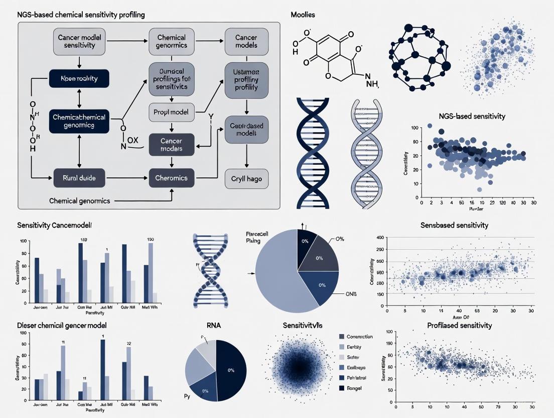
The following workflow diagram illustrates the complete experimental process for NGS-based chemical sensitivity profiling:
Diagram 1: NGS Chemical Sensitivity Profiling Workflow
Essential Research Reagent Solutions
Successful implementation of NGS-based chemical sensitivity profiling requires carefully selected reagents and materials. The following table outlines critical components for establishing robust experimental workflows.
Table 3: Essential Research Reagents for NGS-Based Chemical Sensitivity Profiling
| Reagent Category | Specific Products | Function | Application Notes |
|---|---|---|---|
| Nucleic Acid Extraction | QIAamp DNA FFPE Tissue Kit, DNeasy Blood & Tissue Kit | Isolation of high-quality genomic DNA from various sample types | FFPE-specific kits include cross-link reversal; minimum 20 ng DNA required [7] |
| Library Preparation | Illumina DNA Prep, KAPA HyperPrep Kit | Fragmentation, end-repair, A-tailing, adapter ligation | Incorporates unique dual indexes for sample multiplexing [3] |
| Target Enrichment | Agilent SureSelectXT, Illumina Nextera Flex | Hybrid capture-based selection of genomic regions of interest | Pan-cancer panels (e.g., 544-gene SNUBH panel) provide comprehensive coverage [7] |
| Quality Control | Agilent High Sensitivity DNA Kit, Qubit dsDNA HS Assay | Quantification and size distribution analysis of libraries | Average library size: 250-400 bp; concentration >2 nM [7] |
| Sequencing Reagents | Illumina SBS Chemistry, PacBio SMRTbell | Platform-specific nucleotides and buffers for sequencing reactions | Configuration: 2×150 bp paired-end for Illumina; >10 kb for PacBio HiFi [5] [7] |
Data Analysis and Integration with Sensitivity Metrics
The integration of genomic data with chemical response profiles represents the critical analytical phase that generates biologically actionable insights.
Variant Annotation and Interpretation
- Functional Annotation: Annotate identified variants using SnpEff or similar tools to predict functional consequences (missense, nonsense, splice-site, etc.) [7].
- Variant Classification: Categorize variants according to established guidelines such as the Association for Molecular Pathology (AMP) standards:
- Tier I: Variants of strong clinical significance (FDA-approved therapies, professional guidelines)
- Tier II: Variants of potential clinical significance (investigational therapies, different tumor type indications)
- Tier III: Variants of unknown significance
- Tier IV: Benign or likely benign variants [7]
- Actionability Assessment: Link genomic alterations to therapeutic options using knowledge bases such as OncoKB, CIViC, or Drug-Gene Interaction Database.
Correlation with Chemical Response
- Dose-Response Modeling: Calculate IC50 values and other pharmacodynamic parameters from viability assays following compound treatment.
- Association Analysis: Identify significant relationships between genomic features and sensitivity/resistance using statistical methods including:
- Fisher's exact tests for categorical genomic features
- Linear regression for continuous variables (e.g., TMB, variant allele frequency)
- Multivariate models incorporating clinical covariates
- Pathway Enrichment Analysis: Determine whether specific molecular pathways are enriched in sensitive or resistant models using gene set enrichment analysis (GSEA) or similar approaches.
The following diagram illustrates the analytical workflow for integrating genomic data with chemical sensitivity profiles:
Diagram 2: Genomic and Sensitivity Data Integration
Future Directions and Emerging Technologies
The NGS landscape continues to evolve rapidly, with several emerging technologies and methodologies poised to enhance chemical sensitivity profiling in cancer models.
Multi-omic Integration: The simultaneous analysis of genomic, transcriptomic, epigenomic, and proteomic data from the same samples provides comprehensive molecular portraits that better predict therapeutic response [8] [6]. PacBio's SPRQ chemistry exemplifies this trend, enabling concurrent assessment of DNA sequence and chromatin accessibility [5]. The integration of genetic alterations with gene expression signatures and epigenetic modifications will enable more accurate prediction of chemical vulnerabilities.
Spatial Transcriptomics and In Situ Sequencing: Emerging technologies now enable sequencing of cells within their native tissue context, preserving spatial information that is critical for understanding tumor microenvironment interactions [6]. These approaches will be particularly valuable for profiling heterogeneous tumor models and understanding how spatial relationships influence drug response.
Artificial Intelligence and Machine Learning: AI/ML algorithms are increasingly applied to NGS data to identify complex patterns predictive of chemical sensitivity [8] [6]. Deep learning models like Google's DeepVariant already improve variant calling accuracy, while more sophisticated neural networks can integrate multi-omic features to predict drug response with unprecedented precision [8].
Long-Read Applications: The improving accuracy and decreasing cost of long-read sequencing technologies open new possibilities for characterizing complex genomic regions that influence drug response, including highly repetitive regions, structural variations, and phased haplotypes [5]. These technologies are particularly valuable for resolving complex rearrangement patterns that emerge following chemical treatment.
As these technologies mature, they will increasingly enable researchers to build comprehensive predictive models of chemical sensitivity based on multidimensional molecular data, accelerating both basic cancer research and therapeutic development.
The advent of precision oncology has fundamentally shifted cancer treatment from a one-size-fits-all approach to a targeted strategy based on the unique molecular characteristics of an individual's tumor. This paradigm shift has been enabled by advances in genomic testing technologies, primarily through two distinct approaches: traditional single-gene assays and comprehensive genomic profiling (CGP). Single-gene testing methodologies, such as polymerase chain reaction (PCR), fluorescence in-situ hybridization (FISH), and immunohistochemistry (IHC), focus on identifying alterations in individual genes or limited protein expressions [9] [10]. While these tests have historically formed the foundation of molecular diagnostics, they possess inherent limitations in scope and efficiency when faced with the complex genomic landscape of cancer.
In contrast, comprehensive genomic profiling utilizes next-generation sequencing (NGS) technologies to simultaneously analyze hundreds of cancer-related genes from a single tissue or blood sample [11]. Unlike single-gene tests that are confined to hotspot regions within genes, CGP detects the four main classes of genomic alterations—base substitutions, insertions and deletions, copy number alterations, and rearrangements or fusions—across a broad panel of genes [11]. This comprehensive approach has emerged as a transformative tool in oncology, enabling the identification of clinically actionable biomarkers that might otherwise be missed by sequential single-gene testing approaches. As the number of targeted therapies continues to grow, the limitations of single-gene assays become increasingly pronounced, necessitating a critical examination of the comparative advantages of CGP in both scope and efficiency for modern cancer research and treatment.
Comparative Analytical Scope
Target Range and Alteration Detection
The fundamental distinction between single-gene assays and CGP lies in the breadth of genomic interrogation. Single-gene tests are methodologically constrained to identifying alterations confined to specific genes or hotspot regions, potentially missing clinically relevant mutations in additional genes [11]. For instance, a SNaPshot multiplex PCR panel might target variants in BRAF, EGFR, and KRAS, while FISH testing would be separately required to detect rearrangements in ALK, RET, or ROS1 [10]. This targeted approach becomes increasingly problematic as new biomarkers with clinical utility are discovered.
Comprehensive genomic profiling dramatically expands the detection capability by simultaneously assessing hundreds of cancer-associated genes. The technical advantage of CGP is its ability to identify diverse alteration types across a extensive genomic territory without prior knowledge of which specific gene might be驱动 the cancer. A prime example of this advantage is evident in rare but actionable biomarkers like NTRK fusions, which have been identified in less than 1% of all cancers but have targeted therapies available [11]. These fusions would unlikely be tested for using a single-gene approach due to their low frequency, yet CGP can detect them as part of its comprehensive assessment. Additionally, CGP can identify complex genomic signatures such as microsatellite instability (MSI), tumor mutational burden (TMB), and genomic loss of heterozygosity (gLOH), which have significant implications for immunotherapy response but cannot be adequately assessed through single-gene testing methods [11].
Table 1: Comparative Analysis of Detection Capabilities Between Single-Gene Testing and Comprehensive Genomic Profiling
| Parameter | Single-Gene Testing | Comprehensive Genomic Profiling |
|---|---|---|
| Genes Interrogated | 1 to several genes | 300+ genes simultaneously [10] |
| Variant Types Detected | Limited to methodology (e.g., SNVs by PCR, fusions by FISH) | All four major classes: SNVs, indels, CNAs, fusions [11] |
| Novel Biomarker Discovery | Not possible | Built-in capability for discovery |
| Genomic Signatures | Limited or not available | MSI, TMB, gLOH [11] |
| Actionable Findings in NSCLC | ~25-35% of patients [12] | 46-53% of patients [13] |
Diagnostic and Reclassification Capabilities
Beyond identifying therapeutic targets, comprehensive genomic profiling possesses a unique capability to contribute to diagnostic accuracy and tumor reclassification. In rare cases, CGP has revealed inconsistencies between primary diagnoses and molecular findings, triggering secondary pathological reviews that resulted in diagnostic reclassification [14]. For example, initial diagnoses of non-small cell lung cancer (NSCLC), sarcoma, and neuroendocrine carcinoma have been reclassified to renal cell carcinoma, medullary thyroid carcinoma, and melanoma, respectively, based on molecular findings from CGP [14]. Similarly, CGP has demonstrated significant utility in refining cancers of unknown primary (CUP) origin into distinct diagnostic categories, thereby enabling more precise treatment strategies [14].
These reclassification events have profound therapeutic implications. In one documented case, NGS testing helped correct an inaccurate primary diagnosis of leiomyosarcoma to liposarcoma, leading to indication-matched treatment with improved progression-free survival and quality of life [14]. The biomarkers driving these diagnostic changes include point mutations, gene fusions, and high tumor mutational burden, which provide molecular evidence supporting tumor origin or type. This diagnostic capability remains largely inaccessible through single-gene testing approaches, as they lack the comprehensive genomic context necessary to challenge or refine initial pathological assessments.
Operational Efficiency and Tissue Stewardship
Tissue Conservation and Testing Success
The efficiency of genomic testing is critically dependent on the optimal utilization of often limited tumor tissue. Single-gene testing approaches typically require sequential sectioning of formalin-fixed, paraffin-embedded (FFPE) tissue blocks, with each test consuming valuable material. A comparative analysis revealed that using single-gene testing prior to CGP requires more than 50 slides if all recommended tests are ordered individually, compared with only 20 slides for CGP alone [13]. This substantial difference in tissue requirements directly impacts testing success rates.
The clinical consequences of tissue exhaustion are significant. Research has demonstrated that patients with non-small cell lung cancer who underwent single-gene testing prior to CGP had significantly higher rates of test cancellation due to tissue insufficiency (17% vs. 7%) compared to those who only had CGP [13]. Furthermore, DNA sequencing failures were more common in the single-gene testing first group (13% vs. 8%), highlighting how tissue depletion negatively impacts the quality and success of subsequent comprehensive testing [13]. This is particularly problematic in cancers where biopsy samples are inherently small, such as lung cancer, where one study found that 29% of patients didn't get results from molecular testing because tissue was insufficient [11].
Table 2: Impact of Testing Approach on Tissue Utilization and Success Rates in NSCLC
| Performance Metric | Single-Gene Testing First | CGP Only |
|---|---|---|
| Slide Requirement | >50 slides [13] | ~20 slides [13] |
| Test Cancellation (Tissue Exhaustion) | 17% [13] | 7% [13] |
| DNA Sequencing Failure Rate | 13% [13] | 8% [13] |
| Turnaround Time >14 Days | 62% [13] | 29% [13] |
| Identification of Guideline-Recommended Biomarkers | 46% (after failed SGT) [13] | 53% [13] |
Workflow Efficiency and Turnaround Time
The operational workflow for genomic testing directly impacts clinical decision-making and patient care. Single-gene testing typically involves a sequential process where providers order individual tests based on initial hypotheses, awaiting results before determining subsequent testing needs. This sequential approach inevitably prolongs the time to comprehensive molecular characterization. Data from a prospective study demonstrated that 62% of patients who underwent single-gene testing prior to CGP experienced turnaround times exceeding 14 days, compared to only 29% in the CGP-only group [13].
Comprehensive genomic profiling streamlines this process by consolidating multiple analyses into a single integrated workflow. Advances in NGS technology and bioinformatics have further improved the efficiency of CGP, with some targeted panels now achieving turnaround times as short as 4 days from sample processing to results [15]. This accelerated timeline is crucial in advanced cancer, where timely initiation of appropriate therapy can significantly impact outcomes. The unified reporting structure of CGP also enhances clinical utility by presenting all molecular findings in a single interpretable format, facilitating treatment decision-making based on a complete genomic profile rather than fragmented results from multiple testing modalities.
Clinical Impact and Therapeutic Implications
Identification of Actionable Alterations
The ultimate measure of genomic testing utility lies in its ability to identify clinically actionable alterations that can inform treatment decisions. Comparative studies have consistently demonstrated the superior performance of CGP in this critical dimension. Research across multiple cancer types—including non-small cell lung cancer (NSCLC), cholangiocarcinoma (CCA), pancreatic carcinoma (PC), and gastro-oesophageal carcinoma (GEC)—has shown that tumor profiling with comprehensive NGS panels improved patient eligibility for personalized therapies compared with small panels [12]. The magnitude of this advantage varies by cancer type, with particularly dramatic differences observed in malignancies with diverse genomic drivers.
In gastro-oesophageal carcinoma, comprehensive panels identified actionable targets in 40% of patients, while small panels (≤60 genes) identified none [12]. Similarly, in pancreatic carcinoma, comprehensive profiling increased eligibility for personalized therapies from 3% with small panels to 35% [12]. Even in NSCLC, where testing practices are more established, comprehensive panels identified actionable alterations in 39% of patients compared to 37% with small panels [12]. These findings underscore how CGP expands therapeutic opportunities by casting a wider genomic net, particularly important for cancers with lower mutation frequencies or those lacking dominant driver mutations.
Case series further illustrate this advantage, documenting multiple instances where comprehensive genomic profiling identified highly actionable alterations missed by prior single-gene testing [10]. These included ALK fusions, EGFR exon 20 insertions, and MET exon 14 skipping alterations—all of which have approved targeted therapies [10]. Importantly, 46% of NSCLC patients with negative prior single-gene test results had positive findings for recommended biomarkers when subsequently evaluated by CGP [13], indicating that single-gene testing frequently provides false-negative results rather than truly negative genomic profiles.
Cost-Effectiveness and Resource Utilization
The economic implications of testing strategies represent a significant consideration in healthcare resource planning. While single-gene tests may appear less expensive individually, the cumulative cost of multiple single-gene tests must be weighed against the more comprehensive information obtained from a single CGP test. Research evaluating the overall diagnostic journey cost—from hospital admission through Molecular Tumour Board evaluation—found that the cost per patient to identify someone eligible for personalized treatments varied significantly according to panel size and tumor type [12].
In pancreatic carcinoma, the cost to find a patient eligible for personalized treatments was approximately $27,000 with small panels versus $5,500 with comprehensive panels [12]. The remarkable cost efficiency of comprehensive profiling in this context stems from its higher detection rate of actionable alterations. Similarly, for gastro-oesophageal carcinoma, the cost was immeasurable with small panels (as none of the patients were identified as eligible) versus $5,200 with comprehensive panels [12]. These findings challenge the perception of single-gene testing as a more economical approach and instead position CGP as a superior value proposition in many clinical scenarios.
It is noteworthy that the Molecular Tumour Board discussion component accounted for only 2-3% of the total diagnostic journey cost per patient (approximately €113/patient) [12], suggesting that the interpretive expertise required to implement CGP findings represents a relatively small incremental investment compared to the substantial clinical benefits derived from more comprehensive genomic information.
Experimental Protocols and Applications
Comprehensive Genomic Profiling Wet-Lab Protocol
The following protocol outlines the standard procedure for comprehensive genomic profiling using hybrid capture-based NGS methodology, suitable for implementation in a CLIA-certified laboratory setting:
Sample Requirements and Quality Control:
- Input Material: 50-100ng of DNA extracted from FFPE tissue sections with minimum 20% tumor content [15]
- Quality Assessment: DNA quantification using fluorometric methods (e.g., Qubit), with degradation assessment via fragment analysis
- Sample Tracking: Barcoding of samples to maintain chain of custody throughout the process
Library Preparation:
- DNA Fragmentation: Fragment genomic DNA to ~300bp using acoustic shearing or enzymatic fragmentation
- End Repair and A-Tailing: Repair fragment ends and add adenine overhangs using commercial library preparation kits
- Adapter Ligation: Ligate platform-specific adapters containing unique dual indices to enable sample multiplexing
- Library Amplification: Amplify adapter-ligated fragments using limited-cycle PCR (typically 4-8 cycles)
- Library Quantification: Assess library concentration using qPCR for accurate quantification of amplifiable fragments
Target Enrichment:
- Hybridization: Incubate library pools with biotinylated oligonucleotide probes targeting cancer-related genes (e.g., 324-gene panel [11] or 523-gene panel [10])
- Capture: Bind probe-hybridized fragments to streptavidin-coated magnetic beads
- Wash: Stringent washing to remove non-specifically bound fragments
- Amplification: Limited-cycle PCR to amplify captured libraries
Sequencing:
- Pooling: Normalize and pool enriched libraries based on qPCR quantification
- Cluster Generation: Load pooled libraries onto flow cell for cluster generation (bridge amplification)
- Sequencing: Perform sequencing using Illumina sequencing-by-synthesis technology with paired-end reads (2×100bp or 2×150bp)
- Output: Target sequencing depth of 500-1000x for tissue; minimum 100x unique molecular coverage [15]
Bioinformatic Analysis Pipeline
The computational analysis of NGS data follows a standardized workflow for variant detection and interpretation:
Primary Analysis:
- Base Calling: Convert raw signal data to nucleotide sequences (FASTQ format)
- Quality Control: Assess read quality using tools such as FastQC
- Adapter Trimming: Remove adapter sequences using Trimmomatic or similar tools
Secondary Analysis:
- Alignment: Map reads to reference genome (GRCh38) using optimized aligners such as BWA-MEM or STAR
- Duplicate Marking: Identify and mark PCR duplicates using Picard Tools
- Local Realignment: Perform realignment around indels using GATK
- Base Quality Recalibration: Adjust base quality scores using GATK
Tertiary Analysis:
- Variant Calling: Identify somatic mutations using mutect2, varscan2, or similar variant callers
- Copy Number Analysis: Detect amplifications and deletions using CONTRA, ADTEx, or similar tools
- Structural Variant Detection: Identify gene fusions and rearrangements using DELLY, LUMPY, or Manta
- Annotation: Annotate variants using databases such as dbSNP, COSMIC, ClinVar, and OncoKB
- Filtering: Prioritize variants based on population frequency, functional impact, and clinical relevance
Interpretation and Reporting:
- Pathogenicity Assessment: Classify variants according to AMP/ASCO/CAP guidelines
- Actionability Evaluation: Match genomic alterations to targeted therapies and clinical trials
- Report Generation: Create comprehensive patient reports with evidence-based therapeutic recommendations
Research Reagent Solutions
Table 3: Essential Research Reagents for Comprehensive Genomic Profiling
| Reagent Category | Specific Examples | Function in CGP Workflow |
|---|---|---|
| Nucleic Acid Extraction Kits | QIAamp DNA FFPE Tissue Kit, Maxwell RSC DNA FFPE Kit | Isolation of high-quality DNA from FFPE tissue specimens [15] |
| Library Preparation Kits | Illumina TruSight Oncology 500 HT, Sophia Genetics DDM library kit | Fragmentation, end repair, adapter ligation, and amplification for NGS library construction [15] [10] |
| Target Enrichment Panels | FoundationOne CDx (324 genes), OmniSeq INSIGHT (523 genes) | Hybridization capture of cancer-relevant genomic regions [11] [10] |
| Sequencing Reagents | Illumina NovaSeq 6000 S-Plex, MGI DNBSEQ-G50RS sequencing kit | Sequence generation using sequencing-by-synthesis technology [15] |
| Bioinformatic Tools | Sophia DDM, GATK, OncoKB, Cravat | Variant calling, annotation, and clinical interpretation of genomic data [15] |
| Quality Control Assays | Agilent TapeStation, Qubit dsDNA HS Assay, qPCR libraries quantification | Assessment of nucleic acid quality, quantity, and library preparation success [15] |
Workflow and Signaling Pathway Visualizations
Diagram 1: Comparative workflow analysis between single-gene testing and comprehensive genomic profiling approaches, highlighting differences in tissue utilization, process complexity, and outcomes.
Diagram 2: Biomarker detection capabilities across genomic testing methodologies, illustrating the comprehensive coverage of CGP compared to limited scope of single-gene assays.
The comparative analysis between comprehensive genomic profiling and traditional single-gene assays reveals a consistent pattern of advantages favoring CGP across multiple dimensions—analytical scope, operational efficiency, clinical utility, and economic value. The demonstrated ability of CGP to identify more actionable alterations, conserve precious tissue specimens, provide more rapid and comprehensive results, and ultimately guide more effective treatment decisions positions it as the superior approach for genomic profiling in contemporary oncology practice and research. As the field continues to evolve with an expanding repertoire of targeted therapies and biomarkers, the comprehensive nature of CGP becomes increasingly essential for realizing the full potential of precision oncology.
For research applications, particularly in chemical sensitivity profiling and cancer model development, CGP offers the additional advantage of generating rich genomic datasets that can be mined for discovery purposes beyond immediate clinical applications. The ability to detect novel alterations, identify complex genomic signatures, and contribute to diagnostic refinement makes CGP an invaluable tool for advancing our understanding of cancer biology and therapeutic response mechanisms. While single-gene assays may retain utility in specific, limited contexts where rapid assessment of a single biomarker is sufficient, the weight of evidence supports CGP as the foundational approach for comprehensive cancer genomic characterization in both clinical and research settings.
The management of cancer is increasingly guided by the principle of precision medicine, where treatment strategies are tailored to the specific genetic alterations found in an individual's tumor. Central to this approach is the identification of actionable mutations—somatic genetic alterations that directly influence clinical decision-making by predicting response or resistance to targeted therapies. Next-generation sequencing (NGS) has become the cornerstone technology for comprehensively profiling these alterations across hundreds of cancer-related genes simultaneously, moving beyond single-gene assays to capture the complex genomic landscape of malignancies [9]. The clinical utility of this approach is firmly established; for instance, patients with metastatic castration-resistant prostate cancer (mCRPC) harboring homologous recombination repair gene mutations can be treated with PARP inhibitors, while those with mismatch repair deficiency benefit from immune checkpoint blockade therapies [16].
The biological rationale connecting genetic alterations to therapeutic vulnerabilities stems from the concept of oncogene addiction and synthetic lethality. Oncogene addiction describes the phenomenon where cancer cells become dependent on a single activated oncogene for survival and proliferation, making them uniquely vulnerable to its inhibition. Synthetic lethality occurs when inactivation of either of two genes individually is viable, but simultaneous inactivation results in cell death—a principle exploited by PARP inhibitors in BRCA-deficient tumors. The National Cancer Institute's Molecular Analysis for Therapy Choice (NCI-MATCH) trial exemplifies how this paradigm is operationalized at scale, using NGS to match patients with relapsed or refractory cancers to therapies targeting specific molecular alterations [17]. This framework transforms cancer treatment from a histology-based approach to a genetically-guided strategy.
Methodologies for Mutation Detection and Interpretation
Sample Acquisition and Processing Considerations
Robust identification of actionable mutations begins with appropriate sample acquisition. While formalin-fixed paraffin-embedded (FFPE) tumor tissue remains the gold standard, liquid biopsy approaches using plasma, urine, or other bodily fluids offer non-invasive alternatives when tissue is unavailable [16]. Each sample type presents distinct advantages and limitations. Tumor tissues provide comprehensive genomic information but require invasive procedures. Plasma circulating tumor DNA (ctDNA) detection sensitivity depends heavily on tumor burden and shedding, with studies reporting over 70% of mCRPC patients having ctDNA variant allele frequencies (VAFs) >2%, achieving 90% concordance with tissue-based testing [16]. Urine samples have demonstrated 65.6% detection sensitivity for prostate cancer mutations, while seminal fluid shows potential despite current sampling challenges [16].
The NGS workflow consists of four critical stages: (1) template preparation, (2) sequencing, (3) imaging, and (4) data analysis [18]. For template preparation, three main approaches exist: clonally amplified templates (using emulsion PCR or bridge PCR), single-molecule templates (requiring less material and avoiding amplification bias), and circle templates (reducing error rates for cancer profiling) [18]. The choice of method depends on the application—single-molecule templates are preferred for quantitative analyses like gene expression profiling, while amplified templates are suitable for qualitative mutational analysis despite potential bias in AT-rich and GC-rich regions [18].
Sequencing Technologies and Analytical Validation
Multiple sequencing technologies are available, each with distinct performance characteristics. The Illumina platform uses complementary metal-oxide semiconductor (CMOS) technology with fluorescently labeled reversible terminators, while Ion Torrent employs non-optical sequencing based on detection of hydrogen ions released during DNA polymerase activity [18]. The Oncomine Cancer Panel assay with AmpliSeq chemistry and Personal Genome Machine sequencer has been validated for clinical use in the NCI-MATCH trial, demonstrating 96.98% overall sensitivity for 265 known mutations and 99.99% specificity across four Clinical Laboratory Improvement Amendments (CLIA)-certified laboratories [17].
Analytical validation must establish performance characteristics for each variant type. The NCI-MATCH assay validation established the following limits of detection: 2.8% for single-nucleotide variants (SNVs), 10.5% for small insertions/deletions (indels), 6.8% for large indels (gap ≥4 bp), and four copies for gene amplification [17]. This rigorous validation ensures that reported variants meet quality standards for clinical decision-making. Bioinformatics pipelines for variant calling typically involve quality control of FASTQ files, alignment to reference genomes, and annotation using tools like VarScan2 and ANNOVAR, with filtering thresholds adjusted for different sample types (e.g., VAF ≥1% for tissue, ≥0.3% for plasma) [16].
Table 1: Comparison of NGS Performance Across Different Sample Types
| Sample Type | Detection Sensitivity | Advantages | Limitations |
|---|---|---|---|
| Tumor Tissue (FFPE) | 100% (gold standard) | Comprehensive genomic information; established protocols | Invasive procurement; not always feasible |
| Plasma ctDNA | 67.6% | Non-invasive; enables monitoring | Lower sensitivity for low tumor burden |
| Urine | 65.6% | Completely non-invasive; patient-friendly | Variable DNA concentration |
| Seminal Fluid | 33.3% | High cfDNA concentration in prostate cancer | Sampling challenges post-treatment |
Connecting Mutational Profiles to Therapeutic Opportunities
Interpretation Frameworks for Actionability
The interpretation of genomic alterations follows structured frameworks that classify mutations based on clinical evidence levels. The NCI-MATCH trial established a tiered evidence system: Level 1 includes variants credentialed for FDA-approved drugs in any tissue (e.g., BRAF V600E and vemurafenib); Level 2a comprises variants serving as eligibility criteria for ongoing clinical trials; Level 2b includes variants with evidence from N-of-1 responses; and Level 3 relies on preclinical inferential data supporting treatment selection [17]. This structured approach ensures that treatment assignments are based on rigorously validated biomarkers.
Implementation requires assessment of specific mutation types and their functional consequences. Gain-of-function mutations in oncogenes (e.g., activating mutations in kinases) typically create direct drug targets, while loss-of-function mutations in tumor suppressor genes may indicate sensitivity to targeted therapies through synthetic lethal interactions [17]. For example, nonsense or frameshift variants in 26 tumor suppressor genes are specifically reported in the NCI-MATCH assay, as these truncating alterations may predict response to specific therapeutic classes [17]. Additionally, the mutational landscape provides biological insights—in prostate cancer, mutations in FOXA1, SPOP, and TP53 are commonly detected across sample types, while AR mutations show distinct patterns of prevalence in liquid biopsy samples compared to tissue [16].
Chemical Sensitivity Profiling through Computational Approaches
Emerging computational approaches now enable in silico chemical sensitivity profiling by integrating genomic features with chemical structure information. The ChemProbe model exemplifies this approach, using deep learning to predict cellular sensitivity to hundreds of compounds by combining transcriptomic data with chemical structures [19]. This model employs feature-wise linear modulation (FiLM) layers where chemical features scale and shift gene expression representations, effectively mimicking how chemical substructures modulate biological pathways [19]. This methodology accurately predicted breast cancer patient response in the I-SPY2 trial, achieving a macro-average area under the receiver operating characteristic curve of 0.65 for five therapeutics, demonstrating how computational models can extrapolate from cell line data to clinical predictions [19].
The interpretation of these models provides biological insights into mechanisms of chemical sensitivity. Analysis of learned parameters in ChemProbe revealed that scaling parameters grouped compounds by structural identity, while shifting parameters correlated with compound concentration [19]. Furthermore, gradient-based attribution methods identified transcriptome features reflecting compound targets and protein network modules, successfully identifying genes that drive ferroptosis [19]. This demonstrates how advanced computational approaches not only predict chemical sensitivity but also illuminate underlying biological mechanisms connecting genetic alterations to therapeutic vulnerabilities.
Experimental Protocols and Research Applications
Protocol for NGS-Based Mutation Detection from Tissue and Liquid Biopsies
Sample Collection and DNA Extraction:
- For FFPE tissue samples, use the QIAamp DNA FFPE Tissue Kit (Qiagen) following manufacturer's protocols. Have sections reviewed by a pathologist to assess tumor content [16] [17].
- For liquid biopsy samples (plasma, urine, seminal fluid), isolate circulating tumor DNA using the QIAamp Circulating Nucleic Acid Kit (Qiagen). Collect blood in EDTA tubes and process within 2 hours to prevent genomic DNA contamination [16].
- Extract germline DNA from white blood cells using the DNeasy Blood and Tissue Kit (Qiagen) to serve as a control for identifying somatic mutations and filtering out clonal hematopoiesis variants [16].
Library Preparation and Sequencing:
- Construct sequencing libraries using the KAPA Hyper DNA Library Prep Kit (Roche). For targeted sequencing, enrich 437 cancer-related genes using probe-based hybridization capture panels [16].
- For the NCI-MATCH validated protocol, use the Oncomine Cancer Panel assay with AmpliSeq chemistry on the Personal Genome Machine sequencer. This panel covers 4066 predefined genomic variations across 143 genes [17].
- Sequence on an Illumina HiSeq4000 platform with minimum coverage of 1000× for liquid biopsies and 500× for tissue samples to ensure detection of low-frequency variants [16].
Variant Calling and Annotation:
- Process FASTQ files through quality control using Trimmomatic to remove low-quality bases (quality score below 20) and Picard to remove PCR duplicates [16].
- Align filtered paired-end reads to the human reference genome (GRCh37/hg19) using the Burrows-Wheeler Aligner, followed by realignment around indels with GATK3 [16].
- Identify single nucleotide variants and indels using VarScan2 with minimum thresholds: for tissue samples, unique variant-supporting reads ≥5 and VAF ≥1%; for plasma samples, unique variant-supporting reads ≥3 and VAF ≥0.3% [16].
- Annotate variants using ANNOVAR and filter against population databases (1000 Genomes, ExAC) to exclude common polymorphisms. Compare with matched germline DNA to distinguish somatic mutations [16].
Protocol for Chemical Sensitivity Prediction Using Transcriptomic Data
Data Preprocessing and Model Training:
- Obtain basal transcriptomic profiles from cancer cell lines (e.g., Cancer Cell Line Encyclopedia) and match with drug sensitivity data (e.g., Cancer Therapeutics Response Portal) [19].
- Standardize RNA abundance values using robust z-score normalization across samples. Encode chemical structures using extended-connectivity fingerprints or other molecular representations [19].
- Implement a conditional deep learning model architecture where chemical features modulate gene expression representations through scaling and shifting operations (FiLM layers) [19].
- Train the model using five-fold cross-validation stratified by cell line to ensure generalizability. Use mean squared error loss between predicted and measured viability values optimized with Adam optimizer [19].
Sensitivity Prediction and Interpretation:
- Apply the trained ChemProbe model to new transcriptomic profiles by computing viability values across a range of compound concentrations [19].
- Generate dose-response curves and calculate area under the curve (AUC) values to quantify sensitivity. Establish a decision threshold for responder classification based on distribution of predicted values [19].
- Interpret model predictions using integrated gradients to identify genes driving chemical sensitivity predictions. Perform gene set enrichment analysis on top-weighted genes to identify biological pathways associated with response [19].
- Validate predictions in independent datasets such as clinical trial data (e.g., I-SPY2 trial) to assess translational utility [19].
Table 2: Essential Research Reagents and Solutions for NGS-Based Chemical Sensitivity Profiling
| Reagent/Solution | Function | Example Products/Protocols |
|---|---|---|
| Nucleic Acid Extraction Kits | Isolation of high-quality DNA from various sample types | QIAamp DNA FFPE Tissue Kit, QIAamp Circulating Nucleic Acid Kit, DNeasy Blood and Tissue Kit |
| Target Enrichment Panels | Capture of cancer-relevant genomic regions | Oncomine Cancer Panel (143 genes), Custom panels (437 cancer-related genes) |
| Library Preparation Kits | Construction of sequencing-ready libraries | KAPA Hyper DNA Library Prep Kit, Illumina DNA Prep |
| NGS Platforms | Massive parallel sequencing of captured libraries | Illumina HiSeq4000, Personal Genome Machine, NovaSeq |
| Variant Calling Software | Identification of somatic mutations from sequence data | VarScan2, GATK, Ion Reporter |
| Chemical Sensitivity Databases | Training data for predictive models | CTRP (545 compounds), CCLE (842 cell lines) |
| Deep Learning Frameworks | Implementation of chemical sensitivity models | PyTorch, TensorFlow with FiLM layers |
Implementation in Research and Clinical Settings
The implementation of NGS-based mutation detection and chemical sensitivity profiling requires careful consideration of analytical validation and regulatory compliance. The NCI-MATCH trial established a network of four CLIA-certified laboratories that demonstrated 99.99% mean inter-operator pairwise concordance across laboratories, proving that high reproducibility of complex NGS assays is achievable with standardized protocols [17]. For clinical application, assays must undergo rigorous validation of analytical sensitivity, specificity, reproducibility, and limit of detection for each variant type [17]. This ensures that reported variants meet quality standards for therapeutic decision-making.
Current National Comprehensive Cancer Network guidelines endorse liquid biopsy methodologies when tissue testing fails or is unattainable [16]. The convergence of comprehensive genomic profiling through NGS and computational chemical sensitivity prediction represents the future of precision oncology. These approaches enable the identification of patient-specific therapeutic vulnerabilities based on the unique genetic makeup of their tumors, moving beyond histology-based classification to genetically-guided treatment strategies. As these technologies evolve, they promise to further refine our ability to match the right patient with the right therapy at the right time, ultimately improving outcomes in cancer treatment.
Visualizing Workflows and Biological Relationships
NGS Workflow for Actionable Mutation Detection
Chemical Sensitivity Prediction Framework
Actionable Mutation Clinical Translation Pathway
Next-generation sequencing (NGS) has revolutionized the detection and characterization of drug resistance in cancer by enabling comprehensive genomic analysis of tumors with unprecedented sensitivity and throughput. Unlike traditional Sanger sequencing, which processes DNA fragments individually, NGS allows for massive parallel sequencing, processing millions of fragments simultaneously to identify genetic alterations that drive both primary (innate) and acquired (treatment-emergent) resistance [9]. This capability is transforming precision oncology by moving beyond single-gene assays to capture the complex genomic landscape of resistance mechanisms.
The application of NGS in chemical sensitivity profiling research provides critical insights into the dynamic evolution of tumors under therapeutic pressure. By detecting low-abundance variants and complex resistance patterns, NGS enables researchers to decipher the molecular pathways that allow cancer cells to evade treatment, thereby informing the development of more effective therapeutic strategies and combination regimens to overcome resistance [20] [21].
NGS Methodologies for Resistance Detection
Key Sequencing Technologies and Platforms
Different NGS platforms offer complementary strengths for resistance mechanism studies. Illumina sequencing utilizes sequencing-by-synthesis with fluorescently labeled nucleotides and is widely used for its high accuracy and throughput [2] [9]. Ion Torrent semiconductor sequencing detects hydrogen ions released during DNA polymerization, providing rapid turnaround times [2]. Third-generation technologies like Pacific Biosciences SMRT and Oxford Nanopore enable long-read sequencing, which is particularly valuable for resolving complex structural variations and epigenetic modifications that contribute to drug resistance [2] [22].
The selection of an appropriate NGS approach depends on the specific research objectives. Targeted panels focus on known resistance genes with deep coverage, making them ideal for detecting low-frequency variants. Whole-exome sequencing provides a broader view of coding regions, while whole-genome sequencing captures the complete genomic landscape, including non-coding regions and structural variants [9]. Single-cell sequencing represents a cutting-edge approach that resolves cellular heterogeneity in resistant populations, revealing subclonal dynamics that bulk sequencing might miss [21].
Analytical Considerations for Resistance Studies
The sensitivity of NGS in detecting resistant subclones is critically dependent on sequencing depth and variant calling thresholds. Studies demonstrate that lowering the detection threshold from the conventional 20% to 2% can increase the identification of pretreatment drug resistance by approximately 2.5-fold, revealing clinically relevant low-abundance variants that would otherwise remain undetected [20]. Effective bioinformatics pipelines must integrate variant calling, annotation, and clinical interpretation to distinguish driver resistance mutations from passenger alterations.
Visualization tools like Trackster enable interactive exploration of NGS data, allowing researchers to dynamically adjust parameters and visualize the effects on variant calling in real-time [23]. This integrated visual analysis approach facilitates the identification of optimal analysis settings for resistance mutation detection without the computational burden of repeatedly processing entire datasets.
NGS Applications in Resistance Mechanism Elucidation
Characterizing Primary Resistance Mechanisms
Primary (innate) resistance refers to pre-existing genetic factors that render tumors insensitive to initial treatment. NGS profiling of treatment-naïve tumors has revealed that low-abundance drug-resistant variants present below the detection limit of conventional methods can significantly impact therapeutic outcomes [20]. In HIV research, which provides a model for understanding resistance mechanisms, NGS at a 2% detection threshold revealed a 22.43% prevalence of pretreatment drug resistance compared to 11.08% at the standard 20% threshold [20].
In cancer, primary resistance mechanisms identified through NGS include:
- On-target mutations that alter drug binding sites
- Off-target alterations in parallel signaling pathways
- Activation of compensatory survival pathways
- Pharmacogenomic variants affecting drug metabolism
Mapping Acquired Resistance Evolution
Acquired resistance emerges under selective therapeutic pressure through Darwinian evolution of tumor cell populations. Longitudinal NGS monitoring of patients during treatment captures the dynamic clonal evolution that underlies resistance development. The SPACEWALK study in ALK-positive NSCLC exemplifies this approach, using NGS to identify three distinct resistance mechanisms: on-target (ALK secondary mutations), off-target (bypass pathway activation), and combined mechanisms [24].
In acute myeloid leukemia (AML), deep single-cell multi-omic profiling integrating NGS with ex vivo drug response testing has revealed conserved patterns of venetoclax resistance associated with specific molecular signatures. This integrated approach identified both known and novel mechanisms of innate and treatment-related resistance, including associations with increased proliferation and CD36 expression in resistant blasts [21].
Table 1: NGS Detection of Pretreatment Drug Resistance at Different Sensitivity Thresholds
| Detection Threshold | Overall PDR Prevalence | NNRTI Resistance | INSTI Resistance |
|---|---|---|---|
| 1% | 29.74% | 15.29% | 1.22% |
| 2% | 22.43% | 11.63% | 1.22% |
| 5% | 15.47% | 8.27% | 0.17% |
| 10% | 12.95% | 6.90% | 0.17% |
| 20% | 11.08% | 4.90% | 0.17% |
Data adapted from HIV resistance study demonstrating threshold-dependent mutation detection [20]
Experimental Protocols for NGS-Based Resistance Profiling
Targeted NGS Panel for Resistance Mutation Detection
The following protocol describes the development and validation of a targeted NGS panel specifically designed for comprehensive resistance profiling in solid tumors, based on established methodologies [15]:
Sample Preparation and Quality Control
- Extract DNA from tumor samples (minimum 50 ng input) using validated extraction kits
- Assess DNA quality and quantity via fluorometry and fragment analysis
- For FFPE samples, evaluate DNA degradation and adjust library preparation accordingly
Library Preparation and Target Enrichment
- Fragment DNA to ~300 bp using acoustic shearing or enzymatic fragmentation
- Repair ends and ligate with indexing adapters containing unique molecular identifiers
- Perform hybridization-based capture using biotinylated oligonucleotides targeting 61 cancer-associated genes including known resistance genes (KRAS, EGFR, ERBB2, PIK3CA, TP53, BRCA1)
- Amplify captured libraries with limited-cycle PCR (8-10 cycles)
Sequencing and Data Analysis
- Sequence on DNBSEQ-G50RS platform using cyclic array sequencing
- Generate minimum 2.2 million reads per sample with 469×-2320× median coverage
- Process data through bioinformatics pipeline: base calling > demultiplexing > alignment > variant calling
- Annotate variants using curated resistance databases and interpret clinical significance
Quality Assurance Metrics
- Ensure >98% target coverage at ≥100× molecular coverage
- Maintain sensitivity threshold of 2.9% variant allele frequency
- Achieve >99.99% reproducibility and repeatability in variant detection
- Validate against orthogonal methods and reference standards
Single-Cell Multi-Omic Profiling for Resistance Mechanisms
For comprehensive dissection of heterogeneous resistance mechanisms, the following single-cell protocol integrates genomic, transcriptomic, and functional profiling [21]:
Sample Processing and Single-Cell Isolation
- Collect primary patient samples (blood or bone marrow for hematologic malignancies, tissue for solid tumors)
- Dissociate into single-cell suspensions using gentle enzymatic digestion
- Isoselect viable mononuclear cells using density gradient centrifugation
- Sort or capture individual cells using microfluidic or droplet-based platforms
Multi-Omic Library Preparation
- For genomic analysis: Perform single-cell DNA sequencing to identify copy number variations and mutations
- For transcriptomic analysis: Prepare single-cell RNA sequencing libraries using template-switching chemistry
- For protein analysis: Conduct mass cytometry (CyTOF) with metal-tagged antibodies
- Index cells for multi-modal data integration
Functional Drug Profiling
- Perform single-cell ex vivo drug testing (pharmacoscopy) with resistance-relevant inhibitors
- Incubate cells with drug panels for 24-72 hours across concentration gradients
- Measure cell viability, apoptosis, and signaling responses via automated imaging
- Integrate functional responses with molecular profiles
Data Integration and Analysis
- Process sequencing data using single-cell bioinformatics pipelines (CellRanger, Seurat)
- Map transcriptional phenotypes and identify resistance-associated expression programs
- Correlate genomic alterations with drug response patterns
- Construct phylogenetic trees to model resistance evolution
Technical Specifications and Performance Metrics
Analytical Validation of NGS Resistance Panels
Rigorous validation is essential for reliable resistance mutation detection. The following performance characteristics were demonstrated for a validated 61-gene oncology panel [15]:
Table 2: Performance Metrics of Validated NGS Resistance Panel
| Parameter | Performance Metric | Acceptance Criterion |
|---|---|---|
| Sensitivity | 98.23% (at 95% CI) | >95% |
| Specificity | 99.99% (at 95% CI) | >99.5% |
| Accuracy | 99.99% (at 95% CI) | >99% |
| Precision | 97.14% (at 95% CI) | >95% |
| Reproducibility | 99.99% | >99% |
| Repeatability | 99.99% | >99% |
| Limit of Detection | 2.9% VAF | <5% VAF |
| Minimum DNA Input | 50 ng | ≤50 ng |
Research Reagent Solutions for NGS Resistance Studies
Table 3: Essential Research Reagents for NGS-Based Resistance Profiling
| Reagent Category | Specific Products | Application in Resistance Studies |
|---|---|---|
| Library Preparation | Sophia Genetics Library Kit, Illumina Nextera Flex | Fragment DNA and add adapters for sequencing |
| Target Enrichment | Custom hybridization baits (61-gene panel) | Isolate genomic regions harboring resistance mutations |
| Sequencing | MGI DNBSEQ-G50RS, Illumina MiSeq | Generate high-quality sequencing reads |
| Data Analysis | Sophia DDM, Trackster | Identify and visualize resistance mutations |
| Single-Cell Platforms | 10X Genomics Chromium, BD Rhapsody | Resolve cellular heterogeneity in resistant populations |
| Functional Assays | Pharmacoscopy platform | Correlate genomic findings with drug response |
Data Analysis and Interpretation Framework
Bioinformatics Pipeline for Resistance Mutation Detection
The computational analysis of NGS data for resistance mechanism studies requires a specialized bioinformatics workflow:
Primary Data Analysis
- Base calling and demultiplexing using platform-specific software
- Quality control assessment (FastQC) to evaluate read quality, GC content, and adapter contamination
- Trim low-quality bases and adapter sequences using tools like Trimmomatic or Cutadapt
Secondary Analysis
- Alignment to reference genome (hg19/GRCh38) using optimized aligners (BWA, Bowtie2)
- Post-alignment processing including duplicate marking, local realignment, and base quality recalibration
- Variant calling using mutational-specific algorithms (MuTect2, VarScan2) with sensitivity for low-frequency variants
- Structural variant detection using dedicated tools (Manta, Delly)
Tertiary Analysis and Interpretation
- Annotation of variants using curated databases (OncoKB, CIViC, COSMIC)
- Identification of known resistance mutations and novel putative resistance mechanisms
- Clonal decomposition and evolutionary analysis using phylogenetic methods
- Integration with functional drug response data to validate resistance associations
- Clinical interpretation and reporting using established guidelines
Visualization and Exploration of Resistance Data
Effective visualization is critical for interpreting complex resistance patterns. The Trackster environment enables interactive exploration of NGS data, allowing researchers to dynamically adjust parameters and visualize the effects on resistance variant calling in real-time [23]. This integrated visual analysis approach facilitates the identification of optimal analysis settings without the computational burden of repeatedly processing entire datasets.
Advanced visualization strategies include:
- Circos plots to display genomic alterations across multiple samples
- Fishplot representations of clonal evolution during treatment
- Heatmaps of gene expression patterns in resistant versus sensitive cells
- Pathway diagrams illustrating altered signaling networks in resistance
Visualizing NGS Workflows for Resistance Studies
The following diagrams illustrate key experimental and analytical workflows for NGS-based resistance mechanism studies.
NGS Resistance Profiling Workflow
NGS Resistance Profiling Workflow - This diagram outlines the comprehensive workflow from sample collection through to resistance mechanism interpretation, highlighting key quality control checkpoints.
Resistance Mechanism Classification
Resistance Mechanism Classification - This diagram categorizes the primary resistance mechanisms identifiable through NGS profiling, based on findings from the SPACEWALK study in ALK-positive NSCLC [24].
NGS technologies have fundamentally transformed our ability to decipher the complex molecular mechanisms underlying drug resistance in cancer. The approaches detailed in this application note provide researchers with powerful methodologies to detect both primary and acquired resistance mutations, track clonal evolution under therapeutic pressure, and identify novel resistance pathways. The integration of NGS with functional drug sensitivity profiling creates a particularly powerful paradigm for validating resistance mechanisms and identifying therapeutic vulnerabilities.
Future developments in single-cell multi-omics, long-read sequencing, and artificial intelligence-assisted analysis will further enhance the resolution and predictive power of NGS-based resistance studies. As these technologies continue to mature, they will accelerate the development of more effective therapeutic strategies that anticipate and circumvent resistance mechanisms, ultimately improving outcomes for cancer patients.
Tumor heterogeneity, which fosters tumor evolution, is a fundamental challenge in cancer medicine, as it drives adaptation, metastasis, and therapeutic resistance [25]. Intratumor heterogeneity (ITH) refers to the presence of diverse cellular subpopulations within a single tumor, arising from cumulative genomic alterations and shaped by evolutionary pressures [26]. Tracking this dynamic clonal architecture requires methodologies capable of capturing spatial and temporal complexity. Next-generation sequencing (NGS) has emerged as a pivotal technology for comprehensive genomic profiling, enabling detailed dissection of this heterogeneity across cancer types [9] [27].
Sequential profiling of tumors via NGS provides a powerful strategy for monitoring clonal dynamics during disease progression and in response to therapeutic pressures. This approach moves beyond static molecular snapshots, revealing the patterns and forces that govern tumor evolution, from early clonal expansions to late, complex branching phylogenies [26]. The application of this methodology is particularly relevant in the context of NGS-based chemical sensitivity profiling, as it allows researchers to correlate dynamic genomic landscapes with drug response and resistance mechanisms, thereby informing the development of more effective and enduring treatment strategies.
Key Concepts and Biological Background
Models of Tumor Evolution
Tumor progression is not linear but follows evolutionary patterns that can be inferred from genomic data. Two predominant models explain the genomic landscape of advanced tumors:
- Branching Evolution and Neutral Evolution: In advanced cancers, such as some colorectal cancers, a "Big Bang" model may occur where subclones expand early without subsequent dominant selection, leading to high ITH where numerous subclones coexist without one outcompeting the others [26]. This is characterized by a predominance of passenger mutations in the late phase.
- Darwinian Selection: In contrast, other cancers, like some renal cell carcinomas, exhibit strong natural selection where driver gene alterations (e.g., in MTOR, TSC1) are found in subclones, indicating ongoing selective pressures that shape the tumor architecture [26].
Components of Intratumor Heterogeneity
ITH is fueled by multiple types of genomic alterations, each with distinct clinical implications:
- Ubiquitous (Clonal) Aberrations: Also known as trunk or founder mutations, these are present in all tumor cells and are typically early carcinogenic events. Targeting these clonal events is considered essential for achieving a profound antitumor effect [26].
- Scattered (Subclonal) Aberrations: These are progressor or branch/leaf mutations that are present only in subsets of tumor cells. They contribute significantly to ITH and can be the source of resistant populations following therapy [26].
The degree of ITH varies significantly across cancer types. For instance, lung squamous carcinoma (LUSC) often exhibits higher inter- and intratumor heterogeneity at both the genomic and transcriptomic levels compared to lung adenocarcinoma (LUAD) [28].
Application Note: A Protocol for Sequential Tracking of Clonal Dynamics
This application note provides a detailed protocol for using targeted NGS to track clonal dynamics in solid tumors over time, specifically framed within research using cancer models for drug sensitivity profiling.
The following diagram illustrates the complete workflow for sequential profiling, from sample collection to data interpretation.
Sample Preparation and Study Design
Objective: To collect and process longitudinal tumor samples from cancer models to capture temporal genomic evolution.
Materials:
- Biological Material: Cancer model (e.g., patient-derived xenograft, organoid, or cell line) treated with a compound of interest.
- Sample Types: Fresh-frozen tissue biopsy, formalin-fixed paraffin-embedded (FFPE) tissue, or liquid biopsy (for ctDNA) [29] [30].
- Input Requirements: ≥50 ng of high-quality DNA for targeted sequencing panels. For FFPE samples, input may need to be increased due to DNA fragmentation [15].
Procedure:
- Treatment Arm Design: Establish cancer models and expose them to the chemical compound(s) under investigation. Include appropriate control arms (e.g., vehicle-treated).
- Sequential Sampling: Harvest tumor material at multiple time points:
- T₀: Baseline (pre-treatment)
- T₁: Early during treatment (e.g., 1-2 weeks)
- T₂: At maximal response
- T₃: Upon disease progression/relapse
- Sample Processing:
- For solid tissues, a pathologist should review hematoxylin and eosin-stained slides to mark areas for macrodissection or microdissection, ensuring enrichment of tumor cells and accurate estimation of tumor cell fraction [31].
- Extract genomic DNA using commercially available kits. Assess DNA quantity and quality (e.g., via spectrophotometry and fluorometry).
- Sample Quality Control (QC): Confirm that DNA samples meet pre-established QC metrics (e.g., A260/A280 ratio of 1.8-2.0, DNA integrity number >7 for FFPE samples) before proceeding to library preparation [31].
Library Preparation and Targeted Sequencing
Objective: To prepare sequencing libraries enriched for a defined panel of cancer-associated genes.
Materials:
- Targeted Gene Panel: A validated panel, such as a custom 61-gene oncopanel covering key genes (e.g., KRAS, EGFR, TP53, PIK3CA, BRCA1) [15].
- Library Prep Kit: Hybrid-capture based (e.g., Sophia Genetics) or amplicon-based library preparation kits.
- Instrumentation: Automated library preparation system (e.g., MGI SP-100RS) and benchtop sequencer (e.g., Illumina MiSeq, MGI DNBSEQ-G50RS) [15].
Procedure:
- Library Construction:
- Fragment genomic DNA to a size of ~300 bp and ligate platform-specific adapters. For hybridization-capture methods, use biotinylated oligonucleotide probes designed against the target regions [31].
- Amplify the library via PCR.
- Target Enrichment: Perform hybrid capture or amplicon PCR to isolate the genomic regions of interest. Wash away non-specific fragments.
- Library QC: Quantify the final library using quantitative PCR and assess its size distribution (e.g., via Bioanalyzer).
- Sequencing: Pool barcoded libraries and sequence on the chosen NGS platform. Aim for a minimum mean coverage of >500x to reliably detect low-frequency variants, with a uniformity of >99% across targeted bases [15].
Bioinformatic Analysis and Clonal Deconvolution
Objective: To identify somatic variants and reconstruct clonal architecture from sequential samples.
Computational Tools:
- Sequence Alignment: BWA-MEM for aligning reads to a reference genome (e.g., hg38).
- Variant Calling: MuTect2 for single nucleotide variants (SNVs) and Indels; Control-FREEC or CNVkit for copy number alterations (CNAs) [28].
- Clonal Deconvolution: PyClone or SciClone for inferring clonal populations based on variant allele frequencies (VAFs) and cancer cell fractions.
Procedure:
- Data Preprocessing: Convert raw sequencing data (FASTQ) to aligned reads (BAM format), including duplicate marking and base quality recalibration.
- Variant Calling: Call SNVs, indels, and CNAs from each sample (T₀, T₁, T₂, T₃). Filter variants against population databases to remove germline polymorphisms and retain high-confidence somatic calls.
- Calculate Intratumor Heterogeneity Scores:
- Clustering and Phylogenetic Reconstruction:
- Cluster mutations that share similar cancer cell fractions across samples into putative clones.
- Build a phylogenetic tree depicting the evolutionary relationship between these clones across time points. Trunk mutations are present in all samples, while branch mutations are private to specific time points or samples.
The following diagram visualizes the computational workflow and the logical process of clonal inference.
Expected Results and Data Interpretation
Quantitative Metrics of Tumor Heterogeneity
Sequential profiling generates quantitative data on heterogeneity and clonal dynamics. The table below summarizes key metrics derived from a hypothetical time-course experiment.
Table 1: Representative Data from Sequential Profiling of a Cancer Model Treated with a Targeted Agent
| Time Point | Tumor Burden | Clonal Diversity (ITH Score) | Dominant Clone | Key Resistance Mutation (VAF) | Therapeutic Implication |
|---|---|---|---|---|---|
| T₀ (Baseline) | High | Low (e.g., 0.15) | Clone A (EGFR p.L858R) | Not Detected | Sensitive to EGFR TKI |
| T₁ (Response) | Low | Low (e.g., 0.18) | Clone A (EGFR p.L858R) | Not Detected | Continued sensitivity |
| T₂ (Progression) | High | High (e.g., 0.45) | Clone B | EGFR p.T790M (45%) | Resistance to 1st/2nd gen TKI; potential sensitivity to 3rd gen TKI |
| T₃ (Relapse) | High | High (e.g., 0.48) | Clone C | EGFR p.T790M (5%), MET Amp (90%) | Polyclonal resistance; requires combination therapy |
VAF: Variant Allele Frequency; TKI: Tyrosine Kinase Inhibitor; Amp: Amplification.
Interpretation of Clonal Dynamics
Analysis of the data in Table 1 reveals a classic pattern of adaptive therapeutic resistance:
- Therapeutic Selective Pressure: Treatment effectively suppresses the dominant sensitive clone (Clone A), leading to reduced tumor burden at T₁.
- Emergence of Resistance: A pre-existing or newly acquired subclone (Clone B) harboring a specific resistance mutation (EGFR p.T790M) expands under selective pressure, driving progression at T₂.
- Further Clonal Evolution: Upon further treatment pressure, the tumor ecosystem becomes more complex (high ITH score), with the potential emergence of additional resistant clones (Clone C with MET amplification), indicating polyclonal resistance [26] [30].
These findings directly inform NGS-based chemical sensitivity profiling by identifying the genomic drivers of resistance that should be targeted in subsequent drug combination screens.
The Scientist's Toolkit
Essential Research Reagent Solutions
The table below lists key reagents and materials required for implementing the sequential profiling protocol.
Table 2: Essential Reagents and Materials for Sequential Tumor Profiling
| Item | Function/Description | Example Products/Notes |
|---|---|---|
| Targeted Gene Panel | Focused NGS panel for detecting SNVs, Indels, CNAs, and fusions in cancer genes. | Custom 61-gene pan-cancer panel [15]; Commercial panels (e.g., Illumina TruSight, ThermoFisher Oncomine) |
| NGS Library Prep Kit | Prepares fragmented DNA for sequencing by adding adapters and indices. | Hybrid-capture based kits (e.g., Sophia Genetics, IDT xGen); Amplicon-based kits (e.g., Illumina AmpliSeq) [15] [31] |
| DNA QC Kits | Assess quantity and quality of input DNA, critical for assay success. | Fluorometric assays (e.g., Qubit dsDNA HS), Spectrophotometers (e.g., NanoDrop), Genomic DNA Integrity Number (GDIN) analysis [31] |
| Reference Standards | Validated control materials for assessing assay performance and sensitivity. | Genomic DNA from cell lines with known mutations (e.g., HD701); Seraseq FFPE reference materials [15] [31] |
| Bioinformatics Software | Platform for variant calling, annotation, and clinical interpretation. | Sophia DDM with OncoPortal Plus; Open-source pipelines (GATK, GEMINI) [15] |
Technical Specifications and Validation
For a clinical-grade targeted NGS panel, the following performance metrics should be achieved during validation [15]:
- Sensitivity: >97% for detecting unique variants.
- Specificity: >99.99%.
- Limit of Detection: Ability to reliably detect variants at a Variant Allele Frequency (VAF) of 2.9%-3.0%.
- Reproducibility/Repeatability: >99.99% for both inter-run and intra-run precision.
Sequential profiling of tumors using NGS is an indispensable method for elucidating the complex clonal dynamics that underpin tumor evolution and therapeutic resistance. The protocol outlined here provides a robust framework for integrating these analyses with chemical sensitivity profiling in cancer models. By tracking the rise and fall of specific clones in response to therapeutic pressure, researchers can identify key resistance mechanisms and prioritize effective drug combinations, ultimately accelerating the development of personalized cancer treatment strategies that anticipate and circumvent resistance.
Implementing NGS in Sensitivity Profiling: From Library Preparation to Clinical Decision-Making
In the field of precision oncology, targeted gene sequencing panels have emerged as indispensable tools for comprehensive genomic analysis in cancer models, enabling researchers to identify actionable mutations and biomarkers with high efficiency and precision. These panels represent a strategic middle ground between single-gene assays and broader sequencing approaches, allowing for focused investigation of genes with known or suspected associations with chemical sensitivity and treatment response. For research focusing on NGS-based chemical sensitivity profiling in cancer models, targeted panels offer the practical advantage of producing manageable datasets while achieving the deep sequencing coverage necessary to detect low-frequency variants that may influence chemical response [32] [33].
The fundamental challenge in panel design lies in balancing comprehensive gene coverage against practical considerations including cost, turnaround time, data management, and analytical performance. A well-designed panel must encompass sufficient genomic territory to capture the complex biological networks governing chemical sensitivity while remaining technically and financially viable for implementation across multiple cancer models. This application note outlines evidence-based strategies for designing targeted panels that optimize this balance, with specific consideration to their application in chemical sensitivity profiling research [34] [35].
Core Design Principles: Strategic Gene Selection and Content Balancing
Defining Panel Scope and Objectives
The initial design phase requires precise definition of the panel's intended research application. For chemical sensitivity profiling, this entails identifying genes involved in drug metabolism, resistance mechanisms, and targeted therapy pathways. Two primary approaches exist: using predesigned panels containing established cancer-associated genes, or developing custom panels tailored to specific research questions [31] [33]. Predesigned panels benefit from established validation data and simplified implementation, while custom designs offer flexibility to include emerging biomarkers or pathway-specific genes relevant to particular chemical classes or cancer types [35].
The number of genes included in customized panels for oncology research typically ranges from 20 to over 500 genes, with the optimal size determined by the specific research context [35]. Larger panels provide more comprehensive coverage but require greater sequencing resources and more complex data analysis, while smaller panels offer deeper sequencing at lower costs for focused research questions. For chemical sensitivity applications, the panel must include genes with documented roles in response to therapeutic agents, including those encoding drug targets, metabolizing enzymes, and resistance mediators [32].
Gene Content Selection Strategies
Effective panel design requires a multidisciplinary approach that integrates cancer biology, therapeutic mechanisms, and practical laboratory considerations. The following strategic approaches guide appropriate gene selection:
- Evidence-based selection: Include only genes with established, published relationships to chemical response pathways and resistance mechanisms. This ensures research resources are directed toward biologically relevant targets [36].
- Pathway-centric design: Expand beyond individual genes to encompass complete biological pathways involved in chemical sensitivity, including DNA repair mechanisms, apoptosis signaling, and drug metabolism networks.
- Tiered gene classification: Categorize genes based on their evidence level, with "core genes" having definitive associations with chemical response, and "investigational genes" having emerging evidence requiring further validation [35].
- Pan-cancer applicability: For platforms screening multiple cancer models, include genes with relevance across various cancer types while allowing for customization for specific model systems.
Technical Implementation: Methodologies and Platform Considerations
Target Enrichment Method Selection
The two primary methods for target enrichment in library preparation—hybridization capture and amplicon sequencing—offer distinct advantages for different research scenarios. The choice between these methods significantly impacts panel performance, content flexibility, and practical workflow considerations [31] [33].
Table 1: Comparison of Target Enrichment Methods for Targeted Gene Panels
| Parameter | Hybridization Capture | Amplicon Sequencing |
|---|---|---|
| Ideal Gene Content | Larger panels (>50 genes) [33] | Smaller panels (<50 genes) [33] |
| Variant Detection | Comprehensive for SNVs, indels, CNVs, fusions [31] [33] | Optimal for SNVs and small indels [33] |
| Hands-on Time | Longer [33] | Shorter [33] |
| Turnaround Time | Longer library preparation [33] | Faster workflow [33] |
| Tolerance to Input Quality | Higher tolerance for degraded samples [31] | Requires higher quality input DNA |
| Design Flexibility | High flexibility for custom content [33] | Limited by amplification efficiency |
For chemical sensitivity profiling requiring detection of diverse variant types across multiple pathway genes, hybridization capture often provides the most comprehensive solution. However, for focused questions involving specific chemical-gene interactions with limited sample quantities, amplicon approaches may be preferable [33].
Sequencing Platform and Coverage Considerations
Sequencing depth requirements must align with the specific goals of chemical sensitivity research. For detecting low-frequency variants in heterogeneous cancer models or identifying rare resistant subclones, higher sequencing depths are essential. Recommended coverage exceeds 500×, with some applications requiring 1000× or higher to confidently identify variants present at low allele frequencies [33].
The selection of sequencing platform should consider throughput requirements, read length needs, and error profiles. Major platforms including Illumina, Thermo Fisher's Ion Torrent, and MGI Tech systems each offer distinct advantages for different research scenarios [34] [32]. The DNBSEQ-G50RS platform used in one validated oncopanel achieved median read coverage of 1671× with coverage uniformity >99%, demonstrating the performance achievable with current sequencing technologies [34].
Experimental Protocol: Targeted Panel Validation for Chemical Sensitivity Research
Sample Preparation and Quality Control
Robust sample preparation is foundational to reliable panel performance. The protocol below outlines key steps for processing cancer model samples:
- Input Material Requirements: Use ≥50 ng of high-quality DNA for optimal mutation detection. Lower inputs may compromise sensitivity, particularly for low-frequency variants [34]. Input material can be derived from diverse sources including cell lines, patient-derived xenografts, or fresh-frozen tumor samples.
- Nucleic Acid Extraction: Isolate DNA using standardized methods (spin column kits or magnetic beads) with rigorous quality control. Assess DNA purity via spectrophotometry (A260/280 ratio ~1.8-2.0) and integrity via fragment analysis [32] [37].
- Library Preparation: Fragment DNA to optimal size (200-500 bp) using enzymatic or mechanical methods. Ligate platform-specific adapters, including unique molecular identifiers (UMIs) to distinguish biological variants from PCR artifacts [37].
- Target Enrichment: Perform hybridization capture using biotinylated probes designed against target regions. Optimize hybridization conditions (temperature, duration) to ensure uniform coverage across all targets. Alternatively, employ multiplex PCR amplification for amplicon-based approaches [31] [33].
- Library Quality Control: Quantify final libraries using fluorometric methods (e.g., Qubit) and assess size distribution via microfluidic electrophoresis (e.g., Bioanalyzer). Verify that libraries meet platform-specific concentration requirements before sequencing [37].
Analytical Validation Procedures
Comprehensive validation ensures reliable detection of genomic variants affecting chemical sensitivity. The following procedures establish analytical performance:
- Accuracy and Sensitivity Assessment: Sequence well-characterized reference standards with known variants at different allele frequencies. Calculate sensitivity as TP/(TP+FN) and specificity as TN/(TN+FP), where TP=true positives, TN=true negatives, FP=false positives, FN=false negatives. Aim for >98% sensitivity and >99% specificity for established variants [34] [31].
- Reproducibility Testing: Process replicate samples across different sequencing runs, operators, and instruments. Determine inter-run and intra-run reproducibility, with expected consistency >99% for variant detection [34].
- Limit of Detection (LOD) Determination: Serially dilute samples with known mutations in wild-type background DNA. Establish the minimum variant allele frequency reliably detected by the panel, typically 2-5% for DNA from tumor samples [31].
- Coverage Uniformity Assessment: Verify that ≥98% of target regions achieve coverage ≥100×, with minimal gaps (<0.2% of targets below 0.2× median coverage) [34].
Technical Specifications and Performance Metrics
Establishing and monitoring key performance metrics ensures consistent panel performance across experiments. The following benchmarks represent achievable performance for validated targeted panels:
Table 2: Performance Metrics for Validated Targeted Sequencing Panels
| Performance Metric | Target Specification | Reported Performance |
|---|---|---|
| Sensitivity | >98% for known variants | 98.23% [34] |
| Specificity | >99.9% | 99.99% [34] |
| Reproducibility | >99.9% | 99.98% [34] |
| Coverage Uniformity | >99% | 99.97% [34] |
| On-target Reads | >75% | 78.59% [34] |
| Mean Read Depth | 500-1000× | 1671× (median) [34] |
These metrics should be regularly monitored as part of quality control procedures, with established thresholds for triggering troubleshooting procedures.
Research Reagent Solutions
The following reagents and platforms represent essential components for implementing targeted panel sequencing in chemical sensitivity research:
Table 3: Essential Research Reagents and Platforms for Targeted Panel Sequencing
| Reagent Category | Specific Examples | Function in Workflow |
|---|---|---|
| Library Preparation Kits | Illumina DNA Prep with Enrichment; Sophia Genetics Library Kit [34] [33] | Convert extracted DNA into sequencing-ready libraries with adapters |
| Target Enrichment | Illumina Custom Enrichment Panel v2; AmpliSeq for Illumina Custom Panels [33] | Selectively capture or amplify genomic regions of interest |
| Sequencing Platforms | Illumina NovaSeq; Thermo Fisher Ion Torrent; MGI DNBSEQ-G50RS [34] [32] | Perform high-throughput sequencing of prepared libraries |
| Automation Systems | MGI SP-100RS Library Preparation System [34] | Automate library prep to reduce hands-on time and variability |
| Quality Control Tools | Agilent Bioanalyzer; Qubit Fluorometer; qPCR [32] [37] | Assess nucleic acid quantity, quality, and library integrity |
Workflow Visualization: Panel Design and Implementation
The following diagram illustrates the complete workflow for targeted panel design, validation, and implementation in chemical sensitivity research:
Effective targeted panel design for chemical sensitivity profiling requires strategic balancing of comprehensive gene coverage against practical implementation constraints. By following evidence-based gene selection methods, choosing appropriate technical approaches based on research needs, and implementing rigorous validation protocols, researchers can develop panels that generate biologically meaningful data with optimal resource utilization. The structured approach outlined in this application note provides a framework for designing targeted sequencing panels that successfully bridge the gap between genomic discovery and practical cancer model research, ultimately accelerating the identification of chemical sensitivity patterns and mechanisms of treatment response.
Within the framework of next-generation sequencing (NGS)-based chemical sensitivity profiling, selecting the appropriate sample processing workflow is paramount. The choice between tissue and liquid biopsy approaches significantly impacts the genomic data quality, influencing the accuracy of drug response predictions in cancer models. Tissue biopsies, the historical gold standard, provide direct tumor material but are invasive and may not capture spatial heterogeneity [38]. Liquid biopsies, a minimally invasive alternative, analyze circulating tumor DNA (ctDNA) and other biomarkers from blood, offering a dynamic view of the tumor genome and enabling serial monitoring of treatment response [39]. This application note details the protocols and comparative analytical performance of both workflows to guide researchers in precision oncology.
Workflow Comparison: Technical Specifications and Performance
The selection between tissue and liquid biopsy is guided by specific research objectives, considering their distinct advantages and limitations. The following table summarizes key performance metrics and characteristics critical for experimental design in drug response assessment.
Table 1: Comparative Analysis of Tissue and Liquid Biopsy Workflows for NGS-based Profiling
| Parameter | Tissue Biopsy Workflow | Liquid Biopsy Workflow |
|---|---|---|
| Invasiveness | Invasive surgical procedure [40] | Minimally invasive (blood draw) [38] |
| Turnaround Time (TAT) | ~3 weeks for external services [15] | ~4 days for in-house NGS panels [15] |
| Tumor Heterogeneity | Limited by sampling location; may miss spatial heterogeneity [40] | Captures a broader, systemic representation of heterogeneity [41] |
| Sensitivity (LoD) | High for analyzed tissue region | Varies; e.g., 0.15% VAF for SNV/Indels, 2.11 copies for CNV amplifications in validated assays [42] |
| Specificity | High | >99.9% for multiple variant classes (e.g., fusions, MSI) [42] |
| Primary Analytes | Tumor DNA/RNA from fixed or fresh tissue | Circulating tumor DNA (ctDNA), circulating tumor cells (CTCs), extracellular vesicles (EVs) [38] |
| Ideal Application | Comprehensive genomic profiling from a specific site; histopathological correlation | Longitudinal monitoring of tumor dynamics, minimal residual disease (MRD) detection, assessing resistance mechanisms [39] [38] |
Experimental Protocols for Drug Response Assessment
Protocol A: Tissue Biopsy Processing and NGS Analysis
This protocol is designed for formalin-fixed, paraffin-embedded (FFPE) tissue samples, the most common clinical specimen.
Key Research Reagent Solutions:
- QIAamp DNA FFPE Tissue Kit (Qiagen): For efficient DNA extraction from cross-linked FFPE tissue.
- TTSH-oncopanel or equivalent (Sophia Genetics): A hybrid-capture-based panel targeting 61 cancer-associated genes for comprehensive profiling [15].
- DNBSEQ-G50RS Sequencer (MGI Tech): A sequencing-by-synthesis platform for high-accuracy sequencing [15].
Detailed Procedure:
- Macrodissection: Identify and mark tumor-rich regions (≥20% tumor cellularity) on an H&E-stained slide guided by a pathologist.
- DNA Extraction: a. Cut 2-3 sections of 5-10 µm thickness from the FFPE block corresponding to the marked region. b. Deparaffinize using xylene and ethanol washes. c. Digest tissue with proteinase K for 3 hours at 56°C to reverse cross-links and release DNA. d. Isolate DNA using the QIAamp DNA FFPE Tissue Kit, following the manufacturer's instructions. e. Quantify DNA using a fluorometric method (e.g., Qubit dsDNA HS Assay).
- Library Preparation & Sequencing: a. Input ≥ 50 ng of DNA into the automated MGI SP-100RS library preparation system [15]. b. Perform hybrid-capture-based target enrichment using the custom biotinylated probe panel. c. Sequence libraries on the DNBSEQ-G50RS platform to a median read coverage of >1500x [15].
- Bioinformatic Analysis: a. Align sequencing reads to the reference genome (e.g., Hg19). b. Call somatic variants (SNVs, Indels) and copy number alterations (CNAs) using validated pipelines (e.g., GATK Mutect2) [15]. c. Annotate variants and filter against population databases (e.g., ExAC) to remove common polymorphisms. d. Classify variants using a tiered system (e.g., pathogenic, likely pathogenic) based on guidelines like ACMG/AMP [43].
Protocol B: Liquid Biopsy Processing and Ultra-Sensitive ctDNA Analysis
This protocol focuses on plasma-derived ctDNA for high-sensitivity detection of low-frequency variants, crucial for monitoring minimal residual disease and early treatment response.
Key Research Reagent Solutions:
- Roche Cell-Free DNA Collection Tubes: Contain stabilizers to prevent white blood cell lysis and preserve ctDNA profile during transport [43].
- QIAamp Circulating Nucleic Acid Kit (Qiagen): Optimized for the low yields of ctDNA isolation from plasma [43].
- Twist Library Preparation Kit & Custom Panels (Twist Biosciences): For hybrid-capture-based library preparation targeting relevant genes [42] [43].
- xGEN Dual Index UMI Adapters (Integrated DNA Technologies): Unique Molecular Identifiers (UMIs) to tag original DNA molecules, enabling error correction and accurate variant calling [43].
Detailed Procedure:
- Blood Collection and Plasma Separation: a. Collect 10-20 mL of peripheral blood into cell-stabilizing cfDNA collection tubes. b. Process within 5 days of collection. Centrifuge at 1,600g for 10 min at room temperature to separate plasma from cells. c. Transfer the supernatant to a new tube and perform a second centrifugation at 16,000g for 10 min to remove residual cells and debris. d. Aliquot and store plasma at -80°C until DNA extraction [43].
- ctDNA Isolation: a. Thaw plasma on ice and isolate ctDNA using the QIAamp Circulating Nucleic Acid Kit, eluting in 50 µL. b. Quantify ctDNA concentration using a high-sensitivity assay (e.g., Qubit HS dsDNA kit) [43].
- Library Preparation & Target Enrichment: a. Construct sequencing libraries from ctDNA using the Twist Library Preparation Kit. b. Incorporate xGEN UMI adapters during library construction to label original DNA molecules. c. Perform hybrid capture using a custom panel (e.g., covering 84 genes for comprehensive genomic profiling) [42] [43]. d. Amplify captured libraries and validate quality (e.g., via Bioanalyzer).
- Sequencing and Data Analysis: a. Sequence on an Illumina NovaSeq6000 system with 2x150 bp paired-end reads to achieve a high deduplicated read depth (e.g., ~4,000x) [43]. b. Process bioinformatic data: map reads, group them by UMI families, and perform deduplication to generate consensus sequences, correcting for PCR errors and artifacts. c. Call variants with high confidence using tools like GATK Mutect2, applying stringent filters (VAF > 20x average VAF in healthy controls) to reduce background noise [43]. d. Orthogonally validate low-frequency variants (e.g., <0.5% VAF) using digital droplet PCR (ddPCR) [42].
Workflow Visualization and Biomarker Pathways
The following diagrams illustrate the core procedural workflows and a key biomarker pathway relevant to drug response.
Diagram 1: Tissue vs. Liquid Biopsy NGS Workflows. Key differences include sample origin, the necessity for pathology review in tissue, and the use of UMIs for error correction in liquid biopsy analysis.
Diagram 2: Liquid Biopsy Analytes and Research Applications. Liquid biopsies provide multiple analytes from a single blood draw, each enabling different research applications in drug response monitoring.
Emerging clinical evidence strongly supports an integrated profiling strategy. The ROME trial demonstrated that patients with advanced solid tumors, whose tailored therapy was guided by concordant findings in both tissue and liquid biopsies, experienced significantly improved outcomes. This group showed a median overall survival of 11.05 months versus 7.7 months with standard of care, and a 45% reduction in risk of progression [40] [41]. This underscores that combined profiling captures a more complete genomic picture, optimizing patient selection for targeted therapies.
For NGS-based chemical sensitivity profiling, this translates to a powerful research framework: use the initial tissue biopsy to establish a comprehensive baseline genomic profile, and employ serial liquid biopsies to dynamically monitor the evolution of tumor clones and the emergence of resistance under drug treatment pressure. This synergistic approach, leveraging the depth of tissue and the dynamism of liquid biopsy, provides a robust methodology for accurately assessing drug response and understanding resistance mechanisms in cancer models.
Next-generation sequencing (NGS) has emerged as a pivotal technology in oncology, enabling comprehensive genomic profiling of tumors to identify genetic alterations that drive cancer progression [9]. The detection and interpretation of sequence variants, a process known as variant calling, serves as the critical foundation upon which virtually all downstream analysis and clinical interpretation rely [44]. In the specific context of cancer research, particularly in NGS-based chemical sensitivity profiling, accurate variant calling enables researchers to connect specific genetic alterations with drug response patterns, thereby identifying molecular vulnerabilities that can be targeted therapeutically [9] [45].
Establishing clinically relevant thresholds and signatures for variant calling represents a significant challenge in translational research. This application note addresses the integrated workflows required to detect sequence variants and interpret their biological significance in chemical sensitivity studies, providing detailed protocols and analytical frameworks for implementation in cancer model research.
Background Principles
Variant Calling in the NGS Workflow
Variant calling is a multi-step process that begins with raw sequencing data and culminates in the identification of DNA sequence variations relative to a reference genome. In cancer studies, this typically involves comparing tumor sequences to matched normal tissue to distinguish somatic (acquired) mutations from germline (inherited) variants [44]. The fundamental steps include:
- Sequencing: Generation of short DNA reads from tumor and normal samples
- Alignment: Mapping of reads to a reference genome (e.g., GRCh38)
- Processing: Quality control and refinement of aligned data
- Variant Calling: Computational identification of sequence variants
- Annotation: Functional characterization of identified variants
- Interpretation: Biological and clinical assessment of variant significance [44]
Variant Classification in Cancer Genomics
In the context of chemical sensitivity profiling, variants are categorized based on their potential functional impact and therapeutic implications:
- Single Nucleotide Variants (SNVs): Base substitutions that may alter protein function
- Insertions/Deletions (Indels): Small sequence insertions or deletions that may cause frameshifts
- Copy Number Variants (CNVs): Larger duplications or deletions of genomic regions
- Structural Variants (SVs): Chromosomal rearrangements such as translocations [44]
Table 1: Key Variant Types in Cancer Chemical Sensitivity Profiling
| Variant Type | Detection Method | Potential Impact | Relevance to Chemical Sensitivity |
|---|---|---|---|
| Single Nucleotide Variants (SNVs) | GATK HaplotypeCaller, VarRNA | Altered protein function, activation/inactivation | May predict response to targeted therapies |
| Insertions/Deletions (Indels) | GATK HaplotypeCaller, Platypus | Frameshifts, truncated proteins | Can indicate synthetic lethal opportunities |
| Copy Number Variants (CNVs) | Exome/panel sequencing depth analysis | Gene amplification/deletion | Associated with drug resistance mechanisms |
| Structural Variants (SVs) | Whole-genome sequencing | Gene fusions, regulatory changes | May create novel therapeutic targets |
| Allele-Specific Expression | RNA-Seq variant calling | Preferential allele expression | Can reveal regulatory variants affecting drug metabolism |
Establishing Clinically Relevant Thresholds
Critical Parameters for Variant Calling
Optimal variant detection requires establishing thresholds that balance sensitivity (ability to detect true variants) and specificity (ability to exclude false positives). Key parameters include:
- Sequence Depth: The number of times a genomic position is sequenced, with higher depth enabling more reliable variant calling
- Variant Allele Frequency (VAF): The percentage of sequencing reads containing the variant allele
- Quality Scores: Metrics assessing the confidence of base calls and variant predictions [44]
For somatic variant detection in cancer, VAF thresholds must account for tumor purity and heterogeneity. Subclonal mutations may be present at low VAFs (5-20%), requiring sensitive detection methods [44].
Recommended Thresholds for Clinical Sequencing
Table 2: Established Thresholds for Variant Calling in Clinical Cancer Sequencing
| Parameter | Germline Variants | Somatic Variants | RNA-Seq Variants (VarRNA) |
|---|---|---|---|
| Minimum Read Depth | 30-50x | 100-200x | 50-100x |
| Minimum VAF Threshold | 25-40% (heterozygous) | 5-10% | 10-20% |
| Base Quality Score | ≥ Q20 | ≥ Q20 | ≥ Q20 |
| Mapping Quality | ≥ Q30 | ≥ Q30 | ≥ Q30 |
| Tumor Purity Consideration | Not applicable | Essential | Important for interpretation |
Experimental Protocols
Comprehensive Variant Calling Workflow for Chemical Sensitivity Studies
This protocol outlines an integrated approach for variant detection from DNA and RNA sequencing data, optimized for cancer model systems used in chemical sensitivity profiling.
Sample Preparation and Sequencing
- Input Material: 50-100ng of high-quality DNA (DV200 > 50%) and RNA (RIN > 7) from cancer cell lines or patient-derived models
- Library Preparation: Use validated kits for whole exome sequencing (Illumina Nextera Flex) and RNA sequencing (Illumina TruSeq Stranded mRNA)
- Sequencing: Minimum 100x mean coverage for exome, 50-100 million read pairs for RNA-seq on Illumina platforms [9] [46]
Data Preprocessing and Alignment
- Quality Control: Assess raw sequencing data with FastQC (v0.11.9)
- Adapter Trimming: Remove adapters and low-quality bases with Trimmomatic (v0.39)
- Alignment:
- Post-processing:
- Sort and index BAM files with Sambamba (v0.8.0)
- Mark duplicates with Picard Tools (v2.25.0)
- Perform base quality score recalibration with GATK (v4.2.0) [44]
Variant Calling and Classification
DNA-based Variant Calling:
RNA-based Variant Calling:
Quality Control and Validation
- Sequence Metrics: Verify mean coverage >100x for DNA, >50x for RNA over target regions
- Tumor Purity: Estimate with tools like PureCN (v2.0.0) for somatic variants
- Validation: Confirm variants with orthogonal methods (ddPCR, Sanger sequencing) for clinical reporting [44]
Integration with Chemical Sensitivity Profiling
The connection between genomic variants and chemical sensitivity represents a powerful approach for identifying therapeutic opportunities. Deep learning models such as ChemProbe and DrugS can predict drug response by integrating variant data with transcriptomic profiles and chemical structures [47] [45].
Building Predictive Models of Chemical Sensitivity
Feature Extraction:
- Identify variants in cancer driver genes and drug targets
- Calculate variant allele frequencies and zygosity states
- Integrate with gene expression data from RNA-seq
Model Training:
Interpretation:
The Scientist's Toolkit
Table 3: Essential Research Reagents and Computational Tools for Variant Calling
| Category | Tool/Reagent | Specific Function | Application in Chemical Sensitivity |
|---|---|---|---|
| Alignment Tools | BWA-MEM | DNA-seq read alignment | Foundation for accurate variant detection |
| STAR | RNA-seq read alignment | Enables transcriptome-based variant calling | |
| Variant Callers | GATK HaplotypeCaller | Germline variant detection | Identifies inherited variants affecting drug metabolism |
| GATK Mutect2 | Somatic variant detection | Discovers acquired mutations driving therapeutic resistance | |
| VarRNA | RNA-seq variant classification | Identifies expressed variants and allele-specific expression | |
| Variant Annotation | VEP | Functional consequence prediction | Interprets potential impact of variants on protein function |
| dbNSFP | Pathogenicity scores | Assesses likelihood of variant pathogenicity | |
| Chemical Sensitivity | ChemProbe | Sensitivity prediction from transcriptomes | Links variants to chemical response through gene expression |
| DrugS | Drug response prediction using DNN | Integrates genomic features to predict therapeutic efficacy | |
| Reference Data | Genome in a Bottle | Benchmark variants | Provides gold standard for pipeline validation |
| COSMIC | Cancer mutation database | Annotates variants with known cancer associations |
Advanced Analytical Approaches
Machine Learning for Enhanced Variant Calling
Recent advances in machine learning have significantly improved variant calling accuracy, particularly for challenging variant types and low-frequency mutations:
- VarRNA Implementation: Utilizes two XGBoost models to classify variants as artifact, germline, or somatic from RNA-Seq data alone [46]
- DeepVariant RNA-Seq: Employs convolutional neural networks to genotype candidate positions from RNA-Seq BAM files [46]
- Ensemble Approaches: Combine multiple callers to increase sensitivity while maintaining specificity [44]
Signature Analysis for Chemical Sensitivity Profiling
Variant signatures provide insights into mutational processes that have shaped the cancer genome and may influence therapeutic responses:
Mutational Signature Extraction:
- Decompose variant patterns using non-negative matrix factorization
- Reference COSMIC mutational signatures for known etiologies
- Correlate signature activities with drug sensitivity patterns
Pathway-centric Analysis:
- Aggregate variants at the pathway level rather than individual genes
- Identify coordinated alterations in drug target pathways
- Connect pathway perturbations with chemical vulnerability [45]
Robust variant calling and interpretation form the foundation for connecting genomic alterations with chemical sensitivity patterns in cancer models. By implementing the established thresholds, experimental protocols, and analytical frameworks described in this application note, researchers can reliably identify clinically relevant genomic signatures that predict therapeutic responses. The integration of DNA and RNA-based variant calling approaches, coupled with advanced machine learning methods for chemical sensitivity prediction, provides a comprehensive strategy for advancing personalized cancer treatment and drug development.
As sequencing technologies continue to evolve, the principles of rigorous quality control, appropriate threshold setting, and multimodal data integration will remain essential for extracting biologically meaningful insights from cancer genomic data.
The convergence of comprehensive genomic profiling and high-throughput chemical sensitivity screening represents a transformative approach in oncology research. Next-generation sequencing (NGS) enables the detailed molecular characterization of tumors, while functional drug sensitivity assays provide empirical data on treatment response. Integrating these datasets allows researchers to move beyond correlative observations and establish functional genomic relationships, ultimately identifying predictive biomarkers and advancing personalized cancer therapy. This Application Note details protocols for correlating NGS-based genomic findings with chemical sensitivity data from cancer models, providing a framework for mechanistic insights and drug discovery efforts.
Background
Next-Generation Sequencing in Oncology
NGS technologies have revolutionized cancer genomics by enabling rapid, high-throughput sequencing of entire genomes or targeted genomic regions with unprecedented speed and accuracy [9]. Unlike traditional Sanger sequencing, which processes DNA fragments individually, NGS performs massive parallel sequencing, processing millions of fragments simultaneously, which has significantly reduced the time and cost associated with comprehensive genomic analysis [9]. In clinical oncology, three primary NGS approaches are utilized:
- Whole Genome Sequencing (WGS): Detects genetic alterations across the entire genome, providing deep insight into the DNA sequence with comprehensive analysis of single nucleotide variants (SNVs), insertions/deletions (indels), copy number variations (CNVs), and structural variations (SVs) [29].
- Whole Exome Sequencing (WES): Focuses on the protein-coding regions of the genome (exons), which constitute approximately 1-2% of the genome but harbor the majority of known disease-causing variants, enabling more efficient discovery of functional mutations without the cost of WGS [9] [29].
- Targeted Gene Panels: Sequence key cancer-associated genes or regions of interest to high depth, allowing identification of rare variants with reduced sequencing requirements and computational burden compared to WGS or WES [29].
Chemical Sensitivity Profiling
Chemical sensitivity profiling in cancer models involves screening libraries of chemical compounds against panels of cancer cell lines to determine quantitative measures of drug response. The half maximal inhibitory concentration (IC50) is the standard metric used to quantify drug sensitivity, representing the concentration of a drug required to reduce cell viability by 50% in vitro [48]. High-throughput screening approaches have revealed numerous relationships between genomic alterations and drug responses, providing opportunities to identify genotype-specific vulnerabilities [48].
Integrative Analysis Rationale
The fundamental premise for integrating genomic findings with chemical sensitivity data lies in the hypothesis that somatic alterations in cancer genes (mutations, CNVs, gene fusions) confer specific dependencies that can be targeted with therapeutic compounds. Machine learning approaches that incorporate both genomic features of cancer models and chemical properties of drugs have demonstrated remarkable predictive power for inferring drug sensitivity, achieving coefficients of determination (R²) of 0.72 in cross-validation and 0.64 in blind tests [48]. This integrative framework enables imputation of missing IC50 values, identification of novel drug repositioning opportunities, and provides a computational foundation for personalized medicine by linking genomic traits to drug sensitivity.
Protocols
NGS-Based Genomic Profiling of Cancer Models
Sample Preparation and Quality Control
Table 1: Sample Requirements for NGS Approaches
| NGS Approach | Sample Types | Recommended Quantity | Minimum Amount | Purity (OD260/280) |
|---|---|---|---|---|
| Whole Genome Sequencing | Genomic DNA from blood, fresh-frozen biopsy | >1.5μg, >20 ng/μL | 500 ng | 1.8-2.0 |
| Whole Exome Sequencing | Genomic DNA from blood, fresh-frozen biopsy | >1.5μg, >20 ng/μL | 500 ng | 1.8-2.0 |
| Targeted Sequencing | gDNA and/or RNA from blood, fresh-frozen biopsy; DNA and RNA from FFPE | >1μg, >20 ng/μL | 100 ng | 1.8-2.0 |
Procedure:
- Nucleic Acid Extraction: Isolate high-quality DNA from cancer cell lines or patient-derived models using validated extraction kits. For transcriptome analysis, extract total RNA followed by reverse transcription to generate complementary DNA (cDNA) [9].
- Quality Assessment: Evaluate nucleic acid quality and quantity using spectrophotometry (NanoDrop) or fluorometry (Qubit). Acceptable DNA integrity numbers (DIN) should be >7.0 for optimal library preparation.
- Library Preparation: Fragment genomic DNA to approximately 300bp using physical, enzymatic, or chemical methods. Attach platform-specific adapters to DNA fragments via ligation [9]. For targeted sequencing, enrich coding sequences using PCR with specific primers or exon-specific hybridization probes [9].
- Sequencing: Utilize massive parallel sequencing on platforms such as Illumina, Ion Torrent, or Pacific Biosciences. For WGS, aim for 30-60x coverage; for WES, 100-150x coverage; for targeted panels, >500x coverage to detect low-frequency variants.
Bioinformatics Analysis
Table 2: Genomic Alterations Detectable by NGS
| Variant Type | Detection Method | Clinical Significance |
|---|---|---|
| Single Nucleotide Variants | Alignment to reference genome | Driver mutations in oncogenes and tumor suppressors |
| Insertions/Deletions | Local de novo assembly | EGFR exon 19 deletions, KRAS G12C |
| Copy Number Variations | Read depth analysis | HER2 amplifications, CDKN2A deletions |
| Gene Fusions/Translocations | Split-read analysis | BCR-ABL, EML4-ALK, EWSR1-FLI1 |
| Microsatellite Instability | Analysis of repetitive regions | Predictive marker for immunotherapy |
Variant Calling Pipeline:
- Sequence Alignment: Map raw sequencing reads to reference genome (GRCh38) using aligners such as BWA-MEM or Bowtie2.
- Variant Identification: Call somatic variants using mutational analysis tools like MuTect2 for SNVs, Strelka for indels, and GATK for germline variants.
- Annotation and Prioritization: Annotate variants using databases such as COSMIC, ClinVar, and OncoKB. Filter based on population frequency (<1% in gnomAD), functional impact (missense, nonsense, splice-site), and cancer relevance.
Chemical Sensitivity Assays
High-Throughput Drug Screening
Procedure:
- Cell Culture Preparation: Plate cancer cell lines in 384-well plates at optimized densities (500-2000 cells/well depending on doubling time) in appropriate media. Include controls for normalization (DMSO-only for 100% viability, lysed cells for 0% viability).
- Compound Library Preparation: Prepare drug stocks in DMSO and serially dilute to generate 8-point concentration curves spanning a clinically relevant range (typically 0.1 nM - 100 μM). Include reference compounds with known mechanisms of action as quality controls.
- Drug Treatment and Incubation: Transfer compounds to assay plates using liquid handling systems. Inculture cells for 72-144 hours depending on cell proliferation rates and compound mechanism.
- Viability Assessment: Measure cell viability using ATP-based assays (CellTiter-Glo), resazurin reduction (Alamar Blue), or spectrophotometric methods (MTT). Record luminescence/fluorescence signals using plate readers.
IC50 Calculation and Data Processing
Analysis Workflow:
- Data Normalization: Normalize raw signal values to vehicle control (100% viability) and no-cell background (0% viability).
- Dose-Response Modeling: Fit normalized dose-response data to a 4-parameter logistic model to calculate IC50 values using software such as R drc package or GraphPad Prism.
- Quality Control: Exclude curves with poor fit (R² < 0.8), incomplete inhibition plateau, or insufficient signal-to-noise ratio. Include replicate concordance assessment.
Integrative Data Analysis
Data Integration Framework
Procedure:
- Feature Matrix Construction: Compile genomic features (mutation status, copy number alterations, gene expression) and chemical descriptors (molecular weight, lipophilicity, structural fingerprints) into a unified feature matrix.
- Machine Learning Model Training: Implement neural networks or random forest algorithms to predict IC50 values based on combined genomic and chemical features. Use 827-dimensional input space (689 chemical descriptors + 138 genomic features) as demonstrated in published studies [48].
- Model Validation: Perform k-fold cross-validation (e.g., 8-fold) and blind testing to assess predictive performance. Evaluate using coefficient of determination (R²), Pearson correlation (Rp), and root mean square error (RMSE).
Association Analysis
Statistical Methods:
- Differential Sensitivity Analysis: Identify genomic features associated with drug sensitivity using ANOVA, comparing IC50 values between mutated and wild-type groups. Apply multiple testing correction (Benjamini-Hochberg FDR < 0.2).
- Biomarker Discovery: Perform multivariate regression including tissue type, additional genomic alterations, and technical covariates to identify independent predictors of drug response.
The Scientist's Toolkit
Table 3: Essential Research Reagents and Resources
| Category | Item | Specification/Function |
|---|---|---|
| Sequencing | Illumina NovaSeq Series | High-throughput sequencing platform for WGS, WES, and RNA-Seq |
| Ion Torrent Genexus System | Automated NGS system for targeted sequencing with rapid turnaround | |
| Agilent SureSelect | Hybridization-based target enrichment for exome and custom panels | |
| Cell-Based Assays | CellTiter-Glo | ATP-based luminescent assay for cell viability quantification |
| Alamar Blue | Resazurin-based fluorescent assay for metabolic activity | |
| Corning 384-well Plates | Low-volume, tissue culture-treated plates for HTS | |
| Bioinformatics | GATK | Genome Analysis Toolkit for variant discovery and genotyping |
| MuTect2 | Highly sensitive detection of somatic SNVs and indels | |
| PaDEL-Descriptor | Calculate chemical descriptors and fingerprints from SMILES | |
| Data Resources | GDSC Database | Genomic and drug sensitivity data for 1,000+ cancer cell lines |
| PubChem | Database of chemical molecules and their activities | |
| NORMAN Suspect List Exchange | Curated lists of environmentally relevant chemicals |
Expected Results and Data Interpretation
Predictive Modeling Performance
When properly implemented, the integrative analysis of genomic and chemical sensitivity data should achieve:
- Cross-validation Performance: R² of approximately 0.72 for imputing missing IC50 values across 111 drugs and 608 cell lines [48].
- Blind Test Validation: R² of 0.64 when predicting IC50 values for newly profiled compounds or cell lines not included in model training [48].
- Association Recovery: Capability to recapture 79% of known drug-to-oncogene associations (e.g., BRAF mutations and sensitivity to MEK1/2 inhibitors) using only predicted IC50 values [48].
Biological Validation
Successful implementation should enable:
- Biomarker Discovery: Identification of novel genomic predictors of drug sensitivity beyond established associations.
- Drug Repositioning: Discovery of new therapeutic indications for existing compounds based on genomic context.
- Mechanistic Insights: Elucidation of biological pathways connecting genomic alterations to chemical vulnerability.
Troubleshooting Guide
Table 4: Common Technical Challenges and Solutions
| Problem | Potential Cause | Solution |
|---|---|---|
| Poor NGS Library Complexity | Insufficient input DNA or degradation | Verify DNA quality (DIN >7.0), increase input amount, use fresh extraction |
| Inconsistent IC50 Values | Edge effects in microtiter plates | Use only interior wells for assays, implement plate mapping strategies |
| Weak Genotype-Drug Associations | Underpowered sample size | Increase cell line panel diversity, utilize public datasets (GDSC, CTRP) |
| Model Overfitting | High-dimensional feature space | Apply regularization (L1/L2), feature selection, or dimensionality reduction |
| Batch Effects | Technical variability between screens | Implement normalization methods (ComBat, SVA), include reference standards |
Next-generation sequencing (NGS) has fundamentally transformed personalized cancer medicine by enabling comprehensive genomic analysis of tumors. This technology allows clinicians to identify specific cancer-driving genomic alterations, facilitating informed treatment recommendations based on the tumor's unique biomarker status [49]. The integration of NGS-based molecular profiling into clinical workflows is a crucial component of modern cancer care, enabling the selection of U.S. Food and Drug Administration (FDA)-approved targeted therapies and the identification of patients eligible for clinical trials based on specific biomarkers [49] [50]. Several studies have demonstrated clear benefits of this approach; for instance, patients diagnosed with metastatic breast cancer (mBC) who received NGS testing and subsequent targeted therapy showed prolonged progression-free survival compared to patients who did not receive NGS testing [49].
Major clinical guideline bodies, including the National Comprehensive Cancer Network (NCCN) and the American Society of Clinical Oncology (ASCO), now recommend comprehensive somatic genomic profiling for many cancer patients. For example, patients diagnosed with HR+/HER2− metastatic breast cancer should receive profiling to identify candidates for established targeted therapies [49]. The European Society for Medical Oncology (ESMO) Precision Medicine Working Group also advocates for NGS-based molecular profiling as a routine clinical practice in patients with advanced cancers [49]. Despite these recommendations, challenges remain in ensuring optimal utilization of NGS testing across diverse clinical settings.
NGS Methodology and Analytical Validation
NGS Workflow and Platform Considerations
The NGS workflow comprises four critical steps: (1) nucleic acid isolation, (2) library preparation, (3) clonal amplification and sequencing, and (4) bioinformatic data analysis [51]. Each step requires rigorous quality control to ensure reliable results. For nucleic acid isolation, factors such as yield, purity, and integrity are paramount, especially when working with challenging sample types like formalin-fixed, paraffin-embedded (FFPE) tissues or cell-free DNA [51].
Table 1: Key Steps in NGS Data Analysis
| Stage | Undertaking |
|---|---|
| Processing | Base calling, determination of read numbers and lengths, application of filters, trimming of adapter sequences, demultiplexing of samples |
| Analysis | Mapping or alignment to a reference sequence, visualization of mapped sequences, removal of duplicate mapped reads, detection of sequence/nucleotide variants |
| Interpretation | Seeking insights into sequenced genes, analysis of biological pathways, identification of biomarkers and drug targets, discovery of new genes and transcripts |
Library preparation involves fragmenting nucleic acids and ligating platform-specific adapters. The choice between targeted gene panels, whole exome sequencing (ES), or whole genome sequencing (GS) depends on the clinical or research question. Targeted gene panels interrogate known disease-associated genes and allow for greater depth of coverage, increasing analytical sensitivity and specificity. ES attempts to cover all protein-coding regions (~1-2% of the genome), while GS covers both coding and noncoding regions [52].
For clinical applications, targeted NGS panels have emerged as an effective tool for comprehensive genomic analysis in cancer. These panels overcome limitations of single-gene assays while providing higher coverage and more confident identification of somatic mutations compared to whole exome or genome sequencing, which may yield more variants of uncertain significance [15].
Analytical Validation and Performance Metrics
Robust validation of NGS methods is essential for clinical implementation. The American College of Medical Genetics and Genomics (ACMG) has established standards for clinical laboratory validation of NGS methods to ensure quality results [52]. Key performance metrics include:
- Sensitivity: The ability to detect true positive variants
- Specificity: The ability to exclude false positive variants
- Precision: Both repeatability (intra-run) and reproducibility (inter-run)
- Accuracy: The concordance with known reference materials
A recent validation of a targeted 61-gene oncopanel demonstrated exemplary performance, with sensitivity of 98.23%, specificity of 99.99%, precision of 97.14%, and accuracy of 99.99% at 95% confidence intervals [15]. The assay also showed 99.99% repeatability and 99.98% reproducibility [15].
Table 2: Analytical Performance Metrics of a Validated 61-Gene NGS Panel
| Performance Measure | Result | Confidence Interval |
|---|---|---|
| Sensitivity | 98.23% | 95% CI |
| Specificity | 99.99% | 95% CI |
| Precision | 97.14% | 95% CI |
| Accuracy | 99.99% | 95% CI |
| Repeatability | 99.99% | 95% CI |
| Reproducibility | 99.98% | 95% CI |
The validation process also established optimal DNA input requirements (≥50 ng) and limit of detection (2.9% variant allele frequency for both SNVs and INDELs) [15]. These parameters are critical for ensuring reliable mutation detection in clinical samples with variable tumor content and DNA quality.
Clinical Decision Support and Interpretation of Genomic Findings
Precision Oncology Platforms
The interpretation of NGS data requires specialized knowledge platforms that connect genomic findings to clinical actionable information. Several precision oncology platforms have been developed to aid clinical decision-making by consolidating data from multiple sources into standardized, accessible formats [50]. These platforms can be categorized based on their primary utility:
- Clinical Reasoning Guiding Genomic Databases: Platforms such as MyCancerGenome, OncoKB, and the VICC Variant Interpretation of Cancer Consortium allow users to search for gene mutations and obtain information on cancer type associations and corresponding therapies.
- Therapy Guiding Genomic Databases with Limitations: Platforms including Precision Medicine Knowledgebase and cBioPortal provide therapeutic guidance but may have limitations such as paid memberships or variable data completeness.
- Cancer Research Guiding Databases: Resources like the Mitelman Database of Chromosome Aberrations and the Cancer Cell Line Encyclopedia are primarily focused on research applications [50].
Among these, MyCancerGenome and OncoKB were identified as comprehensive, mostly open-access platforms that are particularly useful for clinicians, providing up-to-date information on the clinical significance of somatic mutations and corresponding therapeutic implications [50].
Clinical Actionability Across Cancer Types
The proportion of tumors harboring clinically actionable mutations varies significantly across cancer types. Data from the 100,000 Genomes Project, which analyzed whole-genome sequencing data from 13,880 solid tumors, revealed that over 50% of tumors in certain cancer types (including glioblastoma multiforme, low-grade glioma, skin cutaneous melanoma, and colon adenocarcinoma) harbored one or more mutations in genes recommended for standard-of-care testing [53]. Other cancer types, such as pancreatic, prostate, esophageal, and stomach adenocarcinomas, demonstrated actionable mutations in less than 20% of cases [53].
Table 3: Actionable Mutation Prevalence Across Selected Cancer Types
| Cancer Type | Percentage with Actionable Mutations |
|---|---|
| Glioblastoma Multiforme | >94% (small variants), >58% (copy number aberrations) |
| Low-Grade Glioma | >50% |
| Skin Cutaneous Melanoma | >50% |
| Colon Adenocarcinoma | >50% |
| Lung Adenocarcinoma | >50% |
| Breast Invasive Carcinoma | 20-49% |
| Ovarian High-Grade Serous Carcinoma | 20-49% |
| Pancreatic Adenocarcinoma | <20% |
| Prostate Adenocarcinoma | <20% |
Notably, comprehensive genomic profiling can identify actionable findings beyond those currently indicated for specific cancer types. For example, the National Genomic Test Directory for Cancer (NGTDC) in the UK's National Health Service specifies which genomic tests are commissioned for different cancer indications, but WGS may reveal additional mutations that could enable recruitment into clinical trials or prompt further review within a multidisciplinary Molecular Tumor Board [53].
Experimental Protocols
Protocol: Targeted NGS Panel for Solid Tumors
Principle: This protocol describes the use of a targeted NGS panel for identification of clinically relevant mutation profiles in solid tumours, enabling personalized treatment selection [15].
Materials:
- Tumor tissue (fresh-frozen or FFPE) or liquid biopsy sample
- DNA extraction kits (e.g., QIAamp DNA FFPE Tissue Kit, QIAamp Circulating Nucleic Acid Kit)
- Hybridization-capture based target enrichment kit (e.g., Sophia Genetics)
- Library preparation system (e.g., MGI SP-100RS)
- Sequencing platform (e.g., MGI DNBSEQ-G50RS)
- Bioinformatics analysis software (e.g., Sophia DDM with OncoPortal Plus)
Procedure:
- Nucleic Acid Isolation: Extract DNA from tumor samples using appropriate methods. Assess DNA quality and quantity using fluorometric methods and gel electrophoresis.
- Library Preparation: Fragment DNA to desired size (100-500 bp) using mechanical or enzymatic methods. Ligate platform-specific adapters to fragment ends. Perform library amplification if starting with low DNA quantities.
- Target Enrichment: Use hybridization capture with custom biotinylated oligonucleotides targeting 61 cancer-associated genes. Include barcoding to enable sample multiplexing.
- Sequencing: Load prepared libraries onto the sequencing platform. Perform clonal amplification and sequence using sequencing-by-synthesis technology. Aim for median coverage of 1000x or higher.
- Data Analysis: Process raw sequencing data through bioinformatic pipeline including base calling, read alignment, variant calling, and annotation. Filter variants based on quality metrics and minimum variant allele frequency threshold (2.9%).
- Interpretation: Classify somatic variations using a four-tiered system (e.g., tiers 1-4 based on clinical significance). Generate clinical report highlighting actionable mutations.
Performance Metrics: Validate assay performance by establishing sensitivity (>98%), specificity (>99.9%), precision (>97%), and accuracy (>99.9%) using reference standards and replicate samples.
Technical Notes:
- Minimum DNA input: 50 ng
- Minimum VAF detection: 2.9% for SNVs and INDELs
- Turnaround time: 4 days from sample processing to report generation
- Quality metrics: >98% of target regions should have coverage ≥100x
Protocol: Clinical Interpretation of NGS Results Using Precision Oncology Platforms
Principle: This protocol outlines a systematic approach for interpreting NGS results using precision oncology platforms to guide treatment decisions [50].
Materials:
- NGS sequencing report with identified variants
- Access to precision oncology platforms (e.g., OncoKB, MyCancerGenome, CIViC)
- Patient clinical information (cancer type, stage, prior treatments)
- Access to clinical trial databases
Procedure:
- Variant Prioritization: Filter variants based on quality metrics, population frequency, and predicted functional impact. Prioritize variants in known cancer driver genes.
- Clinical Annotation: For each prioritized variant, query precision oncology platforms to determine:
- Known or predicted pathogenicity
- Association with specific cancer types
- FDA-approved therapies
- Investigational therapies in clinical trials
- Resistance implications
- Evidence Assessment: Evaluate the level of evidence supporting each biomarker-therapy association using standardized frameworks (e.g., ESMO Scale for Clinical Actionability of Molecular Targets).
- Clinical Context Integration: Consider patient-specific factors including cancer type, stage, performance status, prior therapies, and comorbidities.
- Multidisciplinary Review: Present findings at a Molecular Tumor Board for collaborative decision-making.
- Treatment Selection: Based on the integrated analysis, identify appropriate targeted therapies, immunotherapies, or clinical trial options.
Technical Notes:
- Use multiple platforms to cross-verify information
- Pay special attention to oncogenic fusions and copy number alterations that may be missed by some platforms
- Consider both tissue-agnostic and tissue-specific indications for targeted therapies
- Document the level of evidence for each therapeutic recommendation
Research Reagent Solutions
Table 4: Essential Research Reagents for NGS-Based Cancer Profiling
| Reagent/Category | Function | Examples/Specifications |
|---|---|---|
| Nucleic Acid Isolation Kits | Extract high-quality DNA from various sample types | Kits optimized for FFPE tissue, circulating tumor DNA, single cells |
| Library Preparation Kits | Prepare sequencing libraries from extracted DNA | Hybridization-capture or amplicon-based kits, MGI SP-100RS system |
| Target Enrichment Panels | Enrich for cancer-relevant genomic regions | Custom panels targeting 61+ cancer-associated genes |
| Sequencing Platforms | Perform massively parallel sequencing | MGI DNBSEQ-G50RS, Illumina MiSeq, ThermoFisher Ion S5 |
| Bioinformatics Software | Analyze sequencing data, call variants | Sophia DDM, platforms with machine learning capabilities |
| Reference Standards | Validate assay performance | HD701 and other commercially available reference materials |
Workflow and Pathway Diagrams
NGS Clinical Workflow
Clinical Decision Pathway
Optimizing NGS Assays for Reliable Sensitivity Profiling: Addressing Technical Challenges and Limitations
Within the framework of NGS-based chemical sensitivity profiling in cancer models, the reliability of genomic data is paramount for drawing accurate conclusions about compound efficacy and mechanisms of action. A foundational, yet often overlooked, factor influencing this reliability is the quality and quantity of input DNA used in next-generation sequencing (NGS) library preparations. Suboptimal DNA input can lead to biased variant detection, compromised library complexity, and ultimately, misleading research outcomes [54]. This application note details the establishment of minimum DNA input requirements, providing validated protocols to ensure the generation of robust and reproducible NGS data in chemical sensitivity assays.
The Critical Link Between DNA Input and Data Integrity
In the context of chemical sensitivity profiling, the goal is to accurately identify genomic changes—such as somatic mutations, copy number alterations, or epigenetic modifications—induced by therapeutic compounds. The library complexity, defined as the number of unique DNA molecules represented in an NGS library, is a direct function of the input DNA's quality and quantity [54]. When DNA input is insufficient or degraded, the ensuing library suffers from low complexity. This results in high levels of PCR duplicates (multiple sequencing reads derived from the same original DNA fragment) during amplification, which does not provide new informational content [37].
Consequently, even with high sequencing depth, the effective coverage of the genome is reduced, impairing the detection of low-frequency variants. This is particularly critical when profiling cancer models after chemical exposure, where detecting subclonal populations or low-prevalence resistance mutations can determine the perceived success or failure of a compound [54]. Furthermore, fluctuations in library complexity due to variable input can lead to technical replicates with vastly different estimates of variant allelic fraction, undermining the statistical validity of dose-response relationships [54].
Establishing Minimum DNA Quantity and Quality Standards
Quantitative and Qualitative Benchmarks
Systematic experiments using unique molecular identifiers (UMIs) have demonstrated that reducing DNA input directly compromises library complexity and variant detection sensitivity [54]. Based on empirical data and quality control guidelines from leading organizations, the following minimum requirements are recommended for reliable NGS in a research setting.
Table 1: Minimum DNA Quantity and Quality Requirements for NGS Libraries
| Parameter | Minimum Requirement | Method of Assessment | Impact on Sequencing |
|---|---|---|---|
| DNA Quantity | Varies by assay; sufficient to ensure library complexity | Fluorometry (e.g., Qubit Flex with PicoGreen) | Prevents allelic dropout and ensures sufficient unique reads [54] [55]. |
| Purity (A260/280) | ~1.8 | Spectrophotometry (e.g., Infinite PRO 200) | Lower ratios indicate protein/phenol contamination that inhibits enzymes [55]. |
| Purity (A260/230) | >2.0 | Spectrophotometry | Lower ratios indicate contaminants (salts, carbohydrates) that interfere with reactions [55]. |
| DNA Integrity | Intact, high molecular weight (>50 kb), without smearing | Agarose Gel Electrophoresis or Bioanalyzer | Sheared or degraded DNA leads to short fragments, biasing assembly and coverage [55]. |
| RNA Contamination | Absent | Agarose Gel Electrophoresis or Bioanalyzer | Inflates DNA quantification, leading to under-inputting and low library yield [55]. |
Detailed Protocol: DNA QC and Input Normalization
Principle: To accurately quantify and qualify genomic DNA extracted from cancer models (e.g., cell lines, patient-derived xenografts) prior to NGS library construction for chemical profiling studies.
Materials:
- Extracted genomic DNA
- Quant-iT PicoGreen dsDNA Assay Kit (Thermo Fisher Scientific) or equivalent
- Qubit Flex Fluorometer or equivalent
- Infinite PRO 200 plate reader or equivalent spectrophotometer
- Agarose gel electrophoresis system or Bioanalyzer/TapeStation
Procedure:
- Fluorometric Quantification:
- Prepare a dilution series of the DNA standard according to the PicoGreen kit protocol.
- Dilute 2 µL of each DNA sample in 98 µL of TE buffer.
- Add 100 µL of PicoGreen working solution to each well containing the standard or sample.
- Incubate for 5 minutes at room temperature, protected from light.
- Measure fluorescence on the Qubit Flex or plate reader. Use the standard curve to determine the concentration (ng/µL) of each sample. This is the value used for input normalization.
Purity Assessment via Spectrophotometry:
- Dilute 2 µL of DNA in 98 µL of nuclease-free water.
- Measure absorbance at 230 nm, 260 nm, and 280 nm.
- Calculate the A260/280 and A260/230 ratios. Proceed only if ratios meet the criteria in Table 1.
Integrity and Contamination Check:
- Load 100-200 ng of DNA (as determined by fluorometry) onto a 0.8-1% agarose gel.
- Run the gel at 5-6 V/cm for 45-60 minutes.
- Visualize under UV light. High-quality DNA should appear as a single, tight high-molecular-weight band with no smearing (indicating degradation) and no lower band in the well (indicating RNA contamination).
Input Normalization:
- Based on the fluorometric quantification, calculate the volume required to deliver the desired mass of DNA for the specific NGS library prep protocol (e.g., 100 ng for whole-genome sequencing).
- Dilute all samples to the same concentration in nuclease-free water to ensure consistent input volumes across samples.
Workflow: From Sample to Sequencer
The following diagram illustrates the critical steps for ensuring DNA quality and quantity from sample extraction to sequencing, highlighting key decision points.
The Scientist's Toolkit: Essential Research Reagents
Table 2: Key Research Reagent Solutions for DNA QC in NGS
| Item | Function | Example Product(s) |
|---|---|---|
| Fluorometric DNA Quantification Kit | Selective, accurate quantification of dsDNA; unaffected by RNA or contaminants. | Quant-iT PicoGreen dsDNA Assay Kit [55] |
| Automated Nucleic Acid Extraction System | Standardized, high-throughput purification of high-quality DNA from various sample types. | Hamilton Company, Covaris, and Labcorp collaboration systems [56] |
| Microvolume Spectrophotometer | Rapid assessment of DNA sample purity (A260/280 and A260/230 ratios). | Infinite PRO 200 plate reader [55] |
| Automated Electrophoresis System | Precise evaluation of DNA integrity and size distribution. | Agilent Bioanalyzer or TapeStation systems |
| NGS Library Prep Kit with UMIs | Enables tracking of unique molecules, allowing for accurate assessment of library complexity and removal of PCR duplicates. | Kits supporting Unique Molecular Identifiers (UMIs) [54] |
Establishing and adhering to stringent DNA input quality and quantity standards is a non-negotiable prerequisite for generating reliable NGS data in chemical sensitivity profiling. By implementing the fluorometric and qualitative QC protocols outlined herein, researchers can confidently build complex, representative sequencing libraries. This rigorous approach minimizes technical artifacts, ensures sensitive and accurate detection of genomic alterations, and ultimately fortifies the conclusions drawn about a compound's effect on cancer models, thereby accelerating robust drug discovery.
Within the framework of research on NGS-based chemical sensitivity profiling in cancer models, determining the optimal Variant Allele Frequency (VAF) threshold is a critical pre-analytical step that directly influences the sensitivity, specificity, and ultimate clinical utility of the data. VAF, calculated as the fraction of sequencing reads supporting a specific variant, serves as a proxy for the heterogeneous cell population within a sample [57]. Setting the VAF threshold too high risks missing biologically and clinically relevant low-frequency variants, such as emerging resistant subclones, while setting it too low increases false positives from technical artifacts, thereby increasing validation costs and potentially misleading research conclusions [58] [59]. This document outlines evidence-based protocols and application notes for establishing robust VAF thresholds in the context of cancer model research and drug development.
Current Landscape of VAF Thresholds in Clinical Research
The determination of a VAF threshold is not a one-size-fits-all process; it is influenced by the sequencing methodology, sample type, disease context, and the specific genes under investigation. The following table summarizes recommended VAF thresholds from recent studies across various applications.
Table 1: Recommended VAF Thresholds from Recent Clinical Studies
| Application / Context | Recommended VAF Threshold | Key Supporting Findings | Citation |
|---|---|---|---|
| Medical Exome Sequencing (Germline) | ~0.30 (30%) | Analysis of 13,122 curated variants found all 278 clinically reported SNPs had a VAF between 0.33 and 0.63. A VAF cutoff of <0.33 filtered out 82% of technical artifacts. | [59] |
| Whole Genome Sequencing (Germline) | ≥ 0.25 (25%) | Caller-agnostic thresholds (DP≥15, AF≥0.25) achieved 100% sensitivity and 6.0% precision in a validation study of 1756 WGS variants, effectively isolating all unconfirmed variants into the "low-quality" bin. | [60] |
| TP53 in Chronic Lymphocytic Leukemia (CLL) | ≥ 0.05 (5%) | A validated diagnostic algorithm for NGS demonstrated reliable detection and reporting of pathogenic TP53 variants with VAFs as low as 5%, with 100% concordance using a second NGS panel. | [58] |
| Liquid Biopsy (Plasma ctDNA) | ≥ 0.003 (0.3%) | For plasma-based NGS in prostate cancer, a minimum VAF threshold of 0.3% was used, coupled with a requirement for ≥3 unique variant-supporting reads. | [61] |
| Tumor Tissue (Prostate Cancer) | ≥ 0.01 (1%) | For tissue-based NGS in prostate cancer, a more stringent threshold of VAF ≥1% and ≥5 unique variant-supporting reads was applied. | [61] |
A critical concept in hematological malignancies is the conversion between VAF (a bulk measurement) and the putative cancer cell fraction (CCF). The ISCN nomenclature recommends this conversion to provide an intuitive "proportion of the sample" figure, akin to conventional cytogenetic techniques like FISH [57]. The relationship is particularly important in cancers like CLL, where a VAF of 5% may not represent a 5% CCF. For instance, in a case with a TP53 mutation, if the other allele is deleted [del(17p)], the VAF can approach 100% even if only half the cells carry the mutation. Conversely, in a diploid region without loss of heterozygosity, the maximum expected VAF for a heterozygous mutation is 50% [58] [57]. Therefore, a reported VAF of 5% in a diploid region suggests a CCF of approximately 10%.
Experimental Protocols for VAF Threshold Determination and Validation
Protocol: Establishing a Lab-Specific VAF Threshold for NGS-Based Assays
This protocol provides a framework for wet-lab researchers to empirically determine the optimal VAF cutoff for a specific NGS workflow in cancer model studies.
1. Principle: To create a dilution series of DNA with known variants at defined allele frequencies. By sequencing these controls, the point where variant detection becomes unreliable (the limit of detection) can be identified, informing the minimum reportable VAF.
2. Research Reagent Solutions: Table 2: Essential Materials for VAF Threshold Validation
| Item | Function/Explanation |
|---|---|
| Reference DNA | Commercially available DNA with known pathogenic variants (e.g., from Coriell Institute). Serves as the positive control. |
| Wild-type DNA | DNA from a healthy donor or cell line confirmed to be wild-type for the genes of interest. Used for creating dilutions. |
| NGS Library Prep Kit | Kit compatible with your sample type (e.g., KAPA HyperPrep for tissue, QIAamp Circulating Nucleic Acid Kit for liquid biopsy [61]). |
| Targeted Gene Panel | A panel of cancer-related genes (e.g., 437-gene panel [61] or a custom panel for your cancer models). |
| ddPCR Assay | For orthogonal validation of low VAF variants detected by NGS, providing a digital count of variant molecules [58]. |
3. Procedure:
- Step 1: Prepare Dilution Series. Create a dilution series of the reference DNA into the wild-type DNA to simulate variants at specific VAFs (e.g., 10%, 5%, 2%, 1%, 0.5%, 0.1%).
- Step 2: NGS Library Preparation and Sequencing. Process all samples in the dilution series, including a negative control (wild-type DNA only), using your standard NGS protocol. Ensure a minimum read depth of 1000x for targeted panels to confidently detect low-frequency variants [58].
- Step 3: Bioinformatic Processing and Variant Calling. Align sequences to the reference genome and call variants using your standard pipeline (e.g., Burrows-Wheeler Aligner for alignment, GATK for realignment, VarScan2 for variant calling [61]). Apply minimal filters at this stage.
- Step 4: Data Analysis for LOD. For each known variant in the reference DNA, plot the observed VAF against the expected VAF. The limit of detection (LOD) is the lowest VAF at which the variant is consistently called with 100% concordance in all replicates. This LOD forms the basis for your minimum VAF threshold.
- Step 5: Orthogonal Validation. Select variants around your proposed threshold (both above and below) for confirmation using an orthogonal method like ddPCR or a second, independent NGS panel [58].
The following workflow diagram illustrates the key steps in this validation protocol:
Protocol: Optimizing Variant Prioritization in Rare Disease and Cancer Gene Discovery
For research aimed at discovering novel genetic drivers of chemical sensitivity, efficient variant prioritization is essential. This protocol leverages the Exomiser/Genomiser suite, optimized based on analyses from the Undiagnosed Diseases Network (UDN).
1. Principle: To systematically filter and rank variants from Whole Exome/Genome Sequencing (WES/GS) by integrating genotypic and phenotypic evidence, thereby surfacing the most promising candidates for further experimental validation in cancer models.
2. Procedure:
- Step 1: Data Input Preparation.
- VCF File: Provide a multi-sample VCF file from WES or GS of your cancer models and controls.
- Phenotype Data: Encode the sensitivity profile or phenotypic response of your cancer models using Human Phenotype Ontology (HPO) terms [62]. For example, "sensitivity to ABL inhibitor (HP:0031441)."
- Pedigree File: If using multi-generational cell lines or patient-derived xenografts, include a PED file specifying biological relationships.
- Step 2: Parameter Optimization for Exomiser/Genomiser. Based on UDN benchmarks, the following optimizations significantly improve performance over default settings:
- Variant Frequency: Filter against population databases (gnomAD) with a threshold tailored to your mode of inheritance and disease prevalence.
- Variant Pathogenicity: Utilize a combination of in-silico predictors (e.g., REVEL, CADD).
- Phenotype-Gene Scoring: Use the
PHIVEorHIPHIVEalgorithm to compute gene-phenotype association scores. Providing a comprehensive and accurate HPO list is critical.
- Step 3: Execution and Output Analysis. Run Exomiser. The tool generates a ranked list of candidate genes/variants based on a combined score. For WGS data, run Genomiser in parallel to prioritize non-coding regulatory variants.
- Step 4: Candidate Selection. Focus initial validation efforts on variants ranked in the top 10. With optimized parameters, 88.2% of coding diagnostic variants in ES and 85.5% in GS were ranked within the top 10 in the UDN cohort [62].
The logical flow of data and decisions in this prioritization pipeline is as follows:
The determination of VAF thresholds is a balance between sensitivity and specificity, heavily dependent on the clinical or research context. In germline genetic testing, as used for identifying hereditary cancer risk, higher thresholds (e.g., 25-30%) are effective and efficient for filtering artifacts while retaining true heterozygous variants [59] [60]. In contrast, for somatic variant detection in cancer, particularly in liquid biopsies or for monitoring minimal residual disease, lower thresholds (0.3%-5%) are necessary to capture the biologically and clinically relevant subclonal architecture [58] [61].
A critical finding from recent literature is the significant inter-laboratory variability in NGS sensitivity, which can differ up to four-fold due to differences in bioinformatic pipelines rather than wet-lab procedures [63]. This underscores that a VAF threshold is not just a number but the culmination of a rigorously validated end-to-end workflow. The use of standardized bioinformatic pipelines, such as the DRAGEN system, has been shown to improve sensitivity and reduce false positives, identifying 1.3 to 1.7 times more variants than some in-house methods [63].
For researchers employing NGS-based chemical sensitivity profiling, it is therefore imperative to:
- Empirically determine the VAF threshold for their specific NGS assay using controlled experiments.
- Contextualize the VAF within the genetic landscape of the sample (e.g., ploidy, copy number state) to estimate the true cancer cell fraction.
- Implement optimized variant prioritization tools like Exomiser to efficiently navigate the large datasets generated, ensuring that critical drivers of drug sensitivity are not overlooked.
By adopting these evidence-based protocols, researchers can enhance the reproducibility, accuracy, and clinical relevance of their findings in the field of oncology and drug development.
Addressing Low Tumor Purity and Host DNA Contamination in Sensitive Detection
In the context of NGS-based chemical sensitivity profiling in cancer models, achieving sensitive and reliable genomic detection is paramount for accurately determining compound efficacy and resistance mechanisms. A significant technical obstacle in this research is the frequent occurrence of low tumor purity in patient-derived xenograft (PDX) models and clinical specimens, coupled with high levels of host (human or mouse) genomic DNA contamination. These factors substantially reduce the effective sequencing depth for tumor-derived variants, potentially obscuring critical driver mutations and leading to false negatives in drug response assessment. This Application Note details standardized protocols to mitigate these issues through optimized wet-lab procedures and bioinformatic processing, ensuring robust variant calling for therapeutic sensitivity profiling.
Core Challenges and Quantitative Impact
Host nucleic acid contamination and low tumor purity directly compromise NGS data quality. The following table summarizes their primary impacts on sensitive detection for chemical profiling studies.
Table 1: Impact of Low Tumor Purity and Host Contamination on NGS Sensitivity
| Challenge Factor | Primary Effect on NGS Data | Impact on Chemical Sensitivity Profiling |
|---|---|---|
| High Host DNA Background | Dramatically reduces the proportion of sequencing reads originating from the tumor; requires deeper overall sequencing to achieve sufficient coverage for tumor variants [64]. | Increases per-sample sequencing costs and computational burden; can mask low-frequency, therapy-resistant subclones. |
| Low Tumor Purity | Lowers the variant allele frequency (VAF) of true somatic mutations, bringing them closer to the background sequencing error rate [64]. | Threatens the accurate identification of bona fide oncogenic drivers used to assign targeted therapies, leading to incorrect sensitivity predictions. |
| Contamination from Background Microbes | Introduces non-human sequences that can be misclassified as pathogens or confound bioinformatic analysis if not properly filtered [64]. | Can cause false associations between microbial presence and compound efficacy, confounding research conclusions. |
Integrated Workflow for Mitigation
A combined experimental and computational approach is required to overcome these challenges. The overarching strategy involves depleting host nucleic acids during sample preparation, applying specialized bioinformatic filters to distinguish signal from noise, and validating findings with orthogonal methods.
Experimental Protocols for Sample and Library Preparation
Protocol: Selective Host Cell Lysis and DNA Isolation for PDX Models
This protocol is designed to preferentially lyse contaminating host (e.g., mouse stromal) cells, which are often more fragile than cancer cells, thereby enriching the tumor DNA fraction prior to extraction [64].
- Tissue Homogenization: Mechanically dissociate the PDX tumor sample in a suitable buffer (e.g., PBS or a mild detergent solution) on ice to create a single-cell suspension.
- Differential Lysis: Add a pre-optimized volume of a low-concentration detergent lysis solution (e.g., 0.1% Triton X-100 or SDS). Incubate on ice for 3-5 minutes with gentle agitation. The goal is to lyse the stromal cells while leaving the cancer cells intact.
- Reaction Stopping: Add a large excess of ice-cold PBS or a serum-containing solution to quench the lysis reaction.
- Centrifugation: Pellet the intact cells (enriched for cancer cells) by centrifugation at a low speed (e.g., 300-500 x g for 5 minutes).
- Supernatant Removal: Carefully decant the supernatant, which contains the lysed stromal cell DNA. This step is crucial for depleting host DNA.
- Washing: Wash the pellet once with ice-cold PBS.
- DNA Extraction: Proceed with standard genomic DNA extraction from the pellet using a validated kit (e.g., TIANGEN kits), ensuring high molecular weight and purity [65].
Protocol: Commercial Host DNA Depletion
For samples where differential lysis is not feasible (e.g., FFPE), use commercial kits designed to selectively remove host nucleic acids.
- Nucleic Acid Extraction: Perform total DNA/RNA extraction from the sample using a robust nucleic acid extraction kit [65].
- Probe Hybridization: Incubate the extracted nucleic acids with biotinylated oligonucleotide probes that are complementary to highly repetitive sequences in the host genome (e.g., mouse or human specific repeats).
- Removal of Probe-Target Complexes: Add streptavidin-coated magnetic beads to the mixture. The probes hybridized to host DNA will bind to the beads.
- Magnetic Separation: Place the tube on a magnetic stand to separate the bead-probe-host DNA complexes from the supernatant.
- Recovery: The supernatant, now enriched for non-host (e.g., tumor) nucleic acids, is carefully recovered and purified.
- Quality Control: Assess the depletion efficiency using qPCR for a host-specific single-copy gene and measure the final DNA yield.
Protocol: Targeted Sequencing Library Construction
Targeted panels focus sequencing power on genes of interest, maximizing coverage depth for a given sequencing output, which is critical for detecting low-VAF variants [66].
- Library Preparation: Construct NGS libraries from the enriched tumor DNA using a standard kit (e.g., TIANGEN NGS library prep solutions). This involves end-repair, A-tailing, and adapter ligation [65].
- Hybrid Capture-Based Enrichment:
- Hybridization: Denature the library and incubate with biotinylated probes covering the target regions (e.g., a cancer drug sensitivity gene panel).
- Capture: Bind the probe-hybridized fragments to streptavidin beads.
- Washing: Perform stringent washes to remove non-specifically bound fragments.
- Amplification: Perform a PCR amplification to enrich the captured target libraries.
- Library QC and Pooling: Quantify the final libraries using fluorometry (e.g., Qubit) and qualify them using a bioanalyzer. Pool libraries at equimolar ratios.
- Sequencing: Sequence the pooled libraries on an appropriate NGS platform to a depth that accounts for the expected tumor purity and desired VAF sensitivity (e.g., >500x mean coverage for 10% purity).
Bioinformatic Analysis for Enhanced Specificity
The bioinformatic workflow must be rigorously designed to handle data from low-purity tumors. Key steps include stringent quality control, host sequence subtraction, and the use of variant callers robust to low VAFs.
Protocol: Optimized Bioinformatics Pipeline
This protocol aligns with the standardized NGS analysis framework [67] [66] but emphasizes steps critical for low-purity tumors.
Initial Quality Control and Adapter Trimming:
Host Sequence Subtraction:
- Align the quality-filtered reads to the host reference genome (e.g., GRCm38 for mouse) using a sensitive aligner like
bwa-mem. - Extract reads that do not align to the host genome. These are considered "enriched" tumor reads for subsequent analysis [64].
- Align the quality-filtered reads to the host reference genome (e.g., GRCm38 for mouse) using a sensitive aligner like
Tumor Genome Alignment and QC:
Variant Calling with Low-Frequency Sensitivity:
- Process the aligned BAM file using
GATK Best Practices, including MarkDuplicates and Base Quality Score Recalibration (BQSR) [66]. - Perform variant calling using a tool like
GATK HaplotypeCallerin cohort mode or a specialized low-frequency caller (e.g.,VarDict,MuTect2with--af-of-alleles-not-in-resource). These tools use probabilistic models to distinguish true low-VAF variants from sequencing errors [66].
- Process the aligned BAM file using
Stratified Variant Filtering:
- Implement strict filters based on:
- Depth: Minimum total depth and alternate allele read depth.
- VAF: Set a rational VAF threshold based on expected tumor purity (e.g., 2-5x the expected false positive rate).
- Quality Metrics: QD, FS, MQ, and SOR for
GATKcalls.
- Annotate and prioritize variants using databases like OncoKB or ESCAT for clinical actionability in the context of chemical sensitivity [68].
- Implement strict filters based on:
The Scientist's Toolkit: Essential Research Reagents and Materials
The following table lists key reagents and kits instrumental in implementing the protocols described above.
Table 2: Key Research Reagent Solutions for Host Depletion and Sensitive Detection
| Reagent/Kits | Primary Function | Application Note |
|---|---|---|
| Differential Lysis Buffers | Selective lysis of non-malignant stromal cells in mixed samples. | Critical for PDX model research; requires empirical optimization of detergent concentration and incubation time [64]. |
| Commercial Host Depletion Kits | Probe-based depletion of host (e.g., human or mouse) nucleic acids from total extract. | Ideal for FFPE and liquid biopsy samples; effectively increases tumor sequencing depth [65]. |
| Targeted Hybrid Capture Panels | Enrichment of specific genes (e.g., cancer drug targets) prior to sequencing. | Maximizes sequencing depth on genes of interest; essential for cost-effective low-VAF detection [66]. |
| High-Fidelity DNA Polymerases | Accurate amplification during library preparation and target enrichment. | Reduces PCR-induced errors which are a major confounder in low-VAF variant detection [67]. |
| Validated Reference Materials | Genomic DNA from characterized cell lines with known low-VAF variants. | Serves as essential positive controls for benchmarking pipeline sensitivity and specificity [67]. |
Addressing the dual challenges of low tumor purity and host DNA contamination is non-negotiable for generating reliable data in NGS-based chemical sensitivity profiling. By implementing the integrated wet-lab and computational protocols outlined in this document—including differential lysis, commercial depletion technologies, deep targeted sequencing, and bioinformatic pipelines optimized for low-VAF calling—researchers can significantly enhance the sensitivity and specificity of their genomic analyses. This rigorous approach ensures that critical drug sensitivity and resistance mutations are accurately identified, thereby de-risking the drug discovery and development process.
Next-generation sequencing (NGS) has revolutionized oncology research, enabling comprehensive genomic profiling that informs chemical sensitivity testing in cancer models. A critical component of this workflow is variant calling, the computational process of identifying mutations in a cancer sample compared to a reference genome. The accuracy of this process directly impacts downstream analyses, including the identification of predictive biomarkers and the understanding of drug resistance mechanisms [1].
However, achieving high-fidelity variant calling remains challenging, particularly within complex genomic regions. These regions, characterized by repetitive sequences, low complexity areas, and structural variations, are often problematic for standard bioinformatics pipelines [69]. Inaccuracies in these areas can lead to false positives or missed mutations, compromising the validity of chemical sensitivity profiles derived from cancer models. This application note details optimized protocols and best practices to overcome these hurdles, ensuring reliable and precise variant detection for robust research outcomes.
Key Challenges in Complex Genomic Regions
Complex genomic regions present specific analytical difficulties that confound conventional variant calling algorithms. The primary challenges include:
- Repetitive Sequences and Low-Complexity Regions (LCRs): Short-read technologies (e.g., Illumina) struggle to uniquely map reads to these areas, leading to ambiguous alignments and missed variant calls [69].
- Structural Variants (SVs): Large-scale deletions, duplications, insertions, inversions, and translocations are major drivers of cancer but are difficult to detect with short reads alone due to their size and complexity [69].
- Pseudogenes and Homologous Sequences: The high degree of sequence similarity between genes and their pseudogenes can cause misalignment, resulting in incorrect variant calls [69].
- GC-Rich Regions: Extreme GC content can cause coverage non-uniformity during library preparation and sequencing, creating gaps that obscure variants [1].
Table 1: Impact of Genomic Region Complexity on Variant Calling
| Genomic Region Type | Impact on Short-Read Variant Calling | Consequence for Cancer Research |
|---|---|---|
| Repetitive Regions/LCRs | High misalignment rates; low confidence calls | Missed driver mutations in regulatory elements |
| Structural Variant Breakpoints | Incomplete detection of large insertions/deletions; imprecise breakpoint resolution | Inaccurate assessment of oncogene activation or tumor suppressor loss |
| Homologous Pseudogenes | False positive SNVs/Indels due to mis-mapped reads | Incorrect genotyping of pharmacologically relevant genes (e.g., CYP family) |
| GC-Extreme Regions | Significant drop in sequencing coverage | Failure to detect clinically actionable mutations |
Optimized Variant Calling Strategies
Leveraging Multi-Technology Sequencing Data
Integrating complementary sequencing technologies significantly enhances variant calling accuracy.
- Hybrid Short- and Long-Read Sequencing: Combining Illumina short-read data with PacBio or Oxford Nanopore long-reads leverages their respective strengths. Short reads provide high base-level accuracy, while long reads resolve complex SVs and improve mapping in repetitive zones [70]. Benchmarking studies show that a hybrid approach at 5x-10x long-read depth can reduce variant calling errors by over 50% compared to using short or long reads alone at 30x-35x coverage [70].
- Specialized SV Callers: Use tools specifically designed for structural variants. For short-read data (srWGS), DRAGEN v4.2 and Manta demonstrate high accuracy. For long-read data (lrWGS), Sniffles2 is a top performer [69].
Computational and Workflow Enhancements
Pipeline optimization extends beyond algorithmic choice to encompass computational strategy and workflow management.
- Machine Learning for Resource Allocation: Implement ML models that predict the execution time of different pipeline stages based on input data characteristics (e.g., sequence size, read quality, percentage of duplicates). This allows for optimal job scheduling on GPU-enabled machines, achieving up to a 2x speedup over greedy scheduling approaches [71].
- Distributed Computing for Large-Scale Data: For large datasets, such as those from whole-genome sequencing, employ distributed computing frameworks like Apache Spark. These systems partition computational workloads (e.g., building and traversing De Bruijn graphs) across a cluster, enabling scalable reference-free variant calling [72].
- Advanced Alignment and Reference Genomes: The choice of aligner (e.g., minimap2 for long reads) significantly impacts downstream SV calling [69]. Using a graph-based pangenome reference instead of a linear reference improves alignment accuracy in polymorphic or complex regions, leading to more confident variant calls [69] [72].
Experimental Protocol for Benchmarking Variant Calling Performance
This protocol provides a step-by-step methodology for evaluating the performance of a variant calling pipeline in complex genomic regions, using a validated benchmark sample.
Sample Preparation and Sequencing
- Reference Sample: Utilize the Genome in a Bottle (GIAB) consortium reference sample HG002 (Ashkenazim Trio). Its extensively validated variant set serves as a ground truth for benchmarking [69].
- Sequencing Data Generation/Acquisition:
- Short-Read Data: Sequence the sample on an Illumina platform to achieve a minimum of 30x coverage. Ensure read lengths of 150 bp in paired-end mode [69].
- Long-Read Data: Sequence the same sample on a PacBio (HiFi) or Oxford Nanopore platform to achieve a minimum of 20x coverage [69] [70].
- Alternative: Publicly available FASTQ files for HG002 can be downloaded from the GIAB consortium portal.
Bioinformatics Pipeline Execution
- Pipeline Setup:
- Computing Environment: Use a high-performance computing (HPC) cluster or a GPU-enabled machine for computationally intensive steps [71].
- Software Containers: Employ Docker or Singularity containers for the tools listed below to ensure version control and reproducibility.
- Execution Steps:
- Data Preprocessing: Quality control of raw FASTQ files using FastQC. Adapter trimming and quality filtering using Trimmomatic or Cutadapt.
- Alignment:
- Variant Calling:
- Variant Refinement: Filter variant calls based on quality scores, read depth, and other sequencing metrics.
Performance Evaluation and Analysis
- Benchmarking Against Truth Set:
- Download the GIAB benchmark variant call set (VCF) for HG002 aligned to GRCh38.
- Use the hap.py tool (https://github.com/Illumina/hap.py) to calculate precision, recall, and F1-score for each variant caller.
- Complex Region Analysis:
- Obtain a BED file defining Low-Complexity Regions (LCRs) from a source like the 10X Genomics repository [69].
- Use BEDTools to intersect your variant calls with the LCR BED file.
- Calculate performance metrics (precision/recall) specifically within these regions to assess performance in complex areas.
Table 2: Key Research Reagent Solutions for Variant Calling
| Reagent / Resource | Function / Description | Application in Protocol |
|---|---|---|
| GIAB HG002 DNA | Reference material with a highly validated set of germline variants. | Gold standard for benchmarking pipeline accuracy and sensitivity. |
| Illumina DNA PCR-Free Library Prep Kit | Prepares sequencing libraries without PCR amplification bias. | Generation of high-quality short-read whole-genome sequencing data. |
| PacBio SMRTbell Prep Kit | Prepares libraries for long-read, single-molecule real-time sequencing. | Generation of long-read data for resolving complex genomic regions. |
| GRCh38 Reference Genome | The primary coordinate system for aligning human sequencing reads. | Used by all alignment and variant calling software in the pipeline. |
| Docker/Singularity Containers | Standardized, portable computing environments for bioinformatics tools. | Ensures pipeline reproducibility and simplifies software dependency management. |
Application in Cancer Chemical Sensitivity Profiling
Accurate variant calling is not an endpoint but a critical foundation for reliable downstream oncology research applications.
- Linking Genotypes to Drug Response: In cancer models like cell lines or patient-derived organoids, precise genomic profiling enables the correlation of specific mutations with drug sensitivity or resistance. For instance, accurate identification of a BRAF V600E mutation predicts sensitivity to BRAF and MEK inhibitors, while detecting KRAS G12C mutations can guide the use of KRAS G12C inhibitors [73] [7].
- Machine Learning for Sensitivity Prediction: High-quality variant data from optimized pipelines serves as essential input for machine learning models. These models integrate genomic features (mutations, CNVs, SVs) with chemical properties of drugs to predict IC50 values and identify new drug repositioning opportunities [48].
- Monitoring Clonal Evolution: During long-term chemical treatment of cancer models, resistant subclones often emerge. Optimized variant calling, especially from complex regions, allows researchers to track these minor subpopulations and understand the dynamics of resistance development [1] [9].
Optimizing bioinformatics pipelines for accurate variant calling in complex genomic regions is a critical, multi-faceted endeavor in modern cancer research. By integrating multi-technology sequencing data, employing specialized computational tools, and implementing robust benchmarking protocols, researchers can significantly enhance the fidelity of their genomic data. This reliable genetic foundation is indispensable for building accurate models of chemical sensitivity, ultimately accelerating the discovery of novel therapeutic strategies and advancing the field of precision oncology.
Next-generation sequencing (NGS) has revolutionized cancer research, enabling unprecedented resolution in chemical sensitivity profiling of cancer models. However, the transition from discovery to robust, reproducible biomarkers demands rigorous quality control (QC) frameworks. Inconsistent results across platforms and laboratories remain a significant bottleneck in translating genomic findings into reliable clinical applications [74] [75]. This document outlines standardized QC metrics and experimental protocols designed to ensure reproducible NGS-based chemical sensitivity profiling, providing a critical foundation for therapeutic development.
Essential Quality Control Metrics for NGS Experiments
Implementing a core set of QC metrics is fundamental for assessing the technical quality of NGS data and ensuring its suitability for downstream analysis. The following parameters should be monitored at each stage of the workflow.
Table 1: Core NGS QC Metrics for Library Preparation and Sequencing
| Metric | Target Value | Measurement Method | Importance in Profiling |
|---|---|---|---|
| DNA/RNA Integrity Number (RIN/DIN) | RIN > 8.0, DIN > 7.0 | Bioanalyzer/TapeStation | Ensures input nucleic acid quality, reduces false positives in variant calling [74]. |
| Library Concentration | As per platform spec (e.g., > 2 nM) | qPCR (dsDNA) | Ensures adequate cluster density during sequencing, prevents under/over-loading. |
| Fragment Size Distribution | Sharp peak at expected size | Bioanalyzer/Fragment Analyzer | Confirms successful library construction and target enrichment. |
| Cluster Density | Within 10% of platform optimum | Sequencing Platform QC | Optimizes data yield and quality; deviations indicate library or flow-cell issues. |
| Q-Score (% bases ≥ Q30) | > 75% (Illumina) | FastQC, MultiQC | High confidence in base calls, essential for detecting true somatic variants [75]. |
Table 2: In-Process and Post-Sequencing QC Metrics
| Metric | Target Value | Measurement Method | Importance in Profiling |
|---|---|---|---|
| Raw Read Count | ≥ 4 million reads/sample (targeted) | FastQC, MultiQC | Provides statistical power for sensitive variant detection and reliable CNA calls [74]. |
| Mapping Rate | > 95% (for human genome) | BWA, STAR | Indifies efficient alignment to reference; low rates suggest contamination or poor library prep. |
| Duplication Rate | < 20% (WGS), < 50% (targeted) | Picard MarkDuplicates | High rates indicate low library complexity, limiting detection sensitivity. |
| On-Target Rate | > 60% (targeted panels) | Picard CalculateHsMetrics | Measures capture efficiency; critical for determining true coverage in panel sequencing [74]. |
| Mean Coverage Depth | ≥ 200X (somatic variants) | SAMtools, GATK | Ensures sufficient reads per base to detect low-frequency variants with confidence. |
| Coverage Uniformity | > 95% of targets at ≥ 100X | Picard CollectHsMetrics | Prevents "dropouts" in genomic regions, ensuring comprehensive profiling. |
| Inter-Laboratory Concordance | > 95% for variant calls | Cross-site validation | Ultimate test of protocol robustness and analytical standardization [75] [76]. |
Experimental Protocol: Validating a Targeted NGS Panel for Chemical Sensitivity Profiling
This protocol is adapted from the Unique Molecular Assay (UMA) panel validation for multiple myeloma and multi-institutional NSCLC studies, providing a template for establishing reproducible, in-house NGS testing in a cancer model context [74] [76].
Sample Preparation and Quality Control
- Starting Material: Use DNA extracted from cancer cell lines or patient-derived xenograft (PDX) models treated with compounds of interest. A minimum of 100 ng of DNA is recommended.
- Quality Assessment:
- Quantify DNA using a fluorometric method (e.g., Qubit) for accuracy.
- Assess integrity using a genomic DNA assay on a Bioanalyzer or TapeStation. Accept samples with a DIN > 7.0.
- Library Preparation:
- Fragment DNA to a target size of 200–250 bp using a validated sonication or enzymatic method.
- Proceed with library construction using a kit compatible with your targeted capture panel, incorporating dual-indexed adapters to enable sample multiplexing and prevent cross-contamination.
- Perform a post-library construction QC check to confirm a clean peak at the expected size and adequate concentration.
Target Enrichment and Sequencing
- Hybridization Capture:
- Pool up to 8 libraries in equimolar amounts for a single capture reaction to maximize efficiency while maintaining complexity.
- Hybridize the pooled libraries with biotinylated probes targeting your gene panel (e.g., a custom panel covering cancer drivers, drug resistance genes, and pharmacogenomic markers).
- Wash the captured libraries stringently to remove non-specific binding and reduce off-target reads.
- Pre-Sequencing QC:
- Quantify the final captured library by qPCR.
- Validate library quality and size profile.
- Sequencing:
- Normalize and pool the final libraries for sequencing.
- Sequence on an Illumina platform (or equivalent) using a paired-end run (e.g., 2 x 150 bp) to achieve a minimum of 200x median coverage across all targeted bases. A minimum of 4 million reads per sample is a typical requirement for targeted panels [74].
Data Analysis and Variant Calling
- Primary Analysis (Base Calling and Demultiplexing): Use the sequencing manufacturer's native software (e.g., Illumina's
bcl2fastq) to generate FASTQ files. - Secondary Analysis (Alignment and Variant Calling):
- Quality Control: Use
FastQCandMultiQCto generate a summary report of raw read quality. - Adapter Trimming: Trim adapter sequences and low-quality bases using tools like
TrimmomaticorCutadapt. - Alignment: Align reads to the human reference genome (e.g., GRCh38) using a splice-aware aligner like
BWA-MEMorSTAR. - Post-Alignment Processing: Sort aligned BAM files, mark PCR duplicates, and perform base quality score recalibration using the
Genome Analysis Toolkit (GATK)best practices workflow. - Variant Calling: Call single nucleotide variants (SNVs) and small insertions/deletions (indels) using a caller like
GATK Mutect2(for somatic variants) orVarScan2. Call copy number alterations (CNAs) from targeted NGS data using tools likecn.MOPSorCopywriter.
- Quality Control: Use
- Tertiary Analysis (Annotation and Interpretation):
- Annotate variants using tools like
ANNOVARorSnpEff, integrating information from databases such as COSMIC, dbSNP, and ClinVar. - Correlate specific genetic alterations (e.g., mutations, amplifications) with experimentally derived chemical sensitivity or resistance data from the same models.
- Annotate variants using tools like
Inter-Laboratory Validation Protocol
To establish the reproducibility of the entire workflow across platforms, a formal inter-laboratory validation is essential [74] [76].
- Sample Exchange: Select a subset of samples (e.g., 20-30 DNA samples from characterized cancer models) to be analyzed in at least two independent, proficient laboratories.
- Blinded Analysis: Ensure each laboratory processes the samples blinded, using the same standardized protocol outlined in sections 3.1 to 3.3.
- Data Comparison: Compare the final variant calls (SNVs, Indels, CNAs) and coverage metrics between the two sites.
- Concordance Calculation: Calculate the percentage concordance for variant calls. A well-validated assay should achieve >95% inter-laboratory concordance for key genomic alterations [76]. Calculate the correlation coefficient (R²) for quantitative metrics like variant allele fraction (VAF) between the two datasets, which should show a strong correlation (R² > 0.94) [76].
The Scientist's Toolkit: Essential Reagents and Materials
Table 3: Key Research Reagent Solutions for Reproducible NGS Profiling
| Item | Function | Example |
|---|---|---|
| Targeted Capture Panel | Hybridization-based enrichment of genomic regions of interest (e.g., cancer genes, pharmacogenomic markers). | Custom UMA Panel [74], Commercial Panels (Thermo Fisher, Illumina) |
| NGS Library Prep Kit | Converts fragmented DNA into sequencing-ready libraries with platform-specific adapters. | Illumina DNA Prep, KAPA HyperPrep |
| Barcoded Adapters | Enables multiplexing of samples, reducing per-sample cost and batch effects. | Illumina TruSeq, IDT for Illumina |
| Nucleic Acid QC Kits | Assesses quality and quantity of input DNA and final libraries. | Agilent Bioanalyzer/TapeStation kits, Qubit dsDNA HS Assay |
| Hybridization Buffers | Provides optimal conditions for specific probe-target binding during capture. | Included in capture kit |
| qPCR Quantification Kit | Accurately quantifies amplifiable library molecules for pooling. | KAPA Library Quantification Kit |
| Reference Genomes | Standardized sequence for read alignment and variant calling. | GRCh38 (human) from GENCODE |
| Curated Variant Databases | For annotation and interpretation of called variants. | COSMIC, dbSNP, ClinVar, PharmGKB |
The implementation of rigorous, standardized QC metrics and experimental protocols is non-negotiable for achieving reproducible NGS-based chemical sensitivity profiling in cancer models. By adhering to the detailed metrics, validation protocols, and utilizing the essential tools outlined in this document, research teams can generate robust, reliable data that accelerates the discovery of predictive biomarkers and informs rational drug development.
Validating NGS-Based Predictions: Comparative Performance Across Platforms and Methodologies
Next-generation sequencing (NGS) has become a cornerstone of precision oncology, enabling comprehensive genomic profiling of tumors to guide therapeutic decisions [9]. For these molecular findings to reliably inform clinical action and research outcomes, especially in chemical sensitivity profiling of cancer models, establishing the analytical validity of NGS data through concordance studies is paramount [15] [77]. These studies verify the accuracy and reliability of NGS results by comparing them with those from established orthogonal methods and by correlating molecular findings with observed clinical or phenotypic outcomes. This application note details the experimental protocols and analytical frameworks for conducting robust concordance studies, providing a standardized approach for researchers and drug development professionals.
Quantitative Concordance Data from Validation Studies
Rigorous validation against reference standards and orthogonal methods generates key performance metrics for any NGS assay. The following tables summarize quantitative data from recent studies, illustrating expected performance benchmarks.
Table 1: Overall Performance Metrics of a Targeted 61-Gene NGS Panel (TTSH-Oncopanel) [15]
| Performance Measure | Result (%) | Confidence Interval |
|---|---|---|
| Sensitivity | 98.23 | 95% CI |
| Specificity | 99.99 | 95% CI |
| Precision | 97.14 | 95% CI |
| Accuracy | 99.99 | 95% CI |
| Repeatability (Intra-run Precision) | 99.99 | 95% CI |
| Reproducibility (Inter-run Precision) | 99.98 | 95% CI |
Table 2: Analytical Performance of a Liquid Biopsy NGS Panel (HP2 Assay) for ctDNA Analysis [77]
| Variant Type | Sensitivity (%) | Specificity (%) | Allele Frequency Threshold |
|---|---|---|---|
| SNVs and Indels | 96.92 | 99.67 | 0.5% |
| Gene Fusions | 100 | 100 | 0.5% |
Experimental Protocols for Concordance Studies
Protocol 1: Verification of NGS Results Using Orthogonal Methods
This protocol outlines the steps to validate NGS-derived variants against established, non-NGS technologies.
- Primary Materials: NGS-generated variant call format (VCF) files, matched patient samples (DNA/RNA from FFPE tissue or cell lines), and orthogonal platforms (e.g., Sanger sequencing, digital PCR) [15] [9].
- Procedure:
- Variant Prioritization: From the NGS data, select a representative set of variants for confirmation, focusing on clinically actionable mutations (e.g., in genes like KRAS, EGFR, PIK3CA, TP53) and different variant types (SNVs, Indels) [15].
- Orthogonal Assay Design:
- For SNVs and small Indels, design PCR primers to amplify a 100-300 bp region spanning the variant of interest.
- For fusions or copy number variations (CNVs), employ FISH or microarray-based methods as appropriate [78].
- Experimental Execution:
- Perform Sanger sequencing or digital PCR on the same sample material used for NGS.
- For Sanger sequencing, purify PCR products and sequence using dye-terminator chemistry. Analyze chromatograms for the presence of the variant [9].
- For digital PCR, partition the sample into thousands of individual reactions and count the positive reactions for the wild-type and mutant alleles to calculate variant allele frequency (VAF) with high precision [77].
- Concordance Analysis: Compare the results from the orthogonal method with the NGS calls. A result is considered concordant if the orthogonal method confirms the presence (or absence) of the variant called by NGS. Calculate the positive percent agreement (sensitivity) and negative percent agreement (specificity) [15].
Protocol 2: Correlating NGS Profiles with Clinical and Phenotypic Outcomes
This protocol describes a framework for linking genomic data from NGS to observable endpoints, such as drug response in cancer models.
- Primary Materials: NGS profiling data (e.g., from whole exome, whole genome, or targeted panels), annotated with clinical data or in vitro drug sensitivity data (e.g., IC50 values from chemical sensitivity assays) [78] [9].
- Procedure:
- Cohort Definition: Define a cohort of cancer models (e.g., patient-derived xenografts, cell lines) with available NGS data and corresponding drug sensitivity profiles for a panel of chemotherapeutic and targeted agents.
- Data Integration: Create a unified database linking specific genomic alterations (e.g., BRCA1 mutations, ERBB2 amplifications) to the measured phenotypic response for each drug [78].
- Association Analysis:
- For a predefined gene-drug pair (e.g., PIK3CA mutations and AKT inhibitors), group the models into "altered" (mutant) and "wild-type" groups based on NGS data.
- Compare the distribution of drug sensitivity (e.g., log-transformed IC50 values) between the two groups using a statistical test such as a t-test or Mann-Whitney U test.
- Correct for multiple hypothesis testing using methods like the Benjamini-Hochberg procedure [9].
- Outcome Validation in Clinical Datasets: Where possible, validate findings from model systems against clinical outcomes. For instance, correlate the presence of an ESR1 mutation detected by NGS in circulating tumor DNA (ctDNA) with clinical progression on an aromatase inhibitor therapy in patients [78] [77].
Workflow Visualization
The following diagram illustrates the logical flow and key decision points in a comprehensive concordance study, integrating both technical validation and clinical correlation.
The Scientist's Toolkit: Research Reagent Solutions
Table 3: Essential Materials for NGS Concordance Studies
| Item | Function/Description |
|---|---|
| Formalin-Fixed, Paraffin-Embedded (FFPE) Tissue | A common source of clinical DNA/RNA; requires specialized extraction protocols and quality control due to potential nucleic acid fragmentation and cross-linking [15]. |
| Reference Standard Controls | Commercially available genomic DNA with known mutations at defined allele frequencies; essential for establishing assay sensitivity, specificity, and limit of detection [15] [77]. |
| Liquid Biopsy Kits | Reagents for the extraction of circulating tumor DNA (ctDNA) from plasma; critical for non-invasive monitoring and assessing tumor heterogeneity [77]. |
| Hybrid-Capture or Amplicon-Based Library Prep Kits | Kits for target enrichment (e.g., for 61-gene or 500+ gene panels); choice impacts uniformity of coverage and ability to detect fusions/CNVs [15] [79]. |
| Digital PCR (dPCR) Systems | An orthogonal method for absolute quantification of variant allele frequency; offers high sensitivity and is ideal for validating low-frequency variants in liquid biopsies [77]. |
| Bioinformatic Pipelines & Databases | Software for base calling, alignment, variant calling, and annotation (e.g., Sophia DDM); databases like ClinVar and COSMIC are used for interpreting clinical significance of variants [15] [79] [80]. |
Concordance studies form the critical bridge between NGS data generation and its reliable application in chemical sensitivity profiling and precision oncology. By implementing the standardized protocols and validation frameworks outlined in this document, researchers can ensure their genomic findings are analytically sound and biologically relevant, thereby accelerating robust drug discovery and development.
Next-generation sequencing (NGS) has become a cornerstone of modern cancer research, enabling precise characterization of tumor genomes and transcriptomes. For chemical sensitivity profiling in cancer models, the choice of sequencing platform directly influences the resolution, accuracy, and scope of the findings. This application note provides a comparative evaluation of three major sequencing technologies—Illumina, MGI, and Oxford Nanopore Technologies (ONT)—focusing on their performance metrics, experimental protocols, and suitability for applications in drug sensitivity and resistance research. We frame this evaluation within the critical need for comprehensive genomic profiling to identify biomarkers of drug response [81].
Platform Performance and Technical Specifications
The selection of a sequencing platform involves balancing key performance parameters, including output, read length, accuracy, cost, and run time. Each technology offers distinct advantages: Illumina is renowned for high accuracy and throughput, MGI offers competitive cost-efficiency, and ONT provides long reads and real-time analysis [82].
Table 1: Comparative Technical Specifications of Major NGS Platforms
| Feature | Illumina (e.g., iSeq 100) | MGI (e.g., DNBSEQ-T7) | Oxford Nanopore (e.g., MinION, PromethION) |
|---|---|---|---|
| Sequencing Technology | Sequencing-by-Synthesis (SBS) | DNA Nanoball (DNB) & Combinatorial Probe Anchor Synthesis (cPAS) | Nanopore-based Electronic Sensing |
| Maximum Output | 1.2 Gb (iSeq 100) [83] | Up to 6 Tb (DNBSEQ-T7) | Varies by device (10-300 Gb for MinION, >10 Tb for PromethION) |
| Typical Read Length | Short-read (2x150 bp for iSeq 100) [83] | Short-read | Long-read (up to 2+ Mb); any length possible |
| Run Time | 9.5–19 hours (iSeq 100) [83] | ~1-3 days for high-throughput runs | 1-72 hours; real-time data streaming |
| Key Strengths | High base-level accuracy (~99.9%), established workflows [84] | High throughput at lower cost, DNBSEQ technology | Long reads for structural variation, direct RNA sequencing, real-time analysis, portability [82] [85] |
| Reported Error Rate | ~0.1% (primarily substitutions) [82] | Comparable to Illumina | Varies; ~1-5% with latest chemistry (R10.4.1), higher indels, especially in homopolymers [82] [86] |
| Ideal Application in Sensitivity Profiling | Single Nucleotide Variant (SNV) calling, targeted gene panels, miRNA profiling | Whole Genome Sequencing (WGS), large-scale transcriptomics | Structural Variant (SV) detection, phage-resolved analysis, complex rearrangement mapping, direct epigenetic modification detection [85] [87] |
Recent advancements have significantly narrowed the performance gap between platforms. Notably, the latest ONT R10.4.1 flow cell chemistry, featuring a dual reader head and improved basecalling, has demonstrated accuracy comparable to Illumina sequencing for single-nucleotide polymorphism (SNP)-based phylogeny in bacterial outbreak investigations [82]. This enhancement is particularly relevant for calling variants in homopolymer regions, a historical weakness of nanopore technology. For cancer models, this translates to improved confidence in detecting point mutations and small indels in driver genes.
Application in Cancer Model Sensitivity Profiling
Different research questions in chemical sensitivity profiling necessitate different sequencing approaches. The platforms complement each other in constructing a comprehensive molecular picture.
Illumina is the established leader for targeted sequencing panels, such as the TruSight Oncology 500 series, which enables comprehensive genomic profiling from a small tissue sample. This assay assesses hundreds of genes across all variant classes, including SNVs, indels, copy number variations (CNVs), fusions, and immuno-oncology biomarkers like tumor mutation burden (TMB) and microsatellite instability (MSI). The recent v2 update incorporates homologous recombination deficiency (HRD) status using a gold-standard algorithm, providing a critical biomarker for PARP inhibitor sensitivity research [81]. Its high accuracy makes it the gold standard for validating somatic mutations in treated cancer models.
Oxford Nanopore Technologies excels in applications where long-range genomic context is paramount.
- Gene Fusion Detection: A study comparing a short-read fusion panel to ONT sequencing demonstrated that ONT not only confirmed known fusions but also discovered 20 novel gene fusions in panel-negative samples, with a significantly reduced turnaround time of under 48 hours [87]. This capability is vital for identifying novel resistance mechanisms and oncogenic drivers.
- Epigenetic Profiling: ONT natively sequences DNA without PCR amplification, allowing for the direct detection of epigenetic modifications like 5-methylcytosine (5mC) and 5-hydroxymethylcytosine (5hmC). Researchers have identified distinct hydroxymethylation patterns in cell-free DNA from the cerebrospinal fluid of patients with lung cancer brain metastases, revealing a potential biomarker for disease monitoring and therapy response [85].
- Structural Variant Analysis: Long reads are uniquely suited for mapping large-scale genomic rearrangements, insertions, and deletions that are often invisible to short-read technologies. This is crucial for understanding genomic instability, a hallmark of cancer and a driver of chemoresistance [87].
Table 2: Recommended Platforms for Key Profiling Applications
| Research Application | Recommended Platform(s) | Justification |
|---|---|---|
| Targeted Mutation Profiling | Illumina, MGI | High accuracy for SNV and small indel calling in predefined gene sets. |
| Gene Fusion Discovery | Oxford Nanopore | Long reads span breakpoints, enabling discovery of novel fusions without prior knowledge [87]. |
| Pharmacogenomics & HRD Scoring | Illumina (TSO 500 v2) | Integrated, validated pipelines for complex biomarkers like HRD [81]. |
| DNA Methylation & Epigenetics | Oxford Nanopore | Direct, single-molecule detection of base modifications without bisulfite conversion [85]. |
| Rapid, In-Field Profiling | Oxford Nanopore (MinION) | Portability and rapid turnaround enable near-real-time analysis [87]. |
| Whole Genome/Transcriptome | MGI (cost), Illumina (established), ONT (completeness) | Choice depends on priority: MGI for cost-effectiveness, Illumina for established pipelines, ONT for complete transcript assembly and isoform detection. |
Experimental Protocols for Sensitivity Profiling
Below are generalized protocols for comprehensive genomic profiling using Illumina and Oxford Nanopore platforms, adaptable for cancer cell lines or patient-derived xenograft (PDX) models treated with chemical libraries.
Protocol 1: Targeted Profiling with the Illumina TruSight Oncology 500 Assay
This protocol is designed for comprehensive genomic and immuno-oncology biomarker discovery from formalin-fixed paraffin-embedded (FFPE) tissue or cell line DNA/RNA [81].
- Step 1: Nucleic Acid Extraction. Extract DNA and RNA from your cancer model (e.g., 100-200 mg of tissue or 1x10^6 cells). Use a vacuum concentrator to dry RNA. Quantify using a fluorometer.
- Step 2: Library Preparation.
- DNA Library: Fragment 40-100 ng of DNA, perform end-repair and A-tailing, and ligate with Illumina adapters. Enrich the library via a hybrid-capture step using TSO 500 biotinylated probes.
- RNA Library: Convert 40-100 ng of RNA to cDNA, followed by fragmentation, end-repair, A-tailing, and adapter ligation. Enrich using the same probe panel.
- Step 3: Library Quantification and Pooling. Quantify final libraries using qPCR. Pool DNA and RNA libraries in an equimolar ratio.
- Step 4: Sequencing. Denature and dilute the pooled library to 1.2-1.8 pM. Load onto a compatible Illumina sequencer (e.g., NextSeq 550/1000/2000, NovaSeq 6000). A typical run configuration is 2x150 bp paired-end sequencing.
- Step 5: Data Analysis. Use the integrated DRAGEN Bio-IT Platform for secondary analysis, which includes alignment, variant calling (SNV, indel, CNV, fusion), and biomarker assessment (TMB, MSI, HRD).
Protocol 2: Whole-Transcriptome Sequencing for Fusion Detection with Oxford Nanopore
This protocol leverages long reads to identify known and novel gene fusions and full-length RNA isoforms from cancer model RNA [87].
- Step 1: RNA Extraction and QC. Extract total RNA, ensuring an RNA Integrity Number (RIN) >7.0. Quantify using a fluorometer.
- Step 2: cDNA Library Preparation. Use the ONT cDNA-PCR Sequencing Kit (SQK-PCS109). Perform first-strand cDNA synthesis using reverse transcriptase and a dNTP/primer mix. Synthesize the second strand to create double-stranded cDNA.
- Step 3: cDNA Purification and Amplification. Purify the double-stranded cDNA using AMPure XP beads. Amplify the cDNA via PCR (typically 12-14 cycles) using ONT barcoded primers for multiplexing.
- Step 4: Library Finalization and QC. Purify the final PCR product with AMPure XP beads. Quantify the library using a fluorometer.
- Step 5: Adapter Ligation and Sequencing. Dilute the library, then ligate ONT sequencing adapters using the provided buffer and enzyme mix. Load the library onto a MinION R10.4.1 flow cell. Run sequencing for up to 72 hours, initiating basecalling in real-time.
- Step 6: Data Analysis. For fusion detection, align basecalled reads to the human reference genome (e.g., with minimap2). Use tools like JAFFAL or FusionCatcher (optimized for long reads) to identify fusion transcripts supported by reads that span the breakpoint.
Diagram 1: Generic NGS Profiling Workflow. The process begins with nucleic acid extraction from treated cancer models, followed by platform-specific library preparation and sequencing, culminating in bioinformatic analysis.
The Scientist's Toolkit: Essential Reagents and Materials
Table 3: Key Research Reagent Solutions for NGS-based Profiling
| Item | Function/Benefit | Example Product/Assay |
|---|---|---|
| Comprehensive Genomic Profiling Assay | Simultaneously profiles hundreds of cancer-related genes for multiple variant types and biomarkers from a single sample. | Illumina TruSight Oncology 500 [81] |
| Long-Read cDNA Kit | Generes full-length cDNA sequences for accurate isoform quantification, fusion detection, and novel transcript discovery. | Oxford Nanopore cDNA-PCR Sequencing Kit (SQK-PCS109) [87] |
| Automated Library Prep System | Reduces hands-on time, improves reproducibility, and accelerates turnaround time for complex NGS workflows. | Illumina NeoPrep System (or equivalent) |
| Bioinformatic Analysis Suite | Provides integrated, automated secondary analysis for variant calling, annotation, and biomarker reporting. | DRAGEN Bio-IT Platform (Illumina) [81] |
| Native Barcoding Kit | Allows for high-throughput multiplexing of samples on Oxford Nanopore platforms, reducing cost per sample. | Oxford Nanopore Native Barcoding Kit 96 (SQK-NBD109) [88] |
The landscape of NGS platforms offers powerful, complementary tools for chemical sensitivity profiling in cancer models. Illumina provides highly accurate, targeted solutions ideal for standardized biomarker panels, while Oxford Nanopore's long-read and real-time capabilities unlock novel discoveries in gene fusions, structural variants, and the epitranscriptome. The emerging parity in accuracy between leading platforms means that the choice depends increasingly on the specific biological question. An integrated, multi-platform approach will likely provide the most comprehensive insights into the complex mechanisms of drug response and resistance, ultimately accelerating the development of personalized cancer therapies.
Next-generation sequencing (NGS) technologies have revolutionized cancer research by enabling detailed genomic characterization, which is crucial for understanding drug sensitivity and resistance mechanisms. However, the translational potential of NGS-based chemical sensitivity profiling in cancer models depends critically on the reproducibility of results across different laboratories. Inter-laboratory reproducibility ensures that findings are reliable, comparable, and applicable in multi-center studies, which is essential for robust biomarker discovery and therapeutic development [74] [31].
Establishing standards for multi-center validation addresses a critical challenge in precision oncology: the variability introduced through different laboratory protocols, instrumentation, bioinformatics pipelines, and analytical interpretations. The Association of Molecular Pathology and College of American Pathologists emphasize that improperly validated pipelines may generate inaccurate results with significant consequences for patient care [89]. This application note provides a standardized framework for achieving reproducible NGS-based chemical sensitivity profiling across multiple research centers, with specific focus on cancer model applications.
Background: The Reproducibility Challenge in NGS Studies
The complexity of NGS methodologies introduces multiple potential sources of variability across laboratories. Targeted NGS approaches for oncology applications must reliably detect diverse genomic alterations including single nucleotide variants (SNVs), small insertions and deletions (indels), copy number alterations (CNAs), and structural variants (SVs) [31]. Each variant type presents unique analytical challenges that can affect reproducibility if not properly standardized.
Recent studies highlight both the challenges and possibilities of achieving inter-laboratory reproducibility in NGS workflows. Research on CRISPR/Cas9 genome edited oil seed rape demonstrated that targeted NGS data reproducibility remains very high between independent service providers when sufficient read depth is maintained [90]. Similarly, the Unique Molecular Assay (UMA) panel for multiple myeloma achieved a balanced accuracy of over 93% in detecting CNA and immunoglobulin heavy chain translocations across two laboratories, demonstrating that robust inter-laboratory results are achievable with proper standardization [74].
For chemical sensitivity profiling specifically, genomic features identified through NGS must reliably predict drug responses across different research settings. Deep learning models like DrugS utilize gene expression and mutation data from cancer cell lines to predict drug responses, but their utility depends on consistent genomic data generation across laboratories [45]. The professional standards from the American College of Medical Genetics and Genomics (ACMG) provide a foundational framework for clinical NGS validation that can be adapted to research settings for chemical sensitivity profiling [52].
Standardized Experimental Protocols
Study Design and Sample Preparation
Reference Material Selection:
- Implement commercially available reference cell lines with extensively characterized genomic profiles
- Utilize synthetic spike-in controls for variant detection limits
- Include positive controls for expected genomic alterations relevant to chemical sensitivity profiling
- Establish a panel of normal samples to distinguish somatic from germline variants [74]
Sample Quality Metrics:
- Minimum tumor cellularity: 20% for solid tumors, with macro-dissection or micro-dissection to enrich tumor content
- DNA input: ≥50ng with concentration measured by fluorometric methods
- DNA quality: A260/A280 ratio of 1.8-2.0, DNA integrity number (DIN) ≥7.0
- RNA integrity: RIN ≥8.0 for expression-based chemical sensitivity profiling [31]
Cell Line Authentication:
- Perform short tandem repeat (STR) profiling for all cancer cell models
- Conduct mycoplasma testing every three months
- Maintain detailed culture condition documentation across participating laboratories
Library Preparation and Target Enrichment
Two major approaches are available for targeted NGS library preparation, each with specific standardization requirements:
Hybrid Capture-Based Method:
- Uses biotinylated oligonucleotide probes complementary to regions of interest
- Advantages: Tolerates mismatches in probe binding sites, minimizing allele dropout
- Standardized probe concentration: 0.5-1.0 pmol per reaction
- Hybridization temperature: 65°C ± 2°C with precisely controlled incubation times [31]
Amplicon-Based Method:
- Employs multiplexed PCR to amplify target regions
- Advantages: Requires less input DNA, faster protocol
- Standardized PCR cycle number: 22-28 cycles to minimize amplification bias
- Primer concentration: 50-100nM each with validation of primer specificity
Target Region Design: The UMA panel design strategy effectively balances comprehensive genomic coverage with practical considerations for reproducibility [74]. For chemical sensitivity profiling in cancer models, the target region should include:
- 80-100 cancer driver genes with known therapeutic implications
- Intronic regions for detection of clinically relevant fusion events
- Control regions for copy number variation analysis
- The total panel size should be optimized to 0.4-0.5 Mbp to enable sufficient depth of coverage while controlling costs [74]
Sequencing and Data Generation
Sequencing Parameters:
- Minimum coverage: 250x mean depth with ≥95% of targets at 100x coverage
- Read length: 2x100bp or 2x150bp depending on platform
- Sequence output: ≥4 million reads per sample for targeted panels [74]
- Base quality score: Q30 ≥ 80% for all bases
Platform Standardization: While multiple sequencing platforms may be used across centers, each participating laboratory must:
- Perform initial cross-platform comparability studies
- Establish platform-specific quality thresholds
- Implement standardized run quality metrics (cluster density, phasing/pre-phasing rates)
- Use identical sequencing chemistry versions throughout the study period
Table 1: Required Sequencing Performance Metrics Across Participating Laboratories
| Parameter | Minimum Requirement | Optimal Performance | Inter-lab CV Target |
|---|---|---|---|
| Mean Depth of Coverage | 200x | 250x | <15% |
| Uniformity of Coverage | >90% at 0.2x mean depth | >95% at 0.2x mean depth | <10% |
| On-target Rate | >60% | >75% | <20% |
| Duplication Rate | <15% | <10% | <25% |
| Q30 Score | >75% | >80% | <5% |
Bioinformatics Pipeline Standardization
Bioinformatics analysis represents a significant source of variability in NGS studies. The Association for Molecular Pathology recommends comprehensive validation of all bioinformatics components [89].
Pipeline Components and Standardization:
Base Calling:
- Use platform-specific base calling algorithms with standardized parameters
- Implement dual-base calling approaches for critical low-frequency variants
Read Alignment:
- Reference genome: GRCh38 with standardized decoy sequences
- Alignment algorithm: BWA-MEM with identical parameter settings across centers
- Perform coordinate-sorted alignment files with duplicate marking
Variant Calling:
- Establish minimum variant allele frequency thresholds based on validation studies
- Implement ensemble calling approaches combining multiple algorithms
- Standardize filtering parameters for false positive reduction
Variant Annotation:
- Use consistent annotation databases (e.g., dbSNP, gnomAD, COSMIC)
- Standardize versioning across all annotation resources
- Implement identical pathogenicity prediction algorithms and thresholds
Validation Requirements:
- Demonstrate ≥99% sensitivity for SNVs at 15% VAF and 95% sensitivity for indels at 15% VAF
- Achieve ≥99% positive predictive value for all variant types
- Validate lower limit of detection for low-frequency variants relevant to chemical sensitivity profiling [31]
Multi-center Validation Framework
Analytical Validation Design
A robust multi-center validation study should implement an error-based approach that identifies potential sources of errors throughout the analytical process and addresses these through test design, method validation, or quality controls [31].
Sample Exchange Program:
- Circulate 20-30 well-characterized samples across all participating laboratories
- Include samples with variant allele frequencies spanning 5-50% to assess sensitivity limits
- Incorporate samples with challenging genomic contexts (GC-rich regions, homopolymers)
- Blind laboratories to expected results to prevent bias
Reference Data Sets:
- Establish truth sets using orthogonal validation methods (Sanger sequencing, digital PCR)
- Include variants with clinical relevance to chemical sensitivity profiling
- Ensure representation of all variant types (SNVs, indels, CNAs, fusions)
Statistical Analysis for Concordance:
- Calculate positive percentage agreement (PPA) and positive predictive value (PPV) for each variant type
- Determine inter-laboratory concordance using Cohen's kappa coefficient for categorical data
- Assess quantitative measurements (VAF, copy number ratios) using intraclass correlation coefficients
Table 2: Multi-center Validation Performance Metrics for Chemical Sensitivity Profiling
| Variant Type | Positive Percentage Agreement | Positive Predictive Value | Inter-lab Concordance (Kappa) | VAF Correlation (ICC) |
|---|---|---|---|---|
| SNVs | ≥99% | ≥99% | ≥0.95 | ≥0.98 |
| Indels (<50bp) | ≥95% | ≥95% | ≥0.90 | ≥0.95 |
| CNAs | ≥90% | ≥90% | ≥0.85 | ≥0.90 |
| Gene Fusions | ≥95% | ≥95% | ≥0.90 | N/A |
| Expression Levels | ≥90% | ≥90% | ≥0.85 | ≥0.95 |
Quality Monitoring and Continuous Assessment
Ongoing quality monitoring is essential for maintaining inter-laboratory reproducibility throughout long-term studies.
External Quality Assessment (EQA):
- Implement quarterly sample exchanges between participating centers
- Utilize commercially available EQA schemes when available
- Establish rapid feedback mechanisms for protocol adjustments
Control Charts:
- Monitor key performance indicators using statistical process control methods
- Establish warning and action limits for quality metrics
- Implement corrective action procedures when metrics deviate from established ranges
Data Review Process:
- Conduct regular inter-laboratory data review meetings
- Establish a molecular professional oversight committee [89]
- Implement blinded re-analysis of a subset of samples
Application to Chemical Sensitivity Profiling in Cancer Models
The reproducibility framework established above enables reliable NGS-based chemical sensitivity profiling which correlates genomic features with drug response patterns.
Chemical-Genetic Interaction Profiling
Advanced approaches like the PROSPECT platform demonstrate how chemical-genetic interaction (CGI) profiling can elucidate small molecule mechanisms of action [91]. In this method:
- Hypomorphic strains are engineered for essential genes
- Pooled mutant screens are exposed to chemical compounds
- Next-generation sequencing quantifies hypomorph-specific barcode abundance changes
- CGI profiles serve as fingerprints for mechanism of action prediction
Adapting this approach to cancer models involves:
- Creating isogenic cancer cell lines with defined genetic alterations
- Profiling chemical sensitivity across genetic backgrounds
- Building reference CGI profiles for known anticancer compounds
- Using similarity metrics to predict mechanisms of action for novel compounds
Integration with Drug Response Prediction Models
Deep learning models like DrugS utilize genomic features to predict drug responses in cancer models [45]. Standardized NGS data across laboratories enhances model performance and generalizability through:
- Training on consistently generated multi-center data
- Reducing batch effects that confound predictive features
- Enabling external validation across independent datasets
- Facilitating model sharing and implementation across research centers
The Scientist's Toolkit: Essential Research Reagents and Materials
Table 3: Key Research Reagent Solutions for Reproducible NGS-based Chemical Sensitivity Profiling
| Reagent/Material | Function | Standardization Requirements | Quality Controls |
|---|---|---|---|
| Reference Cell Lines | Inter-laboratory calibration | STR authentication, mycoplasma testing | Viability >90%, passage number <20 |
| Targeted Capture Panels | Genomic region enrichment | Identical probe sets and versions | Validation against reference materials |
| Library Preparation Kits | NGS library construction | Lot-to-lot performance verification | Input DNA quality and quantity checks |
| Sequencing Controls | Process monitoring | Spike-in controls for variant detection | Limit of detection validation |
| Bioinformatic Pipelines | Data analysis | Version control, parameter standardization | Reproducibility across compute environments |
| Chemical Compound Libraries | Sensitivity profiling | Concentration verification, solubility testing | Purity >95%, stability monitoring |
Establishing standards for inter-laboratory reproducibility in NGS-based chemical sensitivity profiling requires systematic approach addressing pre-analytical, analytical, and post-analytical phases. The framework presented enables reliable multi-center studies by implementing standardized protocols, comprehensive validation designs, and continuous quality monitoring.
Implementation Recommendations:
Pre-Study Harmonization: Conduct initial inter-laboratory comparison studies before initiating large-scale projects to identify and address major sources of variability.
Documentation and Transparency: Maintain detailed standard operating procedures for all technical processes and make them accessible across participating centers.
Data Sharing Infrastructure: Implement centralized data repositories with standardized formatting requirements to facilitate collaborative analysis.
Professional Oversight: Engage molecular professionals with bioinformatics expertise to oversee pipeline validation and ongoing quality assessment [89].
Iterative Improvement: Establish regular review intervals to incorporate technological advances and refine protocols based on performance metrics.
As NGS technologies continue to evolve and play increasingly important roles in drug discovery and development, the standards outlined herein provide a foundation for generating reproducible, reliable genomic data that accelerates our understanding of chemical sensitivity patterns in cancer models.
Next-generation sequencing (NGS)-based chemical sensitivity profiling represents a transformative approach in oncology research, enabling the development of personalized cancer therapies. Central to this paradigm is the acquisition of high-quality tumor material for molecular profiling. While tissue biopsy has long been the gold standard for tumor diagnosis and characterization, liquid biopsy has emerged as a complementary approach that analyzes tumor-derived components from peripheral blood or other bodily fluids [92] [93]. This application note provides a detailed comparative analysis of these two modalities for monitoring treatment response in cancer models research, with specific emphasis on technical protocols, performance characteristics, and implementation frameworks suitable for research and drug development applications.
Comparative Analytical Performance
Technical Parameter Comparison
Table 1: Comparative analytical parameters of liquid versus tissue biopsy for treatment response monitoring
| Parameter | Liquid Biopsy | Tissue Biopsy |
|---|---|---|
| Invasiveness | Minimally invasive (blood draw) [38] | Invasive surgical procedure [40] |
| Sampling Frequency | Allows serial sampling and longitudinal monitoring [38] [92] | Limited by procedural risk and patient tolerance |
| Turnaround Time | Rapid processing (potentially 4 days for NGS) [15] | Extended processing (3+ weeks for external NGS) [15] |
| Tumor Heterogeneity | Captures heterogeneity from multiple tumor sites [38] | Limited to sampled region; may miss spatial heterogeneity [40] |
| Analytical Sensitivity | Variable (VAF detection limit ~2.9-5% for NGS) [15] | High (direct tumor analysis) |
| Half-life of Analytes | Short (ctDNA: ~2 hours; CTCs: 1-2.5 hours) [92] [93] | N/A (single-timepoint snapshot) |
| Tumor Fraction | ctDNA typically 0.01-5% of total cfDNA [93] | 100% tumor tissue (when properly sampled) |
| Key Biomarkers | ctDNA, CTCs, exosomes, EVs, microRNA [38] [92] [93] | Tumor tissue, DNA, RNA, protein |
Clinical Performance Metrics
Table 2: Clinical performance evidence for biopsy modalities in treatment monitoring
| Application Context | Liquid Biopsy Performance | Tissue Biopsy Performance | Evidence Source |
|---|---|---|---|
| Immunotherapy Monitoring | Identified progression up to 5 months earlier than imaging; 80% ctDNA reduction associated with 75% lower progression risk [94] | Reference standard but delayed response assessment | RADIOHEAD Study (n=1070) [94] |
| Tailored Therapy Selection | Concordant with tissue in 49.2% of cases; liquid-only detection in 16% [40] | Identified exclusive actionable alterations in 34.7% of cases [40] | ROME Trial (n=1794) [40] |
| Overall Survival Benefit | T+L tailored therapy: 11.05 months OS [40] | Tissue-only tailored therapy: 9.93 months OS [40] | ROME Trial [40] |
| Progression-Free Survival | T+L tailored therapy: 4.93 months PFS [40] | Tissue-only tailored therapy: 3.06 months PFS [40] | ROME Trial [40] |
Experimental Protocols for Treatment Response Monitoring
Integrated Liquid-Tissue Biopsy Workflow for NGS-Based Profiling
Protocol 1: Tissue Biopsy Processing for NGS-Based Profiling
Objective: Isolate high-quality tumor DNA for comprehensive genomic profiling to establish baseline mutational status and identify actionable targets.
Materials:
- Fresh frozen or FFPE tumor tissue samples
- DNA extraction kits (e.g., QIAamp DNA FFPE Tissue Kit)
- TTSH-oncopanel or similar (61 cancer-associated gene panel) [15]
- Library preparation system (e.g., MGI SP-100RS) [15]
- Sequencing platform (e.g., MGI DNBSEQ-G50RS) [15]
Procedure:
- Macrodissection: Select tumor-rich areas (>20% tumor content) from tissue sections
- DNA Extraction: Process samples using validated extraction methods with input ≥50 ng DNA [15]
- Library Preparation: Utilize automated systems to minimize contamination risk and improve consistency
- Target Enrichment: Apply hybridization-capture with custom biotinylated oligonucleotides
- Sequencing: Perform on appropriate platform (e.g., semiconductor sequencing)
- Variant Calling: Use bioinformatics pipelines with minimum 100x coverage and 2.9% VAF threshold [15]
Quality Control:
- Assess DNA quality and quantity by spectrophotometry
- Verify library size distribution by bioanalyzer
- Ensure >98% of target regions achieve ≥100x unique molecular coverage [15]
Protocol 2: Liquid Biopsy Processing for Treatment Response Monitoring
Objective: Serial monitoring of ctDNA dynamics during treatment to assess early response and emerging resistance.
Materials:
- Blood collection tubes (Streck, EDTA, or specialized collection tubes)
- Plasma separation equipment (centrifuge)
- cfDNA extraction kits (e.g., QIAamp Circulating Nucleic Acid Kit)
- Guardant Reveal or similar methylation-based ctDNA assay [94]
- PCR or NGS platforms for detection
Procedure:
- Blood Collection: Draw 10-20ml peripheral blood into appropriate collection tubes
- Plasma Separation: Centrifuge within 6 hours of collection at 3000×g for 15min [95]
- cfDNA Extraction: Isolate cfDNA from 2-4ml plasma using validated methods
- Target Enrichment: Utilize PCR-based or hybridization-capture approaches
- Mutation Analysis: Employ highly sensitive technologies (e.g., Guardant Reveal) capable of detecting VAF as low as 2.9% [15] [94]
- Quantitative Assessment: Measure tumor fraction and specific mutations over time
Quality Control:
- Process samples within 6 hours of collection [95]
- Monitor for hemolysis which can interfere with analysis
- Include control samples for extraction and amplification
- Track sample-specific metrics including cfDNA yield and fragment size
Protocol 3: Integrated Data Analysis for Chemical Sensitivity Profiling
Objective: Integrate tissue and liquid biopsy data to create comprehensive molecular profiles for treatment sensitivity prediction.
Procedure:
- Variant Annotation: Classify somatic variations using tiered system (e.g., OncoPortal Plus) [15]
- Concordance Analysis: Identify overlapping and unique alterations between tissue and liquid biopsies
- Tumor Evolution Tracking: Monitor clonal dynamics through serial liquid biopsies
- Resistance Mechanism Identification: Detect emerging mutations associated with treatment resistance
- Report Generation: Integrate findings with clinical data for sensitivity prediction
Analysis Tools:
- Sophia DDM software with machine learning capabilities [15]
- Custom bioinformatics pipelines for temporal tracking
- Statistical packages for response correlation
The Scientist's Toolkit: Research Reagent Solutions
Table 3: Essential research reagents and platforms for biopsy-based treatment monitoring
| Reagent/Platform | Application | Performance Characteristics | Research Utility |
|---|---|---|---|
| TTSH-Oncopanel (61-gene panel) [15] | Targeted NGS of solid tumors | Sensitivity: 98.23%, Specificity: 99.99%, VAF detection: ≥2.9% [15] | Comprehensive tumor profiling with 4-day TAT |
| Guardant Reveal [94] | Methylation-based ctDNA monitoring | Early response detection (up to 5 months before imaging) [94] | Immunotherapy response monitoring in advanced cancers |
| CellSearch System [92] | CTC enumeration and analysis | FDA-approved for prognostic assessment in breast cancer [92] | Correlation of CTC dynamics with treatment response |
| Sophia DDM Software [15] | NGS data analysis with ML capabilities | Four-tiered clinical significance classification [15] | Automated variant interpretation and visualization |
| FoundationOne Liquid CDx & Tissue [40] | Paired tissue-liquid analysis | Identified concordant actionable alterations in 49.2% of cases [40] | Integrated profiling for tailored therapy selection |
Decision Framework for Biopsy Selection
The integration of liquid and tissue biopsy approaches provides a powerful framework for NGS-based chemical sensitivity profiling in cancer models research. While tissue biopsy remains essential for comprehensive baseline characterization, liquid biopsy offers unparalleled capabilities for dynamic monitoring of treatment response. The superior outcomes observed in the ROME trial for patients with concordant tissue-liquid findings (11.05 months OS vs 7.7 months with standard care) underscore the clinical value of integrated approaches [40]. As technologies advance, the research community should prioritize standardized protocols, validation of multi-analyte platforms, and development of sophisticated computational tools to fully leverage the complementary strengths of both modalities for precision oncology.
Next-generation sequencing (NGS) has revolutionized oncology by enabling comprehensive genomic profiling of tumors, identifying genetic alterations that drive cancer progression [9]. A critical application of this technology lies in predictive chemical sensitivity profiling, which aims to correlate tumor genomic findings with susceptibility to specific chemical compounds and targeted therapies. This approach forms the foundation of precision oncology, allowing for the development of personalized treatment plans that target specific mutations, thereby improving patient outcomes [9] [7].
The convergence of high-throughput sequencing and chemical sensitivity screening in cancer models provides a powerful platform for identifying biomarker-driven treatment strategies. While traditional single-gene assays have been valuable for detecting mutations in known oncogenes, they cannot capture the genomic complexity of tumors and may miss opportunities for optimized treatments [9]. Advanced genomic approaches now enable researchers to connect tumor vulnerability patterns to therapeutic mechanisms, facilitating more effective drug discovery and clinical translation.
Key Genomic Platforms and Technologies for Sensitivity Profiling
Next-Generation Sequencing Modalities
Multiple NGS platforms and approaches support chemical sensitivity profiling, each with distinct advantages for different research applications:
Table 1: NGS Platforms and Their Applications in Chemical Sensitivity Research
| Sequencing Technology | Key Features | Applications in Sensitivity Profiling | References |
|---|---|---|---|
| Targeted Gene Panels | Focuses on cancer-associated genes; high coverage depth; cost-effective | Identifies mutations in druggable pathways; clinical actionability | [15] [7] |
| Whole Exome Sequencing (WES) | Captures protein-coding regions; balances breadth and depth | Discovers novel biomarkers; comprehensive mutation profiling | [96] |
| Whole Genome Sequencing (WGS) | Sequences entire genome; identifies structural variants | Detects non-coding variants; structural alterations | [9] [96] |
| RNA Sequencing | Analyzes transcriptomic profiles; gene expression patterns | Correlates basal gene expression with chemical sensitivity | [97] [19] |
Analytical Performance of NGS Assays
Robust analytical performance is essential for reliable correlation of genomic findings with chemical sensitivity patterns. Validation data from a 61-gene oncopanel demonstrated exceptional performance metrics, including:
- Sensitivity: 98.23% for unique variants
- Specificity: 99.99%
- Accuracy: 99.99% at 95% confidence interval
- Precision: 97.14% for variant calling
- Limit of Detection: 2.9% variant allele fraction for SNVs and INDELs [15]
These performance characteristics ensure that genomic findings used for chemical sensitivity predictions are analytically valid and reproducible across different laboratory settings and sample types.
Methodological Framework for Correlating Genomic and Sensitivity Data
Foundational Approaches
The correlation between basal gene expression patterns and chemical sensitivity represents a powerful approach for identifying mechanisms of action (MoA) and predictive biomarkers. Researchers have successfully applied this strategy across hundreds of cancer cell lines, demonstrating that differential basal gene expression correlates with patterns of small-molecule sensitivity [97].
A landmark study analyzed sensitivity patterns of 481 compounds with ~19,000 basal transcript levels across 823 different human cancer cell lines, identifying selective outlier transcripts that yielded novel mechanistic insights including activation mechanisms, cellular transporters, and direct protein targets [97]. This approach successfully identified that ML239, originally identified in a phenotypic screen for selective cytotoxicity in breast cancer stem-like cells, most likely acts through activation of fatty acid desaturase 2 (FADS2) [97].
Computational Modeling Approaches
Advanced computational models have been developed to predict chemical sensitivity based on genomic features. The ChemProbe model represents a significant advancement by learning to combine transcriptomes and chemical structures to predict cellular sensitivity [19].
This conditional deep learning model uses the formula (y=f({x|n})), where:
- (y) = cellular viability
- (x) = matrix of standardized RNA abundance values
- (n) = matrix of chemical features
- (f) = neural network parameters [19]
The model demonstrated strong predictive performance with an R² of 0.7173 ± 0.0052 when trained on CTRP data (842 cancer cell lines screened against 545 compounds) combined with CCLE transcriptomes [19]. This approach enables in silico chemical screening of biological models and provides mechanistic interpretation of learned gene dependencies without requiring biological priors.
Figure 1: Computational workflow for integrating genomic and chemical data to predict sensitivity and derive mechanistic insights.
Experimental Protocols for Genomic Chemical Sensitivity Profiling
Protocol 1: Targeted NGS Panel for Actionable Mutation Detection
Objective: Identify clinically actionable mutations in solid tumors to guide chemical sensitivity predictions.
Materials:
- QIAamp DNA FFPE Tissue Kit (Qiagen) [7]
- Agilent SureSelectXT Target Enrichment System [7]
- Illumina NextSeq 550Dx or MGI DNBSEQ-G50RS platforms [15] [7]
- SNUBH Pan-Cancer v2.0 Panel (544 genes) or custom panels [7]
Procedure:
- Sample Preparation: Extract genomic DNA from FFPE tumor specimens with minimum input of 50 ng [15]
- Library Preparation: Use hybrid capture-based target enrichment with customized probes
- Sequencing: Perform paired-end sequencing (2 × 150) to at least 300× average depth [98]
- Variant Calling: Apply Mutect2 for SNVs/INDELs, CNVkit for copy number variations, LUMPY for fusions [7]
- Variant Annotation: Classify variants using AMP guidelines into Tiers I-IV [7]
Validation: Ensure detection of all expected variants in control materials with ≥98.23% sensitivity and ≥99.99% specificity [15]
Protocol 2: Correlation of Basal Gene Expression with Chemical Sensitivity
Objective: Establish correlations between basal transcript levels and compound sensitivity patterns across cancer cell lines.
Materials:
- Cancer Cell Line Encyclopedia (CCLE) gene expression data [97] [19]
- Cancer Therapeutics Response Portal (CTRP) sensitivity data [97] [19]
- 860 human cancer cell lines spanning 23 lineages [97]
- 481 tool compounds, probes, and drugs [97]
Procedure:
- Data Collection: Compile basal genome-wide expression data from shared stocks of cancer cell lines [97]
- Sensitivity Profiling: Measure cell line response to each compound over 16-point concentration ranges, calculating area under the curve (AUC) [97]
- Statistical Analysis: Calculate Pearson correlation coefficients between AUC values and expression of 18,543 transcripts [97]
- Stratification: Analyze correlations across all cell lines and within lineage-specific subsets [97]
- Validation: Assess false discovery rates using independent test sets [99]
Validation Metric: Gene-compound pairs with correlation coefficient >0.6 demonstrate approximately 5% false discovery rate [99]
Table 2: Essential Research Tools for Genomic Chemical Sensitivity Profiling
| Category | Specific Product/Resource | Application | Reference |
|---|---|---|---|
| Sequencing Panels | TTSH-oncopanel (61 genes) | Targeted mutation profiling | [15] |
| SNUBH Pan-Cancer v2.0 (544 genes) | Comprehensive genomic profiling | [7] | |
| Data Resources | Cancer Cell Line Encyclopedia (CCLE) | Basal gene expression data | [97] [19] |
| Cancer Therapeutics Response Portal (CTRP) | Chemical sensitivity data | [97] [19] | |
| Cancer Therapeutics Response Portal (public) | Correlation analysis tools | [97] | |
| Cell Models | NCI60 cell lines | Standardized sensitivity screening | [99] |
| 860 cancer cell lines (CTRP) | Large-scale chemical profiling | [97] | |
| Software Tools | Sophia DDM | Variant analysis with machine learning | [15] |
| ChemProbe Model | Sensitivity prediction from transcriptomes | [19] |
Data Interpretation and Clinical Translation
Actionable Mutation Rates Across Cancer Types
The clinical utility of genomic chemical sensitivity profiling is demonstrated by the frequency of actionable alterations identified across various malignancies:
Table 3: Actionable Genomic Alterations in Different Cancer Populations
| Patient Population | Sample Size | Actionable Alteration Rate | Most Frequently Altered Genes | Reference |
|---|---|---|---|---|
| Advanced Solid Tumors (South Korea) | 990 patients | 26.0% (Tier I variants) | KRAS (10.7%), EGFR (2.7%), BRAF (1.7%) | [7] |
| Childhood/AYA Solid Tumors (Meta-analysis) | 5,207 samples | 57.9% (pooled proportion) | BRAF, ALK, EGFR, FGFR, NTRK fusions | [96] |
| Unselected Solid Tumors | 450,000 patients | 21.6% (pathogenic variants) | Diverse across 556 genes | [98] |
Clinical Implementation and Outcomes
The translation of genomic chemical sensitivity findings to clinical practice demonstrates significant potential for improving patient outcomes:
- Therapy Modification: 22.8% of childhood/AYA solid tumor cases had NGS results that informed clinical decision-making [96]
- Response Rates: 37.5% of patients with measurable lesions who received NGS-based therapy achieved partial response, with 34.4% achieving stable disease [7]
- Treatment Duration: Median duration of NGS-guided therapy was 6.4 months (95% CI: 4.4-8.4) [7]
Figure 2: Clinical translation pathway from tumor genomic profiling to treatment outcomes using chemical sensitivity databases.
Technical Challenges and Limitations
Despite substantial advances, several technical challenges remain in correlating genomic findings with chemical sensitivity patterns:
Variant Detection Limitations
Conventional short-read NGS methods have limitations in detecting technically challenging variants:
- 13.8% of pathogenic variants (17,561/127,140) in clinical testing were classified as technically challenging [98]
- Variant types difficult to detect include large indels, small copy-number variants, complex alterations, and variants in low-complexity or segmentally duplicated regions [98]
- In an interlaboratory study, only 2 of 13 challenging variants were detected by all 10 NGS workflows, with just 3 workflows detecting all 13 [98]
Analytical Considerations
Several factors complicate the correlation between genomic markers and chemical sensitivity:
- Lineage-specific patterns: Hematopoietic and lymphoid cell lines generally show different sensitivity patterns than solid tumors [97]
- Background similarity: Compounds sharing no targets still show correlation similarity (ρm ~ 0.53) across all cell lines [97]
- Standardization needs: Significant heterogeneity exists in sequencing methodologies, tumor sampling strategies, and actionability definitions [96]
The integration of next-generation sequencing with chemical sensitivity profiling represents a transformative approach in precision oncology. Through methodical correlation of genomic alterations with compound sensitivity patterns across validated model systems, researchers can identify biomarker-driven treatment strategies with increasing predictive accuracy. The standardized protocols, analytical frameworks, and curated resources outlined in this document provide a foundation for advancing this promising field toward more effective and personalized cancer therapeutics.
Conclusion
NGS-based chemical sensitivity profiling represents a transformative approach in precision oncology, integrating comprehensive genomic data with therapeutic response predictions. The foundational principles establish how NGS identifies actionable targets and resistance mechanisms, while methodological advances enable practical implementation in both research and clinical settings. Addressing technical challenges through optimized workflows and rigorous validation ensures reliable, reproducible results that can guide treatment decisions. As NGS technologies continue to evolve with improved sensitivity, reduced turnaround times, and lower costs, their integration with functional drug screening and multi-omics approaches will further enhance predictive accuracy. Future directions should focus on standardizing analytical frameworks, expanding liquid biopsy applications for dynamic monitoring, and developing AI-driven models that integrate genomic profiles with chemical sensitivity data to advance personalized cancer therapy and accelerate drug development.
References
- 1. Next-Generation Sequencing: A Review of Its Transformative ... [https://pmc.ncbi.nlm.nih.gov/articles/PMC12523276/]
- 2. Next-Generation Sequencing Technology: Current Trends ... [https://pmc.ncbi.nlm.nih.gov/articles/PMC10376292/]
- 3. Next Generation Sequencing Platform: Ultimate Guide 2025 [https://lifebit.ai/blog/next-generation-sequencing-platform/]
- 4. NGS for Beginners | Learn the basics of NGS [https://www.illumina.com/science/technology/next-generation-sequencing/beginners.html]
- 5. A Primer on NGS Technologies and Their Specs (2025) [https://blog.latch.bio/p/a-primer-on-ngs-technologies-and]
- 6. 2025 Trends: NGS [https://www.genengnews.com/topics/genome-editing/2025-trends-ngs/]
- 7. Clinical implementation of next-generation sequencing ... [https://www.nature.com/articles/s41598-024-84909-9]
- 8. 2025 and Beyond: The Future of Genomic Data Analysis ... [https://blog.crownbio.com/2025-and-beyond-the-future-of-genomic-data-analysis-and-innovations-in-genomics-services]
- 9. Next-generation sequencing in cancer diagnosis and ... [https://pmc.ncbi.nlm.nih.gov/articles/PMC12009796/]
- 10. Case report: Single gene testing and comprehensive ... [https://www.frontiersin.org/journals/oncology/articles/10.3389/fonc.2024.1445668/full]
- 11. Why Comprehensive Genomic Profiling? [https://www.foundationmedicine.com/resource/why-comprehensive-genomic-profiling]
- 12. Impact of comprehensive genomic profiling and molecular ... [https://pmc.ncbi.nlm.nih.gov/articles/PMC12086881/]
- 13. The Impact of Prior Single-Gene Testing on ... [https://pmc.ncbi.nlm.nih.gov/articles/PMC11187032/]
- 14. Tumor diagnosis recharacterization enabled by ... [https://www.nature.com/articles/s41698-025-00942-5]
- 15. A targeted next-generation sequencing panel for ... [https://www.nature.com/articles/s41598-025-08039-6]
- 16. Evaluating sensitivity of NGS-based mutation detection ... [https://pmc.ncbi.nlm.nih.gov/articles/PMC12486888/]
- 17. Analytical Validation of the Next-Generation Sequencing ... [https://pmc.ncbi.nlm.nih.gov/articles/PMC5397672/]
- 18. An NGS Workflow Blueprint for DNA Sequencing Data and Its ... [https://pmc.ncbi.nlm.nih.gov/articles/PMC4827795/]
- 19. Learning chemical sensitivity reveals mechanisms of ... [https://www.nature.com/articles/s42003-024-06865-4]
- 20. HIV-1 Drug Resistance Detected by Next-Generation ... [https://pmc.ncbi.nlm.nih.gov/articles/PMC10893194/]
- 21. Single-cell landscape of innate and acquired drug ... [https://www.nature.com/articles/s41467-024-53535-4]
- 22. Application of next-generation sequencing in the detection ... [https://www.sciencedirect.com/science/article/abs/pii/S0009898125003997]
- 23. NGS analyses by visualization with Trackster - PMC [https://pmc.ncbi.nlm.nih.gov/articles/PMC3889118/]
- 24. Research into Acquired Drug Resistance in Patients with ... [https://go2.org/blog/research-into-acquired-drug-resistance-in-patients-with-alk-nsclc/]
- 25. Clonal Heterogeneity and Tumor Evolution: Past, Present ... [https://www.sciencedirect.com/science/article/pii/S0092867417300661]
- 26. Cancer evolution and heterogeneity - PMC [https://pmc.ncbi.nlm.nih.gov/articles/PMC6139712/]
- 27. Recommendations for the use of next-generation ... [https://www.sciencedirect.com/science/article/pii/S092375342400111X]
- 28. Single-cell profiling of tumor heterogeneity and the ... [https://www.nature.com/articles/s41467-021-22801-0]
- 29. Next-generation Sequencing (NGS)-based Tumor Profiling [https://www.creative-biolabs.com/immuno-oncology/next-generation-sequencing-ngs-based-tumor-profiling.htm]
- 30. Next-generation sequencing in liquid biopsy - Human Genomics [https://humgenomics.biomedcentral.com/articles/10.1186/s40246-019-0220-8]
- 31. Guidelines for Validation of Next-Generation Sequencing– ... [https://pmc.ncbi.nlm.nih.gov/articles/PMC6941185/]
- 32. Targeted Gene Panels: Revolutionizing Genomic Testing ... [https://blog.crownbio.com/targeted-gene-panels-genomic-testing-oncology]
- 33. Targeted Gene Sequencing Panels [https://www.illumina.com/techniques/sequencing/dna-sequencing/targeted-resequencing/targeted-panels.html]
- 34. A targeted next-generation sequencing panel for ... [https://pmc.ncbi.nlm.nih.gov/articles/PMC12214661/]
- 35. Good practices for NGS panel customization [https://www.news-medical.net/whitepaper/20250117/Good-practices-for-NGS-panel-customization.aspx]
- 36. Guidelines for diagnostic next-generation sequencing [https://www.nature.com/articles/ejhg2015226]
- 37. Sample Preparation for NGS – A Comprehensive Guide [https://frontlinegenomics.com/sample-preparation-for-ngs-a-comprehensive-guide/]
- 38. Liquid biopsy – a narrative review with an update on current ... [https://pmc.ncbi.nlm.nih.gov/articles/PMC12333414/]
- 39. Cancer in a Drop: Liquid biopsy insights from AACR 2025 [https://pmc.ncbi.nlm.nih.gov/articles/PMC12223554/]
- 40. Patient Outcomes May Improve With Tailored Treatment ... [https://www.aacr.org/about-the-aacr/newsroom/news-releases/patient-outcomes-may-improve-with-tailored-treatment-guided-by-tissue-plus-liquid-biopsies-vs-individually/]
- 41. AACR 2025: Tailored Treatment Guided by Combined ... [https://www.pharmacytimes.com/view/tailored-treatment-guided-by-combined-tissue-and-liquid-biopsies-may-improve-outcomes-in-advanced-solid-tumors]
- 42. Validation of a liquid biopsy assay with increased sensitivity ... [https://pmc.ncbi.nlm.nih.gov/articles/PMC12447423/]
- 43. Clinical utility of liquid biopsy next-generation sequencing ... [https://www.nature.com/articles/s41598-025-13667-z]
- 44. Best practices for variant calling in clinical sequencing [https://genomemedicine.biomedcentral.com/articles/10.1186/s13073-020-00791-w]
- 45. Deep learning-driven drug response prediction and ... [https://www.nature.com/articles/s41598-025-91571-2]
- 46. Variant calling from RNA-Seq data reveals allele-specific ... [https://www.nature.com/articles/s43856-025-00901-y]
- 47. Learning chemical sensitivity reveals mechanisms of ... [https://pmc.ncbi.nlm.nih.gov/articles/PMC10491110/]
- 48. Machine Learning Prediction of Cancer Cell Sensitivity to Drugs Based on Genomic and Chemical Properties - PMC [https://pmc.ncbi.nlm.nih.gov/articles/PMC3640019/]
- 49. Perceived Barriers to NGS-Based Molecular Profiling ... [https://pmc.ncbi.nlm.nih.gov/articles/PMC12564138/]
- 50. Precision oncology platforms: practical strategies for genomic ... [https://pmc.ncbi.nlm.nih.gov/articles/PMC11566986/]
- 51. Next-Generation Sequencing Illumina Workflow–4 Key Steps [https://www.thermofisher.com/us/en/home/life-science/cloning/cloning-learning-center/invitrogen-school-of-molecular-biology/next-generation-sequencing/illumina-workflow.html]
- 52. ACMG clinical laboratory standards for next-generation ... [https://pmc.ncbi.nlm.nih.gov/articles/PMC4098820/]
- 53. Insights for precision oncology from the integration of ... [https://www.nature.com/articles/s41591-023-02682-0]
- 54. Impact of Reducing DNA Input on Next-Generation ... [https://www.sciencedirect.com/science/article/pii/S1525157820300428]
- 55. Evaluating the quality of DNA for Next Generation ... [https://www.tecan.com/blog/evaluating-the-quality-of-dna-for-next-generation-sequencing]
- 56. Fully automated extraction of high-quality total nucleic ... [https://www.sciencedirect.com/science/article/pii/S247263032500010X]
- 57. What the VAF? A guide to the interpretation of variant allele ... [https://pmc.ncbi.nlm.nih.gov/articles/PMC12239336/]
- 58. Five Percent Variant Allele Frequency Is a Reliable ... [https://pmc.ncbi.nlm.nih.gov/articles/PMC9348859/]
- 59. Establishing a Variant Allele Frequency Cutoff for Manual ... [https://www.sciencedirect.com/science/article/pii/S1525157824002502]
- 60. Sanger validation of WGS variants | Scientific Reports [https://www.nature.com/articles/s41598-025-87814-x]
- 61. Evaluating sensitivity of NGS - based mutation detection across... [https://diagnosticpathology.biomedcentral.com/articles/10.1186/s13000-025-01697-0]
- 62. An optimized variant prioritization process for rare disease ... [https://pmc.ncbi.nlm.nih.gov/articles/PMC12539062/]
- 63. Enhancing Clinical Applications by Evaluation of Sensitivity ... [https://4bases.ch/4basespaper-of-the-month-february-2025-enhancing-clinical-applications-by-evaluation-of-sensitivity-and-specificity-in-whole-exome-sequencing/]
- 64. 《mNGS共识》主要执笔者解读编写思路及亮点 [https://www.kanhuxi.com/documents/%E5%AD%A6%E6%9C%AF/550/]
- 65. TIANGEN病原微生物mNGS解决方案助力感染性疾病的精准 ... [https://www.tiangen.com/content/details_18_24158.html]
- 66. NGS流程简介_基于二代测序的基因组突变检测 - 华为云 [https://support.huaweicloud.com/bestpractice-eihealth/eihealth_32_0002.html]
- 67. [J]. 中国癌症防治杂志, 2024, 16(6): 638-649. [http://www.caca.org.cn/system/2025/01/09/030124432.shtml]
- 68. ESMO精准医学工作组报告:转移性肿瘤患者使用NGS的建议 [https://www.liangyihui.net/doc/81834]
- 69. A Hitchhiker Guide to Structural Variant Calling [https://pmc.ncbi.nlm.nih.gov/articles/PMC12383524/]
- 70. A Novel and Accelerated Method for Integrated Alignment ... [https://www.frontiersin.org/journals/bioinformatics/articles/10.3389/fbinf.2025.1691056/full]
- 71. Optimizing the Variant Calling Pipeline Execution on ... [https://arxiv.org/abs/2509.09058]
- 72. A scalable distributed pipeline for reference-free variants calling [https://bmcgenomics.biomedcentral.com/articles/10.1186/s12864-025-11722-7]
- 73. Frontiers | Editorial: Next generation sequencing and cancer: new steps towards personalized medicine [https://www.frontiersin.org/journals/genetics/articles/10.3389/fgene.2025.1675753/full]
- 74. Unique molecular assay (UMA): a next-generation ... [https://pmc.ncbi.nlm.nih.gov/articles/PMC12485320/]
- 75. Reproducibility of next-generation-sequencing-based ... [https://pmc.ncbi.nlm.nih.gov/articles/PMC10562171/]
- 76. Impact and Reproducibility of In-House Targeted Next ... [https://www.sciencedirect.com/science/article/pii/S152515782500042X]
- 77. Analytical Validation of a Pan-Cancer NGS Assay for In- ... [https://www.sciencedirect.com/science/article/pii/S152515782500220X]
- 78. Next-generation sequencing in breast cancer [https://pmc.ncbi.nlm.nih.gov/articles/PMC12502106/]
- 79. Next Generation Sequence Testing: Essential 2025 Guide [https://lifebit.ai/blog/next-generation-sequence-testing/]
- 80. Next-Generation sequencing transforming clinical practice ... [https://www.sciencedirect.com/science/article/abs/pii/S0009898123003704]
- 81. Illumina enhances flagship assay to accelerate access to ... [https://www.illumina.com/company/news-center/press-releases/2025/249e5ce3-32b2-4e97-98fb-4a40323b92fa.html]
- 82. An evolution of Nanopore next-generation sequencing ... [https://pmc.ncbi.nlm.nih.gov/articles/PMC11077973/]
- 83. iSeq 100 Sequencing System [https://www.illumina.com/systems/sequencing-platforms/iseq.html]
- 84. Comparison of Oxford Nanopore Technologies and ... [https://www.nature.com/articles/s41598-023-36101-8]
- 85. Science unlocked: publication picks from August 2025 [https://nanoporetech.com/blog/science-unlocked-publication-picks-from-august-2025]
- 86. Relative Performance of MinION (Oxford Nanopore ... [https://pmc.ncbi.nlm.nih.gov/articles/PMC6803294/]
- 87. Science unlocked: publication picks from July 2025 [https://nanoporetech.com/blog/science-unlocked-publication-picks-from-july-2025]
- 88. Comparative evaluation of sequencing platforms [https://pmc.ncbi.nlm.nih.gov/articles/PMC12365774/]
- 89. Standards and Guidelines for Validating Next-Generation ... [https://www.sciencedirect.com/science/article/pii/S1525157817303732]
- 90. Reproducibility of next-generation-sequencing-based ... [https://www.sciencedirect.com/science/article/pii/S2666566223000229]
- 91. Reference-based chemical-genetic interaction profiling to ... [https://www.nature.com/articles/s41467-025-64662-x]
- 92. Liquid biopsy in cancer: current status, challenges and ... [https://www.nature.com/articles/s41392-024-02021-w]
- 93. Liquid biopsies in cancer - PMC [https://pmc.ncbi.nlm.nih.gov/articles/PMC11923355/]
- 94. Blood-Based Monitoring Assay Predicts Immunotherapy ... [https://www.targetedonc.com/view/blood-based-monitoring-assay-predicts-immunotherapy-response-in-advanced-cancers]
- 95. Comparison of diagnostic accuracy of QuantiFERON‐TB Gold ... [https://pmc.ncbi.nlm.nih.gov/articles/PMC9540107/]
- 96. The Clinical Utility of Next-Generation Sequencing in ... [https://www.mdpi.com/2072-6694/17/8/1292]
- 97. Correlating chemical sensitivity and basal gene expression ... [https://pmc.ncbi.nlm.nih.gov/articles/PMC4718762/]
- 98. One in seven pathogenic variants can be challenging to ... [https://www.nature.com/articles/s41436-021-01187-w]
- 99. Gene expression patterns within cell lines are predictive of ... [https://bmcgenomics.biomedcentral.com/articles/10.1186/1471-2164-9-74]