Navigating the Data Maze: A Comprehensive Guide to Multi-Omics Imputation and Normalization
This article provides a systematic guide for researchers and bioinformaticians tackling the critical preprocessing steps in multi-omics analysis: imputation and normalization.
Navigating the Data Maze: A Comprehensive Guide to Multi-Omics Imputation and Normalization
Abstract
This article provides a systematic guide for researchers and bioinformaticians tackling the critical preprocessing steps in multi-omics analysis: imputation and normalization. It establishes the foundational importance of these steps for robust data integration, details current methodological strategies from classical to AI-driven approaches, offers practical solutions for common pitfalls, and outlines frameworks for rigorous validation. By synthesizing the latest computational advancements and best practices, this guide aims to equip professionals with the knowledge to enhance data quality, ensure biological validity, and accelerate discoveries in precision medicine and drug development.
Why Preprocessing is the Keystone of Reliable Multi-Omics Analysis
In multi-omics research, the integration of diverse molecular datasets—genomics, transcriptomics, proteomics, and metabolomics—is fundamentally complicated by two pervasive technical challenges: missing values and technical noise. These data imperfections represent a significant bottleneck that can obscure true biological signals, introduce biases in statistical analysis, and ultimately compromise the validity of scientific conclusions and biomarker discovery. The inherent heterogeneity of multi-omics data types, each with distinct biochemical properties and measurement technologies, creates multiple avenues for data loss and systematic technical variation. Understanding the origins, characteristics, and methodological approaches to mitigate these issues is a critical prerequisite for any robust multi-omics study. This document delineates the nature of these challenges and provides structured experimental protocols to address them, ensuring data quality and reliability in downstream integrative analyses.
The Multi-Faceted Nature of Missing Values
Origins and Classification of Missing Data
Missing values occur systematically across all omics layers due to a combination of technical and biological factors. The mechanism of data loss is critical for selecting the appropriate imputation strategy and can be categorized as follows:
- Missing Completely at Random (MCAR): The absence of data is unrelated to any observed or unobserved variables. Example: a sample is lost during preparation due to a random pipetting error.
- Missing at Random (MAR): The probability of missingness depends on observed data but not on the missing value itself. Example: a specific metabolite is undetectable in a particular batch of samples due to a known instrument calibration issue that affects all measurements in that batch equally.
- Missing Not at Random (MNAR): The missingness is related to the unobserved missing value itself. This is the most problematic mechanism. Example: a protein's abundance falls below the detection limit of the mass spectrometer [1] [2].
The following table summarizes the quantitative impact and common causes of missing data across different omics modalities, as observed in large-scale studies.
Table 1: Characteristics and Prevalence of Missing Values Across Omics Layers
| Omics Layer | Typical Missing Value Rate | Primary Causes | Data Type |
|---|---|---|---|
| Metabolomics | 10-30% | Abundance below instrument detection limit [1] | Continuous intensity |
| Proteomics | 15-40% | Low-abundance proteins, inefficient peptide detection [1] | Continuous intensity / counts |
| Lipidomics | 10-25% | Low abundance, extraction inefficiencies [1] | Continuous intensity |
| Transcriptomics | 5-20% | Lowly expressed genes, library preparation biases [3] | Counts (RNA-seq) |
| Genomics | <1-5% | Low sequencing coverage, variant calling filters | Discrete genotypes |
Consequences of Unaddressed Missing Data
Ignoring missing values through complete-case analysis (i.e., removing any sample or variable with missing data) is a statistically flawed approach that can severely compromise a study. In multi-omics data, where missingness is widespread, this leads to a drastic reduction in statistical power and the introduction of significant bias, as the remaining "complete" dataset may no longer be representative of the true biological population [4] [5]. Furthermore, many advanced machine learning and network inference algorithms require a complete data matrix to function. Without careful imputation, these models may fail entirely or produce spurious and non-reproducible findings, wasting valuable resources and potentially misleading the scientific community.
The Pervasive Challenge of Technical Noise
Technical noise, or unwanted non-biological variation, is introduced at every stage of the multi-omics workflow, from sample collection to data acquisition. Unlike missing data, noise affects every single measurement to some degree, inflating variance and masking true biological effects. The major sources of noise include:
- Batch Effects: Systematic technical biases introduced when samples are processed in different groups (batches). These can be caused by different reagents, different personnel, instrument recalibration, or day-to-day environmental fluctuations [3] [2]. Batch effects can be strong enough to completely obscure the biological signal of interest.
- Sample Preparation Variability: Inconsistencies during nucleic acid or protein extraction, purification, and quantification can lead to significant technical variation.
- Instrument Noise: In mass spectrometry-based platforms (proteomics, metabolomics, lipidomics), this includes electronic noise and ion suppression effects. In sequencing-based platforms (genomics, transcriptomics), it includes base-calling errors and optical noise [6].
- Background and Interference: Non-specific binding in immunoassays or cross-hybridization in microarray technologies contributes to background signal.
Quantitative Impact of Normalization
The choice of normalization strategy is critical for mitigating technical noise. Different methods are optimized for specific data types and noise structures. The effectiveness of a normalization technique is typically evaluated based on its ability to improve Quality Control (QC) sample consistency and preserve biological variance.
Table 2: Evaluation of Normalization Methods for Mass Spectrometry-Based Omics
| Normalization Method | Optimal Omics Application | Key Performance Metric | Impact on Biological Variance |
|---|---|---|---|
| Probabilistic Quotient (PQN) | Metabolomics, Lipidomics, Proteomics [1] | High improvement in QC feature consistency [1] | Preserves treatment-related variance |
| LOESS (on QC samples) | Metabolomics, Lipidomics [1] | High improvement in QC feature consistency [1] | Preserves time-related variance |
| Median Normalization | Proteomics [1] | Good improvement in QC feature consistency | Preserves treatment-related variance |
| TMM | Transcriptomics (RNA-seq) [3] | Corrects for sequencing depth and composition | Maintains differential expression accuracy |
| Quantile Normalization | Microarray Transcriptomics [3] | Forces identical distributions across samples | Can be aggressive; may remove weak biological signals |
| SERRF (Machine Learning) | Metabolomics [1] | Can outperform in some datasets | Risk of masking treatment-related variance [1] |
Experimental Protocols for Data Cleansing
Protocol 1: A Standard Preprocessing Pipeline for Omics Data
This protocol outlines a generalized workflow for data cleaning, normalization, and missing value imputation, adaptable for various omics data types.
I. Materials and Reagents
- Raw data files (e.g., .txt, .csv, .mzML, .bam)
- Computing environment (e.g., R, Python, MATLAB)
- Reference databases (e.g., HMDB for metabolomics, UniProt for proteomics)
II. Procedure
Data Loading and Quality Control (QC)
- Load the raw data matrix (samples × features).
- Perform initial QC visualization: Generate box plots of raw intensities/log-counts and sample correlation heatmaps to identify severe outliers.
- Criteria: Remove samples with consistently low intensity or correlation coefficients below a threshold (e.g., < 0.5) with other samples in their group [3].
Low-Abundance Filtering
- Remove features (genes, proteins, metabolites) with a high proportion of missing values or low signal.
- Criteria for RNA-seq: Filter out genes where fewer than 10% of samples have a count ≥ 5 [3].
- Criteria for Metabolomics: Filter out metabolites present in less than 80% of samples in any experimental group.
Normalization
- Select and apply a normalization method from Table 2 suited to your data type.
- Example for RNA-seq (in R): Use the
edgeRpackage to perform TMM normalization and transform counts to log2-CPM (Counts Per Million) [3]. - Example for Metabolomics (in R): Use the
pqnfunction from theNOREpackage to perform Probabilistic Quotient Normalization.
Missing Value Imputation
- Choose an imputation method based on the suspected missingness mechanism (see Section 2.1).
- For MCAR/MAR data, use K-Nearest Neighbors (KNN) imputation. It imputes a missing value by averaging the values from the
kmost similar samples (default k=5 is often effective) [3] [7]. - For MNAR data (e.g., left-censored data below detection limit), use methods like Minimum Imputation, Bayesian Principal Component Analysis (BPCA), or model-based methods that account for the detection limit.
Batch Effect Correction
- If a batch structure is known, use a correction method like ComBat (from the
svaR package) [3]. - Provide the normalized data matrix and batch covariate to the ComBat algorithm to remove systematic batch-related variation while preserving biological signal.
- If a batch structure is known, use a correction method like ComBat (from the
III. Data Analysis
- The resulting cleaned, normalized, and imputed data matrix is now suitable for downstream statistical analysis, including differential analysis, clustering, and machine learning.
Protocol 2: Handling Missing Data in Multi-Omics Integration Studies
This protocol specifically addresses the challenge of integrating multiple omics datasets where different samples may have missing layers, a common scenario in consortium studies [4] [5].
I. Materials
- Multiple cleaned and normalized omics datasets (e.g., Genotype, Methylation, Transcriptomics, Proteomics).
- Phenotypic and clinical data.
- Software: BayesNetty or a similar tool capable of handling mixed discrete/continuous data with missing values [4] [5].
II. Procedure
Data Filtering and Variable Selection
Model-Based Imputation within a Bayesian Framework
- This method does not impute values in a separate step. Instead, it fits a Bayesian network model directly to the incomplete data.
- The algorithm uses available data to infer the joint probability distribution of all variables.
- It calculates a posterior distribution for the parameters, which accounts for the uncertainty introduced by the missing values.
- An average network is computed over the posterior distribution, which robustly represents the causal relationships between variables despite the incomplete data [4] [5].
Network Interrogation
- The final average Bayesian network can be queried to infer possible associations and causal relationships between variables of interest (e.g., genotype -> protein -> clinical outcome) [5].
Visualizing the Experimental Workflow
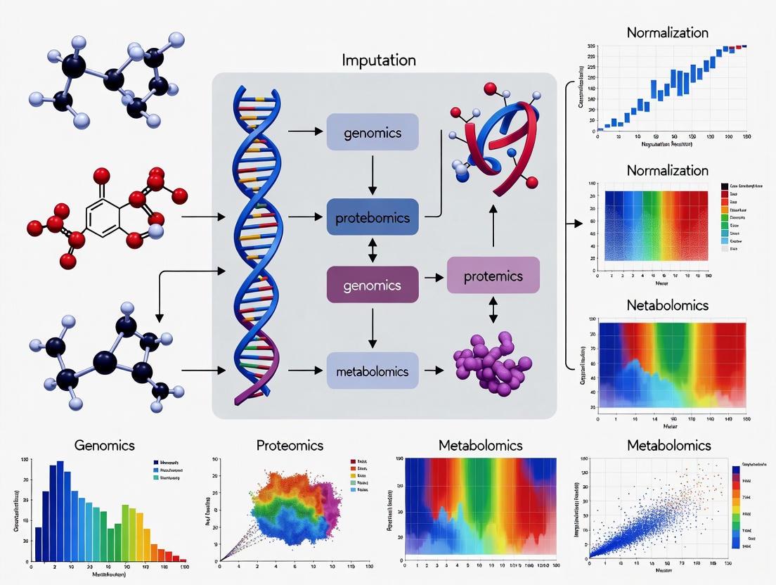
The following diagram illustrates the logical relationships and sequential steps in the standard multi-omics data preprocessing workflow.
Diagram 1: Standard Multi-Omics Preprocessing Workflow.
The Scientist's Toolkit: Essential Research Reagents and Computational Solutions
Table 3: Key Research Reagents and Computational Tools for Data Cleansing
| Item / Tool Name | Function / Description | Application Context |
|---|---|---|
| QC Reference Samples | Pooled samples from all groups, run repeatedly to monitor instrument stability and for LOESS normalization [1]. | All mass spectrometry and sequencing experiments. |
| Internal Standards (IS) | Chemically similar, stable isotope-labeled analogs of target analytes added to correct for sample prep variability and ion suppression. | Targeted metabolomics, lipidomics, proteomics. |
| edgeR / DESeq2 (R packages) | Statistical packages for normalizing and analyzing RNA-seq count data (e.g., TMM method in edgeR) [3]. | Transcriptomics data analysis. |
| BayesNetty | Software package for fitting Bayesian networks to mixed discrete/continuous data with missing values, enabling causal inference [4] [5]. | Multi-omics integration with incomplete data. |
| ComBat / sva (R package) | Algorithm for adjusting for batch effects in high-dimensional data, preserving biological variance [3]. | Multi-omics data from multiple batches or centers. |
| KNN Imputation | k-Nearest Neighbors algorithm; fills a missing value using the average from the k most similar samples. A versatile, general-purpose method [3] [2]. | General imputation for MCAR/MAR data across omics layers. |
| WDL (Workflow Description Language) | A language for describing complex data processing workflows in a portable and scalable manner, ensuring reproducibility [8]. | Deploying standardized preprocessing pipelines on HPC systems. |
| Singularity / Docker | Containerization technologies that package software and dependencies into a portable, reproducible unit [8]. | Ensuring consistent software environments for analysis. |
The High-Dimension Low Sample Size (HDLSS) Problem and its Impact on Downstream Analysis
The High-Dimension, Low Sample Size (HDLSS) regime, where the number of features (p) far exceeds the number of observations (n, presents significant statistical and computational challenges for multi-omics research. This Application Note examines the theoretical foundations of the HDLSS problem and its profound impact on downstream analyses, including classification, clustering, and data integration. We detail robust experimental and computational protocols for data normalization, imputation, and dimensionality reduction specifically designed for HDLSS settings. Within the broader context of multi-omics data imputation and normalization research, these protocols are essential for ensuring the reliability of biological interpretations and the success of subsequent drug development efforts.
In modern bioinformatics, technological advances in high-throughput biology have enabled the simultaneous measurement of tens of thousands to millions of features (e.g., genes, proteins, metabolites) across a relatively small number of biological samples [9] [10]. This scenario is aptly termed the High-Dimension, Low Sample Size (HDLSS) paradigm. A pivotal characteristic of HDLSS data is that the dimensionality p is significantly larger than the sample size n, often denoted as p ≫ n [11].
This paradigm presents unique challenges that run counter to classical statistical intuition. For instance, in the limit as the dimension d → ∞ with a fixed sample size n, a standard Gaussian sample exhibits geometric properties where data vectors tend to lie on the surface of a growing sphere, and the angles between pairs of vectors approach 90 degrees, leading to a phenomenon of random rotation [9]. This inherent geometry can severely degrade the performance of traditional statistical methods, leading to overfitting, model instability, and spurious correlations [12] [13] [11]. In multi-omics studies, these challenges are compounded by the need to integrate heterogeneous data types (genomics, transcriptomics, proteomics, etc.), each with its own HDLSS characteristics, missing value patterns, and technical noise [10] [14] [15]. Addressing the HDLSS problem through principled normalization, imputation, and dimensionality reduction is therefore a critical prerequisite for any meaningful multi-omics integrative analysis.
Impact on Downstream Analysis
The HDLSS problem fundamentally compromises the validity and reliability of downstream analytical tasks. Understanding these impacts is crucial for developing appropriate corrective methodologies.
Table 1: Impact of HDLSS on Key Downstream Analyses
| Analytical Task | Impact of HDLSS | Consequence |
|---|---|---|
| Classification | High misclassification rate due to overfitting and increased variance of discriminant functions [13]. | Reduced accuracy in disease subtyping, sample diagnosis, and biomarker identification. |
| Clustering | Apparent formation of clusters in high-dimensional space that may not represent true biological groups [9]. | Misleading interpretation of cell types or disease subtypes, invalidating biological conclusions. |
| Principal Component Analysis (PCA) | Sample eigenvectors fail to converge to their population counterparts; they instead converge to a cone, creating a systematic angle bias [9]. | Inaccurate data visualization and incorrect identification of primary sources of variation. |
| Feature Selection | Standard methods assume feature independence; HDLSS exacerbates the difficulty in identifying truly relevant features from a sea of irrelevant ones [11]. | Selection of redundant or irrelevant features, hindering biomarker discovery and biological insight. |
| Data Fusion & Integration | The "curse of dimensionality" affects each omics view uniquely, complicating the creation of a unified, low-dimensional representation [12]. | Failure to capture true inter-omics relationships, leading to an incomplete or distorted biological picture. |
Methodological Framework for HDLSS Data
A robust analytical framework for HDLSS multi-omics data must incorporate specialized procedures for normalization, imputation, and dimensionality reduction to mitigate the adverse effects previously described.
Normalization Strategies for Multi-Omics Data
Normalization is a critical pre-processing step to control systematic biases and minimize technical variation, making different samples and omics layers comparable [16] [14]. The choice of normalization method is particularly sensitive in HDLSS settings, where technical artifacts can easily overwhelm subtle biological signals.
Table 2: Evaluation of Normalization Methods for MS-Based Multi-Omics Data
| Normalization Method | Underlying Assumption | Performance in Multi-Omics Context |
|---|---|---|
| Total Ion Current (TIC) | Total feature intensity is consistent across all samples [14]. | Can be biased by highly abundant features; performance varies across omics types. |
| Probabilistic Quotient Normalization (PQN) | The overall distribution of feature intensities is similar across samples [14]. | Identified as optimal for metabolomics and lipidomics; also excels in proteomics. Robust for temporal studies. |
| Median Normalization | The median feature intensity is constant across samples [14]. | Excels for proteomics data; a simple and stable method. |
| LOESS (QC-based) | Assumes balanced proportions of up/down-regulated features; uses quality control (QC) samples to model systematic error [14]. | Top performer for metabolomics and lipidomics; effective at preserving time-related variance in proteomics. |
| Variance Stabilizing Normalization (VSN) | Feature variance depends on its mean and can be transformed to be constant [14]. | Applied to proteomics; transforms data distribution. |
| SERRF (Machine Learning) | Uses Random Forest on QC samples to learn and correct for systematic errors like batch effects [14]. | Can outperform others but risks overfitting and masking true biological variance. |
Protocol 1: Two-Step Pre-Acquisition Normalization for Tissue-Based Multi-Omics Application: This protocol is designed for MS-based analysis of proteins, lipids, and metabolites extracted from the same tissue sample, minimizing technical variation prior to instrumental analysis [16].
- Tissue Homogenization: Weigh frozen tissue samples and homogenize in a methanol-water mixture (e.g., 5:2, v:v) at a consistent ratio (e.g., 0.06 mg tissue per μL solvent) [16].
- Multi-Omics Extraction: Perform a simultaneous extraction of biomolecules using a method like the Folch extraction (using methanol, water, and chloroform at a ratio of 5:2:10, v:v:v) [16].
- Post-Extraction Protein Quantification: Measure the protein concentration from the extracted protein pellet using a colorimetric assay (e.g., DCA assay) [16].
- Volume Adjustment: Normalize the volumes of the lipid and metabolite fractions based on the measured protein concentration before drying and LC-MS/MS analysis [16]. Rationale: Normalizing first by tissue weight and then by post-extraction protein concentration has been shown to generate the lowest sample variation, thereby best revealing true biological differences in subsequent analyses [16].
Multi-Omics Normalization Workflow
Advanced Imputation for Missing Data
Missing values are inevitable in omics datasets and are particularly problematic in HDLSS contexts, as they can constitute a significant portion of the already limited sample information. Integrative imputation techniques that leverage correlations across multi-omics datasets outperform methods relying on single-omics information alone [10] [17].
Table 3: Deep Learning Models for Omics Data Imputation
| Deep Learning Model | Key Principle | Strengths | Weaknesses | Suitable Omics Data |
|---|---|---|---|---|
| Autoencoder (AE) | Learns a compressed data representation (encoder) to reconstruct original data (decoder) [17]. | Excels at learning complex, non-linear relationships; relatively straightforward to train. | Prone to overfitting; latent space can be less interpretable. | scRNA-seq, bulk transcriptomics [17]. |
| Variational Autoencoder (VAE) | A probabilistic generative model that learns a latent variable distribution [17]. | More interpretable latent space; mitigates overfitting; good for modeling uncertainty. | More complex training due to KL divergence loss and sampling. | Transcriptomics, multi-omics integration [17]. |
| Generative Adversarial Networks (GANs) | Uses a generator and a discriminator in an adversarial game to produce realistic data [17]. | Highly flexible; can generate diverse, high-quality samples. | Training is unstable (mode collapse, hyperparameter sensitivity). | Image-based omics data (e.g., histology) [17]. |
| Transformer | Utilizes self-attention mechanisms to weigh the importance of all elements in a sequence [17]. | Captures long-range dependencies in data; highly parallelizable. | Computationally intensive (quadratic complexity with sequence length). | Genomics, proteomics (sequence data) [17]. |
Protocol 2: Autoencoder-Based Imputation for scRNA-seq Data Application: This protocol uses an overcomplete autoencoder to impute missing values in a sparse gene expression matrix, minimizing alterations to biologically uninformative values [17].
- Input: A sparse gene expression matrix
Rwith missing values represented as zeros orNA. - Model Architecture:
- Encoder (
E): A neural network that maps the input dataRto a lower-dimensional bottleneck layer. - Decoder (
D): A neural network that reconstructs the full expression matrix from the bottleneck layer.
- Encoder (
- Loss Function: Minimize the following objective:
min E,D ||R - Dσ(E(R))||₀² + λ/2 (||E||F² + ||D||F²)where||・||₀implies the loss is calculated only for non-zero counts inR,σis the sigmoid activation function, andλis a regularization coefficient to prevent overfitting [17]. - Training: Train the model using backpropagation until the reconstruction error converges.
- Imputation: The output of the trained autoencoder,
Dσ(E(R)), is the imputed gene expression matrix.
Autoencoder Imputation Process
Dimensionality Reduction and Feature Selection
Direct analysis in the original high-dimensional space is often infeasible. Therefore, reducing dimensionality while preserving biological signal is paramount.
Protocol 3: Hybrid Feature Selection for HDLSS Datasets Application: This metaheuristic method combines filtering and wrapper techniques to select a minimal set of informative features from HDLSS data, enhancing prediction model performance [11].
- Phase 1: Gradual Permutation Filtering (GPF)
- Input: All
pfeatures from the HDLSS dataset. - Ranking: Evaluate features based on permutation importance within a model (e.g., a classifier). This involves randomly shuffling a single feature and measuring the decrease in model performance.
- Iterative Filtering: Repeatedly (e.g., 50 times) measure permutation importance and eliminate features with importance near zero. Recalculate importance after each elimination step to minimize bias.
- Output: A ranked list of features that have survived the filtering process.
- Input: All
- Phase 2: Heuristic Tribrid Search (HTS)
- Forward Search: Start with a "first-choice feature" from the GPF-ranked list. Incrementally add the next feature from the list that most improves a performance metric (e.g., LCM).
- Consolation Match: If performance plateaus, attempt to swap a single feature between the selected and unselected pools to escape local optima.
- Backward Elimination: Remove the least important feature from the selected set if it does not degrade performance.
- Stopping Criterion: The process stops when no further improvement is found via swapping or elimination.
Data Fusion in HDLSS Settings
A universal approach for learning in an HDLSS setting involves multi-view mid-fusion [18]. When inherent data views (e.g., separate omics) are not available, this technique artificially constructs them by splitting high-dimensional feature vectors into smaller subsets. Each subset is then treated as an independent "view," and a mid-fusion integration model is applied to learn from these views simultaneously, effectively improving performance in the HDLSS context [18].
The Scientist's Toolkit: Essential Reagents & Materials
Table 4: Key Research Reagent Solutions for Multi-Omics Experiments
| Item | Function/Application | Example/Details |
|---|---|---|
| Folch Extraction Solvents | Simultaneous extraction of proteins, lipids, and metabolites from the same biological sample [16]. | Methanol, Water, Chloroform at ratio 5:2:10 (v:v:v). |
| Internal Standards (I.S.) | Spiked into samples before LC-MS/MS analysis to correct for technical variation during sample preparation and instrument run. | Metabolomics: 13C515N Folic Acid. Lipidomics: EquiSplash mixture [16]. |
| Colorimetric Protein Assay | Quantification of total protein concentration for sample normalization prior to proteomic analysis. | DCA (Dichloroacetic Acid) Assay or similar (e.g., BCA, Bradford) [16]. |
| LC-MS/MS Grade Solvents | Used as mobile phases for liquid chromatography to ensure minimal background noise and high sensitivity in mass spectrometry. | MS-grade Water with 0.1% Formic Acid (FA); Acetonitrile (ACN) with 0.1% FA [16]. |
| Quality Control (QC) Pool | A pooled sample created by combining small aliquots of all study samples, used to monitor and correct for instrumental drift. | Injected at regular intervals throughout the LC-MS/MS sequence for post-acquisition normalization (e.g., LOESS QC) [14]. |
The HDLSS problem is a central challenge in contemporary multi-omics research, directly impacting the veracity of downstream analytical results. Success in this context hinges on the rigorous application of specialized protocols for data pre-processing. As detailed in this note, a combination of robust two-step normalization, advanced deep learning-based imputation, and careful dimensionality reduction or feature selection forms a defensible strategy to mitigate the perils of high-dimensionality and low sample size. Adherence to these protocols ensures that subsequent data integration and modeling efforts are built upon a reliable foundation, thereby accelerating the discovery of robust biomarkers and therapeutic targets in precision medicine.
In multi-omics research, data heterogeneity presents a fundamental challenge for integrative analysis. This heterogeneity manifests primarily through different scales (e.g., read counts for transcriptomics versus intensity values for proteomics), varying distributions (binomial for transcript expression, bimodal for methylation data), and disparate modalities (continuous, categorical, and right-censored data) originating from platforms including genomics, transcriptomics, proteomics, metabolomics, and epigenomics [19]. The core objective of multi-omics integration is to synthesize these heterogeneous datasets, measured on the same biological samples, to achieve a holistic understanding of biological systems and complex diseases such as cancer and neurodevelopmental disorders [20] [21]. Successfully reconciling these differences is critical for uncovering hidden patterns and complex phenomena that are not apparent from single-omics analyses alone [21].
The sources of heterogeneity are both technical and biological. Technical variance arises from differences in sample handling, reagents, instrumentation, and operator, leading to batch effects that can obscure true biological signals [22]. Biologically, different omics layers may produce complementary or occasionally conflicting signals, as seen in colorectal carcinomas where methylation profiles linked to genetic lineages showed inconsistent connections to transcriptional programs [19]. Furthermore, cohort differences in sex, age, ancestry, disease severity, and comorbidities introduce additional variance that is not disease-related, complicating the distinction between technical noise and biological signal [22]. Addressing these challenges requires robust normalization, batch correction, and specialized statistical frameworks designed to handle high-dimensional, sparse data with complex covariance structures [22].
Understanding the Dimensions of Heterogeneity
The heterogeneity in multi-omics data stems from multiple, interconnected sources. Understanding these dimensions is the first step toward developing effective integration strategies.
- Platform-Induced Heterogeneity: Different omics technologies inherently produce data with distinct characteristics. For instance, RNA-sequencing (RNA-seq) data for transcriptomics is typically count-based and follows a negative binomial distribution, while mass spectrometry-based proteomics generates continuous intensity measurements that often require variance-stabilizing normalization [22]. Methylation data, particularly for CpG islands, displays a characteristic bimodal distribution [19]. These inherent differences in measurement scales and distributions must be reconciled before integration.
- Batch Effects and Confounders: Technical batch effects are a major source of unwanted variation, introduced by differences in sample processing dates, reagent lots, sequencing lanes, or mass spectrometry runs [22]. Biological confounders, such as age, sex, post-mortem interval (for brain tissue), and cell type heterogeneity, can also introduce systematic biases that are unrelated to the biological question of interest. In neurodevelopmental disorder studies, for example, case-control imbalances or developmental stage effects are common and must be carefully adjusted for [22].
- Dimensionality and Sparsity: Omics datasets are typically "wide," with thousands to hundreds of thousands of features (e.g., genes, proteins) measured across a relatively small number of samples. This "large p, small n" scenario increases the risk of overfitting and spurious associations [22]. Additionally, data sparsity is a concern, particularly in proteomics and metabolomics, where many features may be missing not at random but due to being below the detection limit.
Impact of Heterogeneity on Downstream Analysis
Failure to adequately address data heterogeneity has profound consequences on the reliability and interpretability of multi-omics studies.
- Reduced Statistical Power and False Discoveries: Unaccounted for technical variation and confounders can severely compromise downstream inference, inflating false positive rates or obscuring true biological signals [22]. Poor quality control, such as the inclusion of outlier samples due to RNA degradation, can distort differential expression analyses and bias integrative modeling.
- Impaired Integration Performance: Data heterogeneity directly challenges integration algorithms. Without proper harmonization, methods may fail to identify concordant signals across omics layers or may group samples based on technical artifacts rather than biological similarity. This can lead to inaccurate patient stratifications, unreliable biomarker discovery, and flawed molecular subtyping [19].
- Limited Reproducibility and Generalizability: Findings from multi-omics analyses that do not properly account for cohort heterogeneity (e.g., differences in ancestry, medication status) often fail to generalize to independent populations, undermining their clinical and translational potential [22].
Table 1: Key Dimensions of Data Heterogeneity in Multi-Omics Studies
| Dimension of Heterogeneity | Description | Exemplary Data Types | Primary Challenge |
|---|---|---|---|
| Scale and Distribution | Differences in data range (e.g., counts, intensities) and underlying statistical distribution. | RNA-seq (count, negative binomial), Methylation (beta-values, bimodal) | Incomparable feature variances that can dominate integration. |
| Modality | Differences in the fundamental type of data generated. | Genomic (categorical), Proteomic (continuous), Clinical (mixed) | Requires flexible algorithms that can handle diverse data structures. |
| Dimensionality | Differences in the number of features measured per omics layer. | Mutation data (highly sparse), Gene expression (dense) | "Large p, small n" problem, risk of overfitting. |
| Technical Noise | Non-biological variation introduced by experimental procedures. | Batch effects, Library preparation, Platform differences | Can confound biological signal if not corrected. |
Quantitative Benchmarks and Guidelines for Multi-Omics Study Design
Recent large-scale benchmarking studies on datasets from The Cancer Genome Atlas (TCGA) have provided evidence-based recommendations for designing robust multi-omics studies that can effectively manage data heterogeneity. These guidelines address key computational and biological factors to enhance the reliability of integration results [19].
A central finding is the critical importance of feature selection. Selecting a smaller subset of biologically relevant features (e.g., less than 10% of omics features) has been shown to improve clustering performance by up to 34% by reducing noise and mitigating the curse of dimensionality [19]. Furthermore, sample size and balance are crucial. Benchmarks recommend a minimum of 26 samples per class to achieve robust cancer subtype discrimination. Maintaining a class balance under a 3:1 ratio of sample sizes is also advised, as high imbalance can skew integration results [19].
The resilience of integration methods to noise is another key consideration. Studies suggest that analytical workflows should be designed to handle noise levels of up to 30%, beyond which performance can degrade significantly [19]. Adherence to these benchmarks provides a structured framework for researchers to optimize their analytical approaches.
Table 2: Evidence-Based Guidelines for Multi-Omics Study Design (MOSD)
| Factor | Category | Recommended Guideline | Impact on Analysis |
|---|---|---|---|
| Sample Size | Computational | ≥ 26 samples per class | Ensures sufficient statistical power for robust clustering. |
| Feature Selection | Computational | < 10% of omics features | Can improve clustering performance by 34%; reduces noise. |
| Class Balance | Computational | Balance ratio < 3:1 | Prevents skewed results and biased model training. |
| Noise Characterization | Computational | Noise level < 30% | Maintains model performance and reliability. |
| Omics Combinations | Biological | Gene Expression + Methylation often perform well | Provides complementary signals for patient stratification. |
| Clinical Correlation | Biological | Integrate molecular & clinical features (e.g., stage, age) | Validates biological relevance and clinical significance. |
Experimental Protocols for Data Reconciliation
This section provides detailed, step-by-step protocols for normalizing and harmonizing heterogeneous multi-omics data, a critical prerequisite for successful integration.
Protocol 4.1: Multi-Omics Data Preprocessing and Normalization
Objective: To transform raw data from each omics layer into a clean, normalized, and batch-corrected dataset ready for integration.
Materials:
- Computing Environment: R (v4.0+) or Python (v3.8+).
- Software Packages: R:
DESeq2,edgeR,sva,limma. Python:scikit-learn,pandas,numpy,scanpy(for single-cell data). - Input Data: Raw feature matrices (e.g., count matrix for RNA-seq, intensity matrix for proteomics).
Procedure:
- Quality Control (QC) and Filtering:
- Perform per-sample QC: Assess metrics such as sequencing depth (for RNA-seq), mapping rates, and number of detected features. Exclude outlier samples with signs of degradation or low quality [22].
- Perform per-feature Filtering: Remove features with excessive missingness (e.g., genes not expressed in a sufficient number of samples) or low variance, as they contribute little information.
Platform-Specific Normalization:
- For RNA-seq Data: Apply methods that account for library size and composition bias. Use the median-of-ratios method in
DESeq2or the trimmed mean of M-values (TMM) method inedgeR[22]. - For Proteomics Data: Apply variance-stabilizing normalization, quantile normalization, or use internal reference standards to correct for technical variation in mass spectrometry data [22].
- For Methylation Data: Perform background correction and normalization using methods from the
minfiorChAMPpackages.
- For RNA-seq Data: Apply methods that account for library size and composition bias. Use the median-of-ratios method in
Batch Effect Correction:
- Identify known batch variables (e.g., processing date, sequencing run).
- Apply a batch correction algorithm such as
ComBat(from thesvapackage) orremoveBatchEffect(from thelimmapackage) to remove systematic technical variation while preserving biological heterogeneity [22]. - For more complex batch structures, consider advanced methods like Mutual Nearest Neighbors (MNN) or deep learning-based approaches, especially in single-cell omics [22].
Handling Missing Data:
- For proteomics and metabolomics data, impute missing values using methods like k-nearest neighbors (KNN) imputation, minimum value imputation, or model-based approaches (e.g.,
MissForest), depending on the assumed mechanism of missingness.
- For proteomics and metabolomics data, impute missing values using methods like k-nearest neighbors (KNN) imputation, minimum value imputation, or model-based approaches (e.g.,
Protocol 4.2: Concatenation-Based Integration with DIABLO
Objective: To integrate multiple omics datasets using a multi-block supervised framework to identify correlated components that discriminate between pre-defined sample classes and predict clinical outcomes.
Materials:
- Software: R package
mixOmics[21]. - Input Data: Normalized and batch-corrected matrices from at least two omics layers (e.g., gene expression, methylation) and a sample phenotype/class vector.
Procedure:
- Data Preparation: Ensure all normalized matrices are transformed and scaled appropriately. The
mixOmicspipeline often includes internal log-transformation for count data and standardization. - Model Design:
- Specify the omics blocks (X) and the outcome vector (Y) that represents the phenotype or class to be discriminated.
- Define the model design matrix, which controls the level of integration between different omics layers. A common starting point is a full design where all blocks are connected.
- Parameter Tuning:
- Use the
tune.block.splsdafunction to perform a cross-validation grid search for the optimal number of components and the number of features to select per component and per omics block. This step is crucial for building a robust model.
- Use the
- Model Fitting:
- Run the final
block.splsda(DIABLO) model using the tuned parameters. - The model will identify a set of components—latent variables—that maximize the covariance between the omics blocks and the correlation with the outcome.
- Run the final
- Result Interpretation:
- Sample Plot: Visualize sample clustering in the latent space to assess class discrimination.
- Circus Plot: Generate a circos plot to visualize the correlations between selected features from different omics layers, revealing multi-omics biomarker networks.
- Loadings: Examine the variable loadings to identify the top features from each omics platform that drive the integration and class separation.
Protocol 4.3: Deep Learning-Based Integration with Flexynesis
Objective: To leverage a flexible deep learning toolkit for integrating bulk multi-omics data for various prediction tasks, including classification, regression, and survival analysis, in a modular and accessible framework.
Materials:
- Software: Flexynesis, available via PyPi, Bioconda, or Galaxy Server [23].
- Input Data: Normalized and batch-corrected matrices from multiple omics layers. The framework supports single-task and multi-task learning.
Procedure:
- Data Preprocessing with Flexynesis:
- Use the accessory pipeline provided with Flexynesis to streamline data processing, including feature selection and hyperparameter tuning.
- Model Configuration:
- Choose from available deep learning architectures (e.g., fully connected or graph-convolutional encoders).
- Select the supervision task(s): attach Multi-Layer Perceptron (MLP) "supervisor" heads for regression (e.g., drug response), classification (e.g., cancer subtype), or survival modeling (Cox Proportional Hazards) [23].
- Training and Validation:
- Implement standard training/validation/test splits. Flexynesis automates hyperparameter optimization.
- Train the model. The framework allows joint training on multiple outcome variables, shaping the sample embedding space (latent variables) with complementary information, even when some labels are missing [23].
- Model Evaluation and Biomarker Discovery:
- Evaluate performance on the held-out test set using task-specific metrics (e.g., AUC for classification, C-index for survival).
- Use the model's interpretability features to identify key input features (biomarkers) that contribute most to the predictions.
The Scientist's Toolkit: Essential Research Reagents and Computational Solutions
Table 3: Key Research Reagent Solutions and Computational Tools for Multi-Omics Integration
| Item / Tool Name | Type | Function in Multi-Omics Integration |
|---|---|---|
| DESeq2 / edgeR | R Software Package | Performs normalization and differential expression analysis for RNA-seq data; addresses library size and composition bias [22]. |
| ComBat (sva package) | R Software Package | Empirical Bayes method for correcting batch effects in high-dimensional data, preserving biological signal while removing technical artifacts [22]. |
| Flexynesis | Python Deep Learning Toolkit | Provides modular, reusable deep learning architectures for bulk multi-omics integration tasks like classification, regression, and survival analysis [23]. |
| DIABLO (mixOmics) | R Software Package | A supervised multi-block framework to identify highly correlated features across multiple omics datasets that discriminate between sample classes [21]. |
| Mutual Nearest Neighbors (MNN) | Computational Algorithm | A batch correction method that identifies pairs of cells (or samples) that are nearest neighbors across batches, used to align datasets and remove technical variation [22]. |
| Internal Reference Standards | Wet-Lab Reagent | Used in proteomics and metabolomics experiments; a set of known, stable isotopically labeled compounds spiked into samples to correct for technical variation during mass spectrometry [22]. |
| Single-Cell Multi-Omics Assays | Wet-Lab Protocol | Enables simultaneous measurement of genomic, transcriptomic, and epigenomic information from the same cell, resolving cellular heterogeneity without inference [24]. |
| Long-Read Sequencing | Technology Platform | Enables full-length transcript sequencing and access to complex genomic regions, improving the resolution of structural variants and isoform diversity [24]. |
In multi-omics research, the raw data generated from high-throughput technologies are never analysis-ready. They contain inherent technical artifacts that, if unaddressed, would obscure true biological signals and lead to spurious findings. Two of the most critical preprocessing steps are imputation and normalization, each serving distinct but complementary purposes. Imputation focuses on handling missing data values that arise from technical limitations, while normalization addresses systematic technical variations that prevent fair comparisons across samples or datasets [2] [25]. The confusion between these processes often stems from their shared position in data preprocessing workflows, yet their methodological approaches and ultimate goals differ fundamentally. Within the context of multi-omics data integration for precision medicine and drug development, applying these techniques appropriately is paramount for generating biologically valid, reproducible results that can inform clinical decision-making and therapeutic discovery [2] [24].
Defining the Core Concepts
The Goal of Imputation: Handling Data Gaps
Imputation is the process of estimating and filling in missing values in a dataset. In multi-omics studies, missing data is a pervasive issue arising from various sources. Technical limitations can prevent the detection of low-abundance proteins in proteomics or metabolites in metabolomics [25]. Analytical platform sensitivities vary, with some technologies failing to detect molecules present at concentrations below their detection thresholds. Biological constraints also contribute, as certain molecules may be expressed in a tissue-specific manner and thus absent in other sample types [25]. The primary goal of imputation is to create a complete data matrix suitable for downstream statistical analyses and machine learning algorithms, most of which require complete datasets. By addressing these gaps, imputation helps prevent biased parameter estimates, loss of statistical power, and reduced generalizability of findings [4].
The Goal of Normalization: Enabling Fair Comparison
Normalization is the process of removing unwanted technical variation to enable fair comparisons across samples and datasets. Multi-omics data are contaminated by numerous non-biological variances including differences in sample preparation, extraction efficiency, instrumental noise, sequencing depth, and reagent batches [26] [25]. These technical artifacts can create systematic differences between samples that obscure genuine biological signals. The core objective of normalization is to eliminate these technical biases so that biological differences can be accurately discerned. This process is particularly crucial when integrating datasets from different studies, laboratories, or platforms, as it ensures that observed differences reflect true biological variation rather than technical inconsistencies [26] [27].
Table 1: Fundamental Distinctions Between Imputation and Normalization
| Aspect | Imputation | Normalization |
|---|---|---|
| Primary Goal | Handle missing data points | Remove technical variation |
| Problem Addressed | Incomplete data matrices | Systematic technical biases |
| Trigger Condition | Missing values detected | Sample-to-sample technical variability |
| Key Challenge | Preserving biological relationships in estimated values | Removing technical noise without removing biological signal |
| Common Methods | Bayesian networks, matrix factorization, k-NN | Probabilistic Quotient Normalization (PQN), LOESS, Median normalization |
Technical Protocols and Methodologies
Experimental Protocol for Data Normalization
Title: Protocol for Normalizing Mass Spectrometry-Based Multi-Omics Data in Temporal Studies
Background: This protocol outlines a robust strategy for normalizing metabolomics, lipidomics, and proteomics datasets derived from mass spectrometry, particularly suited for time-course experiments where preserving temporal biological variance is critical [26].
Reagents and Materials:
- Raw mass spectrometry data files (.raw, .mzML)
- Quality Control (QC) samples (pooled from all experimental samples)
- R statistical environment (v4.3.0 or higher)
- Normalization packages:
limma(for LOESS, Median, Quantile),vsn(for VSN)
Procedure:
- Data Preparation: Process raw files using appropriate software (Compound Discoverer for metabolomics, MS-DIAL for lipidomics, Proteome Discoverer for proteomics) to generate feature intensity tables [26].
- Quality Control Assessment: Calculate the coefficient of variation (CV) for features across QC samples to assess initial data quality.
- Method Selection: Apply multiple normalization methods in parallel:
- For metabolomics and lipidomics: Implement Probabilistic Quotient Normalization (PQN) and LOESS using QC samples (LOESS QC)
- For proteomics: Implement PQN, Median, and LOESS normalization
- Effectiveness Evaluation: Assess each method's performance based on:
- Improvement in QC feature consistency (target: CV reduction >20%)
- Preservation of treatment and time-related variance (variance explained should not decrease >15% post-normalization)
- Method Implementation: Apply the optimal method(s) identified in step 4 to the entire dataset.
Troubleshooting:
- If biological variance decreases substantially post-normalization, consider adjusting parameter settings or trying an alternative method
- If QC consistency does not improve, check for outlier samples that may need exclusion prior to normalization
Experimental Protocol for Data Imputation
Title: Protocol for Bayesian Network Imputation of Multi-Omics Data with Missing Values
Background: This protocol describes a Bayesian network approach to handle missing data in multi-omics datasets, particularly effective for exploring causal relationships in incomplete datasets such as those generated from type 2 diabetes studies [4].
Reagents and Materials:
- Multi-omics dataset with missing values (genomics, proteomics, metabolomics, clinical variables)
- BayesNetty software package
- High-performance computing cluster (for datasets >1,000 samples)
Procedure:
- Data Preprocessing: Filter the full variable set (e.g., from ~16,000 to 260 variables) based on biological relevance and data quality.
- Network Setup: Initialize a Bayesian network structure incorporating prior biological knowledge where available.
- Model Fitting: Use the novel imputation method implemented in BayesNetty to fit the network to the incomplete data.
- Imputation Execution: Estimate missing values based on the conditional probability distributions within the fitted network.
- Validation: Assess imputation quality through:
- Comparison of distribution patterns pre- and post-imputation
- Sensitivity analysis using different network starting points
Troubleshooting:
- If convergence issues occur, consider reducing the number of variables or increasing iterations
- If imputed values show extreme deviations from expected ranges, check for violations of distributional assumptions
Comparative Analysis of Methods
Normalization Method Performance
Recent systematic evaluation of normalization strategies for mass spectrometry-based multi-omics datasets revealed distinct performance patterns across different omics types. The study analyzed metabolomics, lipidomics, and proteomics data from primary human cardiomyocytes and motor neurons exposed to acetylcholine-active compounds over time [26] [1].
Table 2: Performance of Normalization Methods Across Omics Types
| Omics Type | Recommended Methods | Performance Metrics | Methods to Avoid |
|---|---|---|---|
| Metabolomics | PQN, LOESS QC | Enhanced QC consistency, preserved time-related variance | SERRF (masks treatment variance) |
| Lipidomics | PQN, LOESS QC | Improved feature consistency, maintained treatment effects | SERRF (inconsistent performance) |
| Proteomics | PQN, Median, LOESS | Preserved treatment and time-related variance | VSN (over-correction) |
The study found that machine learning-based approaches like Systematical Error Removal using Random Forest (SERRF) showed inconsistent performance—while it outperformed other methods in some metabolomics datasets, it inadvertently masked treatment-related variance in others [26]. This highlights the importance of validating normalization method performance for specific experimental contexts rather than relying on generalized assumptions.
Imputation Method Performance
Bayesian network imputation methods have demonstrated particular utility for multi-omics datasets with complex missingness patterns. Applied to a type 2 diabetes dataset comprising genotypes, proteins, metabolites, gene expression measurements, and clinical variables from 3,029 individuals, the method enabled the construction of a large average Bayesian network from which putative causal relationships could be identified [4]. This approach effectively handled the reality that no individual had complete data for all variables, making standard complete-case analysis impossible. The success of this method stems from its ability to leverage conditional relationships between variables to estimate missing values, preserving the underlying biological structure within the data.
Integrated Workflow and Visualization
Logical Workflow for Multi-Omics Data Preprocessing
The sequential relationship between imputation and normalization, along with their position in the overall data preprocessing pipeline, can be visualized through the following workflow:
Diagram 1: Multi-omics data preprocessing workflow showing the relationship between quality control, normalization, and imputation.
Decision Framework for Method Selection
The selection of appropriate imputation and normalization strategies depends on specific data characteristics and experimental designs. The following decision framework guides researchers in selecting optimal methods:
Diagram 2: Decision framework for selecting appropriate imputation and normalization methods based on data characteristics.
Research Reagent Solutions
Table 3: Essential Research Reagents and Tools for Multi-Omics Preprocessing
| Reagent/Tool | Function | Application Context |
|---|---|---|
| Compound Discoverer 3.3 | Processes raw metabolomics data | Metabolomics feature detection and alignment [26] |
| MS-DIAL 5.1 | Processes lipidomics data | Lipid identification and quantification [26] |
| Proteome Discoverer 3.0 | Processes proteomics data | Protein identification and quantification [26] |
| R limma package | Implements normalization methods | LOESS, Median, and Quantile normalization [26] |
| R vsn package | Variance stabilization | Normalization for proteomics data [26] |
| BayesNetty software | Bayesian network analysis | Handling missing data in multi-omics datasets [4] |
| Quality Control (QC) samples | Monitoring technical variance | Assessment of normalization effectiveness [26] |
Imputation and normalization serve fundamentally distinct yet complementary roles in multi-omics data preprocessing. While imputation addresses data incompleteness by estimating missing values, normalization enables fair comparison by removing technical biases. The confusion between these processes can lead to inappropriate method selection and compromised research outcomes. For normalization, method performance varies significantly across omics types, with PQN and LOESS QC showing particular promise for metabolomics and lipidomics, while PQN, Median, and LOESS excel for proteomics [26]. For imputation, Bayesian network approaches offer powerful solutions for handling missing data while preserving biological relationships [4]. Researchers must carefully consider their specific data types, experimental designs, and analytical goals when selecting and implementing these preprocessing techniques. By applying these methods appropriately and sequentially—typically normalization followed by imputation—researchers can ensure that their multi-omics analyses yield biologically valid, reproducible insights that advance precision medicine and therapeutic development.
In the era of high-throughput biology, multi-omics data integration has become a cornerstone for advancing our understanding of complex biological systems, from disease mechanisms to therapeutic discovery [28]. The promise of integrating genomics, transcriptomics, proteomics, and epigenomics data lies in obtaining a comprehensive picture of biological processes that single-omics approaches cannot capture [29]. However, this promise remains contingent on a critical yet often underestimated prerequisite: rigorous data preprocessing. Poor preprocessing practices introduce systematic distortions that propagate through the entire analytical pipeline, ultimately compromising biological interpretation and undermining scientific reproducibility. This article examines the specific consequences of inadequate preprocessing across multi-omics workflows and provides structured guidelines to mitigate these pervasive challenges.
The Critical Role of Preprocessing in Multi-Omics Research
Data preprocessing transforms raw, complex biological data into clean, analysis-ready datasets. This foundational step is not merely technical "janitor work" but constitutes an essential scientific procedure that determines the validity of all subsequent findings. In multi-omics studies, preprocessing must address the unique characteristics of each data layer while ensuring their eventual compatibility for integration [30].
Traditional manual curation of multi-omics data consumes 60-80% of a computational biologist's time, creating a significant bottleneck in research velocity [30]. This intensive process is necessary because each omics modality presents distinct preprocessing requirements—from genotype imputation and quality control for GWAS data to adapter trimming and read mapping for RNA-seq [31] [32]. Without standardized, automated preprocessing pipelines, studies risk generating irreproducible results that cannot be translated into reliable biological insights.
Table 1: Multi-Omics Data Types and Their Preprocessing Particularities
| Omics Data Type | Key Preprocessing Steps | Primary Challenges |
|---|---|---|
| Genomics (GWAS) | Genotype imputation, quality control (call rate, HWE, MAF), additive encoding [31] | High dimensionality, polygenic architecture, population stratification |
| Transcriptomics (RNA-seq) | Quality control (FastQC), adapter trimming, read mapping, normalization (TPM) [33] [32] | Library size differences, multi-mapping reads, alternative splicing |
| Epigenomics (EWAS) | Background correction, normalization, probe filtering [31] | Cell type heterogeneity, technical variation, confounding |
| Proteomics | Peptide spectral match quantification, normalization, imputation [33] | Missing data, dynamic range compression, batch effects |
Consequences of Inadequate Preprocessing
Technical Artifacts Masquerading as Biological Signals
One of the most pernicious consequences of poor preprocessing is the failure to distinguish technical artifacts from genuine biological signals. Batch effects—systematic technical variation introduced by different processing dates, technicians, or instruments—can completely overwhelm true biological signals if not properly addressed [30]. In one documented case, the first principal component in an integrated multi-omics analysis of leukemia separated samples by sequencing vendor rather than disease subtype, misleading researchers about the fundamental structure of their data [29].
AI models excel at pattern recognition but cannot inherently distinguish real biological differences from technical artifacts. Without specialized correction methods like ComBat or advanced deep learning models, batch effects masquerade as discoveries, invalidating conclusions and leading research down unproductive paths [30].
Spurious Correlations and Functional Misinterpretation
When preprocessing fails to account for the distinct statistical properties of each omics layer, integrated analyses produce spurious correlations that misinterpret functional relationships. Studies often expect high correlation between mRNA and protein expression, but frequently find only weak associations due to legitimate post-transcriptional regulation [29]. Analysts unaware of this biological reality may misinterpret low correlations as meaningful or selectively report stronger pairs, creating distorted networks of molecular interaction.
In one real-world example, an integrated plot showed a correlation of 0.3 between ATAC-seq peaks and RNA for a set of genes, but half the peaks were located >50kb away from the gene body with no supporting regulatory logic [29]. Such oversights lead to incorrect assignment of regulatory elements and misinterpretation of gene regulatory networks.
Compromised Machine Learning Performance
The high-dimensionality of omics data, where the number of features (e.g., genes, variants) vastly exceeds sample size, makes machine learning models particularly vulnerable to poor preprocessing [31]. Feature selection methods like ridge regression, lasso, and elastic-net—while powerful—are not recommended for low sample sizes without appropriate preprocessing and can cause severe overfitting [31].
The curse of dimensionality combined with technical noise leads to models that memorize artifacts rather than learning biology. This fundamentally limits the translational potential of predictive models for clinical applications like treatment response prediction or disease diagnosis [31] [30].
Table 2: Quantitative Impact of Poor Preprocessing on Research Efficiency
| Metric | Traditional Manual Curation | Optimized Preprocessing | Business Impact |
|---|---|---|---|
| Time-to-Harmonization | 6-8 Weeks [30] | <48 Hours [30] | Accelerates insight generation by two months |
| R&D Productivity | Constrained (60-80% time on cleaning) [30] | Increased by 15-30% [30] | Quadruples researcher focus on high-value discovery |
| Data Fidelity | Dependent on human error [30] | Auditable, ontology-bound [30] | Essential for regulatory compliance and model trust |
Resolution Mismatch and Cellular Heterogeneity Oversights
Integrating data of different resolutions without appropriate preprocessing creates fundamental misinterpretations of cellular biology. Comparing bulk RNA-seq with single-cell ATAC-seq, for example, fails when analysts don't account for missing cellular anchors or compositional differences [29]. In one case study, integration of bulk proteomics and scRNA-seq from brain tissue led to misleading correlations because proteins expressed in glial cells were not properly captured in the scRNA-seq clustering [29].
These resolution mismasks are particularly problematic in complex tissues, where cellular heterogeneity drives biological function but requires specialized preprocessing approaches to resolve across omics layers.
Figure 1: Consequences of poor preprocessing practices cascade through the analytical pipeline, ultimately compromising biological interpretation and experimental reproducibility.
Experimental Protocols for Robust Multi-Omics Preprocessing
Protocol 1: Preprocessing of GWAS Data for Predictive Modeling
This protocol outlines the standardized preprocessing of genome-wide association study (GWAS) data, based on established methodologies for constructing predictive models of disease outcomes [31].
Materials:
- Raw genotype data (e.g., Illumina Infinium Global Screening Array)
- High-performance computing environment
- PLINK 1.9 software [31]
- Michigan Imputation Server access [31]
- Haplotype Reference Consortium (HRC) reference panel [31]
Procedure:
- Quality Control (Pre-Imputation): Submit genotypes to Michigan Imputation Server for automated QC. Exclusion criteria: variants with low allelic frequency (<0.2), low call rate (<0.95), repeated variants, or variants without information [31].
- Genotype Imputation: Perform imputation using Minimac 4 method from HRC reference panel (GRCh37/hg19 genomic annotation) [31].
- Quality Control (Post-Imputation): Execute second QC analysis with PLINK 1.9. Exclusion criteria: low imputation quality (R²<0.9); variants violating Hardy-Weinberg equilibrium (HWE P>10⁻⁶); low minor allele frequency (MAF<0.01) [31].
- Data Encoding: Encode GWAS data according to additive model using dosage format to indicate presence/absence of risk or reference allele in each SNP [31].
Validation:
- Assess final variant count and sample retention
- Verify MAF distribution meets study requirements
- Confirm HWE equilibrium in control populations
Protocol 2: Integrated RNA-seq and Proteomics Preprocessing
This protocol provides a streamlined workflow for simultaneous preprocessing of paired transcriptome and proteome data to enable comparative molecular subgroup identification [33].
Materials:
- RNA-seq count data (e.g., from STAR aligner and featureCounts)
- Proteomic peptide spectral match (PSM) data from TMT mass spectrometry
- R statistical environment (v3.6.3 or later) [33]
- Required R packages: tidyverse, edgeR, limma, vsn, biomaRt [33]
Procedure:
- Data Input and Structure:
- Read transcriptomic data (COUNT and TPM formats) ensuring rectangular structure with features in rows and samples in columns [33].
- Read proteomic PSM data, maintaining compatible sample organization.
Normalization and Scaling:
Batch Effect Correction:
Biology-Aware Feature Selection:
- Remove non-informative features: mitochondrial/ribosomal genes, unannotated peaks, proteins with >30% missing data [29].
- Focus on features with known biological relevance to system studied.
- Validate integration with pathway-level coherence assessment.
Validation:
- Compare molecular subgroups identified from each modality
- Examine RNA-protein correlation patterns in known marker genes
- Assess cluster stability and biological interpretability
Figure 2: Strategic preprocessing workflow showing three-phase approach transforming raw multi-omics data into analysis-ready datasets.
The Scientist's Toolkit: Essential Research Reagent Solutions
Table 3: Key Computational Tools for Multi-Omics Preprocessing
| Tool/Resource | Function | Application Context |
|---|---|---|
| PLINK 1.9 [31] | Whole-genome association analysis | Quality control and analysis of GWAS data |
| Michigan Imputation Server [31] | Genotype imputation | HRC reference-based genotype completion |
| FastQC/MultiQC [32] | Quality control check | QC of raw sequencing data across multiple samples |
| edgeR/limma [33] | Differential expression | RNA-seq data normalization and analysis |
| ComBat/Harmony [30] [29] | Batch effect correction | Removing technical variation across datasets |
| MOFA+ [29] | Multi-omics integration | Factor analysis for integrated omics datasets |
| ColorBrewer [34] | Color palette selection | Accessible data visualization design |
| Chroma.js Palette Helper [34] | Color palette testing | Color blindness simulation and palette optimization |
The consequences of neglecting proper preprocessing in multi-omics research extend far beyond technical inconveniences—they fundamentally compromise biological interpretation and scientific reproducibility. Poor preprocessing practices introduce systematic biases that distort analytical outcomes, leading to spurious findings, wasted resources, and lost opportunities for genuine discovery. The protocols and guidelines presented here provide a framework for implementing rigorous, standardized preprocessing approaches that transform multi-omics data chaos into reliable, interpretable biological insight. As multi-omics technologies continue to evolve and integrate into clinical and pharmaceutical applications, establishing robust preprocessing foundations becomes not merely a methodological preference but an ethical imperative for reproducible science.
A Practical Toolkit: Modern Imputation and Normalization Strategies
Missing data represents a pervasive challenge in multi-omics research, significantly impeding analytical capabilities and decision-making processes across various domains including healthcare, bioinformatics, and precision oncology [35]. The inherent complexity of multi-omics data, characterized by high dimensionality, heterogeneity, and technical variability, creates formidable obstacles for integration and analysis [25]. The "four Vs" of big data—volume, velocity, variety, and veracity—pose particular challenges for conventional biostatistical methods, which often lack the flexibility to model non-linear interactions across different biological scales [25].
Imputation methodologies have evolved substantially from classical statistical approaches to contemporary machine learning and deep learning techniques, each with distinct advantages for handling different missingness patterns and data structures [35]. This progression reflects an ongoing effort to address the unique characteristics of omics data, including massive dimensionality disparities (from millions of genetic variants to thousands of metabolites), temporal heterogeneity, platform-specific technical variability, and pervasive missingness arising from both biological and technical constraints [25]. The selection of appropriate imputation strategies is crucial for maximizing the discovery of meaningful biological differences while minimizing the introduction of artifacts that could compromise downstream analyses.
Table 1: Fundamental Categories of Missing Data in Multi-Omics Studies
| Category | Description | Common Causes | Typical Impact |
|---|---|---|---|
| Missing Completely at Random (MCAR) | Missingness unrelated to any variables | Sample processing failures, random technical errors | Reduces statistical power but introduces minimal bias |
| Missing at Random (MAR) | Missingness related to observed variables but not unobserved data | Batch effects, platform-specific detection limits | Can introduce bias if missingness mechanisms are ignored |
| Missing Not at Random (MNAR) | Missingness related to the unobserved values themselves | Low-abundance molecules falling below detection thresholds | Potentially severe bias requiring specialized handling |
| Structured Missingness | Systematic patterns across samples or features | Incomplete modality acquisition, sample quality issues | Complicates integration and requires strategic imputation |
Classical and Statistical Imputation Methods
Classical imputation approaches form the foundational methodology for handling missing data, with development dating back to the 1930s [35]. These methods typically rely on statistical principles and assumptions about data distribution, offering interpretability and computational efficiency, particularly for datasets with limited missingness.
k-Nearest Neighbors (k-NN) and Regression-Based Approaches
k-NN imputation operates on the principle that samples with similar expression patterns across observed features will likely have similar values for missing features [36]. The method identifies the k most similar samples based on distance metrics (typically Euclidean or cosine distance) and imputes missing values as weighted averages of the neighbors' values. The key advantage of k-NN is its intuitive implementation and minimal assumptions about data distribution. However, performance deteriorates with high-dimensional data where distance metrics become less meaningful, and computational costs increase substantially with dataset size [36].
Regression-based methods model each feature with missing values as a function of other observed features, using techniques ranging from simple linear regression to more sophisticated regularized variants (ridge, lasso) [36]. These methods can capture linear relationships effectively but struggle with the complex non-linear interactions prevalent in biological systems. The emergence of multi-omics research has highlighted the limitations of these approaches for handling the completely missing modalities common in multi-platform studies [36].
Matrix Factorization and Completion
Matrix factorization approaches, particularly Non-negative Matrix Factorization (NMF), have gained significant traction for omics data analysis due to their ability to uncover latent structures and handle high-dimensional datasets [37] [36]. NMF decomposes a non-negative data matrix V (n×m) into two lower-dimensional non-negative matrices W (n×k) and H (k×m), such that V ≈ WH, where k represents the number of latent components [37].
The fundamental assumption underlying matrix completion is that the original data matrix has a low-rank structure, meaning that most features can be represented as linear combinations of a smaller number of latent factors [35]. This assumption frequently holds true for omics data, where coordinated biological processes generate strong dependencies among molecular features. For single-cell multi-omics data clustering, approaches like PLNMFG (Pseudo-label guided Non-negative Matrix Factorization with Graph constraint) integrate unified latent representation learning with cluster structure learning in a joint framework [37]. These methods perform adaptive imputation to handle dropout events while using prior pseudo-labels as constraints during collective factorization, resulting in more robust latent representations that preserve similarity information [37].
Table 2: Classical Imputation Methods and Their Applications
| Method Category | Key Algorithms | Strengths | Limitations | Optimal Use Cases |
|---|---|---|---|---|
| k-NN Based | k-NN, Ensemble k-NN | Simple implementation, preserves local structure | Computationally intensive for large datasets, sensitive to distance metrics | Small to medium datasets with limited missingness (MCAR) |
| Regression Based | Linear Regression, MICE | Models feature relationships, provides uncertainty estimates | Assumes linear relationships, may not capture complex biology | Datasets with strong linear correlations between features |
| Matrix Factorization | NMF, PMF, MNAR | Discovers latent structure, handles high-dimensional data | Requires rank selection, may struggle with complex patterns | Multi-omics integration, feature extraction, co-clustering |
| Statistical Models | EM Algorithm, Bayesian Networks | Handles uncertainty, provides probabilistic framework | Computationally intensive, convergence issues | Complex missingness patterns (MAR, MNAR), causal inference |
Modern Machine Learning Approaches
Modern machine learning methods for imputation leverage more sophisticated algorithms to capture complex patterns in multi-omics data, often outperforming classical approaches, particularly for large-scale datasets with complex missingness patterns.
Random Forests and Ensemble Methods
Tree-based ensemble methods like Random Forests handle missing data through sophisticated internal imputation mechanisms that leverage feature relationships. The missForest algorithm, for instance, imputes missing values by training a random forest model on observed values and predicting missing ones, iterating until convergence [35]. These methods are particularly effective for mixed data types (continuous and categorical) and can capture non-linear relationships without strong distributional assumptions. For mass spectrometry-based multi-omics datasets, the SERRF (Systematical Error Removal using Random Forest) method uses correlated compounds in quality control samples to correct systematic errors, including batch effects and injection order variation [14].
Graph-Based Imputation
Graph neural networks (GNNs) have emerged as powerful tools for imputation in biological networks, leveraging the inherent relational structure of omics data [35]. By representing samples and features as nodes in a graph, GNNs can propagate information from observed to unobserved nodes through message-passing mechanisms, effectively imputing missing values based on topological similarities. This approach is particularly valuable for single-cell multi-omics data, where graph Laplacian constraints can preserve local neighborhood structures during imputation [37]. Methods like PLNMFG incorporate graph constraints to maintain the intrinsic structure of multi-omics data during the clustering process, demonstrating how topological information can guide accurate imputation [37].
Deep Generative Models
Deep generative models represent the cutting edge of imputation methodology, leveraging neural networks with complex architectures to model the underlying data distribution and generate plausible imputations, even for challenging scenarios with extensive missingness.
Autoencoders and Variational Autoencoders
Autoencoders learn compressed representations of input data through an encoder-decoder architecture, with the bottleneck layer capturing essential features [35] [38]. For imputation tasks, the model is trained to reconstruct complete data from corrupted inputs, learning to infer missing values based on the observed patterns. Variational Autoencoders (VAEs) introduce a probabilistic framework by learning the parameters of the data distribution rather than direct representations [38].
In multi-omics analysis, VAEs like multiDGD provide a probabilistic framework to learn shared representations of transcriptome and chromatin accessibility, demonstrating outstanding performance on data reconstruction without feature selection [39]. Unlike standard VAEs, multiDGD uses no encoder to infer latent representations but learns them directly as trainable parameters, employing a Gaussian Mixture Model (GMM) as a more complex and powerful distribution over latent space [39]. This architecture improves the model's ability to capture clustered structures in biological data while maintaining computational efficiency.
Generative Adversarial Networks (GANs)
GANs frame imputation as a generative modeling problem, where a generator network produces plausible imputations while a discriminator network distinguishes between observed and imputed values [36] [38]. Through this adversarial training process, the generator learns to produce imputations that are statistically indistinguishable from real observations. The GAIN (Generative Adversarial Imputation Nets) framework introduced a landmark approach by incorporating a hint mechanism to guide the generator toward realistic imputations [35] [36].
For multi-omics applications, frameworks like OmicsNMF integrate GANs with Non-negative Matrix Factorization to impute completely missing omics profiles [36]. This hybrid approach uses the source omics profile as input to the generator instead of random noise, encouraging the imputed values to retain sample-specific characteristics. The incorporation of NMF loss enables the model to leverage missing samples during training by comparing cluster centroids to pre-calculated centroids from available samples, enhancing the modeling of translation from source to target omics [36].
Emerging Architectures: Transformers and Diffusion Models
Transformer architectures, originally developed for natural language processing, have shown remarkable performance in multi-omics integration tasks due to their ability to model long-range dependencies and complex interactions [25] [40]. Their self-attention mechanisms can capture global relationships across omics features, making them particularly suitable for integrating diverse data types. Meanwhile, diffusion models have recently been adapted for missing data imputation, progressively adding noise to observed data and learning to reverse this process for accurate reconstruction [35]. These approaches show particular promise for handling complex missingness patterns in large-scale multi-omics studies.
Experimental Protocols and Applications
Protocol 1: GAN with NMF for Cross-Modality Imputation
The OmicsNMF framework provides a robust protocol for imputing completely missing omics modalities using a combination of Generative Adversarial Networks and Non-negative Matrix Factorization [36].
Materials and Reagents:
- Source Omics Profile: Complete dataset from one modality (e.g., transcriptomics)
- Target Omics Profile: Incomplete dataset from another modality (e.g., proteomics)
- Computational Environment: Python with PyTorch/TensorFlow, GPU acceleration recommended
Procedure:
- Data Preprocessing: Normalize both source and target omics profiles using z-score or quantile normalization. Handle any remaining missing values in the source modality using k-NN imputation.
Model Architecture Setup:
- Generator Network: Implement a fully connected network with batch normalization and ReLU activations. The input dimension matches the source omics feature count, while output dimension matches the target omics feature count.
- Discriminator Network: Design a critic network using the Wasserstein GAN architecture with gradient penalty for stable training.
- NMF Module: Implement non-negative matrix factorization to decompose the target omics data into basis and coefficient matrices.
Training Procedure:
- Initialize generator and discriminator weights using Xavier initialization.
- For each training iteration:
- Sample mini-batch of source and corresponding target omics data.
- Generate synthetic target data using the generator.
- Compute adversarial loss using the discriminator's assessments.
- Calculate NMF loss by comparing cluster centroids of generated data with precomputed centroids from available target samples.
- Combine adversarial loss, NMF loss, and mean squared error loss using adaptive weighting.
- Update generator and discriminator parameters alternately using Adam optimizer.
- Continue training until convergence, monitored by reconstruction loss on validation set.
Imputation Phase:
- For samples with missing target modality, feed corresponding source omics profiles through trained generator.
- The generator output provides the imputed target omics profile.
- Combine imputed samples with originally available target samples for downstream analysis.
Validation:
- Assess imputation quality by predicting breast cancer subtypes using the imputed data.
- Perform survival analysis to verify that imputed omics profiles retain prognostic power for both overall survival and disease-free status [36].
Protocol 2: Bayesian Network Imputation for Multi-Omics Integration
Bayesian networks offer a principled framework for handling missing data while modeling causal relationships in multi-omics datasets [4].
Materials:
- Multi-omics Dataset: Mixed discrete and continuous variables from genomics, transcriptomics, proteomics, metabolomics
- Software: BayesNetty package or equivalent Bayesian network software
- Prior Knowledge: Established biological pathways and relationships for network structure initialization
Procedure:
- Data Preparation and Filtering:
- Filter variables from an initial set of >16,000 down to ~260 based on data quality and biological relevance.
- Handle obvious outliers using winsorization or truncation.
- Partition data into training, validation, and test sets while preserving missingness patterns.
Network Structure Learning:
- Initialize network structure using known biological pathways where available.
- Apply constraint-based algorithms (PC algorithm) or score-based methods (Bayesian Information Criterion) to learn network structure from data.
- Use bootstrap aggregation to assess stability of learned edges.
Parameter Estimation with Missing Data:
- Implement the Expectation-Maximization (EM) algorithm for parameter estimation in the presence of missing data.
- In the E-step, compute expected sufficient statistics given current parameter estimates.
- In the M-step, update parameter estimates using the expected sufficient statistics.
- Iterate until convergence of the log-likelihood.
Imputation via Probabilistic Inference:
- For each sample with missing values, compute the posterior distribution of missing variables given observed variables.
- Impute missing values using the posterior mean for continuous variables or posterior mode for discrete variables.
- Alternatively, generate multiple imputations to account for uncertainty.
Causal Relationship Identification:
- Analyze the completed dataset to identify putative causal relationships between multi-omics variables.
- Validate identified relationships using instrumental variable analysis or through literature mining.
Application Note: This approach has been successfully applied to type 2 diabetes datasets comprising genotypes, proteins, metabolites, gene expression measurements, and clinical variables from 3029 individuals, identifying putative causal relationships despite extensive missingness [4].
Table 3: Research Reagent Solutions for Multi-Omics Imputation
| Reagent/Resource | Type | Function | Example Applications |
|---|---|---|---|
| TCGA Multi-omics Data | Reference Dataset | Provides benchmark data for method development and validation | Pan-cancer multi-omics integration, cross-platform imputation |
| SERRF Normalization | Computational Tool | Corrects systematic errors using Random Forest and QC samples | Mass spectrometry-based metabolomics, lipidomics, proteomics |
| BayesNetty Software | Bayesian Package | Fits Bayesian networks to mixed data with missing values | Causal inference, multi-omics network modeling, MNAR data |
| multiDGD Model | Deep Generative Model | Learns shared representations of transcriptome and chromatin accessibility | Single-cell multi-omics, paired data integration, feature association |
| OmicsNMF Framework | Hybrid Method | Combines GAN and NMF for cross-modality imputation | Missing sample imputation, subtype prediction, survival analysis |
Normalization Strategies for Multi-Omics Data
Effective imputation requires appropriate data normalization to minimize technical variation while preserving biological signals. Different omics types exhibit distinct characteristics that influence normalization strategy selection [14].
For mass spectrometry-based multi-omics datasets, evaluation of normalization methods using data from the same biological samples has identified optimal approaches for different data types. Probabilistic Quotient Normalization (PQN) and Locally Estimated Scatterplot Smoothing (LOESS) QC normalization were identified as optimal for metabolomics and lipidomics, while PQN, Median, and LOESS normalization excelled for proteomics [14]. These methods consistently enhanced quality control feature consistency while preserving time-related or treatment-related variance in temporal studies.
The machine learning-based SERRF normalization uses correlated compounds in quality control samples to correct systematic errors, including batch effects and injection order variation [14]. While SERRF outperformed other methods in some datasets, it inadvertently masked treatment-related variance in others, highlighting the importance of method evaluation for specific experimental contexts.
The taxonomy of imputation methods for multi-omics data spans from classical approaches like k-NN and matrix factorization to modern deep generative models, each with distinct strengths for handling different data types and missingness patterns. Method selection should be guided by dataset characteristics, including missingness mechanism, data dimensionality, and biological context. Hybrid approaches that combine complementary strategies, such as OmicsNMF's integration of GANs with NMF, often provide superior performance by leveraging the strengths of multiple paradigms [36].
Future directions in multi-omics imputation include the development of privacy-preserving methods through federated learning approaches, improved handling of temporal and spatial dependencies, and enhanced model interpretability through explainable AI techniques [25] [40]. As multi-omics technologies continue to evolve, imputation methodologies will play an increasingly critical role in enabling comprehensive integration of diverse molecular data types, ultimately advancing precision oncology and therapeutic development.
In mass spectrometry (MS)-based multi-omics research, normalization stands as a critical pre-processing step to control for systematic biases and minimize unwanted technical variability, thereby ensuring that observed differences genuinely reflect biological truth rather than experimental artifact [41] [42]. The integration of multiple omics layers—such as proteomics, lipidomics, and metabolomics—from the same sample presents a unique challenge, as traditional normalization methods are often applied independently to each data type [41]. Selecting an appropriate normalization strategy is paramount for the accurate quantification of biomolecules and the valid integration of multi-omics datasets [42]. This Application Note delineates detailed protocols and evaluations for three fundamental normalization techniques—scaling by tissue weight, protein concentration, and library size—within the context of a broader research thesis on methods for multi-omics data imputation and normalization. It is designed to provide researchers, scientists, and drug development professionals with practical methodologies to enhance the reliability of their biological comparisons.
Normalization techniques can be broadly categorized into pre-acquisition methods, applied during sample preparation, and post-acquisition methods, applied during data analysis [41]. Pre-acquisition normalization aims to standardize the quantity of starting material across samples, while post-acquisition normalization uses computational approaches to adjust for technical variation after data collection [43]. The choice of method is highly data-dependent, and no single approach is optimal for all datasets [42]. The following table summarizes the core characteristics, advantages, and limitations of the three focal normalization techniques discussed in this protocol.
Table 1: Comparison of Key Pre-Acquisition Normalization Techniques for Multi-Omics Studies
| Normalization Technique | Principle | Typical Application | Key Advantages | Key Limitations |
|---|---|---|---|---|
| Tissue Weight | Standardizes the initial amount of tissue used for extraction [41]. | Lipidomics, Metabolomics from solid tissues [41]. | Simple, direct measure of starting material; does not require specialized assays post-collection. | Assumes homogeneous biomolecule distribution; does not account for extraction efficiency variations. |
| Protein Concentration | Adjusts samples to the same total protein amount, typically via a colorimetric assay [41] [44]. | Proteomics, and as a secondary normalizer for lipidomics/metabolomics [41]. | Directly relevant for cellular content; well-established assays (e.g., DCA assay) [41]. | Not all molecules correlate with protein content; potential interference from detergents in assays. |
| Library Size (Total Intensity) | Scales data so the total intensity (sum of all features) is the same across samples [44]. | Post-acquisition normalization for Proteomics, Lipidomics, Metabolomics [42] [44]. | Simple computational approach; assumes most features do not change. | Sensitive to high-abundance features; performance degrades with large numbers of true differential abundances. |
Evaluation studies on tissue-based multi-omics have demonstrated that a two-step normalization procedure, which first normalizes by tissue weight before extraction and then by protein concentration after extraction, results in the lowest sample variation, thereby maximizing the ability to reveal true biological differences [41].
Detailed Experimental Protocols
Protocol 1: Normalization by Tissue Weight
This protocol is ideal for initial standardization of solid tissue samples prior to multi-omics extraction, particularly for lipidomics and metabolomics where total analyte concentration is unknown [41].
Materials:
- Frozen tissue samples
- Laboratory scale (precision ± 0.1 mg)
- Lyophilizer (e.g., Labconco)
- Tissue homogenizer (e.g., Kimble tissue grinder)
- HPLC-grade water
- Bath sonicator (e.g., Qsonica)
Procedure:
- Tissue Preparation: Briefly lyophilize frozen mouse brain hemisphere tissues (e.g., 2 min under 10 torr) to remove residual moisture or perfusate like PBS [41].
- Weighing: Cut the lyophilized tissue into small pieces using a micro-scissor in a pre-weighed 2 mL tube kept on ice. Accurately weigh the tissue mass.
- Homogenization: Add an appropriate volume of HPLC-grade water to achieve a consistent tissue concentration. For example, homogenize at a ratio of 800 μL of water per 25 mg of tissue [41].
- Solubilization: Sonicate the tissue-water slurry on ice for 10 minutes using a bath sonicator with a cycle of 1 minute on and 30 seconds off. Vortex briefly to ensure homogeneity. The sample is now ready for multi-omics extraction.
Protocol 2: Normalization by Protein Concentration
This protocol uses total protein content, a robust indicator of cellular material, for normalization. It can be applied before extraction (on a tissue slurry) or after extraction (on the recovered protein pellet) [41].
Materials:
- Tissue homogenate (from Protocol 1) or extracted protein pellet
- Protein quantification assay kit (e.g., DCA Assay, Bio-Rad)
- Lysis buffer (e.g., 8 M urea, 50 mM ammonium bicarbonate, 150 mM sodium chloride) [41]
- Spectrophotometer or plate reader
Procedure: A. Pre-Extraction Normalization (Method A from [41]):
- Quantification: Measure the protein concentration of the tissue-water slurry from Protocol 1, Step 4, using a colorimetric protein assay (e.g., DCA assay) according to the manufacturer's instructions.
- Volume Adjustment: Calculate the volume of homogenate required to contain the desired mass of protein (e.g., 1 mg) for downstream multi-omics extraction. Use this standardized protein mass for all subsequent steps.
B. Post-Extraction Normalization (Method C from [41]):
- Multi-omics Extraction: Perform a multi-omics extraction (e.g., Folch method) on samples normalized by tissue weight (Protocol 1). The protein will be recovered as a pellet.
- Protein Reconstitution and Quantification: Reconstitute the dried protein pellet in lysis buffer. Sonicate on ice for 30 minutes and clarify by centrifugation. Measure the protein concentration of the resultant solution using a colorimetric assay.
- Downstream Normalization: Use the measured post-extraction protein concentration to standardize the volumes of the lipid and metabolite fractions before drying and LC-MS/MS analysis. For example, if Sample A has twice the protein concentration of Sample B, then use half the volume of Sample A's lipid fraction for analysis to effectively normalize by protein content [41].
Protocol 3: Normalization by Library Size (Total Intensity)
This is a post-acquisition computational method applied to the feature intensity matrix derived from MS data.
Materials:
- Raw abundance matrix (samples × features) from LC-MS/MS processing
- Computational environment (e.g., R, Python)
Procedure (MaxSum Normalization) [44]:
- Data Input: Load a data matrix where rows represent samples and columns represent identified biomolecules (e.g., peptides, lipids, metabolites) with their raw intensity values.
- Calculate Total Intensity per Sample: For each sample (i), calculate the sum of all feature intensities:
Total_Intensity_i = sum(feature1_i, feature2_i, ..., featureN_i). - Identify Scaling Factor: Find the maximum total intensity value across all samples:
Max_Total = max(Total_Intensity_1, ..., Total_Intensity_M). - Scale Samples: For each intensity value in each sample, apply the following transformation:
Normalized_Value = (Raw_Value / Total_Intensity_i) * Max_Total. - Log Transformation: Perform a log2 transformation on the entire normalized matrix to stabilize variance and make the data more symmetrical [43] [44].
Integrated Workflow for Multi-Omics Studies
For a comprehensive multi-omics analysis, these protocols can be integrated into a single, cohesive workflow. The following diagram visualizes the two-step pre-acquisition normalization strategy that has been shown to minimize sample variation effectively [41].
The Scientist's Toolkit: Research Reagent Solutions
Successful implementation of these normalization protocols relies on specific laboratory reagents and computational tools. The following table details essential materials and their functions.
Table 2: Essential Research Reagents and Tools for Normalization Protocols
| Item Name | Function / Application | Example Vendor / Tool |
|---|---|---|
| DCA Protein Assay Kit | Colorimetric quantification of total protein concentration for pre- or post-extraction normalization. | Bio-Rad [41] |
| Folch Extraction Solvents | Chloroform:methanol mixture for sequential extraction of proteins, lipids, and metabolites from a single sample. | Standard chemical suppliers [41] |
| Internal Standards (I.S.) | Spiked-in compounds for post-acquisition normalization and quality control; corrects for variability in extraction and ionization. | e.g., EquiSplash (Lipidomics), 13C515N Folic Acid (Metabolomics) [41] |
| LC-MS/MS System | High-resolution platform for identifying and quantifying proteins, lipids, and metabolites. | e.g., Vanquish UHPLC coupled to Q-Exactive HF-X [41] |
| R or Python Environment | Computational environment for implementing post-acquisition normalization (Total Intensity, Z-score, Quantile). [43] | RStudio, Jupyter Notebook |
| Evaluation Workflow (PCA & AUC) | A simple DIY workflow to evaluate normalization performance using Principal Component Analysis (PCA) and supervised classification Area Under the Curve (AUC) [42]. | In-house R/Python scripts |
Performance Assessment and Method Selection
Choosing the optimal normalization strategy is an empirical process. A recommended best practice is to evaluate the performance of different methods on your specific dataset [42] [44]. A straightforward evaluation workflow involves two key metrics:
- Unsupervised Assessment (PCA): Visually compare PCA plots of the raw and normalized data. Effective normalization should reduce technical batch effects and enhance clustering of biological replicates [42].
- Supervised Assessment (Classification Accuracy): Train a classifier (e.g., Random Forest) to distinguish known biological groups. The normalization method that yields the highest classification accuracy or Area Under the Receiver Operating Characteristic Curve (AUC) is often the most effective at minimizing within-group variation and maximizing between-group differences [42].
No single normalization method is universally superior. The optimal choice depends on the data type, experimental design, and the specific biological question. For tissue-based multi-omics, the evidence strongly supports a two-step pre-acquisition normalization approach using tissue weight followed by protein concentration to achieve the most reliable biological comparisons [41].
Multi-omics research provides a comprehensive framework for understanding biological systems by integrating data across molecular layers. The effectiveness of any multi-omics study, particularly for downstream data imputation and normalization, is fundamentally dependent on the quality and consistency of the initial, modality-specific experimental procedures. This application note provides detailed, tailored protocols for genomics, transcriptomics, proteomics, and metabolomics, with a specific focus on standardizing these foundational steps to enhance the reliability of subsequent integrated bioinformatics analyses.
Genomics: Whole Genome Sequencing Protocol
Genomic data forms the foundational blueprint upon which other omics layers are built. A standardized protocol for bacterial whole genome sequencing (WGS) is detailed below, ensuring high-quality data for genomic imputation and variant analysis [45].
Experimental Protocol: Bacterial Whole Genome Sequencing
Day 1: DNA Extraction and Purification
- Cell Lysis: Pellet 200 µl of liquid bacterial culture by centrifuging at 8000 g for 8 minutes. Resuspend the pellet in 600 µl of phosphate-buffered saline (PBS). Add 30 µl of lysozyme (50 mg/ml), vortex, and incubate at 37°C for 1 hour [45].
- DNA Purification: Follow the DNeasy Blood and Tissue Kit protocol for DNA extraction. Elute the DNA in 100 µl of elution buffer [45].
- RNA Removal: Treat the eluted DNA with 2 µl of RNase (100 mg/ml) and incubate at room temperature for 1 hour [45].
- Final Purification: Purify the RNase-treated DNA using the High Pure PCR Template Preparation Kit. Modify the protocol by performing only 4 DNA spin-wash steps instead of the recommended 9. Add 100 µl of binding buffer to the DNA, incubate at 70°C for 10 minutes, add 50 µl of 2-Propanol, and transfer to a spin column. Centrifuge at 8000 g for 1 minute. Wash with 500 µl of wash buffer, perform a final spin, and elute the purified DNA in 50 µl of pre-heated elution buffer [45].
Day 2: Library Preparation and Sequencing
- DNA Quantification: Use the Qubit dsDNA HS Assay Kit. For each sample, mix 198 µl of Qubit working solution with 2 µl of DNA. Vortex and incubate for 2 minutes before reading. Adjust the DNA concentration to 0.2 ng/µl with distilled water [45].
- Tagmentation: In a PCR tube, combine 2.5 µl of input DNA (0.2 ng/µl) with 5 µl of tagmentation DNA buffer and 2.5 µl of amplification tagmentation mix. Vortex briefly and run on a thermocycler at 55°C for 5 minutes, then hold at 10°C [45].
- Neutralization: Immediately after tagmentation, add 2.5 µl of neutralization buffer to the tagmented amplicon, vortex, and incubate at room temperature for 5 minutes [45].
- PCR Amplification: To the neutralized tagment amplicon, add 3.75 µl of index 1 (i7), 3.75 µl of index 2 (i5), and 18.75 µl of Nextera PCR master mix. Amplify using the following program: 72°C for 3 minutes; 95°C for 30 seconds; 12 cycles of 95°C for 10 seconds, 55°C for 30 seconds, and 72°C for 1 minute; then hold at 10°C [45].
- Library Clean-up and Normalization: Clean the amplified DNA using Agencourt AMPure XP beads. Normalize the libraries to ensure equal DNA concentration for sequencing [45].
- Sequencing: Pool the normalized libraries and sequence on an Illumina MiSeq platform using a v2 (300-cycle) reagent kit [45].
Table 2.1: Key Research Reagents for Whole Genome Sequencing
| Reagent/Kits | Function | Example Product |
|---|---|---|
| DNA Extraction Kit | Purifies high-molecular-weight genomic DNA | DNeasy Blood and Tissue Kit (Qiagen) [45] |
| DNA Quantification Kit | Precisely measures DNA concentration | Qubit dsDNA HS Assay Kit [45] |
| Library Prep Kit | Fragments DNA and adds adapters/indexes | Nextera XT DNA Library Preparation Kit [45] |
| Solid Phase Reversible Immobilization (SPRI) Beads | Purifies and size-selects DNA fragments | Agencourt AMPure XP beads [45] |
| Sequencing Kit | Provides reagents for sequencing-by-synthesis | MiSeq Reagent Kit v2 (300-cycles) [45] |
Workflow Visualization
Diagram 1: Genomic DNA Sequencing Workflow
Transcriptomics: Single-Cell and Spatial Approaches
Transcriptomics has evolved from bulk analysis to high-resolution single-cell and spatial methods, which are crucial for understanding cellular heterogeneity and its spatial context in tissues.
Key Technologies and Experimental Considerations
Single-cell RNA sequencing (scRNA-seq) detects unusual and transient cell states that are obscured in bulk analyses, revealing cell subtypes, regulatory relationships, and tumor heterogeneity [46]. However, a key limitation is the loss of critical spatial information regarding the original location of cells within the tissue architecture [47].
Spatial transcriptomics overcomes this by enabling the precise localization and quantitative measurement of gene expression in situ [47]. Key technologies include:
- Image-based in situ Transcriptomics: Includes methods like multiplexed error-robust FISH (MERFISH) and sequential FISH (seqFISH), which use fluorescently labeled probes to detect hundreds to thousands of RNA species, and in situ sequencing (ISS) methods like fluorescent in situ sequencing (FISSEQ) and spatially resolved transcript amplicon readout mapping (STARmap), which directly read nucleotide sequences within tissues [47].
- Spatial Barcoding with NGS: This approach involves capturing mRNA molecules on a glass slide coated with position-coded oligo-dT barcodes. The tissue section is placed on the slide, mRNA is captured and reverse-transcribed, and the resulting cDNA library is sequenced, allowing gene expression to be mapped back to its spatial origin [47].
Workflow Visualization
Diagram 2: Single-cell vs Spatial Transcriptomics
Proteomics: Mass Spectrometry-Based Workflows
Proteomics involves the large-scale study of proteins, including their expression levels, post-translational modifications (PTMs), and interactions. Mass spectrometry (MS) is the cornerstone technology for proteomic analysis.
Experimental Protocol: Sample Preparation for MS
Robust sample preparation is critical for successful proteomic analysis and requires meticulous attention to prevent contamination and ensure MS compatibility.
Key Pre-Analytical Considerations:
- Keratin Contamination Prevention: Keratin from skin, hair, and dust is a major contaminant. Always wear gloves and a clean lab coat, use freshly cleaned surfaces and equipment, and consider maintaining a separate stock of "keratin-free" chemicals [48].
- MS-Incompatible Compounds: Remove interfering compounds before analysis.
- Volatile Buffers: Use volatile buffers in the final purification step to avoid ion suppression during MS. Recommended buffers include ammonium bicarbonate (pH 8), ammonium acetate (pH 4-6), and triethylammonium acetate (pH 6-7) [48].
In-Gel Digestion Protocol:
- Gel Electrophoresis: Separate proteins using SDS-PAGE (preferably ≤12% acrylamide). Use fresh colloidal Coomassie stain (e.g., GelCode Blue) or non-fixative fluorescent/silver stains compatible with MS [48].
- Band Excision: Excise the protein band of interest with minimal extraneous gel material. Dice the gel slice into 1 mm³ cubes [48].
- Destaining: For Coomassie-stained gels, wash gel pieces with 50% methanol in 50 mM ammonium bicarbonate until the blue color is removed.
- Reduction and Alkylation: Add enough 10 mM dithiothreitol (in 100 mM ammonium bicarbonate) to cover gel pieces and incubate at 56°C for 30 minutes. Remove solution, add enough 55 mM iodoacetamide (in 100 mM ammonium bicarbonate) to cover, and incubate at room temperature for 20 minutes in the dark.
- Tryptic Digestion: Remove the solution, wash gel pieces with 50 mM ammonium bicarbonate, and dehydrate with acetonitrile. Add a solution of sequencing-grade trypsin (e.g., 12.5 ng/µl in 50 mM ammonium bicarbonate) to rehydrate the gel pieces. Incubate at 37°C for 4-16 hours.
- Peptide Extraction: Add 50-100 µl of 50% acetonitrile/5% formic acid to the gel pieces and sonicate for 15 minutes. Transfer the supernatant (containing extracted peptides) to a new tube. Repeat extraction once and combine the supernatants. Dry down the peptides in a speed-vac for subsequent MS analysis [48].
Alternative Protocols: For complex or membrane protein samples, the S-trap micro kit protocol is recommended as a solution-based digestion method [48]. For multiplexed quantitative proteomics, the TMT isobaric mass tag labeling protocol enables deep-scale proteome and phosphoproteome analysis of multiple samples simultaneously [49].
Table 4.1: Key Research Reagents for Proteomics
| Reagent/Kits | Function | Example Product / Protocol |
|---|---|---|
| MS-Compatible Detergent | Solubilizes proteins while being removable for MS | RapiGest SF (Waters), ProteaseMAX (Promega) [48] |
| Volatile Buffer | Maintains pH without suppressing MS ionization | Ammonium Bicarbonate [48] |
| Protease | Digests proteins into peptides for analysis | Sequencing-Grade Trypsin [48] |
| Multiplexing Kit | Labels peptides from multiple samples for multiplexed MS | Tandem Mass Tag (TMT) Kit [49] |
| Sample Prep Kit | For efficient digestion of membrane proteins | S-trap Micro Kit [48] |
Workflow Visualization
Diagram 3: Proteomics Sample Preparation Workflow
Metabolomics: Mass Spectrometry-Based Profiling
Metabolomics focuses on the comprehensive analysis of small molecules, providing a direct readout of cellular activity and physiological status.
Experimental Protocol: Sample Processing and Metabolite Extraction
The sensitivity of the metabolome to external stimuli necessitates a highly controlled and rapid workflow from sample collection to analysis [50].
Day 1: Sample Collection and Quenching
- Collection: Collect samples (cells, tissue, biofluids) consistently to minimize variability. Use sterile techniques and appropriate containers. For biofluids like blood, collect in tubes containing anticoagulants (e.g., EDTA) [50].
- Quenching: Rapidly quench metabolism immediately after collection to preserve the in vivo metabolic state.
Day 2: Metabolite Extraction
- Internal Standards: Add a known amount of internal standard (e.g., stable isotope-labeled metabolites) to the extraction solvent prior to sample processing to correct for technical variability [50].
- Liquid-Liquid Extraction: This is the most common method for comprehensive metabolite recovery.
- Biphasic Extraction (for Polar and Non-Polar Metabolites): Use the methanol/chloroform/water system. For example, add 1:1 (v/v) methanol:chloroform to the sample, vortex, and centrifuge to separate phases. Polar metabolites partition into the methanol/water (upper) phase, while lipids partition into the chloroform (lower) phase [50].
- Monophasic Extraction (for Broad Polar Metabolites): Using 100% methanol or 80% methanol in water is effective for a wide range of polar metabolites [50].
- Quality Control (QC): Prepare a pooled QC sample by combining a small aliquot of every experimental sample. This QC pool is analyzed intermittently throughout the MS run to monitor instrument performance and correct for signal drift [51].
Analytical Platforms and Data Preprocessing
The two primary platforms for metabolomics are Mass Spectrometry (MS) and Nuclear Magnetic Resonance (NMR) spectroscopy [51]. MS is more widely used due to its higher sensitivity and ability to characterize a wider range of metabolites, especially when coupled with chromatographic separation like Liquid Chromatography (LC-MS) or Gas Chromatography (GC-MS) [51].
The data analysis workflow involves:
- Preprocessing: Raw data from LC/GC-MS is processed using software (e.g., XCMS, MZmine) for peak detection, alignment, and integration to create a data matrix of metabolite features [51].
- Compound Identification: Metabolites are identified by matching MS/MS spectra and retention times to authentic standards in in-house or public databases (e.g., Human Metabolome Database). The Metabolomics Standards Initiative (MSI) defines confidence levels for identification [51].
- Statistical Analysis: Multivariate statistics (e.g., PCA, PLS-DA) and pathway analysis are used to identify differentially abundant metabolites and dysregulated metabolic pathways [51].
Table 5.1: Key Research Reagents for Metabolomics
| Reagent/Kits | Function | Application Note |
|---|---|---|
| Quenching Solvent | Halts metabolic activity instantly | Chilled Methanol (-80°C) [50] |
| Extraction Solvent | Extracts metabolites and precipitates proteins | Methanol/Chloroform/Water (Biphasic) [50] |
| Internal Standards | Corrects for technical variability in extraction and analysis | Stable Isotope-Labeled Metabolites [50] |
| Quality Control (QC) Pool | Monitors instrument stability and performance | Pooled from all experimental samples [51] |
| Derivatization Reagents | Makes metabolites volatile for GC-MS analysis | MSTFA, MOX [51] |
Workflow Visualization
Diagram 4: Metabolomics Analysis Workflow
In multi-omics research, the quality and completeness of data are foundational for generating reliable biological insights. Data scarcity, sparsity, and noise present significant challenges, often stemming from limited samples, costly experiments, and technical variations in sequencing platforms [52] [40]. These issues can severely compromise the reproducibility and translational potential of findings in precision medicine and drug discovery [52] [2].
Deep generative models, particularly Variational Autoencoders (VAEs) and Generative Adversarial Networks (GANs), offer a powerful framework to address these challenges. These models learn the underlying probability distribution of complex, high-dimensional multi-omics data, enabling sophisticated data denoising and augmentation [53] [54]. By generating structurally realistic synthetic data and refining noisy measurements, they facilitate more robust imputation and normalization, which are critical for accurate multi-omics data integration and analysis [40] [2].
This article details practical protocols and applications of VAEs and GANs, providing a guide for researchers aiming to enhance their multi-omics datasets.
Model Fundamentals and Quantitative Performance
VAEs and GANs possess distinct strengths that make them suitable for different aspects of data processing. The table below compares their core characteristics and typical performance in omics data tasks.
Table 1: Comparison of VAE and GAN Models for Omics Data Tasks
| Feature | Variational Autoencoder (VAE) | Generative Adversarial Network (GAN) |
|---|---|---|
| Core Architecture | Encoder-Decoder with a probabilistic latent space | Generator-Discriminator in an adversarial game |
| Primary Strength | Stable training; smooth, interpretable latent space | Generation of high-fidelity, sharp data instances |
| Typical Denoising Performance | High (Effective at capturing data manifold for reconstruction) | Variable (Can be superior but may require stabilization techniques) |
| Typical Augmentation Utility | High for enriching data structure and simulating data | Superior for generating realistic, novel samples for training |
| Key Challenge | Can generate blurrier samples compared to GANs | Training instability (mode collapse, non-convergence) |
Hybrid models, such as VAE-GANs, have been developed to leverage the stability of VAEs and the high sample quality of GANs. For instance, the scCross tool, which employs a VAE-GAN framework, has demonstrated superior performance in single-cell multi-omics integration. It achieved comparable or better scores in key metrics like the Adjusted Rand Index (ARI) and Normalized Mutual Information (NMI) compared to established methods like Seurat and Harmony [54]. Furthermore, research has shown that using synthetic data from generative models for augmentation can improve diagnostic accuracy and close the fairness gap for underrepresented subgroups by generating balanced synthetic examples [55].
Application Notes & Experimental Protocols
Protocol 1: Data Imputation and Denoising for Single-Cell Omics using a VAE
This protocol is designed to handle the high sparsity and noise inherent in single-cell RNA-seq data, which can arise from amplification bias and dropout events [54].
Workflow Diagram: VAE for Data Denoising
Key Research Reagents & Solutions
Table 2: Essential Materials for VAE Denoising Protocol
| Item Name | Function/Description | Example/Notes |
|---|---|---|
| Single-Cell Dataset | Raw input data for training and validation. | A matrix of genes (features) by cells (samples). Public datasets from TCGA or single-cell atlases can be used [40] [54]. |
| VAE Software Framework | Provides the neural network architecture and training logic. | scCross (Python-based) [54], or custom models built with PyTorch/TensorFlow. |
| High-Performance Computing (HPC) Node | Executes the computationally intensive model training. | A computing node with a modern GPU (e.g., NVIDIA A100) and ≥32GB RAM [2]. |
| Normalization Tool | Preprocesses raw count data to remove technical artifacts. | Tools for log(CPM+1) transformation or SCTransform. Integrated into scCross [54]. |
Step-by-Step Procedure:
- Data Preprocessing: Begin with a count matrix of gene expression. Apply quality control to filter out low-quality cells and genes. Normalize the data using a method like Log-Normalization (log(CPM+1)) or SCTransform to correct for library size differences [54].
- Model Architecture Definition: Construct the VAE:
- Encoder: A neural network (e.g., multi-layer perceptron) that maps input data
xto the parameters of a Gaussian distribution (meanμand log-variancelogσ²) in the latent space. - Sampling: The latent vector
zis sampled using the reparameterization trick:z = μ + σ ⋅ ε, whereε ~ N(0, I). - Decoder: A neural network that maps the latent vector
zback to the reconstructed datax'.
- Encoder: A neural network (e.g., multi-layer perceptron) that maps input data
- Loss Function and Training: Train the model by minimizing the Evidence Lower Bound (ELBO) loss, which contains two components:
- Reconstruction Loss: Measures how well the output matches the input (e.g., Mean Squared Error or Bernoulli loss).
- KL Divergence: Acts as a regularizer, encouraging the latent distribution to be close to a standard normal distribution. This helps learn a smooth and continuous latent space [54].
- Imputation and Denoising: Pass the noisy or incomplete input data through the trained VAE. The output from the decoder is the denoised and imputed dataset, where missing values have been filled and noise has been reduced based on the model's learned data manifold.
Protocol 2: Data Augmentation for Single-Cell Multi-Omics Integration with a VAE-GAN
This protocol uses a hybrid VAE-GAN model, like scCross, to augment scarce omics modalities and integrate them into a unified latent space, which is crucial for analyzing unmatched multi-omics data [54].
Workflow Diagram: VAE-GAN for Multi-Omics Augmentation & Integration
Key Research Reagents & Solutions
Table 3: Essential Materials for VAE-GAN Augmentation Protocol
| Item Name | Function/Description | Example/Notes |
|---|---|---|
| Multi-Omics Datasets | Input data from multiple layers (e.g., transcriptomics, epigenomics). | Must include at least one modality with sufficient cells (e.g., scRNA-seq) to act as a reference [54]. |
| Mutual Nearest Neighbors (MNN) Algorithm | Identifies biologically similar cells across different modalities for alignment. | A core component of the scCross workflow to guide the integration in the latent space [54]. |
| VAE-GAN Software Platform | Provides the integrated architecture for joint training. | The scCross tool is specifically designed for this purpose [54]. |
| Evaluation Metric Suite | Quantifies the success of integration and generation. | Includes Adjusted Rand Index (ARI), Normalized Mutual Information (NMI), and FOSCTTM [54]. |
Step-by-Step Procedure:
- Modality-Specific Encoding: Train individual VAEs for each available omics modality (e.g., scRNA-seq and scATAC-seq). Each VAE learns to compress its respective data into a low-dimensional embedding.
- Latent Space Alignment: Integrate the embeddings from all modalities into a shared latent space. This alignment can be guided by Mutual Nearest Neighbors (MNNs), which act as anchors to correctly pair cells across modalities, even in unmatched data [54].
- Adversarial Training: Introduce a Discriminator network. The goal of the generator (the decoder part of the VAE) is to produce data from the latent space that is indistinguishable from real data. The discriminator's goal is to correctly classify real and generated data. This adversarial process sharpens the quality of the generated outputs [54].
- Cross-Modal Generation and Augmentation: Once trained, the model can perform cross-modal generation. For example, to augment a scarce ATAC-seq dataset, one can encode abundant RNA-seq data into the shared latent space and then use the ATAC-decoder to generate corresponding synthetic ATAC-seq profiles [54]. This effectively increases the size and diversity of the scarce modality.
The Scientist's Toolkit
Table 4: Key Reagent Solutions for Generative AI in Multi-Omics
| Category | Item | Explanation |
|---|---|---|
| Software & Algorithms | scCross | A comprehensive tool using VAE-GAN and MNN for single-cell multi-omics integration, cross-modal generation, and in silico perturbation [54]. |
| Emergent SOM (ESOM) | A model-free generative approach based on self-organizing maps, ideal for small biomedical datasets where underlying distributions are unknown [52]. | |
| Diffusion Models | Used for generating high-fidelity medical images and data to improve model robustness and fairness under data distribution shifts [55]. | |
| Data Resources | The Cancer Genome Atlas (TCGA) | A primary source for cancer omics data, commonly used for training and benchmarking multi-omics integration models [40]. |
| Large-Scale Biobanks | Resources like the Human Cell Atlas provide population-level multi-omics and EHR data for training robust generative models [2]. | |
| Computational Infrastructure | Federated Learning Platforms | Frameworks like Lifebit's allow analysis across institutions without sharing raw data, addressing privacy concerns in multi-omics research [2]. |
| High-Performance Computing (HPC) | Cloud or on-premise clusters with GPUs are essential for training deep generative models on large-scale omics datasets [2]. |
Discussion and Future Perspectives
The integration of VAEs and GANs into multi-omics workflows marks a significant shift from traditional imputation and normalization methods. These models move beyond simple statistical assumptions to learn the complex, non-linear relationships inherent in biological systems, enabling more biologically meaningful data refinement and augmentation [52] [2].
However, challenges remain. The "black box" nature of these models can limit interpretability, and their performance is often contingent on careful hyperparameter tuning and architecture design [56] [40]. Furthermore, as highlighted in studies on deep learning for medical time-series, there can be a disconnect between high statistical imputation accuracy and clinically meaningful data reconstruction, underscoring the need for closer integration of clinical expertise into model development [56].
Future directions point towards more hybrid and foundation models. Combining the structure-awareness of ESOMs with the representational power of VAEs and GANs is a promising avenue [52]. Furthermore, the rise of large, pre-trained generative models for biology, similar to GPT in language, could enable powerful transfer learning, where models pre-trained on massive public datasets are fine-tuned for specific tasks with limited private data [40] [53]. Finally, the integration of generative AI with quantum computing holds potential for simulating biological systems and pharmacokinetics at an unprecedented scale, further accelerating drug discovery and personalized medicine [57].
The integration of multi-omics data represents a paradigm shift in biomedical research, enabling a systems-level understanding of complex biological processes and diseases. Multi-omics data integration harmonizes diverse molecular measurements—including genomics, transcriptomics, proteomics, and metabolomics—to uncover relationships not detectable when analyzing individual omics layers in isolation [58]. However, the promise of multi-omics is tempered by formidable computational challenges stemming from the intrinsic heterogeneity of data structures, dimensional disparities, and varied noise profiles across different omics platforms [25] [58].
The critical importance of integration-aware preprocessing cannot be overstated, as the choice of data fusion strategy directly dictates specific preprocessing requirements for optimal model performance. Different deep learning-based fusion methods—early, intermediate, and late fusion—have distinct data structure requirements and sensitivity to technical artifacts [59] [60]. Without careful preprocessing and integration tailored to the specific fusion approach, technical noise can lead to misleading conclusions and compromise biological interpretation [58]. This protocol provides a comprehensive framework for preprocessing multi-omics data in an integration-aware manner, with specific guidelines for each fusion paradigm.
Multi-Omics Fusion Strategies and Their Implications
Characterization of Fusion Approaches
Multi-omics integration methods can be broadly categorized into three fusion strategies based on the stage at which data integration occurs, each with distinct implications for data preprocessing:
Early Fusion combines raw or preprocessed omics data at the input level, creating a concatenated feature vector that is processed by a single model [59]. This approach is considered simple and well-studied but is sensitive to differences in distributions across omics and may not fully exploit inter-omics complementarity [59]. Benchmark studies have identified early fusion methods such as efVAE, efmmdVAE, and efNN that demonstrate competitive performance in both classification and clustering tasks [60].
Intermediate Fusion represents a more flexible approach where modality-specific encoders first process each omics type separately, with integration occurring in the latent feature space before final prediction [59]. This strategy can effectively capture complex inter-modal relationships through mechanisms like cross-attention, which computes interactions between modality pairs based on known regulatory links [59]. Methods such as moGAT have shown superior classification performance, while approaches like CrossAttOmics excel particularly when few paired training examples are available [59] [60].
Late Fusion processes each omics type through separate models and combines the results at the prediction level, similar to ensemble methods [59]. While this approach avoids issues with distribution mismatches, it may not capture complex interactions between modalities and can achieve sub-optimal performance if errors between modalities are correlated [59]. Late fusion variants include lfAE, lfDAE, and lfNN, with efmmdVAE and lfmmdVAE demonstrating promising clustering performance across diverse contexts [60].
Workflow Visualization
The following diagram illustrates the conceptual workflow and data flow relationships for the three primary fusion strategies in multi-omics integration:
Diagram 1: Multi-omics fusion strategies showing data flow through different integration approaches.
Integration-Aware Preprocessing Protocols
General Preprocessing Requirements
All multi-omics integration approaches require rigorous foundational preprocessing to address the "four Vs" of big data in oncology: volume, velocity, variety, and veracity [25]. The following protocol outlines the critical initial steps that form the foundation for all subsequent integration-specific processing:
Protocol 3.1.1: Foundational Data Preprocessing
Data Quality Control and Trimming
- Perform per-omics quality assessment using established packages (e.g., FastQC for sequencing data, Progenesis for proteomics).
- Apply modality-specific filtering: remove genes with zero counts across >90% of samples in RNA-seq data; filter proteins with >50% missing values in proteomics data.
- Document sample-level and feature-level attrition rates for audit trails.
Batch Effect Correction
- Identify technical batch effects using Principal Component Analysis (PCA) with coloring by known batch variables (sequencing run, processing date).
- Apply ComBat or Harmony algorithms for batch effect adjustment, preserving biological signal while removing technical variance.
- Validate correction efficacy through PCA visualization post-adjustment.
Missing Value Imputation
- Implement modality-specific imputation strategies: K-nearest neighbors (KNN) for transcriptomics data; left-censored methods like MinProb for proteomics data with abundance-dependent missingness.
- For multi-omics specific imputation, consider multi-layer methods such as MOGSA or Similarity Network Fusion (SNF) that leverage cross-omics relationships.
- Perform sensitivity analysis to assess imputation impact on downstream results.
Normalization and Scaling
- Apply distribution-based normalization: DESeq2 median-of-ratios for RNA-seq; quantile normalization for microarray-based transcriptomics and methylation data; vsn for proteomics.
- Ensure normalization preserves biological variance while removing technical artifacts.
- Validate normalization using distribution plots and mean-variance relationships.
Fusion-Specific Preprocessing Requirements
The preprocessing workflow must be tailored to the specific integration strategy employed. The following table summarizes the critical data preparation requirements for each fusion approach:
Table 1: Fusion-specific preprocessing requirements and methodological considerations
| Fusion Type | Data Structure Requirements | Critical Preprocessing Steps | Dimensionality Considerations | Recommended Tools |
|---|---|---|---|---|
| Early Fusion | Concatenated feature matrix | Cross-omics normalization, Batch alignment, Feature scaling | High dimensionality (>20,000 features), Feature selection essential | MOFA, DIABLO, MCIA |
| Intermediate Fusion | Matched multi-omics samples | Modality-specific encoding, Cross-attention mapping, Latent space alignment | Moderate dimensionality, Group-based feature reduction | CrossAttOmics, MOMA, MOGONET |
| Late Fusion | Separate omics matrices | Individual normalization, Independent feature selection, Result calibration | Flexible dimensionality, Modality-specific optimization | Similarity Network Fusion (SNF), Ensemble methods |
Protocol 3.2.1: Early Fusion Preprocessing
Data Concatenation and Harmonization
- Perform cross-omics normalization to address scale disparities between different data types (e.g., read counts vs. intensity values).
- Apply quantile normalization across platforms or utilize mutual information to align distributions.
- Create unified data matrix with samples as rows and all omics features as columns.
Dimensionality Reduction
- Implement multi-stage feature selection: first within each omics layer, then across the integrated dataset.
- Apply variance-based filtering (removing low-variance features) followed by biological relevance filtering (pathway-informed selection).
- Use unsupervised methods like MOFA to identify latent factors that capture shared variance structure.
Validation and Robustness Checks
- Perform cross-validation with different feature subsets to assess stability of identified patterns.
- Apply permutation testing to evaluate significance of integrated features.
- Validate with external datasets when available.
Protocol 3.2.2: Intermediate Fusion Preprocessing
Modality-Specific Encoding Preparation
- Process each omics type through specialized encoders (e.g., AttOmics for self-attention within modalities).
- Randomly split high-dimensional features into groups (e.g., 5-15 groups per modality) to enable efficient attention computation.
- Project each feature group into lower-dimensional space using fully connected layers.
Cross-Modality Interaction Mapping
- Define directed interaction graph based on known regulatory links (e.g., transcription factor to target genes, miRNA-mRNA interactions).
- Implement cross-attention modules to compute interactions between source and target modalities.
- Apply multi-head attention to capture different types of cross-modal relationships.
Latent Space Alignment
- Utilize contrastive learning or correlation-based losses to align representations across modalities.
- Ensure shared latent space maintains modality-specific information while capturing shared patterns.
- Validate alignment through correlation analysis of latent representations.
Protocol 3.2.3: Late Fusion Preprocessing
Independent Omics Processing
- Normalize and preprocess each omics dataset independently using modality-optimal methods.
- Perform feature selection separately for each omics type based on domain-specific criteria.
- Train single-omics models using architectures tuned for each data type.
Prediction Integration and Calibration
- Implement weighted combination schemes based on modality reliability or confidence scores.
- Apply ensemble methods (stacking, Bayesian model averaging) to combine predictions.
- Calibrate probability outputs across modalities to ensure comparable confidence estimates.
Cross-Validation Strategy
- Employ nested cross-validation to optimize modality-specific parameters and integration weights.
- Assess complementarity by comparing integrated performance with individual modality performance.
- Evaluate correlation of errors across modalities to ensure diverse perspectives.
Experimental Design and Benchmarking
Multi-Omics Study Design Factors
Successful multi-omics integration requires careful consideration of both computational and biological factors in study design. Based on comprehensive benchmarking across TCGA cancer datasets, the following guidelines ensure robust analytical outcomes:
Table 2: Evidence-based recommendations for multi-omics study design
| Factor Category | Factor | Recommended Threshold | Performance Impact |
|---|---|---|---|
| Computational | Sample size | ≥26 samples per class | Ensures statistical power for subtype discrimination |
| Computational | Feature selection | <10% of omics features | Improves clustering performance by 34% |
| Computational | Class balance | <3:1 ratio between classes | Prevents bias toward majority class |
| Computational | Noise characterization | <30% noise level | Maintains signal integrity |
| Biological | Omics combinations | GE + ME + MI or GE + CNV | Optimal for cancer subtyping accuracy |
| Biological | Clinical feature correlation | Integration of molecular subtypes, stage, age | Enhances biological interpretability |
Protocol 4.1.1: Experimental Optimization Protocol
Sample Size Determination
- Conduct power analysis based on pilot data or literature effect sizes.
- Ensure minimum of 26 samples per class for robust cancer subtype discrimination.
- For rare cancer types, consider cross-study integration or transfer learning approaches.
Feature Selection Implementation
- Apply multi-stage feature selection: first remove low-variance features, then select based on biological relevance or association with phenotypes.
- Limit final feature set to less than 10% of original features to reduce dimensionality while preserving signal.
- Validate feature selection stability through bootstrap resampling.
Noise Robustness Assessment
- Characterize noise profiles for each omics platform through technical replicates.
- Apply Gaussian noise at varying levels (0-50% variance) to assess method robustness.
- Select integration methods that maintain performance up to 30% noise contamination.
Benchmarking and Validation Framework
Protocol 4.2.1: Method Performance Assessment
Classification Task Evaluation
- Assess performance using accuracy, F1 macro, and F1 weighted scores.
- Implement stratified cross-validation to account for class imbalance.
- Compare against unimodal baselines to quantify integration benefit.
Clustering Task Evaluation
- Evaluate using Jaccard index, C-index, silhouette score, and Davies-Bouldin index.
- Assess stability through subsampling approaches.
- Validate biological relevance through survival analysis and clinical annotation enrichment.
Biological Validation
- Perform survival analysis using Kaplan-Meier curves and log-rank tests.
- Assess enrichment for known biological pathways and processes.
- Correlate identified patterns with clinical outcomes and therapeutic responses.
The Scientist's Toolkit
Research Reagent Solutions
Table 3: Essential computational tools and resources for multi-omics data integration
| Tool/Resource | Function | Application Context |
|---|---|---|
| MOFA | Unsupervised factorization using Bayesian framework | Early fusion, dimensionality reduction |
| DIABLO | Supervised integration using multiblock sPLS-DA | Early fusion, biomarker discovery |
| SNF | Similarity Network Fusion using patient graphs | Late fusion, clustering |
| CrossAttOmics | Cross-attention based intermediate fusion | Intermediate fusion, small sample sizes |
| MOGONET | Graph convolutional networks | Late fusion, classification |
| MCIA | Multiple Co-Inertia Analysis | Early fusion, visualization |
| Omics Playground | Integrated analysis platform with UI | All fusion types, exploratory analysis |
| TCGA/ICGC | Curated multi-omics cancer datasets | Data sources, benchmarking |
Workflow Integration and Decision Framework
The following diagram provides a comprehensive workflow for selecting and implementing the appropriate fusion strategy based on data characteristics and research objectives:
Diagram 2: Decision framework for selecting multi-omics fusion strategies based on data characteristics.
Beyond the Default Settings: Solving Common Multi-Omics Preprocessing Pitfalls
Batch effects are technical variations introduced during experimental procedures that are unrelated to the biological factors of interest. In multi-omics studies, these effects are particularly problematic as they can compound across data layers (e.g., transcriptomics, proteomics, metabolomics), leading to increased variability, obscured biological signals, and spurious findings [61] [62]. When biological and technical factors are confounded, the risk of false conclusions is especially high [61] [63]. This document provides application notes and detailed protocols for identifying and correcting for batch effects that compound across omics layers, enabling more reliable data integration and biological interpretation.
Understanding Batch Effects in Multi-Omics Data
Batch effects arise from diverse sources throughout a multi-omics study:
- Study Design: Non-randomized sample collection or confounded designs where batch correlates with a biological variable of interest [62] [63].
- Sample Preparation: Variations in library prep, reagents (e.g., different lots of fetal bovine serum), operators, or protocols [62] [64].
- Data Generation: Differences in sequencing runs, platforms, labs, or analysis pipelines [62] [63].
- Data Integration: Technical variations compounded when integrating data from different omics types, each with distinct distributions and scales [62].
The negative impacts are profound, including the introduction of misleading artifacts, masking of true biological signals, reduced statistical power, and ultimately, irreproducible findings that can invalidate research conclusions and waste resources [62] [64]. In severe cases, batch effects have led to incorrect patient classifications and retracted publications [62].
The Challenge of Compounding Effects
In multi-omics studies, batch effects from individual omics layers (e.g., transcriptomics, proteomics) can interact and compound, creating complex technical artifacts that are greater than the sum of their parts. This compounding effect occurs because each omics type has its own sources of noise and technical variation [64]. When these are integrated without proper correction, the resulting dataset can be dominated by technical rather than biological variance, making it nearly impossible to discern true cross-layer biological relationships [62] [64].
Key Methodologies for Batch Effect Correction
Ratio-Based Scaling Methods
Ratio-based methods, particularly those using reference materials, have demonstrated superior performance for batch effect correction in multi-omics data, especially when batch effects are completely confounded with biological factors [61].
Principle: Expression profiles of study samples are transformed to ratio-based values using expression data from concurrently profiled reference material(s) as denominators. This approach effectively eliminates batch-induced variations while preserving biological signals [61].
Experimental Protocol: Ratio-Based Correction Using Reference Materials
- Experimental Design: Include appropriate reference materials (e.g., Quartet multiomics reference materials) in each batch during study planning [61].
- Sample Processing: Process reference materials alongside study samples using identical protocols in each batch.
- Data Generation: Generate multiomics data (transcriptomics, proteomics, metabolomics) for both reference and study samples across all batches.
- Ratio Calculation: For each feature (gene, protein, metabolite) in each study sample, calculate ratio-based values relative to the corresponding feature in the reference material using the formula:
- Data Transformation: Apply log2 transformation to the ratio matrix to stabilize variance.
- Validation: Assess correction effectiveness using clustering visualization and quantitative metrics.
The following workflow diagram illustrates the ratio-based correction process:
The TAMPOR Algorithm
The Tunable Median Polish of Ratio (TAMPOR) approach provides a flexible framework for batch effect correction and harmonization of multi-batch omics datasets, particularly effective for proteomics data but applicable to other omics types [65].
Principle: TAMPOR implements a two-step median polish, first calculating ratios to bring data from different batches toward a common denominator (row-wise centering), then centering sample-wise medians at zero (column-wise centering), iterating until convergence [65].
Mathematical Formulation: For each row i (analyte), sample j, and batch k(1:n) over n batches:
Where Mkn = median(abundancesiϵjkn) / median(abundances_iϵjkn) [65]
Experimental Protocol: TAMPOR Implementation
- Data Preparation: Organize abundance data into a matrix with analytes as rows and samples as columns, with batch information annotated.
- Initial Ratio Calculation: Apply the TAMPOR equation to compute initial ratios for all abundance values.
- Log Transformation: Transform the ratio matrix using log2.
- Sample-wise Centering: Subtract the median value from each sample (column), centering at log2 ratio of 0.
- Anti-log and Multiplication: Anti-log the centered data and multiply each row by its pre-TAMPOR median abundance.
- Iteration: Repeat steps 2-5 until convergence (difference in Frobenius norm < 10^-8) or up to 250 iterations [65].
- Quality Control: Assess correction using:
- Mean-SD plots: Should show variance reduction across all protein abundance ranks.
- MDS plots: Should show batch clusters merging into a single focus.
- Convergence tracking: Frobenius norm difference should decrease progressively.
Other Prominent Correction Algorithms
Several other algorithms are commonly used for batch effect correction, each with specific strengths and limitations:
ComBat: Uses empirical Bayes framework to adjust for batch effects, effective when batch and biological factors are not completely confounded [61] [63].
Harmony: Leverages principal component analysis and iterative clustering to integrate datasets, performing well in single-cell RNAseq data [61].
RemoveBatchEffect (limma): Linear model-based approach that removes batch effects while preserving biological signals of interest [63].
Surrogate Variable Analysis (SVA): Identifies and adjusts for unknown sources of variation, useful when batch factors are not fully documented [61].
Comparative Performance Assessment
Quantitative Evaluation of Correction Methods
The table below summarizes the performance of different batch effect correction algorithms based on comprehensive assessment using multi-omics reference materials:
Table 1: Performance Comparison of Batch Effect Correction Algorithms
| Algorithm | Omics Applicability | Balanced Design Performance | Confounded Design Performance | Key Strengths | Key Limitations |
|---|---|---|---|---|---|
| Ratio-Based (Reference Materials) | Transcriptomics, Proteomics, Metabolomics | Excellent [61] | Excellent [61] | Effective even with completely confounded designs; Broadly applicable across omics types | Requires concurrent profiling of reference materials |
| TAMPOR | Proteomics, other omics | Excellent [65] | Good [65] | Flexible tuning with GIS; Effective variance reduction; Handles multiple batches | May not converge for all datasets; Requires careful quality control |
| ComBat | Transcriptomics, Proteomics | Good [61] [63] | Poor [61] | Empirical Bayes framework; Handles known batch effects | Struggles with confounded designs; May over-correct biological signals |
| Harmony | scRNA-seq, bulk transcriptomics | Good [61] | Fair [61] | Effective for single-cell data; Integration through clustering | Primarily optimized for transcriptomics |
| SVA | Transcriptomics | Good [61] | Fair [61] | Adjusts for unknown sources of variation | Complex implementation; May capture biological variation |
| RemoveBatchEffect (limma) | Transcriptomics, Proteomics | Good [63] | Poor [63] | Linear model-based; Preserves biological signals | Limited to documented batch factors |
Protocol for Method Evaluation
To objectively assess batch effect correction performance in multi-omics data:
- Dataset Selection: Use well-characterized multi-omics reference materials (e.g., Quartet project materials) with known biological ground truth [61].
- Experimental Design: Create both balanced and confounded scenarios:
- Balanced: Equal representation of biological groups across batches
- Confounded: Biological groups completely separated by batch [61]
- Correction Application: Apply each BECA to both scenarios.
- Performance Metrics:
- DEF Identification Accuracy: Precision in identifying differentially expressed features with known differences.
- Predictive Model Robustness: Performance consistency of models trained on corrected data.
- Clustering Accuracy: Ability to correctly group samples by biological origin rather than batch [61].
- Visualization Assessment: Use PCA and t-SNE plots to visually inspect batch merging and biological preservation.
Implementation Framework for Multi-Omics Studies
Integrated Correction Workflow
For comprehensive handling of batch effects across multiple omics layers, implement the following workflow:
The Scientist's Toolkit: Essential Research Reagents and Materials
Table 2: Key Research Reagent Solutions for Batch Effect Correction Studies
| Reagent/Material | Function | Application Context |
|---|---|---|
| Quartet Reference Materials | Matched DNA, RNA, protein, and metabolite reference materials from four family members; provides biological ground truth for method validation [61] | Performance assessment of BECAs; Quality control in multi-omics studies |
| Global Internal Standards (GIS) | Standard replicate samples included in every batch; enables ratio calculation and data harmonization across batches [65] | TAMPOR implementation; Bridging samples in multi-batch studies |
| Fetal Bovine Serum (FBS) | Cell culture supplement; known source of batch effects due to variability between lots [62] | Studying reagent-induced batch effects; Quality control for cell culture studies |
| RNA-Extraction Solutions | Reagents for RNA isolation; different lots can introduce technical variations affecting downstream analyses [62] | Identifying sources of batch effects; Optimizing sample preparation protocols |
| Multi-Omics QC Platforms | Integrated platforms (e.g., Omics Playground, Pluto Bio) providing multiple BECAs with visualization capabilities [64] [63] | Streamlined batch effect correction without coding; Comparative method assessment |
Best Practices and Recommendations
- Study Design Priority: Implement balanced designs where biological groups are equally distributed across batches whenever possible [63].
- Reference Material Integration: Include appropriate reference materials in each batch for optimal correction, particularly for confounded designs [61].
- Correction Validation: Always assess correction effectiveness using multiple metrics (clustering, DEF identification, predictive modeling) rather than relying on single measures [61].
- Biological Signal Preservation: Verify that correction methods preserve known biological relationships while removing technical artifacts [64].
- Multi-Layer Consideration: Address batch effects individually per omics layer before integration, then assess for residual cross-layer batch effects [62].
For researchers implementing these protocols, the ratio-based methods using reference materials generally provide the most robust correction across diverse scenarios, particularly for confounded designs commonly encountered in longitudinal and multi-center studies [61]. The TAMPOR algorithm offers a powerful alternative for proteomics-focused studies with flexible implementation options depending on available standards [65].
Strategies for Handling Unmatched Samples and Misaligned Data Resolution
Multi-omics studies provide unprecedented opportunities for advancing precision medicine by offering a holistic perspective of biological systems. However, the integration of data from diverse molecular layers—including genomics, transcriptomics, proteomics, and metabolomics—presents significant analytical challenges. Two particularly formidable obstacles are the presence of unmatched samples (where different omics data types originate from different sets of samples) and misaligned data resolution (where datasets exhibit different dimensionalities, scales, or measurement units) [66] [58].
These challenges are inherent in multi-omics research due to technological limitations, experimental constraints, and cost considerations. The inability to properly address these issues can lead to spurious correlations, biased conclusions, and reduced statistical power. This application note provides detailed protocols and strategic frameworks for handling these integration challenges within the broader context of multi-omics data imputation and normalization research.
Understanding the Core Challenges
Classification of Multi-Omics Data Integration Scenarios
Multi-omics data integration can be broadly categorized based on sample alignment, which directly influences the choice of analytical strategies [58]:
- Matched multi-omics: Multiple omics profiles are acquired from the same set of samples, enabling "vertical integration" that maintains biological context.
- Unmatched multi-omics: Data originates from different, unpaired samples, requiring "diagonal integration" to combine omics from different technologies, cells, and studies.
Impact of Data Resolution Misalignment
Misaligned data resolution manifests in several dimensions, creating integration barriers that must be addressed [2]:
- Dimensional heterogeneity: Different omics layers contain vastly different numbers of features (e.g., thousands of transcripts vs. hundreds of proteins).
- Distributional differences: Each data type follows distinct statistical distributions (e.g., binomial for transcript expression, bimodal for methylation).
- Measurement scale variation: Data ranges and units differ significantly across platforms.
Table 1: Characteristics of Multi-Omics Data Types Affecting Integration
| Omics Layer | Typical Features | Data Distribution | Common Normalization Needs |
|---|---|---|---|
| Genomics (DNA) | ~3 billion base pairs (WGS) | Discrete, categorical | Reference-based alignment |
| Transcriptomics (RNA) | 20,000-25,000 genes | Negative binomial | TPM, FPKM, TMM |
| Proteomics | Thousands of proteins | Log-normal | Median normalization, LOESS |
| Metabolomics | Hundreds to thousands | Mixed distributions | PQN, TIC normalization |
Computational Strategies for Unmatched Samples
Diagonal Integration Approaches
Diagonal integration strategies are specifically designed for scenarios where samples are not matched across omics datasets. These methods focus on identifying shared patterns rather than direct sample-to-sample matching [58].
Similarity Network Fusion (SNF) constructs sample-similarity networks for each omics dataset separately, where nodes represent samples and edges encode similarity between samples. These datatype-specific matrices are then fused via non-linear processes to generate a comprehensive network that captures complementary information from all omics layers [58]. The resulting fused network strengthens robust similarities while dampening technical noise.
Multi‐Omics Factor Analysis (MOFA) employs an unsupervised Bayesian framework that infers a set of latent factors capturing principal sources of variation across data types. MOFA decomposes each datatype-specific matrix into a shared factor matrix (representing latent factors across all samples) and weight matrices for each omics modality, plus residual noise terms [58]. This approach effectively handles unmatched samples by identifying shared patterns without requiring direct sample alignment.
Deep Learning Frameworks
scMODAL represents a cutting-edge deep learning framework specifically designed for single-cell multi-omics data alignment with limited known feature relationships. The framework uses neural networks as encoders to map cells from different modalities to a shared latent space, with generative adversarial networks (GANs) employed to align cell embeddings [67].
The protocol for scMODAL implementation involves:
- Input Processing: Cell-by-feature matrices from different technologies ((X1) and (X2)) with potentially different numbers of cells and features.
- Feature Linking: Compilation of known positively correlated features across modalities into matrices ((\widetilde{X}1) and (\widetilde{X}2)).
- Neural Network Encoding: Nonlinear encoders ((E1) and (E2)) map full feature matrices to a shared latent space (Z) to preserve biological information.
- Adversarial Alignment: A discriminator network minimizes distributional differences between modalities in the latent space.
- Anchor Guidance: Mutual nearest neighbor (MNN) pairs identified from linked features regularize the embedding process.
- Topology Preservation: Geometric regularization maintains dataset-specific structures during integration [67].
Network-Based Integration
Network-based methods provide a powerful approach for unmatched sample integration by representing relationships rather than direct measurements. These methods transform each omics dataset into biological networks (e.g., gene co-expression, protein-protein interactions) which are then integrated to reveal functional relationships and modules that drive disease [2]. This intermediate integration strategy effectively handles resolution mismatches by operating on derived relationship matrices rather than raw data.
Protocols for Handling Misaligned Data Resolution
Data Normalization Strategies
Appropriate normalization is crucial for addressing scale and distributional differences across omics platforms. The selection of normalization methods should be guided by data characteristics and the specific integration goals [26].
Table 2: Normalization Method Performance Across Omics Types
| Normalization Method | Underlying Assumption | Metabolomics | Lipidomics | Proteomics |
|---|---|---|---|---|
| Probabilistic Quotient (PQN) | Overall intensity distribution similarity | Optimal | Optimal | Top Performer |
| LOESS | Balanced up/down-regulated features | Top Performer | Top Performer | Effective |
| Median Normalization | Constant median intensity | Variable | Variable | Top Performer |
| Quantile | Identical distribution percentiles | Effective | Effective | Less Effective |
| Variance Stabilizing (VSN) | Variance depends on mean | Not Recommended | Not Recommended | Proteomics-Specific |
Protocol: Multi-Omics Normalization Workflow
- Data Quality Assessment: Evaluate intensity distributions, missing value patterns, and QC sample consistency for each omics dataset separately.
- Method Selection: Choose platform-appropriate normalization methods based on empirical evaluations (see Table 2).
- Parameter Optimization: Determine optimal parameters for each normalization method using quality control metrics.
- Cross-Platform Harmonization: Apply ComBat or other batch-effect correction methods to remove technical biases while preserving biological variation [26] [2].
- Validation: Assess normalization effectiveness through:
- QC feature consistency improvement
- Preservation of biological variance structure
- Enhancement of cross-omics correlation patterns
Feature Selection and Dimensionality Reduction
High-dimensionality poses significant challenges for multi-omics integration, particularly with unmatched samples. Strategic feature selection improves integration performance by 34% according to benchmark studies [19].
Protocol: Multi-Stage Feature Selection
- Univariate Filtering: Remove low-variance features within each omics dataset (retain <10% of omics features).
- Biological Relevance Filtering: Prioritize features with established disease associations or functional importance.
- Multi-Omic Correlation Screening: Identify feature sets with cross-omics correlation patterns, even in unmatched samples.
- Embedding-Based Selection: Use autoencoders or variational autoencoders to compress high-dimensional data into lower-dimensional representations that capture essential biological patterns [66] [2].
Matrix Factorization Techniques
Matrix factorization methods effectively handle resolution mismatches by identifying shared and dataset-specific patterns in multi-omics data.
Joint Non-negative Matrix Factorization (jNMF) decomposes multiple omics datasets into a shared basis matrix and specific omics coefficient matrices [66]. The objective function is formulated as minimizing the Frobenius norm between original matrices and their factorizations, with non-negativity constraints ensuring biologically interpretable components.
Integrative Non-negative Matrix Factorization (intNMF) extends NMF for clustering analysis of multi-omics data. Once the shared matrix is computed, samples are associated with clusters based on the highest entries in the coefficient matrix, effectively handling dimensional heterogeneity across platforms [66].
Experimental Design Considerations
Sample Size and Balance Requirements
Robust multi-omics integration requires careful consideration of sample characteristics, particularly with unmatched data. Evidence-based guidelines recommend [19]:
- Minimum sample size: 26 or more samples per class for reliable pattern identification
- Class balance: Sample balance under a 3:1 ratio between groups
- Noise tolerance: Maintain noise levels below 30% through rigorous quality control
Multi-Omics Study Design (MOSD) Framework
A structured framework addressing nine critical factors significantly enhances integration outcomes [19]:
Computational Factors
- Sample size adequacy
- Feature selection stringency
- Preprocessing strategy compatibility
- Noise characterization and management
- Class balance maintenance
- Number of classes complexity
Biological Factors
- Cancer subtype combinations
- Omics combinations compatibility
- Clinical feature correlation strength
Visualization of Integration Strategies
Workflow for Unmatched Sample Integration
scMODAL Architecture for Cross-Modality Alignment
Research Reagent Solutions
Table 3: Essential Research Reagents and Computational Tools for Multi-Omics Integration
| Resource Category | Specific Tools/Methods | Primary Function | Application Context |
|---|---|---|---|
| Integration Algorithms | MOFA, DIABLO, SNF | Multi-omics data factorization | Unmatched sample integration, biomarker discovery |
| Deep Learning Frameworks | scMODAL, VAEs, GANs | Nonlinear data alignment | Single-cell multi-omics, cross-modality mapping |
| Normalization Packages | limma, vsn, PQN | Technical variation removal | Mass spectrometry data, cross-platform harmonization |
| Feature Selection Tools | MNN, canonical correlation | Dimensionality reduction | High-dimensional data, pattern identification |
| Visualization Platforms | Omics Playground, UMAP | Results interpretation | Biological insight generation, quality assessment |
Effective handling of unmatched samples and misaligned data resolution requires a multifaceted approach combining appropriate normalization strategies, sophisticated computational frameworks, and careful experimental design. The protocols outlined in this application note provide researchers with evidence-based methodologies for overcoming these challenges in multi-omics studies. As integration methods continue to evolve, particularly with advances in deep learning and network-based approaches, the potential for extracting biologically meaningful insights from complex, heterogeneous multi-omics datasets will continue to expand, ultimately advancing precision medicine and therapeutic development.
High-throughput single-cell and multi-omics technologies have revolutionized biomedical research by enabling the comprehensive profiling of cellular components at multiple molecular layers. Feature selection, the process of identifying and selecting a subset of relevant features from high-dimensional data, serves as a critical preprocessing step for downstream analyses. While simple variance filters like highly variable gene selection have become commonplace, especially in single-cell RNA sequencing (scRNA-seq) analyses, they often overlook biological context and experimental design. This application note frames biology-aware feature selection within a broader thesis on multi-omics data imputation and normalization, providing researchers and drug development professionals with advanced methodologies that integrate biological knowledge to extract more meaningful insights from complex datasets.
The limitations of standard approaches are increasingly evident. Recent benchmarking demonstrates that feature selection methods significantly impact the performance of scRNA-seq data integration and querying, affecting batch correction, biological variation preservation, and ability to detect unseen cell populations [68]. Biology-aware methods address these limitations by incorporating experimental design, biological pathways, and multi-omics relationships into the selection process, thereby enhancing biological interpretability and analytical performance.
The Limitations of Standard Variance-Based Filtering
Simple variance filters operate on the assumption that features with high variability across datasets are most likely to be biologically interesting. While computationally efficient, these approaches present significant limitations:
- Biological Context Ignorance: They fail to distinguish between technical artifacts, batch effects, and true biological variation, potentially selecting misleading features [68].
- Batch Effect Sensitivity: Highly variable features may reflect batch-specific technical artifacts rather than meaningful biology, compromising integration across datasets [68].
- Lineage Blindness: They do not prioritize features relevant to specific biological lineages or pathways, potentially obscuring important biological signals in heterogeneous samples [68].
- Multi-omics Incapacity: Simple variance measures cannot integrate information across different molecular layers (e.g., genomics, transcriptomics, proteomics), limiting their utility for integrated multi-omics analyses [2].
Table 1: Key Limitations of Simple Variance Filters in Omics Studies
| Limitation | Impact on Analysis | Potential Consequence |
|---|---|---|
| Insensitivity to batch effects | Poor data integration | Technical artifacts mistaken for biological signals |
| Disregard for biological pathways | Reduced biological interpretability | Failure to identify functionally relevant features |
| Inability to handle multi-modal data | Suboptimal multi-omics integration | Missed cross-layer interactions |
| Lack of lineage specificity | Poor resolution of cellular heterogeneity | Important rare cell populations overlooked |
Biology-Aware Feature Selection Strategies
Batch-Aware Feature Selection
Batch-aware methods explicitly account for technical variability across experiments while preserving biological signals. The core principle involves distinguishing features that vary due to true biological factors from those affected by technical artifacts:
Methodology: These approaches utilize statistical models that decompose variance into biological and technical components, often employing mixed models or linear regression frameworks that regress out batch-associated variation before selecting features [68]. Implementation typically involves calculating variance contributions from batch variables and biological variables of interest, then selecting features with high biological variance relative to technical variance.
Application Context: Particularly crucial for integrating datasets from multiple centers, protocols, or timepoints, as commonly encountered in large-scale consortia and atlas-building projects [68].
Lineage- and Trajectory-Aware Selection
Lineage-specific feature selection prioritizes genes or features relevant to particular cell lineages or developmental trajectories, especially valuable in heterogeneous tissues and developmental systems:
Methodology: These methods typically require prior biological knowledge about lineage markers or pseudotemporal ordering. Approaches include:
- Differential expression testing across predefined lineages or along pseudotime
- Weighting schemes that prioritize known lineage markers
- Supervised selection using labeled subsets of cells [68]
Biological Rationale: Different cell types exhibit distinct molecular signatures; focusing on lineage-informative features enhances resolution of relevant biological processes while reducing noise from irrelevant pathways.
Multi-Omics Integration Approaches
Biology-aware feature selection for multi-omics data leverages relationships across molecular layers to identify robust biomarkers and functional elements:
Network-Based Integration: Maps features from different omics layers onto shared biological networks (e.g., protein-protein interactions, metabolic pathways) to select features with strong cross-omics connections [2].
Bayesian Approaches: Implement Bayesian networks to model probabilistic relationships across omics layers, identifying features with putative causal relationships [4] [5]. These methods can handle mixed discrete/continuous data with missing values, a common challenge in multi-omics studies.
AI-Driven Selection: Employs deep learning models like autoencoders and graph convolutional networks to learn latent representations that integrate information across multiple omics modalities before feature selection [2] [23].
Table 2: Biology-Aware Feature Selection Strategies and Their Applications
| Strategy | Methodological Approach | Best-Suited Applications |
|---|---|---|
| Batch-Aware | Variance decomposition, mixed models | Multi-center studies, data integration |
| Lineage-Aware | Differential expression, marker weighting | Developmental biology, heterogeneous tissues |
| Multi-Omics Network | Biological network mapping, Bayesian networks | Pathway analysis, biomarker discovery |
| Deep Learning | Autoencoders, graph convolutional networks | Complex disease modeling, predictive biomarker identification |
Experimental Protocols and Implementation
Protocol 1: Batch-Aware Feature Selection for Single-Cell Genomics
This protocol implements biology-aware feature selection for scRNA-seq data integration, based on benchmarked methods showing superior performance for atlas-level analyses [68].
Materials and Reagents:
- scRNA-seq count matrix (cells × genes)
- Batch metadata (e.g., sample origin, processing date)
- Biological covariates (e.g., condition, cell type labels if available)
Computational Tools:
- R/Python environment with scRNA-seq packages (Seurat, Scanpy)
- Statistical computing libraries (limma, variancePartition)
Procedure:
- Data Preprocessing:
- Normalize counts using SCTransform (Seurat) or pp.normalize_total (Scanpy)
- Perform initial quality control to remove low-quality cells and genes
Variance Decomposition:
- Fit a linear mixed model for each gene:
Expression ~ (1|Batch) + (1|Biological_Condition) - Extract variance components attributable to batch and biological condition
- Calculate intraclass correlation coefficient (ICC) for biological variance
- Fit a linear mixed model for each gene:
Feature Ranking and Selection:
- Rank genes by biological ICC (prioritizing high biological, low batch variance)
- Apply false discovery rate (FDR) correction for multiple testing
- Select top N genes (typically 2,000-5,000) based on adjusted p-values and effect sizes
Validation:
- Assess integration quality using batch correction metrics (Batch ASW, iLISI)
- Evaluate biological preservation using clustering metrics (ARI, cLISI) [68]
Protocol 2: Multi-Omics Feature Selection Using Bayesian Networks
This protocol employs Bayesian networks for biology-aware feature selection in multi-omics datasets, adapted from methods successfully applied to type 2 diabetes data [4] [5].
Materials:
- Multi-omics datasets (e.g., genotypes, proteins, metabolites, clinical variables)
- Prior biological knowledge (pathway databases, known interactions)
Computational Tools:
- BayesNetty software or equivalent Bayesian network package
- R/Python environment for preprocessing
Procedure:
- Data Preprocessing and Filtering:
Network Structure Learning:
- Define initial priors based on biological knowledge (e.g., KEGG pathways)
- Learn network structure using constraint-based (PC algorithm) or score-based methods
- Perform bootstrap analysis to assess edge confidence
Feature Selection:
- Identify features with high centrality in the integrated network
- Select features with strong probabilistic dependencies across omics layers
- Prioritize features situated in putative causal pathways to outcomes of interest
Validation:
- Assess predictive performance using cross-validation
- Compare with known biological pathways for functional consistency
- Evaluate robustness through network stability analysis
Diagram 1: Multi-Omics Feature Selection Workflow. This workflow illustrates the integrated process for biology-aware feature selection across multiple omics layers.
The Scientist's Toolkit: Essential Research Reagents and Computational Solutions
Successful implementation of biology-aware feature selection requires both wet-lab reagents and computational resources:
Table 3: Essential Research Reagent Solutions for Biology-Aware Feature Selection Studies
| Item | Function | Example Applications |
|---|---|---|
| Single-cell multi-ome kits (10x Genomics) | Simultaneous measurement of transcriptome and epigenome | Lineage-aware selection in heterogeneous samples |
| Protein quantification assays (Olink, SomaScan) | High-throughput proteomic profiling | Multi-omics network feature selection |
| Metabolic profiling platforms (Metabolon) | Comprehensive metabolomic coverage | Bayesian network analysis across omics layers |
| Cell hashing reagents (BioLegend) | Sample multiplexing for batch effect reduction | Batch-aware feature selection implementation |
| Pathway databases (KEGG, Reactome) | Source of prior biological knowledge | Biology-informed network construction |
Validation and Benchmarking Strategies
Rigorous validation is essential when implementing biology-aware feature selection methods. The following approaches ensure selected features capture biologically meaningful signals:
Computational Validation Metrics
Benchmarking should assess both technical performance and biological relevance:
Integration Metrics:
- Batch removal: Batch ASW (Average Silhouette Width), iLISI (Integration Local Inverse Simpson's Index) [68]
- Biological conservation: cLISI (Cell-type LISI), isolated label F1 score [68]
- Mapping quality: For reference-based workflows, assess query mapping accuracy [68]
Biological Plausibility:
- Enrichment in relevant pathways and gene ontology terms
- Concordance with established biological knowledge
- Literature validation of top-ranked features
Experimental Validation Approaches
Wet-lab validation provides ultimate confirmation of selected features' biological relevance:
- Spatial validation using spatial transcriptomics or multiplexed immunofluorescence
- Perturbation studies (CRISPR, siRNA) to test functional importance
- Orthogonal assays to confirm findings across different technological platforms
Diagram 2: Multi-Modal Validation Strategy. Comprehensive validation of biology-aware feature selection requires both computational metrics and experimental confirmation.
Biology-aware feature selection represents a paradigm shift from purely statistical approaches to methods that incorporate biological knowledge and experimental context. By moving beyond simple variance filters to batch-aware, lineage-specific, and multi-omics integrated approaches, researchers can extract more meaningful biological insights from high-dimensional data. The protocols and strategies outlined in this application note provide a framework for implementing these advanced methods within the broader context of multi-omics data normalization and imputation research. As multi-omics technologies continue to evolve and computational methods become more sophisticated, biology-aware feature selection will play an increasingly critical role in translational research and therapeutic development.
Addressing the Static vs. Dynamic Signal Mismatch in Temporal Multi-Omics Studies
The integration of data from multiple molecular layers—genomics, transcriptomics, proteomics, metabolomics, and epigenomics—has become a cornerstone of modern biological research. However, a fundamental challenge persists in temporal multi-omics studies: the static versus dynamic signal mismatch. This mismatch arises from the vastly different timescales and turnover rates at which various molecular layers operate and are measured. For instance, metabolic processes can occur in seconds to minutes, while transcriptional changes unfold over hours, and epigenetic modifications can persist for days or longer [69]. When analyses treat these temporally misaligned signals as synchronous, they generate misleading biological interpretations and obscure genuine causal relationships within and across molecular layers [29] [70].
This Application Note addresses the critical computational and experimental challenges posed by temporal mismatches in multi-omics studies. We provide a structured framework for identifying, managing, and interpreting dynamic biological signals, with a specific focus on practical solutions for researchers designing time-course experiments. The protocols outlined here are situated within a broader thesis on advancing multi-omics data imputation and normalization, emphasizing methods that preserve temporal integrity and biological meaning. By implementing the strategies described, researchers can significantly enhance the reliability of network inference, biomarker discovery, and dynamical systems modeling in complex biological investigations.
Core Challenges in Temporal Multi-Omics Integration
The integration of temporal multi-omics data is fraught with specific technical hurdles that, if unaddressed, systematically compromise analytical validity. These challenges are interconnected and often compound each other, making their individual identification and management essential.
Table 1: Key Challenges in Temporal Multi-Omics Data Integration
| Challenge | Description | Common Consequence |
|---|---|---|
| Timescale Separation | Different molecular layers (e.g., metabolites vs. transcripts) evolve at vastly different rates [69]. | Incorrect inference of regulatory causality; failure to detect true relationships. |
| Misaligned Sampling | Data for different omics layers are collected from the same biological system at different, non-overlapping time points [29]. | Signals are treated as synchronous, creating a false integrated picture. |
| Asynchronous Dynamic Curves | The peak of a dynamic process (e.g., chromatin opening) occurs and decays before the correlated process (e.g., gene expression) begins [70]. | Key regulatory events are missed; biological narratives are inverted or misattributed. |
| Improper Normalization | Using normalization strategies designed for single-omics or static data on time-series data [1] [71]. | Introduction of artificial temporal trends; masking of true biological variance. |
A particularly illustrative example of these challenges is found in brain development research. A study on the developing human hippocampus and prefrontal cortex revealed that the remodeling of DNA methylation is temporally separated from chromatin conformation dynamics. During the differentiation of radial glia into astrocytes, a stage of rapid chromatin conformation remodelling occurs first, followed by a notably protracted maturation of the CG methylome that extends into adulthood [70]. Analyses that fail to account for this temporal separation would fundamentally misunderstand the regulatory sequence of events.
Computational and Statistical Approaches
Addressing temporal mismatch requires computational methods specifically designed for dynamic, multi-layered data. The field has moved beyond simple concatenation of datasets towards sophisticated models that explicitly incorporate time and cross-omic interactions.
Methodologies for Dynamic Network Inference
Several advanced methodologies have been developed to infer regulatory networks while accounting for temporal dynamics and timescale separation.
The MINIE Framework for Multi-Omic Network Inference The MINIE (Multi-omIc Network Inference from timE-series data) method addresses timescale separation by integrating single-cell transcriptomic (slow layer) and bulk metabolomic (fast layer) data through a Bayesian regression framework [69]. Its core innovation lies in using a model of Differential-Algebraic Equations (DAEs). The slow transcriptomic dynamics are modeled with differential equations, while the fast metabolic dynamics are represented as algebraic constraints under a quasi-steady-state assumption ((\dot{{\boldsymbol{m}}}(t)\approx 0)) [69]. This approach avoids the instability of stiff ordinary differential equation (ODE) solvers and provides a more biologically accurate representation.
Key Protocol Steps for MINIE:
- Input Data Preparation: Format time-series data for transcripts (({\boldsymbol{g}}(t))) and metabolites (({\boldsymbol{m}}(t))).
- Transcriptome-Metabolome Mapping: Solve the sparse regression problem to infer matrices (A{mg}) (gene-metabolite interactions) and (A{mm}) (metabolite-metabolite interactions) based on the algebraic equation: (0\approx A{mg}{\boldsymbol{g}} + A{mm}{\boldsymbol{m}} + {{\boldsymbol{b}}}_{m}) [69].
- Regulatory Network Inference: Use Bayesian regression to infer the final network topology, incorporating the timescale-separated DAE model: (\dot{{\boldsymbol{g}}} = {\boldsymbol{f}}({\boldsymbol{g}}, {\boldsymbol{m}}, {{\boldsymbol{b}}}_{g}; {\boldsymbol{\theta }}) + {\boldsymbol{\rho }}({\boldsymbol{g}}, {\boldsymbol{m}}){\boldsymbol{w}}).
- Validation: Validate inferred networks against curated biological knowledge and using synthetic datasets with known ground truth.
Bayesian Networks for Causal Inference For complex, heterogeneous datasets with missing values, such as those from clinical cohorts, Bayesian networks offer a powerful alternative. The BayesNetty software package can handle mixed discrete/continuous data with missing values, allowing for the inference of putative causal relationships from incomplete multi-omics datasets [4] [5]. This method was successfully applied to a Type 2 diabetes dataset, integrating genotypes, proteins, metabolites, and clinical variables to identify possible mediating proteins and genes [5].
Table 2: Computational Tools for Temporal Multi-Omics Analysis
| Tool/Method | Primary Approach | Data Type | Key Feature |
|---|---|---|---|
| MINIE [69] | Differential-Algebraic Equations (DAEs) & Bayesian Regression | Time-series scRNA-seq & Bulk Metabolomics | Explicitly models timescale separation between omics layers. |
| BayesNetty [4] [5] | Bayesian Network Inference with Imputation | Mixed (genotypes, proteins, metabolites, clinical) | Handles missing data and infers putative causal relationships. |
| SERRF [1] | Machine Learning (Random Forest) for Normalization | Metabolomics, Lipidomics, Proteomics time-course | Reduces systematic error while preserving treatment-related variance. |
| EBSeq-HMM [71] | Auto-regressive Hidden Markov Model | RNA-Seq time-course (single-series) | Models expression levels as dependent on previous time points. |
| ImpulseDE2 [71] | Iterative Optimization Clustering | RNA-Seq & ChIP-Seq dynamics (single- or two-series) | Characterizes temporal transitions, initial peaks, and steady states. |
Normalization Strategies for Time-Course Data
Normalization is a critical pre-processing step, and using methods designed for static data on time-course experiments can introduce severe biases. A systematic evaluation of normalization strategies for mass spectrometry-based multi-omics (metabolomics, lipidomics, proteomics) in a temporal study found that the optimal methods preserve time-related variance [1].
Recommended Normalization Protocol:
- For Metabolomics and Lipidomics: Probabilistic Quotient Normalization (PQN) and Locally Estimated Scatterplot Smoothing (LOESS) QC were identified as optimal. These methods consistently enhanced QC feature consistency without masking treatment-related variance [1].
- For Proteomics: PQN, Median, and LOESS normalization performed best [1].
- Caution with Machine Learning: The study noted that while the machine learning method SERRF outperformed others in some datasets, it inadvertently masked treatment-related variance in others, highlighting the need for careful method validation [1].
Experimental Protocol for Integrated Temporal Multi-Omics
The following protocol provides a step-by-step guide for designing and executing a temporal multi-omics study that minimizes static vs. dynamic signal mismatch, from experimental design through data interpretation.
Experimental Workflow Diagram
The diagram below outlines the core workflow for a robust temporal multi-omics study, integrating wet-lab and computational steps to mitigate temporal mismatch.
Figure 1: Integrated workflow for temporal multi-omics studies, highlighting critical steps to address temporal mismatch from design through analysis.
Step-by-Step Protocol
Step 1: Matched Experimental Design
- Objective: Ensure that all omics layers are generated from the same set of matched biological samples collected over a coherent time series.
- Procedure:
- Avoid combining data from different labs or cohorts where sample pairing is impossible [29].
- Create a "matching matrix" to visualize which samples are available for each modality and their degree of overlap before analysis.
- Define the time axis and sampling frequency based on the expected dynamics of the fastest-evolving omics layer in the study (e.g., metabolomics). For a system responding to a stimulus, include frequent early time points.
Step 2: Biology-Aware Data Preprocessing
- Objective: Normalize and clean data in a way that removes technical artifacts without obscuring temporal biological variance.
- Procedure:
- Apply modality-specific normalization methods validated for time-course data (see Section 3.2) [1].
- Perform biology-aware feature selection: filter out uninformative features (e.g., mitochondrial genes, unannotated peaks, proteins with high missing data rates) to focus on interpretable features relevant to the biological system [29].
- Conduct cross-modal batch effect correction after initial alignment of omics layers, and verify that biological signals dominate the integrated structure [29].
Step 3: Dynamics-Aware Data Integration
- Objective: Integrate the multi-omics data using methods that explicitly account for temporal structure and timescale separation.
- Procedure:
- For systems with timescale separation (e.g., transcriptomics & metabolomics): Use methods like MINIE that implement DAEs [69].
- For clinical cohorts with mixed data types and missing values: Use tools like BayesNetty to fit Bayesian networks and infer causal relationships [4].
- Map all measurements to a unified temporal axis. If time points are misaligned, use interpolation, trajectory alignment, or latent time modeling (pseudotime) [29] [70].
Step 4: Validation and Biological Interpretation
- Objective: Ensure that integrated results are biologically plausible and highlight both concordant and discordant signals across omics layers.
- Procedure:
- Do not overinterpret weak correlations (e.g., a 0.3 correlation between an ATAC-seq peak and a distant gene). Only analyze regulatory links when supported by distance, enhancer maps, or transcription factor binding motifs [29].
- Explicitly report and investigate discordances (e.g., high chromatin accessibility without corresponding gene expression), as these can reveal important post-transcriptional regulation or chromatin remodeling without immediate transcriptional output [29].
- Validate key findings using orthogonal methods such as single-molecule fluorescence in situ hybridization or functional assays [70].
The Scientist's Toolkit: Research Reagent Solutions
The table below lists essential computational tools and resources for implementing the protocols described in this note.
Table 3: Research Reagent Solutions for Temporal Multi-Omics Studies
| Tool/Resource | Type | Function | Application Context |
|---|---|---|---|
| MINIE Software [69] | Computational Algorithm | Infers cross-omic regulatory networks from time-series data. | Integrating scRNA-seq and bulk metabolomics; modeling timescale separation. |
| BayesNetty [4] [5] | Software Package | Fits Bayesian networks to mixed, incomplete data. | Exploratory causal analysis of clinical cohorts with missing multi-omics data. |
| snm3C-seq3 [70] | Experimental Assay | Jointly profiles chromatin conformation and DNA methylation in single nuclei. | Studying temporally distinct epigenomic dynamics in development and disease. |
| Hierarchical Gaussian Filter (HGF) [72] | Computational Model | Generates trial-by-trial trajectories of precision-weighted prediction errors. | Computational modeling of brain responses in perceptual learning experiments. |
| PQN & LOESS Normalization [1] | Data Preprocessing Method | Normalizes mass spectrometry-based data while preserving temporal variance. | Pre-processing metabolomics, lipidomics, and proteomics time-course data. |
The static vs. dynamic signal mismatch presents a significant but surmountable challenge in temporal multi-omics biology. Success hinges on a conscious shift from a static, synchronous data view to a dynamic, temporally explicit framework. This requires careful experimental design with matched samples, the application of time-course-aware normalization methods, and, most critically, the use of computational models like MINIE and Bayesian networks that are purpose-built for dynamic, multi-layered data. By adopting the protocols and tools outlined in this Application Note, researchers can more accurately reconstruct the causal temporal relationships that underlie complex biological systems, thereby accelerating discovery in fundamental research and drug development.
Optimizing Computational Workflows for Scalability with Large-Scale Datasets
Workflow Optimization Strategies for Large-Scale Data
The exponential growth in the scale of multi-omics data presents significant computational challenges, necessitating optimized workflows to maintain research efficiency and feasibility. Effective strategies focus on reducing computational load without sacrificing the biological integrity of the data.
Dynamic Data Pruning
The Scale Efficient Training (SeTa) framework addresses computational inefficiency by dynamically identifying and removing low-value samples during model training. This approach is particularly valuable for large-scale synthetic or web-crawled datasets often encountered in multi-omics research [73].
The method operates in two primary phases:
- Phase 1: Stratified Sampling. Initial random pruning eliminates redundant samples. The remaining samples are then clustered based on their learning difficulty, proxied by sample-wise loss values that adapt to the model's evolving state [73].
- Phase 2: Progressive Curriculum Learning. A sliding window strategy progressively transitions training focus from easier to harder sample clusters throughout the training process. This curriculum learning approach is complemented by an annealing mechanism in the final epochs, where a portion of the full dataset is randomly sampled to ensure robust convergence and minimize potential bias [73].
Empirical evaluations on datasets containing millions of samples (e.g., ToCa, SS1M, ST+MJ) demonstrate that SeTa can reduce training costs by up to 50% while maintaining or even improving model performance, with minimal degradation observed even at 70% cost reduction [73].
Scalable Computing Architectures
For datasets too large for single-machine processing, distributed computing frameworks are essential.
- Distributed Computing: Frameworks like Apache Spark distribute data and computation across multiple nodes, enabling parallel processing that significantly accelerates the training of complex models [74].
- Data Sharding: This technique partitions large datasets into smaller, more manageable shards. Range-based sharding partitions data based on a specific key (e.g., patient ID or genomic region), while Hashed sharding uses a hash function to distribute records evenly across shards, facilitating efficient data access and management [74].
- Batch Processing and Online Learning: Dividing data into smaller batches makes the training process more manageable and helps prevent overfitting. For continuously streaming data or datasets too large for memory, online learning (incremental learning) updates model parameters one data point at a time, allowing the model to adapt to evolving data distributions [74].
Table 1: Computational Strategies for Scalable Multi-Omics Analysis
| Strategy | Core Principle | Advantage | Suitability |
|---|---|---|---|
| Dynamic Pruning (SeTa) | Removes redundant & low-value samples during training | Losslessly reduces training time by up to 50% [73] | Large-scale model training (e.g., NN on >3M samples) |
| Data Sharding | Horizontally partitions datasets into shards | Enables parallel processing & improves data access [74] | Managing ultra-large datasets across distributed systems |
| Distributed Computing | Distributes computation across multiple machines | Speeds up analysis of complex models on massive data [74] | Computation-intensive tasks (e.g., whole-genome analysis) |
| Online Learning | Updates model incrementally with each data point | Adapts to streaming data and avoids full-dataset reloads [74] | Real-time data streams or memory-intensive datasets |
Data Normalization and Imputation Protocols
Data normalization is a critical preprocessing step in multi-omics integration, reducing systematic technical variation and maximizing the discovery of true biological signals. This is especially crucial in time-course studies where preserving temporal variance is paramount [26].
Normalization Method Evaluation and Selection
A systematic evaluation of normalization methods for mass spectrometry-based metabolomics, lipidomics, and proteomics data from the same biological lysate provides a robust protocol for method selection. The effectiveness of normalization should be assessed based on two key criteria: improvement in Quality Control (QC) feature consistency and the preservation of treatment and time-related biological variance after normalization [26].
The following workflow provides a detailed protocol for this evaluation, identifying optimal methods for different omics types in temporal studies.
Based on this evaluation workflow, the following methods have been identified as optimal for temporal multi-omics studies [26]:
Table 2: Recommended Normalization Methods for Temporal Multi-Omics Studies
| Omics Type | Recommended Methods | Key Evaluation Metric | Technical Note |
|---|---|---|---|
| Metabolomics | Probabilistic Quotient Normalization (PQN), LOESS QC | Improved QC consistency, preserved time variance [26] | PQN adjusts distribution based on a reference spectrum ranking [26]. |
| Lipidomics | Probabilistic Quotient Normalization (PQN), LOESS QC | Improved QC consistency, preserved time variance [26] | LOESS assumes balanced up/down-regulated features [26]. |
| Proteomics | Probabilistic Quotient Normalization (PQN), Median, LOESS | Preserved time-related or treatment-related variance [26] | Median normalization assumes constant median intensity [26]. |
A critical finding is that sophisticated machine learning-based normalization methods, such as Systematic Error Removal using Random Forest (SERRF), can sometimes overfit the data or inadvertently mask treatment-related biological variance. Therefore, their application requires careful validation against the aforementioned criteria [26].
Handling Missing Data
Missing data is a common challenge in multi-omics studies, as a patient might have genomic data but lack proteomic measurements. Incomplete datasets can introduce significant bias if not handled properly [2]. Robust imputation methods are required to address this, such as:
- k-Nearest Neighbors (k-NN) imputation, which estimates missing values based on similar samples.
- Matrix factorization techniques, which model the underlying data structure to predict missing values [2].
Multi-Omics Integration and AI-Driven Analysis
After preprocessing and normalization, integrating the diverse data types is the next critical step. The choice of integration strategy dictates how relationships across biological layers are discovered.
Integration Strategies
There are three primary paradigms for data integration, each with distinct advantages and challenges [2].
Table 3: Multi-Omics Data Integration Strategies
| Integration Strategy | Timing | Advantages | Disadvantages |
|---|---|---|---|
| Early Integration | Before analysis | Captures all cross-omics interactions; preserves raw information [2] | Extremely high dimensionality; computationally intensive [2] |
| Intermediate Integration | During analysis change | Reduces complexity; incorporates biological context via networks [2] | Requires domain knowledge; may lose raw information [2] |
| Late Integration | After individual analysis | Handles missing data well; computationally efficient [2] | May miss subtle cross-omics interactions [2] |
AI and Machine Learning Techniques
AI and machine learning are indispensable for deciphering complex, high-dimensional multi-omics data. Key techniques include [2]:
- Similarity Network Fusion (SNF): Creates and fuses patient-similarity networks from each omics layer into a single comprehensive network, improving disease subtyping.
- Autoencoders (AEs): Unsupervised neural networks that compress high-dimensional data into a lower-dimensional "latent space," making integration computationally feasible.
- Graph Convolutional Networks (GCNs): Designed to learn from biological networks (e.g., protein-protein interactions), making them powerful for integrating multi-omics data structured as graphs.
Experimental Protocols
Protocol 1: Evaluating Normalization Methods for Multi-Omics Time-Course Data
This protocol is adapted from a study evaluating normalization strategies using metabolomics, lipidomics, and proteomics data generated from the same cell lysates [26].
1. Sample Preparation and Data Acquisition:
- Culture primary human cells (e.g., cardiomyocytes, motor neurons).
- Expose cells to treatments over a time series (e.g., collect samples at 5, 15, 30, 60, 120, 240, 480, 720, 1440 minutes post-exposure).
- Process samples for metabolomics, lipidomics, and proteomics using standard mass spectrometry platforms (e.g., HILIC/RP chromatography for metabolomics, MS-DIAL for lipidomics, Proteome Discoverer for proteomics) [26].
2. Data Pre-processing:
- Process raw data using standard software (e.g., Compound Discoverer for metabolomics).
- Apply consistent filtering and missing value imputation across all datasets.
- Prepare a data matrix with features (e.g., compounds, proteins) as rows and samples as columns for each omics type [26].
3. Apply Normalization Methods:
- Apply a suite of normalization methods to each omics dataset. Key methods to include are:
- Probabilistic Quotient Normalization (PQN)
- LOESS Normalization (and LOESS QC)
- Median Normalization
- Total Ion Current (TIC) Normalization
- Quantile Normalization
- Machine Learning-based methods (e.g., SERRF) [26]
4. Effectiveness Evaluation:
- Criterion A: QC Feature Consistency. Assess the improvement in the consistency of Quality Control sample measurements after normalization.
- Criterion B: Preservation of Biological Variance. Using variance analysis, determine if the normalization method preserves variance attributable to the time course and treatment, rather than removing it as noise [26].
5. Method Selection:
- Select the normalization method that best improves QC consistency while preserving or enhancing the time/treatment-related biological variance for each specific omics type [26].
Protocol 2: Implementing Dynamic Sample Pruning (SeTa) for Model Training
This protocol outlines the steps to integrate the SeTa framework to reduce computational costs during model training on large datasets [73].
1. Initialization:
- Begin with the full large-scale dataset
D. - Define the number of clusters
kfor difficulty stratification and the schedule for the sliding window.
2. Phase 1 - Random Pruning and Difficulty Clustering:
- Random Pruning: Randomly remove a subset of samples to eliminate initial redundancy.
- Difficulty Stratification: For the remaining samples, compute the loss for each sample using the current model.
- Use k-means clustering on these loss values to partition the dataset into
kclusters, ordered from easiest (lowest average loss) to hardest (highest average loss) [73].
3. Phase 2 - Sliding Window Curriculum Training:
- Throughout the training epochs, maintain a dynamic training subset
S_t. - Implement a sliding window that selects a contiguous set of clusters for training, starting with the easier clusters.
- Progressively shift the window to include harder clusters as training advances, following an easy-to-hard curriculum.
- Iterate this sliding process multiple times based on the total number of epochs and clusters [73].
4. Final Annealing Phase:
- In the final training epochs, deactivate the sliding window.
- Randomly sample a portion of the full dataset
Dfor training to ensure stability, reduce potential bias from the pruning strategy, and promote robust convergence [73].
The following diagram illustrates the key stages and data flow of the SeTa protocol.
The Scientist's Toolkit: Key Research Reagents and Computational Solutions
Table 4: Essential Reagents and Resources for Scalable Multi-Omics Workflows
| Item / Solution | Function / Purpose | Example / Note |
|---|---|---|
| Compound Discoverer | Software for processing raw metabolomics mass spectrometry data [26] | Standardizes data pre-processing from .raw files to feature tables. |
| MS-DIAL | Open-source software for lipidomics data identification and quantification [26] | Handles data from tandem MS; critical for lipidome characterization. |
| Proteome Discoverer | Software suite for identifying and quantifying proteins from MS/MS data [26] | Integrates search engines and provides statistical analysis. |
| Apache Spark | Distributed computing framework for large-scale data processing [74] | Enables parallelized analysis of omics data across a compute cluster. |
| SERRF | Machine learning tool (Random Forest) for systematic error removal in metabolomics [26] | Uses QC samples to correct for batch effects and injection order. |
| SeTa Framework | Dynamic sample pruning method to reduce deep learning training time [73] | Can be integrated into training pipelines with minimal code changes. |
| Knowledge Graph | Data structure for representing entities (genes) and relationships (interactions) [75] | Organizes multi-omics data for integrative analysis and GraphRAG. |
| R/Bioconductor (limma) | Statistical package for analyzing omics data, includes normalization methods [26] | Provides functions for LOESS, Quantile, and Median normalization. |
Measuring Success: How to Validate and Benchmark Your Preprocessing Pipeline
In multi-omics data imputation and normalization research, accurately evaluating method performance presents a fundamental challenge: without knowledge of the true values, assessing imputation accuracy remains circular. Establishing ground truth through carefully designed missing data simulation provides the only objective framework for benchmarking imputation methods. This protocol details rigorous techniques for simulating missing data in multi-omics datasets, enabling direct quantification of imputation error and method selection based on empirical evidence rather than theoretical considerations. These approaches allow researchers to mimic various missingness mechanisms—Missing Completely At Random (MCAR), Missing At Random (MAR), and Missing Not At Random (MNAR)—that occur in real-world omics experiments, from single-cell RNA sequencing to large-scale proteomic and metabolomic studies [76] [77]. By implementing these simulation techniques, researchers can transform fully-observed datasets into controlled testbeds where missing values are artificially introduced, known values are held out as ground truth, and imputation methods can be rigorously evaluated against this known standard.
Fundamental Mechanisms of Missing Data in Omics Research
Understanding the patterns by which data becomes missing is essential for designing realistic simulation experiments. The following table summarizes the three primary missingness mechanisms recognized in statistical literature and their manifestations in omics studies:
Table 1: Missing Data Mechanisms in Omics Research
| Mechanism | Statistical Definition | Omics Research Example | Simulation Approach |
|---|---|---|---|
| MCAR (Missing Completely At Random) | Missingness does not depend on observed or unobserved data | Sample loss due to technical errors; random tube failures | Random selection of values to remove regardless of their magnitude or other variables |
| MAR (Missing At Random) | Missingness depends on observed data but not unobserved data | Lowly expressed genes more likely to have missing values; detection bias based on expression levels | Remove values with probability based on observed covariates (e.g., overall expression level) |
| MNAR (Missing Not At Random) | Missingness depends on the unobserved values themselves | Limit of detection issues where low abundance proteins are undetectable; threshold-based missingness | Remove values with probability based on the value itself (e.g., lower values more likely missing) |
The distinction between these mechanisms is crucial for benchmarking, as imputation method performance varies significantly across different missingness patterns [76]. MCAR represents the simplest case for imputation, while MNAR presents the most challenging scenario as the missingness mechanism directly depends on the unobserved values themselves.
Experimental Protocols for Missing Data Simulation
Protocol 1: Random Holdout for Method Validation
Purpose: To evaluate imputation method performance under ideal conditions without technical or biological confounding factors.
Materials:
- Complete omics dataset (e.g., gene expression matrix with no missing values)
- Computing environment with statistical software (R/Python)
- Imputation methods for comparison
Procedure:
- Begin with a complete omics dataset (e.g., CITE-seq dataset with paired transcriptomic and proteomic data) [78]
- Randomly partition the dataset into training set (e.g., 50% of cells) and test set (remaining 50%)
- In the test set, retain only the transcriptomic data to simulate scRNA-seq data
- Apply imputation methods to predict protein abundances in the test set using the training set
- Compare imputed values to the held-out true protein abundances using accuracy metrics (PCC, RMSE)
- Repeat the process multiple times (e.g., 5 iterations) with different random splits to account for variability [78]
Validation: Calculate Pearson Correlation Coefficient (PCC) and Root Mean Square Error (RMSE) between imputed values and ground truth.
Protocol 2: Training Data Size Sensitivity Analysis
Purpose: To determine how imputation method performance depends on the amount of available training data.
Materials:
- Complete omics dataset with sufficient sample size
- Computational resources for multiple imputation runs
Procedure:
- Use a complete dataset as the foundation for simulation
- Create progressively smaller training subsets (e.g., 90%, 70%, 50%, 30%, 10% of original data)
- Maintain a constant test set across all conditions
- Apply imputation methods to each training subset size
- Measure accuracy metrics for each training size condition
- Plot accuracy versus training size to identify performance thresholds and diminishing returns [78]
Validation: Identify critical training data requirements for each method and determine which methods maintain performance with limited data.
Protocol 3: Cross-Platform and Cross-Tissue Validation
Purpose: To assess method generalizability across different experimental conditions, tissues, or measurement platforms.
Materials:
- Multiple omics datasets from different tissues, experimental conditions, or platforms
- Data integration capabilities
Procedure:
- Select datasets from different biological sources (e.g., PBMCs, bone marrow, different cancer types)
- Use one dataset as training set and a different dataset as test set
- Mask the proteomic data in the test set
- Apply imputation methods trained on the first dataset to the test dataset
- Compare imputed values to held-out ground truth [78]
- Repeat with different dataset pairs to assess consistency
Validation: Calculate robustness composite scores (RCS) that consider both mean performance and variability across different dataset pairs [78].
Advanced Simulation Design for Longitudinal Omics Data
Longitudinal multi-omics studies present unique challenges for missing data simulation, as temporal patterns must be preserved. The LEOPARD framework addresses this by using representation disentanglement and temporal knowledge transfer [79]. For longitudinal simulations:
Procedure:
- Factorize longitudinal omics data into content (omics-specific) and temporal representations
- Introduce missing views (complete absence of all features from a certain view) at specific timepoints
- Implement cross-timepoint imputation where knowledge from observed timepoints informs missing views at other timepoints
- Validate using temporal consistency metrics in addition to standard accuracy measures [79]
Validation: Beyond standard metrics like Mean Squared Error, incorporate case studies assessing preservation of biological variations in age-associated metabolite detection or disease prediction tasks [79].
Implementation Framework and Visualization
Experimental Workflow for Missing Data Simulation
The following diagram illustrates the comprehensive workflow for establishing ground truth in imputation benchmarking:
Holdout Validation Scenario
For the random holdout scenario specifically, the process can be visualized as:
The Scientist's Toolkit: Essential Research Reagents and Computational Solutions
Table 2: Research Reagent Solutions for Imputation Benchmarking
| Resource Category | Specific Tool/Solution | Function in Benchmarking | Implementation Notes |
|---|---|---|---|
| Benchmarking Frameworks | Seurat v4 (PCA) [78] | Mutual nearest neighbors-based imputation for cross-modality prediction | Demonstrates exceptional performance in protein expression imputation; sensitive to training data size |
| Deep Learning Architectures | sciPENN [78] | Deep neural network mapping between transcriptomic and proteomic data | Directly learns complex relationships between omics layers; requires careful hyperparameter tuning |
| Encoder-Decoder Methods | TotalVI [78] | Joint latent representation learning for transcriptomic and proteomic data | Uses encoder-decoder framework; models uncertainty in imputations |
| Longitudinal Specific Methods | LEOPARD [79] | Representation disentanglement for multi-timepoint omics data | Specifically designed for temporal data; transfers knowledge across timepoints |
| Validation Metrics | Pearson Correlation Coefficient (PCC) [78] | Measures linear relationship between imputed and true values | Sensitive to outliers; values range from -1 to 1 |
| Validation Metrics | Root Mean Square Error (RMSE) [78] | Measures average magnitude of imputation error | Scale-dependent; useful for comparing methods on same dataset |
| Robustness Assessment | Robustness Composite Score (RCS) [78] | Combines mean and variability of performance across experiments | Evaluates consistency across different biological conditions |
| Classical Benchmarks | missForest [79] | Random forest-based imputation for mixed data types | Serves as baseline comparison for novel methods |
Establishing ground truth through systematic missing data simulation represents a foundational component of rigorous imputation methodology development. By implementing the protocols outlined in this application note—from basic random holdout to complex longitudinal missing view completion—researchers can generate empirical evidence for method selection specific to their experimental context and data characteristics. The benchmarking insights gained from these approaches, particularly when applied across multiple missingness mechanisms and biological contexts, provide the critical evidence base needed to advance robust and biologically meaningful imputation methods for multi-omics research. As the field progresses, these simulation frameworks will enable more nuanced method evaluations that account for the complex realities of omics data generation while maintaining statistical rigor in performance assessment.
In the era of high-throughput biology, the selection of appropriate performance metrics has become a critical determinant of success in multi-omics research. These metrics serve as quantitative compasses, guiding the interpretation of complex analytical outcomes across clustering, classification, and biomarker discovery applications. Within the broader context of multi-omics data imputation and normalization research, proper metric selection ensures that preprocessing enhancements translate into genuine biological insights rather than statistical artifacts. The inherent complexity of multi-omics data—characterized by high dimensionality, heterogeneous data types, and significant missing value challenges—demands a sophisticated understanding of how different metrics capture performance aspects relevant to specific biological questions [10]. This protocol provides a structured framework for selecting, applying, and interpreting key performance metrics across major analytical domains in multi-omics studies.
The integration of multiple omics layers (genomics, transcriptomics, proteomics, metabolomics) creates unique challenges for performance evaluation, as metrics must accommodate diverse data distributions, scales, and biological interpretations. Furthermore, in the biomarker discovery pipeline, statistical considerations must be carefully addressed to control for false discoveries while maintaining power to detect biologically meaningful signals [80]. This document establishes standardized approaches for metric application, ensuring consistent evaluation across studies and enabling meaningful comparisons between methodological innovations in the multi-omics field.
Performance Metrics for Classification Tasks
Core Metric Definitions and Applications
Classification represents a fundamental task in multi-omics research, with applications ranging from disease subtype identification to treatment response prediction. The selection of appropriate classification metrics must align with both the data characteristics and the biological question under investigation.
Accuracy: Measures the overall proportion of correct predictions, calculated as (TP+TN)/(TP+TN+FP+FN), where TP=True Positives, TN=True Negatives, FP=False Positives, and FN=False Negatives [81]. While intuitively simple, accuracy can be misleading with imbalanced class distributions, where the majority class dominates performance [82].
Precision: Quantifies the reliability of positive predictions, calculated as TP/(TP+FP) [81]. Precision is particularly valuable when the cost of false positives is high, such as in diagnostic biomarker applications where false positive results may lead to unnecessary treatments [82].
Recall (Sensitivity): Measures the ability to identify all relevant positive cases, calculated as TP/(TP+FN) [81]. Recall becomes a priority when missing positive cases (false negatives) carries significant consequences, such as in cancer screening applications [81].
F1 Score: Represents the harmonic mean of precision and recall, calculated as 2×(Precision×Recall)/(Precision+Recall) [82]. This metric provides a balanced assessment when both false positives and false negatives carry consequences, and is particularly useful for imbalanced datasets where accuracy may be misleading [83].
ROC AUC: Measures the ability of a classifier to distinguish between classes across all possible classification thresholds, with values ranging from 0.5 (random guessing) to 1.0 (perfect separation) [82]. ROC AUC is appropriate when overall ranking performance is of interest and class distributions are relatively balanced [83].
PR AUC: The area under the Precision-Recall curve provides a more informative picture than ROC AUC for imbalanced datasets where the positive class is rare, as it focuses specifically on the performance of positive class prediction without incorporating true negatives into the assessment [83].
Table 1: Classification Metrics and Their Applications in Multi-Omics Research
| Metric | Calculation | Optimal Range | Primary Use Case | Considerations for Multi-Omics Data |
|---|---|---|---|---|
| Accuracy | (TP+TN)/(TP+TN+FP+FN) | 0.7-1.0 | Balanced datasets with equal importance of all classes | Misleading with imbalanced omics class distributions; requires careful interpretation |
| Precision | TP/(TP+FP) | 0.7-1.0 | When false positives are costly (e.g., diagnostic biomarkers) | Critical for biomarker verification studies; depends on disease prevalence |
| Recall (Sensitivity) | TP/(TP+FN) | 0.7-1.0 | When false negatives are dangerous (e.g., cancer screening) | Important for safety-critical applications; trade-off with precision |
| F1 Score | 2×(Precision×Recall)/(Precision+Recall) | 0.7-1.0 | Imbalanced datasets requiring balance of precision and recall | Preferred over accuracy for most multi-omics classification tasks |
| ROC AUC | Area under ROC curve | 0.8-1.0 | Overall ranking performance across thresholds | May be overly optimistic for imbalanced multi-omics data |
| PR AUC | Area under Precision-Recall curve | 0.7-1.0 | Imbalanced datasets where positive class is rare | More informative than ROC for rare molecular events in omics data |
Experimental Protocol: Comprehensive Evaluation of Multi-Omics Classifiers
Purpose: To systematically evaluate classification models in multi-omics studies using appropriate performance metrics that account for data imbalance and biological relevance.
Materials and Reagents:
- Multi-omics dataset with known class labels (e.g., disease vs. healthy)
- Computational environment (Python/R)
- Machine learning libraries (scikit-learn, LightGBM, XGBoost)
- Visualization tools (Matplotlib, Seaborn)
Procedure:
Data Preparation:
- Partition data into training (70%), validation (15%), and test (15%) sets, maintaining consistent class distributions across splits [84]
- Apply previously established imputation and normalization methods to handle missing values and technical variation
- For multi-omics integration, employ feature selection or dimensionality reduction techniques specific to each omics layer
Model Training:
- Implement at least three different classification algorithms (e.g., random forest, gradient boosting, neural networks)
- Perform hyperparameter optimization using cross-validation on the training set
- For multi-omics integration, compare early fusion (feature concatenation), intermediate fusion (shared representations), and late fusion (model stacking) approaches [84]
Threshold Selection:
- Generate probability predictions from the trained model
- Evaluate precision-recall trade-offs across classification thresholds from 0.1 to 0.9
- Select optimal threshold based on study priorities: higher threshold for precision-critical applications, lower threshold for recall-critical applications [83]
Metric Calculation:
- Compute all metrics from Table 1 using the held-out test set
- Generate confusion matrices for each model at the selected operating threshold
- Plot ROC and precision-recall curves to visualize performance across thresholds
Statistical Validation:
- Perform 100× bootstrapping on test set predictions to estimate confidence intervals for all metrics
- Compare models using paired statistical tests (e.g., McNemar's test for classification performance)
- Apply false discovery rate correction when comparing multiple models or conditions [85]
Troubleshooting:
- If metrics show inconsistent patterns across folds, check for data leakage between training and validation splits
- If precision is unacceptable despite high recall, increase classification threshold or implement cost-sensitive learning
- If performance varies significantly across omics layers, consider weighted fusion approaches that account for the reliability of each data type
Performance Metrics for Clustering Applications
Metric Selection for Unsupervised Learning
Clustering represents a fundamental analytical approach in multi-omics research for discovering novel disease subtypes, molecular patterns, and biological archetypes without predefined labels. Unlike classification metrics, clustering evaluation presents unique challenges due to the absence of ground truth labels in truly unsupervised scenarios.
Silhouette Score: Measures how similar an object is to its own cluster compared to other clusters, with values ranging from -1 (poor clustering) to +1 (excellent clustering) [82]. This internal validation metric is particularly valuable for assessing cluster compactness and separation in the absence of external labels.
Calinski-Harabasz Index: Ratio of between-clusters dispersion to within-cluster dispersion, with higher values indicating better defined clusters [82]. This metric performs well for identifying clusters with similar densities and Gaussian distributions.
Davies-Bouldin Index: Measures average similarity between each cluster and its most similar counterpart, with lower values indicating better separation [82]. This metric provides an intuitive assessment of cluster distinctness without assumptions about cluster shape.
Adjusted Rand Index (ARI): Measures similarity between two clusterings, adjusted for chance, when external validation labels are available [82]. ARI provides a normalized measure of clustering accuracy against a reference standard.
Normalized Mutual Information (NMI): Measures mutual information between cluster assignments and true labels, normalized by the entropy of each [82]. NMI is effective for comparing clusterings across different datasets and algorithms.
Table 2: Clustering Metrics and Their Applications in Multi-Omics Research
| Metric | Calculation Basis | Optimal Range | Primary Use Case | Multi-Omics Considerations |
|---|---|---|---|---|
| Silhouette Score | Intra-cluster vs inter-cluster distance | 0.5-1.0 | Assessing cluster compactness and separation | Sensitive to high-dimensional omics data; benefits from dimensionality reduction |
| Calinski-Harabasz Index | Between/within cluster dispersion | Higher values better | Identifying well-separated, dense clusters | Assumes similar cluster densities; may perform poorly with varied omics cluster sizes |
| Davies-Bouldin Index | Average cluster similarity | 0-1 (lower better) | Evaluating distinctness between clusters | Works well with non-spherical clusters common in transcriptomic data |
| Adjusted Rand Index | Agreement with reference labels | 0.7-1.0 | External validation against known classes | Requires ground truth; useful for benchmarking against established subtypes |
| Normalized Mutual Information | Information theory-based comparison | 0.7-1.0 | Comparing clusterings across algorithms | Robust to different numbers of clusters; good for cross-platform comparisons |
Experimental Protocol: Evaluation of Multi-Omics Clustering
Purpose: To identify molecular subtypes and validate cluster quality in multi-omics data using internal and external validation metrics.
Materials and Reagents:
- Normalized multi-omics dataset (e.g., transcriptomics, proteomics, metabolomics)
- Clustering algorithms (k-means, hierarchical clustering, DBSCAN, spectral clustering)
- Dimensionality reduction tools (PCA, UMAP, t-SNE)
- Reference labels (if available) for external validation
Procedure:
Data Preprocessing:
- Apply previously optimized imputation methods to address missing values across omics layers
- Normalize each omics dataset using platform-specific methods (e.g., TPM for RNA-seq, quantile normalization for proteomics)
- Integrate multi-omics data using established integration methods (e.g., MOFA+, iClusterBayes) [86]
Multi-Omics Clustering:
- Determine optimal cluster number using elbow method, silhouette analysis, and gap statistic
- Apply multiple clustering algorithms to assess robustness of identified patterns
- For longitudinal multi-omics data, employ specialized clustering approaches that incorporate temporal patterns
Metric Calculation:
- Compute internal validation metrics (silhouette score, Calinski-Harabasz, Davies-Bouldin) for each clustering solution
- If reference labels exist, calculate external validation metrics (ARI, NMI)
- Compare clustering solutions across different algorithms and parameters
Biological Validation:
- Perform differential analysis between clusters to identify defining molecular features
- Conduct pathway enrichment analysis to assess functional coherence of clusters
- Evaluate clinical relevance by associating clusters with patient outcomes or phenotypes [87]
Troubleshooting:
- If internal metrics disagree on optimal cluster number, prioritize biological interpretability and stability
- If clusters show poor separation, consider alternative integration methods or feature selection approaches
- If clusters lack biological coherence, revisit data preprocessing and normalization steps
Performance Metrics for Biomarker Discovery
Statistical Framework for Biomarker Evaluation
Biomarker discovery represents a critical translational application of multi-omics research, with distinct statistical considerations for evaluation. The validation of biomarkers requires rigorous assessment of both analytical performance and clinical utility.
Sensitivity and Specificity: Fundamental measures of diagnostic accuracy, with sensitivity measuring the true positive rate and specificity measuring the true negative rate [80]. These metrics form the foundation for clinical biomarker evaluation.
Area Under Curve (AUC): Comprehensive measure of overall discriminative ability across all classification thresholds [80]. AUC provides a single metric summarizing the trade-off between sensitivity and specificity.
Positive and Negative Predictive Values (PPV/NPV): Clinical utility metrics that incorporate disease prevalence, with PPV indicating the probability that a positive test represents true disease and NPV indicating the probability that a negative test represents true absence of disease [80].
False Discovery Rate (FDR): Expected proportion of false positives among all significant findings, calculated as E[V/R] where V=false discoveries and R=total discoveries [85]. FDR control is essential in high-dimensional biomarker discovery to manage multiple testing burden.
Hazard Ratio (HR): Measure of effect size in survival analysis, particularly relevant for prognostic biomarkers that predict clinical outcomes [80]. HR quantifies the relationship between biomarker levels and time-to-event outcomes.
Table 3: Biomarker Validation Metrics and Their Applications
| Metric | Calculation | Interpretation | Clinical Context | Multi-Omics Considerations |
|---|---|---|---|---|
| Sensitivity | TP/(TP+FN) | Ability to correctly identify true cases | Critical for screening biomarkers; minimizes missed diagnoses | Varies across omics platforms; requires platform-specific thresholds |
| Specificity | TN/(TN+FP) | Ability to correctly exclude non-cases | Important for confirmatory testing; minimizes false alarms | Technical specificity must distinguish biological signal from platform artifacts |
| AUC | Area under ROC curve | Overall discriminative ability | Comprehensive performance summary | Integrated AUC can assess multi-omics biomarker panels |
| PPV/NPV | TP/(TP+FP); TN/(TN+FN) | Clinical relevance considering prevalence | Informs clinical decision-making | Dependent on population characteristics; requires validation in target population |
| FDR | E[V/R] | Proportion of false discoveries | Controls false positives in high-throughput discovery | Critical for genomic, transcriptomic, and proteomic screens with multiple testing |
Experimental Protocol: Biomarker Validation Framework
Purpose: To establish analytical and clinical validity of candidate biomarkers identified through multi-omics discovery pipelines.
Materials and Reagents:
- Independent validation cohort with appropriate sample size
- Assay platforms for biomarker measurement (targeted MS, qPCR, immunoassays)
- Clinical data for outcome assessment
- Statistical software for biomarker evaluation
Procedure:
Biomarker Assay Development:
- Establish standardized operating procedures for biomarker measurement
- Determine analytical precision, accuracy, and linear range for each assay
- Establish sample collection and processing protocols to minimize pre-analytical variability
Validation Study Design:
- Calculate required sample size based on expected effect size and variability
- Select appropriate patient population representing intended use context
- Pre-specify statistical analysis plan including primary endpoints and success criteria [80]
Statistical Analysis:
- Calculate sensitivity, specificity, and AUC with confidence intervals
- Determine positive and negative predictive values in the target population
- For prognostic biomarkers, perform survival analysis using Cox proportional hazards models
- For predictive biomarkers, test treatment-biomarker interaction in randomized trial data [80]
Multi-Omics Integration:
- Develop integrated biomarker panels combining multiple omics features
- Compare performance of multi-omics panels against single-omics biomarkers
- Assess clinical utility through decision curve analysis or similar frameworks
Troubleshooting:
- If biomarker performance differs between discovery and validation cohorts, check for cohort differences or overfitting in discovery
- If clinical utility is marginal despite statistical significance, consider refinement of biomarker thresholds or combination with established markers
- If multi-omics integration fails to improve performance, reassess contribution of individual omics layers and integration methodology
Integrated Workflow for Multi-Omics Metric Application
Decision Framework for Metric Selection
The application of performance metrics in multi-omics research requires careful consideration of study objectives, data characteristics, and biological context. The following workflow provides a structured approach to metric selection and interpretation:
Diagram 1: Metric Selection Workflow for Multi-Omics Studies
Table 4: Essential Resources for Multi-Omics Performance Evaluation
| Category | Specific Tools/Resources | Primary Function | Application Context |
|---|---|---|---|
| Programming Environments | Python (scikit-learn, pandas), R (caret, pROC) | Metric calculation and statistical analysis | General-purpose analysis across all omics types |
| Multi-Omics Integration Platforms | MOFA+, MOGLAM, DIABLO | Integrated analysis of multiple omics datasets | Identifying cross-omics patterns and biomarkers [84] |
| Visualization Tools | ggplot2, Matplotlib, Seaborn, ComplexHeatmap | Result visualization and interpretation | Communicating multi-dimensional results |
| Statistical Packages | statsmodels, limma, survival | Advanced statistical testing | Differential analysis, survival modeling, multiple testing correction |
| Benchmark Datasets | TCGA, CPTAC, Human Phenotype Project | Method validation and benchmarking | Comparing algorithm performance across standardized datasets [86] [87] |
| Cloud Computing Resources | Terra, Seven Bridges, BioData Catalyst | Scalable computation for large datasets | Processing and analyzing large-scale multi-omics data |
The rigorous evaluation of analytical performance through appropriate metrics represents a critical component of robust multi-omics research. This protocol has established standardized approaches for metric selection, calculation, and interpretation across classification, clustering, and biomarker discovery applications. By aligning metric choice with specific biological questions and data characteristics, researchers can ensure that methodological advances in multi-omics data imputation and normalization translate into meaningful biological insights. The integrated workflow and decision framework provided herein offer a practical roadmap for implementing these metrics in diverse multi-omics research contexts, ultimately enhancing the reproducibility, interpretability, and translational impact of multi-omics studies.
Comparative Analysis of Popular Tools and Packages (e.g., mixOmics, INTEGRATE)
The advent of high-throughput technologies has generated vast amounts of biological data from different molecular layers, including genomics, transcriptomics, proteomics, and metabolomics [88]. While each omic provides valuable data alone, integrating multiple omics in concert can reveal new biological insights, such as novel cell subtypes, interactions between omic layers, and gene regulatory mechanisms leading to phenotypic outcomes [89]. Multi-omics integration enables researchers to capture a more comprehensive molecular profile of biological systems and complex diseases, facilitating discoveries in precision medicine [90].
However, integrating heterogeneous omics datasets presents significant computational challenges due to variations in data scale, noise ratios, preprocessing requirements, and the inherent biological relationships between different molecular layers [89]. The disconnect between how different modalities correlate—for instance, high gene expression does not always correlate with abundant protein levels—makes integration particularly difficult [89]. Furthermore, technical limitations often result in missing data across omics layers, and the varying breadth of omics technologies means some datasets may have limited features compared to others [89].
This application note provides a comparative analysis of popular multi-omics integration tools and packages, with a specific focus on mixOmics and other prominent frameworks. We present structured comparisons, detailed experimental protocols, and visualization of workflows to guide researchers in selecting and implementing appropriate integration strategies for their multi-omics studies.
Tool Comparison and Selection Guide
Multi-omics integration tools can be broadly categorized based on their integration capacity and the type of data they can handle. The selection of an appropriate tool depends on whether the multi-omics data is matched (profiled from the same cell) or unmatched (profiled from different cells) [89]. Matched integration, also called vertical integration, uses the cell itself as an anchor to integrate varying modalities [89]. Unmatched integration, or diagonal integration, requires projecting cells into a co-embedded space to find commonality between cells when they cannot be directly linked [89].
Table 1: Multi-Omics Integration Tools and Their Characteristics
| Tool Name | Year | Methodology | Integration Capacity | Data Types | Ref. |
|---|---|---|---|---|---|
| mixOmics | 2017 | Multivariate projection | Matched & unmatched | mRNA, proteomics, metabolomics, microbiome | [88] |
| MOFA+ | 2020 | Factor analysis | Matched | mRNA, DNA methylation, chromatin accessibility | [89] |
| Seurat v4 | 2020 | Weighted nearest-neighbor | Matched | mRNA, spatial coordinates, protein, chromatin | [89] |
| TotalVI | 2020 | Deep generative | Matched | mRNA, protein | [89] |
| GLUE | 2022 | Variational autoencoders | Unmatched | Chromatin accessibility, DNA methylation, mRNA | [89] |
| LIGER | 2019 | Integrative non-negative matrix factorization | Unmatched | mRNA, DNA methylation | [89] |
The mixOmics R package deserves special attention as it provides a comprehensive toolkit for multivariate analysis of biological datasets with a specific focus on data exploration, dimension reduction, and visualization [88]. It adopts a systems biology approach, providing a wide range of methods that statistically integrate several datasets at once to probe relationships between heterogeneous omics datasets [88]. Its recent methods extend Projection to Latent Structure (PLS) models for discriminant analysis, for data integration across multiple omics data or across independent studies, and for the identification of molecular signatures [88].
mixOmics Framework and Capabilities
mixOmics offers both unsupervised and supervised multivariate analysis methods designed to answer specific biological questions [88]. The package contains eighteen different multivariate projection-based methods for different analysis frameworks, as summarized in Table 2.
Table 2: Multivariate Methods Available in mixOmics
| Framework | Sparse | Function Name | Predictive Model | |
|---|---|---|---|---|
| Single omics | Unsupervised | - | pca, ipca | - |
| ✓ | spca | - | ||
| Supervised | - | plsda | ✓ | |
| ✓ | splsda | ✓ | ||
| Two omics | Unsupervised | - | rcca, pls | - |
| ✓ | spls | ✓ | ||
| N-integration | Unsupervised | - | wrapper.rgcca, block.pls | - |
| ✓ | wrapper.sgcca, block.spls | ✓ | ||
| Supervised | - | block.plsda | ✓ | |
| ✓ | block.splsda (DIABLO) | ✓ | ||
| P-integration | Unsupervised | - | mint.pls | ✓ |
| ✓ | mint.spls | ✓ | ||
| Supervised | - | mint.plsda | ✓ | |
| ✓ | mint.splsda | ✓ |
mixOmics provides two novel frameworks for different integration scenarios: DIABLO and MINT [88]. DIABLO (Data Integration Analysis for Biomarker discovery using Latent cOmponents) enables the integration of the same biological samples measured on different omics platforms (N-integration), while MINT (Multigroup INtegration) enables the integration of several independent datasets or studies measured on the same predictors (P-integration) [88]. Both frameworks aim to identify biologically relevant and robust molecular signatures to suggest novel biological hypotheses.
Experimental Protocols for Multi-Omics Analysis
Sample Normalization for Mass Spectrometry-Based Multi-Omics
Introduction: Sample normalization is a critical step in mass spectrometry (MS)-based omics studies to control systematic biases and minimize variability [16]. For multi-omics analysis, normalization becomes more complex as different omics experiments have traditionally been normalized independently using different methods [16]. Proper normalization is essential for reliable biological comparison, particularly in tissue-based studies.
Table 3: Comparison of Normalization Methods for MS-Based Multi-Omics
| Normalization Method | Underlying Assumption | Pros | Cons | Recommended For |
|---|---|---|---|---|
| Total Ion Current (TIC) | Total feature intensity is consistent across samples | Simple to implement | May be skewed by highly abundant features | Initial preprocessing |
| Probabilistic Quotient (PQN) | Overall distribution of feature intensities is similar across samples | Robust to dilution effects | Requires reference spectrum | Metabolomics, lipidomics, proteomics |
| Median Normalization | Constant median feature intensity across samples | Robust to outliers | May not handle systematic biases well | Proteomics |
| LOESS | Balanced proportions of upregulated and downregulated features | Handles technical variation well | Requires QC samples | Metabolomics, lipidomics |
| SERRF | Systematic errors can be corrected using machine learning | Corrects for multiple technical factors | May overfit and mask biological variance | Metabolomics with caution |
Protocol: Two-Step Normalization for Tissue-Based Multi-Omics
Tissue Preparation and Homogenization
- Weigh frozen mouse brain tissues and lyophilize briefly (2 min under 10 torr) to remove residual PBS [16].
- Cut tissues into small pieces using a micro-scissor in 2 mL tubes kept on ice.
- Add methanol-water mixture (5:2, v:v) to the tissue at a concentration of 0.06 mg of tissue per microliter of solvent mixture [16].
- Homogenize tissue using a tissue grinder and sonicate on ice for 10 minutes using a bath sonicator with 1 min-on and 30 s-off cycles [16].
Multi-Omics Extraction
- Perform multi-omics extraction for proteins, lipids, and metabolites using the Folch method [16].
- Add methanol, water, and chloroform to the tissue sample at a volume ratio of 5:2:10 (v:v:v).
- Incubate the tissue sample in extraction solvents on ice for 1 hour with frequent vortexing.
- Centrifuge at 12,700 rpm and 4°C for 15 minutes.
- Transfer the organic solvent layer (lipid fraction) into a new tube, dry, and reconstitute in MeOH:CHCl3:H2O mixture (18:1:1, v:v:v) for lipidomics analysis.
- Transfer the aqueous layer (metabolite fraction) into a new tube, dry, and reconstitute in MS-grade water with 0.1% formic acid for metabolomics analysis.
- Retain the protein pellet, dry, and reconstitute in lysis buffer (8 M urea, 50 mM ammonium bicarbonate, and 150 mM sodium chloride) for proteomics analysis [16].
Two-Step Normalization Procedure
- First normalization: Normalize tissue samples based on tissue weight before multi-omics extraction [16].
- Second normalization: After extraction, measure protein concentration from the extracted protein pellet using a DCA assay.
- Normalize the volumes of lipid and metabolite fractions based on the measured protein concentration before drying [16].
- This two-step method (tissue weight followed by protein concentration) has been shown to generate the lowest sample variation to reveal true biological differences [16].
Applications: This normalization protocol was successfully applied to investigate multi-omics profiles of mouse brains lacking the GRN gene, revealing molecular changes and pathways related to lysosomal dysfunction and neuroinflammation [16]. The method is applicable to all tissue-based multi-omics studies, ensuring reliable and accurate biomolecule quantification for biological comparisons.
Data Imputation for Missing Values in Multi-Omics Datasets
Introduction: Missing values are common in omics data due to various reasons, including measurement errors, poor sample quality, technology limitations, and data pre-processing steps [77]. Since most statistical analyses cannot be applied directly to incomplete datasets, imputation is typically performed to infer missing values [91]. Integrative imputation techniques that leverage correlations and shared information among multi-omics datasets can outperform approaches relying on single-omics information alone [91].
Table 4: Deep Learning Methods for Omics Data Imputation
| Method | Architecture | Pros | Cons | Best Suited Omics Types |
|---|---|---|---|---|
| Autoencoder (AE) | Encoder-decoder with bottleneck | Learns intricate relationships, relatively straightforward to train | Prone to overfitting, less interpretable latent spaces | Gene expression, proteomics |
| Variational Autoencoder (VAE) | Probabilistic latent space | More interpretable, mitigates overfitting, models uncertainty | Complex training (KL divergence), requires sampling | Transcriptomics, multi-omics integration |
| Generational Adversarial Networks (GANs) | Generator-discriminator | Flexible, generates diverse samples | Unstable training, mode collapse | Image-formatted omics data |
| Transformer | Self-attention mechanisms | Captures long-range dependencies | Computationally intensive, requires large data | Sequential omics data |
Protocol: Deep Learning-Based Imputation for Multi-Omics Data
Data Preprocessing
- Filter out variables with large amounts of missing values (e.g., >20% missing) [92].
- Normalize the datasets using appropriate methods for each omics type.
- For mixOmics analyses, consider pre-filtering data to less than 10,000 variables per modality to reduce computational time [88]. The
nearZeroVarfunction can identify variables with low variance for removal [92].
Autoencoder Implementation for Imputation
- Architecture Design: Construct an autoencoder with:
- Input layer matching the dimensions of your omics data
- Bottleneck layer with significantly reduced dimensions to capture essential patterns
- Output layer with the same dimensions as the input layer
- Training Procedure:
- Use the omics data with artificial missing values as input
- Train the model to reconstruct the original complete data
- Employ appropriate regularization techniques (e.g., dropout) to prevent overfitting
- Imputation Phase:
- For cells with missing values, forward propagate through the encoder to obtain latent representations
- Decode these representations to generate imputed values for the missing entries
- Iterate until convergence for optimal imputation [77]
- Architecture Design: Construct an autoencoder with:
Validation and Quality Control
- Perform cross-validation to assess imputation accuracy by artificially introducing missing values into complete regions and comparing imputed values with actual values.
- Check that imputation preserves biological relationships and correlations between features.
- For downstream analysis with mixOmics, note that methods such as (s)PLS, (s)PLS-DA and (s)PCA can use the NIPALS algorithm to impute small amounts of missing values (less than 20% of total values in the dataset) during analysis [92].
Applications: Deep learning-based imputation has been successfully applied to various omics data types, including genomics, transcriptomics, proteomics, and metabolomics [77]. For example, autoencoder models have been used to impute missing values in gene expression datasets, where the model learns patterns from complete samples to estimate missing values in incomplete samples [77].
Workflow Visualization and Experimental Design
Multi-Omics Integration Workflow
The following diagram illustrates a comprehensive workflow for multi-omics data integration, covering key steps from experimental design to biological interpretation:
Diagram 1: Comprehensive Multi-Omics Integration Workflow. This workflow outlines key stages from experimental design to biological interpretation, highlighting critical steps at each phase.
mixOmics-Specific Analytical Framework
The following diagram details the specific analytical framework within mixOmics for handling different integration scenarios:
Diagram 2: mixOmics Analytical Framework Decision Tree. This diagram illustrates the process for selecting appropriate mixOmics methods based on data structure and research objectives.
Research Reagent Solutions
Table 5: Essential Research Reagents and Materials for Multi-Omics Studies
| Reagent/Material | Function | Example Application | Considerations |
|---|---|---|---|
| Folch Extraction Solvents (Methanol, Chloroform, Water) | Simultaneous extraction of proteins, lipids, and metabolites from same sample | Tissue-based multi-omics extraction [16] | Maintain 5:2:10 ratio (v:v:v); use HPLC-grade solvents |
| Internal Standards (13C515N folic acid, EquiSplash) | Normalization and quality control for mass spectrometry | Lipidomics and metabolomics quantification [16] | Spike before drying aqueous and organic layers |
| Lysis Buffer (8M urea, 50mM ammonium bicarbonate, 150mM sodium chloride) | Protein extraction and denaturation | Proteomics sample preparation [16] | Use fresh urea solutions to prevent carbamylation |
| Quality Control (QC) Samples | Monitoring technical variation and instrument performance | Normalization reference (PQN, LOESS) [14] | Create by pooling small aliquots of all experimental samples |
| DCA Assay Reagents | Colorimetric quantification of protein concentration | Sample normalization for multi-omics [16] | Compatible with urea-containing buffers |
Based on our comparative analysis of multi-omics integration tools and protocols, we recommend the following best practices for researchers designing multi-omics studies:
Study Design Considerations: For robust multi-omics analysis, ensure adequate sample size (26 or more samples per class), select less than 10% of omics features through careful filtering, maintain sample balance under a 3:1 ratio between classes, and control noise levels below 30% [19]. These parameters have been shown to significantly improve clustering performance and analytical reliability.
Tool Selection Guidelines: Choose integration tools based on your data structure and research objectives. For matched multi-omics data from the same samples, consider mixOmics DIABLO framework, MOFA+, or Seurat [88] [89]. For unmatched data from different cells, GLUE, LIGER, or mixOmics MINT framework may be more appropriate [89]. For studies focusing on biomarker discovery and molecular signature identification, mixOmics provides a comprehensive set of multivariate methods with feature selection capabilities [88].
Normalization Strategy: Implement a two-step normalization approach for tissue-based multi-omics studies: first normalize by tissue weight before extraction, then normalize by protein concentration after extraction [16]. For mass spectrometry-based data, PQN and LOESS normalization methods consistently perform well across metabolomics, lipidomics, and proteomics datasets [14].
Data Imputation: For datasets with limited missing values (<20%), leverage built-in imputation in mixOmics methods such as (s)PLS-DA which uses the NIPALS algorithm [92]. For more extensive missing data, consider deep learning approaches like autoencoders or VAEs that can model complex patterns in high-dimensional omics data [77] [91].
By following these guidelines and selecting appropriate tools and protocols for their specific research questions, researchers can effectively overcome the challenges of multi-omics data integration and extract meaningful biological insights from complex, heterogeneous datasets.
The integration of multi-omics data is crucial for unraveling the complexity of biological systems and advancing precision oncology. The choice between classical machine learning and deep learning methods presents a significant challenge for researchers, necessitating comprehensive benchmarking studies to guide tool selection. These evaluations systematically assess performance across critical metrics such as clustering accuracy, clinical relevance, and computational efficiency to determine the optimal approach for specific research contexts. This article synthesizes findings from recent benchmarking efforts, providing structured protocols and recommendations for multi-omics data analysis in biomedical research.
Performance Benchmarking: Key Quantitative Comparisons
Benchmarking studies reveal that the performance of multi-omics integration methods varies significantly across different data types, cancer types, and evaluation metrics. The table below summarizes quantitative findings from recent large-scale assessments.
Table 1: Performance Benchmarking of Multi-Omics Integration Methods
| Method | Type | Key Performance Metrics | Best Use Cases | Limitations |
|---|---|---|---|---|
| iClusterBayes [93] | Classical Statistical | Silhouette score: 0.89 (at optimal k) | General-purpose clustering | Performance varies by data combination |
| Subtype-GAN [93] | Deep Learning | Execution time: 60 seconds; Silhouette: 0.87 | Large datasets requiring speed | May underperform on small datasets |
| SNF [93] | Classical ML | Silhouette score: 0.86; Execution time: 100s | Balanced performance needs | Moderate computational efficiency |
| NEMO [93] | Classical ML | Overall composite score: 0.89; High clinical significance (log-rank p-value: 0.78) | Clinically relevant subtyping | - |
| LRAcluster [93] | Classical ML | Robustness: NMI 0.89 under noise | Noisy or real-world data | - |
| MOFA+ [94] | Statistical | F1-score: 0.75 (nonlinear model); 121 relevant pathways identified | Feature selection & biological interpretation | Unsupervised approach |
| MoGCN [94] | Deep Learning | 100 relevant pathways identified; Lower F1-score | Scenarios requiring nonlinear feature detection | Lower feature selection performance |
These findings demonstrate that no single method universally outperforms others across all scenarios. Classical methods like NEMO and iClusterBayes frequently excel in clustering accuracy and clinical relevance, while deep learning approaches like Subtype-GAN offer superior computational efficiency for large-scale datasets.
Experimental Protocols for Benchmarking Multi-Omics Methods
Protocol 1: Comprehensive Cancer Subtyping Benchmark
Table 2: Essential Research Reagents and Computational Tools
| Resource/Tool | Function | Application Context |
|---|---|---|
| TCGA Data [93] [94] | Source of multi-omics patient data | Provides genomics, transcriptomics, epigenomics, proteomics |
| MOFA+ [95] [94] | Unsupervised factor analysis | Statistical integration for feature selection |
| MoGCN [96] [94] | Graph Convolutional Network | Deep learning-based integration |
| cBioPortal [94] | Data access and visualization | Repository for cancer genomics datasets |
| ComBat/Harman [94] | Batch effect correction | Data preprocessing to remove technical artifacts |
| Scikit-learn [94] | Machine learning library | Implementation of classification models |
Experimental Workflow:
Data Curation and Preprocessing:
- Source multi-omics data from repositories like TCGA via cBioPortal [94]. The dataset should include multiple omics layers (e.g., transcriptomics, epigenomics, microbiomics).
- Perform batch effect correction using established methods like ComBat for transcriptomics and microbiomics, and Harman for methylation data [94].
- Filter features, discarding those with zero expression in >50% of samples to reduce noise [94].
Method Application:
- Apply both classical and deep learning methods to the processed data. For statistical approaches like MOFA+ [94]:
- Use the MOFA+ package (R) for unsupervised integration.
- Train the model over 400,000 iterations with a convergence threshold.
- Select Latent Factors (LFs) explaining a minimum of 5% variance in at least one data type.
- Extract feature loadings from the most relevant LF.
- For deep learning approaches like MoGCN [94]:
- Process each omics type through separate encoder-decoder pathways.
- Use hidden layers with 100 neurons and a learning rate of 0.001.
- Calculate feature importance scores by multiplying absolute encoder weights by the standard deviation of each input feature.
- Apply both classical and deep learning methods to the processed data. For statistical approaches like MOFA+ [94]:
Feature Selection:
- Standardize the number of selected features for fair comparison (e.g., top 100 features per omics layer) [94].
- For MOFA+, select features based on absolute loadings from the latent factor explaining the highest shared variance [94].
- For MoGCN, use the built-in autoencoder-based feature extractor to select top features based on importance scores [94].
Evaluation and Validation:
- Clustering Performance: Apply t-SNE visualization and calculate metrics like Calinski-Harabasz index (higher=better) and Davies-Bouldin index (lower=better) [94].
- Classification Performance: Evaluate selected features using both linear (Support Vector Classifier) and nonlinear (Logistic Regression) models with 5-fold cross-validation, using F1-score to account for class imbalance [94].
- Biological Relevance: Perform pathway enrichment analysis on selected transcriptomic features using tools like OmicsNet 2.0 and the IntAct database (significance threshold p<0.05) [94].
- Clinical Relevance: Assess association between selected features and clinical variables (tumor stage, lymph node involvement, etc.) using curated databases like OncoDB, with FDR-corrected p-values <0.05 considered significant [94].
Figure 1: Experimental workflow for benchmarking multi-omics integration methods, covering data curation, method application, feature selection, and evaluation.
Protocol 2: Robustness and Efficiency Testing
Experimental Design:
Robustness Assessment:
- Introduce increasing levels of artificial noise (Gaussian noise) to the input data [93].
- Evaluate method performance using Normalized Mutual Information (NMI) scores across noise levels.
- Calculate performance degradation rates to identify the most resilient methods (e.g., LRAcluster maintaining NMI=0.89 under noise) [93].
Computational Efficiency Benchmarking:
- Execute all methods on standardized hardware configurations.
- Measure execution time from initialization to completion.
- Record memory usage peaks during processing.
- Compare computational demands relative to dataset size and complexity [93].
Data Combination Optimization:
- Test all possible combinations of available omics types (e.g., 11 combinations for 4 omics types) [93].
- Evaluate performance metrics for each combination to identify optimal data configurations.
- Note that combinations of 2-3 omics types frequently outperform 4+ omics due to reduced noise and redundancy [93].
Integration Strategies and Data Considerations
Multi-omics integration approaches are broadly categorized into three paradigms, each with distinct advantages and limitations for classical versus deep learning methods.
Figure 2: Multi-omics integration strategies show different advantages for classical and deep learning methods.
The choice of integration strategy significantly impacts method performance. Deep learning approaches generally excel at intermediate integration through architectures that can learn complex relationships across modalities [38] [96]. Classical methods often prove more effective for late integration scenarios where separate analyses are combined [97].
Discussion and Recommendations
Context-Dependent Method Selection
Benchmarking results consistently demonstrate that optimal method selection depends on specific research objectives and data characteristics:
- For clinical applications requiring interpretability and biological relevance, classical methods like MOFA+ and NEMO are preferable due to their superior performance in identifying clinically significant subtypes and biologically meaningful features [93] [94].
- For large-scale datasets where computational efficiency is paramount, deep learning approaches like Subtype-GAN offer significant advantages in processing speed [93].
- When working with noisy real-world data, robust classical methods like LRAcluster maintain performance better than deep learning alternatives [93].
- For feature selection tasks in specific contexts like breast cancer subtyping, statistical approaches like MOFA+ outperform deep learning methods like MoGCN in identifying discriminative features [94].
Practical Implementation Guidelines
Based on comprehensive benchmarking evidence, researchers should:
- Conduct pilot studies comparing 2-3 top-performing methods from different categories on their specific data types before committing to a full analysis.
- Balance data complexity by selecting optimal omics combinations (often 2-3 types) rather than incorporating all available data, as increased dimensionality often introduces noise without improving performance [93].
- Prioritize biological interpretability alongside statistical performance, using pathway analysis and clinical correlation assessments to validate findings [94].
- Consider computational resources when selecting methods, as deep learning approaches may require specialized hardware for large datasets despite their theoretical advantages [38] [93].
The field continues to evolve with emerging approaches like multi-label guided learning [96] and multi-scale attention fusion networks [96] addressing current limitations in generalizability and cross-omics interaction capture.
Biological validation serves as a critical checkpoint in multi-omics research, ensuring that computational preprocessing outcomes—including imputation and normalization—accurately reflect underlying biological reality rather than technical artifacts. The proliferation of multi-omics studies, which explore interactions between multiple types of biological factors, provides significant advantages over single-omics analysis by offering a more holistic view of biological processes and uncovering causal and functional mechanisms for complex diseases [10]. However, multi-omics datasets frequently present challenges including missing values, heterogeneous data types, and the curse of dimensionality [10]. Since most statistical analyses cannot be applied directly to incomplete datasets, imputation is typically performed to infer missing values, with integrative imputation techniques that leverage correlations and shared information among multi-omics datasets outperforming approaches that rely on single-omics information alone [10].
The fundamental premise of biological validation is that properly processed multi-omics data should reinforce known biological pathways and mechanisms when analyzed. When preprocessing results in data that contradict well-established biological knowledge, it signals potential issues with the computational methods. For example, after imputing missing values in transcriptomic data, the completed dataset should show coherent expression patterns for genes participating in known metabolic pathways or signaling cascades. Similarly, integrated multi-omics profiles should preserve expected regulatory relationships between epigenetic modifications, transcription factor binding, and gene expression outcomes. This verification process is particularly crucial in drug development, where decisions based on flawed data interpretation can have significant financial and clinical consequences.
Key Validation Approaches and Methodologies
Pathway Enrichment Analysis
Pathway enrichment analysis provides a statistical framework for determining whether known biological pathways are overrepresented in processed omics data. The methodology operates by testing whether genes or proteins showing significant changes in expression or abundance in preprocessed data are clustered within specific pathways more than would be expected by chance.
Experimental Protocol: Conducting Pathway Enrichment Analysis
- Generate Gene List: From the preprocessed dataset, create a ranked list of features (genes, proteins, metabolites) based on their differential expression or abundance statistics between experimental conditions.
- Select Pathway Databases: Choose appropriate biological pathway resources relevant to your experimental context. Common choices include:
- KEGG (Kyoto Encyclopedia of Genes and Genomes)
- Reactome
- Gene Ontology (GO) Biological Processes
- Perform Statistical Testing: Apply enrichment methods such as Fisher's exact test, Gene Set Enrichment Analysis (GSEA), or over-representation analysis to identify pathways significantly enriched in your data.
- Correct for Multiple Testing: Adjust p-values using Benjamini-Hochberg or similar procedures to control false discovery rates.
- Interpret Results: Identify pathways with significant enrichment and assess whether these align with biological expectations for your experimental system.
The following workflow diagram illustrates the key steps in pathway enrichment analysis for biological validation:
Cross-Platform Consistency Checking
Cross-platform consistency checking validates preprocessing outcomes by comparing results across complementary omics platforms measuring related biological entities. This approach is particularly valuable for multi-omics integration, where different layers of biological information should reflect coherent biological stories.
Experimental Protocol: Cross-Platform Validation
- Data Collection: Process paired samples through multiple omics platforms (e.g., transcriptomics and proteomics from the same biological specimens).
- Independent Preprocessing: Apply standardized preprocessing pipelines to each omics dataset separately, including platform-specific quality control, normalization, and imputation methods.
- Correlation Analysis: Calculate correlation coefficients between expression changes of genes and their corresponding protein products across experimental conditions.
- Expectation Mapping: Compare observed correlations with established biological relationships (e.g., transcription factors and their known target genes).
- Discrepancy Investigation: Identify and investigate instances where cross-platform correlations contradict expected biological relationships, which may indicate preprocessing artifacts.
Table 1: Expected Correlation Patterns for Biological Validation
| Omics Pair | Expected Relationship | Validation Metric | Typical Range |
|---|---|---|---|
| Transcriptomics vs. Proteomics | mRNA-protein abundance correlation | Pearson correlation | 0.4-0.7 [98] |
| Epigenomics vs. Transcriptomics | Open chromatin & gene expression | Statistical association | p < 0.05 with FDR correction |
| Genomics vs. Transcriptomics | eQTL effects | Effect size | Varies by locus |
| Metabolomics vs. Proteomics | Enzyme abundance & metabolite levels | Pathway coherence | Qualitative assessment |
Known Positive Control Validation
The known positive control approach utilizes well-established biological responses as internal standards to validate preprocessing methods. This method is especially powerful when evaluating new imputation or normalization techniques.
Experimental Protocol: Positive Control Validation
- Select Positive Controls: Identify genes, proteins, or metabolites with well-characterized responses to your experimental conditions based on published literature.
- Intentional Data Perturbation: Systematically introduce missing values or noise into a complete dataset to simulate common data quality issues.
- Apply Preprocessing: Implement the imputation or normalization method being validated on the perturbed dataset.
- Recovery Assessment: Quantify how effectively the preprocessing method restores the expected patterns in positive controls.
- Performance Benchmarking: Compare the performance of different preprocessing methods based on their ability to recover positive control signals.
Multi-Omics Network Analysis
Network-based validation examines whether preprocessing preserves known functional relationships between biomolecules, which should manifest as coherent network structures in the analyzed data.
Experimental Protocol: Network-Based Validation
- Reference Network Construction: Compile known molecular interactions from curated databases (e.g., STRING, HumanNet).
- Empirical Network Inference: Calculate correlation or association networks from the preprocessed multi-omics data.
- Topological Comparison: Compare the topological properties of the empirical network with the reference network.
- Module Preservation: Test whether known functional modules (e.g., protein complexes, metabolic pathways) are preserved in the empirical network.
- Hub Conservation: Verify that established hub genes or proteins maintain their central positions in the empirical network.
The following diagram illustrates the network analysis workflow for biological validation:
Research Reagent Solutions for Biological Validation
Table 2: Essential Research Reagents and Computational Tools for Biological Validation
| Reagent/Tool | Function | Application Example |
|---|---|---|
| XCMS [99] | Peak detection & alignment in LC-MS data | Processing metabolomics data prior to pathway validation |
| scGALA [100] | Graph-based cell alignment for single-cell data | Aligning cells across omics layers before biological validation |
| miRBase [101] | Curated miRNA sequence database | Providing reference sequences for miRNA-seq analysis validation |
| Cutadapt [101] | Adapter trimming for sequencing data | Preprocessing raw sequencing reads before quality assessment |
| Bowtie [101] | Short read aligner for sequencing data | Aligning reads to reference genomes for expression quantification |
| KEGG PATHWAY | Curated pathway database | Reference pathways for enrichment analysis validation |
| Cytoscape [101] | Network visualization and analysis | Visualizing molecular interactions for network validation |
| SpaIM [102] | Style transfer learning for spatial transcriptomics | Imputing missing genes in spatial data while preserving patterns |
Case Studies and Performance Metrics
Spatial Transcriptomics Imputation Validation
A recent study on SpaIM, a style transfer learning framework for spatial transcriptomics imputation, demonstrated rigorous biological validation. The method integrates scRNA-seq data to impute unmeasured gene expression in ST data [102]. The validation approach included:
Experimental Protocol: Spatial Transcriptomics Validation
- Data Simulation: Start with a complete spatial transcriptomics dataset containing 14,630 genes, then artificially restrict analysis to only 500 highly variable genes to create a sparse input.
- Imputation Application: Apply SpaIM to impute an additional 1,050 genes using reference scRNA-seq data.
- Structure Preservation Assessment: Compare the cell type structure recovered from imputed genes with the original complete data using UMAP visualization and clustering metrics.
- Marker Gene Recovery: Calculate Pearson correlation between imputed and true expression values for cell type-specific marker genes.
- Functional Capacity Evaluation: Perform Gene Ontology Biological Process (GOBP) enrichment analysis to determine if imputed data preserves and enhances pathway detection capability.
Table 3: Spatial Transcriptomics Imputation Validation Metrics
| Validation Metric | Performance Result | Biological Interpretation |
|---|---|---|
| Cell Type Structure (ARI) | 0.49 (imputed) vs. 0.52 (real data) | Imputation successfully recovers underlying biological structure |
| Marker Gene Correlation | Pearson r = 0.96 | Cell type identity is preserved through accurate marker expression |
| GOBP Enrichment Correlation | r = 0.87 | Functional annotation is maintained in imputed data |
| Pathway Detection | Enhanced compared to sparse input | Biological discovery potential is improved through imputation |
Single-Cell Multi-Omics Integration Validation
The scGALA framework provides an exemplary case of biological validation in single-cell multi-omics integration. This method reformulates cell alignment as a graph link prediction problem, using graph attention networks and score-driven optimization [100].
Validation Outcomes:
- Batch Correction: scGALA enhanced Seurat's batch correction capability, improving Adjusted Rand Index (ARI) by 14.7% and Normalized Mutual Information (NMI) by 7.7% while preserving biological signals.
- Cross-Modal Prediction: When generating RNA expression profiles from ATAC-seq data, scGALA achieved a Pearson correlation of 0.93 with true RNA measurements across different cell types.
- Biological Network Conservation: CellChat analysis showed strong correlation (r = 0.94) between cell-cell interaction predictions from generated and real RNA data, indicating preservation of communication networks.
These validation results demonstrate that scGALA successfully maintains biological fidelity while performing technically challenging integration tasks, making it suitable for studying complex biological systems where multiple omics layers must be considered simultaneously.
Standardized Validation Reporting Framework
To ensure consistent and comprehensive reporting of biological validation in multi-omics studies, we recommend the following standardized framework:
Experimental Protocol: Standardized Validation Reporting
- Positive Control Specification: Explicitly list the known pathways, molecular relationships, or biological responses used as positive controls.
- Preprocessing Parameters: Document all preprocessing steps, including software versions, algorithms, and parameter settings.
- Validation Metrics: Specify quantitative metrics used for validation (e.g., enrichment statistics, correlation coefficients, cluster quality indices).
- Reference Data Sources: Cite the databases and resources used as biological reference (e.g., KEGG version, publication dates).
- Benchmarking Results: Report performance compared to alternative methods or established benchmarks.
- Discrepancy Documentation: Note any cases where preprocessing results contradict expected biology and provide potential explanations.
This systematic approach to biological validation ensures that computational preprocessing methods for multi-omics data produce biologically meaningful results that can be trusted for downstream analysis and interpretation. By implementing these protocols, researchers can confidently proceed with the conviction that their analytical outcomes reflect genuine biology rather than computational artifacts, thereby accelerating discovery in basic research and drug development.
Conclusion
Effective imputation and normalization are not mere technical preludes but are foundational to extracting truthful biological narratives from multi-omics data. This guide has underscored that a one-size-fits-all approach is inadequate; success hinges on selecting strategies attuned to data modality, integration goals, and biological context. The future points toward more automated, AI-driven preprocessing pipelines embedded within federated analysis platforms. By rigorously applying these principles, researchers can transform complex, noisy data into a robust foundation, thereby unlocking the full potential of multi-omics to redefine precision medicine and therapeutic discovery.
References
- 1. Evaluation of normalization strategies for mass ... [https://pubmed.ncbi.nlm.nih.gov/40593232/]
- 2. Integrating Multi-Omics for Precision Medicine: 2025 Guide [https://lifebit.ai/blog/integrating-multi-modal-genomic-and-multi-omics-data-for-precision-medicine/]
- 3. 在MATLAB中对基因表达数据进行数据预处理 [https://juejin.cn/post/7576628528843751433]
- 4. Bayesian network imputation methods applied to multi-omics ... [https://journals.plos.org/plosgenetics/article?id=10.1371/journal.pgen.1011776]
- 5. Bayesian network imputation methods applied to multi ... [https://pubmed.ncbi.nlm.nih.gov/40663565/]
- 6. Evaluating sample normalization methods for MS-based ... [https://pubs.rsc.org/en/content/articlelanding/2025/an/d4an01573h]
- 7. 数据预处理_数据清理(缺失值、噪声等详细处理) 转载 [https://blog.csdn.net/weixin_42144636/article/details/81584372]
- 8. 14 案例系列3:自研平台| 校级高性能计算中心建设与管理指南 [https://apps.hpc.sjtu.edu.cn/whitepaper/innovation.html]
- 9. The Statistics and Mathematics of High Dimension Low ... [https://pmc.ncbi.nlm.nih.gov/articles/PMC5173295/]
- 10. A Review of Integrative Imputation for Multi-Omics Datasets [https://pmc.ncbi.nlm.nih.gov/articles/PMC7594632/]
- 11. An effective heuristic for developing hybrid feature selection in ... [https://bmcbioinformatics.biomedcentral.com/articles/10.1186/s12859-024-06017-9]
- 12. Intrinsic-dimension analysis for guiding dimensionality ... [https://www.sciencedirect.com/science/article/pii/S0933365724002914]
- 13. DBSDA : Lowering the Bound of Misclassification Rate for ... [https://pubmed.ncbi.nlm.nih.gov/30047901/]
- 14. Evaluation of normalization strategies for mass ... [https://link.springer.com/article/10.1007/s11306-025-02297-1]
- 15. 6 practices to begin with multi-omics data integration [https://www.pythiabio.com/post/6-practices-to-begin-with-multi-omics-data-integration]
- 16. Evaluating sample normalization methods for MS-based multi ... [https://pmc.ncbi.nlm.nih.gov/articles/PMC11851094/]
- 17. Deep Learning Methods for Omics Data Imputation [https://www.mdpi.com/2079-7737/12/10/1313]
- 18. Multi-view mid fusion: a universal approach for learning in ... [https://arxiv.org/abs/2507.06026]
- 19. A review on multi-omics integration for aiding study design of ... [https://bmcgenomics.biomedcentral.com/articles/10.1186/s12864-025-11925-y]
- 20. Integrating Multi-Omics Data: Methods and Applications in ... [https://www.sciencedirect.com/science/article/pii/S2215017X25000657]
- 21. A Comprehensive Protocol and Step-by-Step Guide for ... [https://pubmed.ncbi.nlm.nih.gov/40853860/]
- 22. Statistical Methods for Multi-Omics Analysis in ... [https://pmc.ncbi.nlm.nih.gov/articles/PMC12562612/]
- 23. Flexynesis: A deep learning toolkit for bulk multi-omics data ... [https://www.nature.com/articles/s41467-025-63688-5]
- 24. 2025 Trends: Multiomics [https://www.genengnews.com/topics/omics/2025-trends-multiomics/]
- 25. AI-driven multi-omics integration in precision oncology [https://pmc.ncbi.nlm.nih.gov/articles/PMC12634751/]
- 26. Evaluation of normalization strategies for mass spectrometry ... [https://pmc.ncbi.nlm.nih.gov/articles/PMC12214035/]
- 27. Comparison of the effectiveness of different normalization ... [https://www.nature.com/articles/s41598-024-57670-2]
- 28. Ten quick tips for avoiding pitfalls in multi-omics data ... [https://pmc.ncbi.nlm.nih.gov/articles/PMC10325053/]
- 29. Why Multi-Omics Data Integration Often Fails [https://www4.accurascience.com/blogs_44_0.html]
- 30. Multi-Omics Data Preprocessing: AI Solutions for Life ... [https://www.encora.com/insights/multi-omics-data-chaos-building-ai-ready-foundations]
- 31. Omics Data Preprocessing for Machine Learning: A Case ... [https://pmc.ncbi.nlm.nih.gov/articles/PMC9956296/]
- 32. Introduction to RNA-seq sequencing: Preprocessing - StatOmics [https://statomics.github.io/SGA/sequencing_intro.html]
- 33. Protocol for analysis of RNA-sequencing and proteome ... [https://pmc.ncbi.nlm.nih.gov/articles/PMC9133752/]
- 34. How to choose colors for data visualizations [https://www.atlassian.com/data/charts/how-to-choose-colors-data-visualization]
- 35. An Interdisciplinary and Cross-Task Review on Missing ... [https://arxiv.org/html/2511.01196v1]
- 36. Optimizing multi-omics data imputation with NMF and GAN ... [https://pmc.ncbi.nlm.nih.gov/articles/PMC11639186/]
- 37. PLNMFG: Pseudo-label guided non-negative matrix ... factorization [https://journals.plos.org/ploscompbiol/article?id=10.1371/journal.pcbi.1013375]
- 38. Deep learning–driven multi-omics analysis: enhancing cancer ... [https://pmc.ncbi.nlm.nih.gov/articles/PMC12392270/]
- 39. multiDGD: A versatile deep generative model for multi- ... [https://www.nature.com/articles/s41467-024-53340-z]
- 40. Challenges in AI-driven multi-omics data analysis for ... [https://www.sciencedirect.com/science/article/pii/S2352914825000681]
- 41. Evaluating sample normalization methods for MS-based multi ... [https://pubs.rsc.org/en/content/articlehtml/2025/an/d4an01573h]
- 42. Workflow for Evaluating Normalization Tools for Omics ... [https://pmc.ncbi.nlm.nih.gov/articles/PMC10919320/]
- 43. Chapter 7 - Data Normalization — Bioinforomics [https://introduction-to-bioinformatics.dev.maayanlab.cloud/lectures/07-data-normalization/intro.html]
- 44. Proteomics Normalization Guide: Methods and Tips [https://bigomics.ch/blog/proteomics-normalization-guide-and-methods/]
- 45. A step-by-step beginner's protocol for whole genome ... [https://pmc.ncbi.nlm.nih.gov/articles/PMC6706130/]
- 46. Single-cell transcriptomics: background, technologies, ... [https://pubmed.ncbi.nlm.nih.gov/38689046/]
- 47. Spatial multi-omics: deciphering technological landscape of integration... [https://jhoonline.biomedcentral.com/articles/10.1186/s13045-024-01596-9]
- 48. Protocols & Guidelines | Department of Molecular Biology [https://molbio.princeton.edu/core-facilities/shared-research-facilities/proteomics-mass-spec/protocols-guidelines]
- 49. Protocols [https://www.broadinstitute.org/proteomics/protocols]
- 50. Mass-spectrometry based metabolomics: an overview of ... [https://proteomesci.biomedcentral.com/articles/10.1186/s12953-025-00241-8]
- 51. Guide to Metabolomics Analysis: A Bioinformatics Workflow [https://pmc.ncbi.nlm.nih.gov/articles/PMC9032224/]
- 52. Augmenting small biomedical datasets using generative AI ... [https://pmc.ncbi.nlm.nih.gov/articles/PMC11631343/]
- 53. A Comprehensive Survey for Generative Data Augmentation [https://arxiv.org/html/2310.00277v2]
- 54. scCross: a deep generative model for unifying single-cell multi ... [https://genomebiology.biomedcentral.com/articles/10.1186/s13059-024-03338-z]
- 55. Generative models improve fairness of medical classifiers ... [https://www.nature.com/articles/s41591-024-02838-6]
- 56. How Deep is your Guess? A Fresh Perspective on ... [https://pubmed.ncbi.nlm.nih.gov/40343821/]
- 57. Artificial intelligence and multi-omics in drug discovery [https://www.sciencedirect.com/science/article/pii/S3051062725000112?dgcid=rss_sd_all]
- 58. Multi-Omics: Challenges and Solutions [https://bigomics.ch/blog/key-challenges-in-multi-omics-data-integration/]
- 59. multiomics data integration with cross-attention - PMC [https://pmc.ncbi.nlm.nih.gov/articles/PMC12141196/]
- 60. A benchmark study of deep learning-based multi-omics data ... [https://genomebiology.biomedcentral.com/articles/10.1186/s13059-022-02739-2]
- 61. Correcting batch effects in large-scale multiomics studies ... [https://pmc.ncbi.nlm.nih.gov/articles/PMC10483871/]
- 62. Assessing and mitigating batch effects in large-scale omics ... [https://genomebiology.biomedcentral.com/articles/10.1186/s13059-024-03401-9]
- 63. What are Batch Effects in Omics Data and How to Correct ... [https://bigomics.ch/blog/the-tricky-problem-of-batch-effects-in-biological-data/]
- 64. Stop Wasting Time: Correcting Batch Effects in Multi-Omics ... [https://pluto.bio/resources/Learning%20Series/batch-effects-multi-omics-data-analysis]
- 65. Batch correction and harmonization of –Omics datasets ... [https://www.frontiersin.org/journals/systems-biology/articles/10.3389/fsysb.2023.1092341/full]
- 66. A technical review of multi-omics data integration methods [https://pmc.ncbi.nlm.nih.gov/articles/PMC12315550/]
- 67. scMODAL: a general deep learning framework for ... [https://www.nature.com/articles/s41467-025-60333-z]
- 68. Feature selection methods affect the performance of scRNA-seq data integration and querying | Nature Methods [https://www.nature.com/articles/s41592-025-02624-3]
- 69. Multi-omic network inference from time-series data [https://www.nature.com/articles/s41540-025-00591-1]
- 70. Temporally distinct 3D multi-omic dynamics in the ... [https://www.nature.com/articles/s41586-024-08030-7]
- 71. Temporal Dynamic Methods for Bulk RNA-Seq Time Series Data [https://pmc.ncbi.nlm.nih.gov/articles/PMC7997275/]
- 72. Visual Mismatch and Predictive Coding: A Computational ... [https://pmc.ncbi.nlm.nih.gov/articles/PMC6705923/]
- 73. Scale Efficient Training for Large Datasets [https://arxiv.org/html/2503.13385v1]
- 74. Scaling Machine Learning Models with Large Datasets and ... [https://www.sapien.io/blog/scaling-machine-learning-models-with-large-datasets-and-data-labeling]
- 75. How to Structure Your Multi-Omics Data: Knowledge Graph + ... [https://blackthorn.ai/blog/how-to-structure-your-multi-omics-data/]
- 76. A Benchmark for Data Imputation Methods [https://www.frontiersin.org/journals/big-data/articles/10.3389/fdata.2021.693674/full]
- 77. Deep Learning Methods for Omics Data Imputation - PMC [https://pmc.ncbi.nlm.nih.gov/articles/PMC10604785/]
- 78. Benchmarking single-cell cross-omics imputation methods for ... [https://genomebiology.biomedcentral.com/articles/10.1186/s13059-025-03514-9]
- 79. missing view completion for multi-timepoint omics data via ... [https://www.nature.com/articles/s41467-025-58314-3]
- 80. Biomarker Discovery and Validation: Statistical Considerations [https://pmc.ncbi.nlm.nih.gov/articles/PMC8012218/]
- 81. Classification: Accuracy, recall, precision, and related metrics [https://developers.google.com/machine-learning/crash-course/classification/accuracy-precision-recall]
- 82. Evaluation Metrics in Machine Learning [https://www.geeksforgeeks.org/machine-learning/metrics-for-machine-learning-model/]
- 83. F1 Score vs ROC AUC vs Accuracy vs PR AUC [https://neptune.ai/blog/f1-score-accuracy-roc-auc-pr-auc]
- 84. Integration of multi-omics data using adaptive graph ... [https://www.sciencedirect.com/science/article/abs/pii/S0010482523007680]
- 85. False Discovery Rate [https://www.publichealth.columbia.edu/research/population-health-methods/false-discovery-rate]
- 86. Multi-omics strategies for biomarker discovery and application ... [https://pmc.ncbi.nlm.nih.gov/articles/PMC12638490/]
- 87. Phenome-Wide Multi-Omics Integration Uncovers Distinct ... [https://arxiv.org/html/2510.12384v2]
- 88. mixOmics: An R package for 'omics feature selection and ... [https://pmc.ncbi.nlm.nih.gov/articles/PMC5687754/]
- 89. A Guide to Multi-omics Integration Strategies [https://frontlinegenomics.com/a-guide-to-multi-omics-integration-strategies/]
- 90. A guide to multi-omics data collection and integration for ... [https://pmc.ncbi.nlm.nih.gov/articles/PMC9747357/]
- 91. A Review of Integrative Imputation for Multi-Omics Datasets [https://www.frontiersin.org/journals/genetics/articles/10.3389/fgene.2020.570255/full]
- 92. FAQ [https://mixomics.org/faq/]
- 93. Benchmarking Machine Learning Methods for Multi-Omics ... [https://www.sciencedirect.com/science/article/pii/S1877050925016199]
- 94. Comparative analysis of statistical and deep learning-based ... [https://translational-medicine.biomedcentral.com/articles/10.1186/s12967-025-06662-5]
- 95. Multitask benchmarking of single-cell multimodal omics ... [https://www.nature.com/articles/s41592-025-02856-3]
- 96. A multi-omics integration framework using multi-label guided ... [https://pmc.ncbi.nlm.nih.gov/articles/PMC12461718/]
- 97. A Comprehensive Review of Deep Learning Applications ... [https://pmc.ncbi.nlm.nih.gov/articles/PMC12191839/]
- 98. 生物研究中多组学整合的综合方案和分步指南 [https://www.jove.com/cn/t/66995/a-comprehensive-protocol-step-step-guide-for-multi-omics-integration]
- 99. A review and research prospects on the application of the ... [https://pmc.ncbi.nlm.nih.gov/articles/PMC12093214/]
- 100. scGALA:基于图链接预测的细胞对齐新方法推动单细胞多组 ... [https://www.ebiotrade.com/newsf/2025-11/20251127034527435.htm]
- 101. 使用R 进行MiRNA-Seq 数据处理和生物信息学分析的经过 ... [https://www.jove.com/cn/t/68760/a-verified-workflow-for-mirna-seq-data-processing-bioinformatics]
- 102. SpaIM:单细胞空间转录组数据插补- 张俊鹏的博文 [https://blog.sciencenet.cn/blog-571917-1502637.html]