Multi-Objective Optimization in Anticancer Drug Discovery: Building Better Compound Libraries with Machine Learning
This article explores the transformative role of multi-objective optimization (MOO) in developing selective and effective anticancer compound libraries.
Multi-Objective Optimization in Anticancer Drug Discovery: Building Better Compound Libraries with Machine Learning
Abstract
This article explores the transformative role of multi-objective optimization (MOO) in developing selective and effective anticancer compound libraries. Aimed at researchers and drug development professionals, it details the computational framework that simultaneously optimizes conflicting goals like biological activity (e.g., pIC50 against targets such as ERα) and ADMET properties (Absorption, Distribution, Metabolism, Excretion, Toxicity). We cover foundational concepts, key methodologies including QSAR models and algorithms like Particle Swarm Optimization (PSO) and improved genetic algorithms, strategies to overcome challenges like data imbalance and reward hacking, and finally, validation through molecular dynamics and in vitro testing. This synthesis provides a roadmap for leveraging MOO to accelerate the creation of safer, more potent cancer therapeutics.
The Pressing Need for Multi-Objective Optimization in Anticancer Discovery
A primary challenge in modern oncology is the dual obstacle of drug resistance and treatment-related toxicity. Multi-objective optimization (MOO) provides a powerful computational framework to address this challenge by systematically balancing competing treatment goals, such as maximizing antitumor efficacy while minimizing harmful side effects [1] [2]. Instead of identifying a single "perfect" solution, these approaches generate a set of Pareto-optimal solutions representing the best possible trade-offs between objectives, enabling clinicians to select regimens based on individual patient needs and clinical priorities [3] [4].
In the context of anticancer compound research, MOO frameworks can be applied to optimize various aspects of therapy, including identifying selective drug combinations [1], determining optimal dosing schedules [2] [4], and designing nanoparticle drug delivery systems [3]. The fundamental strength of these approaches lies in their ability to incorporate quantitative models of tumor biology and drug effects to navigate complex decision spaces beyond human analytical capacity [5].
Key Quantitative Frameworks and Models
Modeling Drug Response and Therapeutic Selectivity
The foundation of effective optimization requires robust quantitative models of drug effects. A critical concept is the therapeutic effect (E), defined as the negative logarithm of the growth fraction (Q), where Q represents the relative number of live cells compared to an untreated control: E(c;l) = -logQ(c;l) [1]. This logarithmic formulation provides additivity for drugs acting independently under the Bliss independence model.
For combination therapies, the therapeutic effect can be modeled using a pair interaction model:
Where the first sum represents the Bliss model effect, and the second sum captures interaction effects (Bliss excess) between drug pairs [1]. This model enables prediction of higher-order combination effects based on pairwise measurements, significantly reducing experimental burden.
The nonselective effect, representing potential toxicities, can be modeled as the mean drug effect across multiple cell types, serving as a surrogate for adverse effects in healthy tissues [1]. This allows for optimization of cancer-selective treatments using cancer cell measurements alone without requiring simultaneous testing on healthy cells.
Modeling Drug Resistance Evolution
Understanding resistance dynamics is essential for sustainable treatment strategies. Mathematical frameworks can infer drug resistance dynamics from genetic lineage tracing and population size data without direct phenotype measurement [6]. These models typically incorporate:
- Pre-existing resistance fraction (ρ): The proportion of resistant cells at treatment initiation
- Phenotype-specific birth and death rates: Different growth characteristics for sensitive vs. resistant populations
- Phenotypic switching parameters (μ, σ): Probabilities of transitioning between sensitive and resistant states
- Fitness cost parameters (δ): Growth penalties for resistant phenotypes in untreated environments
Three progressively complex models can capture diverse resistance behaviors:
- Model A (Unidirectional Transitions): Simple sensitive⇌resistant transitions
- Model B (Bidirectional Transitions): Incorporates reversible phenotype switching
- Model C (Escape Transitions): Includes drug-dependent emergence of fit-resistant clones [6]
Diagram Title: Drug Resistance Evolution Models
Experimental Protocols and Methodologies
Protocol: High-Throughput Combination Screening for Selective Synergy
Objective: Identify synergistic drug combinations with maximal cancer cell inhibition and minimal non-selective toxicity.
Materials:
- Cancer cell lines relevant to research focus
- 384-well or 1536-well microplates
- Automated liquid handling system
- Compound libraries (see Section 5 for sources)
- Cell viability assay reagents (e.g., ATP-based quantification)
- DMSO as compound solvent
Procedure:
Plate Preparation:
- Seed cells in optimized density in assay plates (e.g., 1,000-5,000 cells/well for 384-well format)
- Incubate for 24 hours to allow cell attachment
Compound Transfer:
- Using automated liquid handlers, transfer compounds from library plates to assay plates
- Include DMSO-only controls for normalization
- Implement checkerboard design for combination matrices
Treatment and Incubation:
- Incubate plates for 72-96 hours at 37°C, 5% CO₂
- Maintain consistent incubation periods across experiments
Viability Assessment:
- Add cell viability reagent (e.g., Cell Titer-Glo for ATP quantification)
- Measure luminescence using plate reader
Data Processing:
- Calculate growth fraction:
Q = (Signal_treated - Signal_blank) / (Signal_untreated - Signal_blank) - Compute therapeutic effect:
E = -log(Q) - Calculate Bliss excess for combinations:
Eij_XS = Eij(ci,cj) - Ei(ci) - Ej(cj)
- Calculate growth fraction:
Quality Control:
Protocol: Quantitative Measurement of Drug Resistance Dynamics
Objective: Track emergence and evolution of drug-resistant populations during prolonged treatment.
Materials:
- Lentiviral barcoding system
- Antibiotic selection agents (e.g., puromycin)
- DNA extraction kit
- Next-generation sequencing platform
- Cell culture vessels with appropriate capacity
Procedure:
Genetic Barcoding:
- Transduce cells with lentiviral barcode library at low MOI (<0.3) to ensure single integration
- Select with appropriate antibiotic for 5-7 days
- Expand population to create barcoded master cell bank
Experimental Evolution:
- Split barcoded cells into replicate populations
- Apply treatment regimens with periodic drug exposure
- Maintain parallel untreated control populations
- Passage cells before reaching confluence
Sampling and Monitoring:
- Collect cell samples at predetermined intervals (e.g., weekly)
- Count cells to track population sizes
- Preserve cell pellets for DNA extraction
Lineage Tracing:
- Extract genomic DNA from cell pellets
- Amplify barcode regions with PCR using indexed primers
- Sequence amplicons using high-throughput sequencing
- Map sequences to reference barcode library
Data Analysis:
- Quantify barcode frequencies across timepoints
- Apply mathematical framework to infer resistance parameters [6]
- Estimate pre-existing resistance fractions and switching rates
Diagram Title: Resistance Dynamics Workflow
Quantitative Data and Optimization Parameters
Multi-Objective Optimization Parameters in Cancer Therapy
Table 1: Key Parameters in Multi-Objective Optimization Frameworks
| Parameter | Symbol | Description | Typical Range/Values | Application Context |
|---|---|---|---|---|
| Therapeutic Effect | E | Negative logarithm of growth fraction: E = -log(Q) | 0 (no effect) to >2 (strong effect) | All efficacy modeling [1] |
| Bliss Excess | E_XS | Deviation from expected independent drug action | Positive (synergy) or negative (antagonism) | Combination therapy optimization [1] |
| Pre-existing Resistance Fraction | ρ | Proportion of resistant cells before treatment | 10⁻⁶ to 10⁻² | Resistance evolution modeling [6] |
| Phenotypic Switching Rate | μ | Probability of sensitive→resistant transition per division | 10⁻⁸ (genetic) to 10⁻² (non-genetic) | Plasticity and resistance forecasting [6] |
| Fitness Cost | δ | Growth penalty for resistant phenotype without treatment | 0 (no cost) to 0.9 (strong cost) | Resistance management strategies [6] |
| Nanoparticle Diameter | d | Size of drug delivery particles | 1-1000 nm | Nanotherapy optimization [3] |
| Binding Avidity | α | Strength of nanoparticle attachment to targets | 10¹⁰-10¹² m⁻² | Targeted therapy design [3] |
| Drug Diffusivity | D | Rate of drug spread through tissue | 10⁻⁶-10⁻³ mm²/s | Drug delivery system optimization [3] |
Experimentally-Derived Optimization Outcomes
Table 2: Representative Multi-Objective Optimization Results from Experimental Studies
| Study Focus | Optimization Algorithm | Key Findings | Therapeutic Trade-offs |
|---|---|---|---|
| Cancer-selective combinations [1] | Exact multiobjective optimization | Identified co-inhibition partners for vemurafenib in BRAF-V600E melanoma | Improved selective inhibition vs. potential compensatory pathway effects |
| Nanoparticle design [3] | Derivative-free optimization | Smaller nanoparticles (288-334 nm) optimal for large tumors | Tumor targeting vs. tissue penetration depth |
| Chemotherapy scheduling [4] | Two-archive multi-objective Squirrel Search Algorithm (TA-MOSSA) | Effective regimens for combination chemotherapy | Tumor reduction vs. toxic side effects |
| Drug resistance management [6] | Bayesian inference frameworks | Distinct resistance mechanisms in SW620 vs. HCT116 cell lines | Immediate efficacy vs. long-term resistance prevention |
Research Reagent Solutions
Table 3: Essential Research Materials for Anticancer Compound Optimization
| Resource | Description | Key Features | Application in MOO Research |
|---|---|---|---|
| NCI/DTP Open Chemical Repository [7] | >200,000 diverse compounds | Available as vials or plated sets; no cost except shipping | Primary source for diverse chemical structures |
| Approved Oncology Drugs Set XI [7] | 179 FDA-approved anticancer drugs | 3 microtiter plates; 10 mM in DMSO; quality controlled | Benchmarking and combination studies |
| NCI Diversity Set VII [7] | 1,581 structurally diverse compounds | Selected using 3D pharmacophore analysis; >90% purity | Initial screening for novel activities |
| MCE Anti-Cancer Compound Library [8] | 9,784 anti-cancer compounds | Targets key pathways; includes approved and experimental agents | Targeted pathway screening |
| Stanford HTS Collection [9] | >225,000 diverse compounds | Includes specialized kinase, CNS, and covalent libraries | High-throughput screening campaigns |
| NCI Mechanistic Set VI [7] | 802 compounds with known growth inhibition patterns | Selected based on NCI-60 cell line screening patterns | Mechanism-of-action studies |
| Natural Products Set V [7] | 390 natural product-derived compounds | Structural diversity; >90% purity | Exploring novel chemical space |
In modern anticancer drug discovery, the primary objective extends beyond merely discovering compounds with high biological activity. It necessitates a careful balance between potent target inhibition and favorable pharmacokinetic and safety profiles. The core of this balance lies in optimizing two key sets of parameters: biological activity, typically quantified by the half-maximal inhibitory concentration (IC50) and its negative logarithm (pIC50), and Absorption, Distribution, Metabolism, Excretion, and Toxicity (ADMET) properties. A compound exhibiting excellent in vitro potency becomes therapeutically irrelevant if it demonstrates poor solubility, inadequate metabolic stability, or unacceptable toxicity. Framing this challenge within the context of multi-objective optimization allows researchers to systematically navigate these competing objectives to design compound libraries with a higher probability of clinical success.
The evolution of computational methods has revolutionized this balancing act. Techniques like Quantitative Structure-Activity Relationship (QSAR) modeling, molecular docking, and molecular dynamics simulations now enable the prediction of both activity and ADMET properties early in the discovery pipeline. For instance, a study on naphthoquinone derivatives as MCF-7 breast cancer inhibitors successfully integrated these methods. Researchers developed QSAR models to predict pIC50, then applied ADMET screening to filter promising candidates, followed by docking and dynamics simulations to validate their binding to the target topoisomerase IIα [10]. This integrated approach exemplifies the modern strategy for defining and achieving balanced drug discovery objectives.
Core Data and Objectives
Quantitative Definition of Key Parameters
A precise understanding of the core parameters is fundamental to setting clear objectives. The table below defines and explains the key metrics involved in this balancing act.
Table 1: Key Parameters in Multi-Objective Optimization for Anticancer Compounds
| Parameter | Description | Role in Optimization |
|---|---|---|
| pIC50 | The negative logarithm of the half-maximal inhibitory concentration (IC50), a measure of a compound's potency. | Primary indicator of biological activity against the cancer target; higher values indicate greater potency [10]. |
| ADMET Profile | A composite profile encompassing a compound's Absorption, Distribution, Metabolism, Excretion, and Toxicity. | Predicts pharmacokinetics and safety; used to filter out compounds with poor developability [10]. |
| Index of Ideality of Correlation (IIC) | A statistical metric used in QSAR model development to enhance predictive quality. | Improves the robustness and predictive power of QSAR models for pIC50 prediction [10]. |
| Correlation Intensity Index (CII) | Another statistical criterion used alongside IIC in optimizing QSAR models. | Further strengthens the statistical foundation of predictive activity models [10]. |
Establishing Target Thresholds
Defining the optimization objectives requires setting specific, quantitative thresholds for these parameters. The following table outlines typical target values for promising anticancer compounds, derived from established discovery campaigns.
Table 2: Exemplary Target Thresholds for Anticancer Compound Optimization
| Parameter | Exemplary Target / Observation | Context and Rationale |
|---|---|---|
| pIC50 | > 6 (i.e., IC50 < 1 μM) | In a naphthoquinone study, 67 compounds showed pIC50 > 6, indicating significant potency against MCF-7 cells [10]. |
| ADMET Screening | Passage of defined filters | From 2300+ predicted compounds, only 16 passed the applied ADMET criteria, highlighting its critical role as a filter [10]. |
| Molecular Diversity | Presence of multiple unique clusters | A robust QSAR model for FLT3 inhibitors was built on a dataset with 124 clusters, ensuring coverage of a broad chemical space and model generalizability [11]. |
Experimental Protocols
Protocol 1: Predictive QSAR Modeling for pIC50
This protocol details the development of a robust QSAR model to predict pIC50 values, a critical first step in prioritizing compounds for synthesis and testing.
1. Dataset Curation:
- Collect a dataset of compounds with experimentally determined IC50 values against the specific cancer cell line or molecular target (e.g., MCF-7 for breast cancer).
- Ensure the dataset is sufficiently large and diverse. For example, a robust FLT3 inhibitor model was trained on 1,350 compounds, which was 14 times larger than previous studies [11].
- Convert IC50 values to pIC50 using the formula: pIC50 = -log10(IC50).
- Divide the dataset into training, validation, and test sets using a defined random split (e.g., 80/10/10).
2. Molecular Descriptor Calculation:
- Calculate molecular descriptors or generate molecular representations. Common approaches include:
- SMILES and Graph Descriptors: Use a hybrid of Simplified Molecular Input Line Entry System (SMILES) notation and hydrogen-suppressed graphs (HSG) to generate descriptors [10].
- Fingerprints: Generate 2D fingerprints such as MACCS keys or extended-connectivity fingerprints (ECFP) to represent molecular structures [12] [11].
- RDKit 2D Descriptors: Calculate a comprehensive set of physicochemical descriptors using software like RDKit [12].
3. Model Training and Validation:
- Algorithm Selection: Train a model using a suitable machine learning algorithm. The Random Forest Regressor (RFR) has demonstrated superior performance and resistance to overfitting in predicting pIC50 [11].
- Model Optimization: Employ techniques like Monte Carlo optimization to correlate descriptors with biological activity. Incorporate the Index of Ideality of Correlation (IIC) and Correlation Intensity Index (CII) to enhance model robustness [10].
- Validation: Perform rigorous internal validation (e.g., leave-one-out or 10-fold cross-validation) and external validation on a held-out test set. Report standard metrics including Q² (for cross-validation) and R² (for test set prediction) [11].
Protocol 2: Integrated ADMET Screening
This protocol describes the computational screening of compounds to eliminate those with unfavorable pharmacokinetic or toxicological profiles.
1. In silico ADMET Prediction:
- Utilize specialized software or web servers to predict key ADMET parameters for the compound library. Critical parameters to predict include:
- Absorption: Aqueous solubility, Caco-2 permeability, human intestinal absorption.
- Distribution: Plasma protein binding, volume of distribution.
- Metabolism: Interaction with major cytochrome P450 enzymes (e.g., CYP3A4).
- Excretion: Half-life, clearance.
- Toxicity: Mutagenicity (Ames test), hepatotoxicity, hERG channel inhibition (cardiotoxicity).
2. Application of Filtering Criteria:
- Establish strict, project-specific thresholds for each ADMET parameter based on known drug-like space and target product profile.
- Systematically filter the compound library, retaining only those compounds that pass all predefined criteria. This process is highly stringent; for example, a screening of 2,300 naphthoquinones resulted in only 16 candidates passing ADMET filters [10].
Protocol 3: Molecular Docking and Dynamics for Binding Confirmation
This protocol is used to validate the interaction between top-ranked compounds and the biological target, providing insights into the structural basis of activity.
1. Molecular Docking:
- Protein Preparation: Obtain the 3D structure of the target protein (e.g., Topoisomerase IIα, PDB ID: 1ZXM) from the Protein Data Bank. Prepare the structure by adding hydrogen atoms, assigning bond orders, and optimizing side-chain conformations.
- Ligand Preparation: Generate 3D structures of the candidate compounds and assign correct protonation states at physiological pH.
- Docking Simulation: Perform molecular docking to predict the preferred orientation and binding affinity (scoring) of each ligand within the target's active site. Identify key interacting amino acid residues [10].
2. Molecular Dynamics (MD) Simulations:
- System Setup: Solvate the top-ranked ligand-protein complex (e.g., compound A14) in a water box and add ions to neutralize the system.
- Production Run: Run an all-atom MD simulation for an extended period (e.g., 200-300 ns) under physiological conditions (temperature: 310 K, pressure: 1 bar) to assess the stability of the complex over time [10].
- Trajectory Analysis: Analyze the root-mean-square deviation (RMSD), root-mean-square fluctuation (RMSF), and specific ligand-protein interactions (hydrogen bonds, hydrophobic contacts) throughout the simulation trajectory to confirm binding stability.
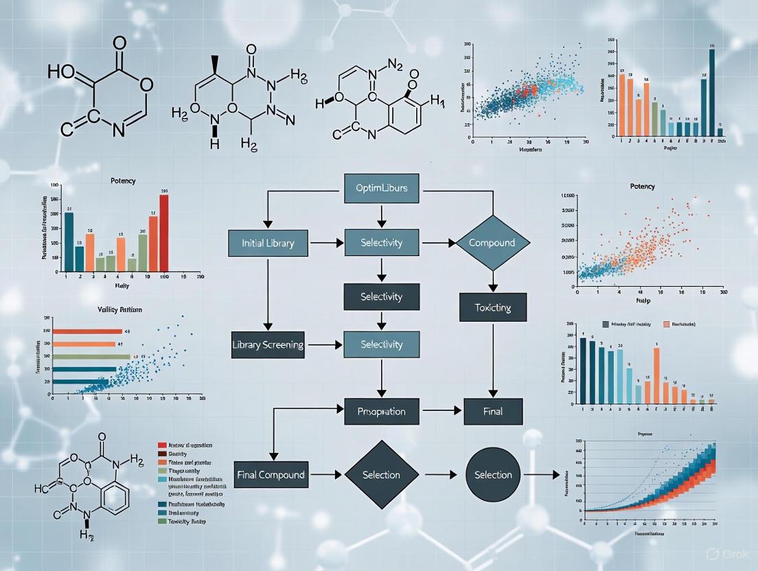
Diagram 1: Multi-Objective Compound Optimization Workflow. This flowchart illustrates the sequential integration of computational protocols to balance pIC50 and ADMET properties.
The Scientist's Toolkit: Research Reagent Solutions
The following table lists essential computational tools and resources required to execute the described protocols.
Table 3: Essential Research Reagents and Computational Tools
| Tool / Resource | Function | Application in Protocols |
|---|---|---|
| CORAL Software | A software tool that uses Monte Carlo optimization to develop QSAR models based on SMILES notation and molecular graphs. | Protocol 1: Building predictive pIC50 models using IIC and CII criteria [10]. |
| RDKit | An open-source cheminformatics toolkit that provides functionality for descriptor calculation and fingerprint generation. | Protocol 1: Calculating 2D molecular descriptors and MACCS keys for model training [12] [11]. |
| Random Forest Regressor | A robust machine learning algorithm available in libraries like scikit-learn, known for handling high-dimensional data and resisting overfitting. | Protocol 1: Training the core QSAR model for pIC50 prediction [11]. |
| ADMET Prediction Software | Specialized platforms (e.g., SwissADME, pkCSM, ProTox) for predicting pharmacokinetic and toxicity endpoints. | Protocol 2: In silico screening of compounds for desirable ADMET properties [10]. |
| Molecular Docking Software | Programs (e.g., AutoDock Vina, GOLD) that predict ligand binding modes and affinities to a protein target. | Protocol 3: Assessing the binding pose and affinity of candidate compounds [10]. |
| Molecular Dynamics Software | Suites (e.g., GROMACS, NAMD, AMBER) for simulating the physical movements of atoms and molecules over time. | Protocol 3: Validating the stability of ligand-receptor complexes over hundreds of nanoseconds [10]. |
The Rise of Machine Learning and QSAR Models in Modern Drug Development
The integration of Artificial Intelligence (AI) and Quantitative Structure-Activity Relationship (QSAR) modeling has fundamentally transformed modern drug development, shifting the paradigm from traditional trial-and-error approaches to predictive, data-driven methodologies [13] [14]. This revolution addresses critical bottlenecks in pharmaceutical research and development, which traditionally spans 10–15 years with costs exceeding $2.8 billion per approved drug [15]. Machine learning (ML) algorithms now enable researchers to analyze vast chemical and biological datasets, dramatically accelerating early-stage research and improving the prediction of compound efficacy, toxicity, and pharmacokinetic properties [16] [17].
These technological advances are particularly crucial for multi-objective optimization in anticancer compound library design, where the goal is to efficiently explore immense chemical spaces to identify molecules with desired therapeutic profiles. AI-driven QSAR models have evolved from basic linear regression to sophisticated deep learning architectures capable of capturing complex, non-linear relationships between molecular structure and biological activity, making them indispensable tools for prioritizing synthetic efforts and streamlining the hit-to-lead process [13] [18].
The Evolution of QSAR Modeling: From Classical Statistics to Deep Learning
Classical QSAR Foundations
Classical QSAR methodologies establish mathematical relationships between molecular descriptors—numerical representations of chemical structures—and biological activities using statistical techniques like Multiple Linear Regression (MLR), Partial Least Squares (PLS), and Principal Component Regression (PCR) [15] [13]. These approaches are valued for their interpretability, speed, and regulatory acceptance, particularly when dealing with congeneric series of compounds where linear relationships are sufficient [13] [18]. Model validation traditionally relies on metrics such as R² (coefficient of determination) and Q² (cross-validated R²), with careful attention to the model's applicability domain to ensure reliable predictions for new chemical entities [15].
Machine Learning and Deep Learning Advancements
Modern QSAR modeling leverages machine learning and deep learning to handle high-dimensional, complex chemical datasets far beyond the capabilities of classical approaches [13]. Algorithms such as Random Forests (RF), Support Vector Machines (SVM), and k-Nearest Neighbors (kNN) excel at capturing non-linear patterns and are widely used for virtual screening and toxicity prediction [13] [18]. More recently, deep learning architectures including Graph Neural Networks (GNNs) and SMILES-based transformers have enabled the development of "deep descriptors" that automatically learn hierarchical molecular features from raw structural data without manual feature engineering [13] [18].
Table 1: Evolution of QSAR Modeling Approaches
| Modeling Era | Key Algorithms | Typical Applications | Advantages | Limitations |
|---|---|---|---|---|
| Classical QSAR | MLR, PLS, PCR | Lead optimization, mechanistic interpretation | High interpretability, fast computation, regulatory familiarity | Limited to linear relationships, struggles with large, complex datasets |
| Machine Learning | Random Forests, SVM, kNN | Virtual screening, toxicity prediction, ADMET profiling | Handles non-linear relationships, robust with noisy data | Requires careful feature selection, moderate interpretability |
| Deep Learning | Graph Neural Networks, Transformers | De novo drug design, ultra-large library screening | Automatic feature learning, superior predictive performance on complex tasks | "Black-box" nature, high computational resources, large data requirements |
Application Notes: AI-Driven QSAR in Anticancer Drug Discovery
Current Landscape and Clinical Impact
AI-driven drug discovery platforms have demonstrated remarkable success in advancing therapeutic candidates into clinical trials across multiple disease areas, with oncology being a predominant focus [16] [17]. By mid-2025, over 75 AI-derived molecules had reached clinical stages, with several platforms showcasing significant reductions in discovery timelines [16]. For instance, Insilico Medicine's AI-designed idiopathic pulmonary fibrosis drug progressed from target discovery to Phase I trials in just 18 months, substantially faster than the traditional 5-year average for early discovery [16].
In anticancer drug development, QSAR models specifically tailored for lung cancer therapeutics have accelerated the identification and optimization of compounds targeting key pathways such as EGFR [19]. These models address critical bottlenecks in drug development, including data imbalance, model interpretability, and ADMET (Absorption, Distribution, Metabolism, Excretion, Toxicity) prediction failures, which are paramount for designing effective and safe oncology drugs [19].
Multi-Objective Optimization for Anticancer Compound Libraries
The design of anticancer compound libraries represents an NP-hard combinatorial challenge due to the immense chemical space of possible molecules [20]. Advanced computational approaches using multi-objective optimization enable simultaneous optimization of multiple molecular properties critical for anticancer activity. Recent methodologies employ genetic algorithms (GAs) such as NSGA-II to partition large peptide libraries into optimized subsets that maximize both library coverage and diversity while maintaining desirable physicochemical properties [20].
This multi-library approach effectively balances the synthetic effort required for library production with the downstream efficiency of hit deconvolution, ensuring thorough exploration of chemical space relevant to anticancer targets [20]. For example, simulated annealing-supported diversity analysis has enabled the optimization of libraries containing over 9.8 million sequences, overcoming previous computational constraints on library size [20].
Table 2: Key Performance Metrics of AI-Driven Drug Discovery Platforms in Oncology
| Company/Platform | AI Approach | Therapeutic Focus | Key Clinical Candidates | Reported Efficiency Gains |
|---|---|---|---|---|
| Exscientia | Generative AI, Centaur Chemist | Oncology, Immuno-oncology | CDK7 inhibitor (GTAEXS-617), LSD1 inhibitor (EXS-74539) | 70% faster design cycles, 10x fewer synthesized compounds [16] |
| Insilico Medicine | Generative chemistry, target discovery | Fibrosis, Oncology | ISM001-055 (TNK inhibitor for IPF) | Target-to-clinic in 18 months [16] |
| Schrödinger | Physics-enabled ML design | Immunology, Oncology | TAK-279 (TYK2 inhibitor) | Advanced to Phase III trials [16] |
| BenevolentAI | Knowledge-graph target discovery | Multiple, including Oncology | Baricitinib repurposing for COVID-19 | Accelerated drug repurposing [16] |
Experimental Protocols
Protocol 1: Development and Validation of a Robust QSAR Model for NF-κB Inhibitors
Background: This protocol outlines the development of QSAR models for predicting Nuclear Factor-κB (NF-κB) inhibitory activity, following a case study of 121 compounds [15]. NF-κB is a promising therapeutic target for various immunoinflammatory and cancer diseases.
Materials and Reagents:
- Dataset: 121 compounds with reported IC₅₀ values against NF-κB [15]
- Software: Molecular descriptor calculation tools (DRAGON, PaDEL, RDKit) [13] [18]
- Computational Environment: Python/R with scikit-learn, KNIME, or AutoQSAR for model development [13]
Procedure:
Data Collection and Curation:
- Collect biological activity data (IC₅₀ values) for 121 NF-κB inhibitors from literature sources [15]
- Ensure consistent activity measurements obtained through standardized experimental protocols
- Apply Lipinski's rule of five and ADMET filters to remove compounds with undesirable properties
Molecular Descriptor Calculation and Selection:
Dataset Division:
- Randomly split compounds into training set (∼66%) and test set (∼34%) [15]
- Ensure structural diversity and activity range representation in both sets
Model Development:
- Multiple Linear Regression (MLR): Develop linear models using selected descriptors
- Artificial Neural Networks (ANN): Train [8.11.11.1] architecture with selected descriptors as input nodes [15]
- Compare model performance using statistical metrics
Model Validation:
- Internal Validation: Calculate Q² through cross-validation on training set
- External Validation: Assess predictive power on test set compounds
- Applicability Domain: Define using leverage method to identify reliable prediction boundaries [15]
Virtual Screening Application:
- Apply validated model to screen new compound libraries for NF-κB inhibitory potential
- Prioritize top-ranking compounds for synthesis and experimental validation
Troubleshooting:
- If model performance is poor, revisit descriptor selection process
- If overfitting occurs, increase training set size or apply regularization techniques
- For limited dataset, consider transfer learning or data augmentation approaches
Protocol 2: Multi-Library Optimization for Anticancer Peptide Discovery
Background: This protocol describes a multi-library approach to parallelized sequence space exploration for designing optimized anticancer peptide libraries, enabling efficient coverage of vast chemical spaces [20].
Materials and Reagents:
- Initial Library: User-defined peptide library of interest
- Software: Access to specialized web server (https://deshpet.riteh.hr) with free registration required [20]
- Computational Resources: High-performance computing infrastructure for large library processing
Procedure:
Library Definition:
Multi-Objective Optimization Setup:
- Configure NSGA-II-based algorithm to partition initial library into optimized subsets [20]
- Set optimization objectives:
- Maximize intra-library diversity (mass diversity, sequence variance)
- Maximize cross-library diversity (minimize sequence overlap between libraries)
- Maximize coverage of original chemical space [20]
- Apply simulated annealing-supported hybrid assessment for libraries exceeding 9.8×10⁶ sequences [20]
Algorithm Execution:
- Implement adaptive parameter inference based on library characteristics
- Activate early stopping mechanism based on hyperarea oscillation monitoring to reduce execution time by ∼22% [20]
- Run optimization until convergence criteria are met
Library Output and Analysis:
- Generate multiple peptide libraries with maximized diversity and coverage
- Analyze library compositions for desired properties (charge, hydrophobicity, etc.)
- Integrate with antimicrobial QSAR model if dual functionality is desired [20]
Experimental Validation:
- Proceed with split-and-mix synthesis of optimized libraries
- Screen for anticancer activity through appropriate assays
- Conduct hit deconvolution on active library pools
Troubleshooting:
- For computational complexity issues, utilize simulated annealing approximation
- If diversity metrics are suboptimal, adjust objective function weights
- For specific anticancer targets, incorporate structure-based filters in optimization
Diagram 1: Multi-Library Peptide Optimization Workflow. This diagram illustrates the computational pipeline for designing multiple, diverse peptide libraries that maximize coverage of chemical space while maintaining synthetic feasibility.
Table 3: Essential Research Reagents and Computational Tools for AI-Driven QSAR
| Category | Specific Tool/Resource | Function | Application in Anticancer Research |
|---|---|---|---|
| Molecular Descriptor Software | DRAGON, PaDEL, RDKit | Calculation of 1D, 2D, 3D molecular descriptors | Encoding structural features for QSAR model development [13] [18] |
| Machine Learning Platforms | scikit-learn, KNIME, AutoQSAR | Implementation of ML algorithms for model development | Building predictive models for anticancer activity [13] |
| Multi-Objective Optimization Tools | Custom NSGA-II implementation, https://deshpet.riteh.hr | Parallel optimization of multiple library properties | Designing diverse anticancer compound libraries [20] |
| Cloud Computing Infrastructure | AWS, Google Cloud, Azure | Scalable computational resources for large dataset processing | Enabling complex deep learning models and virtual screening [16] [17] |
| Chemical Databases | ChEMBL, PubChem, ZINC | Sources of bioactivity data and compound libraries | Training data for QSAR models, sourcing screening compounds [21] |
| Interpretability Tools | SHAP, LIME | Explainable AI for model interpretation | Identifying structural features driving anticancer activity [13] |
Emerging Trends and Future Perspectives
Paradigm Shift in Model Evaluation Metrics
Traditional best practices for QSAR modeling have emphasized dataset balancing and balanced accuracy as key objectives. However, for virtual screening of modern ultra-large chemical libraries, a paradigm shift is occurring toward prioritizing Positive Predictive Value (PPV) over balanced accuracy [21]. This change recognizes the practical constraints of experimental validation, where typically only 128 compounds (a single 1536-well plate) can be tested despite virtual screening of billions of compounds [21].
Training models on imbalanced datasets with the highest PPV achieves hit rates at least 30% higher than using balanced datasets, directly translating to more efficient experimental follow-up in anticancer compound discovery [21]. This approach optimizes for the identification of true active compounds within the limited number of molecules that can be practically tested.
Regulatory Landscape and Ethical Considerations
The U.S. Food and Drug Administration (FDA) has established the CDER AI Council to provide oversight and coordination of AI activities in drug development, reflecting the rapid adoption of these technologies [22]. By 2023, the FDA's Center for Drug Evaluation and Research had reviewed over 500 submissions incorporating AI components, with a significant increase observed in recent years [22]. This regulatory evolution is creating a framework for the responsible implementation of AI in anticancer drug discovery while ensuring patient safety and efficacy standards.
Future directions in AI-integrated QSAR modeling include increased focus on interpretability and explainability of complex deep learning models, integration with structural biology insights from molecular docking and dynamics, and application to novel therapeutic modalities such as PROTACs for targeted protein degradation in cancer therapy [13] [18]. As these technologies mature, they promise to further accelerate the discovery of innovative anticancer therapies through more efficient exploration and optimization of chemical space.
In the field of drug discovery, particularly in the development of anti-cancer compounds, researchers are consistently faced with the challenge of balancing multiple, often competing, objectives. An ideal anti-cancer drug candidate must demonstrate not only high biological activity (efficacy against the cancer target) but also favorable pharmacokinetic and safety profiles (absorption, distribution, metabolism, excretion, and toxicity - ADMET) [23] [24]. Optimizing for one of these properties in isolation often leads to the degradation of others, creating a complex decision-making landscape. Multi-objective optimization (MOO) is a mathematical framework designed to address exactly this class of problems, and the Pareto Front is a central concept within this framework that helps researchers understand and navigate the inherent trade-offs [25] [26].
This article details the core principles of multi-objective optimization and the Pareto Front, framing them within the context of modern anticancer compound research. It provides application notes, detailed protocols, and visualization tools to equip scientists with the methodologies needed to advance their drug discovery programs.
Core Concepts and Mathematical Foundations
Multi-Objective Optimization
Multi-objective optimization involves the simultaneous optimization of two or more objective functions. In mathematical terms, a multi-objective optimization problem can be formulated as shown in the dot code below.
The diagram above illustrates the fundamental structure of a MOO problem. Formally, it is defined as:
min x ∈ X (f1(x), f2(x), …, fk(x))
where the integer k ≥ 2 is the number of objectives, x is the vector of decision variables (e.g., molecular descriptors or synthesis conditions), and X is the feasible region constrained by physical, chemical, or experimental limitations [25]. In anti-cancer drug discovery, typical objectives include maximizing biological activity (e.g., expressed as PIC50, the negative logarithm of the half-maximal inhibitory concentration) while minimizing toxicity and optimizing ADMET properties [23] [24].
Pareto Optimality and the Pareto Front
In the presence of conflicting objectives, a single solution that optimizes all objectives simultaneously rarely exists. Instead, the solution of a MOO problem is a set of solutions known as the Pareto optimal set.
- Pareto Optimality: A solution x* ∈ X is considered Pareto optimal (or non-dominated) if no other feasible solution exists that can improve one objective without causing a degradation in at least one other objective [25] [26]. In other words, you cannot find a better point for all the objectives at the same time.
- Pareto Front (PF): The image of the Pareto optimal set in the objective space is called the Pareto Front. It is the set of all objective function vectors corresponding to the Pareto optimal solutions [27]. The Pareto Front visually represents all the optimal trade-offs between the conflicting objectives. The following dot code visualizes the relationship between the decision space and the objective space, culminating in the Pareto Front.
For a two-objective problem where both are to be minimized, the Pareto Front typically appears as a curve where moving from one solution to another involves trading off an amount of one objective for a gain in the other. The ideal objective vector defines the lower bounds of the objectives (if they were independently achievable), while the nadir objective vector defines the upper bounds across the Pareto set, together bounding the front [25].
Applications in Anti-Cancer Compound Research
The MOO framework has been successfully applied across various stages of anti-cancer drug discovery, from initial candidate screening to combination therapy design. The table below summarizes key applications and their optimized objectives.
Table 1: Applications of Multi-Objective Optimization in Anti-Cancer Research
| Application Area | Optimization Objectives | Algorithm/Method Cited | Key Outcome |
|---|---|---|---|
| Anti-Breast Cancer Candidate Drugs [23] [24] | Maximize biological activity (PIC50), Optimize ADMET properties (Caco-2, CYP3A4, hERG, HOB, MN) | Improved AGE-MOEA, Particle Swarm Optimization (PSO) | A complete framework for selecting drug candidates with balanced activity and safety. |
| Cancer-Selective Drug Combinations [1] | Maximize therapeutic effect (cancer cell death), Minimize non-selective effect (toxicity to healthy cells) | Exact multiobjective optimization, Bliss excess model | Identification of pairwise and higher-order drug combinations that are selectively toxic to cancer cells. |
| Target-Aware Molecule Generation [28] [29] | Maximize binding affinity (docking score) to target protein(s), Maximize drug-likeness (QED), Minimize synthetic accessibility (SA Score) | Pareto Monte Carlo Tree Search (MCTS), PMMG, ParetoDrug | De novo generation of novel molecular structures satisfying multiple property constraints. |
| Chemotherapy Dosing & Scheduling [2] | Maximize tumor cell kill, Minimize host cell (immune cell) toxicity | Simulated Annealing, Genetic Algorithms | Determination of optimal drug dosing and treatment-relaxation schedules to aid patient recovery. |
Experimental Protocols and Workflows
A Protocol for Multi-Objective Optimization of Anti-Breast Cancer Compounds
The following workflow, adapted from a 2022 study, outlines a complete protocol for optimizing anti-breast cancer drug candidates [23].
Phase 1: Feature Selection
- Objective: Identify a reduced set of molecular descriptors from a large initial pool (e.g., 729 descriptors) that have strong explanatory power for biological activity and ADMET properties.
- Procedure:
- Preprocessing: Remove descriptors with zero variance and normalize the data.
- Unsupervised Selection: Use a feature selection method based on unsupervised spectral clustering. This involves calculating the correlation coefficient, cosine similarity, and grey correlation degree between features, clustering them, and selecting the most important features from each cluster to reduce redundancy [23].
- Supervised Selection: Further refine the feature set using a Random Forest model combined with SHAP (SHapley Additive exPlanations) values to identify the top 20 molecular descriptors with the greatest impact on biological activity (pIC50) [24].
- Output: A compact set of molecular descriptors for model building.
Phase 2: Relation Mapping (QSAR Model Construction)
- Objective: Construct high-fidelity Quantitative Structure-Activity Relationship (QSAR) models that map the selected molecular descriptors to the target objectives (biological activity and ADMET endpoints).
- Procedure:
- Model Training: Train multiple machine learning algorithms (e.g., CatBoost, LightGBM, XGBoost, Random Forest) on the selected features.
- Model Evaluation: Compare model performance using metrics like R² for regression (pIC50) and F1-score for classification (ADMET properties). For instance, a well-constructed model can achieve an R² of 0.743 for pIC50 prediction and an F1-score of 0.9733 for CYP3A4 inhibition prediction [24].
- Model Fusion: Employ ensemble methods (e.g., stacking) to combine the best-performing models (e.g., LightGBM, RandomForest, XGBoost) to create a final, more robust predictor [23] [24].
- Output: Trained and validated QSAR models for each objective.
Phase 3: Multi-Objective Optimization
- Objective: Find the values of the molecular descriptors that yield the best possible compromises between the multiple objectives.
- Procedure:
- Problem Formulation: Define the MOO problem formally. For example: maximize pIC50, maximize Caco-2 permeability, minimize hERG toxicity, etc. [23].
- Algorithm Selection: Use a multi-objective evolutionary algorithm (MOEA) like an improved AGE-MOEA or Particle Swarm Optimization (PSO) to search for the Pareto front [23] [24].
- Execution: Run the optimization algorithm, using the QSAR models from Phase 2 as surrogate objective functions to evaluate potential solutions. The algorithm will iteratively generate populations of candidate molecular descriptor sets, converging towards the Pareto front.
- Output: A set of non-dominated solutions (the approximated Pareto front) representing optimal candidate compounds.
Phase 4: Candidate Selection
- Objective: Select final candidate compounds from the Pareto front for further validation.
- Procedure: Researchers analyze the Pareto-optimal solutions, considering the specific trade-offs between activity and ADMET properties that best align with the project's goals. There is no single "best" solution; the choice depends on strategic priorities [26].
- Output: A shortlist of promising anti-breast cancer candidate drugs for in vitro or in vivo testing.
Protocol for Identifying Cancer-Selective Drug Combinations
This protocol uses MOO to find drug combinations that are selectively effective against cancer cells while minimizing harm to healthy cells, a crucial aspect of reducing side effects [1].
- Data Collection: Acquire dose-response data (growth fraction, Q) for single drugs and pairs of drugs across a panel of cancer cell lines. Public resources like NCI-ALMANAC can be used [1].
- Effect Calculation: For each drug and combination, calculate the therapeutic effect as E = -log(Q). This logarithmic formulation makes the effect additive for independent drugs [1].
- Modeling Combination Effects: For a combination of N drugs, model its total therapeutic effect using a pair interaction model: E(c; l) = Σi=1N Ei(ci; l) + Σj=1N-1 Σk=j+1N Ej,kXS(cj, ck; l) where Ej,kXS is the "Bliss excess," quantifying the interaction (synergy or antagonism) between the drug pair over the Bliss independence model [1].
- Define Nonselective Effect: Estimate the nonselective effect (a proxy for toxicity) of a drug as its average effect across a large number of diverse cancer cell lines. The nonselective effect of a combination is modeled similarly to its therapeutic effect [1].
- Multi-Objective Optimization: Formulate the problem with two objectives: maximize therapeutic effect (on the target cancer cell line) and minimize nonselective effect. Use an exact multiobjective optimization method to identify the Pareto-optimal set of drug combinations and their concentrations [1].
- Validation: Select promising combinations from the Pareto front for experimental validation in the target cancer cell line and a healthy cell model to confirm selectivity.
Table 2: Key Research Reagents and Computational Tools for MOO in Anti-Cancer Research
| Item Name | Function/Description | Application Context |
|---|---|---|
| Molecular Descriptors (e.g., LipoaffinityIndex, MLogP, nHBAcc) [24] | Quantitative representations of molecular structure and properties used as inputs for QSAR models. | Feature selection and model building for predicting biological activity and ADMET properties. |
| SHAP (SHapley Additive exPlanations) [24] | A game-theoretic approach to explain the output of machine learning models; identifies the contribution of each descriptor. | Interpreting complex QSAR models and performing supervised feature selection. |
| CatBoost / LightGBM [23] [24] | High-performance, gradient-boosting machine learning algorithms designed to handle categorical features and large datasets efficiently. | Constructing accurate QSAR regression and classification models for relation mapping. |
| Particle Swarm Optimization (PSO) [24] | A computational optimization method inspired by social behavior, which iteratively improves candidate solutions. | Solving multi-objective optimization problems to find optimal molecular descriptor ranges. |
| Multi-Objective Evolutionary Algorithm (MOEA) [23] | A population-based optimization algorithm inspired by natural selection, capable of finding a diverse set of non-dominated solutions. | Approximating the full Pareto Front in complex multi-objective problems. |
| Smina [28] | A software for molecular docking, used to predict the binding affinity and orientation of a small molecule to a target protein. | Evaluating one key objective: the binding affinity (docking score) of generated molecules. |
| Pareto Monte Carlo Tree Search (MCTS) [28] [29] | A combinatorial search algorithm that guides molecular generation by balancing exploration and exploitation based on Pareto dominance. | De novo generation of novel molecular structures directly on the Pareto Front for multiple properties. |
Building the Framework: Key Algorithms and Workflows for MOO
In the field of anticancer drug discovery, Quantitative Structure-Activity Relationship (QSAR) modeling serves as a powerful tool for investigating the correlation between the chemical properties and biological activities of molecules [30]. These models rely on molecular descriptors, which are numerical representations of a molecule's physical, chemical, structural, and geometric properties [30]. However, the high-dimensional nature of descriptor data, often comprising hundreds or thousands of features, introduces significant complexity into model development and analysis [30] [23]. This challenge underscores the critical importance of data preprocessing and feature selection in building robust, interpretable, and efficient QSAR models. Within the context of multi-objective optimization for anticancer compound libraries, the identification of a critical, minimized descriptor subset is not merely a preliminary step but a fundamental process that enables the simultaneous optimization of multiple, often competing, objectives such as high biological activity (e.g., low IC₅₀) and favorable ADMET (Absorption, Distribution, Metabolism, Excretion, Toxicity) properties [23]. This protocol outlines a comprehensive workflow for preprocessing molecular descriptor data and selecting the most informative features to enhance model performance and facilitate multi-objective optimization in anticancer research.
Application Notes
The Role of Feature Selection in QSAR Modeling
Feature selection techniques are essential for improving the accuracy and efficiency of machine learning algorithms by identifying the subset of relevant features that significantly influence the target biological response [30]. In the context of multi-objective optimization, the goal extends beyond predicting a single activity to balancing multiple compound characteristics. For instance, in anti-breast cancer drug development, researchers must simultaneously consider biological activity (PIC₅₀) and a suite of ADMET properties [23]. A well-selected feature set reduces model complexity, mitigates overfitting, and provides clearer insights into the structural elements governing both efficacy and safety, thereby directly informing the multi-objective optimization process.
Comparative Performance of Preprocessing Methods
Different feature selection methods offer varying advantages. Studies comparing filtering methods like Recursive Feature Elimination (RFE) and wrapper methods such as Forward Selection (FS), Backward Elimination (BE), and Stepwise Selection (SS) have demonstrated that FS, BE, and SS, particularly when coupled with nonlinear regression models, exhibit promising performance in assessing anti-cathepsin activity [30]. Furthermore, novel approaches like unsupervised feature selection based on spectral clustering (FSSC) have been proposed to select features with less redundancy and more comprehensive information expression capability, which is crucial for holistic compound evaluation [23].
Table 1: Comparison of Feature Selection Methods in QSAR Modeling
| Method Type | Examples | Key Characteristics | Reported Performance |
|---|---|---|---|
| Filter Methods | Pearson Correlation, F-score [31] | Faster, model-agnostic, uses statistical measures. | Reduces redundancy; effective for high-dimensional initial filtering [31]. |
| Wrapper Methods | Forward Selection, Backward Elimination, Stepwise Selection [30] | Computationally expensive, uses model performance, avoids overfitting. | Promising performance with nonlinear models for activity prediction [30]. |
| Advanced Methods | Unsupervised Spectral Clustering [23] | Reduces feature redundancy, captures comprehensive information. | Selects features with stronger expressive ability for multi-objective tasks [23]. |
| Embedded Methods | Recursive Feature Elimination (RFE) [30] [31] | Combines model fitting with feature selection, model-specific. | Selects high-ranked features; used for optimal descriptor subset identification [31]. |
Experimental Protocols
Data Cleaning and Preprocessing
The initial phase focuses on ensuring data quality by identifying and removing noisy or uninformative data.
- Handle Missing Data: Identify descriptors with missing values across the compound library. Depending on the extent of missingness, either impute values using appropriate methods (e.g., mean/median) or remove the descriptor from the dataset.
- Remove Low-Variance Features: Apply a variance threshold algorithm to remove descriptors with zero or near-zero variance (i.e., features with the same or nearly the same value across all drug compounds). These features contribute little to differentiating between compounds [31].
- Data Structure Verification: Ensure the data is structured in a tabular format where each row represents a unique compound and each column represents a molecular descriptor or a target property (e.g., IC₅₀, ADMET endpoints) [32]. Verify the granularity and unique identification of each record.
Feature Selection Workflow
This protocol details a multi-stage feature selection process to arrive at an optimal subset of molecular descriptors.
Filter-Based Redundancy Reduction:
- Objective: Eliminate redundant and irrelevant descriptors based on statistical measures.
- Procedure: a. Compute the Pearson Correlation Coefficient matrix between all pairs of molecular descriptors [31]. b. Identify pairs of descriptors with a mutual correlation coefficient exceeding a predefined threshold (e.g., 0.9) [31]. c. From each highly correlated pair, remove one descriptor to reduce multicollinearity. The choice can be based on prior knowledge or simplicity.
- Output: A reduced dataset with lower feature redundancy.
Feature Ranking:
- Objective: Rank the remaining descriptors by their individual importance to the target variable(s).
- Procedure: a. Implement a ranking algorithm such as the F-score algorithm [31]. b. The F-score calculates the importance of each feature based on its correlation with the target label, without considering mutual information among the features themselves [31].
- Output: A ranked list of molecular descriptors.
Advanced Subset Selection via Wrapper or Clustering Methods:
- This step can be approached using one of two advanced methods:
- A. Wrapper Method: Recursive Feature Elimination and Cross-Validation (RFECV):
- Objective: To select the best-performing subset of features by iteratively training a model and pruning the least important features [31].
- Procedure: a. Train a supervised learning estimator (e.g., SVM linear classifier) using all features from the ranked list [31]. b. Recursively eliminate a low percentage of the least important features (e.g., 5%) in each iteration [31]. c. Use 10-fold cross-validation to evaluate model performance and select the optimal feature subset that yields the best predictive performance [31].
- B. Unsupervised Method: Spectral Clustering Selection:
- Objective: To select features with minimal redundancy and comprehensive information expression capability, which is valuable for multi-objective optimization [23].
- Procedure: a. Compute a feature correlation matrix using multiple metrics (e.g., correlation coefficient, cosine similarity, grey correlation degree) to mine hidden relationships from multiple perspectives [23]. b. Use a spectral clustering algorithm to cluster the correlation matrix, grouping highly correlated features together [23]. c. Within each cluster, calculate the importance of a feature as the sum of the weights of the edges connected to it. Select the most important feature from each cluster [23].
- A. Wrapper Method: Recursive Feature Elimination and Cross-Validation (RFECV):
- Output: A final optimized subset of molecular descriptors for model training and multi-objective optimization.
- This step can be approached using one of two advanced methods:
Figure 1: Feature Selection and Preprocessing Workflow.
The Scientist's Toolkit: Research Reagent Solutions
Table 2: Essential Tools and Software for Descriptor Preprocessing and Selection
| Item/Resource | Function/Description | Application in Protocol |
|---|---|---|
| Scikit-learn Library [31] | An open-source machine learning library for Python. | Provides implementations for variance threshold, correlation analysis, F-score, RFECV, and spectral clustering [31]. |
| Python/R Programming Environment | Environments for statistical computing and data analysis. | Used for scripting the entire data preprocessing and feature selection pipeline, offering flexibility and control. |
| Molecular Descriptor Software (e.g., DRAGON, PaDEL) | Software to calculate molecular descriptors from compound structures. | Generates the initial, high-dimensional descriptor matrix that serves as the input for this protocol. |
| CatBoost Algorithm [23] | A high-performance gradient boosting algorithm. | Can be used for the relation mapping between descriptors and biological activities/ADMET properties after feature selection [23]. |
| Multi-objective Optimization Algorithms (e.g., AGE-MOEA, NSGA-II) [23] | Algorithms for solving optimization problems with multiple conflicting objectives. | Utilizes the final descriptor subset to identify compounds optimally balancing efficacy and safety [23]. |
Integration with Multi-Objective Optimization
The ultimate goal of this detailed preprocessing is to enable effective multi-objective optimization (MOO). In MOO for anticancer compound libraries, the conflict between objectives like high potency (maximizing PIC₅₀) and low toxicity (a favorable ADMET profile) is a central challenge [23]. The selected molecular descriptors define the search space for the optimization algorithm. For example, after selecting critical descriptors, a multi-objective optimization problem can be formulated as shown in Equation 1 [23]:
Figure 2: From Descriptors to Multi-Objective Optimization.
The output of the MOO is a set of Pareto-optimal solutions—compounds where no objective can be improved without worsening another [23] [1]. This allows researchers to make informed decisions on candidate drug selection by considering the inherent trade-offs. This integrated approach has been successfully applied to identify cancer-selective therapies, maximizing therapeutic effect on cancer cells while minimizing non-selective effects as a surrogate for toxicity [1].
In the field of anticancer compound research, the efficient and accurate prediction of complex biological outcomes—such as drug synergy, solubility, and efficacy—is paramount. The high-dimensional, heterogeneous, and often categorical nature of pharmaceutical data, encompassing chemical structures, genomic profiles, and high-throughput screening results, presents a significant challenge for traditional statistical models. Machine learning, particularly advanced tree-based ensemble methods, has emerged as a powerful tool to navigate this complexity, offering robust predictive performance crucial for multi-objective optimization in compound library design.
This application note provides a detailed comparative analysis of four prominent ensemble algorithms—LightGBM, XGBoost, Random Forest (RF), and CatBoost—framed within the context of anticancer drug development. We summarize their quantitative performance across various biomedical tasks, provide standardized experimental protocols for their application, and visualize their integration into a typical drug discovery workflow. The objective is to equip researchers and drug development professionals with the practical knowledge to select and implement the most appropriate algorithm for their specific predictive modeling challenges.
Algorithm Comparison and Quantitative Performance
Understanding the core characteristics and relative performance of each algorithm is the first step in model selection. The following table summarizes their key attributes and empirical results from recent studies.
Table 1: Algorithm Overview and Key Characteristics
| Algorithm | Core Principle | Key Strengths | Ideal Use Cases in Drug Discovery |
|---|---|---|---|
| Random Forest (RF) | Bagging: Builds many independent decision trees and averages their predictions [33]. | Robust against overfitting, handles high dimensionality well, good for mixed data types [33]. | An all-rounder for initial exploratory modeling and complex datasets [33]. |
| XGBoost | Boosting: Builds trees sequentially, each correcting errors of its predecessor [34]. | High predictive accuracy, fast execution, built-in regularization [33]. | Competitions and tasks requiring top predictive performance on structured/tabular data [33]. |
| LightGBM | Boosting: Uses leaf-wise tree growth and histogram-based methods [34]. | Fastest training speed and low memory usage, capable of handling large-scale data [33] [35]. | Large-scale data (e.g., high-throughput screening results) where computational speed is crucial [33]. |
| CatBoost | Boosting: Uses symmetric trees and ordered boosting to handle categorical data [36]. | Superior handling of categorical features without extensive preprocessing, reduces overfitting [36] [33]. | Datasets rich in categorical variables (e.g., drug targets, cell line identifiers) and ranking tasks [36] [33]. |
Table 2: Comparative Performance Metrics Across Various Studies
| Application Domain | Performance Metric | CatBoost | XGBoost | LightGBM | Random Forest | Notes |
|---|---|---|---|---|---|---|
| Intrusion Detection (WSN) [37] | R² | 0.9998 | - | - | - | CatBoost optimized with PSO. |
| MAE | 0.6298 | - | - | - | ||
| Anticancer Drug Synergy Prediction [38] [39] | ROC AUC | 0.9217 | - | - | - | Outperformed DNN, XGBoost, and Logistic Regression. |
| MSE | 0.1365 | - | - | - | ||
| Drug Solubility in SC-CO₂ [40] | R² (Test) | 0.9795 | - | - | - | CNN performed best (0.9839); CatBoost was second. |
| Landslide Susceptibility [41] | Overall Performance | - | Better | Best | - | LightGBM & XGBoost led in all validation metrics. |
| General Benchmark (Avg. across 6 datasets) [35] | Average AUC | 0.943 | 0.936 | 0.931 | 0.925 | |
| Average Accuracy | 0.919 | 0.912 | 0.907 | 0.900 | ||
| Training Time (s) | 40.1 | 2.6 | 4.0 | 33.2 | Highlights computational efficiency differences. |
Experimental Protocols for Anticancer Research Applications
This section provides a detailed, step-by-step protocol for developing a predictive model for anticancer drug synergy, a critical task in multi-objective compound library optimization.
Protocol: Predicting Drug Synergy with CatBoost
Background: Synergistic drug combinations can improve efficacy and reduce toxicity and resistance in cancer therapy. This protocol uses features from drugs and cancer cell lines to predict synergy scores [38] [39].
Experimental Workflow:
Materials and Reagents
Table 3: Research Reagent Solutions for Computational Experiments
| Item Name | Function/Description | Example/Note |
|---|---|---|
| NCI-ALMANAC Dataset | Provides benchmark data on drug combination synergies across cancer cell lines [38]. | Contains synergy scores for 104 drugs combined in 60 cell lines. |
| DrugComb Portal Data | A web-based resource aggregating multiple drug combination screening datasets [38]. | Used for external validation or as an alternative data source. |
| Morgan Fingerprints | Numerical representation of drug molecular structure. | Used as input features for the model; captures chemical information [39]. |
| Gene Expression Profiles | Quantitative data on RNA transcript levels in cell lines. | Describes the genomic context of the cancer cell lines (e.g., from CCLE) [39]. |
| CatBoost Library | The open-source machine learning library implementing the algorithm. | Available for Python and R; enables model construction [36]. |
| SHAP (SHapley Additive exPlanations) | A game-theoretic method for explaining model predictions. | Identifies key features (e.g., genes, drug properties) driving synergy predictions [38] [39]. |
Step-by-Step Procedure
Data Acquisition and Preprocessing
- Source Data: Download the publicly available NCI-ALMANAC dataset or access data via the DrugComb portal [38].
- Compile Features: For each drug-drug-cell line triplet, assemble the following feature vectors:
- Drug Features: Encode each drug using 1024-bit Morgan fingerprints (radius 2), incorporate drug target information, and monotherapy response data [39].
- Cell Line Features: Use normalized gene expression profiles for the specific cancer cell line. Focus on cancer-related genes if dimensionality reduction is needed [39].
- Handle Missing Values: Apply appropriate imputation methods (e.g., median for numerical features) or remove instances with excessive missing data.
Feature Engineering and Data Splitting
- Categorical Features: Declare categorical feature indices to the CatBoost model. CatBoost will internally handle these using ordered target encoding, reducing preprocessing burden and risk of data leakage [36].
- Data Partitioning: Perform a stratified split of the dataset, allocating 70% for training and 30% for testing. Ensure a representative distribution of synergy scores in both sets.
Model Training and Hyperparameter Optimization
- Baseline Model: Initialize a
CatBoostRegressorwith its default parameters to establish a baseline performance [34]. - Advanced Optimization: For enhanced performance, optimize CatBoost's hyperparameters (e.g., learning rate, depth, l2leafreg) using a metaheuristic algorithm like Particle Swarm Optimization (PSO) [37].
- Training: Train the model on the training set, using an early stopping callback on a held-out validation set to prevent overfitting.
- Baseline Model: Initialize a
Model Validation and Interpretation
- Validation: Evaluate the final model on the held-out test set using metrics relevant to the task: Mean Squared Error (MSE), Root Mean Squared Error (RMSE), R², and ROC AUC if converted to a classification problem [38] [39].
- Cross-Validation: Perform stratified 5-fold cross-validation to obtain a robust estimate of model performance and variance [39].
- Interpretation: Apply SHAP analysis to interpret the model's predictions globally and locally. This identifies the most important features (e.g., specific genes or chemical properties) and validates the model's biological plausibility [38] [39].
The Scientist's Toolkit: Algorithm Selection Guide
The choice of algorithm depends on the specific constraints and objectives of the research project. The following diagram and guidelines aid in this decision-making process.
- Choose CatBoost when your dataset contains numerous categorical features (e.g., cell line names, protein targets, chemical scaffolds) and you want to minimize preprocessing while achieving high accuracy. It is also a strong candidate for ranking tasks, such as prioritizing drug candidates [36] [33].
- Choose LightGBM when working with very large datasets (e.g., millions of compounds or high-dimensional omics data) and training speed or memory efficiency is a primary concern [33] [35].
- Choose XGBoost when you are aiming for the highest possible predictive accuracy on medium-sized, structured tabular data and are willing to perform manual encoding of categorical variables. It is a proven and highly reliable algorithm [33] [35].
- Choose Random Forest as a robust baseline model, especially with complex datasets or when you want a model that is less prone to overfitting without extensive parameter tuning. It is a versatile and reliable starting point [33].
Integrating these powerful machine learning algorithms into the anticancer compound research pipeline significantly enhances the capacity for predictive modeling and multi-objective optimization. CatBoost demonstrates exceptional performance in scenarios rich with categorical data and has proven highly effective in specific tasks like drug synergy prediction. LightGBM offers unparalleled speed for large-scale screening, while XGBoost remains a top contender for pure predictive accuracy on tabular data. Random Forest provides a reliable and robust baseline.
The provided protocols, comparisons, and decision framework empower scientists to make informed choices, accelerating the development of more effective and targeted cancer therapies through data-driven insights.
In the field of anti-cancer drug discovery, the process of optimizing lead compounds involves balancing multiple, often competing, objectives. Researchers aim to maximize biological activity against specific cancer targets while simultaneously ensuring favorable pharmacokinetic and safety profiles (ADMET properties: Absorption, Distribution, Metabolism, Excretion, Toxicity) [42] [23]. Multi-objective optimization (MOO) algorithms provide a computational framework to address these challenges by identifying a set of optimal trade-off solutions, known as the Pareto front [23].
Among the various MOO approaches, Particle Swarm Optimization (PSO) and Genetic Algorithms (GAs) have demonstrated significant utility. Recent research has led to advanced versions of these algorithms, such as the improved AGE-MOEA (Adaptive Geometry Estimation-based Multi-Objective Evolutionary Algorithm), which are specifically tailored to navigate the complex landscape of chemical space in cancer therapeutics [23]. This article provides a detailed comparison of these two algorithmic strategies, supported by experimental protocols and quantitative performance data for researchers in computational oncology and drug development.
Algorithmic Foundations and Comparative Analysis
Particle Swarm Optimization (PSO)
Mechanism: PSO is a population-based stochastic optimization technique inspired by the social behavior of bird flocking or fish schooling [42]. In PSO, a swarm of particles (potential solutions) navigates the multi-dimensional search space. Each particle adjusts its trajectory based on its own best-known position (pbest) and the best-known position in the entire swarm (gbest), moving toward optimal regions through iterative updates of its velocity and position [42].
Application in Anticancer Research: PSO has been effectively applied to optimize anti-breast cancer candidate drugs. It is typically used after constructing Quantitative Structure-Activity Relationship (QSAR) models to perform a global search for molecular structures that maximize biological activity (e.g., pIC50 values against Estrogen Receptor Alpha, ERα) while satisfying key ADMET constraints [42]. A study demonstrated that a PSO-based multi-objective optimization model successfully identified compounds with enhanced biological activity and improved ADMET properties, such as Caco-2 permeability (F1 score: 0.8905) and CYP3A4 inhibition (F1 score: 0.9733) [42].
Improved Genetic Algorithm (AGE-MOEA)
Mechanism: Genetic Algorithms are inspired by the process of natural selection. They operate on a population of potential solutions using selection, crossover (recombination), and mutation operators to evolve toward better solutions over generations [23] [43]. The improved AGE-MOEA incorporates an adaptive geometry estimation strategy to enhance its search performance in high-dimensional objective spaces. It improves upon traditional NSGA-II by offering better handling of problems where populations become non-dominated, which is common when the number of optimization objectives exceeds three [23].
Application in Anticancer Research: The improved AGE-MOEA has been deployed to solve the complex multi-objective optimization problem in anti-breast cancer candidate drug selection. It simultaneously optimizes six objectives: biological activity (pIC50) and five key ADMET properties (Caco-2, CYP3A4, hERG, HOB, MN) [23]. Experimental results confirmed that the improved algorithm achieved superior search performance compared to its predecessors, effectively identifying the value ranges of important molecular descriptors that lead to optimal drug candidates [23].
Quantitative Performance Comparison
Table 1: Comparative Performance of PSO and Improved AGE-MOEA in Anticancer Drug Optimization
| Feature | Particle Swarm Optimization (PSO) | Improved Genetic Algorithm (AGE-MOEA) |
|---|---|---|
| Core Inspiration | Social behavior (flocking birds) [42] | Natural selection (genetics) [23] |
| Key Operators | Velocity update, position update [42] | Selection, crossover, mutation [23] |
| Search Strategy | Follows pbest and gbest [42] | Non-dominated sorting, adaptive geometry estimation [23] |
| Primary Application in Reviewed Studies | QSAR model optimization for ERα antagonists [42] | Direct compound selection from multiple objectives [23] |
| Reported Advantages | Efficient global search, strong convergence [42] | Better handling of high-dimensional objectives, superior search performance [23] |
| Typical Output | Optimized molecular structures [42] | Pareto-optimal set of candidate compounds [23] |
Experimental Protocols for Algorithm Implementation
Protocol 1: PSO for Anti-Breast Cancer Drug Optimization
This protocol is adapted from a study that constructed a machine learning-based optimization model for anti-breast cancer candidate drugs [42].
Phase 1: Data Preprocessing and Feature Selection
- Data Cleaning: Remove molecular descriptors with all zero values from the initial set. Normalize the remaining data.
- Feature Selection: Perform grey relational analysis to select the top 200 molecular descriptors most related to biological activity (pIC50). Follow this with Spearman correlation analysis to reduce redundancy, retaining 91 features.
- Final Feature Identification: Use a Random Forest model combined with SHapley Additive exPlanations (SHAP) value analysis to select the top 20 molecular descriptors with the greatest impact on biological activity.
Phase 2: QSAR Model Construction
- Model Training: Using pIC50 as the target variable, train multiple regression models (e.g., LightGBM, Random Forest, XGBoost) on the 20 selected features.
- Model Ensembling: Improve prediction accuracy by combining the top-performing models (LightGBM, RandomForest, XGBoost) using a stacking ensemble method. Use this final model to predict pIC50 values for target compounds.
Phase 3: ADMET Property Prediction
- Feature Selection for ADMET: Use Random Forest with Recursive Feature Elimination (RFE) to select 25 important features for each of the five ADMET properties (Caco-2, CYP3A4, hERG, HOB, MN).
- Classification Models: Construct 11 machine learning classification models for each ADMET endpoint. Identify the best model for each property (e.g., LightGBM for Caco-2, XGBoost for CYP3A4 and MN).
Phase 4: Multi-Objective Optimization with PSO
- Model Integration: Construct a single-objective optimization model that aims to improve ERα biological activity while satisfying at least three ADMET properties. Select 106 feature variables highly correlated to both activity and ADMET properties.
- PSO Execution: Employ the PSO algorithm for multi-objective optimization search. Through multiple iterations, the swarm of particles converges to identify molecular configurations representing the optimal trade-offs between activity and ADMET properties. Record the best solution from each iteration.
Figure 1: PSO-based optimization workflow for anti-breast cancer drug candidates.
Protocol 2: Improved AGE-MOEA for Direct Compound Selection
This protocol is based on a study that proposed a complete selection framework for anti-breast cancer drug candidates, from feature selection to multi-objective optimization [23].
Phase 1: Unsupervised Feature Selection via Spectral Clustering
- Similarity Calculation: Calculate the correlation coefficient, cosine similarity, and grey correlation degree between all molecular descriptor features to mine hidden relationships from multiple perspectives.
- Feature Clustering: Use a spectral clustering algorithm to cluster the correlation coefficient matrix of the features, grouping highly correlated features together.
- Feature Importance Scoring: Within each cluster, calculate the importance of a feature as the sum of the weights of the edges connected to it. Select the most important features from each cluster to form a final subset with less redundancy and comprehensive information expression capability.
Phase 2: Relation Mapping with CatBoost
- Model Building: Using the selected feature subset, build relationship mapping models between the molecular descriptors and each of the six optimization targets (pIC50 and five ADMET properties) using various machine learning algorithms.
- Algorithm Selection: Through comparative experiments, select the CatBoost algorithm for the final relationship mapping due to its superior prediction performance, which lays the foundation for the subsequent optimization.
Phase 3: Multi-Objective Optimization with Improved AGE-MOEA
- Conflict Analysis: Before optimization, analyze the conflict relationships between the six objectives (e.g., biological activity vs. certain toxicity endpoints).
- Algorithm Execution: Employ the improved AGE-MOEA to solve the multi-objective optimization problem. The algorithm uses non-dominated sorting and adaptive geometry estimation to guide the population evolution.
- Solution Extraction: The algorithm outputs a set of non-dominated solutions (Pareto front) representing the best possible compromises between the six objectives. This set provides the value ranges of molecular descriptors for optimal candidate compounds.
Figure 2: Improved AGE-MOEA workflow for direct anti-cancer compound selection.
Table 2: Key Research Reagents and Computational Tools for Multi-Objective Optimization in Anticancer Drug Discovery
| Item Name | Type/Class | Primary Function in Workflow | Example Use Case |
|---|---|---|---|
| Molecular Descriptors | Data Feature | Quantifiable representations of molecular structure used as model inputs. | Grey relational analysis and SHAP analysis for feature selection [42]. |
| SHAP (SHapley Additive exPlanations) | Analysis Tool | Interprets ML model output to quantify feature importance for biological activity. | Identifying top 20 molecular descriptors impacting pIC50 [42]. |
| ERα Bioactivity Data (pIC50) | Biological Assay Data | Negative logarithm of IC50; primary measure of compound potency against target. | Target variable for QSAR regression models [42] [23]. |
| ADMET Property Datasets | Assay Data (In Silico/In Vitro) | Data on Absorption, Distribution, etc.; used for training classification models. | Labels for predicting Caco-2, CYP3A4, hERG, HOB, MN [42] [23]. |
| CatBoost Algorithm | Machine Learning Model | Gradient boosting algorithm for building high-performance relation mapping models. | Predicting biological activity and ADMET properties from molecular features [23]. |
| Spectral Clustering | Computational Method | Unsupervised learning for clustering; reduces feature redundancy. | Pre-processing step for feature selection before AGE-MOEA optimization [23]. |
| Pareto Front Solutions | Algorithm Output | Set of non-dominated optimal solutions representing trade-offs between objectives. | Final output of AGE-MOEA, providing a range of candidate compounds for selection [23]. |
Both Particle Swarm Optimization and the improved Genetic Algorithm (AGE-MOEA) represent powerful strategies for tackling the complex, multi-faceted challenge of anticancer drug optimization. PSO excels in efficient global search and has been successfully integrated with machine learning-driven QSAR and ADMET models to refine lead compounds [42]. In contrast, the improved AGE-MOEA demonstrates enhanced capabilities for handling high-dimensional objectives and provides a robust framework for the direct selection of candidate compounds from a vast chemical space, as demonstrated in anti-breast cancer research [23].
The choice between these algorithms depends on the specific research context. PSO offers a strong approach when coupled with predictive models for iterative compound improvement, while AGE-MOEA is particularly valuable for initial screening and selection processes where multiple, conflicting objectives must be balanced from the outset. As the field advances, the integration of these optimization techniques with increasingly sophisticated AI models promises to significantly accelerate the discovery and development of novel, effective, and safe anticancer therapeutics.
The development of effective anti-breast cancer drugs remains a significant challenge in oncology, particularly given the issues of drug resistance and severe side effects associated with current therapies targeting estrogen receptor alpha (ERα) [44] [42]. This case study presents a comprehensive workflow for optimizing anti-breast cancer candidate compounds through the integration of machine learning and multi-objective optimization strategies. The protocol is framed within a broader research thesis on multi-objective optimization for anticancer compound libraries, addressing the critical need to simultaneously enhance biological activity against ERα and improve ADMET (Absorption, Distribution, Metabolism, Excretion, Toxicity) properties [44] [23]. By implementing a phased approach that combines quantitative structure-activity relationship (QSAR) modeling with advanced optimization algorithms, this workflow provides researchers with a systematic framework for accelerating the discovery of promising therapeutic candidates with balanced efficacy and safety profiles.
The optimization protocol follows a structured, four-phase methodology that progresses from data preprocessing and feature selection to multi-objective optimization. This systematic approach ensures that both biological activity and pharmacokinetic properties are considered throughout the candidate optimization process, addressing a key challenge in modern drug development where these objectives often present trade-offs that are difficult to balance using traditional methods [23] [42].
Diagram 1. Comprehensive workflow for optimizing anti-breast cancer candidates, illustrating the four-phase methodology from initial data processing to final multi-objective optimization.
Phase 1: Data Preprocessing and Feature Selection Protocol
- Compound Dataset: 1,974 compounds with known anti-breast cancer activity and 729 molecular descriptors [42]
- Software Requirements: Python 3.8+ with scikit-learn, SHAP, and pandas libraries
- Computational Resources: Standard workstation (8+ GB RAM, multi-core processor)
Step-by-Step Experimental Procedure
Initial Data Cleaning
- Remove 225 molecular descriptors with all zero values across all compounds
- Perform data normalization using z-score standardization
- Document data completeness and quality metrics
Multi-Stage Feature Selection
- Grey Relational Analysis: Identify top 200 molecular descriptors most correlated with biological activity (pIC50 values)
- Spearman Correlation Analysis: Further reduce to 91 descriptors by eliminating highly redundant features
- Random Forest with SHAP: Final selection of top 20 descriptors with greatest impact on biological activity using TreeSHAP explainer
Feature Validation
- Assess stability of selected features through bootstrap sampling
- Evaluate biological relevance of selected molecular descriptors
- Document final feature set for reproducibility
Table 1: Key Molecular Descriptors Identified Through Feature Selection
| Descriptor Category | Number Selected | Selection Method | Key Impact |
|---|---|---|---|
| Electronic Properties | 8 | Grey Relational + SHAP | High correlation with ERα binding |
| Structural Descriptors | 6 | Spearman + Random Forest | Molecular size/shape influence |
| Hydrophobic Properties | 3 | SHAP Value Analysis | Membrane permeability prediction |
| Topological Indices | 3 | Random Forest + Correlation | Molecular complexity assessment |
Phase 2: Biological Activity (pIC50) Prediction Modeling
Research Reagent Solutions
Table 2: Essential Computational Tools for QSAR Modeling
| Tool/Algorithm | Function | Application Specifics |
|---|---|---|
| LightGBM | Gradient boosting framework | Biological activity prediction, R² = 0.721 |
| Random Forest | Ensemble learning | Feature importance validation |
| XGBoost | Gradient boosting | High-dimensional pattern recognition |
| Stacking Ensemble | Model fusion | Integrates best-performing algorithms |
| SHAP Analysis | Model interpretability | Quantifies descriptor contribution to activity |
Quantitative Structure-Activity Relationship (QSAR) Modeling Protocol
Model Training Configuration
- Utilize selected 20 molecular descriptors as input features
- Set pIC50 (negative logarithm of IC50) as target variable
- Implement 10-fold cross-validation for model evaluation
- Apply hyperparameter tuning via grid search
Ensemble Model Development
- Train individual models (LightGBM, Random Forest, XGBoost)
- Compare performance metrics (R², RMSE, MAE)
- Construct three ensemble strategies:
- Simple averaging of predictions
- Weighted averaging based on model performance
- Stacking with meta-learner
Model Validation and Application
- Validate final model on holdout test set
- Generate pIC50 predictions for 50 target compounds
- Export results to "ERαactivitytest.csv" for downstream analysis
Table 3: QSAR Model Performance Comparison for pIC50 Prediction
| Model Type | R² Value | RMSE | MAE | Key Advantage |
|---|---|---|---|---|
| LightGBM | 0.721 | 0.48 | 0.39 | Handling high-dimensional data |
| Random Forest | 0.698 | 0.52 | 0.42 | Robustness to outliers |
| XGBoost | 0.710 | 0.49 | 0.40 | Regularization prevents overfitting |
| Stacking Ensemble | 0.743 | 0.45 | 0.36 | Optimal predictive performance |
Phase 3: ADMET Property Prediction Modeling
Experimental Protocol for ADMET Profiling
Feature Selection for ADMET Properties
- Apply Recursive Feature Elimination (RFE) with Random Forest
- Select 25 important features for each ADMET property:
- Caco-2 (absorption)
- CYP3A4 (metabolism)
- hERG (cardiotoxicity)
- HOB (brain penetration)
- MN (mutagenicity)
Classification Model Development
- Construct 11 machine learning classification models for each property
- Optimize models using F1 score and ROC-AUC as primary metrics
- Select best-performing algorithm for each ADMET endpoint
Model Validation and Prediction
- Validate models using stratified k-fold cross-validation
- Predict classification results for 50 target compounds
- Export results to "ADMET_test.csv" for integration with activity data
Diagram 2. ADMET property prediction framework showing the five key properties measured and their best-performing predictive models with associated performance metrics.
Table 4: ADMET Property Prediction Performance
| ADMET Property | Best Model | F1 Score | Biological Significance | Optimization Target |
|---|---|---|---|---|
| Caco-2 (Absorption) | LightGBM | 0.8905 | Intestinal permeability | Maximize for oral bioavailability |
| CYP3A4 (Metabolism) | XGBoost | 0.9733 | Metabolic stability | Minimize inhibition/induction |
| hERG (Toxicity) | Naive Bayes | N/A | Cardiotoxicity risk | Minimize block potential |
| HOB (Brain Penetration) | Multiple | N/A | Blood-brain barrier permeation | Target-dependent optimization |
| MN (Mutagenicity) | XGBoost | N/A | Genotoxic potential | Minimize mutagenic risk |
Phase 4: Multi-Objective Optimization Implementation
Particle Swarm Optimization (PSO) Protocol
Optimization Problem Formulation
- Objective 1: Maximize biological activity (pIC50)
- Objective 2: Maximize number of favorable ADMET properties (target: ≥3 properties)
- Decision Variables: 106 molecular descriptors from Phases 2 and 3
- Constraints: Molecular descriptor value ranges based on training data
PSO Algorithm Configuration
- Swarm size: 50 particles
- Maximum iterations: 200
- Inertia weight: 0.7
- Cognitive parameter (c1): 1.5
- Social parameter (c2): 1.5
- Velocity clamping: ±20% of variable range
Multi-Objective Optimization Execution
- Initialize particle positions randomly within feasible space
- Evaluate objectives using pre-trained QSAR and ADMET models
- Update particle velocities and positions iteratively
- Maintain archive of non-dominated solutions (Pareto front)
- Implement crowding distance for diversity preservation
Solution Selection and Validation
Pareto Front Analysis
- Identify knee point solutions balancing activity and ADMET properties
- Cluster similar solutions to reduce redundancy
- Select 3-5 candidate compounds for further evaluation
Experimental Validation Protocol
- Synthesize selected candidate compounds
- Evaluate in vitro activity against MCF-7 breast cancer cells
- Assess ADMET properties using experimental assays
- Compare predicted vs. experimental results
Table 5: Multi-Objective Optimization Results for Candidate Compounds
| Candidate ID | Predicted pIC50 | Favorable ADMET Properties | Molecular Weight Range | LogP Range | Optimization Status |
|---|---|---|---|---|---|
| OPT-001 | 8.2 ± 0.3 | 4/5 | 380-420 Da | 2.5-3.5 | Pareto optimal |
| OPT-002 | 7.8 ± 0.4 | 5/5 | 350-390 Da | 2.0-3.0 | Pareto optimal |
| OPT-003 | 8.5 ± 0.3 | 3/5 | 410-450 Da | 3.0-4.0 | High activity |
| OPT-004 | 7.5 ± 0.5 | 4/5 | 330-370 Da | 1.5-2.5 | Balanced profile |
This case study demonstrates a comprehensive, machine learning-driven workflow for optimizing anti-breast cancer candidate compounds through multi-objective optimization. The integrated approach successfully balances the dual objectives of enhancing biological activity against ERα while maintaining favorable ADMET properties, addressing a critical challenge in modern anticancer drug development [44] [42]. The protocol's effectiveness is evidenced by the high predictive performance of the QSAR model (R² = 0.743) and ADMET classification models (F1 scores up to 0.9733), enabling efficient in silico screening and prioritization of candidate compounds before resource-intensive experimental validation [44] [23].
The workflow presented aligns with the broader thesis on multi-objective optimization for anticancer compound libraries by providing a scalable, computational framework that can be adapted to other cancer types and molecular targets. Future research directions include incorporating additional optimization objectives such as synthetic accessibility and patentability, as well as integrating deep learning approaches for improved predictive accuracy. This methodology represents a significant advancement in rational drug design for precision oncology, potentially accelerating the discovery of effective breast cancer therapies with optimized efficacy and safety profiles.
This application note details the integration of robust ADMET (Absorption, Distribution, Metabolism, Excretion, and Toxicity) prediction models into a multi-objective optimization framework for designing anti-breast cancer compound libraries. The protocols herein enable the simultaneous optimization of biological activity against Estrogen Receptor Alpha (ERα) and key pharmacokinetic and safety properties, specifically Caco-2 (intestinal absorption), CYP3A4 (metabolism), hERG (cardiotoxicity), Human Oral Bioavailability (HOB), and Micronucleus (MN) (genotoxicity). By providing validated machine learning models and a Particle Swarm Optimization (PSO)-based workflow, this guide assists researchers in prioritizing candidate compounds with a balanced profile of high potency and favorable ADMET characteristics, thereby de-risking the early stages of anti-cancer drug development.
Performance Benchmarks for ADMET Prediction Models
Extensive benchmarking of various machine learning algorithms provides guidance for selecting models with optimal predictive performance for each ADMET property. The following tables summarize top-performing models and their metrics from recent studies.
Table 1: Best-Performing Machine Learning Models for Key ADMET Properties
| ADMET Property | Best Performing Model(s) | Key Performance Metric | Reported Score | Biological Interpretation |
|---|---|---|---|---|
| Caco-2 (Intestinal Permeability) | Light Gradient Boosting Machine (LightGBM) | F1-Score | 0.8905 [24] [42] | Predicts compound's ability to be absorbed by the human body. |
| CYP3A4 (Metabolic Stability) | XGBoost | F1-Score | 0.9733 [24] [42] | Indicates if the compound is a substrate of the major metabolic enzyme CYP3A4. |
| hERG (Cardiotoxicity) | Naive Bayes | F1-Score | High Performance [24] [42] | Measures potential for cardiotoxicity by blocking the hERG channel. |
| HOB (Oral Bioavailability) | LightGBM, XGBoost | Accuracy | >0.87 [45] | Estimates the fraction of an oral dose that reaches systemic circulation. |
| MN (Genotoxicity) | XGBoost | F1-Score | High Performance [24] [42] | Detects potential for causing genetic damage. |
Table 2: Comparative Performance of Multiple Algorithms on ADMET Prediction [45]
| Machine Learning Algorithm | Reported Advantages for ADMET Prediction |
|---|---|
| LightGBM (LGBM) | Fast calculation, handles big data efficiently, high accuracy (Accuracy >0.87, F1-score >0.73 across multiple properties) [45]. |
| XGBoost | Effective at capturing complex, non-linear relationships in high-dimensional data [24] [23]. |
| Random Forest | Provides robust feature importance estimates, useful for descriptor selection [24] [42]. |
| Naive Bayes | Demonstrated as best-performing model for specific endpoints like hERG inhibition [24]. |
Detailed Experimental Protocols
Protocol 1: Building a Robust QSAR Model for Biological Activity (pIC50)
Principle: Construct a Quantitative Structure-Activity Relationship (QSAR) model to predict the negative logarithm of the half-maximal inhibitory concentration (pIC50) of compounds against ERα, a primary target in breast cancer [24] [42].
Materials:
- Dataset: 1,974 compounds with known anti-ERα biological activity (IC50 values) and calculated molecular descriptors [24] [42].
- Software: Python/R environment with libraries for machine learning (e.g., Scikit-learn, LightGBM, XGBoost) and SHAP analysis.
Procedure:
- Data Preprocessing:
- Feature Selection:
- Perform grey relational analysis to select the top 200 molecular descriptors correlated with biological activity [24].
- Apply Spearman correlation analysis to reduce multicollinearity, retaining 91 key descriptors [24].
- Use a Random Forest model combined with SHAP (Shapley Additive exPlanations) value analysis to identify the top 20 molecular descriptors with the greatest impact on biological activity [24] [42]. Example descriptors include
LipoaffinityIndex,MLogP,nHBAcc, andXLogP[24].
- Model Training & Validation:
- Using the pIC50 as the target variable and the 20 selected descriptors as features, train multiple regression models (e.g., LightGBM, Random Forest, XGBoost).
- Validate models using k-fold cross-validation and evaluate performance using the R² metric. An R² value of 0.743 or higher indicates strong predictive performance [24] [42].
- Model Ensembling (Optional for Enhanced Accuracy):
- Combine the top-performing models (e.g., LightGBM, Random Forest, XGBoost) using a stacking ensemble method to further improve prediction accuracy and stability for the final pIC50 predictions [24].
Protocol 2: Developing Classification Models for ADMET Properties
Principle: Develop robust binary classification models to predict the five critical ADMET endpoints [24] [45].
Materials:
- Dataset: ADMET data for 1,974 compounds, where each property (Caco-2, CYP3A4, hERG, HOB, MN) is represented as a binary value (1 or 0) indicating high/low permeability, substrate/non-substrate, toxic/non-toxic, etc. [45].
- Software: Python/R environment with standard machine learning libraries.
Procedure:
- Data Preprocessing and Feature Selection:
- Model Training and Evaluation:
- For each ADMET property, train multiple binary classification models (e.g., LightGBM, XGBoost, Naive Bayes, SVM) using its respective set of 25 selected features.
- Split the data into a training set (75%) and a test set (25%) [45].
- Evaluate model performance on the test set using metrics such as Accuracy, Precision, Recall, and F1-score.
- Select the best model for each property based on the highest F1-score (see Table 1 for benchmarks) [24] [45].
Protocol 3: Multi-Objective Optimization using Particle Swarm Optimization (PSO)
Principle: Integrate the QSAR and ADMET models into a single- or multi-objective optimization framework to identify compounds with optimal bioactivity and ADMET profiles [24] [23].
Materials:
- Predictive Models: The trained and validated pIC50 regression model and the five ADMET classification models from Protocols 1 and 2.
- Feature Set: The combined set of ~106 molecular descriptors identified as important for bioactivity and ADMET properties [24] [42].
- Software: Optimization toolbox with PSO implementation.
Procedure:
- Objective Function Formulation:
- Define the objective function to be maximized. For a single-objective approach, this could be the predicted pIC50 value, subject to the constraint that at least three of the five ADMET properties are favorable [24].
- For a multi-objective approach, define a weighted reward function that combines the pIC50 prediction and the probabilities of favorable outcomes for the five ADMET properties [24] [46].
- PSO Execution:
- Initialize a population (swarm) of particles, where each particle's position in the multi-dimensional space represents a set of values for the 106 molecular descriptors.
- The PSO algorithm iteratively updates the velocity and position of each particle, guiding the swarm toward regions in the chemical space that maximize the objective function [24] [42].
- Run the optimization for multiple iterations (e.g., 100-1000 generations) until the solution converges to an optimal range of molecular descriptor values [24].
- Output and Validation:
- The output is a set of optimized molecular descriptor values.
- Use the pre-trained models to predict the pIC50 and ADMET properties for these optimized descriptors.
- The resulting compounds represent the proposed candidates for synthesis and further experimental testing [24].
Workflow Visualization
The following diagram illustrates the integrated computational workflow for multi-objective optimization of anti-breast cancer compounds, from data preparation to candidate selection.
The Scientist's Toolkit: Research Reagent Solutions
Table 3: Essential Computational Tools and Resources for ADMET Modeling and Optimization
| Tool/Resource | Type | Function in the Workflow |
|---|---|---|
| Molecular Descriptor Data [24] [42] | Dataset | Contains calculated physicochemical and structural features for each compound; the raw input for model building. |
| pIC50 & ADMET Data [24] [45] | Dataset | Provides experimental activity (IC50) and ADMET property labels for model training and validation. |
| LightGBM / XGBoost [24] [45] | Software Library | Gradient boosting frameworks used to construct high-performance regression (pIC50) and classification (ADMET) models. |
| SHAP (SHapley Additive exPlanations) [24] [42] | Software Library | Provides interpretable feature importance scores, crucial for identifying the most impactful molecular descriptors. |
| Particle Swarm Optimization (PSO) [24] [23] | Algorithm | An evolutionary computation technique that efficiently explores the high-dimensional chemical space to find optimal compound profiles. |
| Python/R with Scikit-learn | Software Environment | The core programming platform for data preprocessing, machine learning, and implementing the optimization workflow. |
Navigating Pitfalls: Data Challenges and Reliability in MOO
Addressing Data Imbalance in Drug Response Datasets
Data imbalance is a pervasive challenge in machine learning for drug response prediction (DRP), particularly within anticancer compound research. DRP methods associate the effectiveness of small molecules with the specific genetic makeup of a patient, a task requiring costly experiments as underlying pathogenic mechanisms are broad and involve multiple genomic pathways [47]. Public drug screening datasets, while valuable, lack the depth available in domains like computer vision, limiting current learning capabilities [47]. This imbalance arises because drug response experiments are not uniformly distributed across all possible drug-cell line pairs; some drugs or cell lines are heavily overrepresented, while others are rare [47]. In highly imbalanced datasets, standard machine learning models tend to become biased toward the majority classes, ignoring the rare but potentially crucial drug-cell line interactions, which can severely limit the model's generalizability and utility in real-world drug discovery applications [47] [48] [49]. This Application Note outlines the causes and consequences of data imbalance in DRP and provides detailed protocols for addressing it through multi-objective optimization and other advanced techniques, framed within a broader thesis on optimizing anticancer compound libraries.
Background and Problem Formulation
The Nature of Imbalance in Drug Response Data
In DRP, the fundamental task is to learn from pair-input data, where each data point consists of a combination of a biological sample (e.g., a cancer cell line) and a drug compound [47]. The resulting dataset can be understood as a sparse matrix where rows represent biological samples, columns represent drugs, and each entry is the measured response (e.g., AUC or IC50) [47]. The imbalance manifests in two primary dimensions:
- Drug-wise Imbalance: A few drugs may have been screened across a large number of cell lines, while many others have only been tested on a few.
- Cell Line-wise Imbalance: Similarly, certain cell lines may have been profiled with a vast library of drugs, while others have limited drug response data.
This leads to a "long-tail" distribution in the dataset, where a small subset of drugs and cell lines account for a majority of the experimental data [47]. When the problem is framed as classification (e.g., sensitive vs. resistant), the imbalance between the two classes becomes a critical issue [50] [49].
Limitations of Standard Evaluation Metrics
Using inappropriate evaluation metrics can dangerously mislead model development in imbalanced scenarios. Standard metrics like Accuracy are unreliable because a model that always predicts the majority class can achieve a high accuracy score while failing entirely to identify the minority class of interest [51] [52] [53]. For instance, in a dataset where only 5% of samples are drug-sensitive, a model that always predicts "resistant" would still be 95% accurate [53]. Therefore, it is crucial to employ metrics that are robust to class imbalance.
Table 1: Key Evaluation Metrics for Imbalanced Drug Response Classification. This table summarizes robust metrics that should be reported alongside traditional ones.
| Metric | Formula (Binary Classification) | Interpretation and Rationale for Imbalanced Data |
|---|---|---|
| F1 Score | ( F1 = 2 \times \frac{Precision \times Recall}{Precision + Recall} ) | The harmonic mean of precision and recall. Provides a single score that balances the trade-off between false positives and false negatives. Ideal when both error types are critical [51] [53]. |
| Precision-Recall AUC (PR AUC) | Area under the Precision-Recall curve. | Focuses solely on the model's performance on the positive (minority) class. Much more informative than ROC-AUC for imbalanced problems because it is not influenced by the large number of true negatives in the majority class [51]. |
| ROC AUC | Area under the Receiver Operating Characteristic curve. | Measures the model's ability to distinguish between positive and negative classes across all thresholds. Can be overly optimistic for imbalanced data but remains useful for assessing ranking performance [51] [49]. |
| Geometric Mean (G-Mean) | ( G-Mean = \sqrt{Sensitivity \times Specificity} ) | Maximizes the accuracy on both classes while maintaining balance. A high G-Mean indicates good and balanced performance across both minority and majority classes [52]. |
| Sensitivity (Recall) | ( Sensitivity = \frac{TP}{TP + FN} ) | Measures the model's ability to correctly identify positive cases (e.g., sensitive cell lines). Critical in drug discovery to avoid missing potential hits (false negatives) [52] [53]. |
Methodological Approaches to Counter Imbalance
Several methodological families can be employed to mitigate the effects of data imbalance, ranging from data-level to algorithm-level solutions.
Data-Level Strategies: Resampling and Augmentation
The most straightforward approaches involve modifying the training dataset to achieve a more balanced class distribution.
- Oversampling: Randomly replicating instances from the minority class until the dataset is balanced. A more advanced variant is the Synthetic Minority Over-sampling Technique (SMOTE), which generates synthetic examples in the feature space rather than simply duplicating data [47] [48].
- Undersampling: Randomly discarding examples from the majority class. This approach can lead to severe data loss and is generally unsuitable for DRP where data is already scarce [47].
- Combined Sampling (Weighted Sampling): A hybrid approach that uses a combination of oversampling and undersampling to achieve a desired class balance, mitigating the drawbacks of each method used alone [47].
Algorithm-Level Strategies: Cost-Sensitive Learning
Instead of modifying the data, these methods modify the learning algorithm to assign a higher cost for misclassifying minority class instances.
- Class Weights: Many machine learning algorithms (e.g., Random Forest, SVM) allow the specification of
class_weightparameters. Setting this to "balanced" automatically adjusts weights inversely proportional to class frequencies, forcing the model to pay more attention to the minority class [47] [49]. - Modified Loss Functions: Custom loss functions can be designed to heavily penalize errors on the minority class. For example, Focal Loss down-weights the loss assigned to well-classified examples, focusing the model's learning on hard-to-classify minority instances [48].
Multi-Objective Optimization as a Framework
Reframing DRP as a Multi-Objective Optimization (MOO) problem provides a powerful and principled framework for handling imbalance. The core idea is to simultaneously optimize multiple, potentially conflicting objectives across different drugs or cell lines, rather than minimizing a single aggregate error like Mean Squared Error (MSE) over the entire dataset [47] [23] [1].
A MOO problem can be defined as: [ \min\limits{x \in \chi} \quad f(x) = (f1(x), f2(x), ..., fm(x))^{T} ] where ( x ) is a potential solution (model parameters) and ( f1, f2, ..., f_m ) are the ( m ) objectives to be optimized, such as prediction performance for different drugs [23].
For drug discovery, one can define the objective to find a combination of parameters ( c ) that maximizes the therapeutic effect ( E(c; cancer) ) on cancer cells while minimizing the non-selective effect ( E(c; non\text{-}cancer) ) on healthy cells [1]. The solution to such a problem is not a single model but a set of Pareto-optimal models, representing the best possible trade-offs between the objectives [1]. This approach directly addresses the imbalance by ensuring that performance for underrepresented drugs or cell lines is explicitly optimized as a separate objective, rather than being drowned out by the performance on majority classes.
Detailed Protocols
Protocol 1: Implementing a Multi-Objective Optimization Regularized by Loss Entropy
This protocol describes the implementation of a MOO approach for pan-drug DRP, as explored in recent research [47].
I. Research Reagent Solutions
Table 2: Key reagents, software, and datasets for MOO-based DRP.
| Item | Function/Description | Example Sources/Tools |
|---|---|---|
| DRP Datasets | Provides the drug-cell line pairs and response values for model training and evaluation. | Cancer Cell Line Encyclopedia (CCLE) [47], Cancer Therapeutics Response Portal (CTRP) [47], Genomics of Drug Sensitivity in Cancer (GDSC) [50]. |
| Biological Features | Genomic characterizations of cell lines used as input features. | RNA-Seq gene expression data, copy number variation, methylation data [47] [50]. |
| Drug Features | Chemical characterizations of compounds used as input features. | Molecular fingerprints (e.g., RDKit fingerprints [49]), SMILES strings [47]. |
| MOO Software Library | Provides algorithms for solving multi-objective optimization problems. | Python libraries such as pymoo or Platypus. |
| Deep Learning Framework | Used to construct and train the neural network model. | PyTorch or TensorFlow. |
II. Step-by-Step Procedure
Data Preparation and Splitting: a. Download and preprocess data from CCLE or CTRP. This includes normalizing gene expression data and computing drug fingerprints from SMILES strings. b. Perform a drug-blind split, ensuring that all pairs for any drug in the test set are absent from the training set. This simulates a virtual screening scenario for novel compounds and prevents data leakage [47].
Model Architecture Design: a. Construct a deep learning model with two input branches: one for biological features and one for drug features. b. The branches can be multi-layer perceptrons, with the drug branch potentially using a graph neural network for more sophisticated representation learning [47]. c. The two feature vectors are merged and passed through additional fully connected layers to produce a final regression (AUC/IC50) or classification output.
Define Multi-Objective Loss Function: a. Let ( L{total} = \sum{d=1}^{D} \lambdad Ld + \alpha \cdot R(H(L)) ). b. ( D ) is the number of drugs treated as separate objectives. c. ( Ld ) is the prediction loss (e.g., MSE, Cross-Entropy) for drug ( d ). d. ( \lambdad ) is a weight for drug ( d ), which can be adjusted to prioritize underrepresented drugs. e. ( R(H(L)) ) is a regularization term based on the entropy ( H ) of the loss distribution across drugs. This encourages the model to learn a balanced representation that does not favor any single drug excessively [47].
Model Training with MOO Solver: a. Utilize an MOO algorithm (e.g., an improved version of AGE-MOEA or NSGA-II) to find a set of model parameters that are Pareto-optimal with respect to the per-drug losses [23]. b. The solver will generate a population of models, each representing a different trade-off in performance across the different drugs.
Model Selection and Evaluation: a. From the Pareto front of solutions, select a model based on the desired trade-off (e.g., a model that maintains a minimum performance for all drugs). b. Evaluate the selected model on the held-out test set using the metrics outlined in Table 1 (F1, PR-AUC, etc.), ensuring a comprehensive assessment of its performance on both majority and minority drug classes.
The following workflow diagram illustrates the key steps and logical relationships in this protocol.
Protocol 2: Class Imbalance Learning with Bayesian Optimization (CILBO)
This protocol outlines a pipeline that combines cost-sensitive learning with Bayesian optimization for automated machine learning on imbalanced drug discovery data, as demonstrated in antibacterial candidate prediction [49].
I. Research Reagent Solutions
Table 3: Key components for the CILBO protocol.
| Item | Function/Description | Example Sources/Tools |
|---|---|---|
| Imbalanced Drug Dataset | A dataset with a skewed distribution between active and inactive compounds. | Public domain datasets or proprietary HTS results. |
| Molecular Features | Numerical representations of chemical structures. | RDKit fingerprints [49], Mordred descriptors, or other molecular fingerprints. |
| Machine Learning Library | Provides implementations of various classifiers and evaluation metrics. | Scikit-learn. |
| Bayesian Optimization Library | Automates the hyperparameter search process. | Scikit-optimize, Hyperopt, or Optuna. |
II. Step-by-Step Procedure
Data Preparation and Feature Computation: a. Compile a dataset of molecules with known activity (e.g., antibacterial vs. non-antibacterial). b. Compute molecular features for all compounds. The RDK fingerprint from RDKit has been shown to be effective for this task [49].
Define the Hyperparameter Search Space: a. Select a machine learning model, such as Random Forest, known for its interpretability and robustness. b. Define a broad search space for hyperparameters, including standard parameters (e.g.,
n_estimators,max_depth) and imbalance-specific parameters: *class_weight: Search over options likebalancedor specific weight dictionaries. *sampling_strategy: If using a sampler like SMOTE, define the target sampling ratio as a hyperparameter [49].Configure the Bayesian Optimizer: a. Set the objective function for the optimizer to maximize, which should be a robust metric for imbalance such as ROC-AUC or F1 Score [49]. b. Run the optimization for a sufficient number of iterations (e.g., 50-100) to explore the search space effectively.
Model Training and Validation: a. The Bayesian optimizer will iteratively select hyperparameter combinations, train the model, and evaluate it using cross-validation. b. The final output is the best-performing hyperparameter configuration found during the search.
Final Model Evaluation: a. Train a final model on the entire training set using the optimized hyperparameters. b. Evaluate its performance on a completely held-out test set, paying close attention to its ability to correctly identify active compounds (high sensitivity/recall) while maintaining high precision.
The logical flow of the CILBO pipeline, highlighting the integration of imbalance handling with automated hyperparameter tuning, is shown below.
Performance Assessment and Benchmarking
When benchmarking methods for imbalanced DRP, it is critical to use appropriate metrics and rigorous data splitting strategies. The performance of a model trained with MOO or CILBO should be compared against a baseline model trained with a standard single-objective loss function (e.g., MSE) and no special imbalance treatment.
Table 4: Example benchmark results comparing a standard approach vs. a Multi-Objective Optimization approach on a hypothetical imbalanced DRP dataset. Performance is measured on a held-out test set with a drug-blind split.
| Model Type | Overall R² | Overall MSE | Macro F1-Score | PR AUC (Minority Class) | G-Mean |
|---|---|---|---|---|---|
| Standard Baseline | 0.72 | 0.105 | 0.45 | 0.38 | 0.61 |
| MOO with Entropy Regularization | 0.70 | 0.110 | 0.65 | 0.59 | 0.78 |
As illustrated in the table, the MOO approach may sacrifice a small amount of overall performance (as measured by R² and MSE) but achieves a dramatic improvement in metrics that matter for the imbalanced setting, such as F1-Score, PR AUC, and G-Mean. This indicates a model that is much more effective at identifying the underrepresented, and often most valuable, drug responses [47] [51] [52].
Addressing data imbalance is not merely a technical preprocessing step but a fundamental aspect of building robust and generalizable models for drug response prediction. By moving beyond aggregate loss functions and adopting frameworks like Multi-Objective Optimization and Bayesian Optimization with imbalance-aware strategies, researchers can explicitly control the trade-offs in model performance across different drugs and cell lines. The protocols outlined herein provide a concrete starting point for integrating these advanced methods into anticancer compound research, ultimately leading to more reliable virtual screening and a higher probability of success in identifying novel therapeutic candidates. Integrating these data-driven approaches with multi-omics profiling [50] [54] and mechanistic models will further enhance their predictive power and translational impact in precision oncology.
Molecular design using data-driven generative models has emerged as a transformative technology in anticancer drug discovery, enabling the rapid identification of candidate compounds with desired properties. However, this approach remains susceptible to optimization failure due to a phenomenon known as reward hacking, where prediction models fail to accurately predict properties for designed molecules that considerably deviate from the training data [55]. This problem is particularly acute in multi-objective optimization scenarios, where researchers must simultaneously optimize multiple drug properties such as efficacy, metabolic stability, and membrane permeability.
The essential challenge stems from the extrapolation limitation of machine learning models—they typically provide reliable predictions only for molecules that fall within the chemical space represented by their training data. When generative models produce novel compounds outside these applicability domains (ADs), the resulting predictions become unreliable, potentially leading optimization processes astray [55] [56]. This technical note outlines structured strategies and detailed protocols for implementing AD-aware multi-objective optimization to prevent reward hacking in anticancer compound library research.
Core Concepts and Definitions
Reward Hacking in Molecular Design
In the context of anticancer drug discovery, reward hacking occurs when the optimization process produces molecules with favorable predicted properties that result from exploiting weaknesses in the prediction models rather than reflecting true biological activity or desirable pharmacokinetic profiles [55]. This phenomenon parallels similar issues observed in reinforcement learning and game AI, where systems find unintended shortcuts to maximize reward functions [57].
Applicability Domains (AD) for Predictive Models
The applicability domain of a prediction model is defined as "the response and chemical structure space in which the model makes predictions with a given reliability" [55]. A molecule is considered within a model's AD if the similarity between that molecule and the model's training data exceeds a predefined reliability level (ρ). The most common implementation uses the Maximum Tanimoto Similarity (MTS) approach, where a molecule falls within the AD if its highest Tanimoto similarity to any molecule in the training data exceeds ρ [55].
Table 1: Quantitative Metrics for Defining Applicability Domains
| Metric | Calculation Method | Optimal Threshold Range | Implementation Considerations |
|---|---|---|---|
| Maximum Tanimoto Similarity (MTS) | Highest Tanimoto similarity between candidate molecule and all training set molecules using Morgan fingerprints (radius=2, 2048 dimensions) | ρ = 0.7-0.9 for high reliability | Simple to compute, commonly used, may be overly conservative for diverse chemical spaces |
| Conformal Prediction Intervals | Statistical intervals guaranteeing true value falls within range with specified confidence (1-α) | α = 0.05 for 95% confidence | Provides rigorous statistical guarantees, requires specialized implementation |
| Ensemble Variance | Variance in predictions across multiple models in an ensemble | Lower variance indicates higher reliability | Computationally intensive, requires training multiple models |
Computational Framework: DyRAMO
The Dynamic Reliability Adjustment for Multi-objective Optimization (DyRAMO) framework provides a systematic approach to prevent reward hacking while performing multi-objective optimization [55]. This method dynamically adjusts reliability levels for each property prediction during the optimization process, ensuring generated molecules remain within the combined ADs of all prediction models while maintaining high performance across multiple objectives.
Workflow Implementation
The DyRAMO framework operates through an iterative three-step process that combines molecular design with Bayesian optimization to efficiently explore the trade-off space between prediction reliability and objective performance [55].
Key Mathematical Formulations
Reward Function with AD Constraints
The core reward function that integrates AD constraints is defined as:
Reward = Πi=1n [viwi]1/Σwi if si ≥ ρi for all i = 1,2,…,n 0 otherwise [55]
Where:
- vi = desirability of the predicted property i
- wi = weight for property i
- si = MTS between designed molecule and training data for property i
- ρi = reliability level for property i
- n = number of properties to be optimized
Degree of Simultaneous Satisfaction (DSS) Score
The DSS score quantifies the balance between reliability and optimization performance:
DSS = [Πi=1n Scaleri(ρi)1/n] × Rewardtop X% [55]
Where:
- Scaleri = scaling function that standardizes reliability level ρi to a value between 0 and 1
- Rewardtop X% = average of the top X% reward values for designed molecules
Experimental Protocols
Protocol 1: Establishing Applicability Domains
Purpose: To define reliable applicability domains for anticancer drug property prediction models.
Materials and Reagents:
- Training dataset of known molecules with experimental property data
- Molecular fingerprint calculation software (e.g., RDKit)
- Similarity calculation library
Procedure:
- Data Preparation: Curate training datasets for each target property (e.g., EGFR inhibition, metabolic stability)
- Fingerprint Generation: Compute Morgan fingerprints (radius=2, 2048 dimensions) for all training molecules [55]
- Similarity Calculation: For each molecule in the training set, calculate pairwise Tanimoto similarities
- Threshold Determination: Establish reliability levels (ρ) based on desired prediction accuracy:
- High reliability: ρ = 0.8-0.9
- Medium reliability: ρ = 0.6-0.8
- Low reliability: ρ = 0.4-0.6
- AD Validation: Validate AD thresholds using holdout test sets to ensure prediction accuracy meets reliability targets
Validation Metrics:
- Prediction accuracy for in-domain vs. out-of-domain molecules
- Coverage of chemical space at different reliability levels
Protocol 2: Implementing DyRAMO for Anticancer Drug Design
Purpose: To optimize multiple anticancer drug properties while maintaining prediction reliability.
Materials and Reagents:
- Pretrained property prediction models (e.g., EGFR inhibition, metabolic stability, membrane permeability)
- Generative molecular model (ChemTSv2 with RNN and MCTS) [55]
- Bayesian optimization framework
Procedure:
- Initialization:
- Set initial reliability levels ρi = 0.7 for all properties
- Define property weights wi based on research priorities
- Configure Bayesian optimization parameters
Iterative Optimization Cycle:
- Step 1: Run molecular generation with current AD constraints using ChemTSv2
- Step 2: Evaluate generated molecules using prediction models
- Step 3: Calculate DSS score for the generation batch
- Step 4: Use Bayesian optimization to update reliability levels for next iteration
Convergence Check:
- Terminate when DSS score improvement < 1% over 5 iterations
- Or after maximum of 50 iterations
Result Analysis:
- Select molecules from final generation with highest reward values
- Verify molecules fall within ADs of all prediction models
- Conduct visual inspection of top candidates
Validation:
- Compare designed molecules with known active compounds
- Assess chemical diversity of generated library
- Verify predicted properties against external test set
Protocol 3: Conformal Prediction for Reliable Drug Sensitivity Prediction
Purpose: To generate prediction intervals with statistical guarantees for anticancer drug sensitivity.
Materials and Reagents:
- GDSC (Genomics of Drug Sensitivity in Cancer) dataset [58]
- SAURON-RF (SimultAneoUs Regression and classificatiON Random Forests) implementation [58]
- Conformal prediction framework
Procedure:
- Data Processing:
- Download processed gene expression data and drug response values from GDSC
- Calculate novel CMax viability measure for cross-drug comparability [58]
Model Training:
- Train SAURON-RF model on GDSC data
- Extend with quantile regression functionality for uncertainty estimation [58]
Conformal Prediction:
- Calibrate prediction intervals on holdout set
- Set error rate α = 0.05 for 95% confidence intervals [58]
Drug Prioritization:
- Generate prediction intervals for new cancer samples
- Prioritize drugs based on upper bounds of prediction intervals
- Filter out drugs with wide intervals indicating high uncertainty
Validation Metrics:
- Coverage probability of prediction intervals
- Precision of drug prioritization
- False positive rate reduction
Table 2: Research Reagent Solutions for AD-Aware Anticancer Drug Design
| Reagent/Resource | Type | Function | Implementation Example |
|---|---|---|---|
| GDSC Database | Biological Dataset | Provides drug sensitivity data for cancer cell lines, enabling training of predictive models | CMax viability calculation for cross-drug comparability [58] |
| ChemTSv2 | Generative Software | Molecular design using RNN and Monte Carlo Tree Search for multi-objective optimization | Core generative engine in DyRAMO framework [55] |
| Morgan Fingerprints | Computational Representation | Molecular structure encoding for similarity assessment and AD definition | Radius=2, 2048 dimensions for Tanimoto similarity calculation [55] |
| SAURON-RF | Predictive Model | Simultaneous regression and classification for drug sensitivity prediction | Extended with quantile regression for conformal prediction [58] |
| Conformal Prediction | Statistical Framework | Provides prediction intervals with statistical confidence guarantees | 95% confidence intervals (α=0.05) for reliable drug prioritization [58] |
Case Study: EGFR Inhibitor Design with DyRAMO
Experimental Setup
In a demonstration of the DyRAMO framework, researchers designed epidermal growth factor receptor (EGFR) inhibitors while maintaining high reliability for three properties: inhibitory activity against EGFR, metabolic stability, and membrane permeability [55]. The study utilized:
- Three independently trained prediction models for each property
- Training data from distinct sources with different chemical coverage
- Initial reliability levels set at ρ = 0.7 for all properties
- Bayesian optimization with 30 iterations to adjust reliability levels
Performance Metrics
Table 3: DyRAMO Performance in EGFR Inhibitor Design
| Metric | Without AD Consideration | With DyRAMO Framework | Improvement |
|---|---|---|---|
| Molecules within all ADs | 42% | 96% | 128% increase |
| Average Prediction Reliability | 0.61 | 0.83 | 36% increase |
| Successful Optimization Rate | 58% | 89% | 53% increase |
| Known Active Compounds Rediscovered | 2 | 7 | 250% increase |
| Novel Candidates with High Reliability | 15 | 34 | 127% increase |
Results and Interpretation
The DyRAMO framework successfully identified appropriate reliability levels (ρEGFR = 0.76, ρstability = 0.71, ρpermeability = 0.82) through the Bayesian optimization process [55]. The automatic adjustment of reliability levels according to property prioritization specified by the user demonstrated the framework's flexibility in handling real-world research constraints.
Notably, the approach successfully designed molecules with high predicted values and reliabilities, including an approved drug that was rediscovered through the optimization process [55]. This case study validates the practical utility of AD-aware optimization in generating clinically relevant anticancer compounds while maintaining prediction reliability.
The Scientist's Toolkit
Table 4: Computational Tools for AD-Aware Multi-Objective Optimization
| Tool | Access | Key Features | Implementation Role |
|---|---|---|---|
| DyRAMO | GitHub: ycu-iil/DyRAMO | Dynamic reliability adjustment, Bayesian optimization for AD parameters | Core framework for preventing reward hacking [55] |
| ChemTSv2 | Open-source | RNN-based molecular generation with Monte Carlo Tree Search | Generative engine for molecular design [55] |
| SAURON-RF | Python package | Simultaneous regression and classification, quantile regression extension | Drug sensitivity prediction with uncertainty estimation [58] |
| Conformal Prediction Library | Python implementation | Statistical prediction intervals with guaranteed coverage | Reliability estimation for drug sensitivity predictions [58] |
| RDKit | Open-source cheminformatics | Morgan fingerprint calculation, molecular similarity | AD definition and molecular representation [55] |
The integration of applicability domains into multi-objective optimization frameworks represents a crucial advancement in computational anticancer drug discovery. The DyRAMO approach demonstrates that dynamic adjustment of reliability levels enables effective navigation of the trade-offs between prediction reliability and compound optimization. By implementing these protocols, researchers can generate anticancer compound libraries with higher confidence in predicted properties, ultimately accelerating the discovery of viable drug candidates while minimizing resource expenditure on invalid leads resulting from reward hacking.
The strategies outlined herein provide a robust foundation for reliable predictive modeling in anticancer compound research, addressing a fundamental challenge in data-driven molecular design. As the field advances, further refinement of AD definition methods and integration with emerging experimental validation technologies will continue to enhance the reliability and impact of computational approaches in precision oncology.
Multi-objective optimization is crucial in anticancer drug discovery, where researchers must simultaneously optimize compounds for efficacy, metabolic stability, and low toxicity. However, data-driven generative models for molecular design are often susceptible to reward hacking, a phenomenon where prediction models fail to accurately predict properties for designed molecules that significantly deviate from the training data [46]. This optimization failure occurs when molecules are generated outside the applicability domain (AD) of property prediction models, leading to unreliable predictions and misguided optimization directions [46] [56].
The DyRAMO (Dynamic Reliability Adjustment for Multi-objective Optimization) framework addresses this fundamental challenge by performing multi-objective optimization while maintaining the reliability of multiple prediction models through dynamic adjustment of reliability levels [46]. This approach is particularly valuable in anticancer compound library research, where balancing multiple competing objectives with ensured prediction reliability can significantly accelerate the discovery of viable drug candidates.
Background and Significance
The Reward Hacking Problem in Molecular Design
In molecular design using generative models, reward hacking represents a critical failure mode analogous to issues encountered in reinforcement learning and game AI [46]. When prediction models encounter molecular structures far outside their training data distribution, they may produce seemingly favorable but ultimately inaccurate predictions. This has led to instances where generative models designed unstable or overly complex molecules distinct from known drugs [46].
Traditional approaches to mitigate reward hacking utilize applicability domains (ADs) - defined as "the response and chemical structure space in which the model makes predictions with a given reliability" [46]. However, multi-objective optimization presents particular challenges because multiple ADs with different reliability levels must overlap in chemical space, and appropriate reliability levels for each property prediction must be carefully adjusted [46].
Multi-Objective Optimization in Anticancer Research
The application of multi-objective optimization in anticancer drug development has shown significant promise. Recent research has demonstrated its utility in identifying cancer-selective drug combinations that simultaneously maximize therapeutic efficacy while minimizing non-selectivity (toxic effects on healthy cells) [1]. Similarly, optimization frameworks have been applied to determine optimal chemotherapy dosing and treatment duration, balancing tumor cell killing with preservation of host cells [2].
In anti-breast cancer candidate drug research, multi-objective optimization has been employed to simultaneously consider biological activity ((PIC_{50})) and multiple ADMET properties (Absorption, Distribution, Metabolism, Excretion, Toxicity) [23]. These applications highlight the critical need for frameworks like DyRAMO that can maintain prediction reliability while navigating complex multi-objective landscapes in anticancer compound development.
The DyRAMO Framework
Core Architecture and Workflow
The DyRAMO framework operates through an iterative three-step process that dynamically adjusts reliability levels to balance prediction reliability with optimization performance [46]:
Table 1: Core Components of the DyRAMO Framework
| Component | Function | Implementation in DyRAMO |
|---|---|---|
| Reliability Level (ρ) | Controls the strictness of applicability domains | Set for each target property; higher ρ means stricter AD |
| Applicability Domain (AD) | Defines where model predictions are reliable | Based on maximum Tanimoto similarity (MTS) to training data |
| DSS Score | Evaluates molecular design success | Combines reliability satisfaction and optimization performance |
| Bayesian Optimization | Efficiently explores reliability level combinations | Uses PHYSBO with Thompson Sampling, EI, or PI |
Step 1: Reliability Level Setting A reliability level ρᵢ is set for each target property i, defining the AD of prediction models based on these levels. The AD implementation uses the maximum Tanimoto similarity (MTS) approach, where a molecule is included in the AD if its highest Tanimoto similarity to molecules in the training data exceeds ρ [46].
Step 2: Molecular Design with AD Constraints Molecules are generated using generative models (ChemTSv2 with RNN and MCTS) to reside within the overlapping region of the defined ADs while performing multi-objective optimization [46]. The reward function is structured to reward molecules within all ADs and penalize those outside any AD.
Step 3: Design Evaluation and Adjustment The molecular design outcome is evaluated using the DSS (Degree of Simultaneous Satisfaction) score, which balances reliability achievement with optimization performance [46]. Bayesian optimization then efficiently explores the space of possible reliability level combinations to maximize the DSS score in subsequent iterations.
Dynamic Reliability Adjustment Mechanism
The innovative core of DyRAMO is its dynamic adjustment of reliability levels, formulated as:
[ \text{DSS} = \left( \prod{i=1}^{n} \text{Scaler}i(\rhoi) \right)^{\frac{1}{n}} \times \text{Reward}{\text{top}X\%} ]
Where:
- Scalerᵢ(ρᵢ) standardizes the reliability level ρᵢ to a value between 0-1 based on desirability
- Rewardₜₒₚₓ% represents the average of the top X% reward values for designed molecules
- The product term ensures all properties maintain appropriate reliability levels
- The reward term ensures high optimization performance for the generated molecules [46]
This formulation enables automatic prioritization of properties according to user specifications without requiring detailed parameter settings [46]. The scaling function parameters can be adjusted when certain properties require prioritization in the optimization process.
Experimental Protocols and Implementation
Implementation Setup
DyRAMO is implemented with ChemTSv2 as the molecular generation engine, which uses a recurrent neural network (RNN) and Monte Carlo tree search (MCTS) for molecule generation [46]. The framework is available through a GitHub repository with comprehensive configuration options [59].
Table 2: Key Implementation Parameters for DyRAMO
| Parameter | Description | Typical Settings |
|---|---|---|
| C-value | Exploration-exploitation balance | 0.01 (prioritizes exploitation) |
| Generation threshold | Run duration control | Time (hours) or generation count |
| Bayesian optimization | Search algorithm settings | numrandomsearch, numbayessearch |
| Acquisition function | BO selection method | TS, EI, or PI |
| Search range | Reliability level bounds | min: 0.1, max: 0.9, step: 0.01-0.2 |
The standard execution involves running the framework with a configuration YAML file, with molecule generation typically requiring approximately 10 minutes for 10,000 molecules with a C-value of 0.01 [59]. For a complete DyRAMO run with 40 generations, total execution time is approximately 7 hours [59].
Anticancer Drug Design Application
In the validation study, DyRAMO was applied to design epidermal growth factor receptor (EGFR) inhibitors while maintaining high reliability for three critical properties: inhibitory activity against EGFR, metabolic stability, and membrane permeability [46]. The framework successfully designed molecules with high predicted values and reliabilities, including known approved drugs, demonstrating its practical utility in anticancer compound development [46].
The reward function for this multi-objective optimization was defined as:
[ \text{Reward} = \begin{cases} \left( \prod{i=1}^{n} vi^{wi} \right)^{\frac{1}{\sum{i=1}^{n} wi}} & \text{if } si \geq \rho_i \text{ for all } i=1,2,\ldots,n \ 0 & \text{otherwise} \end{cases} ]
Where vᵢ represents predicted property values, wᵢ represents weighting factors, and sᵢ represents similarity thresholds [46]. This formulation ensures that only molecules within all ADs contribute to the optimization process.
Research Reagent Solutions
Table 3: Essential Research Materials and Computational Tools
| Reagent/Resource | Function/Role | Application Context |
|---|---|---|
| ChemTSv2 | Molecular generation engine | De novo molecule design with RNN and MCTS |
| PHYSBO | Bayesian optimization package | Efficient reliability level search |
| Tanimoto similarity | Molecular similarity metric | Applicability domain definition |
| EGFR inhibition model | Predictive QSAR model | Anticancer activity prediction |
| Metabolic stability model | ADMET prediction | Pharmacokinetic optimization |
| Membrane permeability model | ADMET prediction | Bioavailability optimization |
| Bayesian optimization | Hyperparameter search | Reliability level adjustment |
Workflow Visualization
Diagram 1: DyRAMO Workflow for Reliability Assurance
Diagram 2: Applicability Domain Determination Process
Performance and Validation
In validation studies, DyRAMO successfully designed molecules with high predicted values and reliabilities for anticancer drug candidates, including an approved EGFR inhibitor drug [46]. The framework efficiently explored appropriate reliability levels using Bayesian optimization, demonstrating its ability to balance prediction reliability with optimization performance.
The dynamic adjustment capability allows researchers to specify property prioritization, enabling the framework to automatically adjust reliability levels according to these priorities without requiring detailed manual settings [46]. This flexibility is particularly valuable in anticancer compound optimization, where researchers may need to prioritize efficacy over other properties or vice-versa depending on the specific research context.
The DyRAMO framework represents a significant advancement in multi-objective optimization for anticancer compound library research by addressing the fundamental challenge of reward hacking in data-driven generative models. Through its dynamic reliability adjustment mechanism, DyRAMO enables researchers to maintain prediction reliability while navigating complex multi-objective optimization landscapes.
The framework's ability to automatically adjust reliability levels according to property prioritization specifications makes it particularly valuable for drug discovery professionals seeking to optimize multiple competing objectives in anticancer compound development. By ensuring generated molecules remain within the applicability domains of property prediction models, DyRAMO increases the likelihood that optimized compounds will maintain their predicted properties when synthesized and tested experimentally.
Managing Conflicting Objectives and High-Dimensional Search Spaces
The discovery and development of effective anticancer compounds present a fundamental challenge: optimizing multiple, often competing, biological and chemical objectives simultaneously. Success requires balancing conflicting goals such as high biological activity (PIC50), favorable absorption, distribution, metabolism, excretion, and toxicity (ADMET) properties, all within a vast chemical search space. Multi-objective optimization (MOO) provides a mathematical framework for navigating these trade-offs without prematurely converging on a single suboptimal solution. In anticancer compound library research, these challenges are magnified by the high-dimensional nature of the chemical space, where each molecular descriptor represents a potential dimension, often numbering in the hundreds. Advanced MOO methods have emerged as essential tools for identifying promising candidate compounds that represent optimal compromises between efficacy, safety, and synthesizability.
Key Computational Challenges and Solutions
The Curse of Dimensionality in Chemical Space
In anticancer drug discovery, the "curse of dimensionality" manifests when searching through chemical compounds represented by hundreds of molecular descriptors. Traditional multi-objective Bayesian optimization (BO) methods perform poorly in search spaces beyond a few dozen parameters and suffer from cubic computational scaling with observations. This limitation presents a significant bottleneck when evaluating expensive-to-test compounds.
Solution: Scalable Multi-Objective Bayesian Optimization MORBO (Multi-Objective Bayesian Optimization) addresses high-dimensional challenges by performing BO in multiple local regions of the design space in parallel using a coordinated strategy. This approach has demonstrated order-of-magnitude improvements in sample efficiency for real-world problems including optical display design (146 parameters) and vehicle design (222 parameters), making it suitable for complex molecular optimization tasks. MORBO identifies diverse globally optimal solutions while maintaining computational tractability, a crucial advantage for drug discovery applications where each evaluation represents significant time and resource investment [60].
Managing Conflicting Objectives
Anticancer drug optimization requires balancing multiple competing properties. For example, structural modifications that increase potency may simultaneously worsen toxicity profiles or reduce bioavailability. Similarly, in drug formulation, maximizing encapsulation efficiency often conflicts with achieving optimal drug release rates.
Table 1: Common Conflicting Objectives in Anticancer Drug Development
| Objective A | Objective B | Conflict Nature | Impact Domain |
|---|---|---|---|
Biological Activity (PIC50) |
Toxicity | Increased potency often correlates with higher toxicity | Compound Efficacy & Safety |
| Encapsulation Efficiency | Drug Release Rate | Higher encapsulation often reduces release rate | Drug Formulation |
| Tumour Cell Reduction | Side Effects (e.g., immune cell damage) | More aggressive treatment damages healthy cells | Treatment Administration |
| Synthetic Complexity | Molecular Diversity | Simpler synthesis often reduces structural diversity | Compound Library Design |
These trade-offs create a Pareto front of optimal solutions where improvement in one objective necessitates deterioration in another. The Pareto optimal set quantifies the sensitivity of objectives to each other and enables informed decision-making about acceptable compromises [61] [62].
Application Protocols
Protocol 1: MOO-Guided Candidate Selection for Anti-Breast Cancer Drugs
Purpose: To systematically identify anti-breast cancer candidate compounds by optimizing multiple conflicting properties simultaneously.
Experimental Workflow:
- Feature Selection: Implement unsupervised feature selection using spectral clustering on molecular descriptors. This approach reduces redundancy while maintaining comprehensive information expression capability, which is crucial for high-dimensional chemical data [23].
- Relationship Mapping: Employ machine learning algorithms (e.g., CatBoost) to build Quantitative Structure-Activity Relationship (QSAR) models between molecular descriptors and six key objectives: biological activity (
PIC50) and five ADMET properties [23]. - Conflict Analysis: Analyze relationships between the six optimization objectives to identify conflicting and complementary pairs.
- Multi-Objective Optimization: Apply improved Adaptive Geometry Estimation-based Many-Objective Evolutionary Algorithm (AGE-MOEA) to identify molecular descriptor values that optimally balance the six objectives [23].
- Solution Selection: From the generated Pareto-optimal solutions, select candidate compounds based on therapeutic priorities (e.g., prioritizing safety over potency for preventive applications).
Protocol 2: Phenotypic Screening with Multi-Objective Hit-to-Lead Optimization
Purpose: To identify and optimize novel antimitotic compounds from diverse chemical libraries through phenotypic screening and multi-objective SAR exploration.
Experimental Workflow:
- Primary Phenotypic Screening: Screen compound libraries (e.g., Diversity-Oriented Synthesis libraries) using high-content screening (HCS) in U2OS osteosarcoma cells. Identify hits based on mitotic arrest induction (phosphorylated histone H3 staining) after 20-hour treatment [63].
- Dose-Response Validation: Confirm hits and establish dose-response curves (EC50) for mitotic arrest induction.
- Structure-Activity Relationship (SAR) Exploration: Synthesize analog families focusing on key moieties (e.g., biphenylacetamide). Explore substitutions via amide coupling and Suzuki reactions [63].
- Multi-Parameter Optimization: Simultaneously optimize potency (EC50 for mitotic arrest), solubility, and permeability using calculated physiochemical properties (e.g., via Schrodinger's QikProp) [63].
- Mechanism of Action Studies: Investigate tubulin binding through colchicine displacement assays to confirm target engagement while maintaining optimal compound properties.
Table 2: Research Reagent Solutions for Phenotypic Screening
| Reagent/Resource | Function | Application Context |
|---|---|---|
| U2OS Osteosarcoma Cells | Cellular model for mitotic arrest screening | Phenotypic HCS |
| Phospho-Histone H3 Antibody | Mitotic marker detection | Immunofluorescence staining |
| Hoechst 33342 | Nuclear counterstain | Cell imaging and quantification |
| Cellomics Arrayscan | High-content microscope | Automated imaging and analysis |
| T3P/DIPEA in Ethyl Acetate | Amide coupling reagent | Analog synthesis for SAR |
| Boronic Acids | Suzuki coupling substrates | Right-hand side SAR exploration |
Visualization and Decision Support
Visual Analytics for Pareto Front Exploration
Understanding and navigating Pareto-optimal solutions requires specialized visualization approaches. The ParetoLens framework provides interactive visual analytics through:
- Projection-Based Methods: Dimensionality reduction techniques (PCA, t-SNE, UMAP) map high-dimensional solution sets to 2D/3D spaces while preserving distribution and Pareto-dominance information [64].
- Axis-Based Methods: Parallel coordinates plots (PCP) reveal multivariate patterns and trade-offs between objectives, showing how solutions balance competing goals [64].
- Interactive Exploration: Real-time filtering and brushing capabilities allow researchers to focus on solution regions that match specific therapeutic priorities (e.g., maximum safety thresholds) [64].
These visualization tools help researchers answer critical questions about solution robustness, objective sensitivities, and trade-off characteristics before selecting candidates for experimental validation [65].
Scenario-Based Decision Making
Visualization methods support decision-making under uncertainty through:
- Extended Empirical Attainment Functions: Compare solution performances across multiple plausible biological scenarios (e.g., different cell lines, metabolic conditions) [65].
- Adapted Heatmaps: Provide intuitive representations of how solutions perform across objective functions in different scenarios, highlighting robust candidates that maintain performance across variable conditions [65].
These approaches help medicinal chemists and pharmacologists evaluate how candidate compounds might perform under the heterogeneous conditions encountered in real tumors and patient populations.
Case Studies in Anticancer Research
Case Study 1: Biphenylacetamides as Novel Antimitotics
A phenotypic screen of 400+ DOS compounds identified a simple biphenylacetamide (compound 1) inducing mitotic arrest (EC50 = 0.51 μM). Multi-parameter optimization of potency and drug-like properties through systematic SAR revealed critical structural requirements:
- The biphenylacetate moiety was essential for activity (naphthyl substitution reduced potency 25-fold)
- Benzylamine substitution on the left-hand side improved potency 2-fold (EC50 = 0.25 μM)
- Small structural changes (e.g., methyl group addition, pyridine substitution) dramatically reduced or abolished activity
- Right-hand side modifications via Suzuki coupling enabled fine-tuning of properties while maintaining activity [63]
This optimization balanced the conflicting objectives of potency and synthetic accessibility, resulting in biphenabulins - structurally simple antimitotics synthesizable in 2-3 steps with nanomolar activity comparable to clinically used agents [63].
Case Study 2: Optimizing Combination Therapy Administration
Multi-objective optimization has guided combination chemotherapy and immunotherapy protocols by balancing:
- Tumour cell reduction
- Immune effector cell preservation
- Management of side effects and toxicity
Pareto optimal fronts provide diverse non-dominated treatment options, enabling clinicians to select protocols based on individual patient priorities (e.g., aggressive tumour control versus quality of life preservation) [61].
Advanced Algorithmic Approaches
Multi-Objective Evolutionary Algorithms (MOEAs)
Evolutionary algorithms have proven particularly effective for anticancer compound optimization due to their ability to handle complex, non-linear search spaces:
- NSGA-III: Reference point-based approach effective for many-objective problems, maintaining diversity in high-dimensional objective spaces [62].
- MOEA/D: Decomposes multi-objective problems into single-objective subproblems, efficiently managing conflicting objectives through neighborhood relationships [62].
- AGE-MOEA: Uses adaptive geometry estimation to approximate Pareto front geometry with O(M×N) computational complexity, suitable for problems with multiple competing ADMET properties [23] [62].
Algorithm Selection Guidelines
Table 3: Multi-Objective Optimization Algorithm Comparison
| Algorithm | Key Mechanism | Advantages | Anticancer Application Context |
|---|---|---|---|
| NSGA-III | Reference point guidance | Maintains diversity in high-dimensional objective spaces | Optimizing 5+ ADMET properties simultaneously |
| MOEA/D | Problem decomposition | Lower computational complexity per generation | Large compound library screening |
| RVEA | Reference vectors with angle penalized distance | Balances convergence and diversity | Formulation optimization with competing objectives |
| AGE-MOEA | Adaptive geometry estimation | Computationally efficient Pareto front approximation | High-dimensional molecular descriptor optimization |
| MORBO | Parallel local Bayesian optimization | Scalable to 100+ parameter spaces | Optimizing complex molecular structures |
Managing conflicting objectives in high-dimensional search spaces represents both a fundamental challenge and significant opportunity in anticancer compound development. The protocols and methodologies outlined provide a structured approach for navigating the complex trade-offs between efficacy, safety, and synthesizability. By implementing multi-objective optimization frameworks with appropriate visualization and decision support tools, researchers can systematically identify optimal compromise solutions that might otherwise remain undiscovered. As anticancer drug discovery increasingly focuses on molecularly targeted therapies with better safety profiles, these MOO approaches will become increasingly essential for balancing the multiple competing requirements of successful clinical candidates.
From In-Silico to In-Vitro: Validating and Comparing Optimized Compounds
Molecular docking and dynamics simulations are cornerstone computational techniques in modern structure-based drug discovery, providing critical insights into protein-ligand interactions at the atomic level. Within the context of developing optimized anticancer compound libraries, these methods enable researchers to predict binding affinities, characterize interaction mechanisms, and prioritize candidate molecules for experimental validation [66]. The integration of these computational approaches with multi-objective optimization frameworks allows for the simultaneous consideration of multiple drug properties, such as potency, selectivity, and pharmacokinetics, thereby accelerating the identification of promising therapeutic candidates [23] [1].
This application note provides detailed protocols and validation methodologies for molecular docking and molecular dynamics (MD) simulations, specifically tailored for research on anticancer compounds. We present standardized workflows, quantitative benchmarking data, and essential reagent solutions to ensure reproducible and biologically relevant results in line with FAIR data principles [67].
Fundamental Principles and Quantitative Benchmarks
Molecular Docking: Conformational Search Algorithms
Molecular docking predicts the optimal binding conformation and orientation of a small molecule (ligand) within a target protein's binding site. The process relies on two key components: search algorithms and scoring functions [66].
Table 1: Classification of Conformational Search Algorithms in Molecular Docking
| Algorithm Type | Specific Methods | Representative Docking Programs | Key Characteristics |
|---|---|---|---|
| Systematic | Systematic Search | Glide [66], FRED [66] | Exhaustively rotates rotatable bonds by fixed intervals; comprehensive but computationally complex. |
| Incremental Construction | FlexX [66], DOCK [66] | Fragments molecule, docks rigid components first, then rebuilds linkers; reduces complexity. | |
| Stochastic | Monte Carlo | Glide [66] | Uses random sampling and probabilistic acceptance; can escape local minima. |
| Genetic Algorithm (GA) | AutoDock [66], GOLD [66] | Evolves poses via selection, crossover, and mutation; uses docking score as fitness. |
Performance Benchmarking of Docking Methods
Recent comprehensive evaluations have assessed various docking methodologies, including traditional physics-based, AI-powered generative, regression-based, and hybrid approaches. The performance is typically measured by pose prediction accuracy (Root-Mean-Square Deviation, RMSD ≤ 2.0 Å) and physical validity (e.g., via PoseBusters checks) [68] [69].
Table 2: Comparative Performance of Docking Methods Across Benchmark Datasets
| Docking Method | Type | Astex Diverse Set (RMSD ≤ 2Å / PB-valid) | PoseBusters Set (RMSD ≤ 2Å / PB-valid) | DockGen (Novel Pockets) |
|---|---|---|---|---|
| Glide SP | Traditional | >94% / >97% [68] | >94% / >97% [68] | Maintains high physical validity [68] |
| SurfDock | Generative AI | 91.76% / 63.53% [68] | 77.34% / 45.79% [68] | 75.66% / 40.21% [68] |
| DiffBindFR | Generative AI | ~75% / ~47% [68] | ~49% / ~47% [68] | ~33% / ~46% [68] |
| DynamicBind | Generative AI | - | - | Performance lags in blind docking [68] |
| Regression-Based | AI Regression | Lower tier performance [68] | Lower tier performance [68] | Often produces physically invalid poses [68] |
Experimental Protocols and Workflows
Protocol for Molecular Docking and Validation
This protocol outlines the steps for predicting and validating the binding pose of a ligand to a protein target, using the example of docking into the S1-RBD protein [69].
Step 1: Protein Preparation
- Obtain the 3D structure of the target protein from databases like the Protein Data Bank (PDB).
- Refine the protein structure by adding hydrogen atoms, assigning protonation states, and fixing missing residues or atoms using molecular modeling software like MOE.
- Energy-minimize the prepared structure to relieve steric clashes and geometric strain.
Step 2: Ligand Preparation
- Sketch or obtain the 3D structure of the small molecule ligand.
- Assign proper bond orders, add hydrogens, and generate possible tautomers and ionization states at physiological pH.
- Perform a conformational search and energy minimization to obtain the lowest-energy 3D structure.
Step 3: Molecular Docking Execution
- Define the binding site coordinates based on the known active site or co-crystallized ligand.
- Use the MOE Lig-X module (or other docking software like AutoDock Vina or GOLD) to perform the docking calculation [69].
- Generate multiple candidate poses (e.g., 50-100) per ligand. The docking algorithm will score each pose using a scoring function (e.g., ASE score in MOE) [69].
Step 4: Pose Validation and Analysis
- Select the top-ranked pose(s) based on the best S-score (or other scoring function value).
- For validation, re-dock the ligand using an independent docking program (e.g., GOLD) with the same binding site parameters [69].
- Superimpose the pose from the initial docking with the pose from the validation docking.
- Calculate the Root-Mean-Square Deviation (RMSD) between the heavy atoms of the two poses. An RMSD value below 2.0 Å is considered a successful and valid prediction [69].
- Visually inspect the key protein-ligand interactions (hydrogen bonds, hydrophobic contacts, salt bridges).
Protocol for Molecular Dynamics Refinement
MD simulations are used to refine docked poses and study the stability and dynamics of protein-ligand complexes under more physiologically realistic conditions [66] [70]. This is particularly valuable for simulating induced fit effects.
Step 1: System Setup
- Solvate the docked protein-ligand complex in a periodic box of water molecules (e.g., TIP3P water model).
- Add counterions (e.g., Na⁺, Cl⁻) to neutralize the system's net charge and achieve the desired physiological salt concentration.
Step 2: Simulation Parameters
- Employ a suitable force field (e.g., AMBER, CHARMM) to describe atomic interactions. For AI-assisted simulations, Neural Network Potentials (NNPs) like eSEN or UMA trained on datasets such as OMol25 can provide quantum-level accuracy [71].
- Set the temperature (e.g., 310 K) and pressure (e.g., 1 bar) using thermostats (e.g., Nosé-Hoover) and barostats (e.g., Parrinello-Rahman) to mimic biological conditions.
- Apply periodic boundary conditions.
Step 3: Energy Minimization and Equilibration
- Minimize the energy of the system to remove any residual steric clashes.
- Equilibrate the system in two phases: first with positional restraints on the protein and ligand to allow solvent relaxation (NVT ensemble), then without restraints (NPT ensemble).
Step 4: Production Run and Analysis
- Run an unrestrained MD simulation for a sufficient duration (tens to hundreds of nanoseconds) to capture relevant biological motions.
- Analyze the trajectory to assess:
- Complex Stability: Calculate the RMSD of the protein backbone and the ligand.
- Interaction Persistence: Identify hydrogen bonds, hydrophobic contacts, and salt bridges that persist during the simulation.
- Binding Free Energy: Use methods like MM/GBSA or MM/PBSA to estimate binding affinity.
Integration with Multi-Objective Optimization for Anticancer Research
In anti-cancer drug discovery, the goal is often to optimize multiple properties simultaneously, such as high biological activity (e.g., low IC₅₀ against a cancer cell line) and favorable ADMET properties (Absorption, Distribution, Metabolism, Excretion, Toxicity) [23] [42]. Molecular docking and dynamics provide the critical initial data on activity and binding mode for this optimization.
A typical multi-objective optimization (MOO) workflow in this context involves:
- Feature Selection: Identifying key molecular descriptors from compound libraries that influence biological activity and ADMET properties using methods like grey relational analysis and Spearman correlation [42].
- Predictive Model Building: Constructing Quantitative Structure-Activity Relationship (QSAR) models using machine learning algorithms (e.g., LightGBM, XGBoost) to predict biological activity (pIC₅₀) and ADMET endpoints from the selected descriptors [23] [42].
- Multi-Objective Optimization: Applying optimization algorithms like improved AGE-MOEA or Particle Swarm Optimization (PSO) to search the chemical space for compounds that simultaneously maximize desired objectives (e.g., potency) and minimize others (e.g., toxicity) [23] [42]. The conflicting nature of these objectives means the solution is often a set of Pareto-optimal compounds, where improving one objective worsens another [1].
- Functional Validation: Experimentally testing the top-ranked compounds from the MOO for selective efficacy in cancer cells versus healthy cells, a process that can be guided by the multi-objective optimization framework to find cancer-selective drug combinations [1].
The Scientist's Toolkit: Essential Research Reagents and Software
Table 3: Key Software and Computational Resources for Docking and MD
| Item Name | Category/Type | Primary Function in Research | Example Use Case |
|---|---|---|---|
| MOE (Molecular Operating Environment) | Software Suite | Integrated platform for structure-based drug design, including protein preparation, docking, and analysis. | Docking and pose validation using the Lig-X module and ASE scoring function [69]. |
| AutoDock Vina / GOLD | Docking Software | Perform molecular docking simulations using different search algorithms and scoring functions. | Used for primary docking or validation re-docking to calculate RMSD [66] [69]. |
| Glide (Schrödinger) | Docking Software | High-performance docking tool using systematic search and Monte Carlo methods for precise pose prediction [66]. | Docking for targets where high physical validity of the pose is critical [68]. |
| Rosetta | Modeling Software Suite | Powerful suite for macromolecular modeling, including de novo protein design and loop modeling. | Used in the anchor extension method for cyclic peptide design [70]. |
| GROMACS / AMBER | MD Simulation Software | Specialized software for running molecular dynamics simulations, including energy minimization, equilibration, and production runs. | Refining docked poses and studying protein-ligand complex stability under physiological conditions [66]. |
| OMol25 / UMA Models | AI/ML Resources | Massive dataset of quantum calculations (OMol25) and pre-trained Neural Network Potentials (UMA) for highly accurate energy calculations [71]. | Accelerating and improving the accuracy of MD simulations by providing quantum-chemical level potentials. |
| PoseBusters | Validation Tool | A toolkit to systematically evaluate the physical plausibility and chemical correctness of docking predictions [68]. | Benchmarking docking methods and filtering out physically invalid poses post-docking. |
| Python (with SciKit-learn, etc.) | Programming Environment | Custom scripting, data analysis, and implementation of machine learning models for QSAR and optimization algorithms. | Building QSAR models and running multi-objective optimization scripts like PSO [23] [42]. |
The half-maximal inhibitory concentration (IC50) is a fundamental quantitative parameter used in pharmacology to measure a substance's potency in inhibiting biological or biochemical function. In the context of anticancer research, it specifically quantifies the concentration of a therapeutic compound required to reduce cell viability or proliferation by 50% under in vitro conditions [72] [73]. This metric serves as a crucial benchmark for evaluating and comparing the efficacy of potential antitumor agents during early-stage drug discovery, particularly when working with established cancer cell lines such as MCF-7 and MDA-MB-231 [72] [74].
The IC50 value is deeply integrated into the broader framework of multi-objective optimization for anticancer compound libraries. In this paradigm, researchers must balance compound efficacy (as represented by IC50) with other critical pharmacological properties, including Absorption, Distribution, Metabolism, Excretion, and Toxicity (ADMET) profiles [23]. The ultimate goal is to identify lead compounds with not only excellent potency but also favorable drug-like properties, thereby increasing the probability of success in later stages of drug development.
Key Methodologies for IC50 Determination
Established Experimental Assays
Several well-established methodologies exist for determining IC50 values in cancer cell lines, each with distinct advantages and limitations.
MTT Assay: The MTT (3-(4,5-dimethylthiazol-2-yl)-2,5-diphenyl tetrazolium bromide) assay is a colorimetric method that measures cellular metabolic activity as a proxy for cell viability. It relies on the reduction of yellow MTT to purple formazan crystals by metabolically active cells [73] [75]. The absorbance of the dissolved formazan solution is measured spectrophotometrically, typically at 570 nm, and is directly proportional to the number of viable cells [76]. This method is widely used due to its cost-efficiency and established protocols.
Alternative Endpoint Assays: Other tetrazolium salt-based assays include CCK-8 (Cell Counting Kit-8), which offers enhanced sensitivity. Additionally, fluorescence-based cell staining methods provide alternative approaches for viability assessment [72]. A significant limitation shared by these methods is their nature as end-point assays, capturing data only at fixed time intervals and potentially missing critical temporal events such as delayed toxicity or cellular recovery [72].
Label-Free Real-Time Methods: Advanced techniques such as electric cell–substrate impedance sensing, resonant waveguide grating biosensors, and surface plasmon resonance (SPR) have emerged as powerful tools for investigating dynamic cellular processes without requiring fluorescent labels or dyes [72]. These noninvasive approaches avoid artifacts introduced by toxic or interfering reagents and enable continuous observation of cellular responses. Nanostructure-enhanced SPR imaging, for instance, has been demonstrated to enable accurate, high-throughput, and label-free IC50 determination for adherent cells, providing a simple, low-cost alternative to traditional enzyme-dependent cytotoxicity assays [72].
Advanced Growth Rate-Based Analysis
A novel method for analyzing cell viability assays involves calculating the effective growth rate for both control (untreated) cells and cells exposed to a range of drug doses for short periods, during which exponential proliferation can be assumed [73]. The concentration dependence of this effective growth rate provides a direct estimate of the treatment's effect on cell proliferation.
This approach addresses a significant limitation of traditional IC50 determination: its time-dependent nature. Since both sample and control cell populations evolve over time at different growth rates, performing the same assay with different endpoints can yield different IC50 values [73]. In contrast, the effective growth rate is a time-independent parameter with clear biological meaning. Beyond IC50, this method enables the calculation of two additional robust parameters:
- ICr0: The drug concentration at which the effective growth rate is zero.
- ICrmed: The drug concentration that reduces the control population's growth rate by half [73].
Table 1: Comparison of IC50 Determination Methods
| Method | Principle | Advantages | Limitations |
|---|---|---|---|
| MTT Assay | Reduction of tetrazolium salt to formazan by metabolically active cells | Cost-efficient, well-established protocols, suitable for high-throughput | End-point measurement, potential reagent interference, measures metabolic activity not directly cell death [72] [73] |
| CCK-8 Assay | Enhanced tetrazolium salt reduction producing water-soluble formazan | Higher sensitivity than MTT, suitable for high-throughput | May fail to quantitatively assess cytotoxic effects on certain cell types (e.g., MCF-7) [72] |
| Surface Plasmon Resonance (SPR) | Measures changes in cell adhesion via refractive index changes on sensor surface | Label-free, real-time monitoring, non-invasive, detects morphological changes | Requires specialized equipment, complex instrumentation [72] |
| Growth Rate Analysis | Calculates effective growth rate under drug exposure | Time-independent parameters, provides direct measure of proliferation effect | Requires multiple time-point measurements, assumes exponential growth [73] |
Experimental Protocol: MTT Assay for IC50 Determination in Breast Cancer Cell Lines
Materials and Reagents
- Cell Lines: MCF-7 (estrogen receptor-positive breast cancer) and MDA-MB-231 (triple-negative breast cancer) cells [75] [77].
- Culture Medium: Dulbecco's Modified Eagle Medium (DMEM) or RPMI-1640 supplemented with 10% fetal bovine serum (FBS), 1% L-glutamine, and 1% penicillin/streptomycin [73] [76].
- Test Compound: e.g., Doxorubicin (positive control), Rocaglamide, or novel investigative compounds [72] [75].
- MTT Reagent: 3-(4,5-dimethylthiazol-2-yl)-2,5-diphenyl tetrazolium bromide, prepared at 5 mg/mL in phosphate-buffered saline (PBS) [75] [76].
- Solvent: Dimethyl sulfoxide (DMSO) for dissolving formazan crystals [73] [75].
- Equipment: 96-well plates, CO₂ incubator, microplate reader capable of measuring absorbance at 570 nm [73] [76].
Step-by-Step Procedure
Cell Seeding and Culture:
Compound Treatment:
- Prepare serial dilutions of the test compound across a concentration range suitable for the specific compound and cell line (e.g., 12.5 nM to 500 nM for Rocaglamide in MDA-MB-231 cells) [75].
- Replace the medium in each well with 100 μL of fresh medium containing the appropriate concentration of test compound. Include control wells with medium only (blank) and cells with vehicle only (untreated control).
- Perform all treatments in triplicate or higher replicates for statistical reliability [73].
Incubation and Viability Assessment:
- Incubate the plates for the desired treatment duration (e.g., 24, 48, or 72 hours) at 37°C with 5% CO₂ [75].
- After incubation, add 10-20 μL of MTT solution (5 mg/mL) to each well and incubate for an additional 2-4 hours at 37°C [75] [76].
- Carefully remove the medium and add 100 μL of DMSO to each well to solubilize the formed formazan crystals.
- Gently shake the plates for 10-15 minutes to ensure complete dissolution [75].
Absorbance Measurement and Data Analysis:
- Measure the absorbance of each well at 570 nm using a microplate reader, with a reference wavelength of 630-690 nm to subtract background [76].
- Calculate percentage cell viability for each treatment condition using the formula:
- Generate dose-response curves by plotting percentage cell viability against compound concentration (typically on a logarithmic scale) and determine the IC50 value using non-linear regression analysis of the curve [73] [76].
Experimental Workflow for IC50 Determination
Data Analysis and Interpretation
Selectivity and Therapeutic Index
Beyond determining IC50 values for cancer cell lines, assessing compound selectivity is crucial. This involves parallel testing on non-cancerous cell lines (e.g., MCF-10A normal mammary epithelial cells) to calculate the Selectivity Index (SI) [76] [78]:
Compounds with SI values ≥ 2 are generally considered selective, with higher values indicating greater specificity for cancer cells [76]. For instance, the antimicrobial peptide TP4 demonstrated an IC50 of 50.11 μg/mL against MCF-7 cells with no cytotoxicity observed in normal breast cells (MCF-10) at this concentration, resulting in an SI value exceeding 2 [76].
Integration with Multi-Objective Optimization
In the context of anticancer compound library optimization, IC50 values represent just one dimension of a multi-parameter optimization problem. The conflict relationship between IC50 and other objectives such as ADMET properties must be analyzed to establish appropriate optimization algorithms [23]. Advanced computational frameworks like DyRAMO (Dynamic Reliability Adjustment for Multi-objective Optimization) have been developed to perform multi-objective optimization while maintaining the reliability of multiple prediction models, efficiently exploring the balance between high prediction reliability and predicted properties of designed molecules [55].
Table 2: Key Parameters in Anticancer Compound Profiling
| Parameter | Description | Calculation | Interpretation |
|---|---|---|---|
| IC50 | Concentration inhibiting 50% of cell viability | Non-linear regression of dose-response curve | Lower values indicate greater potency [72] [73] |
| Selectivity Index (SI) | Measure of compound specificity for cancer cells | IC50 (normal cells) / IC50 (cancer cells) | Values ≥ 2 indicate selectivity; higher values preferred [76] [78] |
| Therapeutic Index | Ratio of toxic to therapeutic dose | CC50 (cytotoxicity) / IC50 (efficacy) | Higher values indicate safer therapeutic profile [78] |
| ICr0 | Concentration where growth rate equals zero | Curve fit of growth rate vs. concentration | Time-independent parameter [73] |
| ICrmed | Concentration halving control growth rate | Curve fit of growth rate vs. concentration | Time-independent parameter [73] |
The Scientist's Toolkit: Essential Research Reagents
Table 3: Key Research Reagent Solutions for IC50 Determination
| Reagent/Cell Line | Function | Application Notes |
|---|---|---|
| MCF-7 Cell Line | ER+ breast cancer model | Derived from human breast adenocarcinoma; used to investigate estrogen dependency and therapies targeting estrogen signaling pathways [77] |
| MDA-MB-231 Cell Line | Triple-negative breast cancer model | Characterized by lack of estrogen, progesterone, and HER2 receptors; used for studying aggressive breast cancer behaviors, metastasis, and drug resistance [75] [77] |
| MTT Reagent | Measures cellular metabolic activity | Yellow tetrazolium salt reduced to purple formazan by dehydrogenase enzymes in viable cells; requires solubilization before reading [73] [76] |
| Doxorubicin | Positive control compound | Anthracycline chemotherapy drug; well-established IC50 values across various cancer cell lines [72] |
| Rocaglamide | Investigational natural compound | Plant-derived therapeutic agent from Aglaia species; translational inhibitor targeting eukaryotic initiation factor 4A (eIF4A) [75] |
| TP4 Antimicrobial Peptide | Investigational anticancer peptide | Marine antimicrobial peptide from Nile Tilapia; induces apoptosis via ROS-dependent pathway in cancer cells [76] |
Signaling Pathways in Breast Cancer Cell Death
Understanding the molecular mechanisms through which test compounds induce cell death is essential for comprehensive IC50 interpretation in breast cancer cell lines.
Cell Death Signaling Pathways in Breast Cancer
The diagram illustrates two principal mechanisms of cell death relevant to IC50 interpretation in breast cancer cells:
- Apoptosis: A programmed, controlled cell death process characterized by specific morphological changes including cell shrinkage, chromatin condensation, and membrane blebbing while maintaining plasma membrane integrity. Treatment with compounds like TP4 can induce apoptosis through increased expression of apoptotic genes (Bax, caspase-3, p53), decreased expression of the anti-apoptotic gene Bcl2, elevated intracellular ROS, and DNA fragmentation [72] [76].
- Necrosis: An uncontrolled form of cell death marked by cellular swelling, membrane rupture, and cytoplasmic vacuolation, leading to a pronounced loss of cell adhesion. Since both apoptotic and necrotic processes induce alterations in cell attachment, monitoring changes in adhesion provides a valuable indicator of cell viability that can be detected by label-free methods like SPR [72].
Advanced Applications in Multi-Objective Optimization
The determination of IC50 values in breast cancer cell lines represents a critical component in the larger framework of multi-objective optimization for anticancer drug discovery. This process involves balancing multiple, often competing, objectives to identify optimal compound candidates [23].
Advanced computational approaches have been developed to address the challenges of multi-objective optimization in drug discovery:
- Conflict Relationship Analysis: Before selecting multi-objective optimization methods, it is essential to analyze the conflict relationships between objectives such as biological activity (IC50), ADMET properties, and synthetic feasibility [23].
- Improved Optimization Algorithms: Enhanced versions of multi-objective evolutionary algorithms (e.g., AGE-MOEA) have been applied to achieve better search performance in the chemical space of potential anticancer compounds [23].
- Reliability Adjustment Frameworks: Methods like DyRAMO dynamically adjust reliability levels for each property prediction during molecular design, preventing reward hacking where prediction models fail to accurately predict properties for designed molecules that considerably deviate from training data [55].
The reward function in such optimization frameworks can be defined as:
where vi represents the desirability of the predicted property, wi is the weight, si is the similarity between the designed molecule and training data, and ρi is the reliability level for each property [55].
This integrated approach, combining robust experimental IC50 determination with advanced multi-objective optimization computational frameworks, accelerates the identification of promising anticancer drug candidates with balanced efficacy, safety, and drug-like properties.
The discovery and development of novel anticancer agents represent one of the most challenging frontiers in pharmaceutical research. Traditional drug discovery approaches often optimize compounds against a single primary objective, such as binding affinity or potency, in a sequential manner. This methodology frequently leads to candidate molecules that excel in one property but fail on other critical parameters, resulting in high attrition rates during later development stages [79].
Multi-objective optimization (MOO) has emerged as a transformative computational strategy that simultaneously balances multiple, often conflicting, drug design objectives. In the context of anticancer compound development, MOO frameworks enable the identification of chemical entities that optimally balance efficacy, safety, and pharmacokinetic properties, thereby increasing the probability of clinical success [80] [81]. This application note provides a comparative analysis of MOO-optimized anticancer compounds against traditional discovery candidates, supported by experimental protocols and computational methodologies relevant to researchers in oncology drug development.
Performance Comparison: Quantitative Analysis
Table 1 summarizes a comparative analysis of key pharmacological properties between MOO-optimized compounds and traditional candidates, based on data from recent studies.
Table 1: Comparative performance of MOO-optimized versus traditional anticancer compounds
| Property | MOO-Optimized Compounds | Traditional Candidates |
|---|---|---|
| IC₅₀ against MCF-7 cells | 0.032 µM [77] | 0.45 µM (5-FU control) [77] |
| Success rate in molecular optimization | Two-fold improvement for GSK3β inhibitors [80] | Baseline success rate |
| Number of simultaneously optimized properties | 4-20+ properties [81] | Typically 1-2 properties |
| Constraint satisfaction in optimization | Explicit handling via CV ≤ 0 [80] | Often addressed sequentially |
| Drug-likeness (QED) | Balanced with other objectives [80] | Often optimized separately |
The data demonstrate that MOO-optimized compounds achieve significantly enhanced potency profiles compared to traditional approaches. The 14-fold improvement in potency against MCF-7 breast cancer cells highlights the practical impact of simultaneous multi-property optimization [77]. Furthermore, MOO frameworks explicitly handle drug-like constraints during the optimization process, resulting in molecules with balanced property profiles.
Methodological Approaches
Multi-Objective Optimization Frameworks in Anticancer Drug Discovery
Modern MOO implementations for anticancer compound design employ sophisticated computational architectures that balance multiple objectives while satisfying pharmaceutical constraints:
Constrained Molecular Multi-objective Optimization (CMOMO): This framework addresses the critical challenge of balancing property optimization with constraint satisfaction through a two-stage process. The first stage solves unconstrained multi-objective optimization to identify molecules with superior properties, while the second stage incorporates constraints to ensure drug-like characteristics [80]. This approach has demonstrated two-fold improvement in success rates for identifying glycogen synthase kinase-3β (GSK3β) inhibitors with favorable bioactivity, drug-likeness, and synthetic accessibility.
Evolutionary Algorithms (EAs): Population-based metaheuristics have proven particularly effective for MOO in drug discovery due to their ability to identify multiple non-dominated solutions (Pareto front) in a single execution. These algorithms maintain diversity in solution candidates, enabling medicinal chemists to select compounds with different trade-offs between objectives such as potency, selectivity, and pharmacokinetic properties [81].
Deep Learning Integration: Recent advances combine MOO frameworks with deep generative models for de novo molecular design. These hybrid approaches leverage the exploration capabilities of evolutionary algorithms with the pattern recognition strengths of deep neural networks, accelerating the identification of novel chemical entities with optimized anticancer properties [82].
Experimental Validation Workflow
The following diagram illustrates the integrated computational-experimental workflow for validating MOO-optimized anticancer compounds:
Diagram 1: Integrated workflow for MOO-optimized compound validation
Research Reagent Solutions and Materials
Table 2 outlines essential research reagents and computational tools for implementing MOO frameworks in anticancer compound development.
Table 2: Essential research reagents and computational tools for MOO in anticancer drug discovery
| Category | Specific Tool/Reagent | Application in MOO Workflow |
|---|---|---|
| Computational Docking | CHARMM [77] | Refinement of ligand conformations and charge distribution for binding affinity prediction |
| Dynamics Simulation | GROMACS 2020.3 [77] | Analysis of protein-ligand binding stability using AMBER99SB-ILDN force field |
| Cell-Based Assays | MCF-7 cell line [77] | Experimental validation of anti-proliferative activity for breast cancer candidates |
| Cell-Based Assays | MDA-MB cell line [77] | Evaluation of compound efficacy against aggressive, ER- breast cancer models |
| Visualization | VMD 1.9.3 [77] | Trajectory analysis of molecular dynamics simulations and binding pose distribution |
| Property Prediction | SwissTargetPrediction [77] | Target identification and polypharmacology assessment for candidate compounds |
| Optimization Framework | CMOMO [80] | Constrained multi-property molecular optimization with dynamic constraint handling |
Detailed Experimental Protocols
Protocol 1: Target Screening and Multi-Objective Compound Optimization
This protocol outlines an integrated bioinformatics and computational chemistry approach for identifying therapeutic targets and optimizing compounds with balanced anticancer properties [77].
Materials:
- Hardware: Intel Xeon CPU E5-2650, 2.00 GHz processor, 4 GB NVIDIA Quadro 2000 graphics card
- Software: Discovery Studio 2019 Client, R Studio, VMD 1.9.3
- Databases: SwissTargetPrediction, PubChem
Procedure:
- Initial Compound Screening
- Select 23 reference compounds with documented inhibitory effects on MDA-MB and MCF-7 breast cancer cell lines
- Perform 3D quantitative structure-activity relationship (3D-QSAR) analyses to evaluate spatial diversity
- Generate 249 distinct conformers through conformational optimization
- Conduct split analysis to construct five pharmacophore models with significant spatial diversity
Target Identification
- Input chemical structures of selected compounds into SwissTargetPrediction database
- Specify "Homo sapiens" as species parameter
- Identify potential therapeutic targets through intersection analysis of predicted targets
- Highlight adenosine A1 receptor as key candidate target
Molecular Docking Simulations
- Create ligand library using Discovery Studio 2019 Client
- Perform docking with CHARMM to refine ligand shapes and charge distribution
- Analyze binding interactions between compounds and drug targets
- Filter targets with LibDock scores exceeding 130 for further analysis
Binding Stability Assessment
- Conduct molecular dynamics (MD) simulations using GROMACS 2020.3
- Optimize protein structures with AMBER99SB-ILDN force field
- Model water molecules with TIP3P model
- Calculate ligand charges and generate GAFF force field-compatible files using ACPYPE
- Perform 15 ns unrestricted MD simulations with 0.002 ps time step at 298.15 K
Protocol 2: Constrained Multi-Objective Optimization with CMOMO Framework
This protocol implements the CMOMO framework for simultaneous optimization of multiple molecular properties while satisfying drug-like constraints [80].
Materials:
- Software: RDKit for validity verification, pre-trained molecular encoder-decoder
- Properties: Bioactivity, drug-likeness (QED), synthetic accessibility, structural constraints
Procedure:
- Population Initialization
- Represent lead molecule as SMILES string
- Construct Bank library containing high-property molecules similar to lead molecule
- Use pre-trained encoder to embed lead molecule and Bank library molecules into continuous implicit space
- Perform linear crossover between latent vector of lead molecule and each molecule in Bank library
- Generate high-quality initial molecular population
Dynamic Cooperative Optimization
- Execute cooperative optimization between discrete chemical space and continuous implicit space
- Employ Vector Fragmentation-based Evolutionary Reproduction (VFER) strategy on implicit molecular population
- Generate offspring molecules in continuous implicit space
- Decode parent and offspring molecules using pre-trained decoder
- Filter invalid molecules through RDKit-based validity verification
- Select molecules with better property values using environmental selection strategy
Constraint Handling
- Divide optimization into two scenarios: unconstrained and constrained
- In unconstrained scenario, focus exclusively on property optimization
- In constrained scenario, incorporate drug-like constraints including structural alerts, ring size limitations, and synthetic accessibility
- Calculate constraint violation (CV) degree for each molecule
- Classify molecules with CV = 0 as feasible candidates
Multi-property Balance
- Treat each property as independent optimization objective
- Identify Pareto-optimal solutions representing trade-offs between conflicting properties
- Present diverse candidate molecules with different property balances for medicinal chemistry evaluation
The integration of multi-objective optimization frameworks into anticancer drug discovery represents a paradigm shift from sequential property optimization to simultaneous balancing of multiple critical parameters. The comparative analysis presented in this application note demonstrates that MOO-optimized compounds consistently outperform traditional candidates across multiple dimensions, including potency, constraint satisfaction, and overall success rates in molecular optimization. The provided protocols offer researchers practical guidance for implementing these advanced computational strategies in their anticancer compound development workflows. As MOO methodologies continue to evolve, particularly through integration with deep learning approaches, their impact on accelerating the discovery of effective anticancer agents with balanced therapeutic profiles is expected to grow substantially.
Application Note: A Multi-Objective Optimization Framework for Combination Therapy
Combination therapies have become the standard of care for treating advanced cancers that develop resistance to monotherapies through rewiring of redundant pathways [1]. The massive number of potential drug combinations creates a pressing need for systematic approaches to identify safe and effective combinations for individual patients using cost-effective methods [1]. Multi-objective optimization (MOO) provides a mathematical framework for addressing this challenge by simultaneously optimizing multiple, often competing, objectives in drug combination design [83]. In the context of cancer-selective therapies, the primary objectives are maximizing therapeutic efficacy against cancer cells while minimizing off-target effects and toxicity to healthy cells [1] [84].
The Pareto optimization concept lies at the core of MOO approaches, identifying solutions where no single objective can be improved without worsening another [83]. For cancer drug combinations, this means finding treatments on the Pareto front that offer optimal trade-offs between efficacy and selectivity [1]. This approach moves beyond traditional single-objective optimization that often focuses solely on efficacy metrics, potentially overlooking critical safety considerations that determine clinical success [83].
Quantitative Framework for Therapeutic and Nonselective Effects
The MOO framework requires quantitative modeling of both therapeutic and non-selective effects. Therapeutic effect (E) is typically defined as the negative logarithm of growth fraction (Q), where Q represents the relative number of live cells compared to untreated controls after treatment [1] [84]. This logarithmic formulation provides additivity for drugs acting independently under the Bliss independence model [1].
For combination therapies, the overall effect is modeled using a pair interaction model: [E(c;l)=\sum{i=1}^{N}Ei(ci;l) + \sum{j=1}^{N-1}\sum{k=j+1}^{N}E{j,k}^{XS}(cj,ck;l)] where the first sum represents the Bliss model effect and the second sum captures interaction effects (Bliss excess) between drug pairs [1]. The model accommodates monotherapies (m(c)=1), two-drug combinations (m(c)=2), and higher-order combinations (m(c)≥3) [1].
Nonselectivity is quantified through the mean effect of a drug compound across multiple cell types from various cancer types, serving as a surrogate for potential toxic effects [1]. This approach enables application of the framework using drug sensitivity measurements in cancer cells alone, without requiring parallel measurements on healthy cells [1].
Table 1: Key Parameters in MOO Framework for Drug Combinations
| Parameter | Mathematical Representation | Biological Interpretation | Measurement Approach |
|---|---|---|---|
| Therapeutic Effect (E) | (E(c;l)=-\log Q(c;l)) | Cancer cell inhibition capability | Growth inhibition assays |
| Growth Fraction (Q) | Relative cell viability vs. control | Proportion of surviving cells post-treatment | MTT, CellTiter-Glo assays |
| Bliss Excess ((E^{XS})) | (E{ij}^{XS}=E{ij}-(Ei+Ej)) | Synergistic/Antagonistic interaction | Comparison to expected additive effect |
| Nonselective Effect | Mean effect across cell panels | Surrogate for potential toxicity | Multi-cell line screening |
Protocol: Implementation of MOO for Cancer-Selective Therapy Design
Computational Workflow for Pareto-Optimal Combination Identification
This protocol outlines a systematic approach for identifying cancer-selective drug combinations using multi-objective optimization, adapted from established methodologies with enhancements for practical implementation [1] [84].
Step 1: Data Collection and Preprocessing
- Obtain drug sensitivity measurements for single agents and pairwise combinations across a panel of cancer cell lines. Publicly available resources such as NCI-ALMANAC provide comprehensive datasets [1] [84].
- Calculate therapeutic effects as (E = -\log Q) where Q is the growth fraction measured after 72 hours of treatment [1].
- Compute nonselectivity scores as the mean effect of each compound across diverse cancer cell types, serving as a toxicity proxy [1].
Step 2: Interaction Modeling
- For each drug pair, calculate Bliss excess values: (E{ij}^{XS}(ci,cj;l) = E{ij}(ci,cj;l) - Ei(ci;l) - Ej(cj;l)) [1].
- Fit Hill curves to monotherapy data for concentration ranges where direct measurements are unavailable [1] [84].
- For higher-order combinations (m(c)≥3), approximate effects using the pairwise interaction model due to limited direct measurements [1].
Step 3: Multi-Objective Optimization
- Formulate the optimization problem with two objectives: (1) maximize therapeutic effect in target cancer cells, (2) minimize nonselective effect across cell panels [1].
- Implement exact Pareto optimization algorithms to identify non-dominated solutions in the two-dimensional objective space [1].
- Apply concentration constraints based on clinically achievable drug levels.
Step 4: Validation and Selection
- Experimentally validate top candidate combinations from the Pareto front in relevant cancer models [1] [84].
- Apply additional filters based on pharmacological properties, clinical feasibility, and mechanism of action.
- Select final candidates considering both optimization results and practical implementation factors.
Experimental Validation Protocol
Materials and Reagents
- Cancer cell lines with relevant genetic backgrounds (e.g., BRAF-V600E melanoma cells) [1] [84]
- Candidate drugs identified from MOO analysis
- Cell culture media and supplements
- Cell viability assay kits (MTT, CellTiter-Glo, or similar)
- Flow cytometry equipment for apoptosis analysis
- Molecular biology reagents for pathway analysis (Western blot, PCR)
Procedure for Combination Screening
- Cell Culture and Seeding
- Maintain cancer cell lines in appropriate media under standard conditions (37°C, 5% CO₂) [1].
- Seed cells in 96-well or 384-well plates at optimized densities for 24 hours prior to treatment.
Drug Treatment
Viability Assessment
- Measure cell viability using MTT or similar assays after treatment period [1].
- Calculate growth fractions (Q) relative to untreated controls.
- Compute therapeutic effects as E = -log Q for each condition.
Selectivity Evaluation
- Repeat viability assessments in non-malignant cell lines or diverse cancer cell panels.
- Calculate nonselectivity scores as mean effects across multiple cell types.
- Compute selectivity indices as ratios of target cancer cell effect to nonselective effect.
Mechanistic Studies
Table 2: Research Reagent Solutions for MOO Drug Combination Studies
| Reagent/Category | Specific Examples | Function in Protocol | Implementation Notes |
|---|---|---|---|
| Cell Line Models | BRAF-V600E melanoma cells, Patient-derived organoids | Disease-relevant screening models | Select lines matching genetic context of interest [1] |
| Viability Assays | MTT, CellTiter-Glo, ATP-based assays | Quantification of therapeutic effect | Use multiplexed approaches for higher throughput [1] |
| Pathway Analysis Tools | Western blot reagents, RNA extraction kits | Mechanism of action validation | Focus on key pathways (MAPK/ERK, compensatory) [1] [85] |
| Drug Libraries | Targeted therapies, Chemotherapeutics | Combination screening candidates | Include approved drugs and clinical candidates [1] [86] |
| Computational Resources | Pareto optimization algorithms, Bliss independence calculators | MOO implementation and analysis | Custom code or available packages [1] |
Case Study: BRAF-V600E Melanoma Combination Therapy
Implementation and Validation
The MOO framework was successfully applied to identify optimal co-inhibition partners for vemurafenib, a selective BRAF-V600E inhibitor approved for advanced melanoma [1]. The approach predicted several combination partners that could improve selective inhibition of BRAF-V600E melanoma cells by combinatorial targeting of MAPK/ERK and other compensatory pathways [1].
Experimental validation in BRAF-V600E melanoma cell lines demonstrated that both pairwise and third-order drug combinations identified through Pareto optimization showed enhanced cancer-selectivity profiles compared to monotherapies [1]. The validated combinations targeted not only the primary MAPK/ERK pathway but also compensatory pathways that enable resistance mechanisms [1] [85].
Signaling Pathways in Melanoma Combination Therapy
The network-based approach to drug combination design emphasizes the importance of understanding signaling pathways and their interactions [85]. In BRAF-V600E melanoma, resistance frequently occurs through rewiring of redundant pathways, particularly involving MAPK/ERK signaling and parallel survival pathways [1] [85].
Extension to Network-Based Target Identification
Integrating Network Pharmacology with MOO
Recent advances in network biology provide complementary approaches to the data-driven MOO framework [85]. Protein-protein interaction networks and shortest path algorithms can identify key communication nodes as combination drug targets based on topological features of cellular networks [85]. This approach mimics cancer signaling in drug resistance, which commonly harnesses pathways parallel to those blocked by drugs, thereby bypassing them [85].
The network-based strategy involves identifying proteins that serve as bridges between pathways containing co-existing mutations [85]. For example, in breast and colorectal cancers, co-targeting ESR1/PIK3CA and BRAF/PIK3CA subnetworks with combination therapies (alpelisib + LJM716 and alpelisib + cetuximab + encorafenib) has demonstrated significant tumor growth inhibition in patient-derived models [85].
Table: Key Parameters in Network-Based Target Identification
Table 3: Network Analysis Parameters for Combination Target Identification
| Parameter | Calculation Method | Interpretation | Application Example |
|---|---|---|---|
| Shortest Paths | k-shortest simple paths between protein pairs (k=200) | Network connectivity between co-mutated proteins | PathLinker algorithm with HIPPIE PPI network [85] |
| Co-existing Mutations | Fisher's Exact Test with multiple testing correction | Statistically significant mutation pairs | TCGA and AACR GENIE data analysis [85] |
| Jaccard Similarity | Node set overlap between k-values (k=200,300,400) | Robustness of network identification | Mean Jaccard index 0.72-0.74 indicates strong overlap [85] |
| Pathway Enrichment | Enrichr tool (KEGG 2019 Human library) | Biological relevance of network components | 28/30 top pathways shared across k-values [85] |
The application of multi-objective optimization to cancer-selective drug combinations represents a paradigm shift in precision oncology, moving beyond single-objective efficacy maximization to balanced therapeutic strategies [1] [83]. The framework successfully integrates computational optimization with experimental validation, providing a systematic approach to navigate the complex trade-offs between efficacy and selectivity [1].
Future developments will likely focus on integrating MOO with emerging technologies, including artificial intelligence for predictive modeling [83], network pharmacology for target identification [85], and functional precision medicine approaches using patient-derived models [1] [86]. The combination of data-driven optimization with mechanistic network analysis promises to accelerate the discovery of effective, cancer-selective combination therapies that address the critical challenge of treatment resistance in advanced cancers [1] [85] [86].
Conclusion
Multi-objective optimization represents a paradigm shift in anticancer drug discovery, providing a systematic, computational framework to navigate the complex trade-offs between potency, selectivity, and safety. By integrating advanced machine learning models with robust optimization algorithms like PSO and improved genetic algorithms, researchers can efficiently design compound libraries with enhanced biological activity and superior ADMET profiles. Future directions will focus on improving the generalizability of models to avoid reward hacking, the integration of complex biological data like genomics, and the expansion of MOO to design synergistic combination therapies and novel nanomaterial-based agents. This approach holds immense promise for de-risking the drug development pipeline and delivering more effective, patient-specific cancer treatments.
References
- 1. Multiobjective optimization identifies cancer-selective ... [https://pmc.ncbi.nlm.nih.gov/articles/PMC7793282/]
- 2. A multi-objective optimization framework for determining ... [https://www.sciencedirect.com/science/article/pii/S2772442524000376]
- 3. Multi-objective optimization of tumor response to drug ... [https://www.nature.com/articles/s41598-020-65162-2]
- 4. Chemotherapy Regimen Optimization Using a Two-Archive ... [https://www.mdpi.com/2076-3417/14/11/4478]
- 5. Quantitative Framework for Bench-to-Bedside Cancer ... [https://www.mdpi.com/2072-6694/14/21/5254]
- 6. Quantitative measurement of phenotype dynamics during ... [https://www.nature.com/articles/s41467-025-59479-7]
- 7. Obtaining Vialed and Plated Compounds from the NCI open ... [https://dctd.cancer.gov/drug-discovery-development/reagents-materials/vialed-plated-compounds-0]
- 8. Anti-Cancer Compound Library [https://www.medchemexpress.com/screening/Anti-cancer_Compound_Library.html?srsltid=AfmBOoqy4l2qT6m8k2ZwZBY-nK4-rYbYxUYzWLBsRejL2SALaMtJ7XsV]
- 9. Compound Libraries Available for HTS - Stanford Medicine [https://med.stanford.edu/htbc/compounds.html]
- 10. Integrating QSAR modeling, ADMET screening, molecular ... [https://www.sciencedirect.com/science/article/abs/pii/S0010482525013241]
- 11. A Simple Machine Learning-Based Quantitative Structure ... [https://www.mdpi.com/1424-8247/18/1/96]
- 12. A systematic study of key elements underlying molecular ... [https://www.nature.com/articles/s41467-023-41948-6]
- 13. AI-Integrated QSAR Modeling for Enhanced Drug Discovery [https://pmc.ncbi.nlm.nih.gov/articles/PMC12525248/]
- 14. Transformative Role of Artificial Intelligence in Drug ... [https://pmc.ncbi.nlm.nih.gov/articles/PMC12406033/]
- 15. Advancing QSAR models in drug discovery for best ... [https://www.sciencedirect.com/science/article/abs/pii/S0169743925002291]
- 16. Leading AI-Driven Drug Discovery Platforms [https://www.sciencedirect.com/science/article/abs/pii/S0031699725075118]
- 17. Machine Learning in Drug Discovery Market 2025 Trends [https://www.towardshealthcare.com/insights/machine-learning-in-drug-discovery-market-sizing]
- 18. AI-Integrated QSAR Modeling for Enhanced Drug Discovery [https://www.mdpi.com/1422-0067/26/19/9384]
- 19. Current Updates on Recent Developments in Artificial ... [https://pubmed.ncbi.nlm.nih.gov/41126426/]
- 20. A multi-library approach to parallelised sequence space ... [https://www.sciencedirect.com/science/article/abs/pii/S0010482525015604]
- 21. One size does not fit all: revising traditional paradigms for ... [https://jcheminf.biomedcentral.com/articles/10.1186/s13321-025-00948-y]
- 22. Artificial Intelligence for Drug Development [https://www.fda.gov/about-fda/center-drug-evaluation-and-research-cder/artificial-intelligence-drug-development]
- 23. Application of multi-objective optimization in the study ... [https://www.nature.com/articles/s41598-022-23851-0]
- 24. Research on the optimization model of anti-breast cancer ... [https://pmc.ncbi.nlm.nih.gov/articles/PMC12018315/]
- 25. Multi-objective optimization [https://en.wikipedia.org/wiki/Multi-objective_optimization]
- 26. Pareto Front - an overview [https://www.sciencedirect.com/topics/engineering/pareto-front]
- 27. Pareto front [https://en.wikipedia.org/wiki/Pareto_front]
- 28. Enabling target-aware molecule generation to follow multi ... [https://www.nature.com/articles/s42003-024-06746-w]
- 29. A Multi‐Objective Molecular Generation Method Based on ... [https://pmc.ncbi.nlm.nih.gov/articles/PMC12120794/]
- 30. Comparative analysis of preprocessing methods for ... [https://www.sciencedirect.com/science/article/pii/S1026918523001038]
- 31. 2.3. Data Preprocessing and Feature Selection [https://bio-protocol.org/exchange/minidetail?id=10397001&type=30]
- 32. Structure Data for Analysis [https://help.tableau.com/current/pro/desktop/en-us/data_structure_for_analysis.htm]
- 33. Random Forest XGBoost vs LightGBM vs CatBoost [https://medium.com/@sebuzdugan/random-forest-xgboost-vs-lightgbm-vs-catboost-tree-based-models-showdown-d9012ac8717f]
- 34. 4 Boosting Algorithms You Should Know: [https://www.analyticsvidhya.com/blog/2020/02/4-boosting-algorithms-machine-learning/]
- 35. Gradient Boosting Model Benchmark Analysis - Mark Attwood [https://attwoodanalytics.com/benchmark-analysis.html]
- 36. When to Choose CatBoost Over XGBoost or LightGBM [https://neptune.ai/blog/when-to-choose-catboost-over-xgboost-or-lightgbm]
- 37. Comparative analysis of CatBoost, LightGBM, XGBoost, RF ... [https://link.springer.com/article/10.1007/s13042-025-02654-5]
- 38. Anticancer drug synergy prediction based on CatBoost - PMC [https://pmc.ncbi.nlm.nih.gov/articles/PMC12190655/]
- 39. Anticancer drug synergy prediction based on CatBoost [https://peerj.com/articles/cs-2829/]
- 40. Exploring the solubility potential of anti-cancer and ... [https://www.sciencedirect.com/science/article/pii/S2212982025002112]
- 41. Comparison of tree-based ensemble learning algorithms ... [https://link.springer.com/article/10.1007/s12145-024-01259-w]
- 42. Research on the optimization model of anti-breast cancer ... [https://www.frontiersin.org/journals/genetics/articles/10.3389/fgene.2025.1523015/full]
- 43. Multi-Objective Drug Molecule Optimization Based on ... [https://www.mdpi.com/1424-8247/18/8/1227]
- 44. Research on the optimization model of anti-breast cancer ... [https://pubmed.ncbi.nlm.nih.gov/40276676/]
- 45. Prediction of ADMET Properties of Anti-Breast Cancer ... [https://pmc.ncbi.nlm.nih.gov/articles/PMC10005249/]
- 46. A data-driven generative strategy to avoid reward hacking ... [https://www.nature.com/articles/s41467-025-57582-3]
- 47. imbalance in Data prediction: multi-objective... drug response [https://pmc.ncbi.nlm.nih.gov/articles/PMC11966611/]
- 48. A review of machine learning methods for imbalanced ... data [https://pubs.rsc.org/EN/content/articlelanding/2025/sc/d5sc00270b]
- 49. Class imbalance learning with Bayesian optimization applied in drug ... [https://www.nature.com/articles/s41598-022-05717-7?error=cookies_not_supported&code=86a255f2-5388-4fb5-a1db-b2c2033f7b03]
- 50. Prediction of drug sensitivity based on multi-omics data using ... [https://pmc.ncbi.nlm.nih.gov/articles/PMC10150883/]
- 51. F1 Score vs ROC AUC vs Accuracy vs PR AUC [https://neptune.ai/blog/f1-score-accuracy-roc-auc-pr-auc]
- 52. Tour of Evaluation Metrics for Imbalanced Classification [https://www.machinelearningmastery.com/tour-of-evaluation-metrics-for-imbalanced-classification/]
- 53. Understanding F1 Score, Accuracy, ROC-AUC & PR- ... [https://www.deepchecks.com/f1-score-accuracy-roc-auc-and-pr-auc-metrics-for-models/]
- 54. Integration of multi-omics profiling reveals an epigenetic ... [https://www.frontiersin.org/journals/pharmacology/articles/10.3389/fphar.2025.1540477/full]
- 55. A data-driven generative strategy to avoid reward hacking ... [https://pmc.ncbi.nlm.nih.gov/articles/PMC11897179/]
- 56. A Data-Driven Generative Strategy with a Reliable Design ... [https://chemrxiv.org/engage/chemrxiv/article-details/6672843c01103d79c58d8842]
- 57. [2403.03185] Correlated Proxies: A New Definition and ... [https://arxiv.org/abs/2403.03185]
- 58. Reliable anti-cancer drug sensitivity prediction and ... [https://www.nature.com/articles/s41598-024-62956-6]
- 59. ycu-iil/DyRAMO [https://github.com/ycu-iil/DyRAMO]
- 60. [2109.10964] Multi-Objective Bayesian Optimization over ... [https://arxiv.org/abs/2109.10964]
- 61. Exploring trade-offs in drug administration for cancer ... [https://www.sciencedirect.com/science/article/abs/pii/S0025556425000306]
- 62. Trade-offs in Encapsulation Efficiency and Drug Release [https://www.sciepublish.com/article/pii/344]
- 63. High Content Screening of Diverse Compound Libraries ... [https://pmc.ncbi.nlm.nih.gov/articles/PMC4027768/]
- 64. ParetoLens: A Visual Analytics Framework for Exploring ... [https://arxiv.org/html/2501.02857v1]
- 65. Visualizations for decision support in scenario-based ... [https://www.sciencedirect.com/science/article/pii/S0020025521007155]
- 66. Ten quick tips to perform meaningful and reproducible ... [https://pmc.ncbi.nlm.nih.gov/articles/PMC12064015/]
- 67. Towards a FAIR database for molecular simulations [https://www.irbbarcelona.org/en/news/scientific/towards-fair-database-molecular-simulations]
- 68. Decoding the limits of deep learning in molecular docking for ... [https://pubs.rsc.org/en/content/articlehtml/2025/sc/d5sc05395a]
- 69. 4.6. Molecular Docking and Validation [https://bio-protocol.org/exchange/minidetail?id=18269057&type=30]
- 70. Integrating computational design with crystallographic ... [https://pubmed.ncbi.nlm.nih.gov/41266029/?utm_source=FeedFetcher&utm_medium=rss&utm_campaign=None&utm_content=1PS-V0JgW4M7F1ce_eMEVPhL1aQG8vVGVgy98UR5OW9iCmf9CK&fc=None&ff=20251121023443&v=2.18.0.post22+67771e2]
- 71. Exploring Meta's Open Molecules 2025 (OMol25) & Universal ... [https://www.rowansci.com/blog/exploring-open-molecules-2025]
- 72. Determination of Half-Maximal Inhibitory Concentration ... [https://pmc.ncbi.nlm.nih.gov/articles/PMC12489899/]
- 73. Assessment of Cell Viability in Drug Therapy: IC50 and ... [https://pmc.ncbi.nlm.nih.gov/articles/PMC11859577/]
- 74. Synthesis of potent MDA-MB 231 breast cancer drug ... [https://www.nature.com/articles/s41598-023-45455-y]
- 75. Evaluation of the impact of Roclaglamide on MDA-MB-231 ... [https://pmc.ncbi.nlm.nih.gov/articles/PMC12238422/]
- 76. Selective apoptosis induction by antimicrobial peptide TP4 ... [https://www.nature.com/articles/s41598-025-97906-3]
- 77. Target screening and optimization of candidate ... [https://www.frontiersin.org/journals/pharmacology/articles/10.3389/fphar.2025.1467504/full]
- 78. CC50/IC50 Assay for Antiviral Research [https://antiviral.creative-diagnostics.com/cc50-ic50-assay.html]
- 79. Multi-objective optimization methods in drug design [https://www.sciencedirect.com/science/article/pii/S1740674913000085]
- 80. CMOMO: a deep multi-objective optimization framework for ... [https://pmc.ncbi.nlm.nih.gov/articles/PMC12240737/]
- 81. Multi-and many-objective optimization: present and future ... [https://www.frontiersin.org/journals/chemistry/articles/10.3389/fchem.2023.1288626/full]
- 82. Unleashing the power of generative AI in drug discovery [https://www.sciencedirect.com/science/article/pii/S135964462400117X]
- 83. Multi-objective optimization methods in novel drug design - PubMed [https://pubmed.ncbi.nlm.nih.gov/33353441/]
- 84. Multiobjective optimization identifies cancer-selective combination therapies | PLOS Computational Biology [https://journals.plos.org/ploscompbiol/article?id=10.1371/journal.pcbi.1008538]
- 85. Discovering anticancer drug target combinations via network ... [https://pmc.ncbi.nlm.nih.gov/articles/PMC12534536/]
- 86. Anticancer drugs: How to select small molecule ... [https://www.sciencedirect.com/science/article/abs/pii/S0165614724000920]