From In Silico to In Vitro: A Practical Framework for Validating Chemogenomic Predictions in Drug Discovery
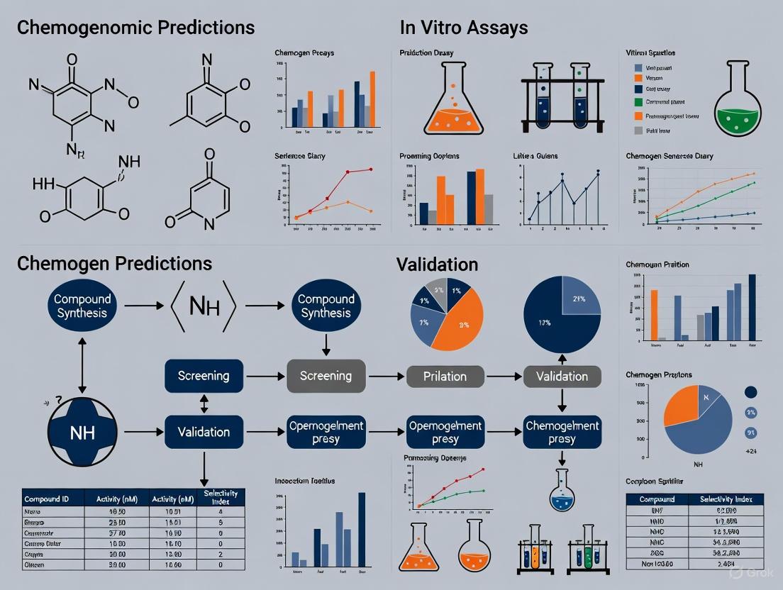
This article provides a comprehensive guide for researchers and drug development professionals on the critical process of validating chemogenomic predictions using robust in vitro assays.
From In Silico to In Vitro: A Practical Framework for Validating Chemogenomic Predictions in Drug Discovery
Abstract
This article provides a comprehensive guide for researchers and drug development professionals on the critical process of validating chemogenomic predictions using robust in vitro assays. It covers the foundational principles of chemogenomics, the selection and development of appropriate methodological approaches, strategies for troubleshooting and optimization, and the final steps for rigorous validation and comparative analysis. By bridging the gap between computational predictions and experimental confirmation, this framework aims to enhance the efficiency and success rate of translating potential drug-target interactions into validated leads, ultimately accelerating the drug discovery pipeline.
Chemogenomics Unveiled: Laying the Groundwork for Predictive Drug Discovery
Chemogenomics represents a paradigm shift in early drug discovery, integrating large-scale genomic data with chemical screening to elucidate interactions between small molecules and biological targets across entire genomes or proteomes. This approach provides a systems-level framework for understanding mechanisms of drug action (MoA), enabling simultaneous exploration of multiple drug-target interactions rather than focusing on single targets in isolation [1] [2]. The fundamental premise of chemogenomics lies in its ability to connect chemical space with biological space, creating a comprehensive map of interactions that accelerates both target identification and validation processes [1].
The drug discovery pipeline has traditionally been a cost-intensive endeavor with high attrition rates, where chemogenomic approaches now offer a strategic advantage. By predicting drug-target interactions (DTIs) early in the discovery process, chemogenomics reduces the target search space, indirectly decreasing the overall cost, time, and labor invested in bringing a drug to market [1]. This is particularly valuable given that conventional drug development processes face a clinical success rate of only 19%, significantly lower than expected rates [1]. Chemogenomic methods have thus gained substantial traction as in silico alternatives to complement traditional wet-lab experiments, supporting data-driven decision-making through the availability of extensive bioinformatics and genetic databases [1].
Core Methodologies in Chemogenomics
Experimental and Computational Approaches
Chemogenomic methodologies can be broadly categorized into experimental screening approaches and computational prediction frameworks, each with distinct advantages and applications.
Experimental chemogenomic profiling utilizes systematic screening of chemical compounds against comprehensive genetic libraries. In model organisms like Saccharomyces cerevisiae, two primary assays form the backbone of these approaches: HaploInsufficiency Profiling (HIP) and Homozygous Profiling (HOP) [2]. The HIP assay exploits drug-induced haploinsufficiency, where heterozygous strains deleted for one copy of an essential gene show specific sensitivity when exposed to a drug targeting that gene product. The complementary HOP assay interrogates nonessential homozygous deletion strains to identify genes involved in drug target biological pathways and those required for drug resistance [2]. The combined HIPHOP chemogenomic profile provides a comprehensive genome-wide view of the cellular response to specific compounds, directly identifying drug target candidates while also revealing resistance mechanisms [2].
For computational prediction, multiple algorithmic strategies have been developed:
Table 1: Comparison of Computational Chemogenomic Approaches
| Method Category | Key Principles | Advantages | Limitations |
|---|---|---|---|
| Similarity Inference Methods | Based on "wisdom of crowd" principle using chemical/structural similarities [1] | High interpretability for justifying predictions [1] | May miss serendipitous discoveries; often uses binary interaction data rather than continuous binding affinity [1] |
| Network-based Methods | Utilize topological features of drug-target bipartite networks [1] | Do not require 3D protein structures or negative samples [1] | Suffer from "cold start" problem for new drugs; biased toward high-degree nodes [1] |
| Feature-based Machine Learning | Use manually extracted features from drugs and targets [1] | Can handle new drugs/targets without similarity information [1] | Feature selection is difficult; class imbalance issues in classification [1] |
| Deep Learning Methods | Employ neural networks for automatic feature learning [1] [3] | Avoid labor-intensive manual feature extraction [1] | Low interpretability; reliability of learned features may not match chemical knowledge [1] |
| Matrix Factorization | Decompose interaction matrices into lower-dimensional representations [1] | Do not require negative samples [1] | Better at modeling linear than non-linear relationships [1] |
Emerging Integrated Frameworks
Recent advances have introduced multitask learning frameworks that simultaneously predict drug-target interactions and generate novel drug candidates. The DeepDTAGen model exemplifies this approach by using shared feature representations for both predicting drug-target binding affinity and generating target-aware drug variants [4]. This integration addresses the intrinsically interconnected nature of these tasks in pharmacological research, potentially increasing clinical success rates by ensuring generated drugs are conditioned on specific target interactions [4].
Another innovative approach is DrugMAN, which integrates heterogeneous biological networks using graph attention networks and mutual attention mechanisms. This method extracts network-specific features for drugs and targets from multiplex functional interaction networks, then captures interaction patterns between them to improve prediction accuracy, particularly in real-world scenarios [3].
Experimental Design and Validation Workflows
Standardized Chemogenomic Profiling Protocols
Robust chemogenomic profiling requires standardized experimental workflows and validation frameworks. For NR4A nuclear receptor research, a comprehensive profiling approach was established using orthogonal assay systems to validate modulator activity [5]. This included:
- Gal4-hybrid-based and full-length receptor reporter gene assays to determine cellular NR4A modulation across all three receptor subtypes (NR4A1, NR4A2, NR4A3)
- Selectivity screening against a representative panel of nuclear receptors outside the NR4A family
- Cell-free binding validation using isothermal titration calorimetry (ITC) and differential scanning fluorimetry (DSF)
- Compound quality control through HPLC purity analysis, mass spectrometry identification, kinetic solubility assessment, and multiplex toxicity monitoring [5]
This multi-layered validation strategy ensures that chemical tools used in functional and phenotypic studies have well-characterized activities and specificities, addressing concerns that incompletely profiled tools can compromise biological findings [5].
The diagram below illustrates a standardized workflow for chemogenomic prediction and validation:
Validation with Benchmark Datasets
Rigorous comparison of prediction methods requires standardized benchmarking. A 2025 systematic evaluation compared seven target prediction methods (MolTarPred, PPB2, RF-QSAR, TargetNet, ChEMBL, CMTNN, and SuperPred) using a shared dataset of FDA-approved drugs [6]. The study employed ChEMBL version 34 as the reference database, containing 15,598 targets, 2,431,025 compounds, and 20,772,701 interactions [6]. To ensure data quality, researchers filtered for high-confidence interactions with a minimum confidence score of 7 (indicating direct protein complex subunits assigned) and excluded non-specific or multi-protein targets [6].
Performance assessment in such benchmarks typically employs multiple metrics including Mean Squared Error (MSE), Concordance Index (CI), R-squared (r²m), and Area Under Precision-Recall Curve (AUPR) for binding affinity prediction, while drug generation tasks are evaluated based on Validity, Novelty, and Uniqueness of generated compounds [4].
Performance Comparison of Prediction Methods
Quantitative Benchmarking Results
Independent comparative studies provide crucial insights into the relative performance of different chemogenomic prediction approaches. A precise comparison study conducted in 2025 revealed that MolTarPred emerged as the most effective method among seven evaluated target prediction tools [6]. The study further optimized MolTarPred by demonstrating that Morgan fingerprints with Tanimoto scores outperformed MACCS fingerprints with Dice scores [6].
For drug-target binding affinity prediction, the DeepDTAGen multitask framework achieved state-of-the-art performance across multiple benchmark datasets:
Table 2: Performance Comparison of DeepDTAGen with Previous Methods on Binding Affinity Prediction
| Dataset | Best Previous Method | DeepDTAGen Performance | Improvement Over Previous Best |
|---|---|---|---|
| KIBA | GraphDTA (CI: 0.891) [4] | MSE: 0.146, CI: 0.897, r²m: 0.765 [4] | 0.67% CI improvement, 11.35% r²m improvement [4] |
| Davis | SSM-DTA (r²m: 0.689) [4] | MSE: 0.214, CI: 0.890, r²m: 0.705 [4] | 2.4% r²m improvement, 2.2% MSE reduction [4] |
| BindingDB | GDilatedDTA (CI: 0.868) [4] | MSE: 0.458, CI: 0.876, r²m: 0.760 [4] | 0.9% CI improvement, 4.1% r²m improvement [4] |
The DrugMAN model demonstrated particularly strong performance in challenging real-world scenarios, showing the smallest decrease in AUROC, AUPRC, and F1-Score from warm-start to cold-start conditions compared to traditional methods like SVM, RF, DeepPurpose, DTINet, and NeoDTI [3]. This robustness highlights the advantage of integrating heterogeneous biological networks, especially when limited chemogenomic data is available for specific targets.
Experimental Validation Case Studies
Robust validation of chemogenomic predictions requires confirmation through experimental assays. In a notable case study, Archetype Therapeutics utilized generative chemogenomics to identify novel and repurposed small molecules for intercepting invasion in lung adenocarcinoma [7]. Their AI-platform screened billions of potential drugs virtually before advancing candidates to experimental validation. The resulting molecules demonstrated significant efficacy in both in vitro and in vivo (GEMM and xenograft) models, substantially outperforming previously published molecules for preventing metastasis in early-stage lung adenocarcinoma [7]. This successful translation from computational prediction to biological validation exemplifies the power of integrated chemogenomic approaches.
For NR4A receptor research, comparative profiling under uniform conditions revealed significant deviations from published activities for several putative ligands, with some compounds showing complete lack of target binding and modulation [5]. This underscores the importance of orthogonal validation, as compounds with flawed characterization data can lead to erroneous biological conclusions. From an initial set of literature-reported NR4A modulators, only eight chemically diverse compounds were validated as direct NR4A modulators suitable for reliable target identification studies [5].
Successful chemogenomics research requires leveraging specialized reagents, databases, and computational tools. The following table summarizes key resources for establishing a chemogenomics research pipeline:
Table 3: Essential Research Resources for Chemogenomics
| Resource Category | Specific Tools/Databases | Key Applications | Technical Considerations |
|---|---|---|---|
| Bioactivity Databases | ChEMBL [6], BindingDB [6], DrugBank [3] | Training data for prediction models; reference for ligand-target interactions | ChEMBL ideal for novel protein targets; DrugBank better for drug indications [6] |
| Chemical Tools | Validated NR4A modulators (agonists/inverse agonists) [5] | Target identification and validation studies | Require orthogonal validation (ITC, DSF, reporter assays) [5] |
| Target Prediction Servers | MolTarPred [6], PPB2 [6], TargetNet [6] | Ligand-centric target fishing | Performance varies; MolTarPred currently top-performing [6] |
| Experimental Models | Yeast HIPHOP platform [2], Cell-based reporter assays [5] | Genome-wide chemogenomic profiling | Yeast systems provide standardized, reproducible fitness signatures [2] |
| Advanced Frameworks | DeepDTAGen [4], DrugMAN [3] | Integrated prediction and generation | DrugMAN excels in cold-start scenarios; DeepDTAGen enables multitask learning [3] [4] |
Chemogenomics has established itself as an indispensable approach in modern drug discovery, effectively bridging the gap between genomic sciences and chemical screening. The integration of diverse methodological approaches—from similarity-based methods to deep learning frameworks—provides researchers with a powerful toolkit for elucidating drug-target interactions across entire biological systems.
The most successful implementations combine computational predictions with orthogonal experimental validation, creating iterative refinement cycles that enhance both target identification and compound optimization. As evidenced by recent advances, future progress in chemogenomics will likely come from increased integration of heterogeneous data sources, development of multitask learning frameworks that simultaneously address prediction and generation tasks, and improved handling of cold-start scenarios for novel target classes.
For researchers embarking on chemogenomic studies, the current evidence supports a strategy that leverages multiple complementary methods rather than relying on a single approach, utilizes high-confidence benchmark datasets for method validation, and incorporates orthogonal experimental assays at early stages to verify computational predictions. This integrated methodology will maximize the potential of chemogenomics to accelerate drug discovery and improve our understanding of complex drug-target interaction networks.
The Crucial Role of In Vitro Validation in the Drug Discovery Pipeline
In the modern drug discovery landscape, where artificial intelligence (AI) and computational methods generate vast numbers of potential targets and candidates, the role of rigorous in vitro validation has never been more critical. These experimental assays form the essential bridge between in silico predictions and clinical success, providing the first real-world test of a molecule's biological activity. This guide examines the performance of various in vitro validation strategies, providing experimental data and protocols to help researchers navigate this complex, high-stakes phase of development.
The Validation Imperative: From In Silico to In Vitro
The first half of 2025 saw continued innovation in oncology therapeutics, with eight novel FDA approvals including targeted therapies, antibody-drug conjugates, and treatments for rare cancers [8]. This progress occurs against a challenging backdrop of persistently high attrition rates (approximately 95%) for novel drug discovery [8]. This high failure rate underscores why in vitro validation is not merely a procedural step, but a crucial strategic filter to mitigate risk before candidates advance to more costly in vivo studies and clinical trials.
The relationship between computational prediction and experimental validation represents a fundamental workflow in modern drug discovery:
Comparative Performance of In Vitro Validation Platforms
Different in vitro models offer varying strengths and limitations for validating chemogenomic predictions. The table below summarizes key performance characteristics of the primary platforms used in contemporary drug discovery pipelines:
| Model Type | Key Applications | Advantages | Limitations | Translational Relevance |
|---|---|---|---|---|
| 2D Cell Lines [8] | - High-throughput cytotoxicity screening- Drug efficacy testing- Initial biomarker hypothesis generation | - Reproducible & standardized- Cost-effective- Large established collections | - Limited tumor heterogeneity- Does not reflect tumor microenvironment | Moderate for initial target validation |
| 3D Organoids [8] | - Investigate drug responses- Evaluate immunotherapies- Predictive biomarker identification | - Faithfully recapitulates original tumor- Preserves tumor architecture- Suitable for HTS | - Complex and time-consuming to create- Cannot fully represent complete TME | High, especially for patient-specific responses |
| PDX-Derived Models [8] | - Biomarker discovery and validation- Clinical stratification- Drug combination strategies | - Most clinically relevant preclinical model- Preserves tumor heterogeneity- Mirrors patient responses | - Expensive and resource-intensive- Not suitable for HTS- Time-consuming | Very High, considered "gold standard" |
Experimental Protocols for Target Validation
Protocol 1: Cellular Target Engagement Using CETSA
The Cellular Thermal Shift Assay (CETSA) has emerged as a leading method for validating direct drug-target interactions in physiologically relevant environments [9].
Workflow Overview:
Detailed Methodology:
- Cell Treatment: Expose intact cells to varying concentrations of the test compound (typically 1-100 µM) for relevant exposure times (e.g., 1-6 hours) to allow cellular penetration and target engagement [9].
- Heat Denaturation: Divide cell suspensions into aliquots and heat at different temperatures (typically 45-65°C gradient) for 3-5 minutes using a precise thermal cycler [9].
- Cell Lysis: Freeze-thaw cycles or mechanical lysis to disrupt cells while preserving protein integrity from engaged targets [9].
- Protein Quantification: Analyze soluble target protein levels via Western blot, mass spectrometry, or ELISA. Recent advances enable high-resolution MS quantification of drug-target engagement, as demonstrated for DPP9 in rat tissue [9].
- Data Analysis: Calculate melting temperature (Tm) shifts. Stabilization of the target protein's thermal profile indicates direct binding and successful target engagement [9].
Protocol 2: Advanced Phenotypic Screening for Malaria Transmission-Blocking Compounds
Recent innovations in phenotypic screening demonstrate the sophistication of modern in vitro validation. A 2025 study established a robust platform for identifying Plasmodium falciparum transmission-blocking drugs using engineered parasites [10].
Key Experimental Steps:
- Parasite Engineering: Utilize transgenic NF54/iGP1_RE9Hulg8 parasites engineered to conditionally produce large numbers of stage V gametocytes expressing a red-shifted firefly luciferase viability reporter [10].
- Compound Exposure: Incubate mature stage V gametocytes with test compounds across a concentration range (typically 0.1 nM-10 µM) for 72 hours [10].
- Viability Assessment: Quantify gametocyte viability through luciferase reporter activity, providing a sensitive, quantitative readout of compound efficacy [10].
- Counter-Screening: Assess specificity by testing compounds against asexual blood stage parasites to identify stage-specific versus pan-active antimalarials [10].
- Secondary Validation: Confirm transmission-blocking activity in Standard Membrane Feeding Assays (SMFA) where mosquitoes feed on compound-exposed gametocytes [10].
The Scientist's Toolkit: Essential Research Reagents
Successful in vitro validation requires specialized reagents and tools. The following table outlines essential solutions for establishing robust validation workflows:
| Research Reagent | Function/Purpose | Application Context |
|---|---|---|
| CETSA Platform [9] | Measures drug-target engagement via thermal stability shifts in intact cells | Mechanistic validation of direct target binding in physiologically relevant systems |
| Engineered Reporter Cell Lines [10] | Express viability or pathway-specific reporters (e.g., luciferase) for compound screening | High-content phenotypic screening (e.g., malaria gametocyte viability assays) |
| Patient-Derived Organoids [8] | 3D cultures that preserve tumor architecture and genetic features | Assessment of tumor-specific drug responses and biomarker discovery |
| PDX-Derived Cells [8] | Cell lines originating from patient-derived xenograft models | Bridge between in vitro and in vivo studies; biomarker hypothesis generation |
| Clinical Database Resources (ChEMBL) [6] | Curated bioactivity data from scientific literature | Benchmarking and validation of target prediction methods |
Strategic Implementation for Pipeline Success
The most effective drug discovery pipelines employ these validation tools not in isolation, but as part of an integrated, multi-stage approach:
This sequential framework enables researchers to leverage the unique advantages of each model. For example, initial biomarker hypotheses generated through high-throughput screening of PDX-derived cell lines can be refined using 3D organoids and ultimately validated in PDX models before clinical trials [8]. This systematic approach builds a robust evidentiary chain that de-risks pipeline progression and increases the probability of clinical success.
Future Directions in Validation Science
The field of in vitro validation continues to evolve rapidly. Several trends are shaping its future development:
- AI Integration: Machine learning models are increasingly used to predict compound efficacy and prioritize molecules for in vitro testing, with recent studies showing 50-fold enrichment rates compared to traditional methods [9].
- Complex Model Systems: As the FDA reduces animal testing requirements for certain drug classes, advanced models like organoids are gaining regulatory acceptance as complementary approaches [8].
- Functional Relevance: Technologies that provide direct, in situ evidence of drug-target interaction, such as CETSA, are transitioning from specialized tools to strategic assets essential for decision-making [9].
In conclusion, while computational methods have dramatically accelerated the initial phases of drug discovery, rigorous in vitro validation remains the critical gatekeeper ensuring that only the most promising candidates advance through the development pipeline. By implementing the comparative frameworks and experimental approaches outlined in this guide, research teams can enhance their decision-making, compress development timelines, and ultimately increase their chances of translational success.
The experimental prediction of drug-target interactions (DTIs) is an expensive, time-consuming, and tedious process, creating a critical bottleneck in modern drug discovery pipelines [1]. Chemogenomic approaches have emerged as powerful computational strategies that leverage both chemical and genomic information to address this challenge, significantly narrowing the search space for interaction candidates that warrant further wet-lab investigation [1] [11]. These methods fundamentally frame DTI prediction as a machine learning problem, utilizing known interactions along with the properties of drugs and targets to train predictive models [11]. The growing importance of polypharmacology—understanding how drugs interact with multiple targets—has further intensified the need for reliable computational methods that can reveal hidden drug-target relationships for drug repurposing and safety profiling [6].
This guide provides a comprehensive comparison of three principal chemogenomic methodologies: ligand-based approaches, molecular docking, and machine learning-based methods. We objectively evaluate their performance characteristics, experimental requirements, and practical implementation considerations, with a specific focus on validating computational predictions through subsequent in vitro assays. As the field progresses, the integration of artificial intelligence with traditional computational methods has begun to transform the drug discovery landscape, enabling rapid screening of billions of compounds and improving the accuracy of binding affinity predictions [12] [13]. Understanding the relative strengths and limitations of each approach is essential for researchers selecting appropriate strategies for specific drug discovery scenarios.
Comparative Analysis of Chemogenomic Approaches
Table 1: Overall comparison of the three main chemogenomic approaches
| Approach | Core Principle | Data Requirements | Strengths | Limitations |
|---|---|---|---|---|
| Ligand-Based | "Wisdom of the crowd" principle using similarity between query molecule and known ligands [6] | Known ligands with annotated targets; compound structures [1] [6] | High interpretability; does not require protein structures; fast predictions [1] [6] | Struggles with novel targets/compounds (cold start problem); limited serendipitous discoveries [1] [6] |
| Molecular Docking | Predicts binding pose and affinity through computational simulation of physical interactions [14] [15] | 3D protein structures; compound structures [1] [15] | Provides structural insights; models physical interactions; can handle novel compounds [14] [15] | Limited by protein structure availability/quality; computationally intensive; scoring function inaccuracies [1] [6] |
| Machine Learning | Learns interaction patterns from known chemogenomic data using algorithms [1] [11] | Known drug-target interactions; compound and protein features [1] [11] | Handles new drugs/targets via features; no negative samples needed for some methods; high accuracy potential [1] [16] | Black-box nature; requires extensive training data; feature selection critical [1] [11] |
Table 2: Performance comparison of specific methods across different evaluation scenarios
| Method | Approach Category | Warm Start Performance | Cold Start Performance | Key Findings |
|---|---|---|---|---|
| ColdstartCPI [16] | Machine Learning (Induced-fit theory) | High performance | Excels, especially for unseen compounds and proteins | Treats proteins/compounds as flexible; outperforms state-of-the-art sequence-based models |
| MolTarPred [6] | Ligand-Centric (2D similarity) | Effective for known chemical space | Limited by ligand similarity | Most effective method in benchmark; performance depends on fingerprint choice |
| EnsemKRR [11] | Machine Learning (Ensemble) | AUC: 94.3% | Not specifically evaluated | Combines dimensionality reduction with ensemble learning |
| CoBDock [15] | Docking (Consensus blind docking) | Superior binding site and mode prediction vs. other blind docking | Not applicable | Machine learning consensus of multiple docking/cavity detection tools |
| ML-Guided Docking [13] | Hybrid (ML + Docking) | Identifies >87% of top-scoring compounds | Not applicable | Reduces docking computation by >1,000-fold for billion-compound libraries |
Experimental Protocols and Workflows
Ligand-Centric Similarity Searching (MolTarPred Protocol)
Ligand-centric methods operate on the principle that chemically similar compounds are likely to share molecular targets [6]. The experimental workflow for implementing similarity-based target prediction involves several standardized steps:
Database Preparation: Compile a comprehensive database of known ligand-target interactions, such as ChEMBL (version 34 contains 2.4 million compounds, 15,598 targets, and 20.8 million interactions) [6]. Filter entries to retain only high-confidence interactions (e.g., confidence score ≥7 in ChEMBL, indicating direct protein complex subunits assigned) and remove duplicates and non-specific targets.
Molecular Representation: Convert query compounds and database molecules into appropriate molecular representations. Common fingerprints include MACCS keys or Morgan fingerprints (hashed bit vector with radius two and 2048 bits) [6].
Similarity Calculation: Compute structural similarity between query molecule and all database compounds using Tanimoto similarity for Morgan fingerprints or Dice scores for MACCS fingerprints [6].
Consensus Prediction: Identify the top similar ligands (typically 1-15 nearest neighbors) from the database and extract their annotated targets. The frequency of target appearances among nearest neighbors indicates prediction confidence [6].
Validation: For experimental validation, select top-predicted targets for in vitro binding assays or functional cellular assays to confirm the computational predictions.
Structure-Based Molecular Docking (CoBDock Protocol)
Molecular docking predicts how small molecules bind to protein targets by exploring binding poses and scoring affinities [14] [15]. The CoBDock protocol implements a consensus blind docking approach:
Target Preparation:
- Input protein structure in PDB format and remove undesired elements (water, free ions, bound ligands) using PyMOL.
- Add hydrogen atoms with Pdb2Pqr software at physiological pH 7.4, using AMBER force field and propka for titration states [15].
Ligand Preparation:
- Input ligand structures in SMILES, PDB, MOL, MOL2, or SDF formats.
- Add hydrogens to polar atoms using Open Babel at pH 7.4 and convert to appropriate formats for docking programs [15].
Parallel Blind Docking and Cavity Detection:
Consensus Binding Site Prediction:
- Superimpose a 10Å-resolution grid over the entire protein surface.
- Assign each predicted binding mode and detected cavity to the closest grid box.
- Use a trained machine learning model to score and rank grid locations based on consensus from all methods [15].
Local Docking and Validation:
- Perform local docking with PLANTS at the top-ranked binding site to generate final pose predictions.
- Experimental validation typically involves X-ray crystallography of protein-ligand complexes to verify binding poses, or binding affinity assays (ITC, SPR) to quantify interaction strength [15].
Workflow for consensus blind docking (CoBDock)
Machine Learning Framework (ColdstartCPI Protocol)
ColdstartCPI represents a modern machine learning approach inspired by induced-fit theory, treating both compounds and proteins as flexible molecules during binding [16]:
Data Collection and Preprocessing:
- Collect compound structures as SMILES strings and protein sequences as amino acid sequences.
- Source known drug-target interactions from databases like ChEMBL, BindingDB, or DrugBank.
- Implement rigorous data splitting strategies (warm start, compound cold start, protein cold start, blind start) to evaluate generalization capability [16].
Feature Extraction:
- Use Mol2Vec to generate substructure feature matrices for compounds, capturing semantic features of drug substructures.
- Apply ProtTrans to create amino acid feature matrices for proteins, encoding structural and functional information [16].
- Generate global representations of compounds and proteins using pooling functions on the feature matrices.
Feature Space Unification:
- Process features through four separate Multi-Layer Perceptrons (MLPs) to unify feature spaces and decouple feature extraction from CPI prediction [16].
Transformer-Based Interaction Modeling:
- Construct a joint matrix representation of the compound-protein pair.
- Feed the joint matrix into a Transformer module to learn compound and protein features by extracting inter- and intra-molecular interaction characteristics [16].
- This flexible representation allows compound features to change depending on binding proteins and vice versa, aligning with induced-fit theory.
Prediction and Experimental Validation:
- Concatenate the final compound and protein features.
- Process through a three-layer fully connected neural network with dropout to predict interaction probability [16].
- Experimental validation includes literature searches for known interactions, molecular docking simulations, binding free energy calculations, and molecular dynamics simulations to verify top predictions [16].
Performance Benchmarking and Experimental Validation
Performance Across Different Scenarios
The generalization capability of chemogenomic methods varies significantly across different validation scenarios, particularly between warm start (where drugs and targets appear in the training set) and cold start (predicting interactions for novel drugs or targets) conditions [16]:
- Ligand-based methods like MolTarPred perform well in warm start scenarios but struggle with cold start problems, particularly for compounds with low similarity to known database entries [1] [6].
- Traditional docking approaches can handle novel compounds but depend heavily on the availability and quality of protein structures [1] [15].
- Advanced machine learning methods like ColdstartCPI demonstrate robust performance across both warm start and cold start conditions, achieving area under receiver operating characteristic curve (AUROC) values exceeding 0.9 in warm start and maintaining competitive performance (AUROC >0.85) in challenging cold start scenarios [16].
Table 3: ColdstartCPI performance across different scenarios
| Evaluation Setting | AUROC | AUPRC | Key Advantage |
|---|---|---|---|
| Warm Start | >0.9 | >0.85 | Benefits from task-relevant feature extraction |
| Compound Cold Start | >0.85 | >0.8 | Handles novel compounds effectively |
| Protein Cold Start | >0.85 | >0.8 | Generalizes to unseen proteins |
| Blind Start | >0.8 | >0.75 | Works with completely novel drug-target pairs |
Validation with Experimental Assays
Computational predictions require rigorous experimental validation to confirm biological relevance. Successful validation strategies include:
Binding Affinity Assays: Surface Plasmon Resonance (SPR) and Isothermal Titration Calorimetry (ITC) provide quantitative measurements of binding strength (Kd values) for predicted interactions [6].
Functional Cellular Assays: Cell-based reporter assays or phenotypic screening confirm whether predicted interactions translate to functional biological effects in relevant cellular contexts [6].
Structural Validation: X-ray crystallography or cryo-electron microscopy of protein-ligand complexes provides atomic-level confirmation of binding modes predicted by docking studies [14] [15].
Drug Repurposing Case Studies: Experimental validation of predictions for specific disease areas demonstrates real-world utility. For example, ColdstartCPI predictions for Alzheimer's Disease, breast cancer, and COVID-19 were validated through literature evidence, docking simulations, and binding free energy calculations [16].
Experimental validation workflow for computational predictions
Research Reagent Solutions
Table 4: Essential research reagents and databases for chemogenomic research
| Resource | Type | Function | Application Context |
|---|---|---|---|
| ChEMBL [6] | Database | Manually curated database of bioactive molecules with drug-like properties | Primary source for ligand-target interactions; training data for machine learning models |
| Protein Data Bank (PDB) [14] | Database | Repository of 3D protein structures determined by X-ray, NMR, Cryo-EM | Source of protein structures for molecular docking studies |
| AutoDock Vina [15] | Software | Molecular docking tool with empirical scoring function | Structure-based virtual screening and binding pose prediction |
| Mol2Vec [16] | Algorithm | Unsupervised machine learning for compound representation | Generates substructure-aware features for machine learning |
| ProtTrans [16] | Algorithm | Protein language model for sequence representation | Generates structural and functional protein features from sequences |
| SPR/Biacore [6] | Instrument | Surface plasmon resonance for binding affinity measurement | Experimental validation of binding affinity (Kd) |
| ITC | Instrument | Isothermal titration calorimetry for thermodynamics | Measures binding affinity and thermodynamic parameters |
| Enamine REAL [13] | Compound Library | Make-on-demand chemical library (70B+ compounds) | Ultralarge virtual screening for hit identification |
The comparative analysis of ligand-based, docking, and machine learning approaches reveals a complementary landscape of chemogenomic methodologies, each with distinct advantages for specific drug discovery scenarios. Ligand-based methods offer interpretability and speed but struggle with novelty, while docking provides physical insights but depends on structural data. Machine learning approaches, particularly recent induced-fit theory-guided models like ColdstartCPI, demonstrate superior performance in cold-start scenarios and show promising generalization capabilities [16].
The emerging trend of hybrid approaches that combine multiple methodologies represents the most promising direction for future research. Machine learning-guided docking screens exemplify this integration, achieving unprecedented efficiency gains—reducing computational requirements by more than 1,000-fold while maintaining high sensitivity in identifying true binders [13]. These integrated workflows enable practical virtual screening of multi-billion compound libraries, dramatically expanding the explorable chemical space for drug discovery.
For researchers validating chemogenomic predictions with in vitro assays, the selection of methodology should align with the specific discovery context: ligand-based approaches for target fishing of compounds with known analogs, docking for structure-enabled targets, and machine learning for scenarios with limited structural information or challenging cold-start problems. As artificial intelligence continues to transform computational drug discovery, the convergence of these approaches with experimental validation will accelerate the identification of novel therapeutic candidates and expand our understanding of polypharmacology.
In the field of chemogenomics, the reliable prediction of drug-target interactions (DTIs) is fundamental to accelerating drug discovery and repurposing efforts. Public bioactivity databases serve as the foundational infrastructure for building predictive computational models. Among these, ChEMBL and DrugBank have emerged as two of the most comprehensive and widely used resources by researchers and drug development professionals. These databases provide curated information on bioactive molecules, their protein targets, and experimentally determined interactions, enabling the training and validation of machine learning models for target prediction. The strategic selection of a database directly impacts the predictive performance of chemogenomic models and the success of subsequent experimental validation [17].
This guide provides an objective comparison of DrugBank and ChEMBL within the context of validating chemogenomic predictions. It details their respective contents, access models, and applicability for different research scenarios, supported by experimental data and methodologies from recent scientific literature.
Database Comparison: ChEMBL vs. DrugBank
The table below provides a detailed, side-by-side comparison of the core characteristics of ChEMBL and DrugBank, highlighting their distinct strategic focuses.
Table 1: Strategic Comparison of ChEMBL and DrugBank
| Feature | ChEMBL | DrugBank |
|---|---|---|
| Primary Focus | Large-scale bioactivity data for drug-like compounds and pre-clinical candidates [18] | Comprehensive drug data, including detailed drug and mechanism-of-action information [19] [18] |
| Core Content | Bioactivity data (e.g., IC₅₀, Kᵢ) from scientific literature and patents; extensive SAR data [20] [18] | FDA-approved and experimental drugs, with rich pharmacological and pharmaceutical data [19] [18] |
| Target Coverage | Extensive, focusing on a broad range of protein targets (e.g., kinases, GPCRs, enzymes) for research [20] [6] | Mappings to primary drug targets, with a focus on established therapeutic mechanisms [19] |
| Data Model | Manually curated bioactivity data integrated with drug information; distinction between research compounds and drugs [18] | Integrated drug and target information, with half of record data devoted to drug and half to pharmacological properties [19] |
| Access Model | Fully open-access [18] [17] | Freely available for non-commercial use; not fully open-access [18] |
| Ideal Use Case | Building generalizable target prediction models for novel compounds and protein targets [6] | Predicting new indications for known drugs and understanding established drug-target pathways [6] |
Performance Evaluation in Target Prediction
Independent, comparative studies are essential for objectively evaluating the utility of databases in practical research. One systematic benchmark study evaluated seven different target prediction methods, many of which are trained on ChEMBL data, on a shared dataset of FDA-approved drugs [6].
Table 2: Performance of ChEMBL-Based Target Prediction Methods
| Prediction Method | Type | Underlying Algorithm | Key Performance Finding |
|---|---|---|---|
| MolTarPred [6] | Ligand-centric | 2D similarity search | Identified as the most effective method in the benchmark study. |
| RF-QSAR [6] | Target-centric | Random Forest | Performance validated on a shared benchmark dataset. |
| CMTNN [6] | Target-centric | Multitask Neural Network | Performance validated on a shared benchmark dataset. |
| EnsemKRR Model [11] | Chemogenomic | Kernel Ridge Regression Ensemble | Achieved highest AUC (94.3%) for DTI prediction using ChEMBL data. |
The study concluded that ChEMBL is more suitable for predicting interactions with novel protein targets due to its extensive chemogenomic data, whereas DrugBank is ideal for predicting new drug indications against known targets because of its focus on drug-related information [6]. Furthermore, a separate study developed an ensemble chemogenomic model using ChEMBL and BindingDB data, reporting that 57.96% of known targets were identified in the top-10 predictions, representing an approximately 50-fold enrichment over random guessing [20].
Experimental Protocols for Validation
Protocol 1: Building an Ensemble Chemogenomic Model
This protocol is adapted from a study that developed a high-performance ensemble model for target prediction [20].
- Data Collection: Extract compound-target interactions with associated bioactivity values (e.g., Kᵢ ≤ 100 nM for positive set) from ChEMBL and other databases like BindingDB.
- Descriptor Calculation:
- Model Training: Construct multiple individual chemogenomic models (e.g., using XGBoost) by combining the different molecular and protein descriptors. Each model learns to differentiate interacting compound-target pairs from non-interacting ones [20].
- Ensemble Construction: Select and combine the best-performing individual models into a final ensemble model to improve overall prediction accuracy and robustness [20].
- Target Prediction: For a query compound, create compound-target pairs with all potential targets in the database. Input these pairs into the ensemble model and rank the targets based on the output interaction scores. The top-k ranked targets are the highest-confidence predictions [20].
Protocol 2: Benchmarking Target Prediction Methods
This protocol outlines the methodology for a fair and precise comparison of different prediction tools, as seen in a recent benchmark study [6].
- Dataset Curation:
- Source a large set of ligand-target interactions from a recent ChEMBL release (e.g., version 34).
- Apply strict filtering: include only bioactivities with standard values (IC₅₀, Kᵢ, EC₅₀) below 10,000 nM; exclude non-specific protein targets; remove duplicate compound-target pairs.
- To prevent bias, create a separate benchmark set of FDA-approved drugs that are excluded from the main training database.
- Model Selection and Execution: Select a representative set of stand-alone and web-server prediction methods (e.g., MolTarPred, PPB2, RF-QSAR). Run these methods on the benchmark dataset.
- Performance Assessment: Evaluate methods based on their ability to recall the known targets for the benchmark drugs, with a focus on top-k prediction accuracy (e.g., whether the true target appears in the top 1 or top 10 predictions) [6].
Workflow Visualization
The following diagram illustrates the logical workflow for building and applying a chemogenomic model, culminating in experimental validation.
The Scientist's Toolkit: Research Reagent Solutions
The table below lists key computational and experimental "reagents" – databases, software, and assays – essential for conducting research in this field.
Table 3: Essential Research Reagents for Chemogenomic Prediction and Validation
| Research Reagent | Type | Function & Application |
|---|---|---|
| ChEMBL Database [18] [6] | Data Resource | Provides a vast, open-access repository of bioactive molecules and curated drug-target interactions for training predictive models. |
| DrugBank Database [19] [18] | Data Resource | Offers comprehensive information on drugs, their mechanisms, and targets, ideal for studies on drug repurposing and established pharmacology. |
| MolTarPred [6] | Software Tool | A ligand-centric, 2D similarity-based prediction method identified as a top-performing tool for target prediction. |
| EnsemKRR [11] | Software/Algorithm | An ensemble learning method that combines multiple classifiers to achieve high accuracy in predicting drug-target interactions. |
| Binding Affinity Assays (e.g., Kᵢ, IC₅₀) [20] [6] | Experimental Assay | Measures the strength of interaction between a compound and a purified target protein, used for experimental validation of computational predictions. |
| Gene Expression Profiling (e.g., CMap) [21] | Experimental/Data Resource | Measures transcriptomic changes in response to drug treatment; can be used for target prediction independent of chemical structure. |
Understanding Forward and Reverse Chemogenomics for Target Validation
Target validation is a critical stage in the drug discovery pipeline, establishing a causal link between the modulation of a target protein and a desired therapeutic effect [1] [22]. Within this process, chemogenomics has emerged as a powerful system-based strategy that utilizes small molecules as probes to elucidate the relationship between a biological target and a phenotypic outcome [23] [24] [22]. This paradigm operates on two complementary axes: forward chemogenomics and reverse chemogenomics. Both approaches are foundational to validating chemogenomic predictions, yet they differ fundamentally in their starting points and methodological workflows [23] [24]. This guide provides an objective comparison of these two strategies, detailing their performance, key experimental protocols, and essential reagent solutions, thereby offering a framework for researchers to select the appropriate methodology for their target validation challenges.
Strategic Comparison: Forward vs. Reverse Chemogenomics
The core distinction between forward and reverse chemogenomics lies in their initial discovery trigger. Forward chemogenomics begins with the observation of a phenotypic change in a cell or organism and aims to identify the molecular target responsible, effectively moving from phenotype to target [23] [24]. Conversely, reverse chemogenomics starts with a specific, isolated protein target and seeks compounds that modulate its activity, subsequently analyzing the resulting phenotype in a biological system, thus moving from target to phenotype [23] [24] [25]. This fundamental difference dictates their respective applications, advantages, and limitations within a research project aimed at in vitro validation.
Table 1: High-Level Strategic Comparison of Forward and Reverse Chemogenomics
| Feature | Forward Chemogenomics | Reverse Chemogenomics |
|---|---|---|
| Starting Point | Phenotypic screen in cells or whole organisms [23] [24] | Specific, known protein target (e.g., enzyme, receptor) [23] [24] |
| Primary Goal | Identify the molecular target(s) underlying an observed phenotype [23] | Find modulators (e.g., inhibitors) for a given target and validate its biological role [23] [24] |
| Typical Screening | Phenotypic assays (e.g., cell growth, morphology) [23] | Target-based in vitro assays (e.g., enzymatic activity, binding) [23] [24] |
| Key Challenge | Deconvoluting the mechanism of action and identifying the specific protein target [23] [24] | Confirming that target modulation produces the desired phenotypic effect in a biologically relevant system [23] |
Table 2: Comparison of Experimental Performance and Output
| Aspect | Forward Chemogenomics | Reverse Chemogenomics |
|---|---|---|
| Target Identification | Directly identifies novel, sometimes unexpected, targets [23] [24] | Requires a pre-selected, hypothesized target [23] |
| Hit Rate for Phenotypic Effect | High, as screening is based on the desired phenotype [23] | Variable; a potent in vitro inhibitor may not yield the desired cellular phenotype [23] |
| Suitability for Orphan Targets | Excellent for elucidating function of uncharacterized targets [23] | Less suitable unless the target is already cloned and available for screening [23] |
| Technical & Computational Demand | High, due to complex target deconvolution steps [23] [25] | Lower initial demand, but requires a robust in vitro assay [23] |
| Risk of Off-Target Effects | Discovered late, after phenotypic confirmation [24] | Can be assessed early via counter-screens and selectivity panels [24] |
Experimental Protocols for Target Validation
The validation of chemogenomic predictions relies on robust and well-established experimental methodologies. Below are detailed protocols for the key assays employed in both forward and reverse chemogenomics approaches.
Forward Chemogenomics: Phenotypic Screening & Target Deconvolution
Objective: To identify small molecules that induce a desired phenotype and subsequently determine their protein target(s) [23] [24]. Workflow Overview:
- Phenotypic Assay Development: A cell-based or whole-organism assay is designed to report on a specific disease-relevant phenotype (e.g., inhibition of tumor growth, alteration in cell morphology, or reversal of a disease marker) [23].
- High-Throughput Screening (HTS): A diverse library of small molecules is screened against the phenotypic assay to identify "hits" that produce the desired effect [23] [24].
- Target Deconvolution: This is the critical and most challenging step. Several methods can be employed:
- Chemogenomic Profiling in Model Organisms: In yeast, for example, a pool of barcoded gene deletion strains is grown competitively in the presence of the hit compound. Strains that are hypersensitive (or resistant) to the compound are identified by sequencing the barcodes, directly implicating the deleted genes in the compound's mechanism of action [25]. This can identify the direct target or genes in pathways that buffer the target.
- Haploinsufficiency Profiling (HIP): Used for essential genes, this method screens a library of heterozygous yeast deletion strains. A strain becomes hypersensitive if the compound inhibits the product of the single remaining gene copy, pointing to the direct target [25].
- Expression-Based Profiling: The genome-wide mRNA expression profile of cells treated with the compound is compared to a reference database of profiles from cells treated with compounds of known mechanism or from genetic mutants. The best-matched profile suggests a similar mechanism of action (guilt-by-association) [25].
- In Vitro Validation: The putative target identified is validated using biochemical assays, such as measuring direct binding (e.g., Surface Plasmon Resonance) or functional inhibition in a purified system [22].
Reverse Chemogenomics: Target-Focused Screening & Phenotypic Validation
Objective: To discover compounds that interact with a predefined protein target and then validate that this interaction produces a relevant biological phenotype [23] [24]. Workflow Overview:
- Target Selection & Assay Development: A purified protein target (e.g., a kinase, protease, or GPCR) is used to develop a high-throughput in vitro assay. This assay measures direct binding or functional modulation of the target (e.g., fluorescence-based enzymatic activity assay) [23] [24].
- High-Throughput Screening (HTS): A focused or diverse chemical library is screened against the target-based assay to identify "hits" that modulate its activity [23].
- Hit Characterization & Optimization: Potent hits are characterized for potency (IC50/Ki), selectivity against related targets, and binding mode (e.g., via crystallography). Medicinal chemistry is often used to optimize lead compounds [23].
- Phenotypic Validation: The optimized compounds are then tested in a cellular or whole-organism model to determine if target modulation produces the anticipated therapeutic phenotype. For example, a kinase inhibitor identified in an enzymatic assay would be tested for its ability to block a downstream signaling pathway or inhibit cancer cell proliferation [23] [24] [22].
Workflow Visualization
The following diagrams, generated using Graphviz DOT language, illustrate the logical workflows and decision processes for both forward and reverse chemogenomics approaches.
Forward Chemogenomics: From Phenotype to Target
Reverse Chemogenomics: From Target to Phenotype
The Scientist's Toolkit: Key Research Reagent Solutions
The execution of chemogenomic studies depends on specialized reagents and tools. The table below details essential materials and their functions for setting up these experiments.
Table 3: Essential Research Reagents for Chemogenomic Target Validation
| Research Reagent / Tool | Function in Chemogenomics | Key Application Notes |
|---|---|---|
| Barcoded Yeast Deletion Libraries (e.g., YKO collection) [25] | Genome-wide competitive fitness profiling in a model organism. Allows for direct target identification via HIP/HOP assays. | Essential for efficient target deconvolution in forward chemogenomics in yeast. Available as homozygous, heterozygous, and DAmP collections [25]. |
| Focused Chemical Libraries [23] | Targeted libraries enriched with compounds known to bind specific protein families (e.g., GPCRs, kinases). | Increases hit rates in reverse chemogenomics. Based on the "privileged structure" concept and SAR homology [23]. |
| Diverse Compound Libraries | Screening a wide array of chemical space to find novel starting points for target modulation or phenotypic effect. | Used in both forward phenotypic screens and reverse target-based screens to identify novel chemotypes [23]. |
| Purified Recombinant Target Proteins | The essential reagent for developing in vitro assays in reverse chemogenomics. | Requires a robust protein production and purification pipeline. Protein quality is critical for assay performance [23]. |
| Phenotypic Reporter Assays | Quantifying complex cellular phenotypes (e.g., pathway activation, cell death, differentiation) in a high-throughput format. | The core of forward chemogenomics screens. Requires careful validation to ensure relevance to the disease biology [23]. |
| Reference Bioactive Compound Sets (e.g., with known MOA) [25] | Used as controls and for building reference profiles in expression-based or fitness-based profiling. | Enables "guilt-by-association" approaches for MOA prediction in forward chemogenomics [25]. |
Forward and reverse chemogenomics represent two powerful, complementary strategies for target validation within drug discovery. The choice between them hinges on the research question and available starting points. Forward chemogenomics is ideal for uncovering novel biology and therapeutic targets from phenotypic observations but faces the significant challenge of target deconvolution. Reverse chemogenomics offers a more direct path to drug development for well-hypothesized targets but carries the risk that target modulation may not yield the desired phenotypic outcome. A modern research program often integrates both approaches, using forward chemogenomics for novel target discovery and reverse chemogenomics for the rational optimization and validation of lead compounds, thereby creating a powerful, iterative cycle for advancing therapeutic candidates.
Building the Bridge: A Methodological Guide to In Vitro Assay Development for Chemogenomics
In modern drug discovery, the journey from a computational prediction to a validated drug candidate is bridged by experimental assays. Chemogenomic models can rapidly identify potential drug-target interactions from millions of possibilities, but these in silico predictions require empirical validation to confirm real-world biological activity [6] [26]. This validation process predominantly relies on two complementary approaches: binding assays, which measure the physical interaction between a compound and its target, and enzymatic activity assays, which quantify the functional modulation of enzyme activity. Understanding the distinction, application, and limitations of these methods is fundamental for researchers aiming to translate computational hypotheses into therapeutic leads effectively.
The choice between binding and activity assays is not merely technical but strategic, impacting the quality, relevance, and ultimate success of a drug discovery campaign. While binding assays determine the affinity and strength of the molecular interaction, enzymatic assays reveal functional consequences, providing critical insights into the mechanism of action and efficacy of potential inhibitors [27] [28]. This guide provides a detailed comparison of these two foundational methods, offering experimental data and protocols to inform assay selection within the context of chemogenomic validation.
Core Principles and Direct Comparison
At their core, these assays answer different but related questions. A binding assay asks, "Does the compound physically bind to the target?" whereas an enzymatic activity assay asks, "Does the compound alter the target's function?"
Fundamental Distinctions
- Binding Assays measure the formation of a physical complex between a small molecule (ligand) and its biological target (e.g., protein, receptor). The key parameter is the equilibrium dissociation constant (Kd), which quantifies the concentration of ligand required to occupy half the binding sites at equilibrium. A lower Kd indicates higher affinity [29] [28].
- Enzymatic Activity Assays measure the catalytic turnover of an enzyme, typically by monitoring the depletion of a substrate or the formation of a product over time. The key parameter is the half-maximal inhibitory concentration (IC50), which measures the potency of an inhibitor. It is related to the inhibition constant (Ki) through the Cheng-Prusoff equation for competitive inhibition:
Ki = IC50 / (1 + [S]/Km), where [S] is the substrate concentration and Km is the Michaelis constant [30] [28].
Comparative Analysis: Strengths, Limitations, and Applications
The table below summarizes the critical characteristics of each assay type to guide initial selection.
| Feature | Binding Assays | Enzymatic Activity Assays |
|---|---|---|
| What It Measures | Physical interaction and affinity (Kd) | Functional modulation of catalytic activity (IC50, Ki) |
| Key Output | Affinity (Kd, Ka), binding kinetics | Potency (IC50), enzyme kinetics (Km, Vmax), mechanism of action |
| Primary Application | Target engagement, affinity screening, binding kinetics | Functional screening, mechanism of action studies, hit validation |
| Throughput | Typically high (e.g., using DSF, SPR) | High, especially with fluorescence/luminescence formats [31] [30] |
| Functional Insight | Indirect; binding does not guarantee inhibition [27] | Direct; measures the functional outcome of binding |
| Correlation to Cellular Activity | Can be weaker, as it ignores cellular permeability and context [28] | Stronger, but can still differ due to cell membrane and intracellular conditions [28] |
| Key Advantage | Can screen inactive kinases or proteins; measures affinity directly. | Confirms compound efficacy and provides mechanistic data. |
| Technical Complexity | Often simpler, label-free options (e.g., DSF) [27] | Can be complex, requiring active enzyme and coupled systems [30] |
Experimental Evidence and Correlation Data
Theoretical distinctions are borne out in experimental data. A seminal study directly compared these methods by screening 244 kinase inhibitors against 15 different kinase constructs using both Differential Scanning Fluorimetry (DSF—a binding assay) and a mobility shift activity assay [27].
Key Experimental Findings
- Baseline Correlation is Weak: Initial comparisons using single-dose activity measurements showed a poor correlation, where only 49% of compounds with a strong binding signal (Tm shift >4°C) also showed potent inhibition (IC50 <0.5 µM) [27].
- Correlation Can Be Improved: The study demonstrated that correlation significantly improves when more precise screening conditions are used. This includes using kinase constructs that include additional regulatory domains beyond just the catalytic domain and determining full IC50 dose-response curves instead of single-point inhibition percentages [27].
- Context is Critical: The functional outcome of binding is highly dependent on the enzyme's conformational state. For example, single-molecule FRET studies on adenylate kinase have shown that ligand binding and conformational dynamics are tightly coupled, and external factors like urea can alter both dynamics and activity without denaturing the enzyme [32].
This evidence underscores that while binding is a prerequisite for inhibition, the relationship is not always straightforward. Enzymatic activity assays are therefore indispensable for confirming that binding leads to the desired functional outcome.
Decision Workflow and Experimental Protocols
Selecting the appropriate assay depends on the research question, stage of the project, and available resources. The following workflow and detailed protocols provide a practical guide for implementation.
Assay Selection Workflow
The diagram below outlines a logical decision-making process for selecting between binding and enzymatic activity assays, particularly in the context of validating computational predictions.
Detailed Experimental Protocols
Protocol 1: Binding Assay using Differential Scanning Fluorimetry (DSF)
DSF is a popular, low-cost binding assay that detects ligand-induced thermal stabilization of a protein [27].
- Principle: A fluorescent dye binds to hydrophobic patches of the protein exposed during thermal denaturation. Ligand binding stabilizes the protein, increasing its melting temperature (Tm).
- Procedure:
- Reaction Setup: In a PCR plate, mix purified protein (e.g., kinase catalytic domain) with the candidate compound and the fluorescent dye (e.g., SYPRO Orange).
- Thermal Ramp: Load the plate into a real-time PCR instrument and gradually increase the temperature (e.g., from 25°C to 95°C) while monitoring fluorescence.
- Data Analysis: Plot fluorescence vs. temperature. Determine the Tm for the protein with and without the compound. A positive ΔTm indicates binding.
- Key Considerations: Run in duplicate or triplicate. Include a DMSO-only control. Compounds with intrinsic fluorescence can interfere [27].
Protocol 2: Enzymatic Activity Assay using Mobility Shift
This is a robust, non-radiometric activity assay that directly measures substrate-to-product conversion [27].
- Principle: The kinase-mediated transfer of a phosphate group to a peptide substrate changes its net charge. Phosphorylated and non-phosphorylated peptides are separated via capillary electrophoresis and quantified by fluorescence.
- Procedure:
- Reaction Setup: Combine the active kinase, ATP (at a concentration of 2-4x its Km), the fluorescently-labeled peptide substrate, and the inhibitor compound in a suitable buffer.
- Incubation and Quenching: Allow the enzymatic reaction to proceed for a set time, then stop it with a quenching buffer.
- Separation and Detection: Load the mixture onto a microfluidics chip (e.g., Caliper system). An electric field separates the substrate and product, which are detected by their fluorescence.
- Data Analysis: Calculate the reaction velocity. Determine the IC50 by fitting the velocity data at varying inhibitor concentrations to a dose-response curve [27].
- Key Considerations: Use ATP concentrations near the Km to ensure sensitivity to competitive inhibitors. Use an active, preferably full-length, kinase construct for physiologically relevant results [27].
The Scientist's Toolkit: Essential Research Reagents and Materials
Successful assay execution relies on high-quality reagents and instruments. The following table details key solutions for setting up binding and enzymatic activity assays.
| Item | Function & Application |
|---|---|
| Purified Protein Target | The isolated enzyme or protein used in both assay types. Full-length constructs including regulatory domains can improve correlation with cellular activity [27]. |
| SYPRO Orange Dye | A fluorescent dye used in DSF binding assays that binds to hydrophobic regions exposed during protein unfolding [27]. |
| Fluorescently-Labeled Peptide Substrate | A custom peptide serving as the phosphate acceptor in kinase activity assays (e.g., mobility shift). Its fluorescence allows for detection post-separation [27]. |
| Adenosine Triphosphate (ATP) | The essential co-substrate for kinase reactions. Its concentration must be carefully optimized (near Km) for sensitive inhibitor detection [27]. |
| Cytoplasm-Mimicking Buffer | A buffer designed to replicate intracellular conditions (high K+, crowding agents, specific pH). It can help align biochemical assay results with cell-based data [28]. |
| High-Throughput Microplates | 384- or 1536-well plates used to miniaturize assay volumes and increase screening throughput for both binding and activity assays [31] [33]. |
Binding and enzymatic activity assays are not competing techniques but rather complementary tools in the drug developer's arsenal. Binding assays offer a direct, function-agnostic measure of target engagement, making them ideal for initial, high-throughput affinity screening of compounds identified through chemogenomic models. Conversely, enzymatic activity assays provide functional validation, confirming that binding translates into the desired pharmacological effect and offering deeper mechanistic insights.
The future of assay development lies in creating more physiologically relevant conditions. As research highlights, performing biochemical assays in buffers that mimic the intracellular environment—considering factors like macromolecular crowding, viscosity, and salt composition—can significantly improve the correlation between biochemical Kd/IC50 values and cellular activity data [28]. This alignment is critical for building more predictive chemogenomic models and accelerating the successful translation of in silico predictions into viable therapeutic candidates. By strategically employing both binding and activity assays, researchers can build a robust and iterative cycle of computational prediction and experimental validation, ultimately de-risking the journey of drug discovery.
The growing complexity of drug discovery, particularly in the era of chemogenomics, demands experimental strategies that can efficiently validate predictions against multiple biological targets or pathways simultaneously. Universal assay platforms address this need by enabling high-content multiplexed analyses from a single sample, thereby accelerating the validation of computational predictions while conserving precious reagents and cellular materials. These platforms are characterized by their ability to integrate multiple data types, such as protein and RNA expression, within a single experimental run, providing a more comprehensive view of cellular responses to perturbation [34]. The drive toward these integrated systems is further underscored by the limitations of traditional, sequential approaches to data collection, which are often inadequate for capturing the complex, interconnected nature of biological systems as identified by chemogenomic analyses.
The core value of these platforms lies in their capacity for multiplexing, defined as the simultaneous evaluation of several experimental elements. This dramatically increases analytical throughput and reduces the time and cost burdens associated with investigating individual components in isolation [34]. For researchers validating chemogenomic models, which often generate vast lists of potential gene-compound interactions, this multiplexing capability is not merely a convenience but a necessity. It allows for the direct experimental interrogation of complex hypotheses regarding multi-target pharmacology and polypharmacology, which are increasingly recognized as fundamental to understanding drug efficacy and safety.
Comparative Analysis of Platform Technologies
This section objectively compares the performance, throughput, and applications of the major high-throughput screening platforms used for multi-target analysis, providing a foundation for selecting the appropriate technology for specific chemogenomic validation goals.
The selection of a universal assay platform involves trade-offs between throughput, content, and physiological relevance. High-Throughput Flow Cytometry (HTFC) excels in single-cell, multiparameter analysis, while integrated digital platforms provide a unified data architecture for the entire discovery workflow. AI-driven predictive models represent a complementary in silico approach that can prioritize experiments.
Table 1: Core Technology Comparison for Multi-Target Screening Platforms
| Platform Technology | Key Strengths | Typical Throughput | Multiplexing Capacity | Primary Applications in Chemogenomic Validation |
|---|---|---|---|---|
| High-Throughput Flow Cytometry (HTFC) | Single-cell resolution; Multi-parameter protein detection; Cell sorting capability | 50,000+ wells/day (384/1536-well) [35] | High (5+ colors, polychromatic) [36] | Immunophenotyping; Signaling profiling; Cell cycle analysis; Intracellular cytokine detection [36] |
| Integrated Digital Discovery Platforms | Unified data model; Workflow harmonization; AI/ML integration; Traceability from sequence to function | Process-wide (Design-Make-Test-Analyze cycles) [37] | Heterogeneous data integration (sequence, binding, expression) [37] | Antibody/biological optimization; Developability assessment; Large-molecule candidate management [37] |
| AI/ML with Metabolic Modeling (e.g., CALMA) | Simultaneous potency/toxicity prediction; Mechanistic interpretability; Pathway-level insight | In silico screening of vast combination spaces [38] | Analyzes multiple metabolic subsystems and pathways concurrently [38] | Prioritizing combination therapies; Identifying synergistic/antagonistic drug interactions; Mitigating toxicity [38] |
Quantitative Performance Metrics
A critical step in platform selection is the evaluation of empirical performance data. The following table summarizes key quantitative benchmarks for flow cytometry and AI-driven approaches, providing a basis for comparing their predictive accuracy and experimental efficiency.
Table 2: Experimental Performance Metrics of Screening Platforms
| Platform & Assay | Validated Prediction Accuracy / Correlation | Key Experimental Readouts | Sample Consumption |
|---|---|---|---|
| HTFC: CAR-T Cytotoxicity (Solid Tumors) | Functional characterization in a single assay [35] | Tumor cell killing; Immune cell activation markers; Cytokine secretion [35] | Adaptable to 384-/1536-well formats [35] |
| HTFC: Primary Cell Profiling | Multiplexed functional readouts in one well [35] | Cell surface markers; Intracellular phospho-proteins; Cytokines [35] | ~1/10th the cells of conventional methods [35] |
| AI Model: CALMA (E. coli) | R = 0.56, p ≈ 10⁻¹⁴ (171 pairwise combinations) [38] | Drug combination potency score; Toxicity score [38] | In silico (uses GEM-simulated flux profiles) [38] |
| AI Model: CALMA (M. tuberculosis) | R = 0.44, p ≈ 10⁻¹³ (232 multi-way combinations) [38] | Drug combination potency score; Treatment regimen efficacy [38] | In silico (uses GEM-simulated flux profiles) [38] |
Detailed Experimental Protocols for Platform Implementation
High-Throughput Flow Cytometry for Multiplexed Cell Signaling
This protocol, adapted from AstraZeneca's integrated systems, is designed for high-content analysis of cell signaling pathways in primary immune cells, enabling validation of chemogenomic predictions on kinase inhibitor function and immune cell activation [35].
Key Research Reagent Solutions:
- Fluorochrome-conjugated Antibodies: For simultaneous detection of cell surface and intracellular epitopes. Selection requires careful panel design to minimize spectral overlap.
- Cell Barcoding Dyes (e.g., Palladium-based): Allows pooling of multiple samples, reducing technical variation and reagent consumption [35].
- Fixation/Permeabilization Buffers: Gentle, proprietary buffers are crucial for preserving surface epitopes while allowing intracellular antibody access.
- PrestoBlue Viability Reagent: A metabolism-based assay multiplexed with other probes to assess cell health [34].
- HyperCyt Sampling System: An automated sampler that serially aspirates samples from microplates, separating them with air bubbles for continuous acquisition, enabling throughput of 50,000+ wells per day [35].
Workflow:
- Cell Preparation and Stimulation: Isolate primary cells (e.g., PBMCs) and plate in 384-well plates. Stimulate with cytokines, inhibitors, or other perturbagens in a dose-response format.
- Cell Barcoding and Staining: Barcode individual wells with unique combinations of fluorescent cell barcoding dyes. Pool wells, then incubate with antibody cocktails against surface markers (e.g., CD3, CD4, CD8). This step significantly reduces hands-on time and inter-well variability.
- Fixation and Permeabilization: Treat cells with a cross-linking fixative followed by a gentle permeabilization buffer. This critical step must be optimized to retain the integrity of surface markers while allowing access to intracellular targets.
- Intracellular Staining: Incubate with antibodies against intracellular targets (e.g., phospho-STAT5, phospho-S6, Ki-67) to quantify signaling pathway activation and cell cycle status.
- HTFC Acquisition: Acquire data using a high-throughput flow cytometer (e.g., IntelliCyt system) equipped with a HyperCyt autosampler. The system automatically parses data from the continuous stream into individual well-based files.
- Data Analysis: Use specialized software (e.g., Genedata Screener) for automated population gating, dose-response curve fitting, and calculation of IC₅₀/EC₅₀ values. The multiparameter data allows for deep analysis of heterogeneous cell responses.
HTFC Multiplexed Signaling Workflow: This diagram outlines the key steps for a high-throughput flow cytometry assay, from cell preparation to automated data analysis.
Integrated AI & Metabolic Modeling for Combination Therapy Prediction
The CALMA (Combinatorial Antibiotic Therapy with Machine Learning) protocol provides a framework for predicting the potency and toxicity of drug combinations, serving as an in silico universal platform to guide experimental validation [38].
Key Research Reagent Solutions:
- Genome-Scale Metabolic Models (GEMs): Computational models (e.g., iJO1366 for E. coli, iEK1008 for M. tuberculosis) containing the organism's full metabolic network [38].
- Chemogenomic/Transcriptomic Data: Used as constraints to simulate organism-specific metabolic states under drug treatment.
- Artificial Neural Network (ANN) Architecture: A customized model where the input layer structure is mapped to GEM subsystems, integrating mechanistic biology with deep learning.
Workflow:
- Flux Simulation under Drug Perturbation: Utilize GEMs to simulate metabolic reaction fluxes at a steady state. Constrain the models with chemogenomic (for E. coli) or transcriptomic (for M. tuberculosis) data from individual drug treatments.
- Joint Profile Feature Engineering: Process the individual reaction flux profiles. Discretize fluxes based on differential activity and generate joint profile features for drug combinations. These features (sigma and delta scores) mathematically represent the similarity and uniqueness of metabolic impacts between drugs.
- ANN Model for Prediction: Input the joint profile features into the custom ANN. The model's architecture groups inputs by metabolic subsystems (e.g., central carbon metabolism, cell wall synthesis), reflecting biological organization. The network then predicts both a potency score (efficacy against the pathogen) and a toxicity score (adverse effect on human cells).
- Experimental Validation: Prioritize drug combinations predicted to be high-potency and low-toxicity for in vitro validation. Use cell viability assays in bacterial cultures and human cell lines to confirm synergistic potency and reduced cytotoxicity, respectively [38].
AI-Driven Combination Therapy Screening: This workflow illustrates the process of using metabolic models and machine learning to predict and validate optimal drug combinations.
The Scientist's Toolkit: Essential Research Reagent Solutions
Successful implementation of universal assay platforms relies on a suite of specialized reagents and tools. The following table details key solutions for enabling multiplexed, high-content analyses.
Table 3: Essential Reagent Solutions for Multi-Target Screening
| Reagent / Tool | Function in Universal Assays | Key Characteristics | Representative Examples / Notes |
|---|---|---|---|
| Fluorescent Cell Barcoding Dyes | Labels individual samples with unique fluorescent signatures for pooling, reducing stain variation and acquisition time. | Cell-permeable or -impermeable; distinct emission spectra. | Palladium-based isotopes (Cell-ID); Allows multiplexing of up to 20+ samples in one tube [35]. |
| PrimeFlow RNA Assay | Simultaneously detects up to 4 RNA targets and protein markers in single cells by flow cytometry. | Branched DNA (bDNA) signal amplification; compatible with immunolabeling. | Enables correlation of gene expression and protein data in heterogeneous cell populations [34]. |
| ViewRNA Cell Plus Assay | Combines FISH and bDNA amplification with antibody-based protein detection for high-content imaging. | Compatible with high-content screening platforms (e.g., Cellinsight CX7). | Allows simultaneous visualization of RNA and protein in single cells within their morphological context [34]. |
| Genome-Scale Metabolic Models (GEMs) | Provides a mechanistic computational framework of metabolism for in silico prediction of drug effects. | Stoichiometric matrix of metabolic reactions; constrainable with omics data. | iJO1366 (E. coli), iEK1008 (M. tuberculosis); used to simulate flux profiles for AI models [38]. |
| Lyo-Comp Antibody Panels | Pre-formulated, lyophilized multicolor antibody panels in microtiter plates. | Minimizes well-to-well and plate-to-plate variability; improves reproducibility. | Custom 96-well format panels standardize immune monitoring across sites and studies [36]. |
| Click-iT Plus TUNEL Assay | Detects DNA fragmentation (apoptosis) in situ and is highly multiplexable with other fluorescent probes. | Gentle reaction conditions; compatible with a wide range of cell types and protein labels. | Can be combined with cell health dyes (e.g., Hoechst 33342) and cytoskeletal stains (e.g., phalloidin) [34]. |
The drug discovery process is inherently costly and time-intensive, involving multiple stages from target identification to clinical trials [1]. In recent years, chemogenomic approaches have gained significant traction for predicting drug-target interactions, serving as a valuable in silico foundation for understanding drug discovery and repositioning [1]. However, the true validation of these computational predictions rests upon reliable experimental methods, primarily biochemical assays. These assays form the critical bridge between theoretical predictions and practical confirmation, translating hypothesized interactions into measurable data [39]. Well-designed biochemical assays can distinguish promising hits from false positives, characterize inhibitor kinetics, and ultimately justify the chemogenomic models that proposed these interactions [39]. This guide provides a comprehensive, step-by-step framework for developing robust biochemical assays, objectively comparing prevalent assay technologies, and contextualizing their application within a chemogenomic validation pipeline.
The Assay Development Workflow: A Step-by-Step Guide
A structured approach to assay development ensures reproducibility, scalability, and data quality, which are paramount when testing specific predictions from chemogenomic models [39].
Step 1: Define the Biological and Chemogenomic Objective
The initial stage requires a clear definition of what the assay intends to measure. This involves identifying the specific enzyme or target, understanding its reaction type (e.g., kinase, protease, methyltransferase), and clarifying the functional outcome to be measured—whether product formation, substrate consumption, or a binding event [39]. Within a chemogenomic context, the objective is often to experimentally verify a predicted interaction between a compound and a protein target, providing ground-truth data for the computational model [1].
Step 2: Select the Detection Method
The choice of detection chemistry is determined by the target's enzymatic product and the required sensitivity, dynamic range, and available instrumentation. The table below compares the most common detection modalities.
Table 1: Comparison of Common Biochemical Assay Detection Methods
| Detection Method | Principle | Best For | Advantages | Disadvantages |
|---|---|---|---|---|
| Fluorescence Polarization (FP) | Measures change in rotational speed of a fluorescent ligand upon binding to a larger protein [39]. | Binding assays, molecular interactions. | Homogeneous ("mix-and-read"), robust, suitable for HTS. | May be sensitive to compound autofluorescence. |
| Time-Resolved FRET (TR-FRET) | Measures energy transfer between two fluorophores in close proximity [39]. | Binding assays, protein-protein interactions. | Reduced short-lived background fluorescence, high sensitivity. | Requires two specific labeling sites, can be more complex. |
| Fluorescence Intensity (FI) | Measures direct change in fluorescence emission intensity. | Enzymatic activity, direct product detection. | Simple, widely compatible with instrumentation. | Susceptible to interference from compounds that quench or fluoresce. |
| Luminescence | Measures light output from a luciferase or other luminescent reaction. | Coupled assays, low abundance targets. | High sensitivity, very low background. | Often requires additional coupling enzymes and substrates. |
Step 3: Develop and Optimize Assay Components
This iterative phase involves determining the optimal concentrations of each assay component. Key parameters to optimize include:
- Substrate and Enzyme Concentrations: Titrating these to find a balance between signal generation and cost, typically using the Michaelis-Menten constant (Km) as a starting point [39].
- Buffer Composition: Optimizing pH, ionic strength, and essential additives like cofactors or divalent cations to stabilize enzyme activity [39].
- Signal-to-Background and Dynamic Range: Adjusting detection reagent ratios and incubation times to maximize the assay window [39].
Step 4: Validate Assay Performance
Before employing the assay for screening or validation, key performance metrics must be evaluated to ensure robustness:
- Z′-factor: A statistical parameter that assesses the quality and suitability of an assay for high-throughput screening (HTS). A Z′ > 0.5 is generally indicative of a robust, excellent assay [39].
- Signal-to-Background Ratio: The ratio of the signal in the positive control to the negative control.
- Coefficient of Variation (CV): A measure of the precision and reproducibility of the assay, both within a single run (intra-assay) and between different runs (inter-assay) [39].
Step 5: Scale, Automate, and Interpret
Once validated, the assay is miniaturized to 384- or 1536-well plates and adapted to automated liquid handlers to support the screening of large compound libraries [39]. The resulting data is then interpreted to generate dose-response curves (e.g., IC₅₀ or EC₅₀ values), advancing structure-activity relationships (SAR) and mechanism of action (MOA) studies [39].
The following workflow diagram summarizes this multi-stage process and its role in the broader chemogenomic context.
Diagram 1: Assay development workflow for chemogenomic validation.
Comparative Analysis of Universal versus Target-Specific Assay Platforms
A critical decision in assay development is choosing between a universal platform that detects a common reaction product or a target-specific assay. This choice significantly impacts the flexibility, development time, and cost of validating multiple targets from a chemogenomic screen.
Table 2: Universal vs. Target-Specific Assay Platforms
| Feature | Universal Assay Platforms | Target-Specific Assays |
|---|---|---|
| Principle | Detects a universal product of an enzymatic reaction (e.g., ADP, SAH) [39]. | Detects a unique product or change specific to a single target. |
| Development Time | Shorter; established protocol requires only optimization of target-specific conditions [39]. | Longer; often requires custom reagent development and extensive optimization. |
| Cost | Lower per target after initial setup; reagents often reusable across projects [39]. | Higher; costs are typically not transferable to other targets. |
| Flexibility | High; applicable to entire enzyme families (e.g., kinases, methyltransferases) [39]. | Low; designed for a single target. |
| Throughput | Excellent; often designed as homogeneous, "mix-and-read" assays compatible with HTS [39]. | Variable; can be limited by complex multi-step protocols. |
| Example Technologies | Transcreener (ADP detection), AptaFluor (SAH detection) [39]. | Custom immunoassays, radiometric assays for unique substrates. |
Supporting Experimental Data: A study comparing method performance is crucial. For instance, a comparison between a semiauto analyzer and a fully automatic analyzer for biochemical parameters like urea and cholesterol showed a strong positive correlation, with a mean difference for urea of -9.85 ± 23.997, indicating that both methods can measure this analyte with relatively small absolute differences [40]. Similarly, when comparing a new assay to a reference method, statistical equivalence testing, such as two one-sided t-tests (TOST), is recommended to demonstrate that the differences between methods fall within pre-defined, clinically acceptable limits [41]. A robust comparison should include at least 40 patient specimens covering the entire working range and be conducted over multiple days to account for variability [42].
The Scientist's Toolkit: Essential Research Reagent Solutions
Successful assay development relies on a core set of reliable reagents and tools. The following table details key components for building and running robust biochemical assays.
Table 3: Essential Research Reagent Solutions for Assay Development
| Reagent / Material | Function & Importance |
|---|---|
| Universal Assay Kits (e.g., Transcreener) | Provides pre-optimized detection reagents for universal products like ADP. Dramatically accelerates development for enzyme families [39]. |
| Quality Enzyme Preparations | The target protein must be of high purity and known specific activity to ensure consistent and interpretable results [43]. |
| Validated Substrates & Cofactors | Substrates (natural or synthetic) and essential cofactors (e.g., ATP, NADH) must be of known purity and concentration to establish a reliable baseline reaction [43]. |
| Detection Tracers & Antibodies | For immunoassay-based detection (e.g., FP, TR-FRET), these reagents must be highly specific and titrated for optimal performance [39]. |
| Optimized Buffer Systems | Stabilize enzyme activity and maintain consistent pH and ionic strength. May include additives like DTT or BSA to prevent non-specific binding [39]. |
| Reference Inhibitors/Compounds | Well-characterized control compounds are essential for validating assay performance and benchmarking new hits [43]. |
Experimental Protocols for Key Assay Types
Protocol 1: Universal ADP Detection Assay for Kinase Targets
This protocol is ideal for validating chemogenomic predictions across multiple kinase targets using a universal platform [39].
- Reaction Setup: In a low-volume assay plate, combine the kinase enzyme, substrate, and test compound in an appropriate reaction buffer.
- Initiation: Start the enzymatic reaction by adding ATP to a final concentration near its apparent Km value.
- Incubation: Allow the reaction to proceed for a predetermined time (e.g., 60 minutes) at room temperature.
- Detection: Stop the reaction and add the detection mix containing the ADP-specific antibody and fluorescent tracer in a "mix-and-read" format.
- Readout: Incubate the detection mix for a stable signal (e.g., 30 minutes) and read the plate using a compatible modality (FP or TR-FRET).
- Data Analysis: Calculate the amount of ADP produced relative to controls (no enzyme, no compound) to determine compound inhibition.
Protocol 2: Comparison of Methods Experiment for Assay Validation
When introducing a new assay to replace an existing one, a formal comparison is necessary to ensure data continuity and validate performance against a benchmark [42].
- Sample Selection: Select a minimum of 40 patient specimens that cover the entire analytical range of the assay and represent the expected sample matrix [42].
- Experimental Design: Analyze each specimen by both the new (test) method and the established (comparative) method. Ideally, perform analyses in duplicate over multiple days to capture routine sources of variation [42].
- Data Analysis:
- Graphical Inspection: Plot the data using a difference plot (test result minus comparative result vs. comparative result) to visually identify outliers and patterns of constant or proportional error [42].
- Statistical Calculation: For data covering a wide range, use linear regression analysis (slope, y-intercept) to estimate systematic error at medically decision concentrations. The correlation coefficient (r) is useful for assessing the adequacy of the data range [42].
- Equivalence Testing: Employ statistical methodologies like the two one-sided t-tests (TOST) to demonstrate that the differences between methods fall within pre-specified, acceptable limits [41].
The following diagram illustrates the logical flow and key decision points in the method comparison process.
Diagram 2: Method comparison and equivalence testing workflow.
Biochemical assay development is a cornerstone of preclinical research, providing the essential experimental foundation for validating in silico chemogenomic predictions [1] [39]. By following a structured, step-by-step process—from defining a clear objective to final automation—researchers can generate high-quality, reproducible data that reliably confirms or refutes computational models. The strategic choice between universal and target-specific assay platforms, guided by the comparative data and protocols outlined in this guide, enables efficient use of resources and accelerates the transition from hit identification to lead optimization. In an era dominated by data-driven drug discovery, robust assay development remains the critical link that transforms promising computational forecasts into tangible therapeutic candidates.
In the landscape of modern drug discovery, phenotypic screening has re-emerged as a powerful strategy for identifying novel therapeutic targets and first-in-class therapies, particularly when applied to complex biological systems that are not fully understood [44]. This approach leverages two primary technological pillars: small molecule screening and genetic perturbation. However, a significant challenge persists in effectively bridging the gap between initial chemogenomic predictions and their subsequent validation in biologically relevant models. Genetically-defined cell panels represent a critical innovation addressing this challenge, serving as a standardized experimental platform to confirm putative mechanisms of action (MOA), identify synthetic lethal interactions, and deconvolve complex phenotypic readouts within a controlled genetic context. By integrating precise genetic modifications with high-content phenotypic profiling, these panels provide the necessary biological context to transform computational predictions into validated therapeutic hypotheses, ultimately enhancing the efficiency of target identification and prioritization in drug development pipelines.
Comparative Analysis of Screening Approaches
The strategic selection of screening methodology fundamentally influences the type and quality of biological insights gained. The table below provides a systematic comparison of the three principal approaches, highlighting their respective capabilities and limitations.
Table 1: Comparative Analysis of Screening Methodologies in Phenotypic Drug Discovery
| Screening Aspect | Small Molecule Screening | Genetic Screening (Functional Genomics) | Genetically-Defined Cell Panels |
|---|---|---|---|
| Target Coverage | Limited to ~1,000-2,000 druggable targets [44] | Broad, theoretically covers all ~20,000 genes [44] | Focused on pre-selected, therapeutically relevant genes/pathways |
| Phenotypic Resolution | High-content, multiparametric profiling possible [45] | Typically lower-content, endpoint-focused | High-content, multiparametric profiling on defined backgrounds [45] |
| Biological Relevance | Pharmacological effects with kinetics & polypharmacology | Acute, complete gene loss-of-function; may not mimic pharmacology [44] | Models specific cancer subtypes or genetic deficiencies with high clinical relevance |
| Primary Application | Identifying chemical starting points & their MOA | Target identification & inferring gene function [44] | Validation of chemogenomic predictions & biomarker discovery |
| Key Limitations | Limited target space, off-target effects, compound permeability [44] | Differences from pharmacological inhibition (e.g., no partial inhibition) [44] | Limited to known genetic variants; panel design constraints |
Core Technologies and Methodologies
Image-Based Cell Profiling: The Phenotypic Engine
Image-based cell profiling is a high-throughput methodology that converts microscopic images of cells into quantitative, multidimensional data profiles summarizing cellular morphology [45]. The workflow involves several critical steps to ensure robust and biologically meaningful data generation.
- Image Analysis and Feature Extraction: The process begins with segmentation, where each cell and its subcellular compartments (e.g., nucleus, cytoplasm) are identified within the image. This can be achieved through model-based approaches (e.g., using CellProfiler) or machine-learning-based methods (e.g., using Ilastik) [45]. Subsequently, hundreds of morphological features are extracted for each cell, falling into several categories:
- Shape Features: Metrics such as area, perimeter, and roundness, computed on the boundaries of cellular compartments [45].
- Intensity Features: Statistics (e.g., mean, maximum) of pixel intensities within each compartment [45].
- Texture Features: Mathematical descriptions of the regularity and patterns of intensity distributions, highlighting internal organization [45].
- Data Quality Control: Automated quality control is essential. At the field-of-view level, metrics like the log-log slope of the power spectrum detect blurring, while the percentage of saturated pixels identifies exposure artifacts [45]. Cell-level quality control filters out outliers from segmentation errors or other technical artifacts.
- Profile Generation: The single-cell data is aggregated per sample to create a morphological profile. A highly effective method involves first performing factor analysis on the cellular measurements, then averaging these reduced dimensions for the cell population. This approach has been demonstrated to correctly predict the mechanism of action for 94% of treatments in a ground-truth set [46].
The Role of Genetically-Defined Panels in Validation
Genetically-defined cell panels are composed of multiple cell lines with well-annotated and engineered genetic backgrounds. Their primary role in chemogenomic validation is to provide a controlled, context-specific testing environment.
- Principle of Genetic Context Dependency: These panels operate on the principle that the effect of a genetic perturbation or compound is not absolute but depends on the cellular genetic background. For example, a compound predicted to be synthetically lethal with a BRCA1 mutation should selectively affect BRCA1-deficient cells within the panel.
- Panel Design Strategies: Panels can be constructed using several strategies:
- Endogenous Variation: Curating naturally occurring cell lines that harbor specific driver mutations (e.g., TP53 wild-type vs mutant).
- Isogenic Engineering: Using CRISPR/Cas9 or other gene-editing tools to introduce or correct a specific genetic alteration in an otherwise identical parental cell line. This is the gold standard for establishing causality.
- Functional Pathway Coverage: Designing panels to encompass diverse alterations across a specific signaling pathway (e.g., receptor tyrosine kinases, MAPK pathway) to map functional dependencies.
The experimental workflow for leveraging these panels in validation is depicted below.
Diagram 1: Validation Workflow for Chemogenomic Predictions
Experimental Data and Performance Benchmarking
The utility of genetically-defined cell panels is demonstrated through their ability to stratify compound responses and validate genetic dependencies based on the underlying genetics of the panel members.
Performance of Profiling Methods
The choice of computational method for constructing profiles from single-cell data significantly impacts the accuracy of downstream analysis, such as predicting a compound's mechanism of action.
Table 2: Performance Benchmarking of Image-Based Profiling Methods for MOA Prediction
| Profiling Method | Description | Reported MOA Prediction Accuracy | Key Advantage |
|---|---|---|---|
| Factor Analysis + Averaging | Performs factor analysis on cellular measurements before population averaging [46] | 94% (on ground-truth set) [46] | High accuracy; accounts for feature covariance |
| Population Means | Averages all scaled features for each sample [46] | Lower than Factor Analysis method [46] | Simplicity and computational speed |
| KS Statistic Profiling | Uses Kolmogorov-Smirnov statistic vs. control for each feature's distribution [46] | Lower than Factor Analysis method [46] | Captures population distribution shapes |
| SVM Hyperplane Normal | Uses normal vector from SVM trained to distinguish from control [46] | Lower than Factor Analysis method [46] | Focuses on most discriminative features |
Key Research Reagents and Solutions
The execution of robust profiling experiments using genetically-defined panels relies on a standardized toolkit of reagents and computational resources.
Table 3: Essential Research Toolkit for Cell Profiling with Genetically-Defined Panels
| Reagent or Solution | Function/Purpose | Example Application in Workflow |
|---|---|---|
| CRISPR/Cas9 Libraries | For precise genetic engineering of isogenic cell lines [44] | Introducing a specific mutation (e.g., BRCA1 KO) into a parental cell line to create an isogenic pair. |
| Cell Painting Assay Kits | Standardized fluorescent dye sets for multiplexed morphological profiling [45] | Staining 8 cellular components (e.g., nucleus, ER, actin, etc.) to generate rich morphological profiles. |
| High-Content Imaging Systems | Automated microscopes for high-throughput, multi-channel image acquisition. | Acquiring thousands of high-resolution images from 96- or 384-well plates. |
| Image Analysis Software (e.g., CellProfiler) | Open-source software for automated segmentation and feature extraction [45] | Identifying individual cells and measuring hundreds of morphological features from each image. |
| Factorial Analysis Code (e.g., in R/Python) | Computational scripts for dimensionality reduction and profile creation [46] | Converting 450+ single-cell features into a concise, per-sample profile for similarity analysis. |
Discussion and Strategic Outlook
The integration of genetically-defined cell panels with image-based profiling represents a powerful framework for validating chemogenomic predictions. This approach directly addresses a key limitation of standalone functional genomics screens: the fundamental difference between genetic knockout and pharmacological inhibition [44]. By testing a compound across a panel with defined genetic vulnerabilities, researchers can observe whether the phenotypic profile of the compound resembles that of a known genetic perturbation (e.g., a BRD4 inhibitor clustering with BRD4 knockout profiles), thereby providing strong evidence for target engagement and mechanism of action.
Future developments in this field are likely to focus on increasing physiological relevance through the use of more complex co-culture systems and patient-derived organoids, and on the integration of artificial intelligence for the predictive design of optimal panel compositions. Furthermore, as the community moves towards more standardized and higher-resolution profiling methods, such as the factor analysis approach that has demonstrated 94% accuracy in MOA prediction [46], the reliability and reproducibility of cross-study validation will be significantly enhanced. The ongoing refinement of these integrated strategies will continue to accelerate the translation of in silico predictions into validated targets and effective therapeutics.
Critical Process Parameters (CPPs) and Critical Quality Attributes (CQAs) in Assay Design
In the context of validating chemogenomic predictions with in vitro assays, the systematic identification and control of Critical Process Parameters (CPPs) and Critical Quality Attributes (CQAs) provides a foundational framework for ensuring reliable, reproducible results. Quality by Design (QbD) has revolutionized pharmaceutical development by transitioning from reactive quality testing to proactive, science-driven methodologies [47]. While originally developed for pharmaceutical manufacturing, the principles of QbD are equally applicable to preclinical assay development, where they enable researchers to build quality into assays from the beginning rather than inspecting for it after execution [48].
This approach is particularly crucial for chemogenomic research, where accurate validation of computational predictions depends entirely on the robustness and reliability of the biological assays used for confirmation. A QbD-developed assay is of keen interest to hit-screeners because the assets identified through these screens form the foundation for further drug development [48]. By implementing a systematic approach to defining CPPs and CQAs, researchers can establish a "design space" – the multidimensional combination and interaction of CPPs and CQAs that ensures acceptable assay quality [48]. This framework provides confidence that small perturbations in assay conditions will not negatively affect the reliability of results, thereby strengthening the validation of chemogenomic predictions.
Theoretical Foundations: Defining CPPs and CQAs
Critical Quality Attributes (CQAs)
A CQA is a physical, chemical, biological, or microbiological property or characteristic that should be within an appropriate limit, range, or distribution to ensure the desired product quality [49]. In the context of assay design, CQAs are the key metrics that define assay performance and reliability. These attributes are closely related to the assay's ability to accurately detect biologically relevant signals and are directly tied to its intended purpose.
According to the FDA's Process Analytical Technology (PAT) framework and ICH Q8 R2 guidelines, CQAs require careful monitoring and control through appropriate analytical methodologies [49]. For chemogenomic validation assays, typical CQAs include precision (measured by coefficient of variation), dynamic range, signal-to-background ratio, and Z'-factor [48] [50]. These metrics collectively define the assay's ability to reliably distinguish between positive and negative controls, thereby ensuring it can accurately validate computational predictions.
Critical Process Parameters (CPPs)
CPPs are terms used in pharmaceutical manufacturing for processes that affect a critical quality attribute (CQA) [51]. In assay development, CPPs represent the variables in the experimental protocol that significantly impact the CQAs. These parameters must be monitored or controlled to ensure the final assay results meet quality specifications.
The process of identifying CPPs involves determining which assay variables, when varied, demonstrate a measurable impact on the CQAs [51]. For cell-based assays used in chemogenomic validation, typical CPPs might include cell passage number, incubation time and temperature, reagent concentrations, DMSO tolerance, and signal development time [48] [50]. A parameter is considered critical when its variability impacts a CQA, and understanding this relationship is fundamental to establishing a robust assay design space [51].
The Interrelationship Between CPPs and CQAs
The relationship between CPPs and CQAs forms the core of the QbD approach to assay design. CPPs represent the inputs that researchers can control, while CQAs represent the outputs that measure assay quality. A well-developed assay understands the cause-and-effect relationship between these elements, allowing researchers to manipulate CPPs within defined ranges to maintain CQAs within acceptable limits.
Table 1: Relationship Between Typical CPPs and CQAs in Cell-Based Assays
| Category | Critical Process Parameters (CPPs) | Impact on Critical Quality Attributes (CQAs) |
|---|---|---|
| Cell Culture Conditions | Cell passage number, Seeding density, Culture duration | Cell viability, Assay window, Signal precision |
| Reaction Conditions | Incubation time, Temperature, Reagent concentration | Signal-to-background ratio, Z'-factor, Dynamic range |
| Compound Treatment | DMSO concentration, Compound incubation time, Agonist/antagonist concentration | Efficacy measurements, Potency values, CV% |
| Detection Parameters | Signal development time, Substrate concentration, Detector settings | Signal intensity, Background noise, Assay linearity |
This framework enables a systematic approach to assay development where CPPs are intentionally varied to understand their effect on CQAs, ultimately defining the operational ranges that ensure reliable assay performance [48].
Implementation Workflow: From Concept to Design Space
Implementing a QbD approach for assay development follows a systematic workflow that transforms theoretical concepts into a practical design space. This methodology ensures that quality is built into the assay from the beginning rather than tested at the end.
Define Quality Target Product Profile (QTPP)
The process begins with establishing a Quality Target Product Profile (QTPP), which outlines the desired quality characteristics of the assay [47] [52]. For chemogenomic validation assays, the QTPP would include specifications such as the required sensitivity to detect predicted compound-target interactions, the ability to distinguish between true positives and false positives, and the robustness to accommodate variations in biological materials.
Identify Critical Quality Attributes (CQAs)
Based on the QTPP, researchers identify the CQAs that are critical to ensuring the assay meets its intended purpose [47]. These are typically determined through risk assessment that considers the impact of each potential attribute on the assay's ability to accurately validate chemogenomic predictions.
Conduct Risk Assessment
A systematic risk assessment evaluates which material attributes and process parameters potentially impact the CQAs [47]. Tools such as Ishikawa diagrams and Failure Mode Effects Analysis (FMEA) help identify and prioritize factors based on their potential impact on assay quality [47].
Design of Experiments (DoE)
DoE is a powerful statistical tool within QbD that systematically examines how process variables affect CQAs [52]. Rather than testing one variable at a time, DoE enables efficient exploration of multiple CPPs simultaneously, revealing interaction effects that might otherwise be missed [48].
Establish Design Space
The design space represents the multidimensional combination of CPPs that have been demonstrated to provide assurance of quality [47] [48]. Working within this established design space provides operational flexibility while maintaining assay quality.
Implement Control Strategy
A control strategy outlines the procedures for monitoring and controlling CPPs to ensure the assay remains within the design space during routine implementation [47]. This includes specifications for reagent quality, equipment calibration, and procedural controls.
Continuous Improvement
The final stage involves continuous monitoring of assay performance and refinement of the design space based on accumulated data [47]. This lifecycle approach ensures ongoing optimization as experience with the assay grows.
The following diagram illustrates this systematic workflow:
Experimental Protocols and Methodologies
Plate Uniformity and Variability Assessment
A fundamental component of assay validation involves assessing plate uniformity and signal variability. This protocol determines the assay's robustness and identifies potential spatial effects across the plate format.
According to established HTS assay validation guidelines, plate uniformity studies should be conducted over multiple days (typically 2-3 days) to assess both intra-day and inter-day variability [53]. The assay is performed using three types of signals:
- "Max" signal: The maximum signal as determined by the assay design
- "Min" signal: The background or minimum signal
- "Mid" signal: A signal point between maximum and minimum, typically using an EC50 concentration of a control compound [53]
The recommended plate layout follows an interleaved-signal format where all three signals are represented on each plate in a systematic pattern. This approach helps identify positional effects and ensures proper statistical design [53]. For a 96-well plate format, the layout typically arranges "Max," "Mid," and "Min" signals in columns across the plate, with this pattern repeated on multiple plates with different signal orders to detect systematic variations.
Table 2: Plate Uniformity Assessment Criteria
| Parameter | Acceptance Criteria | Calculation Method | ||
|---|---|---|---|---|
| Coefficient of Variation (CV) | <20% for all signals | %CV = 100% × (standard deviation/mean) | ||
| Z'-factor | >0.4 | Z' = 1 - (3σ₊ + 3σ₋)/ | μ₊ - μ₋ | |
| Signal Window | >2 | Signal Window = | μ₊ - μ₋ | /(σ₊ + σ₋) |
| Medium Signal SD | <20 (normalized) | Standard deviation of normalized mid-point signal |
Reagent Stability and Storage Testing
Reagent stability directly impacts assay performance and reproducibility. The validation protocol includes comprehensive testing of reagent stability under both storage and operational conditions:
- Determine stability under recommended storage conditions
- Test stability after multiple freeze-thaw cycles if applicable
- Evaluate stability of reagent mixtures when combined
- Assess daily stability for leftover reagents [53]
Time-course experiments establish the acceptable ranges for each incubation step in the assay protocol, providing flexibility in handling timing variations during screening operations [53].
DMSO Compatibility Testing
Since test compounds are typically delivered in DMSO solutions, validating DMSO tolerance is essential for cell-based assays. The compatibility protocol involves:
- Running the assay with DMSO concentrations spanning expected final concentrations (typically 0-10%)
- Determining the maximum DMSO concentration that doesn't interfere with assay performance
- For cell-based assays, maintaining final DMSO concentrations below 1% unless specifically validated at higher levels [53]
All subsequent validation experiments should be performed using the DMSO concentration that will be implemented during actual screening.
Statistical Analysis and Quality Metrics
Rigorous statistical analysis forms the foundation for assessing assay quality. Key metrics include:
Z'-factor: A dimensionless parameter that quantifies the separation between high and low controls, taking into account both the means and standard deviations of the signals [50]. Calculated as: Z' = 1 - (3σ₊ + 3σ₋)/|μ₊ - μ₋|, where values >0.4 indicate acceptable assay quality.
Signal-to-Background Ratio: The ratio between high and low control means (x̄H/x̄L), which should be sufficient to reliably distinguish active compounds from background [50].
Coefficient of Variation (CV): The ratio of standard deviation to mean, expressed as a percentage, which should be less than 20% for all control signals [53].
Comparative Analysis of Assay Types and Their CQAs
Different assay formats present unique challenges and requirements for CPP and CQA definition. The table below compares three common assay types used in chemogenomic research:
Table 3: Comparison of CQAs and CPPs Across Different Assay Formats
| Assay Type | Primary CQAs | Key CPPs | Optimal Z'-factor | Typical CV Range |
|---|---|---|---|---|
| Biochemical Assays | Signal window, Linear range, Substrate conversion rate | Enzyme concentration, Substrate concentration, Incubation time | 0.5-0.8 | 5-10% |
| Cell-Based Reporter Assays | Signal-to-background, Dynamic range, Cell viability | Cell passage number, Transfection efficiency, Induction time | 0.4-0.7 | 8-15% |
| CRISPR Screening Assays | Knockout efficiency, Phenotypic effect size, False discovery rate | gRNA transduction efficiency, Selection pressure, Assay duration | 0.3-0.6 | 10-20% |
| High-Content Imaging Assays | Image quality, Segmentation accuracy, Feature reproducibility | Cell density, Staining intensity, Image acquisition settings | 0.4-0.7 | 12-18% |
The data reveals that while all assay formats share common quality attributes, their relative importance and acceptable ranges vary significantly. Biochemical assays typically achieve higher Z'-factors and lower CVs due to fewer biological variables, while complex functional assays like CRISPR screens naturally exhibit greater variability while still providing biologically relevant results [48].
The Scientist's Toolkit: Essential Research Reagent Solutions
Successful implementation of QbD principles requires appropriate research tools and reagents. The following table outlines essential solutions for developing robust assays for chemogenomic validation:
Table 4: Essential Research Reagent Solutions for QbD-based Assay Development
| Reagent Category | Specific Examples | Function in Assay Development | Quality Considerations |
|---|---|---|---|
| Cell Culture Systems | Reporter cell lines, Isogenic pairs, Primary cell models | Provide biologically relevant systems for target validation | Authentication, Passage number tracking, Mycoplasma testing |
| Detection Reagents | Luminescent substrates, Fluorescent dyes, Antibody conjugates | Enable quantification of biological responses | Batch-to-batch consistency, Stability profiles, Signal intensity |
| Compound Libraries | Chemogenomic collections, Targeted inhibitors, FDA-approved drugs | Source for experimental perturbations in validation studies | DMSO quality, Compound purity, Storage conditions |
| Automation Consumables | 384-well plates, Low-volume tips, Reagent reservoirs | Facilitate miniaturization and high-throughput capabilities | Surface treatment, Well-to-well uniformity, Evaporation control |
| CRISPR Components | gRNA libraries, Cas9 expression systems, Selection markers | Enable genetic perturbations for target validation | Editing efficiency, Off-target effects, Delivery optimization |
Each category represents a critical component where quality control directly impacts the reliability of chemogenomic validation results. Implementing rigorous testing and qualification protocols for these reagents ensures consistent assay performance and strengthens the validity of experimental conclusions.
Application in Chemogenomic Research: A Case Study
The application of CPP/CQA principles in chemogenomic research is illustrated by a case study involving a cell-based CRISPR assay for target validation [48]. In this scenario, computational predictions identified potential gene-disease associations that required experimental validation.
The QTPP was defined as an assay capable of reliably distinguishing between gene knockouts that significantly alter disease-relevant phenotypes from those with minimal effect. Primary CQAs included:
- Z'-factor >0.5
- CV <15% for control samples
- False discovery rate <10%
- Minimum effect size detection of 1.5-fold change
Through systematic DoE approaches, researchers identified critical CPPs including:
- gRNA transduction efficiency (optimized to >70%)
- Cell seeding density (optimized range: 1,000-1,500 cells/well)
- Selection antibiotic concentration (titrated for optimal kill curve)
- Phenotypic readout incubation time (72-96 hours post-transduction)
By establishing a design space that defined acceptable ranges for each CPP, the research team created a robust validation assay that accommodated normal experimental variability while maintaining stringent quality standards [48]. This approach significantly reduced false positive rates compared to traditionally developed assays and increased confidence in the validated chemogenomic predictions.
The application of CPPs and CQAs in assay design represents a paradigm shift from empirical development to systematic, quality-focused approaches. For chemogenomic research, this framework provides the methodological rigor necessary to ensure that computational predictions are validated through biologically relevant, robust, and reproducible experimental systems. By defining critical parameters upfront, establishing statistically derived design spaces, and implementing continuous monitoring, researchers can significantly enhance the reliability of their experimental conclusions.
The integration of QbD principles into preclinical assay development marks an important evolution in research methodology, bridging the gap between computational predictions and experimental validation. As drug discovery increasingly relies on complex in vitro systems for target validation and compound screening, the disciplined application of CPP/CQA frameworks will be essential for generating translatable results that advance therapeutic development.
Optimization and Problem-Solving: Enhancing Assay Robustness and Data Quality
Implementing Quality by Design (QbD) for Preclinical Assay Development
The transition from target-based screening to phenotypic approaches, complemented by an increased focus on polypharmacology and mechanism of action, has underscored the critical need for reliable preclinical assays [6]. Quality by Design (QbD), a systematic framework originally developed for pharmaceutical manufacturing, is now being recognized for its transformative potential in preclinical assay development [48]. This approach is particularly vital for validating chemogenomic predictions—computational forecasts of drug-target interactions—by ensuring that the biological assays used for confirmation are robust, reproducible, and fit-for-purpose [6] [26].
Implementing QbD moves assay development beyond a reactive, "quality-by-testing" paradigm to a proactive strategy where quality is built in from the outset [54]. For researchers relying on in silico target fishing methods like MolTarPred or RF-QSAR, the empirical data generated from QbD-optimized assays provides a reliable foundation for validating computational hits, thereby creating a more efficient and trustworthy discovery pipeline [6].
Core Principles of QbD in Preclinical Development
The QbD Framework and Terminology
The QbD framework for preclinical assays is built upon specific, well-defined components that guide developers from initial concept to a robust, operational assay [48] [55].
- Critical Quality Attributes (CQAs) are the measurable characteristics of an assay that define its performance and quality. These are typically biological or statistical properties that must be controlled within predetermined limits. Common examples in preclinical assays include precision (often measured as %CV), dynamic range, and signal-to-background ratio [48].
- Critical Process Parameters (CPPs) are the input variables of the assay protocol that have a direct, significant impact on the CQAs. These can include factors like incubation time, reagent concentrations, cell seeding density, or temperature [48].
- The Design Space is the multidimensional combination and interaction of CPPs that have been demonstrated to assure the CQAs will meet their required specifications. Operating within the design space provides flexibility and robustness, as small, inadvertent deviations in protocol will not compromise assay quality [48].
- The Target Product Profile (TPP) or Quality TPP (QTPP) is a dynamic, strategic document that outlines the desired attributes and goals of the assay, aligning its development with the overarching project needs [55].
Traditional OFAT vs. QbD Approach: A Paradigm Shift
A fundamental aspect of QbD is its reliance on Design of Experiments (DoE) rather than the traditional One-Factor-At-a-Time (OFAT) approach [48] [56].
Table: Comparison of OFAT and QbD (DoE) Approaches to Assay Development
| Feature | Traditional OFAT Approach | QbD with DoE |
|---|---|---|
| Experimental Strategy | Varies one factor while holding all others constant | Systematically varies multiple factors simultaneously |
| Efficiency | Low; requires many runs to explore few factors | High; efficiently explores the factor space with fewer runs |
| Interaction Effects | Cannot detect interactions between factors | Explicitly models and identifies factor interactions |
| Robustness | Provides a single "optimal" point, vulnerable to drift | Defines a robust operational region (Design Space) |
| Regulatory Flexibility | Low; any change often requires revalidation | High; provides documented flexibility within Design Space |
| Primary Goal | Find a setpoint that "works" | Understand the assay system and build in quality |
The OFAT approach is inherently limited, as it cannot detect interactions between factors and often leads to a narrow, fragile "optimal" setting. In contrast, DoE allows for the efficient construction of a multi-factorial model, enabling the identification of a design space where the assay is known to be robust [56]. This is critical for complex cell-based assays like CRISPR screens, where biological systems introduce inherent variability [48].
Experimental Implementation: A QbD Workflow for Assay Development
The following workflow, adapted for preclinical assays, provides a structured path to implementing QbD principles [48] [55].
Define Target Product Profile and Critical Quality Attributes
The process begins with a clear definition of the assay's purpose within the Target Product Profile (TPP). For a chemogenomic validation assay, the TPP would specify the intended use (e.g., "to confirm primary target engagement for a series of small-molecule inhibitors predicted by MolTarPred"). Based on the TPP, the CQAs are identified. These are the metrics that will determine if the assay is successful [55]. For a hit-validation assay, key CQAs often include:
- Precision (%CV): A %CV of <20% is often a target for robust cell-based assays.
- Signal-to-Background Ratio: A sufficient window to reliably distinguish a true positive signal.
- Z'-factor: A statistical measure of assay robustness and quality, with values >0.5 considered excellent for screening.
Identify and Risk-Assess Critical Process Parameters
Using prior knowledge and tools like Cause-and-Effect (Fishbone) Diagrams or Failure Modes, Effects, and Criticality Analysis (FMECA), the team brainstorms all potential factors that could influence the CQAs [55]. These factors are then risk-assessed to determine which are likely to be the most critical (CPPs). For a cell-based biosensor assay, potential CPPs might include [48]:
- Cell passage number
- Serum concentration in media
- Incubation time with stimulus
- Detection reagent concentration
Design of Experiments and Model Building
A statistically designed experiment (DoE) is executed to systematically explore the impact of the selected CPPs on the CQAs. Common designs include full factorial, fractional factorial, or response surface methodologies like Central Composite Designs [48] [57]. The resulting data is analyzed using multiple regression or other modeling techniques to build a mathematical relationship between the CPPs and each CQA. For example, a simplified model for an assay's Z'-factor might be:
Z' = β₀ + β₁[Cell Density] + β₂[Incubation Time] + β₁₂[Cell Density × Incubation Time]
Establish the Design Space and Control Strategy
The final step is to use the predictive models to establish the design space. This is the region of CPP settings where the probability of meeting all CQA specifications is high (e.g., >90% or >95%) [48]. A control strategy is then implemented, which may include standard operating procedures and in-process controls, to ensure the assay is consistently performed within the defined design space [58].
Diagram 1: The QbD Workflow for Preclinical Assay Development. This flowchart outlines the systematic process from defining objectives to implementing a controlled, robust assay.
Case Study: QbD for a Cell-Based CRISPR Assay
The application of QbD is best illustrated through a real-world scenario, such as the development of a cell-based arrayed CRISPR assay for target validation [48].
- Assay Objective and CQAs: The goal was to identify genes that, when knocked out, induce a specific phenotypic change (e.g., reduced cell viability). Key CQAs were defined as a Z'-factor > 0.5 and a %CV for control wells < 15%.
- CPPs and DoE: Critical process parameters included guide RNA (gRNA) transfection reagent volume, cell seeding density, and duration of assay post-transfection. A fractional factorial DoE was used to efficiently screen these factors.
- Analysis and Design Space: Analysis of the DoE data revealed that cell density and transfection reagent volume had a significant interactive effect on the Z'-factor. The design space was defined as the range of these two parameters where the predicted probability of achieving a Z'-factor > 0.5 was over 90%.
- Outcome: The QbD approach provided the screening team with a validated operational range. This meant that minor, inevitable day-to-day variations in cell counting or pipetting would not invalidate the entire screen, thereby building confidence in the hit genes identified for further study.
The Scientist's Toolkit: Essential Reagents and Solutions
Table: Key Research Reagent Solutions for QbD-driven Assay Development
| Reagent / Solution | Function in QbD Development |
|---|---|
| CRISPR gRNA Library | Provides the genetic perturbations for arrayed or pooled screens; a critical material attribute (CMA) whose quality is essential [48]. |
| Cell Line with Biosensor | Engineered cells (e.g., with cAMP or calcium biosensors) that report on biological activity; a key source of variability and a central component of the assay system [48]. |
| Detection Reagents (e.g., AlphaLISA) | Bead-based or other detection reagents used to quantify a biochemical output; their concentration is often a CPP [48]. |
| Statistical Software (JMP, R) | Essential for designing efficient DoEs and for building statistical models from the experimental data to define the design space [48]. |
QbD as a Bridge for Validating Chemogenomic Predictions
The integration of QbD into preclinical workflows creates a reliable bridge between in silico predictions and empirical validation. Computational methods like MolTarPred or RF-QSAR can rapidly generate hypotheses about drug-target interactions or new indications for existing drugs [6]. However, as noted in Digital Discovery, the "reliability and consistency" of these predictions remain a challenge [6].
A QbD-developed assay acts as a trustworthy validator for these computational hits. When a QbD-based in vitro assay confirms a prediction from a model like MolTarPred, the confidence in that hit is significantly higher because the assay itself has been statistically proven to be robust and reproducible [48] [6]. This creates a powerful, iterative feedback loop: validated results from QbD assays can be fed back into the computational models to refine and improve their future predictions, creating a continuously improving discovery engine [26].
Diagram 2: The QbD-Chemogenomics Validation Cycle. This diagram shows the iterative feedback loop where robust assay data validates and refines computational predictions.
The implementation of Quality by Design in preclinical assay development represents a significant advancement over traditional, empirical methods. By providing a systematic, science-based, and data-driven framework, QbD ensures that assays are not only fit-for-purpose but are also robust and reproducible. This is paramount in an era where drug discovery increasingly relies on the synergy between computational prediction and experimental validation. Adopting QbD empowers scientists to generate high-quality, reliable data, thereby de-risking the decision-making process and accelerating the translation of promising chemogenomic hypotheses into tangible therapeutic candidates.
Accelerating Optimization with Design of Experiments (DoE)
In the field of chemogenomics, researchers leverage large-scale biological data to predict interactions between chemical compounds and biological targets. The transition from in silico predictions to validated results requires rigorous experimental confirmation through in vitro assays. Design of Experiments (DoE) provides a powerful statistical framework for this validation phase, enabling scientists to efficiently optimize assay conditions, understand complex factor interactions, and generate reproducible, statistically-significant data. By systematically exploring multiple variables simultaneously, DoE accelerates the optimization process while providing comprehensive insights into the biological system under investigation, ultimately strengthening the credibility of chemogenomic predictions.
Fundamental Principles of Design of Experiments
DoE moves beyond traditional one-factor-at-a-time (OFAT) approaches by systematically investigating the effects of multiple factors and their interactions on a response variable. This methodology relies on several core principles:
- Factorial Design: Simultaneously varies all factors across specified levels to study main effects and interaction effects efficiently.
- Randomization: Randomizes experimental run order to minimize the effects of confounding variables and uncontrolled environmental factors.
- Replication: Repeats experimental runs to estimate experimental error and improve precision of effect estimates.
- Blocking: Groups experimental runs into homogeneous blocks to account for known sources of variability (e.g., different days, operators, or equipment).
The fundamental advantage of DoE lies in its ability to extract maximum information from a minimal number of experimental runs, making it particularly valuable in resource-intensive in vitro assay development where reagents and time are often limiting factors.
Core Methodological Framework for DoE Analysis
The analysis of DoE data follows a structured workflow to ensure robust and reliable conclusions. According to the National Institute of Standards and Technology (NIST), the analysis proceeds through several key stages [59]:
Figure 1: DoE Analysis Workflow following NIST guidelines
Statistical Foundation
The analytical process incorporates both descriptive and inferential statistics [60]. Descriptive statistics (mean, median, standard deviation, range) characterize the central tendency and variability of the data, while inferential statistics (ANOVA, regression analysis) enable researchers to draw conclusions about population parameters based on sample data. Hypothesis testing forms the backbone of this process, with the p-value (observed probability calculated from sample data) compared against a pre-determined level of significance (typically α = 0.05) to make decisions about factor significance [60].
Statistical errors in hypothesis testing are categorized as:
- Type I Error (α): Rejecting a true null hypothesis (false positive)
- Type II Error (β): Failing to reject a false null hypothesis (false negative)
- Type III Error: Asking the wrong research question
- Type IV Error: Using an incorrect statistical method [60]
Comparative Analysis of DoE Designs for Assay Optimization
Different experimental designs offer varying advantages depending on the research objectives, number of factors, and resource constraints. The selection of an appropriate design significantly impacts the quality and efficiency of assay optimization.
Table 1: Comparison of Common DoE Designs for Assay Development
| Design Type | Key Characteristics | Optimal Application Context | Advantages | Limitations |
|---|---|---|---|---|
| Full Factorial | Tests all possible combinations of factors and levels [61] | Initial screening with limited factors (2-4); studies requiring complete interaction information | Comprehensive data on all main effects and interactions; straightforward interpretation | Number of runs grows exponentially with factors; resource-intensive for many factors |
| Fractional Factorial | Tests a carefully selected subset of full factorial combinations [61] | Screening many factors (5+) when higher-order interactions are negligible | Dramatically reduces experimental runs while maintaining key information; efficient for factor screening | Confounding (aliasing) of some interactions; requires careful design selection |
| Response Surface Methodology (RSM) | Focuses on optimization using curved line patterns [61] | Finding optimal assay conditions after critical factors are identified; mapping response surfaces | Models nonlinear relationships; identifies optimum conditions; characterizes response surfaces | Requires more runs than screening designs; assumes continuous factors |
| Taguchi Arrays | Employs orthogonal arrays to study many factors with minimal runs [61] | Robust parameter design; minimizing variability in assay performance | Highly efficient for many factors; focuses on robustness and noise factors | Limited ability to detect interactions; controversial statistical basis |
| Definitive Screening Design (DSD) | Hybrid design combining advantages of screening and response surface designs [61] | Early-stage experimentation with potential nonlinear effects | Efficient for detecting active factors with curvature; requires relatively few runs | Limited to situations with moderate number of factors; complex analysis |
Performance Comparison in Complex Systems
Research comparing over thirty different DOEs in characterizing complex systems revealed significant performance variations [61]. Some designs, including Central Composite Design (CCD) and certain Taguchi arrays, successfully characterized system behavior, while others failed to capture critical relationships. The extent of nonlinearity in the system played a crucial role in determining the optimal design selection, highlighting the importance of matching design characteristics to system complexity [61].
Experimental Protocols for Key DoE Applications in Assay Validation
Protocol 1: Screening Critical Factors with Fractional Factorial Design
Objective: Identify critical factors influencing assay performance from a large set of potential variables.
Methodology:
- Factor Selection: Identify 5-7 potential factors (e.g., pH, temperature, incubation time, substrate concentration, cofactors).
- Level Definition: Define high (+) and low (-) levels for each factor based on preliminary knowledge.
- Design Selection: Select appropriate resolution fractional factorial design (Resolution IV or higher to avoid confounding of main effects with two-factor interactions).
- Randomization: Randomize run order to minimize confounding with external factors.
- Execution: Perform experiments according to randomized run order.
- Data Analysis:
- Calculate main effects and interaction effects.
- Create half-normal probability plots to identify significant effects.
- Perform ANOVA to assess statistical significance (p < 0.05).
- Validation: Confirm identified critical factors with follow-up experiments.
Protocol 2: Response Surface Optimization with Central Composite Design
Objective: Determine optimal assay conditions and characterize response surface near the optimum.
Methodology:
- Factor Selection: Focus on 2-4 critical factors identified from screening experiments.
- Design Construction: Create Central Composite Design with:
- Factorial points (2^k)
- Axial points (2k) at distance α from center
- Center points (3-6 for error estimation)
- Experimental Execution: Perform runs in randomized order.
- Model Development:
- Fit second-order polynomial model: Y = β₀ + ΣβᵢXᵢ + ΣβᵢᵢXᵢ² + ΣβᵢⱼXᵢXⱼ
- Assess model adequacy using R², adjusted R², and prediction R²
- Optimization:
- Create contour plots and response surfaces
- Use desirability function for multiple responses
- Identify optimum conditions using canonical analysis
- Verification: Confirm predicted optimum with experimental validation runs.
DoE Implementation Framework for Chemogenomic Assay Validation
Implementing DoE within chemogenomic validation requires a systematic approach that bridges computational predictions and experimental verification. The framework below illustrates the integration of these domains:
Figure 2: DoE Implementation Framework for Chemogenomic Validation
Response Selection and Measurement
In chemogenomic validation, response variables should align with the specific predictions being tested. Common responses include:
- Binding affinity (IC₅₀, EC₅₀, Kd)
- Selectivity ratios between target and off-target interactions
- Cellular response metrics (proliferation, apoptosis, gene expression)
- ADME properties (solubility, metabolic stability, membrane permeability) [62] [63]
The selection of appropriate response measurements is critical, as they must be precise, reproducible, and biologically relevant to the chemogenomic predictions being validated.
Research Reagent Solutions for DoE Implementation
Table 2: Essential Research Reagents and Materials for DoE in Assay Validation
| Reagent/Material | Function | Application Context | Key Considerations |
|---|---|---|---|
| High-Quality Chemical Libraries | Source of diverse compounds for screening and validation | Primary screening, structure-activity relationship studies | Purity >95%, structural diversity, known concentration, proper storage conditions |
| Recombinant Proteins & Enzymes | Biological targets for in vitro binding and activity assays | Enzyme inhibition studies, binding affinity measurements | Activity validation, purity assessment, appropriate storage buffers, freeze-thaw stability |
| Cell-Based Assay Systems | Cellular context for functional validation | Cellular efficacy, toxicity, and mechanism studies | Cell line authentication, passage number control, mycoplasma testing, growth condition optimization |
| Analytical Standards | Quantification and method validation | LC-MS/MS, HPLC, and other analytical methods | Certified reference materials, isotopic labeling for internal standards, purity documentation |
| Specialized Buffer Systems | Maintain physiological conditions and compound solubility | All in vitro assay systems | pH optimization, ionic strength, cofactor requirements, compatibility with detection methods |
| Detection Reagents | Signal generation and measurement | Luminescence, fluorescence, and colorimetric assays | Sensitivity, dynamic range, interference testing, stability under assay conditions |
Comparative Performance Data: DoE vs. Traditional Methods
The efficiency gains from proper DoE implementation are substantial and well-documented across multiple studies.
Table 3: Quantitative Comparison of Experimental Efficiency: DoE vs. Traditional Methods
| Performance Metric | Traditional OFAT Approach | DoE Approach | Efficiency Gain |
|---|---|---|---|
| Number of Experiments Required (for 5 factors) | 16-25 experiments | 8-16 experiments | 35-50% reduction |
| Ability to Detect Interactions | Limited to suspected interactions | Comprehensive detection of all two-factor interactions | Significant improvement in system understanding |
| Resource Utilization | Sequential resource allocation | Optimized parallel resource allocation | 40-60% more efficient |
| Time to Conclusion | Lengthy sequential process | Concurrent factor evaluation | 50-70% time reduction |
| Robustness of Conclusions | Vulnerable to confounding | Statistical significance and confidence intervals | More reliable and defensible results |
| Optimal Condition Identification | Limited to tested conditions | Mathematical optimization across continuous space | Higher performance outcomes |
Case Study: DoE in ADME Optimization
In drug discovery, DoE has demonstrated particular value in optimizing ADME (Absorption, Distribution, Metabolism, Excretion) assays [62]. For example, researchers have applied DoE principles to:
- Simultaneously optimize multiple assay conditions for metabolic stability studies
- Develop predictive models for human pharmacokinetics using in vitro data
- Streamline drug-drug interaction assessments in accordance with ICH M12 guidelines [62]
The integration of DoE with modern analytical technologies, including accelerator mass spectrometry (AMS) and PBPK (Physiologically-Based Pharmacokinetic) modeling, has further enhanced the efficiency and predictive power of these optimization efforts [62].
Advanced Applications in Multi-Target Drug Discovery
The principles of DoE find natural application in multi-target drug discovery, where researchers must optimize compounds against multiple biological targets simultaneously [63]. Machine learning approaches for multi-target prediction generate complex hypotheses that require careful experimental validation through designed experiments [63].
Key applications include:
- Optimizing selectivity profiles across related target families (e.g., kinase panels)
- Balancing potency against primary and secondary therapeutic targets
- Minimizing off-target interactions against critical safety targets
- Optimizing ADME properties while maintaining multi-target activity [62] [63]
The combination of machine learning prediction with DoE validation creates a powerful framework for advancing multi-target therapeutics through the development pipeline.
Design of Experiments provides an indispensable framework for accelerating the optimization and validation of chemogenomic predictions. By enabling efficient exploration of complex experimental spaces, facilitating statistical rigor, and providing comprehensive system understanding, DoE significantly enhances the reliability and efficiency of the transition from in silico predictions to experimentally validated results. The systematic application of appropriate experimental designs, coupled with robust statistical analysis, positions researchers to maximize information gain while conserving valuable resources, ultimately accelerating the development of novel therapeutic interventions, particularly in the challenging domain of multi-target drug discovery.
In the validation of chemogenomic predictions, the transition from in silico models to in vitro confirmation relies on robust and reliable assay systems. This guide objectively compares the core metrics used to evaluate assay performance: Z'-factor, Signal-to-Background ratio (S/B), and Dynamic Range. We delineate the appropriate application and interpretation of each parameter, supported by experimental data and detailed protocols. Understanding the strengths and limitations of these metrics is crucial for researchers and drug development professionals to effectively assess the quality of assays designed to confirm computational predictions, such as novel drug-target interactions (DTIs).
Chemogenomics, a field dedicated to the systematic study of the interactions between small molecules and biological targets, increasingly relies on computational models to predict novel drug-target interactions (DTIs) [64] [3]. The validation of these predictions is an indispensable step, typically requiring in vitro assays to confirm binding or functional activity. The quality of these assays directly determines the reliability of the validation. A poorly performing assay can lead to both false positives and false negatives, misdirecting drug discovery efforts. Therefore, quantifying assay robustness using standardized metrics is not just a best practice but a necessity for ensuring that computational predictions are accurately tested. This guide focuses on three key metrics—Z'-factor, Signal-to-Background, and Dynamic Range—that together provide a comprehensive picture of assay quality and suitability for screening purposes.
Defining the Core Metrics
Z'-factor
The Z'-factor (Z'-prime) is a statistical parameter used specifically to assess the quality and robustness of a screening assay by evaluating the separation band between positive and negative controls [65] [66]. Its primary use is during assay development and validation, before any test compounds are screened.
Calculation: The Z'-factor is calculated using the following equation, incorporating the means (µ) and standard deviations (σ) of both the positive (C+) and negative (C-) controls [66] [67]:
Z' = 1 - [3*(σ_C+ + σ_C-) / |μ_C+ - μ_C-|]Interpretation: The resulting value is a unitless number that is interpreted as follows [65] [67] [68]:
- Z' = 1: An ideal, perfect assay (theoretical maximum, never achieved in practice).
- 0.5 ≤ Z' < 1.0: An excellent assay with a large separation band.
- 0 < Z' < 0.5: A marginal assay. A value of 0.4 is often considered the minimum acceptable threshold for a robust assay [67].
- Z' ≤ 0: There is significant overlap between the positive and negative control populations, making the assay unsuitable for screening.
It is critical to distinguish Z'-factor from the related Z-factor. While Z'-factor is calculated using only positive and negative controls to assess the innate quality of the assay platform, the Z-factor is used during or after screening and includes data from test samples to evaluate the assay's actual performance with compounds [66].
Signal-to-Background Ratio (S/B)
The Signal-to-Background Ratio (S/B) is a simpler metric that measures the fold-difference between the mean signal of a positive control (or test sample) and the mean signal of a negative control (background) [67] [68].
Calculation:
S/B = Mean Signal / Mean BackgroundInterpretation: A high S/B ratio indicates a strong signal response compared to the background level. For example, in an agonist-mode assay, this may be reported as Fold-Activation [68]. However, a significant limitation of S/B is that it contains no information regarding data variation [67]. Therefore, an assay can have a high S/B but still be unreliable if the variation in either the signal or background is excessively large.
Dynamic Range
The Dynamic Range of an assay is the range of analyte concentrations over which the assay can provide accurate and quantitative measurements [69]. It is bounded by the Upper Limit of Quantitation (ULOQ) and the Lower Limit of Quantitation (LLOQ).
- Interpretation: A wide dynamic range is essential for detecting and quantifying analytes that may be present in biological samples at concentrations spanning many orders of magnitude. The human plasma proteome, for instance, spans over 10 orders of magnitude, while many detection methods are limited to 3-4 orders [70]. The dynamic range is typically determined from a standard curve and is presented as a range of concentrations (e.g., 0.5 – 100 µg/mL) or, for some sample types, as a range of dilutions (e.g., 6.25% - 100%) [69].
The following diagram illustrates the logical relationship between these three metrics and the data features they capture.
Comparative Analysis of Metrics
The table below provides a direct comparison of the three metrics, highlighting what each measures and their respective advantages and disadvantages.
Table 1: Comparative analysis of assay performance metrics.
| Metric | Measures | Key Advantage | Key Disadvantage | Best Use Case |
|---|---|---|---|---|
| Z'-factor | Separation between positive and negative controls, incorporating variation [65] [67]. | Comprehensive; accounts for both mean separation and data variability from all controls. | Does not evaluate test compounds; can be skewed by outliers [67]. | Primary assessment of assay robustness and suitability for screening [66]. |
| S/B Ratio | Fold-difference between mean signal and mean background [68]. | Simple to calculate and intuitive to understand. | Ignores data variation; a high S/B does not guarantee a robust assay [67]. | Initial, quick check of assay signal strength. |
| Dynamic Range | Concentration range of accurate quantification [69]. | Essential for determining the quantitative capabilities of an assay. | Does not directly inform on well-to-well reproducibility or day-to-day robustness. | Selecting an assay appropriate for the expected analyte concentration. |
Metric Performance in Context
The limitations of relying solely on S/B become clear when comparing two instruments. Two readers can have the same S/B ratio, but if one has high background variability, its Z'-factor will be significantly poorer, correctly identifying it as the less desirable instrument [67]. The Z'-factor is therefore considered a superior metric for assay robustness because it integrates all four critical parameters: mean signal, mean background, signal variation, and background variation [67].
Furthermore, the strict application of a Z'-factor threshold (e.g., > 0.5) requires nuance. For example, while biochemical assays may consistently achieve high Z' values, more complex and biologically relevant cell-based assays are inherently more variable. Insisting on a Z' > 0.5 for all assays may create an unnecessary barrier for essential cell-based screens, and decisions should be made on a case-by-case basis [66].
Experimental Protocols for Metric Determination
Protocol for Determining Z'-factor and S/B Ratio
This protocol is adapted from standard high-throughput screening (HTS) validation procedures [65] [68].
- Plate Design: Seed a microplate with positive control wells (e.g., cells with agonist, activated enzyme reaction) and negative control wells (e.g., cells with buffer only, inactivated enzyme). Use a sufficient number of replicates (e.g., n≥16 for each control) to ensure statistical power.
- Assay Execution: Run the assay according to the established protocol (e.g., add reagents, incubate, read signal) using an appropriate microplate reader.
- Data Collection: Measure the raw signal (e.g., Relative Light Units for luciferase, fluorescence intensity) for every well.
- Statistical Calculation:
- Calculate the mean (µ) and standard deviation (σ) for the positive control (µC+, σC+) and negative control (µC-, σC-) populations.
- S/B Calculation: Compute
S/B = µ_C+ / µ_C-. - Z'-factor Calculation: Compute
Z' = 1 - [3*(σ_C+ + σ_C-) / |μ_C+ - μ_C-|].
Protocol for Determining Dynamic Range in an ELISA
This protocol outlines how to establish the dynamic range for an immunoassay [69].
- Standard Dilution Series: Prepare a series of dilutions of the known standard antigen across a wide range of concentrations, ideally spanning the expected physiological range.
- Assay Execution: Run the ELISA kit protocol for each dilution in replicate, including the recommended blank and background control wells.
- Standard Curve Generation: Plot the measured signal (e.g., absorbance) against the known concentration of the standard for each dilution.
- Linearity Assessment: Identify the range of concentrations where the signal shows a strong, linear correlation with concentration and where the replicates show a low standard deviation.
- Define ULOQ and LLOQ: The Upper and Lower Limits of Quantitation are typically defined as the highest and lowest concentrations on the standard curve that still fall within the linear range and demonstrate acceptable precision and accuracy. The range between the LLOQ and ULOQ is the dynamic range.
The following workflow diagram summarizes the key steps for determining Z'-factor and Dynamic Range.
Application in Research: Validating a DNA-Encoded Library Selection
A study aiming to discover selective peptidic ligands for chromodomains (ChDs) of CBX proteins provides an excellent example of rigorous assay validation prior to a chemogenomic screening campaign [71].
- Challenge: Affinity selection assays from DNA-encoded libraries (DELs) are powerful for ligand discovery, but their robustness is often not validated. The researchers needed to ensure their assay could differentiate ligands of varying affinity for highly similar protein targets.
- Experimental Application: Before screening the full DEL, the team used three known ligands for CBX7 ChD to optimize their affinity selection parameters.
- Use of Z'-factor: Statistical analysis (Z'-factors) was employed to define the ability of the selection assay conditions to both identify and differentiate these ligands of known, varying affinity [71].
- Outcome: By systematically optimizing for Z'-factor, the researchers established a robust assay. This validated assay was then successfully used to screen a DNA-encoded positional scanning library against both CBX7 and CBX8 ChDs, leading to the discovery of novel peptide-based ligands with increased potency and selectivity for CBX8 [71]. This demonstrates a direct line from assay metric validation to successful chemogenomic confirmation.
The Scientist's Toolkit: Essential Research Reagents and Materials
Table 2: Key research reagent solutions for assay development and validation.
| Item | Function in Assay Validation |
|---|---|
| Microplate Readers | Instruments for detecting signals (e.g., luminescence, fluorescence) from assay wells. High sensitivity and low noise are critical for achieving excellent S/B and Z'-factor [66]. |
| Validated Reference Compounds | Known agonists/antagonists (positive controls) and inactive compounds/vehicle (negative controls) are essential for calculating Z'-factor and S/B during assay development [68]. |
| Cell-Based Reporter Assays | Assay systems (e.g., luciferase-based) used to measure functional responses at cellular receptors. Optimization for high Fold-Activation and Z' is critical [68]. |
| DNA-Encoded Libraries (DELs) | Vast libraries of small molecules covalently linked to DNA barcodes, used for affinity-based screening against purified protein targets [71]. |
| qPCR Instrument/Reagents | Used to quantify the recovery of DNA tags from DEL affinity selections, which can indicate successful enrichment of binders [71]. |
| ELISA Kits | Pre-optimized immunoassays used for quantifying specific protein biomarkers. The kit's datasheet provides the validated dynamic range [69]. |
The objective comparison of Z'-factor, Signal-to-Background ratio, and Dynamic Range reveals that each metric provides unique and complementary information about assay performance. For validating chemogenomic predictions, Z'-factor is the paramount metric for assessing assay robustness and screening readiness, as it comprehensively accounts for signal separation and variability. The S/B ratio offers a simple, initial check of signal strength but should not be relied upon alone. Finally, the Dynamic Range defines the quantitative boundaries of an assay, ensuring it is fit for measuring the physiological concentrations of the target analyte. The thoughtful application of all three metrics, as demonstrated in the DEL selection study, provides a solid foundation for translating computational predictions into experimentally validated biological discoveries.
In the pipeline of modern drug discovery, the transition from in silico chemogenomic predictions to confirmed biological activity is fraught with specific, recurrent challenges. Two of the most significant bottlenecks are the cold-start problem—the inability of many models to predict interactions for novel compounds or proteins absent from training data—and artifact interference—false positive signals caused by compounds interfering with assay detection technology rather than genuine biological activity [72] [73]. Effectively addressing these pitfalls is not merely an academic exercise; it is a practical necessity for improving the efficiency and success rate of drug discovery. This guide provides an objective comparison of computational frameworks designed to overcome the cold-start problem and computational tools developed to flag assay artifacts, providing researchers with a clear roadmap for validating predictions with greater confidence in subsequent in vitro assays.
Confronting the Cold-Start Problem in DTI Prediction
The cold-start problem arises when predictive models encounter entirely new entities—a new drug compound or a new protein target—for which no prior interaction data exists. This severely limits the applicability of many powerful data-driven models in real-world discovery scenarios [74]. The following frameworks have been developed specifically to enhance generalization under these challenging conditions.
Comparative Analysis of Cold-Start Capable Frameworks
Table 1: Performance Comparison of Frameworks Addressing the Cold-Start Problem in DTI Prediction
| Framework | Core Methodology | Reported AUC | Reported AUPR | Key Strengths | Experimental Validation |
|---|---|---|---|---|---|
| ColdstartCPI [72] | Induced-fit theory-guided Transformer; unsupervised pre-training (Mol2Vec, ProtTrans). | 0.98 (Average) | Information Missing | Excels in sparse data/low similarity; strong performance for unseen compounds/proteins. | Literature search, molecular docking, binding free energy calculations for Alzheimer's, breast cancer, COVID-19. |
| Hetero-KGraphDTI [75] | Knowledge-integrated Graph Neural Network (GNN); biomedical ontology regularization. | 0.98 (Average) | 0.89 (Average) | High interpretability (identifies salient substructures/motifs); integrates prior biological knowledge. | Prediction of novel DTIs for FDA-approved drugs; experimental confirmation of a high proportion of predictions. |
| Three-Step Kernel Ridge Regression [74] | Kernel-based matrix/tensor factorization. | 0.843 (Hardest cold-start) to 0.957 (Easiest cold-start) | Information Missing | Explicitly formulated for four cold-start subtasks; validated on pharmacovigilance (adverse effect) data. | Illustrative use-case provided for improving post-market surveillance systems. |
Experimental Protocol for Cold-Start Model Validation
The superior generalization claims of cold-start models require rigorous validation through carefully designed experimental protocols. The methodology employed by ColdstartCPI serves as a robust template [72]:
- Dataset and Splitting: Models are trained and evaluated on large-scale public datasets (e.g., BindingDB, BioSNAP). To simulate real-world cold-start conditions, the test sets are constructed to contain only compounds, proteins, or both that are completely absent from the training data.
- Performance Metrics: The standard evaluation involves calculating the Area Under the Receiver Operating Characteristic Curve (AUC-ROC) and the Area Under the Precision-Recall Curve (AUPR). These metrics provide a comprehensive view of the model's ranking and classification performance, especially critical in the typical scenario of imbalanced data (where true interactions are rare).
- Downstream Biochemical Assay: Top-ranking predictions for novel interactions are selected for experimental validation. This typically involves:
- In vitro binding assays to confirm the physical interaction between the compound and target protein.
- Functional assays (e.g., enzyme inhibition, cell viability) to determine if the interaction produces the expected biological effect.
- Secondary validation using techniques like molecular docking simulations and binding free energy calculations to provide a structural and thermodynamic rationale for the predicted interaction.
Identifying and Mitigating Assay Artifact Interference
Assay artifacts, or false positives, represent a major drain on resources in drug discovery. These compounds appear active in primary screens but do not engage the target specifically. Common mechanisms include chemical reactivity (e.g., thiol reactivity, redox cycling), inhibition of reporter enzymes (e.g., luciferase), and autofluorescence [73]. Computational tools that predict these nuisance behaviors before wet-lab experiments can dramatically increase the confidence in HTS hits.
Comparative Analysis of Artifact Prediction Tools
Table 2: Performance Comparison of Computational Tools for Predicting Assay Interference
| Tool / Model | Interference Types Predicted | Reported Balanced Accuracy | Key Strengths | Underpinning Data |
|---|---|---|---|---|
| Liability Predictor [73] | Thiol reactivity, Redox activity, Luciferase (firefly & nano) inhibition. | 58% - 78% (across assays) | More reliable than PAINS filters; based on curated QSIR models from HTS data. | Largest publicly available HTS dataset for chemical liabilities; experimental validation on 256 external compounds per assay. |
| InterPred [76] | Luciferase inhibition, Autofluorescence (Red, Blue, Green). | ~80% (Average) | Web-based tool; predicts interference likelihood for new chemical structures. | Tox21 consortium HTS data; 8,305 unique chemicals screened in cell-free and cell-based formats. |
| PAINS Filters | Various (via substructural alerts). | Not formally reported | Historical widespread use; simple substructure matching. | Known for oversensitivity and high false positive rates; limited predictive power [73]. |
Experimental Protocol for Identifying Assay Interference
To empirically confirm that a hit is not an artifact, orthogonal assays that do not rely on the same detection technology are essential. The protocol for characterizing luciferase inhibitors, as used in developing Liability Predictor, is illustrative [73] [76]:
Cell-Free Luciferase Inhibition Assay:
- Reagents: A mixture containing a buffer (e.g., 50 mM Tris-acetate pH 7.6), D-luciferin substrate, ATP, and the firefly-luciferase enzyme is prepared.
- Procedure: The test compound is introduced into the reaction mixture. After a short incubation, luminescence intensity is measured. A significant decrease in signal compared to a DMSO control indicates direct inhibition of the luciferase enzyme.
- Data Analysis: Concentration-response curves are generated, and IC₅₀ values (concentration for half-maximal inhibition) are calculated to quantify inhibition potency.
Orthogonal Cell-Based Assay:
- To rule out luciferase inhibition and confirm true biological activity, a hit must be active in a non-luciferase-based assay targeting the same pathway. This could be a fluorescence-based reporter assay (though this requires checking for autofluorescence), an ELISA measuring protein levels, or a phenotypic assay like cell viability.
Successfully navigating the pitfalls of cold-start prediction and artifact interference relies on a suite of computational tools, experimental reagents, and data resources.
Table 3: Key Research Reagent Solutions for Validating Chemogenomic Predictions
| Tool / Resource | Type | Primary Function in Validation | Key Features / Examples |
|---|---|---|---|
| Mol2Vec & ProtTrans [72] | Computational Feature Generator | Provides high-quality, unsupervised molecular representations for proteins and compounds, crucial for cold-start models. | Captures semantic features of drug substructures and high-level protein features related to structure/function. |
| DOCK3.7 [77] | Molecular Docking Software | Used in virtual fragment screening to predict binding modes and rank compounds for experimental testing. | Enables evaluation of ultralarge libraries (trillions of conformations); confirmed by X-ray crystallography. |
| Tool Compounds [78] | Chemical Reagents | Serve as high-quality positive controls for assay development and target validation (e.g., JQ-1 for BRD4, Rapamycin for mTOR). | Potent, selective, and have well-characterized mechanisms of action. |
| Firefly-Luciferase & D-Luciferin [76] | Assay Reagents | Essential for running luciferase-reporter assays and the corresponding counter-screens for luciferase inhibition artifacts. | Cell-free kits available for specific interference testing. |
| Curated Liability Datasets [73] | Data Resource | Used to train and benchmark QSIR models for predicting assay interference. | Largest public HTS datasets for thiol reactivity, redox activity, and luciferase inhibition. |
Navigating the challenges of cold-start prediction and assay artifact interference is paramount for robust chemogenomic model validation. As demonstrated, frameworks like ColdstartCPI and Hetero-KGraphDTI offer significant advances in generalizing predictions to novel drug and target spaces, moving beyond the limitations of traditional lock-and-key models. Concurrently, tools like Liability Predictor and InterPred provide critical, data-driven filters to prioritize genuinely bioactive compounds, overcoming the well-documented shortcomings of rule-based alerts like PAINS. An integrated strategy—leveraging these advanced computational tools to generate and triage predictions, followed by rigorous experimental validation using orthogonal assays and high-quality tool compounds—provides a powerful framework for accelerating drug discovery and repurposing efforts.
In the modern drug discovery pipeline, chemogenomic approaches for predicting drug-target interactions (DTIs) have become indispensable, tapering the expensive and time-consuming exploration space for wet-lab experiments [79]. However, the predictive models generated by these computational methods are only as valuable as their demonstrated accuracy and reproducibility in a laboratory setting. The process of analytical method validation provides a rigorous framework to establish that any method, whether computational or experimental, performs as intended for its application [80]. This guide objectively compares the performance of different validation strategies and instrumental techniques used to confirm chemogenomic predictions, providing researchers with a clear framework for ensuring their results are both reliable and actionable.
Core Principles of Analytical Method Validation
Before delving into specific instrumentation, it is critical to establish the foundational performance characteristics of any analytical method used for validation. These principles, drawn from established guidelines (e.g., ICH, FDA), ensure that the methods generating experimental data are themselves reliable [80].
- Accuracy: Defined as the closeness of agreement between an accepted reference value and the value found in a sample. For drug substances, accuracy is measured as the percent of analyte recovered by the assay and is established across the method's range. It is documented from a minimum of nine determinations over three concentration levels [80].
- Precision: This characterizes the closeness of agreement among individual test results from repeated analyses of a homogeneous sample. Precision is further broken down into:
- Repeatability (Intra-assay precision): Results from short-term analysis under identical conditions.
- Intermediate precision: Results from within-laboratory variations (e.g., different days, analysts, equipment).
- Reproducibility: Results from collaborative studies between different laboratories [80].
- Specificity: The ability to measure the analyte of interest accurately and specifically in the presence of other components that may be expected in the sample (e.g., impurities, degradation products). In chromatography, this is commonly demonstrated via resolution and peak-purity tests using photodiode-array (PDA) or mass spectrometry (MS) detection [80].
- Linearity and Range: Linearity is the ability of the method to provide results directly proportional to analyte concentration. The range is the interval between upper and lower concentrations that have been demonstrated to be determined with acceptable precision, accuracy, and linearity. Guidelines typically specify a minimum of five concentration levels to establish this [80].
- Limit of Detection (LOD) and Quantitation (LOQ): The LOD is the lowest concentration that can be detected, while the LOQ is the lowest concentration that can be quantitated with acceptable precision and accuracy. These are often determined via signal-to-noise ratios (e.g., 3:1 for LOD, 10:1 for LOQ) [80].
- Robustness: A measure of the method's capacity to remain unaffected by small, deliberate variations in method parameters, providing an indication of its reliability during normal use [80].
Table 1: Key Validation Parameters and Acceptance Criteria for Analytical Methods [80]
| Performance Characteristic | Definition | Typical Methodology & Acceptance Criteria |
|---|---|---|
| Accuracy | Closeness to the true value | Minimum 9 determinations over 3 concentration levels; reported as % recovery. |
| Precision (Repeatability) | Agreement under identical conditions | Minimum 6 determinations at 100% concentration; reported as % RSD. |
| Specificity | Ability to measure analyte amidst interference | Demonstrated via resolution, plate number, tailing factor, and peak purity tests (PDA/MS). |
| Linearity | Proportionality of response to concentration | Minimum of 5 concentration levels; reported with correlation coefficient (r²). |
| LOD/LOQ | Lowest detectable/quantifiable level | Often via S/N ratios: 3:1 for LOD, 10:1 for LOQ. |
| Robustness | Resilience to parameter changes | Experimental design to monitor effects of small variations (e.g., temperature, flow rate). |
Comparative Analysis of Chemogenomic Prediction Approaches
Computational prediction of DTIs is a critical first step, and the choice of method impacts the validation strategy. Chemogenomic approaches, which integrate information from both drugs and targets, are now central to this effort [79].
Table 2: Comparison of Chemogenomic Methods for Drug-Target Interaction Prediction [1]
| Method Category | Key Principle | Advantages | Disadvantages |
|---|---|---|---|
| Network-Based Inference (NBI) | Uses topology of bipartite DTI network for prediction. | Does not require 3D structures or negative samples. | Suffers from "cold start" for new drugs; biased towards high-degree nodes. |
| Similarity Inference | Based on the principle that similar drugs bind similar targets. | High interpretability of predictions ("wisdom of crowd"). | May miss serendipitous discoveries; typically uses binary interaction data. |
| Feature-Based Methods | Uses machine learning on manually extracted drug/target features. | Can handle new drugs/targets without similarity information. | Feature selection is difficult; class imbalance can be an issue. |
| Matrix Factorization | Decomposes the DTI matrix to latent features for prediction. | Does not require negative samples. | Better at modeling linear than non-linear relationships. |
| Deep Learning | Uses neural networks to automatically learn feature representations. | Surpasses need for manual feature extraction. | Low interpretability; reliability of learned features can be uncertain. |
The performance of these models is typically evaluated using metrics such as area under the curve (AUC), precision-recall, and others, based on benchmark datasets from sources like KEGG, DrugBank, and ChEMBL [1] [79]. The choice of model influences which predicted interactions are prioritized for costly experimental validation.
Experimental Protocols for Validating Predictions
Once a computational prediction is made, it must be validated through experimental assays. The following are detailed protocols for key experimental methods.
In Vitro Binding Affinity Assays (e.g., Surface Plasmon Resonance - SPR)
Objective: To quantitatively measure the binding kinetics (association rate, kᵒₙ; dissociation rate, kₒff) and affinity (KD) between a predicted drug target (protein) and a small molecule ligand (drug) [80].
Detailed Methodology:
- Immobilization: The purified protein target is immobilized onto a sensor chip surface.
- Ligand Injection: A series of concentrations of the drug candidate are flowed over the chip surface in a continuous buffer stream.
- Real-Time Monitoring: The SPR instrument measures the change in refractive index at the chip surface in Real-Time (Response Units, RU) as the ligand binds and dissociates.
- Regeneration: The chip surface is regenerated by flowing a solution that disrupts the binding interaction, preparing it for the next sample cycle.
- Data Analysis: The resulting sensorgrams (plot of RU vs. time) for multiple concentrations are globally fitted to a binding model (e.g., 1:1 Langmuir binding) to calculate the kinetic rate constants and equilibrium dissociation constant (KD = kₒff / kᵒₙ).
Validation Parameters: The method must be validated for specificity (no binding to a reference surface), accuracy (by comparing to a known standard), precision (repeatability of KD values), and LOQ for weak binders [80].
Functional Cell-Based Assays (e.g., Reporter Gene Assay)
Objective: To confirm that a drug-target interaction produces the intended functional effect in a cellular context, moving beyond mere binding.
Detailed Methodology:
- Cell Line Engineering: A cell line expressing the target of interest is engineered to contain a reporter gene (e.g., luciferase) under the control of a pathway-responsive promoter.
- Compound Treatment: Cells are treated with a range of concentrations of the drug candidate, including positive and negative controls.
- Incubation and Detection: After an appropriate incubation period, a reporter substrate is added. The resulting signal (e.g., luminescence) is measured and is proportional to the pathway activity.
- Dose-Response Analysis: The signal is plotted against the logarithm of the compound concentration, and a curve is fitted to determine the half-maximal effective concentration (EC₅₀) for agonists or half-maximal inhibitory concentration (IC₅₀) for antagonists.
Validation Parameters: Key parameters include accuracy (response of controls), precision (inter-assay %RSD of EC₅₀/IC₅₀), specificity (use of pathway-specific inhibitors), and robustness to slight variations in cell passage number or seeding density [80].
Workflow Visualization: From Prediction to Validation
The following diagram illustrates the integrated workflow of computational prediction and experimental validation, highlighting the critical role of instrumentation and validation checks.
The Scientist's Toolkit: Essential Research Reagents and Materials
The following table details key reagents and materials essential for conducting the experiments described in this guide.
Table 3: Essential Research Reagent Solutions for Validation Experiments
| Item | Function/Description | Application Example |
|---|---|---|
| Purified Target Protein | High-purity, functional protein for binding studies. | Immobilization for SPR assays; used in biochemical activity assays. |
| Cell-Based Assay Kits | Commercial kits providing optimized reagents for functional readouts. | Reporter gene assays (luciferase), cell viability assays (MTT). |
| Reference Standards (Active & Inactive) | Compounds with known activity/inaction against the target. | Positive and negative controls for assay validation and benchmarking. |
| Bioinformatics Databases | Structured repositories of chemical and biological data. | Sources for DTI data (KEGG, DrugBank) and compound structures (PubChem). |
| CHEMBL Database | A manually curated database of bioactive molecules with drug-like properties. | Provides data on known drug activities and targets for model training and comparison [79]. |
| STITCH Database | A resource exploring known and predicted interactions between chemicals and proteins. | Aids in understanding polypharmacology and predicting off-target effects [79]. |
The journey from a computational prediction to a validated drug-target interaction is paved with rigorous analytical validation. By applying the established principles of accuracy, precision, specificity, and robustness to both computational models and the instrumental techniques used to test them, researchers can ensure the reproducibility and reliability of their findings. This objective comparison demonstrates that no single method is sufficient; rather, a synergistic strategy combining multiple chemogenomic prediction approaches with orthogonal experimental validation techniques is the most powerful path forward in accelerating drug discovery and development.
Confirming Predictions: Rigorous Validation and Comparative Analysis Frameworks
The paradigm of drug discovery has progressively shifted from traditional single-target approaches towards more holistic strategies that embrace polypharmacology and systems pharmacology [63]. This shift, coupled with the rise of chemogenomic prediction methods, has made the establishment of a robust experimental validation workflow more critical than ever. Computational models, including machine learning and ligand-based similarity methods, can predict numerous potential drug-target interactions (DTIs) [6] [79]. However, the true test of these predictions lies in their experimental validation, a process that bridges the virtual world of algorithms with the physical world of biology and chemistry. This guide objectively compares the performance of various computational prediction tools and outlines the subsequent experimental workflow essential for confirming and qualifying hits, providing a framework for researchers to translate in-silico findings into viable therapeutic candidates.
The foundational step in this pipeline is the accurate prediction of potential DTIs. Computational methods have emerged as indispensable tools for this task, narrowing the search space from millions of compounds to a manageable number of high-probability hits [79]. These methods generally fall into three categories: ligand-centric, which leverage the similarity between a query molecule and known active ligands; target-centric, which use quantitative structure-activity relationship (QSAR) models or molecular docking for specific targets; and modern chemogenomic approaches that integrate both drug and target information [6] [1]. The performance of these methods varies significantly, influencing the quality of the hits entering the validation cascade.
Performance Comparison of Target Prediction Methods
Selecting the optimal computational tool is the first critical decision in the validation pipeline. The performance of these methods directly impacts the hit rate and quality of compounds entering experimental confirmation. A systematic comparison of seven widely used target prediction methods, conducted on a shared benchmark dataset of FDA-approved drugs, provides valuable objective data for this selection [6].
Table 1: Comparative Performance of Target Prediction Methods
| Method | Type | Source | Underlying Algorithm | Key Fingerprint/Descriptor | Reported Performance |
|---|---|---|---|---|---|
| MolTarPred | Ligand-centric | Stand-alone code | 2D similarity | MACCS, Morgan | Most effective in comparative analysis [6] |
| RF-QSAR | Target-centric | Web server | Random Forest | ECFP4 | Evaluated in benchmark [6] |
| TargetNet | Target-centric | Web server | Naïve Bayes | FP2, MACCS, E-state, ECFP2/4/6 | Evaluated in benchmark [6] |
| ChEMBL | Target-centric | Web server | Random Forest | Morgan | Evaluated in benchmark [6] |
| CMTNN | Target-centric | Stand-alone code | ONNX Runtime | Morgan | Evaluated in benchmark [6] |
| PPB2 | Ligand-centric | Web server | Nearest Neighbor/Naïve Bayes/Deep Neural Network | MQN, Xfp, ECFP4 | Evaluated in benchmark [6] |
| SuperPred | Ligand-centric | Web server | 2D/Fragment/3D similarity | ECFP4 | Evaluated in benchmark [6] |
The comparative study concluded that MolTarPred was the most effective method among those tested [6]. The study further explored optimization strategies for MolTarPred, finding that while high-confidence filtering (using a confidence score ≥7 from the ChEMBL database) improves precision, it does so at the cost of reduced recall, making it less ideal for drug repurposing projects where maximizing potential leads is crucial [6]. Furthermore, for this specific tool, the use of Morgan fingerprints with Tanimoto scores was shown to outperform the combination of MACCS fingerprints with Dice scores [6].
Beyond the stand-alone tools listed above, machine learning (ML) and deep learning (DL) frameworks represent a powerful and evolving category for multi-target prediction. These models can integrate heterogeneous data, learn complex non-linear relationships, and predict drug-target interactions at scale [63]. Classical ML models like Random Forests and Support Vector Machines (SVMs) offer interpretability and robustness, while advanced DL architectures like Graph Neural Networks (GNNs) and transformer-based models excel at learning from molecular graphs and biological networks [63]. The choice between a user-friendly web server and a programmable ML framework often depends on the research team's computational expertise and the specific requirements of the project.
Experimental Protocol for Hit Confirmation
Once computational predictions are generated, the hits must enter a rigorous experimental confirmation phase. The primary goal of this stage is to discriminate true pharmacological modulators from the inevitable "by-catch" of compounds that act through off-target or unspecific interference mechanisms [81]. This requires a well-designed screening cascade of tailored assays.
Key Assays for Hit Validation
The hit confirmation process relies on a triad of assay types to ensure specificity and desired mechanism of action (MoA) [81].
- Orthogonal Assays: These are used for positive selection of hits. An orthogonal assay investigates the same target but uses a fundamentally different assay format or technology platform. For example, a hit identified in a biochemical binding assay might be re-tested in a cell-based functional assay. Confirmation of activity across different assay formats significantly increases confidence that the observed effect is real and specific to the target.
- Counter Assays: These are critical for hit de-selection. A counter assay uses the same core assay format but applies it to a different, often unrelated, target. The purpose is to identify and eliminate compounds that generate a positive signal due to assay format-specific interference rather than genuine target engagement (e.g., compounds that auto-fluoresce or are promiscuous enzyme inhibitors).
- Selectivity Assays: These assays evaluate activity against related targets, frequently from the same protein family (e.g., a kinase panel). The goal is to identify compounds with the desired selectivity profile early on, weeding out overly promiscuous binders that could lead to off-target toxicity later in development.
Workflow Visualization: From Prediction to Confirmed Hit
The following diagram illustrates the sequential process of hit confirmation, integrating computational predictions with experimental verification.
Diagram: Hit Confirmation Screening Cascade. This workflow shows the progression from primary screening through orthogonal, counter, and selectivity assays to identify confirmed hits with high specificity.
Experimental Protocol for Lead Qualification
Hit qualification is a critical post-confirmation activity that aims to increase the value delivered with a validated hit list by incorporating early ADME/Tox (Absorption, Distribution, Metabolism, Excretion, and Toxicity) profiling and initial Structure-Activity Relationship (SAR) exploration [81]. This phase transforms a confirmed hit into a qualified lead, a compound with not only specific target activity but also promising drug-like properties.
Key Analyses for Lead Qualification
- Physicochemical and ADMET Profiling: At this stage, confirmed hits are subjected to a battery of tests to evaluate their inherent properties. These include assessing solubility, lipophilicity, chemical stability, metabolic stability, membrane permeability, and plasma protein binding [81]. These data provide an early indication of a compound's likelihood to possess acceptable pharmacokinetic parameters in vivo.
- Chemical Context and SAR Expansion: To move from a solitary hit to a preliminary lead series, medicinal chemistry efforts are initiated. This involves an in-depth analysis of available data based on similarity and relevant substructures. The process typically includes:
- Resynthesis of the original hit to confirm its chemical structure.
- Design and synthesis of "strategic analogues" to probe initial SAR.
- Testing of these analogues not only in the primary activity assay but also in reference counter-assays and basic ADME property tests [81].
- Compound Purification and Integrity Checks: As a final step in hit validation and a prerequisite for qualification, the purity of hit compounds is typically verified by techniques like mass spectroscopy, and the compounds are re-tested from solid material to ensure the observed activity is due to the parent compound and not an impurity [81].
Workflow Visualization: From Confirmed Hit to Qualified Lead
The lead qualification phase builds directly upon the outputs of hit confirmation, adding layers of pharmacological and chemical assessment.
Diagram: Lead Qualification Process. This workflow outlines the key steps to advance a confirmed hit, including integrity checks, ADME/Tox profiling, and initial SAR studies.
Essential Research Reagent Solutions
The successful execution of the validation workflow depends on a foundation of high-quality reagents and robust technological infrastructure. The following table details key materials and solutions essential for the featured experiments.
Table 2: Key Research Reagent Solutions for Validation Workflows
| Reagent / Solution | Function in Workflow | Application Context |
|---|---|---|
| High-Quality Compound Library | Provides a diverse and well-characterized collection of small molecules for screening. | Hit Generation via HTS; source of known ligands for ligand-centric prediction [81] [82]. |
| Assay-Ready Plates | Pre-dispensed compound plates in formats suitable for automated screening. | Enables high-throughput and reproducible primary and secondary assays [81]. |
| Target-Specific Biochemical & Cell-Based Assays | Measures compound activity and interaction with the intended target and cellular pathway. | Primary HTS, Orthogonal Assays, and Selectivity Assays [81]. |
| ADME/Tox Profiling Kits | Standardized kits for evaluating pharmacokinetic and toxicity properties in vitro. | Lead Qualification (e.g., metabolic stability, permeability, cytotoxicity) [81]. |
| CHEMBL / DrugBank Databases | Curated databases of bioactive molecules and drug-target interactions. | Training and validation data for computational prediction methods [6] [63]. |
Establishing a robust validation workflow from hit confirmation to lead qualification is a multi-faceted endeavor that requires the seamless integration of computational and experimental disciplines. The process begins with a critical evaluation of chemogenomic prediction tools, where methods like MolTarPred have demonstrated leading performance in benchmark studies [6]. The subsequent experimental cascade is non-negotiable; it relies on a strategic sequence of orthogonal, counter, and selectivity assays to confirm true pharmacological activity [81]. Finally, qualifying a confirmed hit into a lead demands the early incorporation of ADME/Tox profiling and initial SAR exploration to ensure compounds have not only potency but also promising drug-like properties [81] [82]. By objectively comparing computational tools and adhering to a rigorous, phased experimental protocol, researchers can effectively translate in-silico predictions into qualified lead candidates, de-risking the journey toward new multi-target therapeutics.
In the demanding field of drug discovery, ensuring the reliability of experimental data is paramount. Orthogonal assays, which use methodologies based on fundamentally different principles to measure the same biological effect, have emerged as the gold standard for confirming primary results [83]. This approach is crucial for validating findings from high-throughput chemogenomic predictions, as it mitigates the risk of false positives and instrumental artifacts, providing scientists with the confidence needed to advance costly drug discovery campaigns [83] [84]. This guide explores the implementation and value of orthogonal assays through performance data, standardized protocols, and practical workflows.
Regulatory bodies like the FDA, EMA, and MHRA explicitly recommend using orthogonal methods to strengthen the analytical data underlying drug discovery and development [83] [85]. The core strength of this strategy lies in its ability to cross-verify results using independent mechanisms. For instance, a primary assay based on a luminescent readout might be confirmed by a secondary assay using a different detection technology, such as Amplified Luminescence Proximity Homogeneous Assay (AlphaScreen) or surface plasmon resonance [83] [84]. When these independent methods concur, the resulting data is considered highly trustworthy, forming a solid foundation for critical decision-making in the research pipeline [83].
Performance Data at a Glance
The following tables summarize quantitative performance data from published studies, highlighting how orthogonal strategies are applied to verify results across different fields.
Table 1: Performance Comparison of SARS-CoV-2 Antibody Assays Demonstrating Orthogonal Testing This study compared three automated serologic assays and evaluated an orthogonal testing algorithm that used the Siemens and Roche assays together to achieve the highest positive predictive value in low seroprevalence settings [86].
| Assay Manufacturer | Diagnostic Sensitivity* (%) | Diagnostic Specificity† (%) | Sensitivity for Antibody Detection (%) | Specificity for Antibody Detection (%) |
|---|---|---|---|---|
| DiaSorin | 96.7 | 95.0 | 92.4 | 94.9 |
| Roche | 93.3 | 99.2 | 97.7 | 97.1 |
| Siemens | 100 | 100 | 98.5 | 97.1 |
*Diagnostic Sensitivity: Ability to detect a COVID-19 positive patient ≥14 days after positive PCR. †Diagnostic Specificity: Ability to detect a COVID-19 negative patient [86].
Table 2: Key Reagents and Materials for Orthogonal Assay Development A successful orthogonal workflow relies on a toolkit of reliable research solutions. The following table details essential components used in the featured experiments.
| Research Reagent / Solution | Function in Orthogonal Assays | Example Use-Case |
|---|---|---|
| Luciferase Reporter System | Cell-based assay measuring transcriptional activation via luminescence output. | Measuring YB-1 transcription factor activity on an E2F1 promoter [84]. |
| AlphaScreen System | Bead-based proximity assay detecting molecular interactions in a microplate format. | Detecting inhibition of YB-1 binding to a single-stranded DNA sequence [84]. |
| Sheep Anti-YB-1 Antibody | Captures the target protein in the AlphaScreen assay. | Conjugated to acceptor beads to bind YB-1 protein [84]. |
| Poly-D-Lysine / Agarose | Used for liquid overlay method to generate 3D cell models. | Production of spheroids for multifaceted phenotyping [87]. |
| Nectin-2/CD112 (D8D3F) Antibody | Recombinant monoclonal antibody for target-specific detection. | Validated for Western Blot using orthogonal RNA expression data [88]. |
| Mass Spectrometry | Antibody-independent method for protein identification and quantification. | Orthogonal validation of IHC results via peptide counting [88]. |
Detailed Experimental Protocols
To ensure reproducibility and provide a clear framework for researchers, below are detailed methodologies for key orthogonal assays cited in this guide.
Protocol 1: Cell-Based Luciferase Reporter Gene Assay for Transcription Factor Inhibition
This protocol is designed to identify compounds that interfere with the transcriptional activation properties of a target protein, such as the nucleic acid binding factor YB-1 [84].
Plasmid Transfection:
- Seed HCT116 colon cancer cells into 100 mm culture dishes 12-18 hours prior to transfection.
- Transfect cells with 8 µg of the pGL4.17-E2F1-728 plasmid DNA, which contains a firefly luciferase reporter gene under the control of an E2F1 promoter fragment, using a lipid-based transfection reagent (e.g., Lipofectamine 3000).
- Control Setup: Perform a parallel transfection with the same plasmid plus 5 nmol of a decoy oligonucleotide that sequesters the target transcription factor (e.g., YB-1), modeling inhibition.
Cell Plating and Compound Addition:
- After 6 hours of incubation at 37°C, resuspend the transfected cells and dispense them into a 384-well plate at a density of 8,000 cells per well.
- Eight hours after plating, use robotic dispensing to add small-molecule screening compounds from a library. The final concentration of DMSO (the compound solvent) should not exceed 0.5% v/v. Include control wells with DMSO only.
Luminescence Measurement:
- Thirty-six hours post-transfection, add 30 µL of SteadyGlo Luciferase Substrate to each well.
- Incubate the plate at room temperature for 20 minutes to allow the luminescent signal to develop.
- Read the plate using a multimode plate reader (e.g., PerkinElmer EnSpire) configured for luminescence detection.
Data Analysis:
- Normalize the luminescent readings from compound-treated wells to the control wells.
- Calculate IC50 values by fitting the dose-response data to an appropriate equation.
Protocol 2: AlphaScreen Assay for Protein-ssDNA Interaction Inhibition
This biochemical assay provides an orthogonal method to the cell-based luciferase assay, using a different principle to measure disruption of the same target interaction [84].
Acceptor Bead Conjugation:
- Conjugate a polyclonal sheep anti-YB-1 antibody to AlphaScreen Acceptor Beads according to the manufacturer's instructions.
Assay Setup:
- Perform 50 µL reactions in a 96-well OptiPlate using a PBS buffer supplemented with 0.2% w/v bovine serum albumin (BSA).
- Step 1: Dispense 20 µL of buffer containing purified YB-1 protein (40 fmol/L final concentration) into each well. Add the test compounds or the decoy oligonucleotide control (1 pmol/L) at this stage.
- Step 2: After a 30-minute incubation at room temperature, add 10 µL of buffer containing the antibody-conjugated acceptor beads (20 µg/mL) and the biotinylated single-stranded DNA (ssDNA) target (2.5 fmol/L).
- Step 3: Incubate the plate in darkness for 60 minutes at room temperature to allow the complex to form.
- Step 4: Add 20 µL of buffer containing Streptavidin-coated Donor Beads (20 µg/mL).
Signal Detection and Analysis:
- Following a final 60-minute incubation in the dark, read the plate on a compatible multimode plate reader (e.g., EnSpire) using excitation at 680 nm and emission detection at 570 nm.
- Calculate IC50 values from the dose-response curves of the test compounds.
Strategic Implementation in the Drug Discovery Workflow
Orthogonal assays are not standalone experiments but are strategically integrated throughout the drug discovery pipeline, from initial screening to late-stage lead optimization.
Validating Chemogenomic Predictions
Computational models, like the VirtualKinomeProfiler, can profile millions of compound-kinase interactions to prioritize candidates for experimental testing [89]. The transition from in silico prediction to confirmed hit requires rigorous experimental validation. An orthogonal approach here might use a primary biochemical kinase assay followed by a secondary cell-based viability assay in a relevant cancer cell line. This two-tiered confirmation ensures that the predicted activity translates into a meaningful biological effect, reducing the false-discovery rate associated with single-assay screens [89].
Characterizing Complex Biological Systems
In advanced disease models, such as 3D spheroids, orthogonal phenotyping is essential for a comprehensive understanding. A modular framework of sequential orthogonal assays allows for both longitudinal and endpoint analysis of the same spheroid batch [87]. For example:
- Longitudinal Analysis: Morphometry (size, circularity) can be tracked live using light microscopy, while metabolite consumption and cytokine production are measured from collected supernatant.
- Endpoint Analysis: Spheroids can be dissociated for single-cell RNA sequencing to determine composition and cell state, or they can be fixed and sectioned for immunohistochemistry to analyze spatial protein expression patterns [87]. This multi-faceted data provides a holistic view of the model's biology and drug response.
Regulatory Submissions for Generic Drugs
The U.S. FDA's "Abbreviated New Drug Application" (ANDA) pathway for generic peptide drugs explicitly recommends using orthogonal methods to demonstrate immunological equivalence to the reference product [85]. This involves at least two independent assessment methods, such as:
- In silico immunogenicity screening of peptide impurities for T-cell epitopes.
- Independent in vitro T-cell assays comparing naïve T-cell responses to the active pharmaceutical ingredient and its impurities using blood samples from a diverse donor population [85]. This orthogonal strategy ensures that generic products do not pose an increased immunogenicity risk, safeguarding patient safety.
Visualizing Orthogonal Assay Workflows
The following diagrams illustrate the logical flow and strategic application of orthogonal assays in a drug discovery context.
Orthogonal Assay Validation Logic
Strategic Implementation in Drug Discovery
The consistent application of orthogonal assays across diverse domains—from serology and transcription factor profiling to kinase inhibitor discovery and immunogenicity risk assessment—establishes them as an indispensable component of robust scientific research [86] [84] [85]. By integrating multiple, independent lines of evidence, researchers can decisively eliminate false positives, confirm the activity of lead candidates, and generate the high-quality data required for regulatory submissions and successful therapeutic development. In an era of increasing focus on data reproducibility, the orthogonal approach truly represents the gold standard for confirming primary results.
Validating chemogenomic predictions with in vitro assays is a critical process in modern drug discovery, serving as the essential bridge between theoretical models and practical application. The high cost and frequent failure of traditional drug development, which can exceed $2 billion per successfully marketed drug, have intensified the need for robust and reliable computational platforms [90]. These in silico methods promise to reduce failure rates by prioritizing the most promising candidates for expensive experimental testing [90]. However, the true value of any computational prediction is determined by its performance under rigorous benchmarking against empirical biological data. This guide objectively compares the benchmarking performance of computational drug discovery platforms with experimental results, providing researchers with a structured framework for validation.
Quantitative Benchmarking Data: Computational vs. Experimental Correlation
A core objective of benchmarking is to quantify how well computational predictions correlate with results from established experimental assays. The following table summarizes key performance metrics from various studies, highlighting the validation of computational models against in vitro data.
Table 1: Benchmarking Performance of Computational Models Against Experimental Assays
| Computational Method | Experimental Validation Assay | Key Performance Metric | Reported Result | Interpretation & Context |
|---|---|---|---|---|
| CANDO Platform (Multiscale) [90] | Ground truth from CTD/TTD databases [90] | Recall (Top 10) | 7.4% (CTD), 12.1% (TTD) | Platform performance varies based on the ground truth database used. |
| Umbrella Sampling MD (Permeability) [91] | Parallel Artificial Membrane Permeability Assay (PAMPA) | Quantitative Permeability Profile | Substantially improved agreement with PAMPA | The computational model showed superior predictive power compared to existing methods. |
| DDI Prediction Methods (Various ML/GNN) [92] | Known DDI databases (e.g., DrugBank) | Performance under simulated real-world distribution changes | Significant performance degradation | Most methods lack robustness when drug distribution changes, unlike realistic development. |
Detailed Experimental Protocols for Benchmarking
To ensure the reproducibility of benchmarking studies, it is crucial to detail the methodologies for both computational and experimental procedures.
Computational Protocol for Permeability Prediction
The following workflow outlines the comprehensive protocol for predicting drug membrane permeability using molecular dynamics, as validated in vitro [91].
Experimental Protocol: Parallel Artificial Membrane Permeability Assay (PAMPA)
PAMPA is a high-throughput in vitro method used to validate computational predictions of passive drug permeability across biological membranes [91].
- Principle: The assay uses a microtiter plate with an artificial lipid membrane immobilized on a filter, separating a donor compartment from an acceptor compartment.
- Procedure:
- Sample Preparation: The test compound is dissolved in a suitable buffer solution and placed in the donor compartment. The acceptor compartment contains blank buffer.
- Incubation: The plate is incubated for a predetermined period (e.g., 4-16 hours) to allow for passive diffusion.
- Quantification: After incubation, the concentration of the compound in both the donor and acceptor compartments is quantified, typically using UV spectroscopy or LC-MS/MS.
- Data Analysis: The permeability coefficient (P~e~) is calculated based on the compound's flux from the donor to the acceptor compartment over time. This experimental P~e~ serves as the ground truth for validating computational predictions [91].
The Scientist's Toolkit: Essential Research Reagents and Materials
Successful benchmarking requires specific reagents and tools for both computational and experimental workflows. The following table details key items used in the featured protocols.
Table 2: Essential Research Reagents and Materials for Validation
| Item Name | Function / Description | Application in Benchmarking |
|---|---|---|
| Lipid Bilayer Model (e.g., DOPC) | A computational or physical model of the cell membrane. | Serves as the environment for Molecular Dynamics simulations (in silico) or forms the basis of the artificial membrane in PAMPA (in vitro) [91]. |
| Force Field Parameters (e.g., CHARMM, AMBER) | A set of mathematical functions describing atomic interactions. | Essential for running accurate Molecular Dynamics simulations to predict molecular behavior and properties [91]. |
| PAMPA Plate | A multi-well plate system with a supported artificial membrane. | High-throughput experimental assay for measuring the passive permeability of chemical compounds [91]. |
| Analytical Instrument (e.g., LC-MS/MS) | Equipment for precise chemical quantification. | Used to measure compound concentrations in the donor and acceptor compartments of the PAMPA assay after incubation [91]. |
| Ground Truth Database (e.g., CTD, TTD, DrugBank) | A curated database of known drug-indication or drug-drug interactions. | Provides the validated biological data against which the accuracy of computational predictions is benchmarked [90] [92]. |
Benchmarking Framework for Real-World Generalizability
A critical aspect of benchmarking is evaluating how computational models perform under realistic conditions, such as when predicting interactions for newly developed drugs that may have different chemical properties from known drugs. The following diagram illustrates a robust benchmarking framework designed to simulate these real-world distribution changes.
This framework moves beyond simple random splits. It intentionally introduces a distribution change between the "known drug" set (used for training the model) and the "new drug" set (used for testing), simulating the scenario where a model must predict for novel chemical entities [92]. Studies have shown that while many methods suffer significant performance degradation under these realistic conditions, incorporating drug-related textual information and using large language model (LLM)-based approaches can enhance robustness [92].
Rigorous benchmarking of computational platforms against standardized experimental assays is fundamental to advancing predictive drug discovery. As the field evolves, best practices are shifting towards protocols that not only measure raw accuracy but also evaluate model robustness against the distribution changes inherent in real-world drug development [90] [92]. By adopting comprehensive benchmarking frameworks that include quantitative metrics, detailed protocols, and realistic validation scenarios, researchers can better qualify computational predictions, thereby de-risking the drug development pipeline and accelerating the delivery of new therapeutics.
The COVID-19 pandemic triggered an unprecedented global effort to identify effective therapeutics, with drug repurposing emerging as a critical strategy for rapid response. Computational approaches have played a pivotal role in this endeavor, generating numerous candidate compounds through methods such as molecular docking and machine learning [93]. However, a significant challenge has been the transition from in silico prediction to in vitro and in vivo efficacy, with many proposed candidates lacking experimental validation [94]. This case study examines a specific research campaign that successfully bridged this gap, focusing on the discovery of inhibitors targeting the SARS-CoV-2 main protease (MPro), also known as 3CLpro. We will analyze the complete workflow, from the initial computational screening to the final experimental assays, providing a framework for validating chemogenomic predictions in infectious disease drug discovery.
The SARS-CoV-2 Main Protease (MPro) as a Drug Target
The SARS-CoV-2 main protease (MPro) is an indispensable viral enzyme that processes the polyproteins translated from viral RNA, making it a prime target for antiviral therapy [95]. Its catalytic dyad, consisting of His41 and Cys145, is highly conserved [96]. Crucially, MPro has no close human homolog, which minimizes the risk of off-target toxicity in host cells and makes it an attractive target for selective drug development [94] [95]. The success of specifically developed MPro inhibitors such as nirmatrelvir (component of PAXLOVID) underscores the therapeutic validity of this target [94] [97].
The following diagram illustrates the critical role of MPro in the SARS-CoV-2 life cycle, highlighting why it is a compelling target for therapeutic intervention.
Computational Screening & Candidate Identification
Ligand-Based Ensemble Model Development
The initial screening phase employed a sophisticated ligand-based approach to identify potential MPro inhibitors. Researchers developed an ensemble of quantitative structure-activity relationship (QSAR) models using a curated dataset of known active and inactive compounds [94].
Dataset Curation and Model Training:
- Data Compilation: The dataset was compiled from 18 original research articles and the COVID Moonshot database, comprising 134 active and 281 inactive compounds against MPro [94].
- Standardization: Molecular structures were standardized using the Molecule Validation and Standardization (MolVS) package, selecting the largest organic fragment and removing stereochemistry information [94].
- Descriptor Calculation: The modeling used ~1,613 conformation-independent Mordred molecular descriptors [94].
- Iterative Random Subspace PCA (iRaPCA): The dataset was split into training, test, and validation sets using iRaPCA. This method creates 100 random subsets of 200 descriptors, performs Principal Component Analysis (PCA) on each, and applies K-means clustering to ensure representative sampling and improve model predictivity [94].
- Ensemble Learning: The best-performing individual linear classifiers were combined through selective ensemble learning to enhance the predictive power and robustness of the virtual screening process [94].
This ensemble model was used to screen the DrugBank, Drug Repurposing Hub, and Sweetlead libraries, from which a limited number of top-ranking candidates were selected for experimental validation [94].
Complementary Computational Approaches
Other studies have employed similar or complementary methodologies. One group used molecular docking with AutoDock Vina to calculate binding affinities of 5,903 approved drugs against MPro, followed by machine learning regression models (including Decision Tree Regression and Gradient Boosting Regression) to build QSAR models and predict high-affinity binders [96]. Another approach integrated genetically regulated gene expression (GReX) data with drug transcriptional signatures from the LINCS library to prioritize FDA-approved drugs, which were then tested in vitro [98].
Experimental Validation of Hits
In Vitro Assay Protocols
The transition from computational prediction to biological validation is critical. The following experimental protocols are standard for confirming MPro inhibition and antiviral activity.
4.1.1 MPro Enzyme Inhibition Assay
- Objective: To measure the half-maximal inhibitory concentration (IC50) of candidate drugs against SARS-CoV-2 MPro enzyme activity.
- Protocol Summary: Recombinant MPro enzyme is incubated with candidate compounds across a range of concentrations (e.g., 0-50 µM). Enzyme activity is measured using a fluorogenic or colorimetric substrate peptide that mimics the natural cleavage site. The rate of substrate cleavage is monitored spectrophotometrically. The IC50 value is calculated from the dose-response curve [94].
- Key Controls: Include a positive control (a known MPro inhibitor) and a negative control (DMSO vehicle alone). Specificity can be checked by testing compounds against a related protease, such as SARS-CoV-2 papain-like protease (PLPro) [94].
4.1.2 Kinetic Mechanism Studies
- Objective: To determine the mode of inhibition (e.g., competitive, uncompetitive, irreversible) of confirmed inhibitors.
- Protocol Summary: MPro enzyme kinetics (Michaelis-Menten constants, Km and Vmax) are measured at several fixed concentrations of the inhibitor. The pattern of changes in Km and Vmax, analyzed via Lineweaver-Burk or other kinetic plots, reveals the inhibition mechanism. For example, an acompetitive inhibitor will decrease both Vmax and Km, while an irreversible inhibitor may exhibit time-dependent activity loss [94].
4.1.3 Cell-Based Antiviral Assay
- Objective: To assess the ability of confirmed MPro inhibitors to block SARS-CoV-2 replication in a live cell system.
- Protocol Summary: Permissive cells (e.g., VERO cells) are infected with SARS-CoV-2 at a low multiplicity of infection (MOI) in a Biosafety Level 3 (BSL-3) facility. Infected cells are treated with the candidate compound at non-cytotoxic concentrations. After a set period (e.g., 48-72 hours), viral replication is quantified by plaque assay, qRT-PCR for viral RNA, or immunostaining for viral proteins [94] [98].
Experimental Results & Validation Outcomes
The integrated computational and experimental workflow led to the identification and validation of two clinical drugs as MPro inhibitors. The table below summarizes the key experimental findings for these repurposing candidates.
Table 1: Experimental Validation Data for MPro Repurposing Candidates
| Drug Candidate | Original Indication | MPro IC50 | Inhibition Mechanism | PLPro Specificity (at 25 µM) | Antiviral Activity in VERO cells |
|---|---|---|---|---|---|
| Atpenin | Mitochondrial inhibitor; antifungal agent | 1 µM | Acompetitive [94] | No inhibition [94] | Not effective [94] |
| Tinostamustine | Antineoplastic agent | 4 µM | Irreversible [94] | No inhibition [94] | Not effective [94] |
| Nelfinavir | HIV protease inhibitor | N/A | N/A | N/A | ~95% viral load reduction [98] |
| Saquinavir | HIV protease inhibitor | N/A | N/A | N/A | ~65% viral load reduction [98] |
The finding that atpenin and tinostamustine showed enzyme inhibition but no antiviral activity in cell culture is a critical reminder of the challenges in drug development. This disconnect can arise from numerous factors, including poor cellular uptake, efflux by transporters, metabolic instability, or insufficient intracellular concentration to inhibit the virus [94]. Conversely, drugs like nelfinavir and saquinavir, identified via a genetically informed computational pipeline, demonstrated potent viral replication inhibition in human lung epithelial cells, though their direct interaction with MPro was not confirmed [98].
The Scientist's Toolkit: Essential Research Reagents
Successful execution of the described validation pipeline requires a suite of specialized reagents and tools. The following table details key materials and their functions in MPro-targeted repurposing research.
Table 2: Key Research Reagent Solutions for MPro Inhibitor Validation
| Research Reagent | Function in Validation Pipeline | Specific Examples / Specifications |
|---|---|---|
| Recombinant SARS-CoV-2 MPro | Target protein for primary in vitro enzyme inhibition assays. | >95% purity; catalytic dyad (Cys145-His41) must be functional [94]. |
| Fluorogenic/Cleavable MPro Substrate | Enables quantification of MPro enzymatic activity in real-time. | Peptide substrates with a cleavage site (e.g., TSAVLQ\SGFRK) coupled to a fluorophore/quencher pair [94]. |
| SARS-CoV-2 Virus Isolate | Essential for cell-based antiviral efficacy testing. | Requires handling in BSL-3 containment facilities [94] [98]. |
| Permissive Cell Line | Provides a cellular context for antiviral assays and cytotoxicity testing. | VERO cells (monkey kidney epithelial) or human lung-derived cell lines (e.g., Calu-3) [94] [98]. |
| Transcriptomic Signature Libraries | For computational screening based on gene expression reversal. | Library of Integrated Network-Based Cellular Signatures (LINCS); Connectopedia [98]. |
This case study underscores a critical pathway in modern drug discovery: the integration of computational predictions with rigorous experimental validation. The research demonstrates that ligand-based ensemble models and molecular docking can successfully identify clinically used drugs with previously unknown activity against SARS-CoV-2 MPro [94] [96]. The subsequent in vitro assays are indispensable, confirming target engagement and revealing the pharmacological profile of the hits.
However, the journey from a confirmed enzyme inhibitor to an effective antiviral drug is fraught with challenges. The discordance between the in vitro IC50 of atpenin and tinostamustine and their lack of antiviral efficacy in cell culture highlights the profound impact of cellular pharmacokinetics and the complexity of biological systems [94]. This disconnect serves as a crucial checkpoint, preventing premature advancement of compounds with poor translational potential. It also emphasizes the need for early assessment of absorption, distribution, metabolism, and excretion (ADME) properties, even in repurposing efforts.
The broader lesson from COVID-19 drug repurposing is the value of a multi-pronged screening strategy. While this case focused on MPro, successful repurposing stories have emerged from targeting other viral proteins (e.g., RNA-dependent RNA polymerase with remdesivir) or host pathways (e.g., immunomodulation with dexamethasone and baricitinib) [97] [95]. The future of rapid therapeutic response to emerging pathogens lies in building robust, scalable validation pipelines that can efficiently triage computational hits into viable pre-clinical candidates, thereby accelerating the delivery of life-saving treatments.
Integrating Multi-Omics Data for Comprehensive Target Engagement Analysis
Target engagement analysis represents a critical juncture in modern drug discovery, serving to confirm that a therapeutic compound interacts with its intended biological target and to elucidate the consequent functional effects. The emergence of multi-omics technologies has fundamentally transformed this field, enabling researchers to move beyond single-dimensional analysis to a systems-level perspective. As an essential component of modern drug discovery, drug-target identification is growing increasingly prominent, yet single-omics technologies provide only a partial view of the complex interactions between drugs and biological systems [99]. Multi-omics integration addresses this limitation by combining data from genomics, transcriptomics, proteomics, metabolomics, and other molecular layers to provide a comprehensive understanding of how compounds engage with their targets and modulate downstream biological pathways [99] [100].
The transition from single-omics to integrated multi-omics approaches represents a paradigm shift in target validation. Single-omics studies cannot sufficiently explain how different multi-layered biological processes interact to produce complex phenotypes, as they may be limited by uncertainties related to specificity, selectivity, and biochemical relevance [99]. Multi-omics integration enables researchers to capture comprehensive cellular processes, thereby better understanding the relationship between biological mechanisms and genotypic-phenotypic correlations essential for confirming target engagement [99]. This holistic approach is particularly valuable for validating chemogenomic libraries, where understanding the multidimensional effects of compound-target interactions is crucial for prioritizing lead compounds with genuine therapeutic potential.
Multi-Omics Integration Methodologies: A Comparative Analysis
Categories of Integration Strategies
Multi-omics data integration strategies can be broadly classified into three main categories: early integration (concatenation-based), intermediate integration (transformation-based), and late integration (model-based). Each approach offers distinct advantages and limitations for target engagement analysis, particularly in the context of validating chemogenomic predictions.
Early integration, also known as concatenation-based integration, involves combining multiple omics datasets into a single unified matrix prior to analysis. This approach preserves the original data structure but presents challenges related to the high dimensionality and heterogeneous scales of different omics measurements [101] [102]. While computationally straightforward, early integration may struggle with dominant data types that can overshadow more subtle but biologically important signals from other omics layers.
Intermediate integration methods transform individual omics datasets into a common representative space before integration. Techniques in this category include dimensionality reduction, matrix factorization, and similarity network fusion [101]. These approaches effectively handle data heterogeneity while capturing complex relationships across omics layers. Methods such as Multi-Omics Factor Analysis (MOFA+) use statistical frameworks to identify latent factors that represent shared variations across different omics modalities [103], making them particularly valuable for identifying coherent biological signatures of target engagement.
Late integration, or model-based integration, involves analyzing each omics dataset separately and subsequently combining the results. This approach includes ensemble methods, consensus clustering, and decision fusion strategies [101]. Late integration preserves the unique characteristics of each data type and can effectively handle missing data, but may overlook important inter-omics relationships that are crucial for understanding comprehensive target engagement profiles.
Performance Comparison of Integration Methods
The selection of an appropriate integration method significantly impacts the quality and reliability of target engagement analysis. Recent benchmarking studies have systematically evaluated various integration approaches across multiple cancer types and biological contexts, providing valuable insights for method selection.
Table 1: Comparative Performance of Multi-Omics Integration Methods
| Integration Method | Category | Key Strengths | Limitations | Reported Performance |
|---|---|---|---|---|
| MOFA+ [103] | Statistical-based (Intermediate) | Identifies latent factors across omics; Excellent feature selection | Unsupervised; Requires careful factor interpretation | F1-score: 0.75 (BC subtyping); Identified 121 relevant pathways |
| Similarity Network Fusion (SNF) [101] | Network-based (Intermediate) | Effective for cancer subtyping; Handles noise robustly | Computationally intensive for large datasets | Superior clustering accuracy for certain cancer types |
| iClusterBayes [101] | Statistical-based (Intermediate) | Bayesian framework; Handles missing data | Computationally intensive; Complex implementation | Good clinical significance in subtyping |
| Multi-Omics Graph Convolutional Network (MoGCN) [103] | Deep learning (Intermediate) | Captures non-linear relationships; Powerful feature extraction | Requires large sample sizes; Complex tuning | F1-score: 0.68 (BC subtyping); Identified 100 pathways |
| PriorityLasso [104] | Statistical-based (Late) | Handles noise effectively; Prioritizes informative omics | Requires prior knowledge of data informativeness | Top performer in survival prediction with noise resistance |
| Mean Late Fusion [104] | Deep learning (Late) | Strong noise resistance; Good calibration performance | May miss early-layer interactions | Best overall discriminative performance in survival analysis |
In a comprehensive comparison focused on breast cancer subtyping, MOFA+ demonstrated superior performance in feature selection capability, achieving an F1-score of 0.75 with a nonlinear classification model, compared to 0.68 for the deep learning-based MoGCN approach [103]. Additionally, MOFA+ identified 121 biologically relevant pathways compared to 100 pathways identified by MoGCN, suggesting enhanced capacity for uncovering functional insights relevant to target engagement [103].
For survival prediction tasks, which share analytical challenges with target engagement validation, a systematic evaluation of 12 integration methods revealed that only one deep learning method (mean late fusion) and two statistical methods (PriorityLasso and BlockForest) performed well in terms of both noise resistance and overall discriminative performance [104]. This study highlighted a critical challenge in multi-omics integration: many methods demonstrate performance degradation when integrating larger numbers of omics modalities, emphasizing the importance of selecting only modalities with known predictive value for specific biological contexts [104].
Experimental Design and Workflow for Target Engagement Validation
Integrated Multi-Omics Workflow for Target Validation
A robust experimental workflow for validating target engagement using multi-omics data involves sequential phases of computational analysis and experimental validation. The following diagram illustrates a comprehensive workflow adapted from successful implementations in cancer research:
Experimental Protocols for Multi-Omics Target Validation
The following detailed methodologies are adapted from established protocols for multi-omics target validation, particularly from studies investigating ovarian cancer biomarkers [105]:
Differential Expression Analysis Protocol:
- Dataset Selection: Retrieve multiple gene expression datasets from public repositories (e.g., GEO, TCGA) containing both disease and healthy control samples. Inclusion criteria should encompass human samples, availability of raw/processed expression data, and relevant clinical annotations [105].
- Data Preprocessing: Normalize expression data using quantile normalization and log2 transformation to minimize technical variability. Correct for batch effects using established methods such as ComBat from the Surrogate Variable Analysis (SVA) package [105] [103].
- Statistical Analysis: Perform differential expression analysis using the limma package (Linear Models for Microarray Data) in R. Apply linear modeling with empirical Bayes moderation to obtain moderated t-statistics, log2 fold changes, and adjusted p-values using Benjamini-Hochberg false discovery rate (FDR) correction. Consider genes with adjusted p-value < 0.05 as statistically significant [105].
- Integration of DEGs: Identify robust and consistently dysregulated genes across multiple datasets by intersecting lists of significant differentially expressed genes (DEGs) using the VennDiagram package in R. This cross-dataset validation enhances the reliability of candidate targets [105].
Protein-Protein Interaction Network Analysis:
- Network Construction: Submit common DEGs to the STRING database (v11.5 or higher) with a minimum interaction confidence score of 0.7 to construct protein-protein interaction networks [105].
- Hub Gene Identification: Import the resulting PPI network into Cytoscape software (v3.9.1 or higher) for visualization and topological analysis. Use node degree centrality to identify highly connected genes within the network. Select hub genes based on a combination of high connectivity and biological relevance to the disease context [105].
In Vitro Functional Validation Protocol:
- Cell Culture: Maintain relevant cancer cell lines (e.g., A2780, OVCAR3 for ovarian cancer) in appropriate media (e.g., RPMI-1640 supplemented with 10% fetal bovine serum and 1% penicillin-streptomycin) under standard conditions (37°C, 5% CO₂) [105].
- Gene Knockdown: Perform siRNA-mediated knockdown of identified hub genes using validated siRNA constructs. Transfert cells using appropriate transfection reagents according to manufacturer protocols [105].
- Functional Assays:
- Proliferation Assessment: Measure cellular proliferation at 24, 48, and 72 hours post-knockdown using MTT or CCK-8 assays according to standardized protocols.
- Colony Formation: Seed transfected cells at low density and allow colonies to form for 10-14 days before fixing, staining with crystal violet, and counting.
- Migration Analysis: Evaluate migration capabilities using Transwell assays or wound healing assays according to established protocols [105].
- Expression Validation: Confirm knockdown efficiency using RT-qPCR with SYBR Green Master Mix on a quantitative PCR system. Use the 2^−ΔΔCt method for relative quantification with GAPDH as an internal control [105].
Key Signaling Pathways in Multi-Omics Target Engagement
Multi-omics integration frequently reveals involvement of critical signaling pathways in therapeutic target engagement. The following diagram illustrates key pathways commonly identified through multi-omics approaches:
In ovarian cancer multi-omics studies, hub genes identified through integrated analysis have been strongly implicated in oncogenic pathways including epithelial-mesenchymal transition (EMT), apoptosis, and DNA repair mechanisms [105]. Similarly, breast cancer multi-omics investigations have revealed significant involvement of the Fc gamma R-mediated phagocytosis pathway and the SNARE pathway, offering insights into immune responses and tumor progression [103]. These pathway discoveries not only validate target engagement but also reveal potential mechanisms of action and compensatory pathways that may influence therapeutic efficacy.
Research Reagent Solutions for Multi-Omics Target Validation
Table 2: Essential Research Reagents for Multi-Omics Target Engagement Studies
| Reagent/Category | Specific Examples | Function in Target Validation | Application Notes |
|---|---|---|---|
| Cell Lines | A2780, OVCAR3, SKOV3 (ovarian cancer); MCF-7, MDA-MB-231 (breast cancer) | In vitro models for functional validation of candidate targets | Select lines representing disease heterogeneity; maintain under recommended conditions [105] |
| Gene Expression Analysis | TRIzol reagent, RevertAid cDNA Synthesis Kit, SYBR Green Master Mix | RNA extraction, cDNA synthesis, and quantitative PCR analysis | Use GAPDH as internal control; perform biological triplicates [105] |
| Gene Knockdown | Validated siRNA constructs, Transfection reagents (e.g., Lipofectamine) | Functional validation of target engagement through targeted gene suppression | Optimize transfection efficiency; include appropriate controls [105] |
| Functional Assays | MTT/CCK-8 kits, Crystal violet, Transwell chambers | Assessment of proliferation, colony formation, and migration capabilities | Standardize assay conditions across experiments [105] |
| Bioinformatics Tools | limma, STRING, Cytoscape, MOFA+, SNF | Statistical analysis, network construction, and multi-omics integration | Use latest versions; implement appropriate statistical corrections [105] [101] [103] |
| Databases | GEO, TCGA, cBioPortal, STRING, ClinVar | Access to multi-omics datasets, clinical annotations, and variant interpretation | Verify data quality and clinical annotations [105] [103] [100] |
Discussion and Future Perspectives
The integration of multi-omics data represents a transformative approach for comprehensive target engagement analysis, particularly in the validation of chemogenomic library predictions. The comparative analysis presented in this guide demonstrates that method selection should be guided by specific research contexts rather than assuming that more complex approaches universally outperform simpler ones.
A crucial insight emerging from recent benchmarking studies is the counterintuitive finding that incorporating more omics data types does not necessarily improve predictive performance and may even degrade it in some cases [104] [106]. One large-scale benchmark study focusing on survival prediction across 14 cancer types found that using only mRNA data or a combination of mRNA and miRNA data was sufficient for most cancer types, with additional data types only providing benefit in specific contexts [106]. This highlights the importance of strategic selection of omics modalities based on known biological relevance rather than comprehensive inclusion of all available data types.
Future directions in multi-omics integration for target engagement will likely focus on enhancing noise resistance in integration methods [104], developing standardized workflows for clinical translation [107], and leveraging artificial intelligence for improved pattern recognition across omics layers [100] [108]. The emergence of single-cell multi-omics and spatial multi-omics technologies offers particularly promising avenues for resolving cellular heterogeneity in target engagement analysis [99], potentially enabling the identification of cell-type-specific target interactions that may be obscured in bulk tissue analyses.
As the field advances, the successful implementation of multi-omics approaches for target engagement validation will depend on continued method development, comprehensive benchmarking studies, and the creation of standardized frameworks that enable robust and reproducible integration across diverse biological contexts and therapeutic areas.
Conclusion
The integration of chemogenomic predictions with rigorous in vitro validation represents a powerful paradigm shift in modern drug discovery. A systematic approach—spanning foundational understanding, methodological application, meticulous optimization, and conclusive validation—is essential for translating computational hits into viable therapeutic leads. The adoption of frameworks like Quality by Design and Design of Experiments significantly enhances assay robustness and reliability. Future progress hinges on the continued development of more predictive in vitro models, such as complex cell panels and iPSC-derived systems, and the deeper integration of multi-omics and AI-driven analytics. By solidifying this bridge between in silico and in vitro worlds, researchers can de-risk the development pipeline, improve the predictability of clinical outcomes, and ultimately deliver new medicines to patients more efficiently.
References
- 1. Chemogenomic Approaches for Revealing Drug Target ... [https://pmc.ncbi.nlm.nih.gov/articles/PMC8844939/]
- 2. comparison of large-scale chemogenomic fitness signatures [https://bmcgenomics.biomedcentral.com/articles/10.1186/s12864-022-08395-x]
- 3. Drug–target interaction prediction by integrating ... [https://bmcbioinformatics.biomedcentral.com/articles/10.1186/s12859-024-05976-3]
- 4. DeepDTAGen: a multitask deep learning framework for ... [https://www.nature.com/articles/s41467-025-59917-6]
- 5. Comparative Profiling and Chemogenomics Application of ... [https://pmc.ncbi.nlm.nih.gov/articles/PMC12516683/]
- 6. A precise comparison of molecular target prediction methods [https://pubs.rsc.org/en/content/articlehtml/2025/dd/d5dd00199d]
- 7. Lung Cancer Drug Candidate Results From Archetype ... [https://abzena.com/articles/news/archetype-therapeutics-to-present-at-aacr-2025/]
- 8. Transforming Cancer Therapy: Insights from 2025 H1 FDA ... [https://blog.crownbio.com/transforming-cancer-therapy-insights-from-2025-h1-fda-approvals-and-preclinical-drug-discovery]
- 9. Top 5 Drug Discovery Trends 2025 Driving Breakthroughs [https://www.pelagobio.com/cetsa-drug-discovery-resources/blog/drug-discovery-trends-2025/]
- 10. An all-in-one pipeline for the in vitro discovery and in vivo ... [https://www.nature.com/articles/s41467-025-62014-3]
- 11. Drug-target interaction prediction using ensemble learning ... [https://www.sciencedirect.com/science/article/abs/pii/S1046202317300257]
- 12. Convergence of Computer-Aided Drug Discovery and ... [https://www.sciencedirect.com/science/article/pii/S2773216925000388]
- 13. Rapid traversal of vast chemical space using machine ... [https://www.nature.com/articles/s43588-025-00777-x]
- 14. Docking strategies for predicting protein-ligand interactions ... [https://pmc.ncbi.nlm.nih.gov/articles/PMC11583305/]
- 15. Cobdock: an accurate and practical machine learning-based ... [https://jcheminf.biomedcentral.com/articles/10.1186/s13321-023-00793-x]
- 16. ColdstartCPI: Induced-fit theory-guided DTI predictive model ... [https://pmc.ncbi.nlm.nih.gov/articles/PMC12254244/]
- 17. A Strategic Guide to Chemical Databases in Modern Drug ... [https://www.drugpatentwatch.com/blog/a-comprehensive-review-of-database-resources-in-chemistry/?srsltid=AfmBOop3YbH8kBlv0reUgu2zFjajfjqWs-n31EXbP02dJZKW8qUC5nrU]
- 18. Drug and Clinical Candidate Drug Data in ChEMBL - PMC [https://pmc.ncbi.nlm.nih.gov/articles/PMC12516679/]
- 19. Comparing the Chemical Structure and Protein Content of ... [https://pmc.ncbi.nlm.nih.gov/articles/PMC3916886/]
- 20. In-silico target prediction by ensemble chemogenomic model ... [https://pmc.ncbi.nlm.nih.gov/articles/PMC10123967/]
- 21. Predicting target proteins for drug candidate compounds ... [https://pmc.ncbi.nlm.nih.gov/articles/PMC4683716/]
- 22. TARGET VALIDATION IN CHEMOGENOMICS [https://www.sciencedirect.com/science/article/abs/pii/B9780123693938500031]
- 23. Chemogenomics - Wikipedia [https://en.wikipedia.org/wiki/Chemogenomics]
- 24. Chemogenomics: an emerging strategy for rapid target and drug discovery | Nature Reviews Genetics [https://www.nature.com/articles/nrg1317]
- 25. Chemogenomics - an overview [https://www.sciencedirect.com/topics/medicine-and-dentistry/chemogenomics]
- 26. From Patterns to Pills: How Informatics Is Shaping Medicinal ... [https://pmc.ncbi.nlm.nih.gov/articles/PMC12114992/]
- 27. A Comparison of Protein Kinases Inhibitor Screening Methods ... [https://journals.plos.org/plosone/article?id=10.1371/journal.pone.0098800]
- 28. Comparative Analysis of Biochemical and Cellular Assay ... [https://pmc.ncbi.nlm.nih.gov/articles/PMC12429910/]
- 29. Binding Assays - More Than One Way to Achieve your Goals [https://www.bmglabtech.com/en/blog/binding-assays-more-than-one-way-to-achieve-your-goals/]
- 30. Enzyme Assays: The Foundation of Modern Drug Discovery [https://bellbrooklabs.com/enzyme-assays-and-drug-discovery/?srsltid=AfmBOoqzo5znHXwWSEZgfIRf4BvorpWvCHLXYz45D_NusHyeA6AB39f4]
- 31. Top Enzymatic Assays for Drug Screening in 2025 [https://synapse.patsnap.com/article/top-enzymatic-assays-for-drug-screening-in-2025]
- 32. Interplay between conformational dynamics and substrate ... [https://pubs.rsc.org/en/content/articlehtml/2025/sc/d4sc06819j]
- 33. Enzyme Assay Analysis: What Are My Method Choices? [https://www.thermofisher.com/blog/analyteguru/enzyme-assay-analysis-what-are-my-method-choices/]
- 34. The Power of Multiplexing [https://www.thermofisher.com/blog/behindthebench/the-power-of-multiplexing/]
- 35. High-Powered Multiplexing with Flow Cytometry in High- ... [https://www.slas.org/publications/electronic-laboratory-neighborhood/2018-eln-articles/high-powered-multiplexing-with-flow-cytometry-in-high-throughput-systems/]
- 36. Multiparameter Flow Cytometry: Advances in High ... [https://pmc.ncbi.nlm.nih.gov/articles/PMC3659255/]
- 37. Advancing large-molecule discovery with a unified digital ... [https://pmc.ncbi.nlm.nih.gov/articles/PMC12439557/]
- 38. A mechanistic neural network model predicts both potency ... [https://pmc.ncbi.nlm.nih.gov/articles/PMC11957163/]
- 39. Biochemical Assay Development: Strategies to Speed Up ... [https://bellbrooklabs.com/accelerate-biochemical-assay-development/?srsltid=AfmBOooscVVIAguBFXpXN0VJc9oZE1MKaIkMMZJj9MYPdaEK2xg1Dukh]
- 40. Comparison of test performance of biochemical parameters ... [https://pmc.ncbi.nlm.nih.gov/articles/PMC7586617/]
- 41. Review of statistical methodologies used to compare (bio) ... [https://www.sciencedirect.com/science/article/abs/pii/S1570023209000609]
- 42. The Comparison of Methods Experiment [https://www.westgard.com/lessons/basic-method-validation/lesson23.html]
- 43. Assay Guidance Manual - NCBI Bookshelf - NIH [https://www.ncbi.nlm.nih.gov/books/NBK53196/]
- 44. The limitations of small molecule and genetic screening in ... [https://www.sciencedirect.com/science/article/pii/S2451945625003459?dgcid=rss_sd_all]
- 45. Data-analysis strategies for image-based cell profiling [https://www.nature.com/articles/nmeth.4397]
- 46. Comparison of methods for image-based profiling ... [https://pmc.ncbi.nlm.nih.gov/articles/PMC3884769/]
- 47. Aspects and Implementation of Pharmaceutical Quality by ... [https://pmc.ncbi.nlm.nih.gov/articles/PMC12114689/]
- 48. Quality by Design for Preclinical In Vitro Assay Development [https://pmc.ncbi.nlm.nih.gov/articles/PMC11788254/]
- 49. What are Critical Quality Attributes (CQAs)? [https://www.hamiltoncompany.com/knowledge-base/article/what-are-critical-quality-attributes-cqas?srsltid=AfmBOoqEIJYuh5ag-giQZwlFLU7QnanNyz4CTeQ5bMG3Wvi0OqDhxIO0]
- 50. Assay Validation in High Throughput Screening [https://www.intechopen.com/chapters/48130]
- 51. Regulatory Requirements | CQA and CPP [https://dsinpharmatics.com/what-to-control-cqas-and-cpps/]
- 52. Rethinking Pharmaceutical Industry with Quality by Design [https://link.springer.com/article/10.1208/s12248-025-01079-w]
- 53. HTS Assay Validation - Assay Guidance Manual - NCBI - NIH [https://www.ncbi.nlm.nih.gov/books/NBK83783/]
- 54. Analytical Quality by Design and Analytical Procedure Life Cycle [https://qualitymatters.usp.org/paradigm-shift-drug-development-analytical-quality-design-and-analytical-procedure-life-cycle]
- 55. A hybrid innovation method based on quality by design ... [https://www.nature.com/articles/s41598-025-18181-w]
- 56. Is Analytical QbD the future of Method Development? [https://www.valgenesis.com/blog/is-analytical-qbd-the-future-of-method-development?hs_amp=true]
- 57. Is it advantageous to use quality by design (QbD) ... [https://www.sciencedirect.com/science/article/pii/S0378517324003971]
- 58. Did QbD provide any Regulatory Relief versus the Traditional [https://www.linkedin.com/pulse/did-qbd-provide-any-regulatory-relief-versus-traditional-approach]
- 59. 5.4.1. What are the steps in a DOE analysis? [https://www.itl.nist.gov/div898/handbook/pri/section4/pri41.htm]
- 60. 4. Step 3 of DOE Results by Analyzing the Data [https://www.theopeneducator.com/doe/Introduction-to-Design-of-Experiments/DOE-Results]
- 61. Designing the design of experiments (DOE) [https://www.sciencedirect.com/science/article/pii/S037877882100582X]
- 62. ADME Optimisation 2025 | Accelerate Drug Development [https://www.pharmaron.com/pharmaron-stories/adme-optimisation-2025/]
- 63. Machine Learning for Multi-Target Drug Discovery [https://pmc.ncbi.nlm.nih.gov/articles/PMC12473769/]
- 64. A comparative chemogenic analysis for predicting Drug- ... [https://pmc.ncbi.nlm.nih.gov/articles/PMC7176722/]
- 65. Calculating a Z-factor to assess the quality of a screening ... [https://www.graphpad.com/support/faq/calculating-a-z-factor-to-assess-the-quality-of-a-screening-assay/]
- 66. The Z prime value (Z´) [https://www.bmglabtech.com/en/blog/the-z-prime-value/]
- 67. Better metrics for comparing instruments and assays [https://www.moleculardevices.com/en/assets/app-note/br/better-metrics-for-comparing-instruments-and-assays]
- 68. Understanding Assay Performance Metrics [https://indigobiosciences.com/understanding-assay-performance-metrics/]
- 69. How to interpret the dynamic range of an ELISA [https://www.abcam.com/en-us/technical-resources/guides/elisa-guide/elisa-dynamic-range]
- 70. Extending the dynamic range of biomarker quantification ... [https://pmc.ncbi.nlm.nih.gov/articles/PMC10344875/]
- 71. Robustness of In Vitro Selection Assays of DNA-Encoded ... [https://pmc.ncbi.nlm.nih.gov/articles/PMC5962403/]
- 72. ColdstartCPI: Induced-fit theory-guided DTI predictive ... [https://www.nature.com/articles/s41467-025-61745-7]
- 73. Lies and Liabilities: Computational Assessment of High- ... [https://pmc.ncbi.nlm.nih.gov/articles/PMC11724736/]
- 74. Cold-Start Problems in Data-Driven Prediction of Drug– ... [https://www.mdpi.com/1424-8247/14/5/429]
- 75. Enhancing drug-target interaction prediction with graph ... [https://pmc.ncbi.nlm.nih.gov/articles/PMC12583218/]
- 76. High-Throughput Screening to Predict Chemical-Assay ... [https://www.nature.com/articles/s41598-020-60747-3]
- 77. Virtual fragment screening for DNA repair inhibitors in vast ... [https://www.nature.com/articles/s41467-025-56893-9]
- 78. Comprehensive report on biochemical, pharmacological, and ... [https://pubs.rsc.org/en/content/articlehtml/2025/pm/d4pm00331d]
- 79. A comparative chemogenic analysis for predicting Drug ... [https://www.nature.com/articles/s41598-020-63842-7]
- 80. Analytical Method Validation: Back to Basics, Part II [https://www.chromatographyonline.com/view/analytical-method-validation-back-basics-part-ii]
- 81. From gene to validated and qualified hits [https://axxam.com/from-gene-to-validated-and-qualified-hits/]
- 82. Hit to Lead and Lead Optimization Services [https://bicoll-group.com/technology/hit-to-lead/]
- 83. Therapeutics Discovery with Orthogonal Assay Data [https://revvitysignals.com/article/blog/improving-therapeutics-discovery-orthogonal-assay-data]
- 84. Orthogonal assays for the identification of inhibitors ... [https://pmc.ncbi.nlm.nih.gov/articles/PMC6804448/]
- 85. Immunogenicity of Generic Peptide Impurities [https://link.springer.com/article/10.1007/s11095-025-03843-1]
- 86. Performance of three automated SARS-CoV-2 antibody ... [https://pubmed.ncbi.nlm.nih.gov/33554506/]
- 87. Sequential orthogonal assays for longitudinal and endpoint ... [https://www.nature.com/articles/s41596-025-01150-y]
- 88. Antibody Validation Essentials: Orthogonal Strategy - CST Blog [https://blog.cellsignal.com/hallmarks-of-validation-orthogonal-strategy]
- 89. Chemogenomic Analysis of the Druggable Kinome and Its ... [https://www.sciencedirect.com/science/article/pii/S2451945619302727]
- 90. Strategies for robust, accurate, and generalizable ... [https://pmc.ncbi.nlm.nih.gov/articles/PMC12607264/]
- 91. a Drug's Membrane Permeability: A Predicting Model... Computational [https://pubmed.ncbi.nlm.nih.gov/28453293/]
- 92. Benchmarking Computational Methods for Emerging Drug- ... [https://arxiv.org/html/2410.18583v4]
- 93. COVID-19 Drug Repurposing: a review of computational ... [https://pmc.ncbi.nlm.nih.gov/articles/PMC8049524/]
- 94. Drug repurposing screening validated by experimental ... [https://www.frontiersin.org/journals/drug-discovery/articles/10.3389/fddsv.2022.1082065/full]
- 95. Therapeutic strategies for COVID-19: progress and lessons ... [https://www.nature.com/articles/s41573-023-00672-y]
- 96. Drug repurposing targeting COVID-19 3CL protease using ... [https://www.nature.com/articles/s41598-025-02773-7]
- 97. An update on drugs with therapeutic potential for SARS-CoV-2 ... [https://pmc.ncbi.nlm.nih.gov/articles/PMC8654464/]
- 98. A genetically based computational drug repurposing ... [https://www.medrxiv.org/content/10.1101/2025.01.10.25320348v1]
- 99. Advances in Integrated Multi-omics Analysis for Drug-Target ... [https://pmc.ncbi.nlm.nih.gov/articles/PMC11201992/]
- 100. Genomics and multiomics in the age of precision medicine [https://www.nature.com/articles/s41390-025-04021-0]
- 101. Evaluation and comparison of multi-omics data integration ... [https://pmc.ncbi.nlm.nih.gov/articles/PMC8384175/]
- 102. Genomic prediction powered by multi-omics data [https://www.frontiersin.org/journals/genetics/articles/10.3389/fgene.2025.1636438/full]
- 103. Comparative analysis of statistical and deep learning-based ... [https://translational-medicine.biomedcentral.com/articles/10.1186/s12967-025-06662-5]
- 104. Systematic comparison of multi-omics survival models ... [https://www.sciencedirect.com/science/article/pii/S2667237523000802]
- 105. Multi-Omics analysis and in vitro validation reveal diagnostic ... [https://pmc.ncbi.nlm.nih.gov/articles/PMC12363112/]
- 106. Does combining numerous data types in multi-omics data ... [https://bmcmedinformdecismak.biomedcentral.com/articles/10.1186/s12911-024-02642-9]
- 107. Clinical Validation of a Noninvasive Multi-Omics Method for ... [https://www.sciencedirect.com/science/article/pii/S1525157825001060]
- 108. A systematic mapping study of semantic technologies in ... [https://www.sciencedirect.com/science/article/pii/S1532046425000383]