Cross-Attention in Protein-Ligand Interaction: A New Paradigm for AI-Driven Drug Discovery
This article explores the transformative impact of cross-attention mechanisms in predicting protein-ligand interactions, a cornerstone of modern drug discovery.
Cross-Attention in Protein-Ligand Interaction: A New Paradigm for AI-Driven Drug Discovery
Abstract
This article explores the transformative impact of cross-attention mechanisms in predicting protein-ligand interactions, a cornerstone of modern drug discovery. We begin by establishing the foundational principles of cross-attention and its superiority over traditional methods in capturing complex biomolecular relationships. The discussion then progresses to a detailed analysis of cutting-edge methodologies, including EZSpecificity, CAT-DTI, and KEPLA, which leverage cross-attention for tasks ranging from binding affinity prediction to substrate specificity and binding site identification. We further address critical troubleshooting and optimization strategies to enhance model generalizability and efficiency, tackling challenges like data imbalance and domain shift. Finally, the article provides a rigorous comparative validation of these AI-driven approaches against established benchmarks, demonstrating their significant performance gains. This resource is tailored for researchers, scientists, and drug development professionals seeking to understand and implement state-of-the-art computational techniques in their workflows.
The Foundational Shift: Why Cross-Attention is Revolutionizing Protein-Ligand Prediction
Limitations of Traditional Docking and Machine Learning Methods
Molecular docking, a cornerstone of computational drug discovery, is undergoing a significant transformation driven by artificial intelligence (AI). While traditional methods have served as indispensable tools for predicting protein-ligand interactions, they face substantial limitations in accuracy, physical plausibility, and generalization. The emergence of deep learning (DL) approaches has introduced new capabilities but also revealed novel challenges. This application note systematically examines the limitations of both traditional and DL-based molecular docking methods, contextualized within a research framework utilizing cross-attention mechanisms for protein-ligand interaction studies. We provide a comprehensive analysis of current limitations, quantitative performance comparisons, and detailed protocols for evaluating docking methods, specifically designed for researchers and drug development professionals.
Critical Limitations of Traditional and Deep Learning Docking Methods
Fundamental Constraints of Traditional Docking Approaches
Traditional physics-based docking tools like Glide SP and AutoDock Vina operate on a search-and-score framework, combining conformational search algorithms with scoring functions to estimate binding affinities [1]. These methods face several inherent limitations that constrain their predictive accuracy and practical utility in drug discovery pipelines.
A primary limitation is the oversimplified treatment of molecular flexibility. Most traditional methods allow ligand flexibility while treating the protein receptor as rigid, neglecting critical induced-fit effects where proteins undergo conformational changes upon ligand binding [2]. This simplification becomes particularly problematic in real-world scenarios such as cross-docking (docking to alternative receptor conformations) and apo-docking (using unbound structures), where protein flexibility significantly impacts binding pose accuracy.
The scoring function problem represents another critical limitation. Traditional scoring functions struggle to accurately predict binding affinities because they cannot adequately capture the complex physics of molecular recognition or account for entropic contributions and solvation effects [3]. Consequently, while these functions may successfully identify correct binding poses, they frequently fail in ranking compounds by binding affinity, limiting their utility for virtual screening [3] [4].
From a computational perspective, traditional methods face sampling and efficiency challenges. The computational demand of exploring high-dimensional conformational spaces forces traditional methods to sacrifice accuracy for speed, particularly problematic for large-scale virtual screening against rapidly expanding compound libraries [2] [5].
Emerging Challenges in Deep Learning-Based Docking
Deep learning approaches have introduced transformative capabilities but also revealed distinct limitations. Current DL docking methods can be categorized into generative diffusion models, regression-based architectures, and hybrid frameworks, each with specific strengths and weaknesses [1].
A significant concern is the generalization gap. DL models exhibit performance degradation when encountering novel protein binding pockets, sequences, or ligand scaffolds not represented in their training data [1] [6]. This limitation restricts their applicability in real-world drug discovery targeting unprecedented binding sites.
The physical plausibility problem particularly affects regression-based DL methods, which often generate chemically invalid structures with improper bond lengths, angles, or steric clashes despite favorable root-mean-square deviation (RMSD) scores [1] [2]. Evaluation using the PoseBusters toolkit reveals that many DL methods produce physically implausible structures, with some regression-based methods achieving PB-valid rates below 20% on challenging datasets [1].
Furthermore, biological relevance deficiencies persist even in geometrically accurate predictions. DL models frequently fail to recapitulate key protein-ligand interactions essential for biological activity, limiting their utility for understanding mechanism of action or guiding structure-based optimization [1].
Table 1: Quantitative Performance Comparison Across Docking Method Types
| Method Category | Pose Accuracy (RMSD ≤ 2Å) | Physical Validity (PB-valid) | Combined Success Rate | Virtual Screening Efficacy | Generalization to Novel Pockets |
|---|---|---|---|---|---|
| Traditional Methods | Moderate (e.g., Glide SP: 81.18% on Astex) | High (e.g., Glide SP: >94% across datasets) | High (e.g., Glide SP: 70.59% on Astex) | Moderate | Moderate |
| Generative Diffusion | High (e.g., SurfDock: 91.76% on Astex) | Moderate to Low (e.g., SurfDock: 63.53% on Astex) | Moderate (e.g., SurfDock: 61.18% on Astex) | Variable | Limited |
| Regression-based DL | Variable | Low (often <20% on challenging sets) | Low | Limited | Poor |
| Hybrid Methods | Moderate to High (e.g., Interformer: 81.18% on Astex) | Moderate to High (e.g., Interformer: 72.94% on Astex) | High (e.g., Interformer: 68.24% on Astex) | Promising | Moderate |
Cross-Attention Mechanisms for Protein-Ligand Interaction Modeling
Cross-attention layers offer a promising architectural framework for addressing key limitations in both traditional and DL-based docking approaches. These mechanisms enable explicit, learnable interactions between protein and ligand representations, capturing binding patterns in a ligand-aware manner [7].
The LABind framework exemplifies this approach, utilizing a graph transformer to capture binding patterns within the local spatial context of proteins while employing cross-attention to learn distinct binding characteristics between proteins and ligands [7]. This architecture allows the model to integrate protein sequence and structural information with ligand chemical properties encoded via pre-trained molecular language models, creating a unified representation of the interaction landscape.
Cross-attention mechanisms specifically address the generalization challenge by learning transferable binding patterns across diverse ligand types, including unseen ligands not present in training data [7]. Additionally, they mitigate the biological relevance deficiency by explicitly modeling interaction patterns rather than relying solely on geometric fitting.
Experimental Protocols for Method Evaluation
Protocol 1: Comprehensive Docking Performance Assessment
Objective: Systematically evaluate docking method performance across multiple dimensions including pose accuracy, physical validity, interaction recovery, and generalization.
Materials:
- Benchmark Datasets: Curate evaluation sets including the Astex diverse set (known complexes), PoseBusters benchmark (unseen complexes), and DockGen dataset (novel protein binding pockets) [1] [6]
- Docking Software: Select representative methods from each category: Traditional (Glide SP, AutoDock Vina), Generative Diffusion (SurfDock, DiffBindFR), Regression-based (KarmaDock, QuickBind), and Hybrid (Interformer) [1]
- Evaluation Tools: PoseBusters for physical plausibility assessment [1]
Procedure:
- Dataset Preparation: Prepare protein structures and ligands for each benchmark dataset, ensuring proper formatting and protonation states
- Pose Prediction: Run each docking method with default parameters to generate predicted binding poses
- Accuracy Assessment: Calculate RMSD between predicted and experimental poses using the formula: RMSD = √(Σ(xipred - xiexp)²/N), where xi represents atomic coordinates
- Physical Validity Check: Evaluate poses using PoseBusters to assess chemical and geometric consistency, including bond lengths, angles, stereochemistry, and clash detection
- Interaction Analysis: Compare key protein-ligand interactions (hydrogen bonds, hydrophobic contacts, salt bridges) between predicted and experimental poses
- Generalization Testing: Evaluate performance stratification across datasets of varying difficulty
Expected Outcomes: Traditional methods will demonstrate superior physical validity, while diffusion models will excel in pose accuracy. Hybrid methods are expected to provide the most balanced performance across evaluation metrics [1].
Protocol 2: Cross-Attention Model Training and Validation
Objective: Train and validate a cross-attention based model for ligand-aware binding site prediction.
Materials:
- Protein-Ligand Complex Data: Curated structures from PDBBind with binding site annotations [7]
- Feature Extraction Tools: Ankh protein language model and MolFormer molecular encoder [7]
- Computational Framework: Graph neural network implementation with cross-attention layers (e.g., PyTorch Geometric)
Procedure:
- Data Preprocessing: Extract protein sequences and structures with corresponding ligand SMILES strings from curated datasets
- Feature Generation: Encode protein sequences using Ankh to obtain sequence embeddings and process 3D structures to extract geometric features including angles, distances, and directions between residues [7]
- Ligand Encoding: Generate ligand representations using MolFormer pre-trained on SMILES sequences [7]
- Model Architecture: Implement graph transformer for protein feature extraction with cross-attention mechanism between protein and ligand representations
- Training Protocol: Train model to predict binding residues using multi-task learning objective with evaluation metrics including F1 score, Matthews correlation coefficient (MCC), and area under precision-recall curve (AUPR) [7]
- Validation: Evaluate generalization to unseen ligands and proteins using held-out test sets
Expected Outcomes: The cross-attention model should demonstrate improved binding site prediction accuracy, particularly for novel ligands, by explicitly modeling protein-ligand interactions rather than relying on pattern matching alone [7].
Table 2: Research Reagent Solutions for Docking Method Development
| Reagent Category | Specific Tools | Function | Application Context |
|---|---|---|---|
| Benchmark Datasets | Astex Diverse Set, PoseBusters Benchmark, DockGen | Method evaluation across difficulty levels | Performance validation and comparison |
| Evaluation Toolkits | PoseBusters | Physical plausibility assessment | Quality control for predicted structures |
| Protein Encoders | Ankh, ESMFold | Protein sequence and structure representation | Feature extraction for ML models |
| Ligand Encoders | MolFormer, RDKit | Molecular property calculation and representation | Ligand feature generation |
| Docking Software | Glide SP, AutoDock Vina, SurfDock, DiffBindFR | Traditional and DL-based pose generation | Baseline comparisons and hybrid approaches |
| Analysis Frameworks | Scikit-learn, PyTorch Geometric | Model implementation and evaluation | Custom method development |
Integration Strategies and Future Directions
To address the identified limitations, researchers should adopt integrated strategies that leverage the complementary strengths of different approaches. Hybrid methods that combine traditional conformational sampling with DL-based scoring represent a promising direction, offering improved balance between accuracy and physical plausibility [1]. Additionally, incorporating protein flexibility through molecular dynamics ensembles or specialized flexible docking algorithms can enhance performance for challenging targets with induced-fit effects [2] [8].
The integration of cross-attention mechanisms with physical constraints presents a particularly valuable research direction. By combining the representational power of DL with physics-based priors, these approaches could address both the physical plausibility and generalization challenges simultaneously [7]. Future work should focus on developing unified frameworks that explicitly model the dynamic nature of protein-ligand interactions while maintaining computational efficiency suitable for large-scale virtual screening.
Cross-attention mechanisms are revolutionizing the prediction of pairwise interactions in computational biology, particularly in the critical areas of protein-ligand and protein-protein binding. This architectural innovation enables deep, bidirectional information exchange between molecular entities, moving beyond traditional methods that process proteins and their partners in isolation. By allowing each residue in a protein to dynamically attend to the most relevant atoms or residues in a ligand or partner protein, cross-attention provides a powerful framework for modeling the complex, interdependent nature of molecular recognition events. This application note details the implementation, experimental protocols, and practical applications of cross-attention models, serving as an essential resource for researchers and drug development professionals engaged in structure-based interaction prediction.
The core innovation lies in cross-attention's ability to create a learnable communication channel between two distinct molecular graphs or sequences. In practical terms, this means that when predicting how a protein interacts with a specific ligand, the model doesn't just look at the protein and ligand separately—it enables the protein's representation to be influenced by the ligand's chemical characteristics, and vice versa. This bidirectional flow of information allows the model to capture subtle binding preferences and specific interaction patterns that would be missed by methods treating the interaction partners independently. Implementations such as LABind, Pair-EGRET, KEPLA, and PLAGCA have demonstrated that this approach significantly improves prediction accuracy for binding sites, interaction residues, and binding affinity, providing valuable tools for accelerating drug discovery and understanding fundamental biological processes.
Core Architectural Framework
Fundamental Mechanism of Cross-Attention
At its essence, cross-attention operates as an information-bridging mechanism between two distinct input sources—typically designated as "query" and "key-value" pairs. In protein-ligand interaction contexts, the protein often serves as the query source, while the ligand provides keys and values, or vice versa. The mechanism computes attention weights by comparing each element from the query source against all elements from the key source, determining how much focus to place on different parts of the key source when constructing updated representations for the query elements. These attention weights are then used to create weighted combinations of the value vectors, producing contextually enriched representations that incorporate relevant information from the interaction partner.
The mathematical formulation follows the standard attention mechanism: Attention(Q, K, V) = softmax(QKᵀ/√dₖ)V where Q (queries) originates from one modality (e.g., protein residues), and K (keys) and V (values) originate from the other modality (e.g., ligand atoms or molecular representation). The scaling factor √dₖ stabilizes gradients during training. The resulting output contains transformed query representations that now incorporate the most relevant information from the key-value source, effectively modeling the pairwise dependencies between the two interacting entities.
Implementation Variants in Current Methods
Recent advanced implementations have adapted this core mechanism to various molecular data representations:
Graph-based Cross-Attention: Methods like Pair-EGRET operate on graph representations of protein structures, where each residue forms a node connected to its spatial neighbors. Cross-attention is applied between graphs of interacting protein pairs, allowing interfacial residues to focus on their binding partners across the molecular interface [9]. Similarly, LABind encodes protein structures as graphs with spatial features and applies cross-attention between protein residue representations and ligand representations derived from SMILES sequences [10] [7].
Hierarchical Cross-Attention: KEPLA implements a dual-objective framework where cross-attention operates at both local and global levels. Local cross-attention captures fine-grained interactions between specific protein residues and ligand atoms, while global alignment ensures consistency with broader biochemical knowledge from Gene Ontology and ligand property databases [11].
Multi-Modal Cross-Attention: PLAGCA integrates multiple data types by employing cross-attention between different feature representations—specifically between global sequence features extracted from protein FASTA sequences and ligand SMILES strings, and local structural features derived from 3D molecular graphs of binding pockets [12].
Quantitative Performance Analysis
Binding Site Prediction Accuracy
Table 1: Performance comparison of cross-attention methods for protein-ligand binding site prediction
| Method | Dataset | AUPR | MCC | F1 Score | Key Advantage |
|---|---|---|---|---|---|
| LABind | DS1 | 0.723 | 0.581 | 0.662 | Generalization to unseen ligands |
| LABind | DS2 | 0.695 | 0.554 | 0.641 | Ligand-aware binding characteristics |
| LABind | DS3 | 0.708 | 0.567 | 0.653 | Unified model for small molecules & ions |
| GraphBind | DS1 | 0.642 | 0.492 | 0.583 | Hierarchical GNN without cross-attention |
| DeepSurf | DS1 | 0.587 | 0.451 | 0.539 | Surface-based features only |
| P2Rank | DS1 | 0.601 | 0.468 | 0.551 | Conservation & pocket detection |
LABind demonstrates marked advantages over competing methods across multiple benchmark datasets, with particularly strong performance in AUPR (Area Under Precision-Recall Curve), which is especially informative for imbalanced classification tasks where binding sites represent a small minority of residues [10] [7]. The integration of ligand information through cross-attention enables the model to learn distinct binding patterns for different ligand types while maintaining robustness when applied to ligands not present in the training data.
Interaction Affinity and Interface Prediction
Table 2: Performance of cross-attention methods for affinity prediction and interface residue identification
| Method | Dataset | RMSE | Pearson's r | MAE | Prediction Task |
|---|---|---|---|---|---|
| KEPLA | PDBbind | 0.991 | 0.831 | 0.745 | Binding affinity |
| PLAGCA | PDBbind | 1.028 | 0.815 | 0.768 | Binding affinity |
| Pair-EGRET | DSiB | 0.894* | 0.862* | N/A | Interface residues |
| KEPLA | CSAR-HiQ | 1.124 | 0.812 | 0.853 | Binding affinity |
| *Baseline (no cross-attention) | PDBbind | 1.123 | 0.786 | 0.842 | Binding affinity |
Note: * indicates metrics converted from method-specific evaluation criteria; DSiB refers to partner-specific interaction benchmark [9] [11] [12].
For binding affinity prediction, KEPLA achieves significant improvements, reducing RMSE by 5.28% on PDBbind and 12.42% on CSAR-HiQ compared to state-of-the-art baselines [11]. This enhancement stems from the effective integration of biochemical knowledge with structural information through the cross-attention mechanism. Similarly, Pair-EGRET demonstrates remarkable performance in partner-specific protein-protein interaction site prediction, accurately identifying interfacial residues through learned cross-attention patterns between protein pairs [9].
Experimental Protocols
Protocol 1: Protein-Ligand Binding Site Prediction with LABind
Purpose: To identify binding residues for small molecules and ions in a ligand-aware manner, including generalization to unseen ligands.
Input Requirements:
- Protein structure file (PDB format) or sequence for structure prediction
- Ligand SMILES string
- Optional: Experimental binding site annotations for validation
Procedure:
- Data Preprocessing
- Generate protein graph representation from 3D structure
- Calculate node spatial features: angles, distances, directions from atomic coordinates
- Compute edge spatial features: directions, rotations, and distances between residues
- Extract protein sequence embeddings using Ankh protein language model
- Calculate DSSP features for structural information
- Concatenate sequence embeddings and DSSP features to form protein-DSSP embedding
Ligand Representation
- Input ligand SMILES sequence into MolFormer pre-trained model
- Extract molecular representation capturing chemical properties
- Project representation to compatible dimensionality with protein features
Cross-Attention Implementation
- Implement attention-based learning interaction module:
- Where: Qprotein = learned queries from protein representation, Kligand, V_ligand = keys and values from ligand representation
- Apply multi-head attention (typically 8 heads) to capture different interaction aspects
Binding Site Prediction
- Process cross-attention output through Multi-Layer Perceptron classifier
- Apply sigmoid activation for per-residue binding probability
- Use optimal threshold (maximizing MCC) for binary predictions
Validation & Analysis
- Calculate performance metrics: AUPR, MCC, F1, Recall, Precision
- Compare with ground truth binding residues
- Visualize attention weights to interpret binding determinants
Technical Notes: LABind maintains robust performance even with predicted protein structures from ESMFold or OmegaFold, extending applicability to proteins without experimental structures [10] [7].
Protocol 2: Protein-Protein Interaction Site Prediction with Pair-EGRET
Purpose: To accurately predict interfacial residues in protein-protein complexes using partner-specific modeling.
Input Requirements:
- 3D structures of both interacting proteins (receptor and ligand)
- Optional: PDB file of complex for validation
Procedure:
- Graph Construction
- Represent both proteins as directed k-nearest neighbor graphs
- Define graph nodes corresponding to amino acid residues
- Create directed edges to k closest neighbors based on average inter-atom distances
- Calculate edge features: inter-residue distance and relative orientation
Feature Extraction
- Generate node features using ProtBERT embeddings (1024-dimensional)
- Append physicochemical properties (16 dimensions): hydrophobicity, polarity, flexibility, etc.
- Final node feature dimension: 1040
Cross-Attention Between Protein Pairs
- Implement edge-aggregated graph attention network (GAT)
- Apply cross-attention mechanism between receptor and ligand graphs:
- Where M represents edge feature aggregation from both proteins
- Use learned attention coefficients to weight neighbor contributions
Interface Prediction
- Process final node representations through output layer
- Predict interaction probability for each residue
- Generate pairwise residue interaction predictions if needed
Interpretation & Validation
- Analyze cross-attention matrix to identify critical residue pairs
- Visualize interfacial residues on 3D structure
- Calculate interface region accuracy and pairwise precision
Technical Notes: Pair-EGRET excels at both interface region prediction and specific residue-residue interaction identification, providing comprehensive interaction mapping [9].
Protocol 3: Binding Affinity Prediction with KEPLA
Purpose: To predict protein-ligand binding affinity incorporating biochemical knowledge from Gene Ontology and ligand properties.
Input Requirements:
- Protein amino acid sequence (FASTA format)
- Ligand molecular graph or SMILES string
- Optional: 3D structure for local feature extraction
Procedure:
- Input Encoding
- Protein: Encode sequence using ESM (Evolutionary Scale Modeling)
- Ligand: Encode molecular graph using GCN (Graph Convolutional Network)
- Generate both global and local representations for both molecules
Knowledge Integration
- Retrieve Gene Ontology annotations for protein
- Extract ligand properties: hydrogen bond donors/acceptors, molecular descriptors
- Construct knowledge graph embeddings for both entities
- Align structural representations with knowledge embeddings
Cross-Attention Module
- Implement local interaction mapping between protein and ligand representations
- Apply cross-attention to capture fine-grained interactions:
- Fuse outputs from structural and knowledge pathways
Affinity Prediction
- Process joint representation through MLP decoder
- Output continuous binding affinity value (pKd or pKi)
- Apply regularization to prevent overfitting
Cross-Domain Evaluation
- Implement cluster-based pair split for domain shift simulation
- Train on source domain (60% protein clusters + 60% ligand clusters)
- Evaluate on target domain (remaining clusters)
- Assess generalization capability
Technical Notes: KEPLA's knowledge enhancement provides scientific interpretability through attention visualization and knowledge graph relations, moving beyond black-box predictions [11].
Visualization Framework
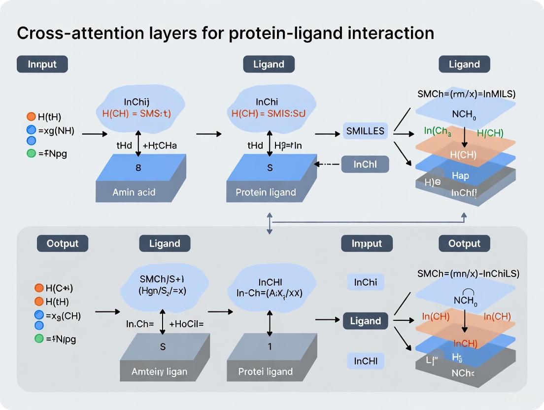
Workflow Diagram: Cross-Attention in Protein-Ligand Interaction
Diagram Title: LABind Cross-Attention Workflow
Architecture Diagram: Cross-Attention Mechanism
Diagram Title: Cross-Attention Mechanism Architecture
Research Reagent Solutions
Table 3: Essential computational tools and resources for cross-attention implementation
| Resource | Type | Application | Access |
|---|---|---|---|
| ProtBERT | Protein Language Model | Generating contextual residue embeddings from protein sequences | HuggingFace Model Hub |
| Ankh | Protein Language Model | Sequence representation in LABind | OpenSource |
| MolFormer | Molecular Language Model | Ligand representation from SMILES strings | NVIDIA NGC Catalog |
| ESMFold/OmegaFold | Structure Prediction | Generating 3D structures from sequences when experimental structures unavailable | OpenSource |
| DSSP | Structural Feature Tool | Calculating secondary structure and solvent accessibility | GitHub Repository |
| PDBbind | Benchmark Dataset | Training and evaluation for affinity prediction | Public Database |
| Gene Ontology | Knowledge Base | Biochemical knowledge integration in KEPLA | Public Database |
| RDKit | Cheminformatics | Molecular descriptor calculation and SMILES processing | OpenSource |
Implementation Considerations
Data Preparation Guidelines
Successful implementation of cross-attention models requires careful data preparation. For protein inputs, ensure consistent preprocessing of 3D structures, including proper hydrogen addition and residue numbering alignment. For ligand inputs, standardize SMILES representation using tools like RDKit to avoid representation variances. When working with binding affinity data, carefully curate the dataset to remove ambiguous complexes and ensure consistent measurement types (Kd, Ki, IC50). Implement rigorous data splitting strategies, such as cluster-based splits that separate proteins and ligands by similarity to prevent data leakage and properly evaluate generalization capability [11].
Computational Requirements and Optimization
Cross-attention models are computationally intensive, particularly for large protein complexes or high-throughput screening. Recommended implementation includes GPU acceleration with at least 16GB VRAM for training, and batch size optimization to balance memory constraints and training stability. For attention computation, consider implementing memory-efficient variants such as factored attention or block-sparse patterns when working with very large inputs. Monitoring attention entropy during training can help identify collapsed attention heads that may require reinitialization or regularization.
Interpretation and Validation Strategies
The cross-attention weights provide inherent interpretability, but require careful analysis. Implement attention visualization tools to map attention patterns onto 3D structures, identifying potential binding hotspots. Validate predictions through multiple metrics beyond overall accuracy, including performance on specific ligand classes and statistical significance testing. For binding site predictions, complement computational validation with experimental literature evidence when available, and consider employing ensemble methods to improve robustness across diverse protein families and ligand types.
In the field of computational drug discovery, accurately predicting how small molecules (ligands) interact with protein targets is a fundamental challenge. Traditional methods often struggle to capture the complex, long-range dependencies that govern these interactions, where atoms distant in sequence can be spatially close and critical for binding. Cross-attention mechanisms, a core component of modern transformer architectures, are emerging as a powerful solution to this challenge. These mechanisms allow for direct, dynamic communication between all elements of a protein and all elements of a ligand, enabling models to identify and weigh the importance of specific inter-molecular relationships regardless of their positional separation. This application note details how cross-attention is revolutionizing protein-ligand interaction research by capturing these non-local dependencies, providing researchers with protocols, data, and tools for implementation.
Quantitative Superiority of Cross-Attention Models
Cross-attention-based models have demonstrated state-of-the-art performance across multiple benchmarks related to protein-ligand interactions, from predicting binding affinity to identifying binding sites.
Table 1: Performance of Cross-Attention Models on Binding Affinity Prediction (CASF-2016 Benchmark)
| Model | Core Principle | Pearson's R (↑) | RMSE (↓) | MAE (↓) | CI (↑) |
|---|---|---|---|---|---|
| DAAP [13] | Distance features + Attention | 0.909 | 0.987 | 0.745 | 0.876 |
| PLAGCA [14] | Graph Cross-Attention | 0.864 | 1.120 | 0.860 | 0.847 |
| LumiNet [15] | Physics-integrated GNN | 0.850 | - | - | - |
Table 2: Performance of Cross-Attention Models on Binding Site Prediction
| Model | Task | Key Metric | Performance |
|---|---|---|---|
| LABind [7] [10] | Ligand-aware Binding Site Prediction | AUPR | Superior to P2Rank, DeepSurf, and DeepPocket |
| EZSpecificity [16] | Enzyme Substrate Specificity | Identification Accuracy | 91.7% (vs. 58.3% for previous model) |
The DAAP (Distance plus Attention for Affinity Prediction) model highlights the power of combining physics-inspired distance features with an attention mechanism, achieving a remarkably high correlation coefficient of 0.909 on the standard CASF-2016 benchmark [13]. Similarly, PLAGCA integrates global sequence features with local 3D structural features via graph cross-attention, demonstrating superior generalization capability and lower computational costs [14]. For binding site identification, LABind utilizes a graph transformer and cross-attention to learn distinct binding characteristics from protein structures and ligand SMILES sequences, enabling it to predict sites even for unseen ligands [7] [10].
Experimental Protocols for Cross-Attention Implementation
Protocol A: Implementing a Graph Cross-Attention Workflow for Affinity Prediction (Based on PLAGCA)
This protocol outlines the procedure for predicting protein-ligand binding affinity by integrating global and local features with cross-attention [14].
1. Input Representation and Feature Extraction: * Protein Global Features: Input the protein's FASTA sequence. Use a self-attention block or a pre-trained protein language model (e.g., Ankh [7]) to generate a global feature representation of the entire protein sequence. * Ligand Global Features: Input the ligand's SMILES string. Use a self-attention block or a pre-trained molecular language model (e.g., MolFormer [7]) to generate a global feature representation of the ligand. * Local Structure Representation: * Generate the 3D structure of the protein's binding pocket and the ligand. * Represent the pocket and ligand as a molecular graph, where nodes are atoms/residues and edges represent bonds or spatial proximity. * Use a Graph Neural Network (GNN) to generate initial atomic-level embeddings for both molecules.
2. Feature Interaction via Graph Cross-Attention: * Input the protein pocket and ligand graph embeddings into a cross-attention module. * In this module, the ligand embeddings serve as the Query, and the protein pocket embeddings serve as the Key and Value (or vice-versa). This allows each ligand atom to attend to and aggregate relevant information from all protein pocket atoms. * The output is a refined ligand representation that is context-aware of the protein pocket's structure.
3. Feature Fusion and Prediction: * Concatenate the protein global features, ligand global features, and the refined local interaction features from the cross-attention module. * Feed the combined feature vector into a Multi-Layer Perceptron (MLP) regressor. * The final output is the predicted binding affinity (e.g., pKd, pKi).
Protocol B: Ligand-Aware Binding Site Prediction with Cross-Attention (Based on LABind)
This protocol describes a method for predicting which protein residues form a binding site for a specific small molecule or ion [7] [10].
1. Input Encoding: * Ligand Encoding: Input the SMILES sequence of the ligand into a pre-trained molecular language model (MolFormer) to obtain a comprehensive ligand representation. * Protein Encoding: * Sequence Features: Input the protein sequence into a pre-trained protein language model (Ankh) to obtain per-residue embeddings. * Structural Features: Process the protein's 3D structure with a tool like DSSP to obtain geometric features (e.g., angles, distances, solvent accessibility). * Graph Construction: Convert the protein structure into a graph where nodes are residues. Node features are a combination of sequence embeddings and DSSP features. Edge features include spatial distances and directions between residues.
2. Protein-Ligand Interaction with Cross-Attention: * Process the protein graph through a graph transformer to capture internal residue-residue relationships and binding patterns. * The ligand representation and the transformed protein residue representations are processed through a cross-attention mechanism. * This mechanism enables the protein residues to "query" the ligand representation, learning the distinct binding characteristics for that specific ligand.
3. Binding Site Classification: * The output representation for each residue, now enriched with protein-ligand interaction information, is fed into an MLP classifier. * The classifier predicts a probability for each residue, indicating its likelihood of being part of a binding site for the query ligand.
Visualizing Workflows and Architectures
Graph 1: Hierarchical Workflow for Affinity Prediction. This diagram illustrates the integration of global sequence features and local 3D structural features through a cross-attention mechanism, as seen in models like PLAGCA [14] and LABind [7].
Graph 2: Core Cross-Attention Architecture. This diagram details the core cross-attention mechanism where the ligand representation queries the protein context, enabling ligand-aware prediction of binding sites, a key feature of LABind [7] [10].
The Scientist's Toolkit: Essential Research Reagents & Materials
Table 3: Key Research Reagent Solutions for Cross-Attention Research
| Item Name | Function/Application | Specific Examples |
|---|---|---|
| PDBbind Database | Provides curated experimental protein-ligand structures and binding affinities for training and benchmarking. | PDBbind v2016, v2020 [13] [14] |
| CASF Benchmark | Standardized benchmark set for rigorous evaluation of scoring power (affinity prediction). | CASF-2016 [13] [15] |
| Pre-trained Language Models | Provides rich, contextualized initial representations for proteins and ligands, boosting model performance. | Ankh (Protein), MolFormer (Ligand) [7] [10] |
| Graph Neural Network (GNN) Libraries | Framework for building models that operate directly on molecular graph structures. | PyTorch Geometric, Deep Graph Library (DGL) [17] [15] |
| Structure Analysis Tools | Extracts secondary structure and solvent accessibility features from protein 3D structures. | DSSP [7] [10] |
| Cross-Attention Implementation | The core algorithmic component that models interactions between protein and ligand representations. | Custom modules in PyTorch/TensorFlow [17] [14] |
The Transition from Sequence-Based to Interaction-Aware Models
The field of computational biology is undergoing a significant paradigm shift, moving from models that analyze biomolecular sequences in isolation to those that explicitly capture the intricate interactions between molecular entities. This transition is particularly transformative in protein-ligand interaction research, where accurately predicting binding affinity and docking poses is crucial for drug discovery. Traditional sequence-based models, which process protein and ligand information through separate encoders, have demonstrated limitations in generalizability and predictive accuracy because they fail to capture the complex, dynamic interactions that occur at the binding interface [12] [18].
The integration of cross-attention layers represents a cornerstone of this evolution, enabling models to learn the conditional relationships between protein residues and ligand atoms directly from data. These attention mechanisms allow for the creation of interaction-aware models that can identify specific non-covalent bonds, such as hydrogen bonds and hydrophobic interactions, which are critical for understanding binding mechanisms and predicting drug efficacy [18]. This application note details this methodological transition, provides experimental protocols for implementing interaction-aware models, and highlights the superior performance of these approaches through quantitative benchmarks.
The Paradigm Shift: From Sequential to Interaction-Aware Modeling
Limitations of Sequence-Based Models
Traditional sequence-based models for protein-ligand interaction have primarily relied on processing protein sequences (e.g., via FASTA) and ligand information (e.g., via SMILES strings) through separate, parallel encoders [12]. These encoders typically utilize convolutional neural networks (CNNs) or recurrent neural networks (RNNs) to extract global features from each molecule independently. The extracted features are then concatenated and passed to a final classifier or regression head to predict binding affinity or other properties.
The fundamental limitation of this architecture is its inability to model intermolecular interactions. By processing protein and ligand features in separate silos, these models lack a dedicated mechanism to identify which protein residues interact with which ligand atoms, or to capture the specific physicochemical nature of these interactions [18]. This often results in models that learn superficial correlations from the training data rather than the underlying binding mechanisms, leading to poor generalization on unseen protein-ligand pairs [12].
The Rise of the Interaction-Aware Paradigm
Interaction-aware models address these limitations by architecturally prioritizing the modeling of inter-molecular relationships. The core innovation is the use of cross-attention mechanisms that allow features from the protein and ligand to dynamically interact and influence each other during the computation of representations.
In this paradigm, the model learns to:
- Attend to relevant protein residues given a specific ligand atom, and vice versa.
- Weight the importance of different potential interactions.
- Integrate this interaction information directly into the molecular representations.
This approach is biologically grounded, as it mirrors the actual process of binding where local and specific interactions collectively determine the binding affinity and pose [18]. Models like Interformer and PLAGCA exemplify this shift, employing graph-transformers and cross-attention layers to explicitly model non-covalent interactions, thereby achieving new state-of-the-art performance in docking and affinity prediction tasks [18] [12].
Architectural Implementation of Cross-Attention Mechanisms
Graph-Transformer Hybrid Architectures
The Graph-Transformer architecture has emerged as a powerful framework for interaction-aware modeling, as demonstrated by the Interformer model [18]. This hybrid design effectively captures both the local connectivity within molecules and the global dependencies between them.
Table 1: Core Components of a Graph-Transformer for Protein-Ligand Interaction
| Component | Function | Implementation in Interformer |
|---|---|---|
| Input Representation | Represents protein binding site and ligand as graphs. | Nodes: atoms; Features: pharmacophore types. Edges: based on Euclidean distance [18]. |
| Intra-Blocks | Updates node features by capturing intra-molecular interactions (within protein or ligand). | Self-attention layers that operate on individual molecular graphs [18]. |
| Inter-Blocks | Captures inter-molecular interactions between protein and ligand atom pairs. | Cross-attention layers where one molecule's nodes attend to the other's, generating an "Inter-representation" [18]. |
| Interaction-Aware MDN | Models the conditional probability of distances for atom pairs, focusing on specific interactions. | Uses mixture density network (MDN) with Gaussian functions to model hydrogen bonds and hydrophobic interactions explicitly [18]. |
The following diagram illustrates the flow of information in a Graph-Transformer architecture like Interformer:
Figure 1: Graph-Transformer Architecture for Docking and Affinity Prediction. Intra-Blocks process individual molecules, while the Inter-Block uses cross-attention to model their interactions.
Hierarchical Cross-Attention for Binding Affinity Prediction
The PLAGCA and CheapNet models showcase another effective pattern: using hierarchical representations with cross-attention for the specific task of binding affinity prediction [12] [19]. These models integrate multiple levels of molecular information to achieve robust performance.
- Global Feature Extraction: PLAGCA uses sequence encoding and self-attention to extract global features from protein FASTA sequences and ligand SMILES strings [12].
- Local Interaction Feature Extraction: Simultaneously, it employs graph neural networks (GNNs) to extract local, 3D structural features from the protein binding pocket and the ligand [12].
- Feature Integration via Cross-Attention: A cross-attention mechanism is applied to these hierarchical representations, allowing the model to focus on the most relevant local features conditioned on the global context, and vice versa. The final prediction is made by a multi-layer perceptron (MLP) on the concatenated features [12].
CheapNet refines this concept by introducing cluster-level cross-attention. It generates hierarchical cluster-level representations from atom-level embeddings via differentiable pooling, which efficiently captures essential higher-order interactions that are critical for accurate binding affinity prediction while maintaining computational efficiency [19].
Quantitative Performance Benchmarks
The transition to interaction-aware models is quantitatively justified by their superior performance on established benchmarks for docking accuracy and binding affinity prediction.
Table 2: Performance Comparison of Interaction-Aware Models on Docking Tasks
| Model | Architecture | Benchmark | Performance (Top-1 Success Rate, RMSD < 2Å) |
|---|---|---|---|
| Interformer [18] | Graph-Transformer + Interaction-Aware MDN | PDBBind Time-Split | 63.9% |
| DiffDock [18] | GNN-based | PDBBind Time-Split | 53.6% |
| GNINA [18] | CNN-based | PDBBind Time-Split | 22.3% |
| Interformer [18] | Graph-Transformer + Interaction-Aware MDN | PoseBusters Benchmark | 84.09% |
Table 3: Performance of Interaction-Aware Models on Affinity Prediction
| Model | Architecture | Key Feature | Performance |
|---|---|---|---|
| PLAGCA [12] | GNN + Cross-Attention | Integrates global sequence and local 3D graph features | Outperforms state-of-the-art methods, superior generalization |
| CheapNet [19] | Hierarchical Cross-Attention | Atom-level and cluster-level interactions | State-of-the-art across multiple affinity prediction tasks |
Experimental Protocols
Protocol 1: Training an Interaction-Aware Docking Model
This protocol outlines the procedure for training a model like Interformer for protein-ligand docking and pose scoring.
A. Input Preparation and Featurization
- Data Source: Obtain protein-ligand complexes with 3D structures from databases like PDBBind.
- Graph Construction:
- For each complex, define a protein graph (binding site residues) and a ligand graph.
- Node Features: Use pharmacophore atom types (e.g., hydrogen bond donor/acceptor, hydrophobic, aromatic) for both protein and ligand atoms [18].
- Edge Features: For intra-molecular edges, use Euclidean distance between atoms. For inter-molecular edges, compute distances between all protein and ligand atom pairs within a specified cutoff (e.g., 5 Å).
B. Model Training Cycle
- Pre-Training (Optional): Pre-train the Intra-Blocks on large-scale molecular datasets using masked atom prediction or related self-supervised tasks.
- Supervised Docking Training:
- Feed the featurized protein and ligand graphs into the model.
- The Intra-Blocks process each graph independently to generate refined atom representations.
- The Inter-Block performs cross-attention between the protein and ligand representations.
- The Interaction-Aware MDN predicts parameters for multiple Gaussian distributions to model distances for different interaction types (general, hydrophobic, hydrogen bond) [18].
- The loss function is a negative log-likelihood loss against the true distances from crystal structures.
- Pose Scoring and Affinity Prediction Head Training:
- Use the generated docking poses (from Monte Carlo sampling using the predicted energy function) as input.
- A virtual node collects information from the pose via a final self-attention layer.
- This virtual node's embedding is used to predict a confidence (pose) score and a binding affinity value (e.g., pIC50) [18].
- Employ a contrastive pseudo-Huber loss, which uses both good (native-like) and poor (decoy) poses to teach the model to discriminate between them [18].
Protocol 2: Assessing Binding Affinity with Cross-Attention
This protocol describes the methodology for training a model like PLAGCA or CheapNet for predicting protein-ligand binding affinity.
A. Multi-Modal Input Processing
- Sequence-Based Inputs:
- Encode the protein primary sequence from its FASTA format into a one-hot or embedding matrix.
- Encode the ligand's SMILES string into a sequential representation.
- Structure-Based Inputs:
- Extract or define the 3D binding pocket from the protein structure.
- Represent the pocket and the ligand as 3D graphs where nodes are atoms and edges are based on spatial proximity.
- Use atom features like element type, charge, and hybridization state.
B. Hierarchical Feature Integration and Prediction
- Global Feature Extraction: Process the protein and ligand sequences through separate encoders (e.g., CNNs or Transformers) to get global feature vectors [12].
- Local Feature Extraction: Process the protein pocket and ligand graphs through a GNN to get a set of local, atom-level features [12] [19].
- Cross-Attention Fusion:
- Apply a cross-attention layer where the global protein features are the query, and the local ligand features are the key and value (and vice versa). This allows the model to identify which local ligand features are most relevant given the global protein context.
- In CheapNet, an additional cross-attention step is performed on hierarchically pooled cluster-level representations to capture higher-order interactions [19].
- Affinity Prediction: Concatenate the final global and local, interaction-aware representations. Feed this combined feature vector into an MLP regressor to predict the binding affinity value [12].
The workflow for this protocol is summarized below:
Figure 2: Binding Affinity Prediction Workflow. The model integrates global sequence information and local 3D structural features via cross-attention.
The Scientist's Toolkit: Research Reagent Solutions
Table 4: Essential Computational Tools and Datasets for Interaction-Aware Research
| Resource Name | Type | Function/Purpose | Relevance to Interaction-Aware Models |
|---|---|---|---|
| PDBBind [18] | Dataset | Curated database of protein-ligand complexes with 3D structures and binding affinity data. | Primary source for training and benchmarking docking and affinity prediction models. |
| PoseBusters Benchmark [18] | Benchmark | Evaluates physical plausibility and correctness of docking poses. | Critical for validating the real-world performance of docking models like Interformer. |
| ESM-2 [20] | Pre-trained Model | Protein Language Model that generates embeddings from amino acid sequences. | Can be used to initialize protein feature encoders, providing evolutionarily informed input representations. |
| Monte Carlo (MC) Sampling [18] | Algorithm | A method for sampling conformational space by making random changes and accepting them based on an energy function. | Used in the docking pipeline (e.g., in Interformer) to generate candidate ligand poses by minimizing a model-predicted energy function. |
| Differentiable Pooling [19] | Algorithm | A method for hierarchically coarsening graph representations in a way that maintains differentiability for gradient-based learning. | Used in models like CheapNet to efficiently generate cluster-level features from atom-level graphs. |
| Spectral-Normalized Neural Gaussian Process (SNGP) [21] | Method | Enhances a model's ability to provide uncertainty estimates for its predictions. | Can be integrated to identify out-of-distribution samples and improve model reliability, though not yet common in interaction-aware models. |
Architectural Deep Dive: Implementing Cross-Attention for Specific Prediction Tasks
Accurately predicting the binding affinity between a protein and a small molecule (ligand) is a cornerstone of structure-based drug discovery, as it directly expresses the effectiveness of the protein-ligand complex and helps in ranking candidate drugs [22]. Traditional computational methods, ranging from molecular dynamics simulations to machine learning-based scoring functions, often face a trade-off between computational overhead and prediction accuracy [22] [23]. Recently, deep learning models have emerged as powerful tools capable of automatically learning complex patterns from protein and ligand data without relying heavily on domain-specific feature engineering [22] [14].
A significant architectural innovation in this domain is the adoption of the cross-attention mechanism. Unlike models that process protein and ligand features in isolation, cross-attention explicitly models the mutual interactions between amino acids in a protein and atoms in a ligand [14]. This allows the model to identify and weigh which specific parts of the protein are most influenced by which parts of the ligand, and vice versa, leading to a more nuanced and physically meaningful representation of the binding interaction [19] [14]. This document details the application and protocols for several state-of-the-art architectures that utilize cross-attention, namely EBA, CheapNet, and PLAGCA, providing a framework for their implementation in drug discovery research.
Comparative Analysis of Architectures
The following table summarizes the core characteristics, strengths, and performance metrics of the key architectures discussed in this protocol.
Table 1: Comparative Analysis of Protein-Ligand Binding Affinity Prediction Architectures
| Architecture | Core Innovation | Input Features | Key Mechanism | Reported Performance (Benchmark) |
|---|---|---|---|---|
| EBA (Ensemble Binding Affinity) [22] | Ensemble of 13 deep learning models | Combinations of 5 simple 1D sequential and structural features | Self-attention & cross-attention layers; model ensembling | CASF-2016: R=0.914, RMSE=0.957 [22] |
| CheapNet [19] [24] | Hierarchical cluster-level interactions | Molecular structures (3D) | Cross-attention between protein and ligand clusters | State-of-the-art across multiple tasks with high efficiency [19] |
| PLAGCA [14] | Integration of global and local features | Protein sequence, ligand SMILES, and 3D pocket structure | Graph cross-attention on local pockets; self-attention on sequences | Outperforms state-of-the-art on PDBBind2016 core set and CSAR-HiQ sets [14] |
| DEAttentionDTA [25] | Dynamic word embeddings | Protein sequence, pocket sequence, ligand SMILES | Self-attention on dynamically embedded sequences | Superior results on PDBBind2020 and CASF benchmarks [25] |
Detailed Architectures and Experimental Protocols
EBA: Ensemble Binding Affinity Prediction
The EBA framework addresses the challenge of low generalization in single-model approaches by leveraging the power of model ensembling. It trains multiple deep learning models, each with different combinations of input features, and combines their predictions to achieve superior accuracy and robustness [22].
Key Components:
- Input Features: EBA utilizes five types of 1D sequential and structural features, including a novel angle-based feature vector for short-range direct interactions, rather than relying on complex 3D complex features [22].
- Model Architecture: Individual models employ both self-attention and cross-attention layers. Self-attention captures long-range interactions within a protein or ligand sequence, while cross-attention layers are specifically designed to capture the interaction between proteins and ligands [22].
- Ensemble Strategy: Thirteen models are trained on various combinations of the five input features. The final prediction is generated by aggregating the outputs of the best-performing ensemble of these models [22].
Experimental Protocol:
- Data Preparation:
- Model Training:
- Train the 13 distinct deep learning models, each with a unique combination of input features.
- Each model should be trained using a regression loss function, such as Mean Squared Error (MSE), to predict the binding affinity (e.g., -logKd, -logKi).
- Ensemble Construction:
- Evaluate all possible combinations (ensembles) of the trained models on a held-out validation set.
- Select the ensemble that achieves the highest Pearson Correlation Coefficient (R) and the lowest Root Mean Square Error (RMSE).
- Evaluation:
- Benchmark the performance of the selected EBA ensemble on standard test sets like CASF-2016 and CSAR-HiQ, comparing R, RMSE, and MAE metrics against state-of-the-art predictors [22].
CheapNet: Cross-Attention on Hierarchical Representations
CheapNet addresses the computational inefficiency and noise associated with atom-level modeling by introducing a hierarchical representation that integrates atom-level and cluster-level interactions [19] [24].
Key Components:
- Hierarchical Representations: CheapNet goes beyond atom-level interactions. It uses differentiable pooling to group atoms into meaningful clusters, thereby capturing higher-order molecular representations that are crucial for binding [19] [24].
- Cross-Attention Mechanism: The core of CheapNet is a cross-attention mechanism that operates between the protein clusters and ligand clusters. This allows the model to focus on biologically relevant binding interactions at a more abstract and efficient level [19].
Experimental Protocol:
- Environment Setup:
- Data Preprocessing:
- Download preprocessed datasets (e.g., for Cross-dataset Evaluation, Diverse Protein Evaluation, or LEP) using the provided commands in the repository, which automatically fetch data from sources like GIGN and ATOM3D [24].
- Training:
- Prediction and Evaluation:
PLAGCA: Graph Cross-Attention for Local Pockets
PLAGCA is designed to integrate both global sequence information and local three-dimensional structural features of the protein binding pocket, addressing the limitation of methods that ignore local interaction features [14].
Key Components:
- Feature Integration: PLAGCA extracts three types of features:
- Global features from protein FASTA sequences and ligand SMILES strings using self-attention blocks.
- Local interaction features from the 3D molecular structures of protein binding pockets and ligands using a graph neural network (GNN) [14].
- Graph Cross-Attention: A graph cross-attention mechanism is applied to the local 3D graphs to learn the critical interaction features between the protein pocket and the ligand, highlighting residues with high contribution to binding [14].
Experimental Protocol:
- Data and Representation:
- Use protein-ligand complexes from PDBbind. Extract the global FASTA sequence and ligand SMILES.
- For local features, generate a molecular graph for the protein binding pocket and the ligand. Nodes represent atoms, and edges represent bonds or distances.
- Model Implementation:
- Implement a dual-pathway network:
- Global Pathway: Use an embedding layer followed by self-attention blocks to process sequences.
- Local Pathway: Use a GNN followed by a graph cross-attention layer to process the 3D graphs.
- Concatenate the output features from both pathways and feed them into a Multi-Layer Perceptron (MLP) for final affinity prediction [14].
- Implement a dual-pathway network:
- Interpretability Analysis:
- Analyze the attention scores from the graph cross-attention layer to identify critical functional residues in the protein pocket that contribute most to the binding affinity prediction [14].
The Scientist's Toolkit: Essential Research Reagents
Table 2: Key Research Reagents and Resources for Implementation
| Resource Name | Type | Description / Function | Example Source / Tool |
|---|---|---|---|
| PDBbind Database | Dataset | Comprehensive collection of protein-ligand complexes with binding affinity data for training and testing. | http://www.pdbbind.org.cn/ [14] |
| CASF Benchmark | Dataset | Well-known benchmark sets (e.g., CASF2016, CASF2013) for standardized performance evaluation. | PDBbind website [22] |
| SMILES String | Data Format | 1D string representation of a ligand's molecular structure. | Open Babel for conversion from SDF [25] |
| GNN & Transformer | Software Library | Libraries for building graph neural networks and attention mechanisms. | PyTorch, PyTorch Geometric, DeepMind's Graph Nets |
| Cross-Attention Module | Algorithmic Component | Core mechanism to model interactions between protein and ligand representations. | Custom implementation in model architectures [19] [14] |
Workflow and Architectural Diagrams
Generic Cross-Attention Model Workflow
The following diagram illustrates a high-level workflow common to many cross-attention based binding affinity prediction models, integrating steps from EBA, CheapNet, and PLAGCA.
Generic Cross-Attention Model Workflow: This diagram outlines the common steps in a cross-attention based pipeline, from data sourcing and feature extraction to encoding, interaction modeling, and final affinity prediction.
CheapNet's Hierarchical Cross-Attention Architecture
CheapNet's Hierarchical Architecture: This diagram details CheapNet's specific two-stage process, which first processes atoms and then groups them into clusters for efficient cross-attention.
CAT-DTI is a deep learning model designed to predict drug-target interactions by effectively capturing the feature representations of drugs and proteins alongside their interaction characteristics. The framework is engineered to enhance generalization capability within real-world scenarios, often characterized by out-of-distribution data. Its primary innovation lies in integrating a cross-attention mechanism with a Transformer-based architecture, possessing domain adaptation capability. This allows the model to efficiently learn the complex relationships between drug molecules and protein targets, a critical task for accelerating drug discovery and reducing development costs [17].
The prediction of drug-target interactions is a cornerstone of computer-aided drug discovery. While traditional methods, such as molecular docking, are often limited by computational inefficiency and relatively low accuracy of scoring functions, deep learning methods have shown significant promise. However, many existing deep learning models fail to fully capture global context information while retaining local features or adequately model the local crucial interaction sites between the drug molecule and target protein. The CAT-DTI framework was proposed to address these specific limitations, achieving superior predictive performance by leveraging a protein feature encoder that combines convolutional neural networks (CNN) with Transformer, and a cross-attention module for feature fusion [17] [26].
The CAT-DTI framework processes drug and target inputs through separate feature encoders before fusing their representations to predict the interaction. The following diagram illustrates the core workflow and architecture of the CAT-DTI model.
Protein Feature Encoder
The protein feature encoder is a critical component that processes the amino acid sequence of a target protein. It employs a convolution neural network (CNN) combined with a Transformer to encode the distance relationship between amino acids within the protein sequence. The CNN is effective at capturing local residue patterns and motifs from the amino acid sequence. The Transformer architecture then leverages self-attention to capture global context and long-distance dependencies between these local subsequences, which is crucial for understanding the full protein structure. This hybrid approach allows the model to consider both local features and global context information simultaneously, addressing a key limitation of models that rely solely on CNN [17] [26].
Drug Feature Encoder
For drug representation, the model begins by converting the drug's SMILES string into a corresponding 2D molecular graph. Each atom node in the graph is initialized with a 74-dimensional integer vector that encapsulates atom attributes such as type, degree, number of implicit hydrogens, formal charge, and hybridization. A three-layer Graph Convolutional Network (GCN) is then used to transmit and aggregate information on the drug molecular structure. Each GCN layer updates the feature representation of each atomic node using the information of its neighboring nodes, thereby effectively capturing the correlation information between adjacent atoms in the drug molecule. The output is a node-level drug feature map, which is retained for subsequent explicit learning of interactions with protein fragments [17].
Cross-Attention Module for Feature Fusion
After obtaining the feature maps for the drug and protein, they are input into a cross-attention module. This module is designed to interact the protein and drug features for feature fusion, rather than simply concatenating them. The mechanism allows the model to capture the interaction relationship between specific drug substructures and protein regions. Specifically, the key and value from the protein attention are swapped with those from the drug attention, enabling a deeper fusion of information. This process helps the model to preserve the internal features of drugs and proteins while simultaneously exploring the interaction information between them, addressing a common oversight in models that focus only on extracting internal features [17].
Domain Adaptation and Decoder
To enhance the model's generalization to novel drug-target pairs in real-world scenarios, CAT-DTI integrates a Conditional Domain Adversarial Network (CDAN). This component is employed to align DTI representations under diverse distributions, facilitating effective knowledge transfer from the source domain (training data) to a target domain with different data characteristics. Finally, the fused and domain-adapted features are processed through a decoder, typically a fully connected neural network, to produce the final DTI prediction [17].
Performance Evaluation and Benchmarking
The performance of CAT-DTI has been rigorously evaluated against multiple baseline models on several public benchmark datasets. The following tables summarize key quantitative results.
Table 1: Performance Comparison of CAT-DTI and Baseline Models on the BindingDB, BioSNAP, and Human Datasets (Values are AUROC)
| Model | BindingDB | BioSNAP | Human |
|---|---|---|---|
| SVM | 0.939 | 0.862 | 0.913 |
| RF | 0.942 | 0.860 | 0.939 |
| DeepConv-DTI | 0.945 | 0.886 | 0.978 |
| GraphDTA | 0.951 | 0.887 | 0.965 |
| MolTrans | 0.952 | 0.895 | 0.981 |
| DrugBAN | 0.960 | 0.903 | 0.981 |
| CAT-DTI | 0.965 | 0.909 | 0.983 |
| NFSA-DTI | 0.965 | 0.909 | 0.987 |
Table 2: Detailed Performance of CAT-DTI on the DrugBank Dataset
| Metric | Performance (Std) |
|---|---|
| Accuracy | 82.02% |
| Precision | 81.90% |
| MCC | 64.29% |
| F1 Score | 82.09% |
As shown in Table 1, CAT-DTI demonstrates robust and superior performance, achieving the highest or tied highest Area Under the Receiver Operating Characteristic Curve (AUROC) across all three benchmark datasets (BindingDB, BioSNAP, and Human). It outperforms other advanced models like DrugBAN and MolTrans, underscoring its effectiveness. The model's strong performance is further confirmed on the DrugBank dataset (Table 2), where it shows robust results across multiple metrics, including accuracy, precision, and F1 score [26] [27].
Experimental Protocol for CAT-DTI Implementation
This protocol provides a detailed methodology for replicating the CAT-DTI training and evaluation process as described in the foundational research.
Data Preprocessing and Preparation
- Drug Molecular Graph Construction: Convert drug SMILES strings into 2D molecular graphs. For each atom, generate a 74-dimensional feature vector encoding atom type, degree, number of implicit Hs, formal charge, number of radical electrons, hybridization, number of total Hs, and aromaticity. Set a maximum number of nodes per graph (e.g.,
m_d = 100) to ensure uniform input size [17]. - Protein Sequence Encoding: Represent protein amino acid sequences as numerical embeddings. The specific method for initial embedding generation (e.g., one-hot encoding, learned embeddings) should be detailed based on the original implementation [17].
- Dataset Splitting: Randomly split the chosen benchmark dataset (e.g., DrugBank, Davis, KIBA) into training, validation, and test sets using a standard ratio such as 8:1:1. This split is crucial for unbiased evaluation [27].
Model Training Procedure
- Initialization: Initialize model parameters, including the GCN for drugs, the CNN-Transformer hybrid for proteins, and the cross-attention modules.
- Loss Function: Use a binary cross-entropy loss function for the DTI classification task.
- Domain Adaptation Integration: Incorporate the Conditional Domain Adversarial Network (CDAN) loss during training. This involves a gradient reversal layer to maximize the domain classifier's loss, thereby encouraging the learning of domain-invariant features [17].
- Optimization: Train the model using a stochastic gradient descent-based optimizer (e.g., Adam). Utilize the validation set for early stopping to prevent overfitting. Monitor key metrics like AUC and AUPR on the validation set.
Model Evaluation and Validation
- Performance Metrics: Evaluate the trained model on the held-out test set. Report standard metrics for binary classification, including:
- Comparative Analysis: Compare the model's performance against established baseline models to contextualize the results.
Table 3: Key Research Reagents and Computational Tools for DTI Research
| Item / Resource | Function / Description |
|---|---|
| SMILES Strings | A standardized line notation for representing molecular structures of drugs, serving as the primary input for the drug encoder. |
| Amino Acid Sequences | The primary structure of the target protein, provided as a string of one-letter codes, serving as input for the protein encoder. |
| Molecular Graphs | A graph representation of a drug molecule where nodes are atoms and edges are bonds; used by GCNs to capture topological information. |
| Graph Convolutional Network (GCN) | A type of neural network that operates directly on graph structures to learn node embeddings by aggregating information from neighbors. |
| CNN-Transformer Hybrid Encoder | A feature extraction module that combines the local feature detection of CNNs with the global context capture of Transformer self-attention. |
| Cross-Attention Mechanism | A neural network layer that enables the model to jointly attend to and fuse information from two different modalities (e.g., drug and protein features). |
| Conditional Domain Adversarial Network (CDAN) | A technique to improve model generalization by aligning feature distributions across different domains (e.g., different experimental settings). |
| Benchmark Datasets (e.g., DrugBank, Davis, KIBA) | Publicly available, curated datasets containing known drug-target interactions used for training and evaluating DTI prediction models. |
Logical Flow of the CAT-DTI Framework
The following diagram illustrates the logical sequence of operations and decision points within the CAT-DTI framework, from input processing to final prediction.
Enzyme substrate specificity, the precise recognition and catalytic action of an enzyme on particular target molecules, is a cornerstone of biological function and a critical parameter in biotechnology and drug discovery [28]. The traditional "lock and key" analogy has been superseded by a more nuanced understanding of induced fit and enzyme promiscuity, where enzymes can dynamically adjust their conformation and even catalyze reactions beyond their primary function [28]. Accurately predicting these interactions has been a persistent challenge, impeding the efficient application of enzymes in fundamental research and industry.
The emergence of artificial intelligence (AI) is revolutionizing this field. This Application Note focuses on EZSpecificity, a novel AI tool that leverages a cross-attention-empowered SE(3)-equivariant graph neural network to achieve unprecedented accuracy in predicting enzyme-substrate pairs [28] [16] [29]. Developed by researchers at the University of Illinois Urbana-Champaign, EZSpecificity represents a significant leap forward, providing researchers with a powerful, freely available online tool to accelerate their work [28] [30].
EZSpecificity: Core Technology and Performance
Architectural Innovation and Training Regime
EZSpecificity's predictive power stems from its sophisticated architecture and the comprehensive dataset on which it was trained. The model is built on a cross-attention graph neural network that operates directly on the 3D structural representations of enzymes and substrates [16] [29]. The cross-attention mechanism is pivotal as it allows the model to learn the specific chemical interactions between amino acid residues in the enzyme's active site and functional groups on the substrate [30]. This SE(3)-equivariant design ensures that the model's predictions are robust to rotations and translations of the input structures, a crucial feature for analyzing molecular interactions [16].
The model was trained on a vast, tailor-made database of enzyme-substrate interactions that integrated both sequence and structural information [16]. To overcome the scarcity of experimental data, the team employed extensive molecular docking simulations, performing millions of calculations to create a large-scale computational dataset of enzyme-substrate pairs [28] [30]. This hybrid training approach, which combined limited experimental data with expansive computational data, was key to building a highly accurate and generalizable model [30].
Quantitative Performance Benchmarking
EZSpecificity's performance was rigorously evaluated against ESP, the existing state-of-the-art model for enzyme substrate specificity prediction. The validation involved benchmark tests across multiple scenarios and experimental follow-up on a challenging enzyme class.
Table 1: Comparative Performance of EZSpecificity vs. ESP Model
| Evaluation Metric | EZSpecificity | ESP (State-of-the-Art) |
|---|---|---|
| Overall Accuracy (Top Prediction) | 91.7% [28] [16] | 58.3% [28] [16] |
| Validation Case | 8 Halogenase enzymes vs. 78 substrates [28] [16] | 8 Halogenase enzymes vs. 78 substrates [28] [16] |
The experimental validation on halogenases, a class of enzymes with poorly characterized specificity that is increasingly used to synthesize bioactive molecules, underscores EZSpecificity's practical utility and superior accuracy in real-world applications [28] [16].
The following diagram illustrates the integrated computational and experimental workflow for developing and validating EZSpecificity:
Application Notes: Harnessing EZSpecificity in Research
Accessing the Tool
EZSpecificity has been developed as a freely available online tool to maximize its accessibility to the research community [28]. Users can access the model through a user-friendly web interface. The researchers have made the tool open source with no restrictions, though a patent has been filed to protect the intellectual property [30]. The official demo can be accessed via the Shukla Group's website or the publication links associated with the Nature paper [29].
Input Requirements and Workflow
To use EZSpecificity, researchers must provide two key pieces of information about the system they wish to analyze:
- Protein Sequence: The amino acid sequence of the enzyme of interest.
- Substrate Structure: The chemical structure of the target substrate molecule [28].
The model processes these inputs through its cross-attention graph neural network to predict the compatibility of the enzyme-substrate pair, outputting a prediction of whether the substrate is likely to be accepted by the enzyme [28] [30].
Practical Use-Cases and Applications
EZSpecificity is designed to accelerate research and development across multiple disciplines:
- Drug Discovery: Identify novel substrates for enzymes involved in biosynthetic pathways of therapeutic agents, or predict off-target effects of drug candidates [28] [30].
- Synthetic Biology and Metabolic Engineering: Determine the optimal enzyme and substrate combinations to efficiently produce desired chemicals, fuels, and materials in engineered biological systems [28] [30].
- Enzyme Engineering and Characterization: Rapidly screen and prioritize enzyme mutants for experimental testing, streamlining the process of designing enzymes with new or improved functions [28]. The tool is particularly valuable for characterizing understudied enzyme families, such as halogenases [16].
Experimental Protocol: Validation of Halogenase Substrate Specificity
The following section details the experimental protocol used to validate EZSpecificity's predictions for halogenase enzymes, a process that can be adapted for testing computational predictions in other enzyme systems.
Reagent Setup
Table 2: Essential Research Reagents for Enzyme Specificity Validation
| Reagent / Material | Function / Description | Example / Comment |
|---|---|---|
| Halogenase Enzymes | Catalyzes the incorporation of halogen atoms into substrates. | Purified recombinant enzymes (e.g., 8 different halogenases) [16]. |
| Substrate Library | Molecules to be tested for enzymatic activity. | A diverse set of potential substrates (e.g., 78 compounds) [28] [16]. |
| Reaction Buffer | Provides optimal pH and ionic conditions for the enzyme. | e.g., 50 mM Tris-HCl, pH 7.5 [31]. |
| Analytical Instrumentation | Detects and quantifies the reaction product. | HPLC-MS or spectrophotometer for measuring product formation [16]. |
Step-by-Step Procedure
- Enzyme Purification: Express and purify the recombinant halogenase enzymes of interest using standard chromatographic methods (e.g., affinity chromatography) to obtain a preparation with high purity and specific activity [16].
- Reaction Assembly: In a final volume of 50 µL, combine the following components:
- Incubation: Incubate the reaction mixture at the enzyme's optimal temperature (e.g., 40°C) for a defined period, typically between 30 to 60 minutes [31].
- Reaction Termination: Stop the reaction by placing the tubes on ice for 10 minutes [31].
- Product Quantification:
- Spectrophotometric Assay: If the reaction produces a chromophoric product (like p-nitrophenol in assays with pNP-substrates), measure the absorbance at the appropriate wavelength (e.g., 410 nm) using a calibration curve to determine the concentration of the released product [31].
- Chromatographic Assay: For non-chromophoric products, use techniques like High-Performance Liquid Chromatography (HPLC) or Mass Spectrometry (MS) to separate and quantify the reaction products [16].
- Control Reactions: Run parallel control reactions to ensure validity. Essential controls include:
- Substrate Control: Water plus substrate (checks for non-enzymatic degradation).
- Enzyme Control: Enzyme plus water (checks for background activity).
- Host Protein Control: Host cell protein lysate (from non-recombinant cells) plus substrate (checks for interference from host proteins) [31].
The logical flow of this validation protocol is summarized below:
Data Analysis and Interpretation
Calculate enzyme activity based on the amount of product formed per unit of time per amount of enzyme. Compare the activities across different substrates to rank substrate preferences. A successful validation is achieved when the substrates predicted by EZSpecificity to be reactive show significantly higher activity than those predicted to be non-reactive.
Technical Specifications and Future Outlook
EZSpecificity sets a new benchmark for enzyme specificity prediction. The developers are committed to its continued enhancement. Key future directions include:
- Expanding Predictive Scope: The team plans to extend the AI framework to predict enzyme selectivity (preference for certain sites on a substrate), which is vital for ruling out undesirable off-target effects in therapeutic applications [28].
- Improving Quantitative Predictions: A major goal is to move beyond binary classification (substrate/non-substrate) to predicting quantitative kinetic parameters, such as reaction rates and binding affinities (e.g., Km and kcat), which would provide a more nuanced understanding of enzyme performance [30].
- Enhancing General Accuracy: While performance on certain enzyme families is exceptional, accuracy for other classes can be lower. The researchers aim to address this by incorporating more experimental and computational data into the training set to create a model of broader applicability and higher general accuracy [30].
Table 3: Key Technical Features of EZSpecificity
| Feature | Specification |
|---|---|
| Core Architecture | Cross-attention empowered SE(3)-equivariant graph neural network [16]. |
| Input Data | Enzyme sequence and substrate structure [28]. |
| Training Data | Comprehensive database integrating sequence, structure, and docking simulations [28] [16]. |
| Key Differentiator | Uses cross-attention to model atomic-level enzyme-substrate interactions [29] [30]. |
| Availability | Freely available online as an open-source tool [28] [30]. |
Protein-ligand interactions are fundamental to numerous biological processes, including enzyme catalysis and signal transduction, and are pivotal in drug discovery and design [10]. Identifying the specific regions on a protein where these interactions occur, known as binding sites, is a critical step. Experimental methods for determining binding sites are resource-intensive, creating a pressing need for robust computational solutions [10]. While existing computational methods exist, they are often limited; they are either tailored to specific ligands and fail on unseen compounds, or they are multi-ligand methods that do not explicitly incorporate ligand information, constraining their accuracy and generalizability [10] [32].
The LABind (Ligand-Aware Binding site prediction) model represents a significant advance by directly addressing these limitations. It is a structure-based method designed to predict binding sites for small molecules and ions in a "ligand-aware" manner [10] [32]. This means LABind explicitly learns the distinct binding characteristics between a protein and a specific ligand, enabling it to generalize effectively to ligands not encountered during its training phase. Its design is situated within a broader thesis that cross-attention mechanisms are uniquely powerful for modeling complex biomolecular interactions, as they allow for deep, learned integration of information from different molecular entities [10] [14].
Computational Framework of LABind
LABind's architecture is engineered to learn interactions between protein structural contexts and ligand chemical properties. Its overall workflow integrates several advanced components to achieve ligand-aware prediction.
Model Architecture and Workflow
The following diagram illustrates the end-to-end workflow of the LABind model, from input processing to final binding site prediction:
The Role of the Cross-Attention Mechanism
The cross-attention module is the core of LABind's ligand-aware capability [10]. It enables the model to dynamically compute the relevance and potential interactions between each residue in the protein graph and the input ligand. Unlike simpler methods that process protein and ligand features in isolation, this mechanism allows the ligand's representation to directly influence and query the protein's structural features. This process learns the "distinct binding characteristics" between the specific protein-ligand pair, which is essential for accurately identifying binding sites for a wide array of ligands, including those that are unseen during training [10]. The success of cross-attention in LABind is part of a growing trend in bioinformatics, with models like PLAGCA also leveraging graph cross-attention to learn local interaction features for predicting binding affinity [14].
Performance Analysis and Benchmarking
LABind's performance has been rigorously evaluated against state-of-the-art methods on multiple benchmark datasets (DS1, DS2, and DS3), demonstrating superior accuracy and generalizability.
Key Performance Metrics
The model was evaluated using standard metrics for imbalanced classification, including Area Under the Precision-Recall Curve (AUPR) and Matthews Correlation Coefficient (MCC), which are particularly informative given the scarcity of binding residues compared to non-binding ones [10].
Table 1: Comparative Performance of LABind on Benchmark Datasets
| Method | Type | AUPR (DS1) | MCC (DS1) | AUPR (DS2) | MCC (DS2) | Generalization to Unseen Ligands |
|---|---|---|---|---|---|---|
| LABind | Ligand-Aware | 0.723 | 0.651 | 0.685 | 0.594 | Yes |
| LigBind | Single-Ligand-Oriented | 0.691 | 0.622 | 0.652 | 0.561 | Limited |
| GraphBind | Single-Ligand-Oriented | 0.645 | 0.580 | 0.621 | 0.540 | No |
| P2Rank | Multi-Ligand-Oriented | 0.598 | 0.532 | 0.578 | 0.501 | No |
| DeepSurf | Multi-Ligand-Oriented | 0.634 | 0.569 | 0.605 | 0.527 | No |
LABind consistently outperforms both single-ligand-oriented methods (e.g., GraphBind, LigBind) and multi-ligand-oriented methods (e.g., P2Rank, DeepSurf) across key benchmarks [10]. Its primary advantage is the maintained high performance on unseen ligands, a scenario where other models struggle.
Performance on Molecular Docking
The practical utility of LABind extends to improving molecular docking tasks. When the binding sites predicted by LABind were used to define the search space for the docking tool Smina, a significant enhancement in the accuracy of the generated docking poses was observed [10].
Table 2: Application in Molecular Docking (Smina)
| Docking Search Space Method | Pose Accuracy (RMSD < 2.0 Å) | Average Docking Time (min) |
|---|---|---|
| LABind-predicted site | 78.5% | 4.2 |
| P2Rank-predicted site | 65.3% | 4.5 |
| Full protein surface scan | 71.1% | 12.8 |
Application Notes and Protocols
This section provides detailed methodologies for implementing and utilizing the LABind model in various research scenarios.
Protocol 1: Standard Structure-Based Binding Site Prediction
This is the primary protocol for predicting binding sites when an experimental protein structure and a ligand of interest are available.
1. Input Preparation:
- Protein Structure File: Obtain the protein's 3D structure in PDB format from the Protein Data Bank (PDB) or via structure prediction tools.
- Ligand SMILES String: Define the ligand using its Simplified Molecular-Input Line-Entry System (SMILES) string, which can be sourced from databases like PubChem.
2. Feature Extraction:
- Protein Features:
- Process the protein sequence through the Ankh protein language model to generate residue-level embeddings [10].
- Analyze the protein structure with DSSP to compute secondary structure and solvent accessibility features [10].
- Convert the protein structure into a graph where nodes represent residues. Node features include spatial attributes (angles, distances), and edge features represent residue-residue interactions.
- Ligand Features: Process the ligand's SMILES string through the MolFormer molecular language model to obtain a comprehensive ligand representation [10].
3. Model Inference:
- Combine the protein graph and ligand representation in the LABind model.
- The model processes the inputs through its graph transformer and cross-attention layers.
- The output is a per-residue probability score indicating the likelihood of that residue being part of a binding site for the specified ligand.
4. Post-processing:
- Apply a threshold (optimized to maximize the MCC score) to the probabilities to generate binary predictions (binding/non-binding) for each residue [10].
- Cluster adjacent binding residues to define one or more discrete binding pockets.
Protocol 2: Sequence-Based Prediction with ESMFold
For proteins without an experimentally determined structure, LABind can be applied using predicted structures.
1. Input Preparation: Provide only the protein's amino acid sequence and the ligand's SMILES string.
2. Protein Structure Prediction: Use a high-accuracy protein structure prediction tool like ESMFold or OmegaFold to generate a 3D model of the protein from its sequence [10].
3. Binding Site Prediction: Use the predicted protein structure as input to the standard LABind pipeline (Protocol 1). Experimental results validate LABind's robustness even when using predicted structures, though a minor performance drop compared to using experimental structures may occur [10].
Protocol 3: Binding Site Center Localization
This protocol is used to identify the precise spatial center of a binding pocket, which is valuable for docking and functional studies.
1. Binding Site Residue Prediction: Execute Protocol 1 or 2 to identify binding site residues.
2. Center Calculation:
- Extract the 3D coordinates of the Cα atoms of all predicted binding residues.
- Calculate the geometric center (centroid) of these coordinates.
- This centroid is reported as the predicted binding site center.
3. Validation Metric: The performance is evaluated using DCC (Distance between the predicted binding site Center and the true binding site Center) and DCA (Distance to the Closest ligand Atom). Lower DCC and DCA values indicate higher prediction accuracy [10].
The Scientist's Toolkit: Research Reagent Solutions
The following table details the key software, databases, and models that are essential for operating the LABind framework.
Table 3: Essential Research Reagents and Resources for LABind
| Resource Name | Type | Function in LABind Protocol | Source/Availability |
|---|---|---|---|
| Ankh | Pre-trained Protein Language Model | Generates evolutionary and semantic embeddings from protein sequences. | Academic Use |
| MolFormer | Pre-trained Molecular Language Model | Generates molecular representations from ligand SMILES strings. | Academic Use |
| DSSP | Bioinformatics Tool | Derives secondary structure and solvent accessibility from 3D coordinates. | Open Source |
| ESMFold/OmegaFold | Protein Structure Prediction Tool | Predicts 3D protein structures from amino acid sequences for protocol 2. | Academic Use |
| PDBbind | Curated Database | Provides benchmark datasets of protein-ligand complexes for training and testing. | http://www.pdbbind.org.cn |
| RDKit | Cheminformatics Library | Handles ligand molecular graphs and conformer generation (used in related methods like LaMPSite) [33]. | Open Source |
| Smina | Molecular Docking Software | Used to validate the utility of LABind-predicted sites in docking tasks. | Open Source |
Advanced Applications and Case Study
Distinguishing Binding Sites for Different Ligands
A powerful application of LABind is its ability to predict distinct binding sites for different ligands on the same protein. The model's cross-attention mechanism allows it to adapt its predictions based on the specific chemical properties of the input ligand. For example, LABind can be used to show how a protein like human serum albumin binds fatty acids differently than it binds drugs like warfarin, by highlighting different residue clusters as the binding site for each ligand type [10]. This capability was validated through visualization of the model's attention patterns.
Case Study: SARS-CoV-2 NSP3 Macrodomain
LABind was successfully applied to predict the binding sites of the SARS-CoV-2 NSP3 macrodomain with unseen ligands [10].
- Objective: To identify potential binding sites for novel compounds that could inhibit the virus's function.
- Method: The protein structure was input into LABind along with the SMILES strings of the novel, unseen ligands.
- Outcome: LABind accurately predicted binding sites that were consistent with known functional regions of the macrodomain, demonstrating its real-world applicability in drug discovery against new targets and for novel chemistries [10].
The following diagram illustrates the logical decision process and functional relationships LABind leverages to handle such real-world cases, including the distinction between structure-based and sequence-based inputs.
Accurate prediction of protein-ligand binding affinity (PLA) is a critical task in computational drug discovery, as it helps determine how strongly a drug candidate (ligand) interacts with a protein target, thereby influencing drug efficacy [11]. While recent deep learning approaches have shown promising results, they often rely solely on the structural features of proteins and ligands, creating performance bottlenecks and lacking scientific interpretability [11]. To overcome these limitations, the KEPLA (Knowledge-Enhanced Protein-Ligand binding Affinity prediction) framework represents a novel approach that explicitly integrates prior biochemical knowledge from Gene Ontology (GO) and ligand properties (LP) to enhance both prediction performance and interpretability [11].
KEPLA is an interaction-free model, meaning it infers binding affinity from lower-dimensional data like protein amino acid sequences and ligand molecular graphs, without requiring known three-dimensional structures of protein-ligand complexes [11]. This gives it a wider application scope than interaction-based methods, especially when facing proteins with unknown 3D structures. The framework's core innovation lies in its deep integration of biochemical factual knowledge through a knowledge graph (KG), moving beyond traditional black-box predictions to provide scientifically grounded insights [11].
Architectural Framework and Methodology
Core Components of KEPLA
The KEPLA framework follows an encoder-decoder paradigm, jointly optimized on two complementary objectives: a knowledge graph embedding objective and a binding affinity prediction objective [11]. The overall architecture and workflow are designed to seamlessly integrate structural data with external knowledge, as shown in Figure 1.
Figure 1. KEPLA Framework Workflow. The diagram illustrates the integration of protein and ligand encoders with knowledge graph embedding and cross-attention mechanisms for binding affinity prediction.
The Role of Gene Ontology and Knowledge Graphs
Gene Ontology provides a systematic framework for describing gene products across three domains: Molecular Function (e.g., kinase activity), Biological Process (e.g., signal transduction), and Cellular Component (e.g., cell membrane) [34]. KEPLA constructs a comprehensive knowledge graph that incorporates GO annotations for proteins and molecular descriptors for ligands, organizing this diverse biochemical knowledge into entity-relation-entity triples that the model can efficiently process [11].
For instance, if a protein's molecular function includes "ATP binding," this GO annotation becomes a node in the knowledge graph, potentially connected to ATP-like ligands through relation edges. This structured representation allows the model to learn that such proteins may exhibit high affinity for ATP-like compounds [11]. Similarly, ligand properties such as the number of hydrogen bond donors and acceptors are incorporated into the knowledge graph, capturing crucial information about potential binding interactions with proteins [11].
Integration with Cross-Attention Mechanisms
While KEPLA itself utilizes cross-attention between local protein and ligand representations to construct fine-grained joint embeddings [11], this approach aligns with broader trends in protein-ligand interaction research. The cross-attention mechanism enables the model to focus on the most relevant substructures between the protein and ligand, mimicking the selective binding nature of molecular interactions [14] [17].
In KEPLA's architecture, the cross-attention module processes the encoded local representations through a local interaction mapping step followed by cross-attention computation, which together generate a joint protein-ligand representation that feeds into the final prediction layer [11]. This approach allows the model to learn which specific amino acids in the protein and which molecular substructures in the ligand contribute most significantly to their binding interaction.
Experimental Protocols and Validation
Data Preparation and Knowledge Graph Construction
Materials and Datasets:
- Protein-Ligand Complexes: Source from PDBbind database (refined set and core set) and CSAR-HiQ dataset [11].
- Gene Ontology Annotations: Obtain from Gene Ontology Consortium database or UniProt [34].
- Ligand Properties: Calculate molecular descriptors using RDKit or similar cheminformatics tools [35].
- Knowledge Graph Construction: Use entity-relation-entity triples to represent protein-GO and ligand-LP relationships [11].
Protocol Steps:
- Data Collection and Curation: Download protein-ligand complexes from PDBbind, ensuring removal of overlapping complexes between training and test sets [11].
- GO Annotation Mapping: Map each protein to its corresponding GO terms using UniProt ID cross-referencing [34].
- Ligand Feature Extraction: Generate ligand properties including hydrogen bond donors/acceptors, molecular weight, and other relevant descriptors [11].
- Knowledge Graph Assembly: Construct the KG using (protein, hasfunction, GOterm) and (ligand, hasproperty, moleculardescriptor) triples [11].
- Dataset Splitting: Implement both random splits for in-domain evaluation and clustering-based pair splits for cross-domain validation [11].
Model Training and Implementation
Implementation Details:
- Protein Encoder: Use ESM (Evolutionary Scale Modeling) for processing protein amino acid sequences [11].
- Ligand Encoder: Implement Graph Convolutional Networks (GCNs) for molecular graph input [11].
- Knowledge Graph Embedding: Apply standard KG embedding techniques such as TransE or ComplEx [11].
- Cross-Attention Module: Implement multi-head cross-attention between local protein and ligand representations [11].
- Training Procedure: Jointly optimize the KG embedding loss and PLA prediction loss using multi-task learning [11].
Training Protocol:
- Initialize protein and ligand encoders with pre-trained weights when available.
- For each batch of protein-ligand pairs:
- Extract global and local representations using respective encoders.
- Compute KG embedding loss by comparing aligned global representations with KG relations.
- Compute interaction features through cross-attention of local representations.
- Generate affinity predictions through MLP decoder.
- Calculate weighted sum of both losses and perform backpropagation.
- Validate on both in-domain and cross-domain test sets.
- Use early stopping based on validation performance to prevent overfitting.
Evaluation Metrics and Experimental Design
Performance Metrics:
- Primary Metric: Root Mean Square Error (RMSE) for binding affinity prediction [11].
- Additional Metrics: Include Pearson correlation coefficient and mean absolute error for comprehensive evaluation [11].
Experimental Strategies:
- In-Domain Evaluation: Random split of protein-ligand complexes with 9:1 training-validation ratio [11].
- Cross-Domain Evaluation: Cluster-based pair split using single-linkage algorithm on protein and ligand features to simulate domain shift [11].
- Ablation Studies: Evaluate contribution of individual components by removing KG embedding objective or cross-attention module [11].
Table 1: KEPLA Performance Comparison on Benchmark Datasets
| Dataset | Evaluation Scenario | Baseline RMSE | KEPLA RMSE | Improvement |
|---|---|---|---|---|
| PDBbind Core Set | In-Domain | 1.41 | 1.34 | 5.28% |
| CSAR-HiQ | In-Domain | 1.53 | 1.34 | 12.42% |
| PDBbind | Cross-Domain | 1.62 | 1.47 | 9.26% |
Table 2: Research Reagent Solutions for KEPLA Implementation
| Reagent Category | Specific Tools/Resources | Function in Framework |
|---|---|---|
| Protein Data | PDBbind Database [11] | Provides protein-ligand complexes with binding affinity data |
| Ontology Resources | Gene Ontology Consortium [34] | Source of functional annotations for knowledge graph |
| Molecular Descriptors | RDKit [35] | Calculates ligand properties for knowledge graph |
| Protein Encoder | ESM (Evolutionary Scale Model) [11] | Generates protein representations from sequences |
| Ligand Encoder | Graph Convolutional Networks [11] | Processes ligand molecular graphs |
| Knowledge Graph Embedding | TransE/ComplEx Algorithms [11] | Aligns representations with biochemical knowledge |
| Interaction Module | Cross-Attention Mechanism [11] | Captures fine-grained protein-ligand interactions |
Interpretation and Analysis Framework
Knowledge Graph Relation Analysis
The knowledge graph in KEPLA provides a natural mechanism for interpretability through analysis of relation strengths and attention patterns. Researchers can identify which GO terms and ligand properties most strongly influence binding affinity predictions by examining:
- Entity Embedding Similarity: Calculate cosine similarity between protein/ligand embeddings and related KG entities to identify influential knowledge elements [11].
- Relation Path Analysis: Trace important relation paths in the KG that connect specific proteins to ligands through shared GO terms or properties [11].
- Attention Visualization: Generate heatmaps of cross-attention weights to identify critical binding residues and molecular substructures [11].
Cross-Attention Visualization Protocol
The cross-attention mechanism provides residue-level and atom-level insights into binding interactions. The protocol for interpreting these patterns includes:
Figure 2. Cross-Attention Interpretation Workflow. The process for visualizing and analyzing attention patterns to identify critical binding determinants.
Visualization Steps:
- Extract Attention Weights: For a given protein-ligand pair, extract the cross-attention weights between all residue-atom pairs [11].
- Generate Interaction Heatmaps: Create two-dimensional heatmaps with protein residues on one axis and ligand atoms on the other, colored by attention weight intensity [11].
- Identify Binding Hotspots: Highlight residues and atoms with consistently high attention weights across multiple forward passes [11].
- Correlate with KG Entities: Map high-attention residues to their GO annotations to provide functional context for the observed interactions [11].
Case Study: Application to Novel Ligand Binding
A practical application of KEPLA's interpretability framework involves predicting binding affinity for proteins with novel ligands. The analysis protocol includes:
- KG Completion: For novel ligands not in the original KG, use similar ligand properties to infer potential relations [11].
- Cross-Attention Pattern Matching: Compare attention patterns with known high-affinity complexes to predict binding modes [11].
- Functional Validation Hypothesis: Generate testable hypotheses about which GO-annotated functions might be most relevant for the novel interaction [11].
This interpretability framework moves beyond traditional black-box predictions, providing researchers with actionable insights into the molecular determinants of binding affinity and facilitating more informed decisions in drug discovery pipelines.
Overcoming Practical Hurdles: Strategies for Robust and Generalizable Models
Addressing Data Imbalance in Pocket Prediction with Focal Loss
In the field of structure-based drug discovery, accurately predicting protein-ligand binding sites is a critical first step. This process is fundamentally hampered by severe class imbalance, where binding residues typically constitute less than 5% of all amino acids in a protein [36]. This skew predisposes standard machine learning models toward the non-binding majority class, resulting in poor predictive performance for the binding sites of primary interest. Within the broader thesis research on using cross-attention layers for protein-ligand interaction studies, addressing this data imbalance is not merely a preprocessing step but a core challenge that must be overcome to leverage the full power of advanced deep-learning architectures.
This Application Note provides a detailed protocol for implementing Focal Loss [36] to mitigate this imbalance in binding site prediction. We situate this solution within a ligand-aware prediction framework that utilizes cross-attention mechanisms to integrate protein and ligand information, enabling the model to learn distinct binding characteristics for different ligands, including those not seen during training [7]. The integration of Focal Loss ensures that the model's attention is effectively directed toward learning from the critical minority class—the binding residues.
Theoretical Foundation and Key Metrics
The Class Imbalance Challenge in Pocket Prediction
Binding site prediction is typically formulated as a per-residue classification task. In a typical protein, the ratio of binding to non-binding residues can be exceptionally low. For instance, in the SJC dataset used to train CLAPE-SMB, the binding sites ratio was reported to be lower than 5% [36]. This imbalance causes models to be dominated by the majority class, making standard evaluation metrics like accuracy misleading and uninformative.
Focal Loss: A Solution to Class Imbalance
Focal Loss (FL) is an extension of the standard cross-entropy loss designed to address class imbalance by down-weighting the loss assigned to well-classified examples and focusing learning on hard, misclassified examples [36]. The loss function is defined as:
Class-Balanced Focal Loss [36]:
L_focal = - ((1-β) / (1-β^(n_y))) * Σ (1 - p_i^t)^γ * log(p_i^t)
Parameters and Their Roles:
p_i^t: The model's estimated probability for the true class.- Modulating Factor
(1 - p_i^t)^γ: This component is the core of Focal Loss. Theγ(gamma) parameter is a focusing parameter. A higherγincreases the relative loss for misclassified examples, forcing the model to focus on them. - Effective Number
(1-β)/(1-β^(n_y)): This term, proposed by Cui et al., handles class imbalance by using a hyperparameterβto re-weight classes based on their effective number of samples.
In practice, Focal Loss is often combined with other objective functions. For example, CLAPE-SMB used a composite loss integrating Focal Loss with Triplet Center Loss (TCL) to better distinguish between binding and non-binding sites in the embedding space [36].
Evaluation Metrics for Imbalanced Data
Standard metrics like accuracy are unsuitable for imbalanced classification. The field has instead adopted a suite of metrics that provide a more realistic picture of model performance, especially for the minority class. The following table summarizes the key metrics used for evaluating binding site predictors.
Table 1: Key Evaluation Metrics for Imbalanced Classification in Pocket Prediction
| Metric | Formula | Interpretation and Utility |
|---|---|---|
| Precision | TP / (TP + FP) |
Measures the reliability of positive predictions; high precision means fewer false positives [37] [7]. |
| Recall (Sensitivity) | TP / (TP + FN) |
Measures the ability to find all positive samples; high recall means fewer false negatives [37] [7]. |
| F1-Score | 2 * (Precision * Recall) / (Precision + Recall) |
The harmonic mean of precision and recall; provides a single balanced metric [37] [7]. |
| AUPR | Area under the Precision-Recall curve | More informative than ROC-AUC for imbalanced data as it focuses on the performance of the positive class [7]. |
| MCC | Matthews Correlation Coefficient | A correlation coefficient between observed and predicted classifications that is generally regarded as a balanced measure [7]. |
| ROC-AUC | Area under the Receiver Operating Characteristic curve | Measures the model's ability to separate classes across all thresholds; a common benchmark (e.g., PocketMiner achieved 0.87) [38] [37]. |
Integrated Protocol: Ligand-Aware Binding Site Prediction with Focal Loss
This protocol details the implementation of a ligand-aware binding site prediction model, LABind [7], incorporating Focal Loss to handle class imbalance. The architecture leverages a cross-attention mechanism to fuse protein and ligand information.
Research Reagent Solutions
Table 2: Essential Materials and Software Tools
| Item Name | Function / Description | Relevance to Protocol |
|---|---|---|
| ESM-2 | A pre-trained protein language model that generates evolutionary-scale sequence embeddings from amino acid sequences [36] [39]. | Used to obtain robust, pre-trained feature representations of the input protein sequence. |
| MolFormer | A pre-trained molecular language model that generates molecular representations from SMILES strings [7]. | Used to encode the ligand's chemical information for the cross-attention mechanism. |
| DSSP | Dictionary of Protein Secondary Structure program; assigns secondary structure and solvent accessibility from 3D coordinates [7]. | Provides crucial structural features (e.g., angles, accessibility) that complement sequence embeddings. |
| Graph Transformer | A neural network architecture that processes data structured as graphs, using self-attention to weigh the importance of nodes and edges [7]. | The core network for processing the protein's 3D structure represented as a graph of residues. |
| Cross-Attention Module | A mechanism that allows representations from different modalities (e.g., protein and ligand) to interact and attend to each other [7]. | Enables the model to be "ligand-aware" by learning specific protein-ligand interaction patterns. |
Workflow and Signaling Pathway
The following diagram illustrates the complete experimental workflow for ligand-aware binding site prediction, from data input to final output.
Step-by-Step Methodology
Step 1: Data Preparation and Feature Extraction
- Input Processing: For a given protein-ligand complex, extract the protein structure (PDB format) and the ligand's SMILES string.
- Protein Feature Generation:
- Process the protein sequence through the ESM-2 model to obtain a 1,280-dimensional embedding for each residue [36].
- Run the DSSP program on the protein structure to compute structural features (e.g., secondary structure, solvent accessibility) for each residue [7].
- Concatenate the ESM-2 embedding and DSSP features to form a comprehensive protein representation.
- Ligand Feature Generation: Process the ligand's SMILES string through the MolFormer pre-trained model to obtain a fixed-dimensional vector representing the ligand's chemical properties [7].
- Graph Construction: Convert the protein 3D structure into a graph where nodes represent residues. Node features are the combined protein representation, and edges represent spatial proximity. Incorporate spatial features like distances, angles, and directions between residues [7].
Step 2: Model Architecture and Training with Focal Loss
- Protein Graph Encoding: Feed the protein graph into a Graph Transformer network. This architecture uses self-attention to capture long-range interactions and the spatial context of the protein's structure, outputting a refined representation for each residue [7].
- Cross-Attention for Protein-Ligand Interaction: The refined protein representations and the ligand representation from MolFormer are passed through a cross-attention layer. This mechanism allows the protein residues to "attend" to the ligand and vice versa, learning the distinct binding characteristics between the specific protein and ligand [7].
- Classification Layer: The output of the cross-attention mechanism for each residue is fed into a Multi-Layer Perceptron (MLP) classifier with a softmax output to predict the probability of each residue being a binding site [7].
- Loss Calculation and Optimization:
- Calculate the Class-Balanced Focal Loss (
L_focal) between the predictions and the true labels [36]. - (Optional) As done in CLAPE-SMB, a contrastive loss like Triplet Center Loss (TCL) can be added to the total loss to further improve the separation between binding and non-binding residue embeddings [36]. The total loss is:
L_total = L_focal + λ * L_tc, whereλis a weighting hyperparameter. - Use a modern optimizer (e.g., AdamW) to minimize the total loss and update the model's weights.
- Calculate the Class-Balanced Focal Loss (
Step 3: Model Evaluation
- Prediction: Run the trained model on the hold-out test set.
- Performance Assessment: Calculate the suite of metrics listed in Table 1 (Precision, Recall, F1-score, AUPR, MCC, ROC-AUC) to comprehensively evaluate model performance, with a particular emphasis on AUPR and F1-score due to the imbalanced nature of the task [37] [7].
Integrating Focal Loss into a modern, ligand-aware deep-learning framework provides a robust solution to the pervasive challenge of class imbalance in protein-ligand binding site prediction. The methodology outlined in this application note, centered on the LABind architecture, demonstrates how to effectively leverage pre-trained language models, geometric deep learning, and cross-attention mechanisms. By forcing the model to focus on hard, minority-class examples, Focal Loss ensures that the sophisticated representations learned by the cross-attention layers are effectively channeled toward the accurate identification of binding residues. This approach significantly enhances the model's utility in a real-world drug discovery pipeline, where correctly identifying a potential binding pocket is the critical first step toward designing novel therapeutics.
Ensemble Methods (EBA) to Boost Generalization Capability
Accurate prediction of protein-ligand binding affinity is a cornerstone of structure-based drug discovery, as it directly influences the efficiency of virtual screening and the ranking of candidate drugs during the drug development process [40]. The strength of this interaction determines the biological effectiveness of the protein-ligand complex and serves as a key metric for initial drug candidate success [40]. While various computational methods have been developed to predict binding affinity, most existing deep learning approaches utilize single models that often suffer from limitations in accuracy and, crucially, generalization capability across diverse datasets [40]. For instance, the CAPLA model demonstrates strong performance on benchmark CASF2016 and CASF2013 datasets but shows poor generalization on CSAR-HiQ test sets [40]. This lack of robustness presents a significant challenge in computational drug discovery.
A promising strategy to enhance generalization involves employing ensemble learning, where multiple models are combined to capture a wider spectrum of characteristics from the data [40]. The Ensemble Binding Affinity (EBA) method addresses the generalization challenge by integrating multiple deep learning models with different feature combinations, utilizing cross-attention and self-attention layers to extract both short and long-range interactions within protein-ligand complexes [40]. This approach moves beyond single-model predictions to create a more robust and reliable framework for binding affinity prediction, ultimately contributing to improved success rates for potential drugs and an accelerated drug development pipeline [40].
Core Methodology of Ensemble Binding Affinity (EBA)
The EBA framework is built upon a systematic approach to model diversification and integration. Its core innovation lies in strategically combining multiple deep learning models, each trained on distinct combinations of input features, to form a powerful ensemble that significantly outperforms any single constituent model [40].
Feature Engineering and Model Diversification
The foundation of EBA's robustness is the diverse set of input features used to train its constituent models. EBA extracts information pertaining to the protein, the ligand, and their interaction using five primary input features [40]. Rather than relying on computationally expensive 3D complex features, EBA utilizes simpler 1D sequential and structural features, making it more efficient while maintaining high accuracy [40]. A key innovation is the generation of a new angle-based feature vector, which is designed to capture short-range direct interactions between proteins and ligands [40]. The models within EBA employ cross-attention layers to effectively capture the interaction between ligands and proteins, and self-attention layers to extract both short and long-range dependencies within the data [40].
In total, thirteen distinct deep learning models are trained using various combinations of the five input features [40]. This deliberate variation in input feature space ensures that the models learn complementary representations and patterns from the data, which is the fundamental prerequisite for a successful ensemble.
Ensemble Construction Strategy
After training the thirteen individual models, the EBA method explores all possible ensembles of these models to identify the optimal combinations [40]. This exhaustive search strategy ensures that the final ensemble is not based on an arbitrary selection but is empirically determined to deliver the best predictive performance. The ensemble's final prediction is achieved by aggregating the outputs of its constituent models, thereby synthesizing their diverse knowledge and compensating for individual model weaknesses. This process results in a more accurate and stable prediction of binding affinity than any single model could achieve [40].
Table 1: Key Research Reagent Solutions for EBA Implementation
| Research Reagent / Resource | Function and Description |
|---|---|
| PDBbind Datasets [40] | Standardized benchmark datasets (e.g., PDBbind2016, PDBbind2020) for training and validating protein-ligand binding affinity prediction models. |
| Protein FASTA Sequences [12] | Provides the primary amino acid sequence of the target protein, used for extracting global sequence features. |
| Ligand SMILES Strings [40] [12] | A line notation for representing ligand molecular structures, used as input for feature extraction. |
| Angle-Based Feature Vector [40] | A custom feature engineered to capture short-range direct interaction geometry between the protein and ligand. |
| Cross-Attention Layers [40] [12] | A neural network mechanism that allows the model to focus on relevant parts of the protein and ligand features when modeling their interaction. |
| Graph Neural Network (GNN) [41] | An alternative framework for representing protein-ligand complexes as graphs to capture topological and interaction features. |
Quantitative Performance Benchmarking
The EBA method has been rigorously evaluated against state-of-the-art predictors across multiple benchmark datasets. Its performance, measured by Pearson Correlation Coefficient (R) and Root Mean Square Error (RMSE), demonstrates a significant and consistent improvement over existing methods.
On the well-known CASF2016 benchmark test set, one of the EBA ensembles achieved a top-tier Pearson R value of 0.857 and an RMSE of 1.195 when trained on the PDBbind2016 dataset [40]. When the training data was scaled up to the PDBbind2020 dataset, the performance of EBA improved further, with the best ensemble achieving a remarkable Pearson R value of 0.914 on the CASF2016 benchmark, setting a new standard for accuracy [40].
The generalizability of EBA is most evident in its performance on the CSAR-HiQ test sets, where it showed a dramatic improvement over the second-best predictor, CAPLA. EBA achieved an increase of more than 15% in R-value and a reduction of over 19% in RMSE on both CSAR-HiQ test sets [40]. This leap in performance on external validation data underscores the effectiveness of the ensemble approach in creating models that generalize well to new, diverse complexes.
Table 2: Performance Benchmarking of EBA on CASF2016 Dataset
| Method | Pearson | RMSE | MAE |
|---|---|---|---|
| EBA (Trained on PDBbind2016) | 0.857 | 1.195 | 0.951 |
| EBA (Trained on PDBbind2020) | 0.914 | 0.957 | Not Reported |
| CAPLA [40] | Lower by >15% | Higher by >19% | Not Reported |
| Other State-of-the-Art Methods [40] | Outperformed across all metrics | Outperformed across all metrics | Outperformed across all metrics |
Table 3: EBA Performance on CSAR-HiQ Test Sets
| Test Set | Performance Metric | EBA Result | Improvement over CAPLA |
|---|---|---|---|
| CSAR-HiQ Dataset 1 | Pearson | Significantly Higher | > 15% |
| CSAR-HiQ Dataset 1 | RMSE | Significantly Lower | > 19% |
| CSAR-HiQ Dataset 2 | Pearson | Significantly Higher | > 15% |
| CSAR-HiQ Dataset 2 | RMSE | Significantly Lower | > 19% |
Experimental Protocols
Protocol 1: Training an Individual Base Model for EBA
This protocol details the procedure for training a single deep learning model that can serve as a component of the EBA ensemble.
1. Input Feature Preparation:
- Proteins: Obtain the protein's FASTA sequence. Encode the sequence into a numerical vector suitable for neural network input [12].
- Ligands: Obtain the ligand's SMILES string. Encode the SMILES string into a numerical representation [40] [12].
- Structural Features: Calculate the angle-based feature vector from the protein-ligand complex structure to capture short-range interactions [40].
- Feature Combination: Select a specific combination from the five available input features for this particular model, as per the EBA diversification strategy [40].
2. Model Architecture Configuration:
- Employ a neural network architecture that incorporates both self-attention and cross-attention layers [40] [12].
- The self-attention layers should be applied to the protein and ligand features independently to capture long-range interactions within each molecule [40].
- The cross-attention layers are critical for modeling the interactions between the protein and ligand features, allowing the model to focus on relevant residues and atoms [40] [12].
- The output of these attention layers should be fed into a regression head (e.g., a Multi-Layer Perceptron) to produce the final binding affinity prediction [12].
3. Model Training:
- Use a standard dataset such as PDBbind2016 or PDBbind2020 for training [40].
- Use a loss function appropriate for regression, such as Mean Squared Error (MSE).
- Train the model until convergence on a held-out validation set, employing an optimizer like Adam.
Protocol 2: Constructing and Validating the EBA Ensemble
This protocol describes the process of combining individual models into an ensemble and evaluating its performance.
1. Base Model Collection:
- Train a total of thirteen individual models, each with a different combination of the five input features, following Protocol 1 [40].
2. Ensemble Construction:
- Perform an exhaustive search by evaluating all possible ensembles of the thirteen trained models on a validation set [40].
- Select the ensemble configuration that delivers the best performance metrics (e.g., highest Pearson R, lowest RMSE) [40].
3. Ensemble Validation & Benchmarking:
- Make final predictions on the test set by aggregating (e.g., averaging) the predictions from all models in the selected ensemble [40].
- Evaluate the ensemble's performance on multiple independent benchmark test sets, including CASF2016, CASF2013, and CSAR-HiQ [40].
- Compare the results against current state-of-the-art methods to confirm the superiority and improved generalization of the EBA ensemble [40].
EBA Ensemble Workflow
Integration with Cross-Attention Research
The EBA framework is intrinsically linked to the broader thesis on using cross-attention layers for protein-ligand interaction research. Cross-attention is not merely a component but a foundational mechanism within the EBA's constituent models, enabling them to effectively capture the critical interactions between proteins and ligands [40] [12].
The cross-attentional mechanism allows a model to dynamically focus on specific residues in the protein and specific atoms in the ligand that are most relevant for their binding interaction [12]. This is a significant advancement over methods that process protein and ligand features in isolation, as it explicitly models the pairwise interactions between the two entities. When this powerful mechanism is replicated across multiple models in an ensemble, each trained on different feature sets, the EBA framework effectively creates a multi-faceted "lens" for examining protein-ligand interactions. Each model in the ensemble learns a slightly different perspective of the interaction landscape via cross-attention, and their combination leads to a more comprehensive and robust understanding, which directly translates to superior generalization capability on unseen test data [40].
Cross-Attention Mechanism
Mitigating Domain Shift with Domain Adversarial Networks (e.g., in CAT-DTI)
Domain shift presents a significant challenge in computational drug discovery, where models trained on one distribution of drug-target pairs often fail to generalize to new data with different characteristics. This technical note explores the integration of Domain Adversarial Networks into drug-target interaction (DTI) prediction models, with specific focus on the CAT-DTI (Cross-Attention and Transformer network with Domain Adaptation) framework. The content is framed within a broader thesis investigating cross-attention mechanisms for protein-ligand interaction research, highlighting how domain adversarial training enhances model robustness and generalizability across diverse biological contexts.
Domain Shift in DTI Prediction: Core Challenges
Domain shift occurs when a model encounters data during deployment that differs significantly from its training data, leading to performance degradation. In DTI prediction, this manifests through several key challenges:
- Variable Assay Conditions: Experimental data collected under different laboratory conditions, temperatures, or pH levels creates distributional shifts
- Diverse Protein Families: Models trained on specific protein classes (e.g., kinases) may not generalize well to other families (e.g., GPCRs)
- Novel Chemical Space: New drug candidates with structural features underrepresented in training data
- Species-Specific Variations: Differences in protein sequences and structures across organisms
The CAT-DTI model addresses these challenges by incorporating a conditional domain adversarial network (CDAN) that aligns feature representations across different domains, enabling more reliable predictions on out-of-distribution data [17] [42].
CAT-DTI employs a multi-component architecture designed to capture complex drug-target interactions while mitigating domain shift:
Feature Extraction Modules
- Drug Molecular Graph Encoding: Molecular graphs from SMILES strings processed through Graph Convolutional Networks (GCNs) to generate drug feature maps ((F_D)) [17]
- Protein Sequence Encoding: Combination of CNN and Transformer architectures capturing both local features and global context in protein sequences ((F_P)) [17] [26]
Cross-Attention Mechanism
The model employs a specialized cross-attention module that swaps keys and values between drug and protein attention mechanisms, enabling explicit learning of interaction features between atomic nodes in drug molecules and residues in protein sequences [17].
Domain Adaptation Component
The conditional domain adversarial network aligns feature distributions between source and target domains using gradient reversal during training, forcing the feature extractor to learn domain-invariant representations [17] [42].
Quantitative Performance Analysis
Table 1: Performance Comparison of CAT-DTI Against Baseline Models on Benchmark Datasets
| Model | BindingDB AUROC | BindingDB AUPRC | BioSNAP AUROC | BioSNAP AUPRC | Human AUROC | Human AUPRC |
|---|---|---|---|---|---|---|
| SVM | 0.939 | 0.928 | 0.862 | 0.864 | 0.913 | 0.905 |
| RF | 0.942 | 0.921 | 0.860 | 0.886 | 0.939 | 0.927 |
| DeepConv-DTI | 0.945 | 0.925 | 0.886 | 0.890 | 0.978 | 0.982 |
| GraphDTA | 0.951 | 0.934 | 0.887 | 0.890 | 0.965 | 0.955 |
| MolTrans | 0.952 | 0.936 | 0.895 | 0.897 | 0.981 | 0.976 |
| DrugBAN | 0.960 | 0.948 | 0.903 | 0.902 | 0.981 | 0.969 |
| CAT-DTI | 0.965 | 0.957 | 0.909 | 0.909 | 0.983 | 0.976 |
Performance metrics demonstrate CAT-DTI's consistent improvement across multiple datasets, particularly in cross-domain scenarios. AUROC: Area Under Receiver Operating Characteristic curve; AUPRC: Area Under Precision-Recall Curve [26].
Table 2: Cross-Domain Generalization Performance
| Model | In-Domain Accuracy | Cross-Domain Accuracy | Generalization Gap |
|---|---|---|---|
| Traditional DTI Models | 0.882 | 0.705 | 0.177 |
| CAT-DTI (with CDAN) | 0.896 | 0.836 | 0.060 |
CAT-DTI demonstrates significantly reduced performance degradation when applied to out-of-distribution data, highlighting the effectiveness of its domain adaptation components [17] [42].
Experimental Protocol: Implementing Domain Adversarial Training
Data Preparation and Preprocessing
- Drug Representation: Convert SMILES strings to molecular graphs with atom-level features (atom type, degree, implicit hydrogens, formal charge, radical electrons, hybridization, total hydrogens, aromaticity) [17]
- Protein Representation: Process amino acid sequences with position-specific encoding, maintaining maximum sequence length of (m_d) residues
- Dataset Splitting: Implement chronological time-split partitioning (training: pre-2019, testing: 2020+) to simulate real-world domain shift scenarios [43]
Model Training Procedure
CAT-DTI Architecture and Domain Adaptation Workflow
Critical Training Parameters
- Optimization: Adam optimizer with learning rate 0.001, batch size 64
- Gradient Reversal: Weight of -0.1 applied during backpropagation to encourage domain-invariant features [17] [44]
- Early Stopping: Monitoring validation loss with patience of 20 epochs
- Regularization: Dropout rate of 0.3 and L2 regularization (λ = 0.0001)
Evaluation Metrics and Cross-Validation
- Primary Metrics: AUROC, AUPRC, Accuracy, Sensitivity, Specificity [26]
- Domain Shift Assessment: Performance comparison between in-domain and cross-domain test sets
- Statistical Validation: 5-fold cross-validation with stratified sampling
The Scientist's Toolkit: Research Reagent Solutions
Table 3: Essential Research Reagents and Computational Tools for DTI with Domain Adaptation
| Category | Specific Tool/Resource | Function | Application in CAT-DTI |
|---|---|---|---|
| Data Resources | PDBbind Database [43] | Curated protein-ligand complex structures | Model training and benchmarking |
| DrugBank [45] | Comprehensive drug and target database | Drug feature extraction and validation | |
| BindingDB [26] | Public database of drug-target interactions | Performance evaluation on diverse compounds | |
| Computational Libraries | RDKit [43] | Cheminformatics and molecular manipulation | SMILES processing and molecular graph generation |
| PyTorch/TensorFlow | Deep learning frameworks | Model implementation and training | |
| Graph Neural Network Libraries | Specialized graph processing | Drug molecular graph encoding | |
| Domain Adaptation Components | Gradient Reversal Layer [17] [44] | Implements adversarial training | Forces domain-invariant feature learning |
| Conditional Domain Adversarial Network [17] | Aligns feature distributions | Handles domain shift in DTI prediction | |
| Evaluation Frameworks | CAPRI Criteria [46] | Standard for protein docking assessment | Model quality assessment in structural contexts |
Implementation Considerations for Research Applications
Practical Deployment Guidelines
When implementing domain adversarial networks for DTI prediction:
- Data Requirements: Minimum of 10,000 diverse drug-target pairs for effective domain adaptation [17]
- Computational Resources: GPU acceleration recommended (NVIDIA Tesla V100 or equivalent) for training times of 6-12 hours
- Hyperparameter Tuning: Focus on gradient reversal weight and feature dimension balancing for optimal performance
Interpretation and Explainability
The cross-attention mechanism in CAT-DTI provides inherent interpretability by:
- Identifying specific atomic contributions in drug molecules to binding interactions
- Highlighting relevant amino acid residues in protein sequences
- Visualizing interaction patterns between drug and protein substructures [17] [26]
Limitations and Alternative Approaches
While CAT-DTI demonstrates strong performance, researchers should consider:
- Alternative Architectures: NFSA-DTI incorporating neural fingerprints and self-attention mechanisms [26]
- Reinforcement Learning Approaches: MoleProLink-RL using geometric transport for domain-policy learning [45]
- Dynamic Docking Methods: DynamicBind for predicting ligand-specific protein conformations [43]
Domain adversarial networks represent a significant advancement in addressing domain shift challenges in drug-target interaction prediction. The CAT-DTI framework successfully integrates cross-attention mechanisms with conditional domain adversarial training to improve model generalizability across diverse biological contexts. The experimental protocols and implementation guidelines provided in this technical note enable researchers to effectively apply these methods in protein-ligand interaction studies, potentially accelerating drug discovery pipelines and improving prediction reliability in real-world scenarios.
Enhancing 3D Geometric Awareness with SE(3)-Equivariant Networks and Curvature Features
The accurate prediction of protein-ligand interactions is a cornerstone of modern drug discovery. Traditional computational methods often struggle to capture the complex three-dimensional geometric and physical principles that govern these interactions. The incorporation of SE(3)-equivariant neural networks—which inherently respect the symmetries of 3D space (rotations and translations)—represents a transformative advancement for structural biology. When enhanced with curvature-aware features and cross-attention mechanisms, these models achieve unprecedented performance in predicting binding affinities, poses, and complex structures. This document provides application notes and experimental protocols for leveraging these technologies within a research framework focused on protein-ligand interactions, offering scientists a practical guide to implementing state-of-the-art geometric deep learning models.
Key Mechanisms and Theoretical Foundations
SE(3) Equivariance in Molecular Modeling
SE(3)-equivariant neural networks are architecturally constrained so that their internal representations and outputs transform predictably under any 3D rotation or translation of the input data. This means that if an input protein-ligand complex is rotated, the predicted binding affinity or pose transforms accordingly, without the model needing to learn this symmetry from data [47]. This is mathematically formalized by ensuring that an equivariant map ( f ) satisfies: [ f(T \cdot x) = T \cdot f(x), \quad \forall T \in \mathrm{SE}(3) ] where ( T ) is a transformation in the SE(3) group [47]. This built-in geometric awareness ensures stability and data efficiency, which is critical in scientific domains where labeled experimental data is scarce.
The Role of Cross-Attention for Hierarchical Interactions
Cross-attention mechanisms enable models to learn the complex relationships between different hierarchical components of a biological system. For instance, in protein-ligand binding, it allows the model to dynamically weigh the importance of different protein residues with respect to specific ligand atoms or molecular fragments. The attention weights ( \alpha_{ij} ) are computed as scalar invariants, ensuring they are unaffected by the global orientation of the molecules [47]. This capability is crucial for moving beyond simple atom-level interactions to capture more complex, cluster-level interactions that drive binding [19].
Curvature and Spectral Features for Geometric Awareness
Incorporating features that describe the local curvature and intrinsic geometry of protein surfaces provides critical information for identifying binding pockets and interaction sites. The Feature-enhanced Multi-scale Network (FMN), for example, uses a Spectral-Vectorized Feature Enhancement module that incorporates the Laplace spectrum to capture the intrinsic shape of molecular structures [48]. These spectral features help the model discriminate between different conformational states and binding propensities that are not apparent from atomic coordinates alone.
Quantitative Performance of SE(3)-Equivariant Models
The following tables summarize the performance of various SE(3)-equivariant models on key tasks in drug discovery, demonstrating their state-of-the-art capabilities.
Table 1: Performance of SE(3)-Equivariant Models on Complex Structure Prediction
| Model | Task | Key Metric | Performance | Reference |
|---|---|---|---|---|
| DeepTernary | Ternary Complex Prediction (PROTAC) | DockQ Score | 0.65 | [49] |
| DeepTernary | Ternary Complex Prediction (MGD) | DockQ Score | 0.21 | [49] |
| DeepTernary | Inference Time | Average per Complex | ~7 sec (PROTAC), ~1 sec (MGD) | [49] |
| EquiCPI | Virtual Screening (DUD-E) | AUC | On par/exceeding state-of-the-art | [50] |
Table 2: Performance on Binding Site and Affinity Prediction
| Model | Task | Key Metric | Performance | Reference |
|---|---|---|---|---|
| LABind | Binding Site Prediction | AUPR | Superior to baseline methods | [10] |
| PLAGCA | Binding Affinity Prediction | Comparative Accuracy | Outperforms other computational methods | [12] |
| CheapNet | Binding Affinity Prediction | Performance vs. Efficiency | State-of-the-art across multiple benchmarks | [19] |
| FMN | Molecular Dynamics (MD17) | Positional Error (MAE) | State-of-the-art results | [48] |
Experimental Protocols
Protocol 1: Predicting PROTAC-Induced Ternary Complexes with DeepTernary
Application Note: This protocol details the procedure for predicting the 3D structure of a ternary complex formed by a PROTAC molecule, an E3 ligase, and a target protein of interest, which is critical for targeted protein degradation drug discovery [49].
Workflow Diagram:
Materials & Reagents:
- Hardware: GPU cluster (recommended NVIDIA A100 or equivalent)
- Software: Python 3.8+, PyTorch, DeepTernary codebase
- Data: TernaryDB dataset (curated from PDB) or custom protein/ligand data
Step-by-Step Procedure:
- Data Curation and Preprocessing:
- Curate a dataset of ternary complexes from the Protein Data Bank (PDB). The DeepTernary study used TernaryDB, a curated set of over 22,000 high-quality complexes containing a small molecule and two proteins [49].
- Apply stringent filters for resolution, sequence similarity, and ligand quality. Exclude known PROTACs and molecular glue degraders (MGDs) from the training set to test generalizability.
- Use MMseqs2 to cluster proteins by sequence similarity. Remove entire clusters containing test-set complexes to prevent data leakage [49].
- Model Input Representation:
- Disassemble each ternary complex into three components: protein 1 (p1), the ligand (lig), and protein 2 (p2).
- Model each component as a graph where nodes represent atoms or residues. Node features should include chemical and physical properties.
- Edge features should capture spatial relationships, such as distances and directions derived from atomic coordinates [49] [10].
- Model Training with SE(3) Equivariance:
- Implement an SE(3)-equivariant graph neural network as the encoder. This ensures all learned features transform correctly under 3D rotations and translations of the input complex [49] [47].
- Employ a novel ternary inter-graph attention mechanism to capture the intricate relationships between the p1, lig, and p2 components.
- Use a query-based pocket points decoder to predict the final 3D coordinates of the assembled ternary complex.
- Train the model using a loss function that combines terms for coordinate accuracy and structural fidelity.
- Validation and Analysis:
- Evaluate the predicted structures against experimentally determined benchmarks using the DockQ score.
- Calculate the Buried Surface Area (BSA) of the predicted complex, as this metric correlates with experimental degradation potency [49].
Protocol 2: Protein-Ligand Binding Affinity Prediction with PLAGCA
Application Note: This protocol describes a method for accurately predicting binding affinity by integrating global protein/ligand features with local, curvature-sensitive, 3D interaction features from the binding pocket [12].
Workflow Diagram:
Materials & Reagents:
- Software: RDKit (for molecular graph generation), PyTorch Geometric or DGL (for GNNs), Transformer libraries (e.g., Hugging Face)
- Data: BindingDB or PDBbind for affinity labels
Step-by-Step Procedure:
- Feature Extraction:
- Global Features: Encode the protein's FASTA sequence and the ligand's SMILES string using self-attention based encoders (e.g., a Transformer) to capture long-range context and chemical structure [12].
- Local 3D Features: From the 3D structure of the protein's binding pocket and the ligand, generate molecular graphs. Use a Graph Neural Network (GNN) to extract features that capture the local atomic environment and geometry.
- Feature Integration via Cross-Attention:
- Implement a graph cross-attention mechanism between the protein pocket graph and the ligand graph. This allows the model to learn which specific protein residues and ligand atoms interact most significantly [12] [10].
- The cross-attention layer computes a weighted sum of the ligand's graph features for each protein residue node, dynamically focusing on the most relevant interaction partners.
- Affinity Prediction:
- Concatenate the global sequence/SMILES features with the local graph interaction features extracted from the cross-attention layer.
- Feed the combined feature vector into a Multi-Layer Perceptron (MLP) to produce the final binding affinity prediction (e.g., pKd, pKi) [12].
- Model Interpretation:
- Analyze the attention weights from the cross-attention layer to identify critical functional residues and ligand substructures that contribute most to the binding. This provides interpretable insights for lead optimization.
Protocol 3: Ligand-Aware Binding Site Prediction with LABind
Application Note: This protocol uses a graph transformer and cross-attention to predict binding sites for small molecules and ions in a ligand-aware manner, enabling generalization to unseen ligands [10].
Materials & Reagents:
- Pre-trained Models: Ankh (protein language model), MolFormer (molecular language model)
- Structural Analysis Tool: DSSP for deriving protein secondary structure features
Step-by-Step Procedure:
- Input Representation Generation:
- Ligand Representation: Input the ligand's SMILES string into the pre-trained MolFormer model to obtain a molecular property representation [10].
- Protein Representation:
- Generate protein sequence embeddings using the Ankh pre-trained language model.
- Compute structural features (e.g., secondary structure, solvent accessibility) from the protein's 3D coordinates using DSSP.
- Convert the protein structure into a graph. Node (residue) spatial features should include angles, distances, and directions. Edge features should include directions, rotations, and distances between residues.
- Concatenate the sequence embeddings and DSSP features to form the initial node features in the protein graph [10].
- Spatio-Structural Encoding:
- Process the protein graph through a graph transformer to capture potential binding patterns in the local spatial context. This step integrates the geometric relationships between residues.
- Ligand-Protein Interaction via Cross-Attention:
- Feed the ligand representation and the protein graph representation into a cross-attention module. This allows the model to "condition" its search for binding sites on the specific chemical characteristics of the query ligand [10].
- Binding Site Identification:
- The output of the cross-attention module is passed to an MLP classifier that performs a per-residue binary prediction: whether the residue is part of a binding site for the given ligand.
- Cluster the predicted binding residues to localize the binding site center.
The Scientist's Toolkit: Essential Research Reagents and Solutions
Table 3: Key Research Reagents and Computational Tools
| Item Name | Type | Primary Function | Example/Reference |
|---|---|---|---|
| TernaryDB | Dataset | Curated dataset of ternary complexes for training models like DeepTernary. | [49] |
| Ankh | Pre-trained Model | Protein language model for generating powerful sequence representations. | [10] |
| MolFormer | Pre-trained Model | Molecular language model for generating ligand representations from SMILES. | [10] |
| ESMFold | Software Tool | Predicts protein 3D structure from amino acid sequence for use when experimental structures are unavailable. | [50] |
| DiffDock-L | Software Tool | Docks ligand structures into protein pockets to generate initial 3D conformations. | [50] |
| SE(3)-Transformer | Model Architecture | Core equivariant network for processing 3D point clouds and graphs with guaranteed symmetry. | [47] |
| Cross-Attention Module | Algorithm | Learns dynamic, data-dependent relationships between different molecular components (e.g., protein and ligand). | [19] [12] [10] |
| Graph Transformer | Model Architecture | Captures long-range interactions in graph-structured data, such as a protein's 3D structure. | [10] |
Computational protein-ligand docking stands as a cornerstone of modern structure-based drug discovery, enabling researchers to predict how small molecule ligands interact with protein targets at atomic resolution [39] [51]. The central challenge in this field lies in balancing predictive accuracy with computational efficiency – two competing demands that often force practitioners to choose between biologically realistic models and practically feasible computation times [39]. While traditional docking methods relied heavily on physics-based simulations and empirical scoring functions, recent advances in deep learning have revolutionized the field through architectures that automatically learn complex patterns from structural data [51].
The emergence of geometric deep learning has particularly influenced this accuracy-speed tradeoff. Frameworks like CWFBind explicitly address this balance by integrating local curvature descriptors and degree-aware weighting mechanisms to enrich geometric representations while maintaining computational efficiency [39]. Similarly, DynamicBind employs equivariant geometric diffusion networks to construct smooth energy landscapes that promote efficient transitions between biological states without exhaustive sampling [43]. These approaches represent a significant departure from traditional methods that often treat proteins as rigid entities or require computationally expensive molecular dynamics simulations [43].
This application note examines the architectural innovations and methodological approaches that enable modern docking frameworks to achieve an optimal balance between accuracy and speed, with particular emphasis on their integration with cross-attention mechanisms for protein-ligand interaction research.
Current Landscape of Protein-Ligand Docking Methods
Methodological Spectrum and Efficiency Considerations
Protein-ligand docking methods can be broadly categorized based on their underlying approach to the structure prediction problem, with significant implications for their computational efficiency and accuracy profiles [39].
Table 1: Classification of Protein-Ligand Docking Methods by Approach
| Method Category | Representative Examples | Accuracy Profile | Efficiency Profile | Key Limitations |
|---|---|---|---|---|
| Generative Model-Based | DiffDock | High accuracy | Low efficiency due to multi-step sampling | Computationally demanding sampling processes |
| Regression-Based | FABind, EquiBind | Moderate accuracy | High computational efficiency | Lags behind generative methods in precision |
| Hybrid Approaches | CWFBind, FABind+ | Balanced accuracy | Moderate to high efficiency | Implementation complexity |
| Traditional Docking | AutoDock Vina, GLIDE | Variable accuracy | Moderate efficiency | Limited handling of protein flexibility |
| Co-folding Models | AlphaFold3, RoseTTAFold All-Atom | High accuracy for certain targets | Computationally intensive | Limited physical understanding [52] |
Quantitative Performance Benchmarks
Recent comparative evaluations provide insight into the practical tradeoffs between different docking approaches. DynamicBind demonstrates a 1.7-fold higher success rate (33% vs. 19%) compared to DiffDock under stringent criteria (ligand RMSD < 2Å, clash score < 0.35) while maintaining computational feasibility [43]. Meanwhile, traditional physics-based methods like AutoDock Vina achieve approximately 60% accuracy when provided with binding sites, significantly lower than AF3's reported 93% accuracy for the same task [52].
Table 2: Comparative Performance Metrics for Selected Docking Methods
| Method | Ligand RMSD < 2Å (%) | Ligand RMSD < 5Å (%) | Clash Tolerance | Computational Time | Reference |
|---|---|---|---|---|---|
| DynamicBind | 33-39% | 65-68% | Moderate | Efficient for flexible docking | [43] |
| DiffDock | 19% (stringent criteria) | ~38% (blind docking) | High | Sampling-intensive | [43] [52] |
| AlphaFold3 | ~81% (blind docking) | ~93% (with binding site) | Moderate | Computationally intensive | [52] |
| AutoDock Vina | N/A | ~60% (with binding site) | Low | Moderate | [52] |
| Traditional MD | High (when converged) | High | Low | Extremely intensive | [43] |
Architectural Innovations in Efficient Docking Frameworks
CWFBind: Geometry-Aware Efficiency
The CWFBind framework incorporates several key innovations specifically designed to enhance computational efficiency without sacrificing accuracy [39]:
Local Curvature Features (LCF)
- Function: Captures multi-dimensional geometric properties of molecular nodes
- Implementation: Leverages Ollivier's Ricci curvature as a statistical descriptor
- Efficiency Advantage: Enriches geometric representation without expensive quantum calculations
- Integration: Combined with evolutionary sequence embeddings from ESM-2 and chemical features from TorchDrug
Degree-Aware Weighting Mechanism
- Function: Dynamically assigns contribution weights to neighboring atoms based on node degree
- Implementation: Embedded directly into the message passing process
- Efficiency Advantage: Suppresses noise from irrelevant connections, reducing unnecessary computation
- Performance Benefit: Enhances capture of spatial structural distinctions and interaction strengths
Ligand-Aware Dynamic Radius Strategy
- Function: Addresses class imbalance in pocket prediction
- Implementation: Employs balanced focal loss with adaptive pocket radius prediction using MLP
- Efficiency Advantage: Identifies single high-confidence pocket per protein, improving interpretability
- Scalability: Dynamically adjusts binding region based on ligand size through atomic count conditioning
DynamicBind: Equivariant Generative Efficiency
DynamicBind employs a significantly different approach focused on handling protein flexibility efficiently [43]:
Equivariant Geometric Diffusion Networks
- Function: Constructs smooth energy landscape for efficient state transitions
- Implementation: Uses morph-like transformation for decoy generation during training
- Efficiency Advantage: Minimally frustrated transitions between biologically relevant states
- Performance Benefit: Accommodates large conformational changes (e.g., DFG-in to DFG-out in kinases)
Iterative Refinement Process
- Function: Gradually improves complex structure through coordinated updates
- Implementation: 20 iterations with progressively smaller time steps
- Efficiency Advantage: Early steps focus on ligand conformation, later steps adjust protein residues
- Computational Optimization: Avoids expensive simultaneous optimization of all degrees of freedom
DynamicBind Workflow Diagram: Illustration of the efficient iterative refinement process that enables DynamicBind to handle protein flexibility while maintaining computational tractability.
Experimental Protocols and Implementation
Protocol 1: Implementing CWFBind for Efficient Docking
Data Preprocessing and Feature Extraction
Protein Representation Preparation
- Extract residue-level graphs from PDB files
- Generate ESM-2 evolutionary sequence embeddings
- Compute local curvature descriptors using Ollivier's Ricci curvature
- Implementation note: Curvature calculation should prioritize spatial neighborhoods within 10Å radius
Ligand Representation Preparation
- Process ligand structures from SMILES or SDF formats
- Extract chemical and topological features using TorchDrug
- Compute molecular graph curvature descriptors
- Generate initial 3D conformations using RDKit
Binding Pocket Pre-identification
- Apply ligand-aware dynamic radius strategy
- Use multi-layer perceptron conditioned on ligand atomic count
- Implement balanced focal loss to address class imbalance
- Output single high-confidence binding pocket
Model Training and Optimization
Architecture Configuration
- Initialize graph neural network with degree-aware weighting
- Integrate local curvature features with chemical and evolutionary embeddings
- Configure message passing with dynamic neighbor weighting
- Set initial learning rate of 0.001 with cosine decay scheduling
Training Procedure
- Use PDBbind v2020 dataset with chronological split
- Implement combined loss function: pocket prediction + docking loss
- Train for 100 epochs with early stopping patience of 15 epochs
- Batch size: 8 protein-ligand pairs based on GPU memory constraints
Efficiency Optimization
- Implement gradient checkpointing for memory efficiency
- Use mixed-precision training (FP16) where numerically stable
- Employ neighbor caching for spatial graph operations
Protocol 2: Benchmarking Efficiency and Accuracy
Experimental Setup
Dataset Preparation
- Curate benchmark set from PDBbind (time-based split)
- Include Major Drug Target (MDT) test set with 599 structures
- Ensure representation of key protein families: kinases, GPCRs, nuclear receptors, ion channels
Evaluation Metrics
- Primary metrics: Ligand RMSD, pocket RMSD, clash scores
- Efficiency metrics: Inference time, memory consumption, throughput
- Success rates: Fraction with RMSD < 2Å and clash score < 0.35
Comparative Methods
- Include generative (DiffDock), regression-based (FABind), and traditional (AutoDock Vina) approaches
- Ensure consistent hardware and software environment
- Use identical input structures (AlphaFold-predicted conformations)
Execution and Analysis
Performance Measurement
- Execute each method on identical test set
- Record computational resources using system monitoring tools
- Calculate aggregate statistics across protein families
Statistical Analysis
- Perform paired t-tests for significance testing
- Compute correlation analyses between efficiency and accuracy metrics
- Generate success rate curves across RMSD thresholds
Core Software Frameworks and Databases
Table 3: Essential Computational Resources for Protein-Ligand Docking Research
| Resource Name | Type | Primary Function | Efficiency Considerations | Citation |
|---|---|---|---|---|
| TorchDrug | Software Library | Chemical and topological feature extraction | Optimized for molecular graph processing | [39] |
| ESM-2 | Protein Language Model | Evolutionary sequence embeddings | Pre-computed embeddings reduce runtime overhead | [39] |
| RDKit | Cheminformatics Library | Ligand conformation generation | Efficient initial pose generation | [43] |
| PDBbind | Curated Dataset | Training and benchmarking | Chronological splits prevent data leakage | [39] [43] |
| PLA15 Benchmark | Evaluation Dataset | Interaction energy validation | Fragment-based decomposition for tractable QC | [53] |
| AlphaFold DB | Protein Structure Database | Source of apo protein structures | Provides consistent input conformations | [43] |
Specialized Computational Methods
Table 4: Advanced Methods for Specific Docking Scenarios
| Method Name | Computational Approach | Best Use Cases | Efficiency Tradeoffs | Reference |
|---|---|---|---|---|
| g-xTB | Semiempirical Quantum Method | Interaction energy validation | Near-DFT accuracy with significantly lower cost (6.1% MAPE) | [53] |
| UMA-medium | Neural Network Potential | Binding affinity prediction | 9.57% MAPE on PLA15 but systematic overbinding | [53] |
| FABind | Regression-Based Docking | Rapid screening scenarios | Unified pocket prediction and docking eliminates external modules | [39] |
| DiffDock | Generative Diffusion Model | High-accuracy pose prediction | Multi-step sampling increases computational demand | [43] |
| Chai-1/Boltz-1 | Co-folding Models | Multi-component complexes | Computational intensive but unified framework | [52] |
Integration with Cross-Attention Research
The efficiency optimizations in frameworks like CWFBind and DynamicBind create opportunities for integration with cross-attention mechanisms, which have shown promise in protein-ligand interaction modeling but often carry significant computational overhead [19].
Hierarchical Cross-Attention for Multi-Scale Modeling
Architectures like CheapNet demonstrate that combining atom-level representations with cluster-level interactions through cross-attention can capture essential higher-order molecular interactions while maintaining reasonable computational efficiency [19]. The key innovation lies in using differentiable pooling of atom-level embeddings to create meaningful cluster representations that reduce the quadratic complexity of attention mechanisms.
Hierarchical Cross-Attention Architecture: Diagram illustrating how atom-level representations are transformed into cluster-level features for efficient cross-attention computation in protein-ligand interaction prediction.
Efficiency-Optimized Attention Mechanisms
The geometric priors and degree-aware weighting in CWFBind can be extended to attention-based models through several efficiency strategies:
- Spatially-Localized Attention: Restricting attention operations to spatially proximal regions based on curvature-informed neighborhoods
- Degree-Aware Attention Heads: Specializing attention heads for different topological contexts using node degree information
- Dynamic Computation Allocation: Applying more sophisticated attention mechanisms only to binding interface regions identified through preliminary geometric analysis
The ongoing development of protein-ligand docking frameworks demonstrates that computational efficiency need not come at the expense of predictive accuracy. Approaches like CWFBind that incorporate geometric awareness through local curvature features and intelligent weighting through degree-aware mechanisms establish a new paradigm for balanced performance [39]. Similarly, DynamicBind's equivariant generative modeling demonstrates that efficient sampling of complex conformational changes is achievable through learned energy landscapes [43].
Future research directions should focus on further integrating physical constraints into efficient architectures, addressing systematic errors in binding affinity prediction [53], and developing better benchmarks for evaluating real-world performance across diverse protein families [43] [52]. The integration of geometric priors with cross-attention mechanisms represents a particularly promising avenue for maintaining the representational power of attention-based models while constraining their computational demands [19].
As these methodologies continue to mature, the balance between accuracy and efficiency will remain central to their practical utility in drug discovery pipelines, where both biological insight and computational tractability are essential for success.
Benchmarking Performance: Rigorous Validation Against State-of-the-Art
Protein-ligand benchmark datasets are foundational for developing and validating computational models in structure-based drug design. The table below summarizes the core characteristics of three key datasets.
Table 1: Key Characteristics of Protein-Ligand Benchmark Datasets
| Dataset | Primary Curation Source | Key Features | Typical Application | Notable Considerations |
|---|---|---|---|---|
| PDBbind | Protein Data Bank (PDB) | Curated complex structures with experimental binding affinities; organized into "general", "refined", and "core" sets. [54] | Training and testing scoring functions (SFs). [54] | May contain structural artifacts; manual curation process is not fully open-source. [54] |
| CASF-2016 | PDBbind (core set) | A standardized benchmark of 285 high-quality complexes for objective SF assessment. [55] | Evaluating scoring, ranking, docking, and screening power of SFs. [55] | Decouples scoring from docking for a more precise performance depiction. [55] |
| CSAR-HiQ | Multiple sources (e.g., BioLiP, Binding MOAD, BindingDB) | A high-quality, non-covalent dataset created to fix common structural artifacts in existing resources. [54] [11] | Developing and validating SFs and other structure-based tools. [54] | Created via an open-source, semi-automated workflow (HiQBind-WF) to ensure reproducibility. [54] |
Experimental Protocols for Dataset Utilization
Protocol: Benchmarking a Scoring Function with CASF-2016
The CASF-2016 benchmark provides a rigorous framework for evaluating scoring functions across four critical metrics. [55]
1. Principle The benchmark decouples the scoring process from the docking process to precisely evaluate the scoring function itself. It uses a high-quality test set compiled from the PDBbind core set. [55]
2. Procedures
- Data Acquisition: Download the complete CASF-2016 benchmark from the PDBbind-CN web server.
- Evaluation Metrics:
- Scoring Power: Calculate the linear correlation (e.g., Pearson's R) between predicted scores and experimental binding constants across the 285 complexes. [55]
- Ranking Power: Assess the capability to rank the binding affinities of different ligands for the same protein target. [55]
- Docking Power: Evaluate the ability to identify the native binding pose from a set of computer-generated decoy poses. [55]
- Screening Power: Measure the success rate in identifying true binders for a specific target from a pool of decoy molecules. [55]
- Execution: Score all provided complexes and poses, then compute the four metrics against the provided experimental data.
3. Workflow Visualization
Protocol: Creating a High-Quality Dataset with HiQBind-WF
The HiQBind workflow is a semi-automated pipeline for curating high-quality protein-ligand datasets, addressing structural issues in original PDB files. [54]
1. Principle The workflow applies a series of algorithms to correct common structural artifacts in protein-ligand complexes from databases like BioLiP and Binding MOAD, resulting in a more reliable dataset (HiQBind) for model training. [54]
2. Procedures
- Data Input: Supply PDB entries from reference datasets (e.g., BioLiP, Binding MOAD).
- Structure Splitting: For each PDB entry, split the structure into three components: ligand, protein, and additives (ions, solvents, co-factors). [54]
- Data Filtration:
- Covalent Binder Filter: Exclude ligands covalently bound to the protein (identified via "CONECT" records). [54]
- Rare Element Filter: Exclude ligands containing elements other than H, C, N, O, F, P, S, Cl, Br, I. [54]
- Small Ligand Filter: Exclude ligands with fewer than 4 heavy atoms. [54]
- Steric Clashes Filter: Exclude structures where any protein-ligand heavy atom pair is closer than 2 Å. [54]
- Structure Correction:
- Ligand Fixing: Ensure correct bond order and reasonable protonation states for the ligand. [54]
- Protein Fixing: Extract and add missing atoms to all protein chains involved in binding. [54]
- Structure Refinement: Simultaneously add hydrogens to both the protein and ligand in their complexed state. [54]
- Output: A curated set of high-quality, non-covalent protein-ligand complex structures with reliable binding affinity annotations. [54]
3. Workflow Visualization
Integration with Cross-Attention Research
Cross-attention mechanisms are increasingly employed in deep learning models to capture fine-grained, interdependent features between proteins and ligands. Benchmark datasets are crucial for developing and validating these architectures.
The Role of Benchmarks in Cross-Attention Model Development
For cross-attention models that learn from protein and ligand representations, the quality and scale of structural and affinity data directly determine how effectively the model can learn interaction patterns. [56] [11]
- Training Foundation: Models like KEPLA use protein sequences and ligand molecular graphs as input. The binding affinity labels in these benchmarks serve as the regression target for training. [11]
- Structured Representation: Benchmarks provide the 3D structural context that informs the construction of 2D graph representations (atoms as nodes, bonds as edges for ligands; residues as nodes for proteins) used in graph-based cross-attention models. [57]
- Interaction Learning: Cross-attention layers allow a model to let each protein residue attend to all ligand atoms, and vice versa, creating a fine-grained interaction map. High-quality complexes from CASF-2016 or HiQBind ensure the model learns physically realistic interactions. [57] [11]
Application Protocol: Training a Cross-Attention Model with PDBbind
1. Principle Leverage the large volume of data in the PDBbind general set to train a model to predict binding affinity by jointly learning from protein and ligand features using a cross-attention mechanism. [57] [11]
2. Procedures
- Data Preprocessing:
- Source: Retrieve complexes from the PDBbind-v2020 general set.
- Structure Preparation: Use tools like Schrödinger's Protein Preparation Wizard to add hydrogens, delete water molecules, and optimize hydrogen bonds. Determine protonation states at pH=7.0 and perform minimal energy minimization. [57]
- Input Representation:
- Model Training:
- Feature Extraction: Encode the ligand graph with a Graph Neural Network (GCN) and the protein sequence/pocket with a Transformer (ESM). [11]
- Cross-Attention Module: Process the local (node-level) representations of the protein and ligand through a cross-attention network. This mechanism computes attention scores between all protein and ligand nodes, generating a weighted, interactive joint representation. [11]
- Knowledge Enhancement (Optional): Incorporate prior biochemical knowledge, such as Gene Ontology for proteins and molecular properties for ligands, via a Knowledge Graph, aligning global representations to enrich the learning process. [11]
- Output and Loss: Decode the joint representation using an MLP to predict binding affinity. Train the model by minimizing the loss (e.g., Mean Squared Error) between predictions and experimental values. [11]
3. Workflow Visualization
The Scientist's Toolkit
Table 2: Essential Research Reagents and Computational Tools
| Tool/Resource | Type | Primary Function | Relevance to Cross-Attention Research |
|---|---|---|---|
| PDBbind [54] | Dataset | Provides a large collection of protein-ligand complexes with binding affinities for model training. | Serves as the primary source of data for training and initial validation of interaction-based and interaction-free models. |
| CASF-2016 [55] | Benchmark | Standardized set for objective evaluation of scoring functions across multiple metrics. | Used for the final, unbiased benchmarking of trained models, ensuring they generalize well. |
| HiQBind-WF [54] | Software Workflow | Curates high-quality, non-covalent protein-ligand datasets by fixing structural artifacts. | Generates improved training data, potentially leading to more robust and accurate cross-attention models. |
| RDKit | Software Library | Open-source cheminformatics for handling molecular data. | Used for ligand graph construction, feature generation (atom/bond types), and molecular descriptor calculation. [57] |
| Schrödinger Suite | Commercial Software | Comprehensive molecular modeling platform. | Used for professional-grade structure preparation (adding H, optimization) and molecular docking studies. [57] |
| PyTorch Geometric | Software Library | Deep learning library for graph neural networks. | Implements the graph-based neural networks (GCNs, Transformers) and cross-attention layers that form the core of modern architectures. [57] |
| Knowledge Graph (GO/LP) [11] | Data Resource | Structured biochemical knowledge (Gene Ontology, Ligand Properties). | Provides external, factual knowledge to enhance model representations and interpretability, moving beyond pure structure-based learning. |
In the field of computational drug discovery, accurately predicting protein-ligand binding affinity is crucial for identifying potential drug candidates. The emergence of sophisticated deep learning architectures, particularly those employing cross-attention mechanisms, has significantly improved prediction capabilities. However, the reliable evaluation of these models depends on the rigorous application of appropriate performance metrics. This document provides detailed application notes and experimental protocols for three essential metrics—Pearson Correlation Coefficient (R), Root Mean Square Error (RMSE), and Area Under the Precision-Recall Curve (AUPR)—within the context of protein-ligand interaction research. The focus is placed on their critical role in validating models that use cross-attention to integrate protein and ligand representations.
Metric Definitions and Theoretical Foundations
Pearson Correlation Coefficient (R)
The Pearson Correlation Coefficient (R) is a measure of the strength and direction of a linear relationship between two variables. In binding affinity prediction, it quantifies how closely the predicted affinities align with the experimental values in a linear fashion [58].
Formula and Calculation: The formula for the sample Pearson correlation coefficient is:
A step-by-step protocol for its hand-calculation is provided in Section 3.1.
- Interpretation: The value of
rranges from -1 to +1. Anrvalue of +1 indicates a perfect positive linear relationship, -1 a perfect negative linear relationship, and 0 indicates no linear relationship [58]. In the context of binding affinity, a higher positive R value is desirable, indicating that the model's predictions consistently rank the binding strength of complexes correctly compared to experimental results. For example, the AK-score model achieved a high Pearson R of 0.827 on the PDBBind core set, demonstrating strong scoring power [59].
Root Mean Square Error (RMSE)
Root Mean Square Error (RMSE) measures the average magnitude of the prediction errors, expressed in the same units as the original variable (typically kcal/mol for binding affinity) [60] [61]. It is a standard measure of accuracy for regression models.
Formula and Calculation: The RMSE is calculated as the square root of the average of the squared differences between predicted values (ŷ) and actual values (y):
Interpretation: RMSE is always non-negative, and a value of 0 represents a perfect fit to the data [60]. Lower RMSE values indicate higher predictive accuracy. Because errors are squared before being averaged, RMSE gives a relatively high weight to large errors. This makes it sensitive to outliers [60] [61]. For instance, in the evaluation of the AK-score ensemble model, an RMSE of 1.293 kcal/mol was reported on the PDBBind core set, reflecting the model's average prediction error [59].
Area Under the Precision-Recall Curve (AUPR)
The Area Under the Precision-Recall Curve (AUPR), also known as Average Precision (AP), is a performance metric for classification tasks, especially under class imbalance [62]. While affinity prediction is a regression problem, AUPR is critical for related tasks like virtual screening, where the goal is to identify true binders (positives) from a large pool of non-binders (negatives).
- Definitions:
- Precision: The fraction of true positives among instances predicted as positive.
Precision = TP / (TP + FP) - Recall (Sensitivity): The fraction of true positives that were correctly identified.
Recall = TP / (TP + FN)[62]
- Precision: The fraction of true positives among instances predicted as positive.
- The PR Curve: This curve plots precision against recall for different classification thresholds. A high area under this curve represents both high recall and high precision [62].
- Interpretation and Challenges: The AUPRC value ranges from 0 to 1, with 1 representing perfect performance. It is a more informative metric than ROC curves when the positive class is rare [63]. However, a critical application note is that different software tools can compute and interpolate the PR curve in different ways (e.g., direct straight-line, discrete expectation, step curves), leading to conflicting and overly-optimistic AUPRC values if not used carefully [63]. Researchers must ensure consistency in the evaluation tools used for comparative studies.
Table 1: Summary of Key Performance Metrics
| Metric | Measures | Value Range | Ideal Value | Primary Use Case | Units |
|---|---|---|---|---|---|
| Pearson R | Linear Correlation | -1 to +1 | +1 | Binding Affinity Prediction | Unitless |
| RMSE | Prediction Accuracy | 0 to ∞ | 0 | Binding Affinity Prediction | kcal/mol |
| AUPR | Classification Quality | 0 to 1 | 1 | Virtual Screening | Unitless |
Experimental Protocols
Protocol: Calculating and Interpreting Pearson R
This protocol outlines the steps to calculate the Pearson Correlation Coefficient for a set of experimental versus predicted binding affinity values.
- Data Preparation: Compile a list of
nexperimental binding affinity values (e.g., pKᵢ, ΔG) and the corresponding predicted values from your model. Label the experimental values as variablexand the predictions as variabley. - Compute Required Sums:
a. Calculate
Σx(sum of experimental values) andΣy(sum of predicted values). b. CalculateΣxy(sum of the product of x and y for each complex). c. CalculateΣx²(sum of squared experimental values) andΣy²(sum of squared predicted values) [58]. - Apply the Formula: Substitute the computed sums into the Pearson R formula provided in Section 2.1.
- Statistical Testing:
a. Formulate hypotheses: Null hypothesis (H₀: ρ = 0); Alternative hypothesis (Hₐ: ρ ≠ 0).
b. Calculate the t-statistic:
t = r * √[(n-2)/(1-r²)]with degrees of freedomdf = n - 2[58]. c. Compare the calculated t-value to the critical t-value from the t-distribution table at a chosen significance level (e.g., α=0.05). If the absolute t-value exceeds the critical value, the correlation is statistically significant. - Interpretation: Report the
rvalue and its statistical significance (p-value). Refer to the guidelines in Section 2.1 to describe the strength of the linear relationship.
Protocol: Calculating and Interpreting RMSE
This protocol details the calculation of RMSE to evaluate the accuracy of a binding affinity prediction model.
- Data Preparation: Compile a list of
nexperimental binding affinity values and the corresponding predicted values from your model. - Calculate Residuals: For each protein-ligand complex
i, compute the prediction error (residual):e_i = y_i - ŷ_i, wherey_iis the experimental value andŷ_iis the predicted value. - Square the Residuals: Calculate
e_i²for each complex. - Compute Mean Squared Error (MSE): Sum all the squared residuals and divide by the number of observations:
MSE = Σ(e_i²) / n. - Take the Square Root: Calculate the RMSE as the square root of the MSE:
RMSE = √MSE[60] [61]. - Interpretation: Report the RMSE value in kcal/mol. A lower RMSE indicates a model with smaller average prediction error. Compare the RMSE to the range of your experimental data to contextualize its magnitude.
Protocol: Computing and Visualizing the PR Curve and AUPR
This protocol uses the scikit-learn library in Python, a tool noted for its use of the linear interpolation method to handle ties in classification scores [63].
- Data Preparation: For a virtual screening task, obtain the true binary labels (1 for binder, 0 for non-binder) and the corresponding prediction scores (e.g., probability of being a binder) from your classifier for a set of test molecules.
- Compute Precision-Recall Pairs:
- Calculate Average Precision (AUPR):
- Visualize the PR Curve:
- Interpretation: Report the AUPR value. A curve that sits in the top-right corner and an AUPR close to 1.0 indicate superior performance. The "chance level" on the plot represents the performance of a random classifier, providing a useful baseline for comparison [62].
The Scientist's Toolkit
Table 2: Essential Research Reagents and Computational Tools
| Item Name | Function/Application | Example Use in Context |
|---|---|---|
| PDBBind Database | A curated database providing protein-ligand complex structures and experimental binding affinity data. | Serves as the standard benchmark dataset for training and evaluating binding affinity prediction models (e.g., using the "refined set" for training and the "core set" for testing) [59] [14]. |
| scikit-learn | A comprehensive machine learning library for Python. | Used to compute standard metrics like Precision-Recall curves, AUPR, and RMSE [63] [62]. |
| RDKit | An open-source toolkit for cheminformatics. | Used for processing ligand structures, generating molecular descriptors (e.g., Morgan fingerprints), and molecular standardization [64]. |
| Graph Neural Network (GNN) | A type of neural network that operates on graph structures. | Used to learn representations from the molecular graphs of ligands or the 3D structure of protein binding pockets [12] [14]. |
| Cross-Attention Mechanism | A deep learning module that allows different representations to interact with each other. | Core component in modern architectures (e.g., PLAGCA, CheapNet) for learning the mutual dependencies between protein pocket residues and ligand atoms to predict affinity [12] [19] [14]. |
Workflow Visualization
The following diagram illustrates the integrated experimental and computational workflow for developing and evaluating a cross-attention-based protein-ligand binding affinity predictor, highlighting where key performance metrics are applied.
Diagram Title: Protein-Ligand Affinity Prediction and Evaluation Workflow
Performance Benchmarking of Cross-Attention Models
The following table summarizes the reported performance of recent advanced methods that utilize cross-attention or related hierarchical mechanisms on standard benchmarks. The AK-score is included as a high-performing baseline that uses a different architecture (3D-CNN ensemble).
Table 3: Benchmarking Performance of Recent Affinity Prediction Models on PDBBind
| Model Name | Core Architecture | PDBBind 2016 Core Set | PDBBind 2016 Core Set | External Test Set | External Test Set |
| Pearson R | RMSE (kcal/mol) | Pearson R | RMSE (kcal/mol) | ||
|---|---|---|---|---|---|
| AK-score-ensemble [59] | 3D-CNN Ensemble | 0.827 | 1.293 | - | - |
| PLAGCA [12] [14] | GNN + Cross-Attention | Reported superior performance vs. state-of-the-art | Reported superior performance vs. state-of-the-art | Strong generalization on CSAR-HiQ | Strong generalization on CSAR-HiQ |
| CheapNet [19] | Hierarchical Rep. + Cross-Attention | State-of-the-art performance | State-of-the-art performance | State-of-the-art performance | State-of-the-art performance |
Application Notes on Benchmarking:
- Dataset Consistency: When comparing models, ensure they are evaluated on the same dataset split (e.g., the PDBBind v.2016 "core set" of 285 complexes) for a fair comparison [59].
- Metric Comprehensiveness: A robust model should excel across all three metrics. Pearson R ensures correct ranking, RMSE guarantees quantitative accuracy, and AUPR (for related tasks) confirms utility in realistic, imbalanced virtual screening scenarios.
- Reproducibility: The issues with inconsistent AUPRC calculation across tools [63] highlight the need to specify evaluation software and parameters in methodological descriptions.
This application note details a case study validating EZSpecificity, a novel cross-attention-empowered SE(3)-equivariant graph neural network, for predicting enzyme-substrate specificity. The study focused on the challenging task of identifying reactive substrates for halogenase enzymes, a class with significant applications in synthetic chemistry and drug development. EZSpecificity achieved a 91.7% accuracy in identifying the single potential reactive substrate from a pool of 78 candidates, significantly outperforming the state-of-the-art model (ESP) at 58.3% accuracy [16] [28]. This demonstrates the transformative potential of cross-attention mechanisms in decoding complex protein-ligand interactions.
Enzyme substrate specificity is a fundamental property in biology, governing the ability of an enzyme to recognize and act on particular substrates [16]. The traditional "lock and key" analogy is insufficient; enzymes are dynamic, with active sites that change conformation upon substrate binding in an "induced fit" [28]. Furthermore, many enzymes exhibit promiscuity, acting on multiple substrates, which complicates prediction [16] [28].
The halogenase family was selected for this validation due to its industrial relevance in introducing halogen atoms into organic compounds—a key step in creating bioactive molecules—and its historically poor characterization [65] [66]. Accurately predicting which substrates a halogenase will accept from a vast chemical space is a formidable challenge, which EZSpecificity was designed to address.
The EZSpecificity Architecture: A Cross-Attention Framework
EZSpecificity's architecture is specifically engineered to model the complex, three-dimensional interactions between enzymes and their substrates.
Core Architectural Components
- SE(3)-Equivariant Graph Neural Network: The model represents enzymes and substrates as graphs where atoms and residues are nodes, and biochemical interactions are edges. The SE(3)-equivariance property ensures the model's predictions are invariant to rotations and translations in 3D space, a critical feature for understanding molecular systems where relative positioning determines function [16] [65].
- Cross-Attention Mechanism: This is the central innovation that enables dynamic, context-sensitive communication between the enzyme and substrate representations. It allows the model to learn the distinct binding characteristics between proteins and ligands by identifying which amino acid residues in the enzyme's active site are most relevant to specific chemical groups on the substrate [16] [7]. This effectively mimics the "induced fit" phenomenon observed in nature [28] [65].
The following diagram illustrates the high-level logical workflow of the EZSpecificity model, from input processing to final prediction.
Experimental Protocol & Validation Methodology
This section outlines the specific experimental design used to validate EZSpecificity's performance with halogenases.
Model Training and Data Curation
The accuracy of EZSpecificity is built upon a comprehensive, tailor-made database of enzyme-substrate interactions [16].
- Training Datasets: The model was trained on a combined dataset integrating:
- Experimental data from public sources and the ESIBank database [30].
- Large-scale computational data generated via molecular docking simulations. The Shukla group performed millions of docking calculations to model atomic-level interactions between enzymes and substrates across various classes, creating a massive database of enzyme-substrate pairs that was purely computational [28] [30]. This provided the missing puzzle piece of interaction information.
- Algorithm: The model uses a cross-attention algorithm that operates on two input sequences (enzyme and substrate). Given an enzyme-substrate complex, the model predicts the specific interactions between amino acids and substrate chemical groups [30].
Halogenase Validation Experiment
The protocol for the benchmark test was as follows:
- Enzyme Selection: Eight (8) halogenase enzymes were selected [16] [66].
- Substrate Library: A diverse set of seventy-eight (78) substrate molecules was compiled [16] [66].
- Prediction Task: For each halogenase, the model was tasked to identify the single potential reactive substrate from the library of 78 candidates.
- Benchmarking: EZSpecificity's performance was compared head-to-head with ESP (Enzyme Substrate Prediction), the leading state-of-the-art model at the time [16] [28].
- Accuracy Metric: Accuracy was defined as the percentage of halogenases for which the model correctly identified the true reactive substrate in its top prediction [16].
Key Results and Performance Data
The experimental validation demonstrated EZSpecificity's superior performance in a direct, head-to-head comparison.
Table 1: Model Performance on Halogenase Substrate Identification
| Model | Architecture | Test Enzymes | Substrate Library | Accuracy |
|---|---|---|---|---|
| EZSpecificity | Cross-attention SE(3)-equivariant GNN | 8 Halogenases | 78 substrates | 91.7% [16] [28] |
| ESP (State-of-the-Art) | Not Specified | 8 Halogenases | 78 substrates | 58.3% [16] |
The results show that EZSpecificity achieved a remarkable 91.7% accuracy, a 33.4-percentage-point increase over the previous best model. This level of accuracy indicates that the model successfully captured the fundamental principles of enzyme specificity rather than merely memorizing training examples [65].
Table 2: Underpinning Data and Resources for EZSpecificity
| Component | Description | Role in Model Performance |
|---|---|---|
| PDBind+ & ESIBank | Comprehensive databases of enzyme-substrate interactions [30]. | Provided the foundational experimental data for training. |
| Molecular Docking Simulations | Millions of computational calculations modeling atomic-level enzyme-substrate interactions [28] [30]. | Expanded the training data beyond experimental limits, providing critical interaction information. |
| Cross-Attention Mechanism | Algorithm that learns specific interactions between enzyme amino acids and substrate chemical groups [16] [30]. | Enabled dynamic, context-sensitive reasoning about binding, mimicking "induced fit". |
The Scientist's Toolkit: Research Reagent Solutions
For researchers seeking to apply or develop similar models, the following key resources are essential.
Table 3: Essential Research Reagents and Resources
| Item | Function/Description | Application in this Study |
|---|---|---|
| EZSpecificity Web Tool | Freely available online interface with a user-friendly input system [28] [66]. | Allows researchers to input an enzyme sequence and substrate structure to receive compatibility predictions. |
| Molecular Docking Software | Computational tools (e.g., AutoDock) to simulate and analyze protein-ligand binding [16]. | Generated a large-scale database of enzyme-substrate interactions for model training. |
| Halogenase Enzymes & Substrate Libraries | Biocatalysts and their potential molecular targets [16] [65]. | Served as the critical experimental validation set for benchmarking model performance. |
| Pre-trained Language Models | Models like Ankh (for proteins) and MolFormer (for ligands) to represent sequence and molecular properties [7]. | Provides advanced feature extraction from protein sequences and ligand SMILES strings. |
Implementation Protocol
This section provides a practical workflow for using EZSpecificity in a research setting, derived from the described methodology.
The following diagram details the step-by-step protocol for employing EZSpecificity to identify enzyme-substrate pairs, from data preparation to result interpretation.
Step-by-Step Protocol:
- Input Preparation: Collect the amino acid sequence of the target enzyme. For the substrate, obtain its structural representation, such as a SMILES (Simplified Molecular Input Line Entry System) string [7] [14].
- Feature Encoding: Input these sequences into the EZSpecificity model. Internally, the model converts this information into graph representations. The enzyme's sequence and potential 3D structure are used to create a graph where nodes are atoms/residues and edges represent biochemical interactions [16] [65].
- Cross-Attention Processing: The model processes the enzyme and substrate graphs through its cross-attention mechanism. This step identifies and weighs the importance of specific interactions between amino acid residues in the enzyme's active site and chemical groups on the substrate [16] [30].
- Specificity Scoring: The model outputs a binding compatibility score or a binary prediction (substrate/not a substrate) for the given pair [28] [66].
- Result Interpretation: For a given enzyme, researchers can screen a library of potential substrates. Rank the results based on the prediction score to identify the most promising substrate candidates for experimental validation [16].
This case study establishes that EZSpecificity, powered by its cross-attention architecture, sets a new standard for predicting enzyme-substrate specificity, as evidenced by its 91.7% accuracy with halogenases. It provides researchers in drug development and synthetic biology with a powerful tool to rapidly identify optimal enzyme-substrate pairs, reducing reliance on tedious and expensive experimental trial-and-error [28] [30].
Future developments will focus on:
- Expanding Accuracy and Generality: Incorporating more experimental data to improve accuracy for a wider range of enzyme classes [30].
- Predicting Quantitative Metrics: Moving beyond binary classification to predict kinetic parameters (e.g., reaction rates, binding energies) [30].
- Incorporating Selectivity: Enhancing the model to predict enzyme selectivity—the preference for a specific site on a substrate—which is vital for minimizing off-target effects in drug development [28] [66].
The accurate prediction of protein-ligand interactions is a cornerstone of modern computer-aided drug design (CADD), directly impacting the efficiency of structure-based drug discovery [67] [68]. For decades, this field has been dominated by conventional scoring functions, which rely on explicit physical equations, empirical data, or statistical potentials to estimate binding affinity [69]. While these methods are computationally efficient, they often struggle with accuracy and generalization across diverse protein-ligand complexes [69] [70].
The advent of deep learning has catalyzed a paradigm shift, introducing models capable of learning complex interaction patterns directly from data [51]. Among these, cross-attention mechanisms have emerged as a particularly powerful architecture. These models dynamically model the mutual influence between protein and ligand features, moving beyond the isolated feature extraction of earlier deep learning approaches [71] [72] [73]. This application note provides a comparative analysis of these two methodologies, detailing their theoretical foundations, performance benchmarks, and practical implementation protocols to guide researchers in selecting and applying these tools effectively.
Background and Key Concepts
Conventional Scoring Functions
Conventional scoring functions are mathematical models used to predict the binding affinity of a protein-ligand complex. They are traditionally categorized into three main types [69]:
- Physics-Based: Calculate binding energy by summing explicit physical interaction terms such as Van der Waals forces, electrostatic interactions, and sometimes solvent effects. They are physically intuitive but computationally expensive [69].
- Empirical-Based: Estimate binding affinity as a weighted sum of energy terms derived from known 3D structures of complexes. The weights are calibrated using regression techniques against experimental data. Examples include FireDock, RosettaDock, and ZRANK2 [69].
- Knowledge-Based: Also known as statistical potentials, these functions derive interaction potentials from the observed frequencies of atom-atom pairwise distances in known structures via Boltzmann inversion. They offer a balance between accuracy and speed [69].
A longstanding concern with these classical methods is their limited accuracy and their struggle to generalize across different types of complexes and tasks (e.g., binding affinity prediction, pose prediction, virtual screening) [67] [69].
Cross-Attention Mechanisms in Drug Discovery
Cross-attention is a neural network mechanism that allows elements from two distinct sequences or sets to interact directly. In the context of protein-ligand interaction prediction [71] [72] [73]:
- The model computes a dynamic, weighted interaction map between protein residues and ligand atoms (or sub-structures).
- These weights determine how much "attention" each protein residue should pay to each ligand component, and vice versa, when forming the final representation used for prediction.
- This enables the model to explicitly capture the mutual interaction and structural complementarity between the binding pocket and the ligand, which is a critical determinant of binding strength [72].
This approach overcomes a key limitation of earlier sequence-based deep learning models, which processed protein and ligand features in detached modules and combined them only via simple concatenation, thereby failing to capture their complex interdependencies [72].
Performance Comparison
The table below summarizes a quantitative comparison between representative cross-attention models and conventional scoring functions on established benchmarks. Performance is measured using standard metrics for binding affinity prediction, including Pearson Correlation Coefficient (R), Root Mean Square Error (RMSE), and Area Under the Receiver Operating Characteristic Curve (AUC).
Table 1: Performance Benchmarking of Selected Models on Public Datasets
| Model | Type | Key Features | Test Set (CASF-2016) | Test Set (CSAR-HiQ) |
|---|---|---|---|---|
| R ↑ / RMSE ↓ | R ↑ / RMSE ↓ | |||
| CAPLA [72] | Cross-Attention | Uses cross-attention between protein pocket and ligand SMILES sequences. | 0.856 / 1.192 | ~0.75 / ~1.40 (est.) |
| EBA (Ensemble) [70] | Cross-Attention (Ensemble) | Ensembles multiple cross-attention models with diverse input features. | 0.914 / 0.957 | ~0.83 / ~1.15 (est.) |
| DeepRLI [67] | Multi-Objective DL | A comprehensive framework using multi-task learning, not solely cross-attention. | Superior comprehensive performance in broad applications | |
| ZRANK2 [69] | Empirical | Linear weighted sum of energy terms (van der Waals, electrostatics, desolvation). | Lower performance compared to DL models | Lower performance compared to DL models |
| RosettaDock [69] | Empirical | Minimizes an energy function summing multiple physical interaction terms. | Lower performance compared to DL models | Lower performance compared to DL models |
| PyDock [69] | Hybrid | Balances electrostatic and desolvation energies. | Lower performance compared to DL models | Lower performance compared to DL models |
The data reveals that cross-attention models, particularly advanced ensembles like EBA, achieve state-of-the-art performance on benchmark datasets [70]. They demonstrate a significant improvement in both the correlation with experimental data and the reduction of prediction error compared to conventional functions. Furthermore, the EBA ensemble's strong performance on the CSAR-HiQ dataset highlights the enhanced generalization capability that can be achieved by integrating multiple feature representations and models [70].
Experimental Protocols
This section outlines detailed methodologies for implementing and evaluating protein-ligand binding affinity prediction using a cross-attention-based approach, using CAPLA as a representative example [72].
Protocol: Training a Cross-Attention Model for Binding Affinity Prediction
Application Note: This protocol describes the procedure for training a model like CAPLA to predict the binding affinity of protein-ligand complexes from their sequence and 1D structural information.
Materials and Reagents:
- Hardware: A computer workstation with a high-performance GPU (e.g., NVIDIA A100, V100, or RTX 4090) and at least 32 GB RAM.
- Software: Python (v3.8+), PyTorch or TensorFlow deep learning framework, and relevant cheminformatics libraries (RDKit, Open Babel).
Procedure:
- Data Curation:
- Obtain the primary dataset from the PDBbind database (e.g., the "general set" from v2016 for training, and the "core set" for independent testing) [72].
- Preprocess the data by: a. Extracting protein and binding pocket sequences from PDB files. b. Converting ligand SDF files to SMILES strings using a tool like RDKit. c. Generating additional protein features, including secondary structure elements (SSE) and residue-level physicochemical properties (e.g., hydrophobicity, charge) using a tool like DSSP [72]. d. Uniformly padding or truncating all protein, pocket, and SMILES sequences to fixed lengths (e.g., 1000, 63, and 150, respectively).
Feature Encoding:
- Protein/Pocket Input: Encode the amino acid sequence as integer indices or embeddings. Concatenate this with the one-hot encoded SSE and the scaled physicochemical properties to form a comprehensive feature vector for each residue [72].
- Ligand Input: Encode the SMILES string into integer indices representing each character.
Model Architecture & Training:
- Feature Extraction: Employ separate dilated convolutional neural networks (CNNs) for the protein, pocket, and ligand inputs to learn multi-scale long-range features from their respective sequences [72].
- Cross-Attention Module: Pass the learned pocket and ligand feature representations through a cross-attention layer. This module allows each residue in the pocket to attend to all atoms in the ligand SMILES string and vice versa, generating a refined, interaction-aware representation for both [72].
- Prediction Head: Concatenate the final feature vectors from the protein, the interaction-refined pocket, and the interaction-refined ligand. Feed this combined vector into a fully connected neural network (FNN) to output the predicted binding affinity (pKd/pKi) [72].
- Optimization: Train the model using a regression loss function (e.g., Mean Squared Error) and an optimizer like Adam, using a standard 80/20 train/validation split on the PDBbind general set for early stopping.
Protocol: Virtual Screening using a Trained Cross-Attention Model
Application Note: This protocol describes the use of a pre-trained cross-attention model to screen a library of small molecules against a specific protein target to identify high-affinity binders.
Materials and Reagents:
- Pre-trained cross-attention model (e.g., CAPLA, EBA).
- The 3D structure of the target protein (from PDB or predicted by AlphaFold2/ESMFold).
- A library of small molecules in SMILES format.
Procedure:
- Target Preparation:
- If a binding pocket is not predefined, use a binding site prediction tool like LABind [7] to identify the key residues.
- Extract the sequence of the target protein and the identified binding pocket.
Ligand Library Preparation:
- Standardize the SMILES strings for all compounds in the library using RDKit.
- Generate the same features as used during model training.
Affinity Prediction and Ranking:
- For each protein-ligand pair, process the protein sequence, pocket sequence, and ligand SMILES through the trained model to obtain a predicted binding affinity.
- Rank the entire library of compounds based on the predicted affinity (from highest to lowest).
- Select the top-ranked compounds for further experimental validation.
Workflow Visualization
The following diagram illustrates the typical workflow of a cross-attention model for protein-ligand binding affinity prediction, integrating steps from the experimental protocols above.
Diagram 1: Cross-Attention Model Workflow for Virtual Screening.
The Scientist's Toolkit
The table below lists key resources, software, and datasets essential for research in computational protein-ligand interaction prediction.
Table 2: Essential Research Reagents and Resources
| Item Name | Type | Function/Application | Access/Reference |
|---|---|---|---|
| PDBbind Database | Dataset | A comprehensive, curated collection of protein-ligand complexes with experimental binding affinity data, used for training and benchmarking. | http://www.pdbbind.org.cn [72] |
| CASF Benchmark | Dataset | A high-quality benchmark set derived from PDBbind, designed for the fair and strict evaluation of scoring functions. | Part of PDBbind [72] [70] |
| RDKit | Software | An open-source cheminformatics toolkit used for processing ligands, converting file formats, and calculating molecular descriptors. | https://www.rdkit.org |
| DSSP | Software | A tool for assigning secondary structure and solvent accessibility from protein 3D structures, used for generating input features. | https://swift.cmbi.umcn.nl/gv/dssp/ [72] |
| LABind | Software/Tool | A ligand-aware binding site predictor based on a graph transformer and cross-attention, useful for target preparation. | PMC Article [7] |
| CAPLA | Model | A reference implementation of a cross-attention model for binding affinity prediction from sequence information. | GitHub Repository [72] |
| EBA Code | Model | The implementation of the ensembling method for affinity prediction, demonstrating state-of-the-art performance. | Referenced in Scientific Reports [70] |
The integration of cross-attention mechanisms represents a significant advancement over conventional scoring functions for predicting protein-ligand interactions. By dynamically modeling the mutual influence between proteins and ligands, these models achieve superior accuracy and enhanced generalization, as evidenced by benchmarks. While conventional functions remain valuable for rapid screening due to their speed, cross-attention models offer a powerful and interpretable tool for critical tasks in drug discovery, such as lead optimization and virtual screening. Future developments will likely focus on integrating these models with geometric deep learning and incorporating protein flexibility more explicitly, further bridging the gap between computational prediction and biological reality [51].
In the field of computational drug discovery, understanding the molecular basis of protein-ligand interactions (PLIs) is crucial for designing effective and safe small-molecule drugs [74]. While traditional methods have often relied on explicit structural information and resource-intensive computations, two powerful, interpretable approaches have recently emerged: the analysis of cross-attention maps from deep learning models and the use of knowledge graphs to encapsulate complex biological and chemical spaces [75] [7] [19]. Cross-attention mechanisms explicitly model the interactions between proteins and ligands, providing a dynamic view into the binding process. Knowledge graphs offer a holistic framework for integrating disparate data types, from protein sequences to gene expression, enabling a systems-level view of PLIs [75] [76]. This application note details how these methodologies can be synergistically employed to gain actionable insights, providing structured protocols, quantitative comparisons, and essential toolkits for researchers.
Theoretical Foundations
Cross-Attention in Multimodal Deep Learning
Cross-attention is a neural mechanism that allows different data types, or modalities, to interact and exchange information directly within a model's architecture [77].
- Core Components: The mechanism operates using three core components: a Query from one modality, Keys from another, and associated Values. In the context of PLIs, the ligand representation often serves as the Query, searching for relevant patterns in the protein's structural or sequential features (the Keys) [77] [19].
- Information Flow: The model calculates attention scores, which measure the relevance between each Query and Key. These scores are then used to create a weighted sum of the Values, effectively allowing the ligand to "attend to" the most relevant parts of the protein, and vice versa [77]. This process generates a cross-attention map, a matrix that visually represents the strength of association between different ligand and protein elements, offering a direct window into the model's perception of the interaction.
Knowledge Graphs for Biological Data Integration
A knowledge graph is a structured data model that represents real-world entities (nodes) and the relationships between them (edges) [76]. This framework is exceptionally well-suited for integrating the complex, multi-scale data inherent in biological research.
- Composition: Knowledge graphs are built from Entities (e.g., a specific protein, a small molecule drug), Relationships (e.g., "binds_to," "inhibits"), and Attributes (e.g., protein sequence, ligand SMILES string, binding affinity value) [76].
- Application to PLIs: In protein-ligand research, a knowledge graph can integrate primary protein sequences, gene expression data, protein-protein interaction networks, and structural similarities between ligands into a single, heterogeneous data structure [75] [78]. This allows models to reason over a rich network of biological knowledge rather than relying on a single data source, thereby capturing latent patterns that are not apparent from structural information alone.
Quantitative Performance of PLI Prediction Methods
The following table summarizes the performance of several advanced PLI prediction methods, highlighting their core approaches and key strengths, particularly regarding interpretability.
Table 1: Performance and Characteristics of Advanced PLI Prediction Methods
| Method Name | Core Methodology | Key Reported Performance Metrics | Interpretability Features |
|---|---|---|---|
| LABind [7] | Graph Transformer with Cross-Attention | Superior F1 score, MCC, and AUC on benchmark datasets (DS1, DS2, DS3) vs. state-of-the-art methods. | Ligand-aware binding; Cross-attention maps show which protein residues interact with a given ligand. |
| G-PLIP [75] [78] | Knowledge Graph Neural Network (GNN) | Competes with or outperforms structure-aware models in binding affinity prediction without using 3D structures. | Provides insights from the integrated biological network (sequence, expression, PPI network). |
| CheapNet [19] | Hierarchical Cross-Attention | State-of-the-art performance across multiple binding affinity prediction benchmarks. | Cross-attention on cluster-level representations captures higher-order interactions. |
Application Notes & Experimental Protocols
Protocol 1: Analyzing Binding Sites with Cross-Attention (LABind)
This protocol outlines the procedure for employing the LABind model to predict ligand-aware binding sites and interpret the results via cross-attention maps [7].
1. Objective: To identify protein binding sites for specific small molecules or ions, including unseen ligands, and gain insights into the interaction mechanisms through attention analysis.
2. Research Reagent Solutions:
Table 2: Essential Reagents for Cross-Attention Analysis
| Item | Function / Description |
|---|---|
| Pre-trained LABind Model | The core deep learning model (graph transformer) for predicting binding sites. |
| Protein Structure/Sequence File | Input data (e.g., PDB file for structure; FASTA for sequence). |
| Ligand SMILES String | A standardized string representing the ligand's chemical structure. |
| MolFormer Model | A pre-trained molecular language model to generate ligand representations from SMILES [7]. |
| Ankh Model | A pre-trained protein language model to generate protein sequence representations [7]. |
| DSSP Software | Generates secondary structure and solvent accessibility features from protein 3D structure [7]. |
3. Workflow:
- Input Preparation:
- Protein: Input the protein's 3D structure (PDB format). If an experimental structure is unavailable, use a predicted structure from tools like ESMFold or OmegaFold [7].
- Ligand: Input the SMILES string of the target small molecule or ion.
- Feature Encoding:
- Process the ligand SMILES string with MolFormer to obtain a numerical ligand representation.
- Process the protein sequence with Ankh and its structure with DSSP. Concatenate these features to form a comprehensive protein representation.
- Graph Construction & Model Inference:
- Convert the protein structure into a graph where nodes are residues and edges represent spatial relationships.
- Feed the protein graph and ligand representation into the LABind model. The core of the model is a cross-attention mechanism that learns the interaction between the protein residues and the ligand.
- Output & Interpretation:
- Binding Site Prediction: The model outputs a per-residue classification, indicating the probability of each residue being part of a binding site.
- Attention Map Visualization: Extract the cross-attention maps from the model. These maps will show which protein residues received the highest attention from the ligand query, directly highlighting residues critical for the binding interaction.
Protocol 2: Predicting Bioactivity with a Knowledge Graph (G-PLIP)
This protocol describes the use of the G-PLIP model for predicting protein-ligand binding affinity without 3D structural information, leveraging a large-scale biological knowledge graph [75] [78].
1. Objective: To predict the binding affinity between a protein and a ligand by utilizing a pre-constructed knowledge graph that encapsulates chemical and proteomic space.
2. Research Reagent Solutions:
Table 3: Essential Reagents for Knowledge Graph-Based Prediction
| Item | Function / Description |
|---|---|
| Pre-trained G-PLIP Model | A lightweight Graph Neural Network trained on a heterogeneous knowledge graph. |
| Heterogeneous Knowledge Graph | A graph database containing proteins, ligands, and relationships (e.g., sequence similarity, PPI, gene expression). |
| Protein Identifier | e.g., UniProt ID, to query the relevant protein node in the graph. |
| Ligand Identifier | e.g., SMILES or ChEMBL ID, to query the relevant ligand node in the graph. |
3. Workflow:
- Graph Construction (Pre-process): This step involves building the foundational knowledge graph. It integrates data from various sources, including:
- Querying the Model:
- Input the protein and ligand identifiers (e.g., UniProt ID and SMILES string) into the G-PLIP model.
- Graph Neural Network Inference:
- The GNN performs message-passing across the knowledge graph. It aggregates information from a node's neighbors, allowing the model to leverage context from the entire network (e.g., similar proteins or ligands) to make a prediction [75].
- Output & Interpretation:
- Affinity Prediction: The model outputs a numerical value predicting the strength of the protein-ligand interaction.
- Insight Extraction: By analyzing which paths in the knowledge graph were most influential for the prediction (e.g., a connection via a functionally similar protein in the PPI network), researchers can generate hypotheses about the biological or chemical determinants of binding.
Integrated Workflow for Enhanced Interpretability
The true power of these approaches is realized when they are used in concert. The following integrated workflow proposes a pipeline for a comprehensive and interpretable analysis of protein-ligand interactions.
Objective: To synergistically use knowledge graphs and cross-attention models for a multi-faceted analysis that provides both systemic and granular insights into PLIs.
Workflow:
- Hypothesis Generation with Knowledge Graph: Use a model like G-PLIP to screen a large set of potential protein-ligand pairs. The knowledge graph context can identify promising candidates based on network properties, even in the absence of 3D structures [75].
- Focused Investigation with Cross-Attention: For top candidates identified in Step 1, employ a structure-based model like LABind or CheapNet. This provides a high-resolution, residue-level view of the predicted binding interaction, validated by the cross-attention mechanism [7] [19].
- Iterative Knowledge Graph Enrichment: The novel insights gained from the cross-attention analysis (e.g., a previously unknown binding motif) can be formalized and fed back into the knowledge graph as new relationships or attributes, enriching the data resource for future predictions [76].
Conclusion
The integration of cross-attention mechanisms marks a significant leap forward in computational drug discovery. By enabling deep, explicit modeling of the interactions between proteins and ligands, these methods have consistently demonstrated superior accuracy and generalizability across critical tasks like binding affinity prediction, binding site detection, and substrate specificity profiling. Key takeaways include the necessity of ensemble methods and domain adaptation for robustness, the power of integrating biochemical knowledge as seen in KEPLA, and the critical role of geometric awareness for spatial accuracy. Future directions point toward more holistic frameworks that seamlessly combine sequence, structure, and kinetic data, improved handling of protein flexibility, and a stronger focus on real-world clinical applicability. As these AI-driven models continue to evolve, they are poised to drastically accelerate the drug discovery pipeline, reducing both time and cost while increasing the success rate of bringing new therapeutics to market.
References
- 1. Decoding the limits of deep learning in molecular docking for ... [https://pmc.ncbi.nlm.nih.gov/articles/PMC12401186/]
- 2. Beyond rigid docking: deep learning approaches for fully ... [https://pmc.ncbi.nlm.nih.gov/articles/PMC12406700/]
- 3. The Truth About Docking: Uses and Limitations [https://averysader.com/the-truth-about-docking-uses-and-limitations/]
- 4. Beware of docking! [https://www.sciencedirect.com/science/article/abs/pii/S0165614714002144]
- 5. Using Machine Learning Surrogate Models to Increase ... [https://nhsjs.com/2025/using-machine-learning-surrogate-models-to-increase-throughput-of-virtual-screening-with-docking/]
- 6. Decoding the limits of deep learning in molecular docking for ... [https://pubs.rsc.org/en/content/articlehtml/2025/sc/d5sc05395a]
- 7. LABind: identifying protein binding ligand-aware sites via ... [https://pmc.ncbi.nlm.nih.gov/articles/PMC12365077/]
- 8. Evaluating ligand docking methods for drugging protein ... [https://jcheminf.biomedcentral.com/articles/10.1186/s13321-025-01067-4]
- 9. Pair-EGRET: enhancing the prediction of protein– ... [https://pmc.ncbi.nlm.nih.gov/articles/PMC11495673/]
- 10. LABind: identifying protein binding ligand-aware sites via ... [https://www.nature.com/articles/s41467-025-62899-0]
- 11. KEPLA: A Knowledge-Enhanced Deep Learning ... [https://arxiv.org/html/2506.13196v3]
- 12. PLAGCA: Predicting protein-ligand binding affinity with the ... [https://pubmed.ncbi.nlm.nih.gov/40139623/]
- 13. Distance plus attention for binding affinity prediction - PMC [https://pmc.ncbi.nlm.nih.gov/articles/PMC11089753/]
- 14. PLAGCA: Predicting protein–ligand binding affinity with the ... [https://www.sciencedirect.com/science/article/abs/pii/S1532046425000450]
- 15. Robust protein–ligand interaction modeling through ... [https://pubs.rsc.org/en/content/articlehtml/2025/sc/d4sc07405j]
- 16. Enzyme specificity prediction using cross-attention graph ... [https://www.nature.com/articles/s41586-025-09697-2]
- 17. CAT-DTI: cross-attention and Transformer network with ... [https://bmcbioinformatics.biomedcentral.com/articles/10.1186/s12859-024-05753-2]
- 18. Interformer: an interaction - aware for... | Nature Communications model [https://www.nature.com/articles/s41467-024-54440-6?error=cookies_not_supported&code=ded5d495-f159-4587-a5a7-8762d35bf62b]
- 19. CheapNet: Cross-attention on Hierarchical representations ... [https://openreview.net/forum?id=A1HhtITVEi]
- 20. PLM-interact: extending protein language models to predict ... [https://www.nature.com/articles/s41467-025-64512-w]
- 21. TUnA: an uncertainty-aware transformer model for ... [https://pmc.ncbi.nlm.nih.gov/articles/PMC11269822/]
- 22. Ensembling methods for protein-ligand binding affinity ... [https://pmc.ncbi.nlm.nih.gov/articles/PMC11489440/]
- 23. DEELIG: A Deep Learning Approach to Predict Protein ... [https://pmc.ncbi.nlm.nih.gov/articles/PMC8274096/]
- 24. [ICLR 2025] CheapNet: Cross-attention on Hierarchical ... [https://github.com/hyukjunlim/CheapNet]
- 25. DEAttentionDTA: protein–ligand binding affinity prediction ... [https://pmc.ncbi.nlm.nih.gov/articles/PMC11193059/]
- 26. NFSA-DTI: A Novel Drug–Target Interaction Prediction ... [https://pmc.ncbi.nlm.nih.gov/articles/PMC11546351/]
- 27. Evidential deep learning-based drug-target interaction ... [https://pmc.ncbi.nlm.nih.gov/articles/PMC12297561/]
- 28. New AI tool helps match enzymes to substrates [https://www.igb.illinois.edu/article/new-ai-tool-helps-match-enzymes-substrates]
- 29. Methods [https://shuklagroup.org/products/]
- 30. UI researchers create AI-powered drug development tool [https://dailyillini.com/news-stories/science-technology/2025/11/15/ui-researchers-create-ai-powered-drug-development-tool/]
- 31. Substrate specificity testing [https://bio-protocol.org/exchange/minidetail?id=810782&type=30]
- 32. LABind: identifying protein binding ligand-aware sites via ... [https://pubmed.ncbi.nlm.nih.gov/40830361/]
- 33. Protein Language Model-Powered 3D Ligand Binding Site Prediction from Protein Sequence [https://arxiv.org/html/2312.03016v1]
- 34. Gene ontology and pathway analysis - Bioinformatics [https://bioinformatics.ccr.cancer.gov/docs/bioinformatics-for-beginners-2025/Module3_Pathway_Analysis/Lesson15/]
- 35. A novel deep learning framework for predicting protein ... [https://www.sciencedirect.com/science/article/abs/pii/S2468111325000465]
- 36. Protein-small molecule binding site prediction based on a pre ... [https://jcheminf.biomedcentral.com/articles/10.1186/s13321-024-00920-2]
- 37. Tour of Evaluation Metrics for Imbalanced Classification [https://www.machinelearningmastery.com/tour-of-evaluation-metrics-for-imbalanced-classification/]
- 38. Predicting locations of cryptic pockets from single protein ... [https://www.nature.com/articles/s41467-023-36699-3]
- 39. Geometry-Awareness for Fast and Accurate Protein-Ligand ... [https://arxiv.org/html/2508.09499v1]
- 40. Ensembling methods for protein-ligand binding affinity ... [https://www.nature.com/articles/s41598-024-72784-3]
- 41. PLAIG: Protein-Ligand Binding Affinity Prediction Using a ... [https://pubmed.ncbi.nlm.nih.gov/40556781/]
- 42. CAT-DTI: cross-attention and Transformer network with ... [https://pubmed.ncbi.nlm.nih.gov/38566002/]
- 43. DynamicBind: predicting ligand-specific protein ... [https://www.nature.com/articles/s41467-024-45461-2]
- 44. Cross-species behavior analysis with attention-based ... [https://www.nature.com/articles/s41467-021-25636-x]
- 45. MoleProLink-RL: geometric transport for domain-policy ... [https://www.nature.com/articles/s41746-025-02158-0]
- 46. Domain-Based Protein Docking with Extremely Large ... [https://www.sciencedirect.com/science/article/abs/pii/S0022283622004399]
- 47. SE(3) Equivariant Neural Architecture [https://www.emergentmind.com/topics/se-3-equivariant-neural-architecture]
- 48. Learning Feature-enhanced Multi-scale Network for SE(3) [https://www.sciencedirect.com/science/article/abs/pii/S0031320325009318]
- 49. SE(3)-equivariant ternary complex prediction towards ... [https://www.nature.com/articles/s41467-025-61272-5]
- 50. EquiCPI: SE(3)-Equivariant Geometric Deep Learning for ... [https://arxiv.org/abs/2504.04654]
- 51. Recent advances in AI-driven protein-ligand interaction ... [https://www.sciencedirect.com/science/article/pii/S0959440X25000387]
- 52. Investigating whether deep learning models for co-folding ... [https://www.nature.com/articles/s41467-025-63947-5]
- 53. Benchmarking Protein–Ligand Interaction Energy | Rowan [https://www.rowansci.com/blog/benchmarking-protein-ligand-interaction-energy]
- 54. A workflow to create a high-quality protein–ligand binding ... [https://pmc.ncbi.nlm.nih.gov/articles/PMC11967698/]
- 55. Comparative Assessment of Scoring Functions: The CASF ... [https://pubmed.ncbi.nlm.nih.gov/30481020/]
- 56. Augmented BindingNet dataset for enhanced ligand ... [https://www.nature.com/articles/s44386-024-00003-0]
- 57. A generalized protein–ligand scoring framework with ... [https://pubs.rsc.org/en/content/articlehtml/2023/sc/d3sc02044d]
- 58. Pearson Correlation Coefficient (r) | Guide & Examples [https://www.scribbr.com/statistics/pearson-correlation-coefficient/]
- 59. AK-Score: Accurate Protein-Ligand Binding Affinity Prediction ... [https://pmc.ncbi.nlm.nih.gov/articles/PMC7697539/]
- 60. Root mean square deviation [https://en.wikipedia.org/wiki/Root_mean_square_deviation]
- 61. Root Mean Square Error (RMSE) In AI: What You Need To ... [https://arize.com/blog-course/root-mean-square-error-rmse-what-you-need-to-know/]
- 62. Precision-Recall [https://scikit-learn.org/stable/auto_examples/model_selection/plot_precision_recall.html]
- 63. Commonly used software tools produce conflicting and ... [https://pmc.ncbi.nlm.nih.gov/articles/PMC11089773/]
- 64. ADMET evaluation in drug discovery: 21. Application and ... [https://pmc.ncbi.nlm.nih.gov/articles/PMC11724520/]
- 65. Predicting Enzyme Specificity with Graph Neural Networks [https://bioengineer.org/predicting-enzyme-specificity-with-graph-neural-networks/]
- 66. AI Tool EZSpecificity Accurately Predicts Enzyme-Substrate ... [https://hyper.ai/en/headlines/88006437db993c266c9c5c19fe1490a2]
- 67. DeepRLI: a multi-objective framework for universal protein – ligand ... [https://pubs.rsc.org/en/content/articlelanding/2025/dd/d4dd00403e]
- 68. Docking strategies for predicting protein-ligand interactions ... [https://pmc.ncbi.nlm.nih.gov/articles/PMC11583305/]
- 69. A comprehensive survey of scoring functions for protein ... [https://bmcbioinformatics.biomedcentral.com/articles/10.1186/s12859-024-05991-4]
- 70. Ensembling methods for protein - ligand binding affinity prediction [https://www.nature.com/articles/s41598-024-72784-3?error=cookies_not_supported&code=aa00d8d2-b9dd-4cb3-b17c-29665e0d531b]
- 71. CAMF-DTI: Enhancing Drug–Target Interaction Prediction via ... [https://pmc.ncbi.nlm.nih.gov/articles/PMC12651166/]
- 72. CAPLA: improved prediction of protein – ligand binding affinity by... [https://pmc.ncbi.nlm.nih.gov/articles/PMC9900214/]
- 73. Enhanced information cross-attention fusion for drug ... [https://peerj.com/articles/cs-3117/]
- 74. Protein–Ligand Interactions: Recent Advances in ... [https://pmc.ncbi.nlm.nih.gov/articles/PMC12525376/]
- 75. G–PLIP: Knowledge graph neural network for structure-free ... [https://www.sciencedirect.com/science/article/pii/S2001037024002289]
- 76. What is a Knowledge Graph? A Comprehensive Guide [https://www.puppygraph.com/blog/knowledge-graph]
- 77. What Is Cross-Attention in Multimodal Models? [https://magai.co/what-is-cross-attention-in-multimodal-models/]
- 78. Knowledge graph neural network for structure-free protein ... [https://pubmed.ncbi.nlm.nih.gov/39108676/]