Beyond the Pixel: Advanced Morphological Feature Extraction for Next-Generation Phenotypic Profiling in Drug Discovery
This article provides a comprehensive overview of modern morphological feature extraction techniques and their transformative impact on phenotypic profiling, particularly in biomedical research and drug development.
Beyond the Pixel: Advanced Morphological Feature Extraction for Next-Generation Phenotypic Profiling in Drug Discovery
Abstract
This article provides a comprehensive overview of modern morphological feature extraction techniques and their transformative impact on phenotypic profiling, particularly in biomedical research and drug development. We explore the foundational shift from traditional, manual analysis to automated, high-content methods like the Cell Painting assay, which uses multiplexed fluorescent dyes to capture intricate cellular details. The piece delves into advanced deep learning methodologies, including variational autoencoders (VAEs) and latent diffusion models, that enable landmark-free, high-dimensional analysis of complex biological shapes. We further address key challenges in model optimization and data reproducibility, offering troubleshooting strategies for real-world applications. Finally, the article presents a rigorous validation and comparative analysis framework, demonstrating how morphological profiling enhances the prediction of mechanisms of action (MOAs) and accelerates phenotypic drug discovery by bridging the gap between cellular appearance and biological function.
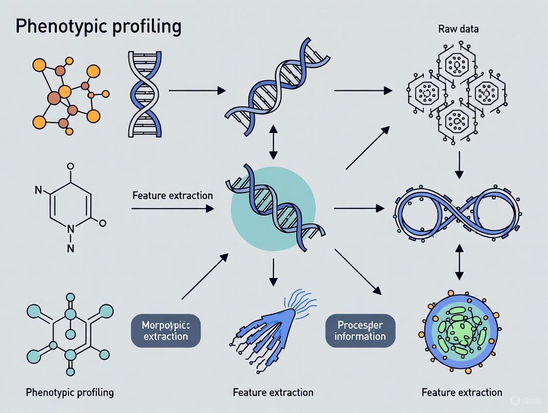
What is Phenotypic Profiling? Unlocking Cellular Secrets Through Morphology
Morphological analysis has undergone a revolutionary transformation, evolving from qualitative microscopic observations to quantitative, high-dimensional machine-driven profiling. This evolution is particularly impactful in phenotypic profiling research, where extracting meaningful morphological features enables researchers to decipher complex biological states and responses to perturbations [1]. The emergence of high-content imaging and artificial intelligence has propelled this field into the phenomics era, allowing for the systematic correlation of cellular and organismal form with function at unprecedented scale and resolution [1] [2]. This Application Note details the protocols and analytical frameworks essential for modern morphological feature extraction, providing researchers with practical methodologies to advance phenotypic drug discovery and functional genomics.
Traditional Morphological Analysis: Qualitative Microscopy
Traditional morphological analysis relied heavily on direct microscopic observation and manual characterization of physical structures. While now supplemented by advanced techniques, these methods remain fundamental for initial specimen characterization and provide the conceptual foundation for quantitative approaches.
Protocol: Light and Electron Microscopy for Preliminary Analysis
Purpose: To conduct initial morphological assessment of biological samples using microscopy techniques. Scope: Applicable to cellular and sub-cellular samples, as well as tissue sections and small organisms.
Materials:
- Biological sample (e.g., cell culture, tissue section, pollen grain [3])
- Microscope slides and coverslips
- Appropriate fixatives (e.g., glutaraldehyde, formaldehyde)
- Staining solutions (e.g., hematoxylin and eosin, DAPI, phalloidin)
- Light Microscope (LM) and/or Scanning Electron Microscope (SEM)
Procedure:
- Sample Preparation: Fix samples in appropriate fixative for 24 hours at 4°C. Dehydrate through a graded ethanol series (30%, 50%, 70%, 90%, 100%).
- Staining: Apply stains to highlight specific cellular or structural components.
- Microscopy:
- For LM: Mount samples on slides and observe under appropriate magnification. Capture images using attached digital camera.
- For SEM: Critical-point dry samples, sputter-coat with gold-palladium, and examine under SEM at accelerating voltages of 5-20 kV [3].
- Qualitative Analysis: Document morphological descriptors (e.g., shape, surface patterning, aperture type [3]).
Table 1: Qualitative Morphological Descriptors in Traditional Analysis
| Descriptor Category | Example Features | Application Example |
|---|---|---|
| Shape | Oblate-spheroidal, prolate, fibrous | Pollen grain identification [3] |
| Surface Pattern | Reticulate, rugulate, fossulate, scabrate | Halophyte plant systematics [3] |
| Aperture Type | Tricolporate, tricolpate, trizonocolporate | Taxonomic delineation in legumes [3] |
| Color/Staining | Eosinophilia, basophilia | Tissue pathology assessment |
| Spatial Arrangement | Clustered, solitary, linear | Cellular organization analysis |
The Shift to Quantitative Morphometrics
The transition to quantitative morphometrics marked a pivotal advancement, replacing subjective descriptions with objective, continuous data. This shift enables robust statistical analysis and phylogenetic comparison [4].
Protocol: Geometric Morphometric Analysis using Landmarks
Purpose: To quantify shape variation using landmark-based data for phylogenetic inference or taxonomic classification. Scope: Suitable for structures with definable homologous points (e.g., skulls, organelles, pollen grains).
Materials:
- Specimen images (2D or 3D)
- Software for landmark digitization (e.g., MorphoJ, tpsDig2)
- Statistical software (e.g., R)
Procedure:
- Landmark Definition: Define Type I (discrete juxtapositions), Type II (maxima of curvature), and Type III (extremal points) landmarks on all specimen images.
- Digitization: Manually place landmarks at corresponding positions across all samples in the dataset.
- Procrustes Superimposition: Scale, translate, and rotate landmark configurations to remove non-shape variation using Generalized Procrustes Analysis (GPA).
- Statistical Analysis: Perform Principal Component Analysis (PCA) on Procrustes coordinates to identify major axes of shape variation.
- Phylogenetic Analysis: Use resulting shape variables (e.g., PC scores) in phylogenetic reconstruction algorithms [4].
Table 2: Comparison of Discrete vs. Continuous Morphological Data in Phylogenetics
| Parameter | Discrete Morphological Data | Continuous Morphometric Data |
|---|---|---|
| Data Type | Categorical character states | Continuous measurements or landmark coordinates |
| Subjectivity | High potential for bias in character coding | More objective, but landmark placement can introduce error [4] |
| Information Content | Can lose continuous variation through arbitrary discretization [4] | Retains full shape information |
| Phylogenetic Signal | Variable; can be misleading due to homoplasy | Can be strong, but often confounded with allometric variation [4] |
| Analytical Methods | Parsimony, Bayesian Mk model | Squared-change parsimony, Bayesian Brownian motion models [4] |
| Performance | Traditional standard for fossil integration | Does not consistently improve resolution; requires specialized models [4] |
Modern High-Content Morphological Profiling
Contemporary phenotypic profiling leverages high-content screening and automated image analysis to extract thousands of quantitative features, creating a high-dimensional morphological profile for each sample.
Protocol: Cell Painting Assay for Morphological Profiling
Purpose: To generate comprehensive morphological profiles of cells under different genetic or chemical perturbations using the Cell Painting assay. Scope: Applicable to in vitro cell cultures for drug discovery and functional genomics.
Materials:
- Cell line (e.g., U2OS, A549, Hep G2 [2] [5])
- Cell Painting staining kit: dyes for DNA, ER, RNA, AGP, and Mito [2]
- High-throughput confocal microscope
- ǂ-well microplates
- Automated liquid handling system
- Image analysis software (CellProfiler [2] [5] or DeepProfiler [2])
Procedure:
- Cell Culture and Plating: Seed cells into ǂ-well plates and incubate for 24 hours.
- Perturbation: Treat cells with chemical compounds or genetic perturbations at optimized concentrations/durations.
- Staining: Follow standardized Cell Painting protocol:
- Fix cells with formaldehyde.
- Permeabilize with Triton X-100.
- Stain with Hoechst (DNA), Concanavalin A (ER), Syto14 (RNA), Phalloidin (AGP), and MitoTracker (Mito) [2].
- Image Acquisition: Image five channels per well using a high-throughput confocal microscope with a 20x objective.
- Feature Extraction:
- Use CellProfiler to identify individual cells and segment subcellular compartments.
- Extract ~1,500 morphological features per cell (e.g., area, shape, intensity, texture) for each channel [5].
- Data Analysis: Normalize features, aggregate per well, and use multivariate statistics (e.g., PCA) to analyze morphological profiles.
The Machine Learning Frontier: Predictive Morphology
The latest evolution involves using deep learning to not only describe but also predict morphological outcomes from molecular data, dramatically accelerating phenotypic screening.
Protocol: Predicting Morphology with Transcriptome-Guided Diffusion Models
Purpose: To predict cell morphological changes under unseen genetic or chemical perturbations using a transcriptome-guided latent diffusion model (MorphDiff) [2]. Scope: For in-silico phenotypic screening and MOA identification when morphological data is unavailable.
Materials:
- L1000 gene expression profiles for perturbations [2]
- Pre-trained MorphDiff model (available from original publication)
- Cell morphology image dataset for training (e.g., JUMP, CDRP, LINCS [2])
- High-performance computing cluster with GPUs
Procedure:
- Data Curation: Collate paired L1000 transcriptomic profiles and Cell Painting morphology images for a set of training perturbations.
- Model Training:
- Train MVAE: Compress high-dimensional morphology images into low-dimensional latent representations using a Morphology Variational Autoencoder (MVAE).
- Train LDM: Train a Latent Diffusion Model (LDM) to generate morphological latent representations conditioned on perturbed gene expression profiles.
- Prediction:
- Mode 1 (G2I): For a novel perturbation with L1000 data, use MorphDiff to generate predicted morphology from random noise.
- Mode 2 (I2I): Transform an unperturbed cell morphology image to the predicted perturbed state using the novel perturbation's L1000 profile as condition [2].
- Validation: Extract features from generated images using CellProfiler and compare to ground-truth morphological profiles.
Table 3: Performance of MorphDiff in Predicting Mechanisms of Action (MOA)
| Evaluation Metric | MorphDiff (G2I) | MorphDiff (I2I) | Ground-Truth Morphology | Gene Expression Only |
|---|---|---|---|---|
| MOA Retrieval Accuracy | Comparable to ground-truth | High | Benchmark | Not Specified |
| Improvement over Baselines | +16.9% | +8.0% | N/A | Baseline |
| Performance on Unseen Perturbations | Accurate prediction | Accurate transformation | N/A | N/A |
The Scientist's Toolkit: Research Reagent Solutions
Table 4: Essential Research Reagents and Materials for Morphological Profiling
| Reagent/Material | Function | Example Application |
|---|---|---|
| Cell Painting Dye Set | Multiplexed staining of major organelles | High-content morphological profiling [2] [5] |
| L1000 Assay Kit | High-throughput gene expression profiling | Generate transcriptomic conditions for MorphDiff [2] |
| Hoechst 33342 | DNA stain; marks nucleus | Cell segmentation and nuclear morphology analysis |
| Phalloidin (Conjugated) | F-actin stain; marks cytoskeleton | Cytoskeletal organization and cell shape analysis |
| MitoTracker | Mitochondrial stain | Mitochondrial morphology and network analysis |
| Concanavalin A (ConA) | Endoplasmic Reticulum (ER) stain | ER structure and distribution analysis |
| SYTO 14 | RNA stain; marks nucleoli and cytoplasm | Nucleolar morphology and granularity assessment |
| Pro-Crush Anti-Fade Mountant | Preserves fluorescence for imaging | Long-term storage of stained samples for microscopy |
Cell Painting is a high-content, image-based assay used for cytological profiling that employs a suite of fluorescent dyes to "paint" and visualize multiple cellular components simultaneously [6]. This multiplexed approach allows researchers to capture a comprehensive image of cellular state and organization by highlighting key organelles and structures. The core principle is that changes in cellular morphology reflect the biological state of a cell and its response to genetic, chemical, or environmental perturbations [7] [8].
Originally developed in 2013 by Gustafsdottir et al., the assay was designed to be a low-cost, single assay capable of capturing numerous biologically relevant phenotypes with high throughput [7] [8]. Over the past decade, the protocol has been optimized and standardized, with recent consortium-led efforts (JUMP-Cell Painting) further refining staining reagents, experimental conditions, and imaging parameters to enhance reproducibility and quantitative performance [7]. The assay's ability to generate rich, high-dimensional morphological profiles has made it particularly valuable in phenotypic drug discovery, toxicology, and functional genomics [7] [9].
The Scientific Principle of Morphological Profiling
Conceptual Framework
At its core, Cell Painting operates on the fundamental premise that cellular morphology is intricately linked to cell physiology, health, and function [7]. When cells undergo genetic or chemical perturbations, these changes manifest as alterations in the size, shape, texture, and spatial organization of cellular components [6]. Unlike targeted assays that measure specific, expected phenotypic responses, Cell Painting takes an untargeted approach to capture a broad spectrum of morphological features in an unbiased manner [10]. This makes it particularly valuable for identifying unexpected effects of perturbations and discovering novel biological connections.
The profiling strategy leverages the concept that compounds or genetic perturbations with similar mechanisms of action (MoA) often produce similar morphological profiles, allowing for functional classification based on phenotypic similarity [10] [7]. This approach has proven powerful for MoA identification of uncharacterized compounds, functional annotation of genes, and discovery of novel biological relationships that might be missed by hypothesis-driven assays [9].
Information Content and Profiling Power
The analytical power of Cell Painting stems from its high information density. From each individually segmented cell, automated image analysis software extracts approximately 1,500 morphological measurements across various categories including size, shape, texture, intensity, and spatial relationships between organelles [11] [9]. This multi-parametric profiling at single-cell resolution enables detection of subtle phenotypes that might be invisible to the human eye and allows resolution of cellular subpopulations within heterogeneous samples [6] [9].
When compared to other profiling technologies, Cell Painting offers complementary advantages. While high-throughput transcriptomic profiling methods like L1000 provide population-level gene expression signatures, Cell Painting delivers single-cell resolution of morphological features at a lower cost per sample [9]. Studies have shown that morphological and gene expression profiling capture partially overlapping but distinct information about cell state, suggesting they are orthogonal and powerful when combined [9].
Core Components of the Cell Painting Assay
Standard Dye Panel and Cellular Targets
The foundational Cell Painting assay uses a carefully selected set of six fluorescent dyes to label eight cellular compartments, imaged across five fluorescence channels [11] [9]. This panel was designed to provide comprehensive coverage of major organelles and structures while maintaining compatibility with standard high-throughput microscopes and minimizing cost by using dyes rather than antibodies [9].
Table 1: Standard Dye Panel for Cell Painting Assay
| Cellular Component | Fluorescent Dye | Staining Target | Imaging Channel |
|---|---|---|---|
| Nucleus | Hoechst 33342 | DNA | Blue/DAPI |
| Endoplasmic Reticulum | Concanavalin A, Alexa Fluor 488 conjugate | Glycoproteins | FITC/Green |
| Nucleoli & Cytoplasmic RNA | SYTO 14 | RNA | FITC/Green (with ER) |
| Actin Cytoskeleton | Phalloidin, Alexa Fluor 568 conjugate | F-actin | TRITC/Red |
| Golgi Apparatus & Plasma Membrane | Wheat Germ Agglutinin, Alexa Fluor 555 conjugate | Glycoproteins | TRITC/Red (with Actin) |
| Mitochondria | MitoTracker Deep Red | Mitochondrial membrane | Cy5/Far Red |
This standardized set of dyes visualizes a diverse array of cellular structures, enabling the detection of a wide spectrum of morphological changes induced by experimental perturbations [11] [9]. In practice, some dyes with non-overlapping emission spectra are often imaged in the same channel (e.g., RNA and ER; Actin and Golgi) to maximize throughput while maintaining coverage of multiple organelles [10] [8].
Experimental Workflow
The Cell Painting assay follows a standardized workflow that can be completed in approximately two weeks for cell culture and image acquisition, with an additional 1-2 weeks for feature extraction and data analysis [9]. The process involves multiple coordinated stages from sample preparation to computational analysis.
Diagram 1: Cell Painting Workflow. The process begins with cell plating and proceeds through treatment, staining, imaging, and analysis stages to generate morphological profiles.
Research Reagent Solutions
Implementation of the Cell Painting assay requires specific reagents and tools designed for high-content screening applications. Commercial kits and individual components are available to support standardized implementation.
Table 2: Essential Research Reagents for Cell Painting
| Reagent/Tool | Function | Application Note |
|---|---|---|
| Image-iT Cell Painting Kit | Pre-optimized dye combination | Simplifies staining with precisely measured reagents for 2 or 10 full multi-well plates [11] |
| Hoechst 33342 | Nuclear DNA stain | Labels nucleus, enables segmentation and nuclear feature extraction [6] [9] |
| MitoTracker Deep Red | Mitochondrial stain | Labels mitochondria, reveals metabolic state and organization [6] [9] |
| Concanavalin A, Alexa Fluor 488 | ER membrane stain | Visualizes endoplasmic reticulum structure and distribution [9] |
| Phalloidin, Alexa Fluor conjugates | F-actin stain | Labels actin cytoskeleton, reveals cell shape and structural changes [9] |
| Wheat Germ Agglutinin, Alexa Fluor conjugates | Golgi and plasma membrane stain | Highlights Golgi apparatus and plasma membrane glycoproteins [9] |
| SYTO 14 green fluorescent nucleic acid stain | RNA stain | Labels nucleoli and cytoplasmic RNA [6] [9] |
| High-content imaging system (e.g., CellInsight CX7) | Automated image acquisition | Designed for multi-well plate imaging at high speed and resolution [11] |
| Image analysis software (e.g., CellProfiler, IN Carta) | Feature extraction | Identifies cells and measures morphological features [6] [8] |
Advanced Methodological Developments
Cell Painting PLUS (CPP): Expanding Multiplexing Capacity
A significant recent advancement is the development of Cell Painting PLUS (CPP), which expands the multiplexing capacity of traditional Cell Painting through iterative staining-elution cycles [10]. This approach enables multiplexing of at least seven fluorescent dyes that label nine different subcellular compartments, including the addition of lysosomes, which are not typically included in the standard assay [10].
The key innovation in CPP is the use of an optimized dye elution buffer (0.5 M L-Glycine, 1% SDS, pH 2.5) that efficiently removes staining signals while preserving subcellular morphologies, allowing for sequential staining and imaging of dyes in separate channels [10]. This eliminates the need to merge signals from multiple organelles in the same imaging channel, thereby improving organelle-specificity and diversity of the phenotypic profiles [10]. The method provides researchers with enhanced flexibility to customize dye panels according to specific research questions while maintaining the untargeted profiling advantages of the original assay.
Alternative Dye Panels and Live-Cell Adaptations
Researchers have explored alternative dye configurations to address specific experimental needs. Recent studies have validated substitutes for standard dyes, including MitoBrilliant as a replacement for MitoTracker and Phenovue phalloidin 400LS for standard phalloidin stains [12]. These substitutions minimally impact assay performance while offering potential advantages such as isolating actin features from Golgi or plasma membrane signals [12].
The development of live-cell compatible dyes such as ChromaLive enables real-time assessment of compound-induced morphological changes, moving the assay from fixed endpoint measurements to dynamic kinetic profiling [12]. This live-cell adaptation provides temporal resolution of phenotypic responses and can be combined with standard Cell Painting to significantly expand the feature space for enhanced cellular profiling [12].
Cell Line Selection and Optimization
While the original Cell Painting protocol was developed using U-2 OS osteosarcoma cells, the assay has been successfully adapted to dozens of biologically diverse cell lines without adjustment to the staining protocol [7] [13]. Studies have systematically evaluated phenotypic profiling across multiple cell types including A549, MCF7, HepG2, and primary cell models [7] [13].
Research has shown that different cell lines vary in their sensitivity to specific mechanisms of action, with some lines better for detecting phenotypic activity (strength of morphological phenotypes) while others excel at predicting mechanism of action (phenotypic consistency with annotated MoAs) [7]. This indicates that cell line selection should be guided by specific screening goals, with some applications benefiting from profiling across multiple cell types to capture complementary biological perspectives [7].
Detailed Experimental Protocol
Sample Preparation and Staining
The Cell Painting protocol begins with plating cells in 96- or 384-well imaging plates at appropriate density to achieve sub-confluent monolayers, typically ranging from 1,000 to 5,000 cells per well depending on cell type [11] [9]. After allowing cells to adhere, they are treated with chemical compounds or genetic perturbations at desired concentrations, followed by incubation for a specified period (typically 24-48 hours) to allow phenotypic manifestation [11].
Staining Procedure:
- Fixation: Aspirate media and fix cells with 4% paraformaldehyde for 20-30 minutes at room temperature [9]
- Permeabilization: Incubate with 0.1% Triton X-100 for 15-30 minutes [9]
- Staining: Apply dye cocktail containing all six fluorescent dyes simultaneously or in sequence according to manufacturer's recommendations [9]
- Washing: Perform multiple washes with PBS or buffer to remove unbound dye [11]
- Storage: Store plates in sealing foil with desiccant at 4°C if not imaging immediately [11]
Critical considerations during staining include maintaining consistent incubation times across plates, protecting light-sensitive dyes from photobleaching, and confirming dye compatibility to avoid precipitation or interactions [9].
Image Acquisition Parameters
Image acquisition is performed using high-content screening (HCS) systems capable of automated multi-well plate imaging [11]. These systems employ fluorescent imaging specifically designed for maximum speed and data throughput, with combinations of widefield and confocal fluorescence capabilities [11].
Table 3: Image Acquisition Specifications
| Parameter | Specification | Notes |
|---|---|---|
| Plate Format | 96- or 384-well | Higher density plates increase throughput |
| Imaging Sites | Multiple positions per well | Ensures adequate cell sampling |
| Magnification | 20x or 40x objective | Balances resolution and field of view |
| Z-dimension | Multiple focal planes | Optional, based on cell thickness |
| Channels | 5 fluorescence channels | Matches dye emission spectra |
| Resolution | ≥ 0.65 μm/pixel (20x) | Sufficient for subcellular features |
| Bit Depth | 12- or 16-bit | Enables quantitative intensity measurements |
Image acquisition time varies based on the number of images per well sampled, sample brightness, and the extent of sampling in the z-dimension [11]. For large-scale screens, acquisition parameters are often optimized to balance data quality with throughput requirements [11].
Image Analysis and Feature Extraction
Image analysis transforms raw microscopy images into quantitative morphological profiles using automated software pipelines. The open-source CellProfiler software is commonly used, though commercial alternatives are also available [8] [9].
Analysis Pipeline:
- Cell Segmentation: Identify individual cells using nuclear stain as primary object followed by cytoplasmic expansion [9]
- Organelle Identification: Detect subcellular compartments within each segmented cell [9]
- Feature Measurement: Extract ~1,500 morphological features per cell across categories [9]
- Data Quality Control: Identify and exclude poor-quality images or segmentation failures [7]
- Data Aggregation: Compile single-cell measurements into population-level profiles [9]
The extracted features encompass multiple measurement categories including intensity (mean, median, standard deviation), texture (Haralick, Zernike features), shape (eccentricity, form factor), size (area, perimeter), and spatial relationships (adjacency, correlation between channels) [9].
Applications in Drug Discovery and Toxicology
Cell Painting has become an invaluable tool in phenotypic drug discovery, where it enables target-agnostic compound evaluation and mechanism of action identification [7]. By clustering compounds based on morphological similarity, researchers can identify novel compounds with desired phenotypic effects, characterize polypharmacology, and detect off-target effects early in the discovery process [7] [9].
In toxicology, Cell Painting has been applied to generate bioactivity profiles for industrial chemicals, with data from over 1,000 chemicals incorporated into the U.S. EPA CompTox Chemicals Dashboard [10] [7]. The assay's sensitivity to diverse cellular stressors makes it particularly valuable for predicting potential hazardous effects of environmental chemicals and understanding their subcellular targets [7].
The integration of Cell Painting with machine learning approaches has further expanded its applications, enabling prediction of compound activities, identification of disease signatures, and discovery of functional gene relationships [7] [8]. Large-scale consortia efforts like JUMP-Cell Painting have generated public datasets of morphological profiles for over 135,000 genetic and chemical perturbations, creating valuable community resources for method development and biological discovery [10] [7].
Technical Considerations and Limitations
Methodological Constraints
Despite its powerful applications, Cell Painting has several technical limitations that researchers must consider. Spectral overlap between fluorescent dyes can constrain multiplexing capacity and necessitate channel sharing, potentially reducing profiling specificity [10] [14]. The requirement for adherent, non-overlapping cells limits application to certain cell types, with non-adherent or compactly growing cells presenting challenges for imaging and analysis [8].
Some biological pathways or targets may not generate detectable morphological changes within the resolution of standard Cell Painting, creating potential biological blind spots in profiling experiments [14]. Additionally, the assay's sensitivity to batch effects from variations in cell culture conditions, staining protocols, or imaging parameters requires careful experimental design and normalization strategies to ensure robust, reproducible results [7] [14].
Computational Challenges
The high-dimensional nature of Cell Painting data presents significant computational challenges. The substantial data storage and processing requirements - with single experiments generating terabytes of images and millions of single-cell measurements - demand robust computational infrastructure [11] [8]. Analysis of high-dimensional feature spaces introduces statistical difficulties including spurious correlations and multiple testing challenges that require appropriate correction methods [8].
Currently, no established routine analytical protocol exists for all applications, requiring researchers to adapt and validate analysis pipelines for specific experimental contexts [8]. The interpretation of morphological profiles in terms of underlying biology can also be non-trivial, as morphological changes may represent integrated responses to multiple underlying molecular events [8].
The future of Cell Painting will likely involve continued integration with emerging computational and experimental techniques [7]. Advances in deep learning for image analysis may enable direct extraction of biologically relevant features from raw images without predefined measurement sets, potentially capturing more subtle and complex phenotypes [7] [8]. The generation of larger public datasets will support training of more powerful models and enable broader biological discoveries [7].
Methodologically, approaches like Cell Painting PLUS that expand multiplexing capacity and improve organelle-specificity represent an important direction for enhancing the resolution and biological interpretability of morphological profiling [10]. Similarly, live-cell adaptations and integration with other omics technologies (transcriptomics, proteomics) will provide more comprehensive views of cellular responses to perturbations [7] [12].
In conclusion, Cell Painting has established itself as a powerful, versatile tool for morphological profiling that continues to evolve through methodological refinements and expanding applications. Its ability to capture rich, high-dimensional information about cellular state in an untargeted manner makes it particularly valuable for phenotypic drug discovery, toxicology, and functional genomics. As the assay becomes more widely adopted and integrated with complementary technologies, it promises to yield further insights into cellular biology and accelerate the development of novel therapeutics.
In phenotypic profiling research, the quantitative analysis of cellular morphology provides a powerful window into cellular state and function. Image-based cell profiling enables the quantification of hundreds of morphological features from populations of cells subjected to chemical or genetic perturbations, creating distinctive "morphological profiles" that can reveal biologically relevant similarities and differences [15]. This approach critically depends on precise and specific labeling of key cellular components—nuclei, endoplasmic reticulum, mitochondria, and the cytoskeleton—to extract meaningful data about cell health, organization, and response to stimuli. These application notes provide detailed protocols and reagent solutions for comprehensive cellular labeling, framed within the context of morphological feature extraction for drug discovery and basic research.
The Scientist's Toolkit: Research Reagent Solutions
Table 1: Essential reagents for labeling key cellular components
| Cellular Component | Reagent/Solution | Function/Application | Example Products |
|---|---|---|---|
| Nuclei | Cell-permeant nucleic acid stains | Label DNA in live or fixed cells; viability assessment | Hoechst stains, DAPI [16] [17] |
| Endoplasmic Reticulum | ER-Tracker dyes | Live-cell staining selective for ER; bind to sulfonylurea receptors | ER-Tracker Blue-White DPX, ER-Tracker Green/Red [17] |
| Endoplasmic Reticulum | CellLight reagents | BacMam vectors encoding fluorescent protein fusions | CellLight ER-GFP/RFP (calreticulin-KDEL fusion) [17] |
| Mitochondria | TMRM (Tetramethylrhodamine, methyl ester) | Cell-permeant dye that accumulates in active mitochondria with intact membrane potential | TMRM [18] |
| Mitochondria | abberior LIVE mito probes | Cristae-specific labeling for super-resolution STED microscopy | abberior LIVE RED/ORANGE mito [19] |
| Golgi Apparatus | Fluorescent ceramide analogs | Selective stains for Golgi apparatus; metabolized to fluorescent sphingolipids | BODIPY FL C5-ceramide, NBD C6-ceramide [17] |
| Cytoskeleton (Actin) | Fluorescent phalloidin conjugates | High-affinity F-actin binding for fixed cells | Not specified in search results |
| Cytoskeleton (Microtubules) | Immunofluorescence reagents | Antibody-based labeling of tubulin in fixed cells | Not specified in search results |
Experimental Protocols for Key Cellular Labeling
Nuclear Staining Protocol
This protocol provides general instructions for labeling cell nuclei using nucleic acid stains, which exhibit minimal fluorescence before binding nucleic acids and significant intensity increases after binding [16].
Materials Required:
- Cells (adherent or suspension)
- Staining medium (complete cell culture medium or saline-based buffer like PBS/HBSS)
- Nuclear stain (e.g., Hoechst stains)
- Fluorescence microscope with appropriate filter set
Procedure:
- Prepare staining solution: Create 1 mL of nuclear dye staining solution at desired concentration. For most nuclear dyes, a 1 μM staining solution diluted from a 1 mM working solution is appropriate. Prepare multiple concentrations if optimizing [16].
- Remove medium: Aspirate existing medium from cells.
- Apply staining solution: Add sufficient staining solution to completely cover the sample.
- Incubate: Incubate for 5-15 minutes at room temperature or 37°C for most nuclear dyes. Some live-cell dyes may require longer incubation [16].
- Optional wash: For live-cell imaging with high-affinity stains, remove staining solution and wash to improve signal-to-background ratio.
- Image cells: Visualize using a fluorescence microscope with filter sets matched to your fluorophore.
Notes: The choice between complete medium and saline-based buffer depends on experimental design. Use complete medium for viability assays in live cell populations, and saline-based buffers for counterstaining during immunolabeling [16].
Endoplasmic Reticulum Staining Protocol
Option A: Using ER-Tracker Dyes for Live-Cell Imaging
ER-Tracker dyes are cell-permeant, live-cell stains highly selective for the endoplasmic reticulum with minimal mitochondrial staining [17].
Materials Required:
- Live cells
- ER-Tracker dye (Blue-White DPX, Green, or Red)
- Pre-warmed live-cell imaging medium
- DMSO for stock solutions
- Fluorescence microscope with appropriate filter sets
Procedure:
- Prepare stock solution: Dissolve ER-Tracker dye in DMSO according to manufacturer's instructions.
- Prepare working solution: Dilute stock solution in pre-warmed imaging medium to recommended working concentration.
- Replace medium: Remove existing cell culture medium and rinse cells with pre-warmed imaging medium.
- Apply staining solution: Add sufficient ER-Tracker working solution to cover cells.
- Incubate: Incubate for 15-30 minutes at 37°C under appropriate CO₂ conditions.
- Wash: Remove staining solution and rinse cells 2-3 times with fresh imaging medium.
- Image: Immediately image live cells using appropriate fluorescence filter sets.
Option B: Using CellLight BacMam Reagents
CellLight reagents provide highly specific ER labeling through BacMam expression of fluorescent protein fusions with ER targeting sequences [17].
Procedure:
- Plate cells: Seed cells in appropriate imaging chamber at least 16 hours before transduction.
- Add reagent: Simply add CellLight ER-GFP or ER-RFP reagent directly to cells.
- Incubate: Incubate for 16-24 hours to allow for gene expression and protein localization.
- Image: Visualize using standard GFP or RFP filter sets.
Validation Considerations: When expressing ER fluorescent reporters, confirm that overexpression does not significantly impact ER morphology by comparing to untransfected cells stained with ER antibodies (e.g., anti-PDI) [20].
Functional Mitochondrial Staining Protocol
This protocol uses TMRM (Tetramethylrhodamine, methyl ester) to detect mitochondria with intact membrane potentials, where signal intensity correlates with mitochondrial activity [18].
Materials Required:
- Live cells
- Complete cell culture medium
- TMRM (Tetramethylrhodamine, methyl ester)
- DMSO for stock solution
- Phosphate-buffered saline (PBS)
- Fluorescence microscope with TRITC filter set
Procedure:
- Prepare stock solution: Make a 10 mM TMRM stock solution in DMSO and store at -20°C [18].
- Prepare intermediate dilution: Create 50 μM intermediate dilution by adding 1 μL of 10 mM TMRM to 200 μL complete medium.
- Prepare staining solution: Make 250 nM staining solution by adding 5 μL of 50 μM TMRM to 1 mL complete medium.
- Remove media: Aspirate medium from live cells.
- Apply staining solution: Add TMRM staining solution to cells.
- Incubate: Incubate for 30 minutes at 37°C.
- Wash: Wash cells 3 times with PBS or other clear buffer.
- Image: Visualize using TRITC filter sets.
Alternative Advanced Protocol: abberior LIVE Mito Probes
For super-resolution imaging of mitochondrial cristae [19]:
- Prepare stock: Dissolve abberior LIVE mito probe in DMF or DMSO to make 1 mM stock.
- Prepare staining solution: Dilute stock in pre-warmed live-cell imaging medium to 250-500 nM final concentration.
- Replace medium: Remove culture medium and rinse with pre-warmed imaging medium.
- Stain cells: Add staining solution and incubate for 45-60 minutes at optimal growth conditions.
- Wash: Rinse 3 times with fresh imaging medium, followed by additional 15-20 minute wash.
- Image: Mount samples and immediately image with appropriate microscope systems.
Cytoskeleton Imaging Approaches
While specific staining protocols for cytoskeletal elements are not detailed in the search results, several imaging modalities and considerations are documented for cytoskeleton visualization.
Actin Cytoskeleton Imaging: The actin cytoskeleton can be visualized in various assembly formations that provide framework for cell shape, motility, and intracellular organization [21]. Imaging approaches include:
- Fixed cells: Fluorescent phalloidin conjugates for F-actin staining
- Live cells: GFP-actin fusion proteins or actin-binding domain probes
Microtubule Imaging: Microtubules are highly dynamic structures composed of α- and β-tubulin heterodimers that radiate from the centrosome [21]. They can be visualized using:
- Immunofluorescence: Antibodies against α- or β-tubulin in fixed cells
- Live-cell imaging: GFP-tubulin fusions or chemical probes
Recommended Microscopy Techniques:
- Spinning disk confocal microscopy (SDCM): Ideal for rapid dynamics of actin filaments and focal adhesion complexes [21]
- Laser scanning confocal microscopy (LSCM): Provides optical sectioning for 3D reconstruction of cytoskeletal architecture [21]
Workflow Integration for Morphological Profiling
The integration of multiple cellular labeling strategies enables comprehensive morphological profiling for phenotypic screening. The diagram below illustrates the complete workflow from sample preparation to data analysis.
Image Analysis and Quality Control for Profiling
High-quality morphological profiling requires rigorous image analysis and quality control to ensure data integrity [15].
Illumination Correction: Correct for inhomogeneous illumination using:
- Retrospective multi-image methods: Build correction functions using all images in experiment for most robust results [15]
- Prospective methods: Use reference images taken at time of acquisition (less recommended)
- Retrospective single-image methods: Calculate correction for each image individually (can alter relative intensity)
Segmentation Approaches:
- Model-based segmentation: Use a priori knowledge of expected object size and shape with algorithms like thresholding and watershed transformation [15]
- Machine learning-based segmentation: Train classifiers on manually labeled ground-truth data for difficult segmentation tasks [15]
Feature Extraction for Profiling:
- Shape features: Perimeter, area, roundness of nuclei, cells, or organelles [15]
- Intensity-based features: Mean intensity, maximum intensity within cellular compartments [15]
- Texture features: Mathematical functions to quantify intensity regularity and patterns [15]
- Microenvironment features: Spatial relationships between cells and organelles [15]
Table 2: Quantitative parameters for mitochondrial membrane potential assessment
| Parameter | Normal Range | Interpretation | Measurement Method |
|---|---|---|---|
| TMRM Intensity | Cell-type dependent | Bright signal indicates intact ΔΨm; dim signal indicates depolarization | Mean fluorescence intensity per cell [18] |
| Incubation Time | 30 minutes at 37°C | Optimal for dye accumulation | Time at 37°C [18] |
| Working Concentration | 250 nM | Balance between signal intensity and potential toxicity | Dilution from stock [18] |
| Incubation Temperature | 37°C | Critical for proper dye uptake and mitochondrial function | Environmental control [20] |
Critical Considerations for Experimental Design
Live Cell Imaging Requirements
For dynamic imaging of organelle interactions and processes, maintain cells under physiological conditions:
- Temperature control: Pre-warm stage warmer or environmental housing for at least 20 minutes before experiments [20]
- Environmental control: Maintain appropriate CO₂, humidity, and pH conditions throughout imaging [19]
- Minimal phototoxicity: Use low dye concentrations and optimize exposure times to reduce cellular stress [19]
Multiplexing and Experimental Validation
- Dye compatibility: Ensure spectral separation between fluorophores for multi-component labeling
- Expression validation: Confirm that fluorescent reporter expression doesn't alter native organelle morphology [20]
- Controls included: Always include appropriate controls (untreated, vehicle-only, and positive controls)
- Morphological assessment: Quantitate fluorescence intensities and correlate with potential structural alterations [20]
Comprehensive labeling of nuclei, endoplasmic reticulum, mitochondria, and cytoskeletal elements provides the foundation for quantitative morphological profiling in phenotypic research. The protocols and reagents detailed in these application notes enable researchers to capture the complex interplay between cellular compartments and extract meaningful data about cellular state in response to genetic, chemical, or environmental perturbations. When properly implemented within a rigorous analytical workflow, these labeling strategies support the generation of high-quality morphological profiles that can reveal novel biological insights and accelerate drug discovery efforts.
Morphological profiling via feature extraction represents a transformative approach in phenotypic screening, enabling the quantification of cellular states induced by genetic or chemical perturbations [22]. This process transforms raw, high-dimensional image data into informative, numerical descriptors that capture essential biological information. By systematically analyzing intensity, texture, shape, and spatial features, researchers can obtain unbiased bioactivity profiles that predict the mode of action (MoA) for unexplored compounds and uncover unanticipated activities for characterized small molecules [22]. These profiles have become indispensable tools in early-stage drug discovery, allowing for the detection of bioactivity in a broader biological context [22]. This protocol details the comprehensive methodology for extracting multifaceted features critical for robust morphological profiling and phenotypic analysis.
Morphological profiling leverages automated imaging and advanced image analysis to record alterations in cellular architecture by detecting hundreds of quantitative features in high-throughput experiments [22]. Feature extraction serves the critical function of transforming raw image data into compact, informative representations, enabling efficient analysis, recognition, and classification in modern image processing and computer vision applications [23]. This process is fundamental for dimensionality reduction, separating crucial features to improve accuracy in classification tasks, and enhancing system performance for real-time applications while effectively reducing noise [24].
In phenotypic profiling, the morphological profile induced by a small molecule provides a rich, rather unbiased description of the perturbed cellular state, creating a distinctive signature that can be compared to profiles of compounds with known mechanisms [22]. The systematic categorization of features includes:
- Geometric Features: Capturing structural relationships and object shapes.
- Statistical Features: Providing quantitative descriptors of intensity distributions.
- Texture-Based Techniques: Highlighting surface characteristics and spatial patterns using methods like Local Binary Patterns (LBP) and Gray Level Co-occurrence Matrix (GLCM) [23].
- Spatial Features: Describing object prominence and organizational context within an image [25].
Comprehensive Feature Taxonomy and Quantitative Comparison
The following tables summarize the core feature categories extracted in morphological profiling, their specific metrics, and their primary biological applications.
Table 1: Core Feature Categories in Morphological Profiling
| Feature Category | Sub-category | Key Metrics | Biological Applications |
|---|---|---|---|
| Intensity | Statistical | Mean, Median, Standard Deviation, Minimum/Maximum Pixel Values | Protein expression levels, drug accumulation, cellular health |
| Histogram-based | Mode, Entropy, Kurtosis, Skewness | Content distribution analysis, phenotype classification | |
| Texture | Statistical | Contrast, Correlation, Energy, Homogeneity (from GLCM) [24] | Cytoskeletal organization, chromatin patterning, organelle distribution |
| Structural | Local Binary Patterns (LBP) [23] | Surface characterization, repetitive pattern identification | |
| Spectral | Gabor Filter responses [24] | Pattern analysis at multiple scales and orientations | |
| Shape | Contour-based | Area, Perimeter, Eccentricity, Major/Minor Axis Length | Nuclear morphology, cell shape analysis, morphological changes |
| Moment-based | Hu Moments, Zernike Moments | Object recognition and orientation | |
| Spatial | Object Prominence | Size, Centeredness, Image Depth [25] | Analyzing cellular organization and relational context |
| Topological | Nearest Neighbor Distance, Voronoi Tessellation, Delaunay Triangulation | Spatial organization analysis, tissue architecture |
Table 2: Computational Characteristics of Feature Extraction Methods
| Extraction Method | Computational Complexity | Noise Sensitivity | Dimensionality of Output | Primary Use Cases |
|---|---|---|---|---|
| Edge Detection (Canny) [24] | Medium | Low-Medium | Variable (edge pixels) | Cell boundary detection, segmentation |
| Corner Detection (Harris) [24] | Low | Medium | Variable (corner points) | Feature point matching, tracking |
| Blob Detection (LoG/DoG) [24] | High | Low | Variable (blob regions) | Spot detection (vesicles, nuclei), counting |
| GLCM Texture [24] | Medium-High | Medium | Fixed (multiple features) | Texture classification, pattern analysis |
| LBP [23] [24] | Low | Low | Fixed (histogram) | Real-time texture classification, face recognition |
| Gabor Filters [24] | High | Low | Fixed (multiple features) | Multi-scale texture analysis, frequency analysis |
Experimental Protocols for Feature Extraction
Protocol 1: Intensity and Texture Feature Extraction
Purpose: To quantify pixel value distributions and textural patterns in cellular images.
Materials:
- Fluorescent or brightfield cellular images
- Image processing software (e.g., Python with OpenCV, ImageJ)
- Segmentation masks identifying cellular regions of interest
Procedure:
- Image Preprocessing:
- Apply Gaussian smoothing with a 3×3 kernel to reduce noise [24].
- Normalize intensity across image sets using histogram equalization.
- Convert to grayscale if working with color images.
Intensity Feature Extraction:
- For each segmented cellular region, calculate:
- Mean, median, and standard deviation of pixel intensities.
- Intensity histogram skewness and kurtosis.
- Minimum and maximum pixel values within the region.
- Record values for statistical analysis.
- For each segmented cellular region, calculate:
Texture Feature Extraction using GLCM:
- Calculate Gray-Level Co-occurrence Matrix for distances [1, 2] and angles [0°, 45°, 90°, 135°].
- Compute Haralick features from GLCM:
- Contrast:
∑(i,j)‖i-j‖²·p(i,j) - Correlation:
∑(i,j)((i-μi)(j-μj)p(i,j))/(σiσj) - Energy:
∑(i,j)p(i,j)² - Homogeneity:
∑(i,j)p(i,j)/(1+‖i-j‖)[24]
- Contrast:
Texture Feature Extraction using LBP:
- For each pixel, compare with its 8 surrounding neighbors.
- Generate binary pattern: 1 if neighbor ≥ center, else 0.
- Convert binary pattern to decimal value.
- Create LBP histogram for the region [24].
Troubleshooting:
- High background intensity: Adjust segmentation thresholds or apply background subtraction.
- Inconsistent staining: Normalize intensities across batches using control samples.
Protocol 2: Shape and Spatial Feature Extraction
Purpose: To quantify morphological characteristics and spatial relationships of cellular structures.
Materials:
- Segmented binary masks of cellular structures
- Computational geometry libraries (e.g., SciPy, scikit-image)
Procedure:
- Shape Feature Extraction:
- For each segmented object, calculate:
- Area: Total number of pixels in the region.
- Perimeter: Distance around the boundary of the region.
- Eccentricity: Ratio of the distance between foci of ellipse and its major axis length.
- Major and Minor Axis Lengths: Dimensions of the fitted ellipse.
- Solidity: Ratio of area to convex hull area.
- For each segmented object, calculate:
Contour-Based Analysis:
- Detect contours using border following algorithms.
- Approximate contours to reduce complexity.
- Calculate contour moments for shape representation.
Spatial Feature Extraction:
- Calculate Object Prominence metrics:
- Size: Normalized area relative to image dimensions.
- Centeredness: Distance from object centroid to image center.
- Saliency: Using saliency detection algorithms to estimate visual attention [25].
- Compute spatial relationships:
- Nearest Neighbor Distances between objects.
- Voronoi Tessellation of object centroids.
- Delaunay Triangulation of object centroids.
- Calculate Object Prominence metrics:
Spatial Statistics:
- Ripley's K-function to analyze clustering or dispersion.
- Pair correlation function for spatial patterns at different scales.
Troubleshooting:
- Overlapping objects: Use watershed segmentation or marker-controlled approaches.
- Small/fragmented objects: Apply morphological operations (closing) before analysis.
Workflow Visualization
Figure 1: Comprehensive workflow for morphological feature extraction from cellular images, showing the sequential process from raw images to analyzable profiles.
Table 3: Essential Resources for Morphological Profiling
| Resource Category | Specific Tool/Reagent | Function/Application |
|---|---|---|
| Image Acquisition | High-content screening microscope | Automated acquisition of cellular images at scale |
| Cell painting assay reagents | Multiplexed staining of multiple organelles | |
| Image Processing | Python/OpenCV [24] | Implementation of feature extraction algorithms |
| ImageJ/Fiji | Open-source image analysis with plugin ecosystem | |
| CellProfiler | Domain-specific software for biological image analysis | |
| Feature Extraction | Scikit-image | Python library for image analysis algorithms |
| Mahotas | Computer vision library for biological image analysis | |
| Data Analysis | R/Python pandas | Data manipulation and statistical analysis |
| Scikit-learn | Machine learning for phenotype classification | |
| Specialized Algorithms | Canny Edge Detector [24] | Reliable boundary detection for cell segmentation |
| Harris/Shi-Tomasi Corner Detector [24] | Interest point detection for tracking | |
| Laplacian of Gaussian (LoG) [24] | Blob detection for vesicles and organelles |
Analysis and Integration of Multimodal Features
Integrating multiple feature types creates a more robust and accurate representation of cellular morphology than any single feature category alone [23]. The fusion of intensity, texture, shape, and spatial features enables a comprehensive phenotypic profile that captures both intrinsic cellular characteristics and their organizational context.
Integrated Analysis Workflow:
- Feature Normalization: Standardize features across different scales using Z-score normalization or min-max scaling.
- Dimensionality Reduction: Apply Principal Component Analysis (PCA) or t-distributed Stochastic Neighbor Embedding (t-SNE) to visualize high-dimensional feature space.
- Clustering Analysis: Use k-means or hierarchical clustering to identify distinct phenotypic clusters.
- Classification: Train supervised classifiers (Random Forest, SVM) to predict treatment classes or mechanisms of action.
- Similarity Scoring: Compute distances between profiles (e.g., Euclidean, Mahalanobis) to identify similar morphological responses.
The prominence of objects within images, as determined by factors like size, centeredness, and saliency, provides crucial contextual information for interpreting morphological features [25]. This spatial context enhances the biological interpretability of profiling data by distinguishing primary phenotypic effects from secondary changes.
Morphological profiling through comprehensive feature extraction provides a powerful framework for quantitative phenotypic analysis in drug discovery and basic research. By systematically quantifying intensity, texture, shape, and spatial characteristics, researchers can create rich, informative profiles that capture subtle biological states induced by genetic or chemical perturbations. The integrated approaches discussed here, combining multiple feature types and considering object prominence, enhance the robustness and biological relevance of analyses performed at scale. As these methodologies continue to evolve, they will further enable the detection of bioactivity in compound collections and the prediction of mechanisms of action, accelerating therapeutic development and fundamental biological discovery.
Image-based phenotypic profiling is a powerful method that combines automated microscopy and computational analysis to identify phenotypic alterations in cell morphology, providing critical insight into a cell's physiological state [26]. This approach quantitatively compares cell morphology after various chemical or genetic perturbations, enabling researchers to identify meaningful similarities and differences in the same way transcriptional profiles are used to compare samples [27]. The fundamental premise is that disturbances in cellular pathways and processes manifest as detectable changes in microscopic appearance, creating a bridge between observable morphology and underlying biology.
The field has progressed significantly through consortium efforts like the JUMP Cell Painting Consortium, which brings together pharmaceutical companies, non-profit institutions, and supporting companies to advance methodological development [27]. These collaborations have enabled the creation of benchmark datasets such as CPJUMP1, containing approximately 3 million images and morphological profiles of 75 million single cells treated with carefully matched chemical and genetic perturbations [27]. Such resources provide the foundation for optimizing computational strategies to represent cellular samples so they can be effectively compared to uncover valuable biological relationships.
Key Concepts and Biological Significance
Core Principles
Phenotypic profiling operates on several core principles. First, different perturbation types targeting the same biological pathway often produce similar morphological changes, creating recognizable profiles. Second, the directionality of correlations among perturbations targeting the same protein can be systematically explored, with some showing positive correlations (similar phenotypes) and others showing negative correlations (opposing phenotypes) [27]. Finally, these morphological profiles are reproducible across experimental replicates and can be detected using appropriate computational methods.
The biological significance of this approach lies in its ability to connect morphological patterns to specific biological states without prior knowledge of the underlying mechanisms. This makes it particularly valuable for identifying mechanisms of action for uncharacterized compounds, discovering novel gene functions, and understanding disease pathologies through comparative analysis of patient-derived cells [27].
Analytical Framework
The analytical framework for phenotypic profiling typically involves several stages: perturbation application, image acquisition, feature extraction, profile generation, and similarity analysis. In the final stage, cosine similarity or its absolute value is commonly used as a correlation-like metric to measure similarities between pairs of well-level aggregated profiles [27]. The statistical significance of these similarities is then assessed using permutation testing with false discovery rate correction to account for multiple comparisons.
Table: Benchmark Performance of Phenotypic Profiling Representations
| Perturbation Type | Cell Type | Time Point | Fraction Retrieved | Key Findings |
|---|---|---|---|---|
| Chemical Compounds | U2OS, A549 | 2 time points | Higher than genetic | Most distinguishable from negative controls |
| CRISPR Knockout | U2OS, A549 | 2 time points | Intermediate | More detectable than overexpression |
| ORF Overexpression | U2OS, A549 | 2 time points | Lower than others | Weakest signal, potentially due to plate layout effects |
Experimental Protocols and Methodologies
Cell Painting Assay Protocol
The Cell Painting assay is the most widely used protocol for phenotypic profiling [27]. The following detailed methodology outlines the key experimental steps:
Materials and Reagents:
- Cell lines (e.g., U2OS osteosarcoma, A549 lung carcinoma)
- Perturbations (chemical compounds, CRISPR guides, ORF constructs)
- Cell culture reagents and media
- Staining dyes: MitoTracker (mitochondria), Phalloidin (actin), Concanavalin A ( endoplasmic reticulum), SYTO 14 (nucleic acids), and Wheat Germ Agglutinin ( Golgi and plasma membrane)
- Fixative: 4% formaldehyde in PBS
- Permeabilization buffer: 0.1% Triton X-100 in PBS
- Washing buffer: 1X PBS
- 384-well imaging plates
Procedure:
- Plate Cells: Seed appropriate cell density in 384-well plates, optimizing for confluency at time of imaging.
- Perturbation Application: Apply chemical or genetic perturbations in replicate wells across multiple plates. Include negative controls (DMSO or empty vector) and positive controls if available.
- Incubation: Incubate cells for predetermined time points (e.g., 24h, 48h, 96h) at 37°C with 5% CO₂.
- Fixation: Aspirate media and add 4% formaldehyde solution for 15-20 minutes at room temperature.
- Permeabilization: Aspirate fixative, add permeabilization buffer for 10-15 minutes.
- Staining: Apply staining cocktail containing all five dyes for 30-60 minutes protected from light.
- Washing: Perform 3×5 minute washes with 1X PBS.
- Storage: Add fresh PBS and store plates at 4°C protected from light until imaging.
- Image Acquisition: Acquire images using high-content microscopy systems with appropriate filters for each fluorescent channel.
Critical Considerations:
- Plate layout should randomize perturbation positions to minimize positional effects
- Include sufficient replicates for statistical power (typically 4-8 replicates per perturbation)
- Maintain consistent imaging parameters across all plates and experimental batches
- Include reference controls for assay quality assessment
Image Analysis and Feature Extraction Workflow
The following workflow transforms acquired images into quantitative morphological profiles:
Detailed Protocol Steps:
Image Preprocessing
- Correct for background fluorescence and uneven illumination
- Apply flat-field correction if required
- Register multiple channels if necessary
- Quality control to exclude out-of-focus images
Cell and Organelle Segmentation
- Use nuclei staining to identify individual cells
- Apply cytoplasm segmentation using actin or plasma membrane markers
- Segment individual organelles using specific channel information
- Validate segmentation accuracy manually for a subset
Feature Extraction
- Extract morphological features for each cell and subcellular compartment
- Include measurements for size, shape, intensity, texture, and spatial relationships
- Calculate both classical hand-engineered features and deep-learning derived features
- Generate population-level statistics for each well
Profile Generation and Normalization
- Aggregate single-cell measurements to well-level profiles
- Apply normalization to remove technical artifacts (plate, batch effects)
- Use control-based normalization (e.g., using in-distribution control experiments)
- Perform feature selection to reduce dimensionality
Data Analysis and Computational Approaches
Feature Representation Strategies
Different computational approaches can be employed to represent the morphological profiles:
Classical Representations: Rely on hand-engineered features carefully developed and optimized to capture cellular morphology variations, including size, shape, intensity, and texture of various stains [27]. These features require post-processing steps including normalization, feature selection, and dimensionality reduction.
Anomaly-Based Representations: Use the abundance of control wells to learn the in-distribution of control experiments and formulate a self-supervised reconstruction anomaly-based representation [26]. These representations encode intricate morphological inter-feature dependencies while preserving interpretability and have demonstrated improved reproducibility and mechanism of action classification compared to classical representations.
Deep Learning Representations: Automatically identify features directly from pixels using representation learning algorithms [27]. These methods can capture more complex patterns but may be harder to biologically interpret without specialized explainability techniques.
Table: Comparison of Feature Representation Methods
| Representation Type | Key Features | Advantages | Limitations |
|---|---|---|---|
| Classical Features | Hand-engineered morphological measurements | Biologically interpretable, established methods | May not capture full complexity of cellular organization |
| Anomaly Representations | Encodes deviations from control morphology | Improved reproducibility, reduces batch effects | Requires sufficient control data for training |
| Deep Learning Features | Learned directly from raw images | Potential to capture novel patterns, minimal preprocessing | Hard to interpret biologically, requires large datasets |
Perturbation Detection and Matching
Two critical analytical tasks in phenotypic profiling are perturbation detection and matching:
Perturbation Detection: Identifies perturbations that produce statistically significant morphological changes compared to negative controls. This is often measured using average precision to retrieve replicate perturbations against the background of negative controls, with statistical significance assessed using permutation testing and false discovery rate correction [27].
Perturbation Matching: Identifies genes or compounds that have similar impacts on cell morphologies. Improved matching enables better discovery of compound mechanisms of action and virtual screening for useful gene-compound relationships [27].
The following diagram illustrates the computational pipeline for these analyses:
Essential Research Reagents and Materials
Successful phenotypic profiling requires carefully selected reagents and materials optimized for consistency and reproducibility:
Table: Essential Research Reagent Solutions for Phenotypic Profiling
| Reagent Category | Specific Examples | Function and Application |
|---|---|---|
| Cell Lines | U2OS (osteosarcoma), A549 (lung carcinoma) | Provide consistent cellular context for perturbation studies; different cell types may show varying sensitivity to perturbations |
| Chemical Perturbations | Drug Repurposing Hub compounds | Well-annotated compounds with known targets enable ground truth for method validation and mechanism of action studies |
| Genetic Perturbations | CRISPR guides, ORF overexpression constructs | Target specific genes to establish causal relationships between gene function and morphological phenotypes |
| Staining Dyes | MitoTracker, Phalloidin, Concanavalin A, SYTO 14, Wheat Germ Agglutinin | Visualize specific subcellular compartments to capture comprehensive morphological information |
| Imaging Plates | 384-well imaging-optimized plates | Provide consistent imaging surface with minimal background fluorescence and optical distortion |
| Reference Controls | DMSO, empty vectors, known pathway modulators | Enable normalization and quality control across experiments and batches |
Applications in Drug Discovery and Functional Genomics
Phenotypic profiling enables several critical applications in biological research and drug development:
Mechanism of Action Identification: By comparing morphological profiles of compounds with unknown mechanisms to those with known targets, researchers can generate hypotheses about compound mechanisms [27]. The availability of datasets with matched chemical and genetic perturbations, where each perturbed gene's product is a known target of at least two chemical compounds, significantly enhances this capability.
Functional Gene Discovery: Clustering large sets of genetically perturbed samples reveals relationships among genes, helping to assign function to uncharacterized genes [27]. Different perturbation mechanisms (CRISPR knockout vs. ORF overexpression) can provide complementary information about gene function.
Disease Mechanism Elucidation: Comparing cells from patients with specific diseases to healthy controls can identify disease-specific morphological signatures and potentially reveal underlying disease mechanisms.
Toxicity Assessment: Detracting morphological changes associated with cellular stress or death can provide early indicators of compound toxicity.
The following diagram illustrates the primary application workflows:
Future Directions and Methodological Advancements
The field of phenotypic profiling continues to evolve with several promising directions:
Integration with Other Data Modalities: Combining morphological profiles with transcriptional, proteomic, or metabolic data provides multi-dimensional views of cellular states.
Improved Representation Learning: Self-supervised and semi-supervised approaches that better leverage unlabeled data or limited annotations may enhance feature learning, particularly anomaly representations that encode morphological inter-feature dependencies [26].
Explainable AI: Developing methods to biologically interpret deep learning models and anomaly representations will be crucial for building trust and extracting biological insights [26].
Standardized Benchmarking: Resources like the CPJUMP1 dataset enable systematic comparison of computational methods and establish benchmarks for the field [27].
As these methodological advancements mature, phenotypic profiling is poised to become an increasingly powerful approach for connecting cellular morphology to underlying biology, accelerating discovery in basic research and drug development.
From Data to Discovery: AI-Powered Methodologies for Morphological Profiling
In the field of phenotypic profiling research, quantitative analysis of cellular and organismal morphology is paramount for deciphering developmental processes, disease states, and drug responses. Traditional morphological analysis has long relied on landmark-based geometric morphometrics, which requires manual annotation of anatomically homologous points by experts. This approach presents significant limitations, including difficulties in comparing phylogenetically distant species, information loss from insufficient landmarks, and inter-researcher variability in landmark placement [28]. To overcome these challenges, the Morphological Regulated Variational AutoEncoder (Morpho-VAE) framework represents a transformative advancement by enabling landmark-free shape analysis through deep learning.
Morpho-VAE constitutes an image-based deep learning framework that combines unsupervised and supervised learning models to reduce dimensionality while focusing on morphological features that distinguish data with different biological labels [28]. This hybrid architecture effectively extracts discriminative morphological signatures without requiring prior anatomical knowledge, making it particularly valuable for large-scale phenotypic screening in drug discovery where manual annotation would be prohibitively time-consuming. By capturing nonlinear relationships in morphological data, Morpho-VAE can identify subtle phenotypic changes induced by genetic or chemical perturbations that might elude conventional analysis methods.
Technical Framework and Architecture
Core Components of Morpho-VAE
The Morpho-VAE architecture integrates two fundamental modules into a cohesive framework for morphological feature extraction:
VAE Module: The foundation employs a variational autoencoder consisting of an encoder that compresses high-dimensional input images into a low-dimensional latent representation (ζ), and a decoder that reconstructs the input from this compressed latent space. This component ensures that morphological information is preserved during the compression process through its reconstruction capability [28].
Classifier Module: A supervised classification component is interconnected with the VAE through the latent variables, guiding the encoder to extract features that are maximally discriminative between specified biological classes (e.g., cell types, treatment conditions, or species) [28].
The mathematical formulation of the Morpho-VAE training objective combines both unsupervised and supervised elements through a weighted total loss function: E_total = (1 - α)E_VAE + αE_C, where E_VAE represents the variational autoencoder loss (reconstruction + regularization), E_C denotes the classification loss, and α is a hyperparameter balancing these objectives. Through cross-validation on primate mandible image data, the optimal α value has been determined to be 0.1, successfully incorporating classification capability without significantly compromising reconstruction quality [28].
Comparative Advantage Over Traditional Methods
Table 1: Performance comparison of Morpho-VAE against traditional morphometric methods
| Method | Cluster Separation (CSI) | Landmark Requirement | Nonlinear Feature Capture | Handling of Missing Data |
|---|---|---|---|---|
| Morpho-VAE | 0.75 (Superior) | No | Excellent | Yes |
| Standard VAE | 1.12 (Moderate) | No | Good | Limited |
| PCA | 1.45 (Poor) | Yes | No | No |
| Landmark-Based GM | Varies | Yes | Limited | No |
The cluster separation index (CSI) quantifies the superiority of Morpho-VAE in distinguishing morphological classes, with lower values indicating better separation. Morpho-VAE achieves a CSI of 0.75, significantly outperforming standard VAE (1.12) and PCA-based approaches (1.45) [28]. This enhanced performance stems from its ability to capture nonlinear morphological relationships that linear methods like PCA cannot represent, while simultaneously focusing on biologically discriminative features through its integrated classifier.
Application Protocols for Phenotypic Profiling
Implementation Workflow for Cellular Morphological Analysis
The following Graphviz diagram illustrates the end-to-end Morpho-VAE workflow for phenotypic profiling:
Experimental Protocol: Morpho-VAE for Drug Response Profiling
Objective: To quantify morphological changes in cell lines in response to compound treatments using the Morpho-VAE framework.
Materials and Reagents:
- Cell lines relevant to research focus (e.g., U2OS, A549)
- Cell culture media and supplements
- Compound libraries for screening
- Cell Painting staining reagents [29]:
- MitoTracker (Mitochondria staining)
- Phalloidin (Actin cytoskeleton)
- WGA (Golgi and plasma membrane)
- Concanavalin A (Endoplasmic reticulum)
- SYTO (Nuclear DNA)
- Fixation and permeabilization buffers
- High-content imaging compatible plates
Procedure:
Sample Preparation
- Seed cells in 96-well or 384-well imaging plates at appropriate density
- After cell attachment, treat with compounds at multiple concentrations
- Include DMSO controls and appropriate positive/negative controls
- Incubate for predetermined time (typically 24-48 hours)
- Fix cells and perform Cell Painting staining protocol [29]
Image Acquisition
- Acquire images using high-content microscope with 20x or 40x objective
- Capture 5 fluorescent channels corresponding to Cell Painting stains
- Acquire multiple fields per well to ensure adequate cell population sampling
- Save images in standard format (TIFF preferred) with appropriate metadata
Image Preprocessing
- Apply illumination correction to correct for uneven field illumination
- Perform background subtraction to remove camera noise
- Resize images to standard dimensions (e.g., 128×128 or 256×256 pixels)
- Apply data augmentation (rotation, flipping) to increase dataset diversity
Morpho-VAE Model Configuration
- Implement encoder network with convolutional layers (4-6 layers)
- Set latent dimension based on complexity (typically 3-50 dimensions)
- Implement decoder network with transposed convolutional layers
- Add classifier network with fully connected layers
- Configure hyperparameters: α=0.1, learning rate=0.001, batch size=32
Model Training
- Split data into training (70%), validation (15%), and test (15%) sets
- Train model for 100-500 epochs with early stopping
- Monitor both reconstruction and classification losses
- Validate cluster separation using quantitative metrics
Feature Extraction and Analysis
- Extract latent representations for all samples
- Apply dimensionality reduction (t-SNE, UMAP) for visualization
- Perform statistical analysis to identify significant morphological responses
- Correlate morphological profiles with treatment conditions
Troubleshooting Notes:
- Poor reconstruction quality may indicate insufficient model capacity or training time
- Inadequate cluster separation may require adjustment of the α parameter
- Overfitting to training classes can be addressed with increased regularization
- Computational requirements can be significant for large datasets; consider cloud resources
Research Reagent Solutions and Computational Tools
Table 2: Essential research reagents and computational tools for Morpho-VAE implementation
| Category | Specific Tool/Reagent | Function in Workflow | Key Features |
|---|---|---|---|
| Cell Staining | Cell Painting Kit | Multiplexed morphological staining | Standardized 5-6 channel staining protocol [29] |
| Microscopy | High-content imagers (e.g., ImageXpress) | Automated image acquisition | Multi-channel, high-throughput capability |
| Image Analysis | CellProfiler [2] | Image preprocessing and feature extraction | Open-source, pipeline-based processing |
| Deep Learning | TensorFlow/PyTorch | Morpho-VAE implementation | Flexible neural network frameworks |
| Feature Extraction | InceptionV3 [29] | Transfer learning for evaluation | Pre-trained on natural images |
| Visualization | UMAP/t-SNE | Latent space visualization | Non-linear dimensionality reduction |
Advanced Applications in Drug Discovery
Predictive Morphological Profiling with MorphDiff
The principles underlying Morpho-VAE have been extended in sophisticated frameworks like MorphDiff, a transcriptome-guided latent diffusion model that predicts cell morphological responses to perturbations [2]. This approach addresses a fundamental challenge in phenotypic drug discovery: the impracticality of experimentally profiling all possible chemical and genetic perturbations.
MorphDiff operates through a two-stage framework:
- Morphology VAE (MVAE): Compresses high-dimensional cell morphology images into low-dimensional latent representations
- Latent Diffusion Model (LDM): Generates perturbed cell morphology representations conditioned on L1000 gene expression profiles [2]
This architecture enables two operational modes: MorphDiff(G2I) generates cell morphology directly from gene expression data, while MorphDiff(I2I) transforms unperturbed cell morphology to predicted perturbed morphology using gene expression as guidance [2]. In benchmark studies, MorphDiff has demonstrated remarkable accuracy in predicting cell morphological changes under unseen perturbations, achieving MOA retrieval accuracy comparable to ground-truth morphology and outperforming baseline methods by 16.9% and 8.0%, respectively [2].
Integration with Transcriptomic Data
The following Graphviz diagram illustrates the MorphDiff framework for transcriptome-guided morphological prediction:
Validation and Benchmarking Framework
Quantitative Performance Metrics
Table 3: Key metrics for evaluating Morpho-VAE performance in phenotypic profiling
| Metric Category | Specific Metric | Interpretation | Ideal Value |
|---|---|---|---|
| Reconstruction Quality | Mean Absolute Error (MAE) | Pixel-wise reconstruction accuracy | Lower is better |
| Structural Similarity Index (SSIM) | Perceptual image similarity | Closer to 1.0 | |
| Latent Space Quality | Cluster Separation Index (CSI) | Separation of biological classes | <1.0 [28] |
| Kullback-Leibler Divergence (KLD) | Latent space regularization | Balanced | |
| Biological Relevance | MOA Retrieval Accuracy | Identification of mechanism of action | Higher is better [2] |
| Feature Correlation | Association with known biology | Statistically significant |
Systematic evaluation of Morpho-VAE and related frameworks requires multiple complementary metrics. For reconstruction quality, mean absolute error (MAE) and structural similarity index (SSIM) provide pixel-level and perceptual assessments, respectively [29]. The cluster separation index (CSI) quantifies how effectively the latent representation separates biological classes, with values below 1.0 indicating good separation [28]. In practical applications, MOA retrieval accuracy serves as the ultimate validation, measuring how well generated morphological profiles identify mechanisms of action in comparison to experimental ground truth [2].
Recent benchmarking studies have demonstrated that general-purpose feature extractors like InceptionV3 can match or surpass domain-specific models in capturing biologically relevant morphological variations [29]. This finding simplifies implementation pipelines by reducing dependency on specialized feature extraction tools. Additionally, the Stable Diffusion VAE has shown promising performance in reconstructing Cell Painting images despite being trained primarily on natural images, validating the transferability of these architectures to biological domains [29].
Concluding Remarks
The Morpho-VAE framework represents a paradigm shift in morphological analysis for phenotypic profiling research. By eliminating the dependency on manual landmarks and capturing nonlinear morphological features directly from images, it enables scalable, quantitative analysis of complex biological systems. The integration of supervised classification directly into the feature learning process ensures that extracted features are biologically discriminative, enhancing utility for drug discovery applications.
As phenotypic profiling continues to evolve, deep learning approaches like Morpho-VAE and its extensions (e.g., MorphDiff) provide the computational foundation for predicting morphological responses to unprecedented numbers of perturbations, ultimately accelerating target identification and drug development. The systematic protocols and analytical frameworks presented here offer researchers comprehensive guidance for implementing these powerful approaches in their phenotypic profiling workflows.
Variational Autoencoders (VAEs) have emerged as a powerful deep learning framework that extends beyond simple classification tasks to enable sophisticated dimensionality reduction and feature extraction from complex morphological data. In phenotypic profiling research, where biological forms represent one of the most visually recognizable phenotypes across all organisms, VAEs provide a landmark-free approach to quantifying and characterizing shape variations that occur during developmental processes and evolve over time [28]. Unlike conventional approaches based on anatomically prominent landmarks that require manual annotations by experts, VAEs can automatically learn compressed, meaningful representations of high-dimensional image data in an unsupervised manner, capturing complex nonlinear patterns that linear methods often miss [28] [30]. This capability is particularly valuable for comparing morphology across phylogenetically distant species or developmental stages where biologically homologous landmarks cannot be defined [28].
The fundamental architecture of a VAE consists of an encoder network that compresses input data into a lower-dimensional latent space representation, and a decoder network that reconstructs the input data from this compressed representation [31]. What distinguishes VAEs from traditional autoencoders is their probabilistic formulation, where the encoder transforms input data into parameters of a probability distribution in the latent space, typically a Gaussian distribution, enabling generative capabilities and learning a continuous, organized latent space [31]. This probabilistic approach allows VAEs to learn disentangled representations where different dimensions in the latent space correspond to semantically meaningful factors of variation in the input data, making them particularly suitable for exploratory biological research where interpretability is crucial [32].
Key Applications in Phenotypic Profiling
Landmark-Free Morphometric Analysis
Conventional morphometric approaches rely on manually annotated landmarks, which present difficulties in objective and automatic quantification of arbitrary shapes. The landmark-based method is unsuitable for comparisons between phylogenetically distant species or distant developmental stages where biologically homologous landmarks cannot be defined [28]. VAEs address this limitation through their ability to learn latent representations directly from image data without manual landmark annotation. Tsutsumi et al. (2023) demonstrated this application through Morpho-VAE, an image-based deep learning framework that conducts landmark-free shape analysis of primate mandible images [28] [30]. Their modified architecture combined unsupervised and supervised learning models to reduce dimensionality by focusing on morphological features that distinguish data with different labels, successfully extracting morphological features that reflected the characteristics of the families to which the organisms belonged [28].
Polygenic Trait Prediction from Genomic Data
VAEs have shown remarkable success in genetic prediction of complex traits, enabling improved polygenic risk scores (PRSs) that aggregate information across the genome for personalized risk prediction. Conventional PRS calculation methods that rely on linear models are limited in their ability to capture complex patterns and interaction effects in high-dimensional genomic data [33]. The VAE-PRS model, a deep-learning method for polygenic prediction, harnesses the power of variational autoencoders to capture genetic interaction effects, outperforming state-of-the-art methods for biobank-level data in 14 out of 16 blood cell traits while being computationally efficient [33]. This approach demonstrates how VAEs can capture complex genetic architectures underlying complex traits through their non-linear representation learning capabilities.
Artificial Patient Generation for Clinical Research
In healthcare applications, VAEs have been successfully applied to generate artificial patients with reliable clinical characteristics, addressing the challenge of data scarcity in medical research. A recent proof-of-concept feasibility study demonstrated that geometry-based VAEs can be applied to high-dimension, low-sample-size (HDLSS) tabular clinical data to generate large artificial patient cohorts with high consistency (fidelity scores >94%) while guaranteeing confidentiality through non-similarity with real patient data [34]. This application is particularly valuable for in silico trials carried out on large cohorts of artificial patients, thereby overcoming the pitfalls usually encountered in in vivo trials, including recruitment challenges and risks to human subjects [34].
Table 1: Performance Comparison of VAE Applications in Biological Research
| Application Domain | Dataset | Key Metric | Performance | Comparison Methods |
|---|---|---|---|---|
| Mandible Shape Analysis [28] | 147 primate mandibles from 7 families | Cluster Separation Index | Superior cluster separation compared to PCA and standard VAE | PCA, Standard VAE |
| Blood Cell Trait Prediction [33] | ~396,000 UK Biobank individuals | Pearson Correlation Coefficient | Outperformed linear methods in 14/16 traits; 56.9% higher PCC vs BLUP | EN, C+T, PRS-CS, BLUP |
| Artificial Patient Generation [34] | 521 real patients with 85 clinical features | Fidelity Score | 97.8% for 5,000 artificial patients | N/A |
Experimental Protocols
Protocol: Landmark-Free Morphological Analysis Using Morpho-VAE
Purpose: To extract meaningful morphological features from biological shape images without manual landmark annotation.
Materials and Reagents:
- High-quality 2D or 3D image data of biological specimens
- Standard computing hardware with GPU acceleration (e.g., NVIDIA RTX series)
- Python deep learning frameworks (PyTorch or TensorFlow)
- Data augmentation pipelines for image preprocessing
Methodology:
- Image Preparation and Preprocessing:
- For 3D morphological data (e.g., mandibles), project from multiple directions (e.g., anterior, lateral, superior) to produce 2D images [28].
- Resize all images to uniform dimensions (e.g., 128×128 pixels).
- Apply standard normalization to pixel values.
Morpho-VAE Architecture Configuration:
- Implement a hybrid architecture combining VAE with a classifier module [28].
- Use a weighted total loss function: Etotal = (1 - α)EVAE + αEC, where EVAE is the standard VAE loss (reconstruction + regularization) and EC is the classification loss [28].
- Set hyperparameter α (typically 0.1 based on cross-validation) to balance reconstruction and classification performance [28].
- Configure encoder with convolutional layers and decoder with deconvolutional layers.
- Set latent space dimension to 3 for visualization or higher for complex shapes.
Model Training:
- Split data into training/validation sets (typically 90/10).
- Train for 100 epochs with early stopping to prevent overfitting.
- Use Adam optimizer with learning rate of 0.001.
- Monitor both reconstruction quality and classification accuracy.
Feature Extraction and Analysis:
- Extract latent representations from trained model.
- Perform cluster analysis in latent space to identify morphological groupings.
- Calculate Cluster Separation Index (CSI) to quantify separation between labeled groups [28].
Troubleshooting Tips:
- If clusters are poorly separated, increase α to emphasize classification performance.
- If reconstruction quality is poor, decrease α or increase latent space dimensions.
- For overfitting, increase regularization strength or employ dropout.
Morpho-VAE Workflow: Integration of VAE with classifier for morphological feature extraction.
Protocol: VAE-PRS for Polygenic Trait Prediction
Purpose: To construct improved polygenic risk scores using VAE-based regression framework for polygenic quantitative trait predictions.
Materials and Reagents:
- Individual-level genotype data from biobank resources (e.g., UK Biobank)
- Phenotypic measurements for target traits
- High-performance computing cluster for large-scale genomic data
- Pre-processed GWAS summary statistics
Methodology:
- Data Preparation:
- Select top 100K GWAS significant variants for computational efficiency [33].
- Perform quality control on genotype data: filter for call rate, minor allele frequency, and Hardy-Weinberg equilibrium.
- Adjust quantitative traits for covariates (age, sex, principal components) and apply inverse normalization [33].
VAE-PRS Architecture Configuration:
- Implement a 3-layer multilayer perceptron (MLP)-based encoder to compress high-dimensional genotype data into lower-dimensional latent space [33].
- Implement a 3-layer MLP-based decoder to reconstruct input genotype matrix from latent representation [33].
- Include a separate regressor operating on the latent space to predict quantitative trait values assuming Gaussian distribution [33].
Model Training:
- Split data into training/validation (90/10) and hold-out test sets.
- Train with early stopping to avoid overfitting.
- Use batch size of 32 and learning rate of 0.001.
- Monitor both genotype reconstruction and trait prediction accuracy.
Performance Evaluation:
- Calculate Pearson correlation coefficient between PRS and measured phenotypes in hold-out test set.
- Compare with traditional methods (C+T, PRS-CS, BLUP) using cross-validation.
- Interpret model via Shapley additive explanations to assess contribution of individual markers [33].
Key Considerations:
- Sample size of ≥150K recommended for optimal performance [33].
- For smaller sample sizes (50K-100K), apply LD pruning and p-value thresholding to reduce input variants [33].
VAE-PRS Architecture: Dual pathway for genotype reconstruction and trait prediction.
Research Reagent Solutions
Table 2: Essential Research Reagents and Computational Tools for VAE-based Morphological Research
| Reagent/Tool | Specification | Application Context | Function |
|---|---|---|---|
| Morpho-VAE Framework [28] | Python-based with PyTorch/TensorFlow | Landmark-free shape analysis | Combines VAE with classifier for morphological feature extraction |
| VAE-PRS Model [33] | 3-layer MLP architecture | Polygenic trait prediction | Captures genetic interaction effects for improved risk scores |
| Structural Equation VAE (SE-VAE) [32] | Measurement-aligned architecture | Tabular data with known indicator-construct structure | Embeds measurement structure directly into VAE design |
| Geometry-Based VAE [34] | Modified Pyraug's training pipeline | Artificial patient generation | Handles high-dimension, low-sample-size tabular data |
| Cluster Separation Index (CSI) [28] | Quantitative separation metric | Morphological cluster analysis | Measures separation between labeled groups in latent space |
Advanced Applications and Methodologies
Structural Equation VAE for Tabular Data
For phenotypic profiling research involving structured tabular data, the Structural Equation VAE (SE-VAE) offers a novel approach that embeds measurement structure directly into the VAE architecture [32]. This method addresses the challenge of learning interpretable latent representations from tabular data by aligning latent subspaces with known indicator groupings and introducing a global nuisance latent to isolate construct-specific confounding variation [32]. The SE-VAE architecture partitions the encoder into multiple parallel sub-encoders, each dedicated to a specific group of observed indicators, enabling modular and factor-aligned encoding particularly valuable for complex phenotypic data where measurements naturally group by biological function or anatomical region [32].
Implementation Protocol:
- Data Structure Definition: Organize tabular data into theorized indicator-construct groupings based on biological knowledge.
- Architecture Configuration: Implement partitioned encoder with dedicated sub-encoders for each indicator group.
- Nuisance Latent Incorporation: Include shared nuisance latent to capture confounding variation across indicators.
- Adversarial Training: Apply leakage loss to prevent nuisance latent from encoding construct-specific information.
- Validation: Assess disentanglement using metrics (MIG, DCI, SAP) against ground truth factors [32].
Multi-Modal Data Integration
VAEs demonstrate remarkable flexibility in integrating multiple data modalities, a crucial capability for comprehensive phenotypic profiling that may include imaging, genetic, and clinical data. The fundamental VAE architecture can be extended through conditional frameworks that enable cross-modal generation and representation learning. For instance, a VAE trained on mandible images can be conditioned on phylogenetic information to investigate evolutionary patterns, or a VAE-PRS model can integrate imaging features with genetic data for enhanced predictive power [28] [33].
Table 3: Performance Characteristics of VAE Models Across Data Types
| Data Type | Sample Size Requirements | Optimal Latent Dimension | Key Evaluation Metrics | Typical Training Time |
|---|---|---|---|---|
| Morphological Images [28] | 100-500 specimens | 3-10 dimensions | Reconstruction loss, CSI, classification accuracy | 2-8 hours on single GPU |
| Genomic Data [33] | >150,000 individuals | 20-100 dimensions | Pearson correlation, R² improvement | 24-72 hours on HPC cluster |
| Clinical Tabular Data [34] | 500-5,000 patients | 10-50 dimensions | Fidelity score, similarity metrics | 1-4 hours on single GPU |
Variational Autoencoders represent a versatile framework that extends far beyond classification tasks to enable powerful dimensionality reduction and feature extraction capabilities for phenotypic profiling research. Through their ability to learn compressed, disentangled representations from high-dimensional data, VAEs facilitate landmark-free morphometric analysis, improved genetic risk prediction, and generation of synthetic research data while maintaining interpretability through their probabilistic framework. The experimental protocols and application notes provided herein offer researchers comprehensive methodologies for implementing these approaches across diverse biological domains, from evolutionary morphology to precision medicine. As deep learning continues to transform biological research, VAEs stand out for their unique combination of representational power, generative capabilities, and interpretability, making them particularly valuable for exploratory research where understanding biological variation is as important as predicting it.
The exploration of cell morphology changes following chemical or genetic perturbations is a cornerstone of phenotypic drug discovery. However, the vast space of possible perturbations makes it experimentally impractical to profile all candidates using conventional high-throughput imaging [2]. MorphDiff represents a transformative approach to this challenge—a transcriptome-guided latent diffusion model that simulates high-fidelity cell morphological responses to perturbations in silico [2] [35]. This protocol details the application of MorphDiff for predicting morphological changes and enhancing mechanisms of action (MOA) retrieval, providing researchers with a powerful tool to accelerate phenotypic screening pipelines.
Core Architecture & Theoretical Basis
MorphDiff operates on the principle that gene expression profiles can direct the synthesis of proteins that ultimately regulate cellular structure and dynamics [2] [36]. Although the relationship between transcriptome and morphology is complex, significant shared information exists between these modalities, making cross-modal prediction feasible [2]. The model's architecture comprises two principal components, illustrated in Figure 1.
Figure 1. MorphDiff Architecture Overview. The framework consists of a Morphology Variational Autoencoder (MVAE) for image compression and a Latent Diffusion Model (LDM) for generating morphological representations conditioned on gene expression profiles [2].
Morphology Variational Autoencoder (MVAE): This component compresses high-dimensional, five-channel cell microscopy images (capturing DNA, ER, RNA, AGP, and mitochondrial compartments) into informative low-dimensional latent representations. The encoder transforms input images into this latent space, while the decoder reconstructs images from these representations, maintaining perceptual fidelity while significantly reducing dimensionality for efficient diffusion modeling [2].
Latent Diffusion Model (LDM): This module learns to generate morphological representations through a controlled denoising process conditioned on L1000 gene expression profiles [2] [36]. The process involves:
- Noising Process: Sequential addition of Gaussian noise to latent representations over multiple steps (0 to T) until reaching a standard Gaussian distribution.
- Denoising Process: Recursive removal of noise from noisy latent representations conditioned on L1000 gene expression data as the timestep decreases from T to 0.
- Implementation: The LDM utilizes a denoising U-Net architecture augmented with attention mechanisms, where gene expression profiles are integrated as conditions through the key and value parameters of the attention layers [2].
Operational Modes
MorphDiff supports two distinct generation modes, enabling flexible application across different experimental scenarios:
MorphDiff(G2I): Generates cell morphology directly from L1000 gene expression profiles by denoising random noise distributions conditioned solely on transcriptomic data [2] [36].
MorphDiff(I2I): Transforms unperturbed (control) cell morphology images into their predicted perturbed states using the target perturbation's gene expression profile as a condition. This approach requires no retraining and enables visualization of continuous morphological transitions [2].
Application Notes & Experimental Protocols
Dataset Preparation & Curation
Required Materials & Data Sources:
Table 1: Essential Research Reagents and Data Resources
| Resource | Specification | Function/Application |
|---|---|---|
| Cell Lines | U-2 OS, A549, HepG2 [37] [5] | Provide cellular context for morphological profiling across diverse biological contexts |
| Perturbation Agents | Chemical compounds (CDRP, LINCS), Genetic perturbations (JUMP) [2] | Induce measurable changes in gene expression and cellular morphology |
| Imaging Platform | Cell Painting assay [37] [5] | High-content morphological imaging using multiplexed fluorescent probes |
| Gene Expression Profiling | L1000 assay [2] [36] | Quantifies transcriptomic responses to perturbations at scale |
| Feature Extraction Tools | CellProfiler [2] [37], DeepProfiler [2] | Extracts quantitative morphological features from microscopy images |
Protocol: Data Collection & Preprocessing
Perturbation Screening: Treat cells with chemical or genetic perturbations across appropriate concentration ranges and timepoints. Include control (unperturbed) conditions for baseline comparisons [37].
Multiplexed Imaging: Implement the Cell Painting protocol using five-channel fluorescence microscopy:
- DNA Channel: Hoechst 33342 (nuclei staining)
- ER Channel: Concanavalin A (endoplasmic reticulum)
- RNA Channel: SYTO 14 (nucleoli/RNA)
- AGP Channel: Wheat Germ Agglutinin (Golgi/apparatus)
- Mito Channel: MitoTracker Deep Red (mitochondria) [37]
Transcriptomic Profiling: Parallel to imaging, perform L1000 gene expression profiling on similarly perturbed samples to capture corresponding transcriptomic responses [2].
Image Feature Extraction: Process acquired images using CellProfiler to extract ~1,500 morphological features per cell, capturing textures, intensities, shapes, and spatial relationships across channels [2] [37].
Data Curation & Splitting: Organize data into appropriate training and evaluation splits:
- In-Distribution (ID) Sets: Perturbations structurally or functionally similar to training data
- Out-of-Distribution (OOD) Sets: Novel perturbations distinct from training data
- Target/MOA Sets: Reserved for specific downstream application testing [2]
Model Training Protocol
Protocol: MorphDiff Implementation
MVAE Pre-training:
- Train the Morphology VAE on control and perturbed cell morphology images
- Optimize reconstruction loss to ensure latent space preserves morphological information
- Validate by comparing reconstructed images with originals using perceptual metrics
Latent Diffusion Model Training:
- Initialize LDM with U-Net architecture with cross-attention layers
- Train using paired data: {(gene expression profile, morphology latent representation)}
- Condition the denoising process by injecting L1000 profiles into attention layers
- Minimize variational upper bound (noise prediction error) across diffusion steps
- Validate generation quality using hold-out perturbation pairs [2]
Hyperparameter Optimization:
- Adjust latent dimension size based on available computational resources
- Optimize diffusion steps (typically 1000) for training stability
- Tune learning rates and batch sizes for efficient convergence
Model Evaluation & Benchmarking
Quantitative Assessment Framework:
Table 2: MorphDiff Performance Benchmarking Across Datasets
| Evaluation Metric | JUMP (Genetic) | CDRP (Drug) | LINCS (Drug) | Performance vs. Baselines |
|---|---|---|---|---|
| Image Quality (FID↓) | 22.3 | 19.7 | 21.5 | Outperforms GAN-based models (IMPA, MorphNet) by ~18% |
| Feature Distribution | 72% match to ground truth | 75% match to ground truth | 70% match to ground truth | >70% features statistically indistinguishable from real |
| MOA Retrieval (Top-k) | 0.89 mAP | 0.92 mAP | 0.85 mAP | +16.9% vs. baselines, +8.0% vs. transcriptome-only |
| OOD Generalization | 84% accuracy | 81% accuracy | 79% accuracy | Robust performance on unseen perturbations |
Protocol: Performance Validation
Generation Quality Metrics:
Biological Relevance Validation:
- Extract CellProfiler features from generated and ground truth images
- Perform statistical testing (KS-test) on feature distributions
- Validate preservation of correlation structure between gene expression and morphology [2]
Downstream Application Testing:
- Implement MOA retrieval pipeline using DeepProfiler embeddings
- Measure mean Average Precision (mAP) for matching perturbations with shared mechanisms
- Compare with baseline methods (IMPA, transcriptome-only) using folds-of-enrichment [2]
Downstream Applications
MOA Retrieval & Phenotypic Screening
The primary application of MorphDiff is accelerating mechanism of action identification through in-silico phenotypic screening. Figure 2 illustrates the complete workflow from perturbation to MOA hypothesis generation.
Figure 2. MOA Retrieval Workflow Using MorphDiff. The process generates morphological profiles for novel perturbations and queries reference databases to identify compounds with similar phenotypic responses, suggesting shared mechanisms of action [2].
Protocol: MOA Retrieval Pipeline
Reference Database Construction:
- Compile morphological profiles for compounds with known MOAs
- Generate DeepProfiler embeddings for all reference treatments
- Organize by mechanism classes for efficient retrieval [2]
Query Processing:
- Generate morphological profile for novel compound using MorphDiff
- Extract DeepProfiler embedding from generated images
- Compute similarity distances (cosine, Euclidean) to reference compounds
Hypothesis Generation:
- Rank reference compounds by morphological similarity
- Apply consensus mechanism assignment from top matches
- Return MOA hypotheses with confidence scores [2]
Interpretable Feature Analysis
Beyond MOA retrieval, MorphDiff enables investigation of specific morphological changes associated with perturbations through interpretable feature extraction.
Protocol: Morphological Feature Analysis
Feature Extraction:
Differential Analysis:
- Compare feature distributions between perturbed and control conditions
- Identify significantly altered morphological compartments
- Relate feature changes to biological pathways using gene expression data [2]
Technical Considerations & Limitations
While MorphDiff demonstrates impressive performance, researchers should consider several practical aspects:
Computational Requirements: The diffusion-based approach requires significant GPU resources for both training and inference. Consider leveraging cloud computing resources for large-scale applications.
Data Dependency: MorphDiff requires perturbed gene expression profiles as input, limiting application to perturbations with available L1000 data. Integration with models that predict gene expression from chemical structures could expand applicability [2] [36].
Biological Context: Current implementation does not explicitly model time or concentration dependencies. Future extensions could incorporate these factors when suitable data becomes available [2].
Validation Strategy: Always validate in-silico predictions with targeted experimental confirmation, particularly for high-value candidate compounds or novel mechanism hypotheses [36].
Modern phenotypic profiling research, particularly in drug discovery, increasingly relies on quantitative assessments of cellular and subcellular morphology to determine the effects of genetic or chemical perturbations [38]. The process of structured feature extraction is fundamental to this approach, transforming complex morphological data into quantifiable, biologically relevant insights. This document details the implementation of multi-scale analyzers, which are designed to capture morphological features across different spatial resolutions and biological hierarchies—from entire glands and tissues down to subcellular components [39] [40] [41]. By framing these methodologies within the context of phenotypic drug discovery (PDD), this protocol provides a standardized framework for researchers and drug development professionals to systematically analyze complex morphologies, thereby supporting target-agnostic therapeutic screening and accelerating the identification of first-in-class medicines [42] [38].
Background & Significance
Morphological feature extraction serves as a critical bridge between raw image data and biologically meaningful conclusions in phenotypic research. The resurgence of Phenotypic Drug Discovery (PDD) has underscored the value of this approach, with analyses revealing that a majority of first-in-class drugs approved between 1999 and 2008 were discovered empirically without a predefined target hypothesis [42]. Modern PDD leverages complex disease models and focuses on modulating disease phenotypes or biomarkers rather than pre-specified molecular targets. This strategy has successfully expanded the "druggable target space" to include unexpected cellular processes such as pre-mRNA splicing, protein folding, trafficking, and degradation [42]. For instance, compounds like risdiplam for spinal muscular atrophy and ivacaftor for cystic fibrosis were identified through phenotypic screens that measured functional improvements in realistic disease models [42].
The power of morphological profiling is magnified when combined with other data modalities. Evidence indicates that chemical structures (CS), image-based morphological profiles (MO) from assays like Cell Painting, and gene-expression profiles (GE) from L1000 assays provide complementary information for predicting compound bioactivity [38]. While each modality alone can predict assay outcomes for 6-10% of assays, their combination can predict up to 21% of assays with high accuracy (AUROC > 0.9)—a 2 to 3-fold improvement over single-modality approaches [38]. This multi-modal integration enables more effective virtual compound screening, significantly reducing the time and resources required for physical screens in the early stages of drug discovery [38].
Multi-Scale Analysis Framework
Complex morphologies exhibit relevant features at different spatial scales, necessitating analytical approaches that can simultaneously capture both global context and local details. The proposed multi-scale framework operates across three primary levels of biological organization.
Macroscopic Scale (Tissue & Organ Level)
At the macroscopic level, analysis focuses on larger structures such as glands, organoids, or entire tissue regions. The key objective is to quantify architectural features like gland arrangement, dropout areas, and spatial distribution patterns [39]. For example, in meibography image analysis, this involves extracting metrics such as gland density, shortening ratio, and inter-gland distances to assess Meibomian gland dysfunction (MGD) [39]. These features often serve as primary indicators of tissue health and function in pathological conditions.
Mesoscopic Scale (Cellular Level)
The mesoscopic scale concentrates on individual cells and their collective organization. This level captures cellular morphology, arrangement patterns, and cell-to-cell interactions [40]. Implementation typically involves segmenting individual cells from microscopy images and extracting features related to size, shape, orientation, and spatial relationships. These measurements provide insights into cellular health, state differentiation, and response to experimental treatments [40].
Microscopic Scale (Subcellular Level)
At the finest resolution, microscopic analysis targets subcellular structures and components. This includes quantifying organelle distribution, cytoskeletal organization, and molecular localization [38] [41]. Advanced imaging techniques like PolSAR (Polarimetric Synthetic Aperture Radar) can leverage both amplitude and phase information to characterize intricate subcellular architectures that may be invisible to conventional imaging [41]. Features at this scale often reveal the earliest indicators of cellular responses to perturbations.
Experimental Protocols
Protocol 1: Multi-Modal Feature Extraction from Meibography Images
This protocol adapts methodologies from automated morphological analysis of Meibomian glands [39], providing a structured approach for quantifying glandular structures.
Sample Preparation and Image Acquisition
- Imaging Systems: Utilize specialized imaging systems such as the LipiView II Ocular Surface Interferometer (TearScience Inc., USA) or the EasyTear View-Plus system (EasyTear, Italy) [39].
- Image Specifications: Acquire images at appropriate resolutions (e.g., 1280×640 pixels for LipiView; 742×445 pixels for EasyTear) [39].
- Sample Handling: For eyelid imaging, gently evert the eyelid to expose the tarsal conjunctiva where Meibomian glands are located. Ensure consistent illumination and focus across all samples.
Region of Interest (ROI) Selection
- Implement a semi-automated ROI selection process where users manually select five points along each eyelid border through an interactive interface [39].
- For upper eyelids: Generate boundary curves using interpolation and construct the ROI as the region enclosed between two ellipses fitted from the user-selected points [39].
- For lower eyelids: Define the upper boundary similarly while using a smoothing curve for the lower boundary to capture the distinct curvature of the region [39].
Image Optimization and Preprocessing
- Brightness and Contrast Normalization: Apply standardization to minimize variability between images and imaging devices [39].
- Contrast Enhancement: Use Contrast Limited Adaptive Histogram Equalization (CLAHE) to enhance local contrast without overamplifying noise [39].
- Gamma Correction: Apply device-specific γ values to optimize image intensity (γ=5.00 for LipiView images; γ=1.25 for noisier EasyTear images) [39].
- Noise Reduction: For noisy images (e.g., from EasyTear systems), apply a constant-time median filter to reduce noise while preserving glandular features [39].
Gland Identification and Segmentation
- Edge Enhancement: Apply two Gaussian kernels in parallel—one with a large standard deviation (30 pixels) to suppress high-frequency spatial noise, and another with a small standard deviation (2 pixels) to highlight intricate details and maintain sharp features [39].
- Binarization: Use adaptive thresholding with high sensitivity to dynamically adjust thresholds based on local pixel intensity, ensuring retention of faint glandular structures [39].
- Artifact Filtering:
- Remove small objects below a pixel size threshold (60 pixels for LipiView; 25 pixels for EasyTear) [39].
- Apply orientation filters to remove objects with angles outside the expected vertical alignment of glands (<40° or >140°) [39].
- Optionally remove objects in nasal and temporal regions that often exhibit imaging irregularities [39].
- Morphological Operations:
Feature Extraction
- Gland-Level Metrics: Extract features for individual glands including length, width, area, tortuosity, and branching patterns [39].
- Image-Level Metrics: Calculate composite measures such as gland density, dropout area, shortening ratio, and spatial distribution patterns [39].
- Implementation: Utilize piecewise linear modeling to derive clinically interpretable metrics that capture key structural characteristics [39].
Protocol 2: Cellpose-Based Analysis of Cellular Morphologies
This protocol extends Cellpose, a state-of-the-art segmentation framework, with feature extraction capabilities for analyzing cellular morphologies [40].
Cell Culture and Staining
- Cell Lines: Use appropriate cell lines such as NIH 3T3 mouse fibroblasts for adhesion studies [40].
- Culture Conditions: Maintain cells under standard sterile conditions in a humidified atmosphere at 37°C with 5% CO₂ using Dulbecco's Modified Eagle Medium (DMEM) supplemented with 10% fetal bovine serum (FBS) and antibiotic/antimycotic solution [40].
- Staining Protocol:
Image Acquisition
- Microscopy: Use a fluorescent microscope (e.g., Leica DMI8) with corresponding software [40].
- Image Specifications: Record images containing approximately 1.08 mm² with 1920×1440 pixels using 10x/0.25 or 20x/0.40 objectives [40].
- Image Formats: Save merged cell images in standard formats (e.g., JPG, TIFF) for subsequent analysis [40].
Cell Segmentation with Cellpose
- Implementation: Utilize Cellpose 2.0 in non-interactive (command-line) mode for batch processing of microscopy images [40].
- Custom Output: Generate custom files (.npy) to store segmentation parameters and segmented masks to allow manual refinement [40].
- Workflow: Adopt a mixed processing workflow with scripts that perform cell segmentation automatically with pre-defined parameters [40].
Feature Extraction and Statistical Analysis
- Morphological Features: Extract features including cell area, perimeter, eccentricity, orientation, and nuclear-cytoplasmic ratios from segmented masks [40].
- Statistical Validation: Perform analysis of variance (ANOVA) to compare samples based on their means and assess significance of differences [40].
- Validation: Compare cell counts obtained by Cellpose with manual operator counts, refining software parameters until standard deviations match [40].
Protocol 3: Multi-Modal Profiling for Compound Bioactivity Prediction
This protocol integrates chemical structures with phenotypic profiles to predict compound bioactivity, leveraging complementary information from multiple data modalities [38].
Data Profile Generation
- Chemical Structure Profiles (CS): Compute using graph convolutional networks to represent molecular structures [38].
- Morphological Profiles (MO): Generate from Cell Painting assays using feature extraction or convolutional neural networks [38].
- Gene-Expression Profiles (GE): Obtain from L1000 assays measuring transcriptomic responses [38].
Assay Selection and Data Compilation
- Assay Selection: Curate a diverse set of assays (e.g., 270 assays) performed over an extended period, filtered to reduce similarity but not selected based on metadata to ensure representativeness [38].
- Compound Library: Select a comprehensive collection of compounds (e.g., 16,170 compounds) with available profiling data [38].
- Data Matrix: Compile a complete matrix of experiment-derived profiles for all compounds across all assays [38].
Model Training and Validation
- Predictor Training: Train assay predictors using a multi-task setting with scaffold-based splits to evaluate ability to predict hits for structurally dissimilar compounds [38].
- Cross-Validation: Implement 5-fold cross-validation using scaffold-based splits to prevent learning assay outcomes for highly structurally similar compounds [38].
- Evaluation Metric: Use AUROC > 0.9 as the primary evaluation metric, following established practices in assay prediction studies [38].
Data Fusion and Integration
- Late Data Fusion: Build assay predictors for each modality independently, then combine output probabilities using max-pooling [38].
- Performance Assessment: Evaluate the number of assays that can be successfully predicted using single modalities versus combined approaches [38].
- Retrospective Assessment: Estimate performance of ideal data fusion by selecting the best predictor for each assay after examining hold-out set performance [38].
Research Reagent Solutions
The following table details essential materials and their functions for implementing the protocols described in this document.
Table 1: Essential Research Reagents and Materials
| Item | Function/Application |
|---|---|
| LipiView II Ocular Surface Interferometer | Specialized imaging system for high-contrast meibography image acquisition [39] |
| EasyTear View-Plus System | Imaging device for meibography, producing raw, unprocessed images [39] |
| Cellpose 2.0 | State-of-the-art cell segmentation framework extensible for feature extraction [40] |
| Dulbecco's Modified Eagle Medium (DMEM) | Standard cell culture medium for maintaining mammalian cells [40] |
| Fetal Bovine Serum (FBS) | Serum supplement for cell culture media providing essential growth factors [40] |
| Fluorescein Isothiocyanate (FITC) | Fluorescent dye for cytoplasmic staining in cellular imaging [40] |
| 4',6-Diamidino-2-Phenylindole (DAPI) | Fluorescent stain for nuclear visualization in fixed cells [40] |
| Poly-3-hydroxybutyrate-co-hydroxyvalerate (P3HBV) | Polymer for creating film substrates for cell adhesion studies [40] |
| L1000 Assay Platform | High-throughput gene expression profiling for transcriptional response measurement [38] |
| Cell Painting Assay Reagents | Kit components for standardized morphological profiling including dyes and fixatives [38] |
Quantitative Data Presentation
Table 2: Performance Comparison of Feature Extraction Modalities in Predicting Compound Bioactivity
| Profiling Modality | Assays Predicted (AUROC > 0.9) | Assays Predicted (AUROC > 0.7) | Key Strengths | Limitations |
|---|---|---|---|---|
| Chemical Structures (CS) | 16/270 (6%) [38] | ~100/270 (37%) [38] | Always available; no wet lab work required; enables virtual screening of non-existent compounds [38] | Limited biological context; activity cliffs; data sparsity issues [38] |
| Morphological Profiles (MO) | 28/270 (10%) [38] | Information not available | Predicts largest number of assays individually; captures relevant biological responses [38] | Requires wet lab experimentation; image analysis complexity [38] |
| Gene Expression Profiles (GE) | 19/270 (7%) [38] | Information not available | Direct measurement of transcriptional responses; proven success for MOA prediction [38] | Requires wet lab experimentation; limited gene coverage [38] |
| Combined CS + MO | 31/270 (11%) [38] | ~173/270 (64%) [38] | 2x improvement over CS alone; leverages complementary information [38] | Increased experimental and computational complexity [38] |
Table 3: Device-Specific Parameters for Meibography Image Analysis
| Parameter | LipiView II System | EasyTear View-Plus System |
|---|---|---|
| Image Resolution | 1280 × 640 pixels [39] | 742 × 445 pixels [39] |
| Image Quality | High-contrast, highly processed [39] | Higher noise levels, raw unprocessed images [39] |
| Gamma Correction (γ) | 5.00 [39] | 1.25 [39] |
| Size Filter Threshold | 60 pixels [39] | 25 pixels [39] |
| Preprocessing Requirement | Minimal noise reduction needed [39] | Constant-time median filter recommended [39] |
Workflow Visualization
Multi-Scale Feature Extraction Workflow
Multi-Modal Data Integration Approach
Elucidating the Mechanism of Action (MOA) of a compound and predicting its bioactivity are critical challenges in modern drug discovery. A complete understanding of a drug's MOA—the biological process by which a pharmacologically active substance produces its effects—is fundamental for understanding its efficacy, potential toxicity, and opportunities for repurposing [43]. Traditional methods for MOA determination are often time-consuming, expensive, and low-throughput. However, recent advances in high-content screening technologies, particularly morphological profiling, coupled with sophisticated computational approaches, are revolutionizing this field by enabling data-driven predictions of compound activity and mechanism. This application note details practical protocols and experimental frameworks for predicting drug MOA and compound bioactivity, with a specific focus on the role of morphological feature extraction for phenotypic profiling.
The Role of Morphological Profiling in MOA Prediction
Image-based phenotypic profiling quantitatively captures changes in cell morphology induced by genetic or chemical perturbations, providing deep insight into a cell's physiological state [26] [27]. The Cell Painting assay is a prominent example, using fluorescent dyes to label multiple cellular components and automated microscopy to capture morphological changes [27] [38]. This approach generates high-dimensional morphological profiles that serve as informative fingerprints for the biological activity of tested compounds.
Key Reagent Solutions for Image-Based Profiling
Table 1: Essential Research Reagents for Morphological Profiling
| Reagent/Resource | Function in MOA Prediction |
|---|---|
| Cell Painting Assay Kits | Standardized dye sets for staining organelles (nucleus, cytoplasm, mitochondria, etc.) to generate comprehensive morphological profiles [27]. |
| CPJUMP1 Dataset | A public benchmark dataset of ~3 million images from cells treated with matched chemical and genetic perturbations, enabling method development and validation [27]. |
| Classical Image Analysis Software (e.g., CellProfiler) | Extracts "hand-engineered" morphological features (size, shape, texture) from images, forming the current standard profile [27]. |
| Deep Learning Representation Models | Automatically learns informative feature representations directly from raw image pixels, capturing complex morphological patterns [27]. |
| Anomaly-based Representation Algorithms | A self-supervised method that encodes intricate morphological inter-feature dependencies, improving reproducibility and MOA classification [26]. |
Experimental Protocols for MOA and Bioactivity Prediction
This section outlines detailed protocols for key experiments in the field, ranging from large-scale morphological screening to computational target prediction.
Protocol: Image-Based Profiling for MOA Hypothesis Generation
This protocol describes the process of using the Cell Painting assay to generate morphological profiles and compare them to reference compounds for MOA prediction.
Workflow Diagram: Image-Based Profiling for MOA
Cell Seeding and Compound Treatment:
- Seed appropriate cell lines (e.g., U2OS, A549) in 384-well plates.
- Treat cells with the test compound(s) and a panel of reference compounds with known MOAs. Include negative controls (DMSO) in each plate.
- Incubate for a predetermined time (e.g., 24, 48 hours). The JUMP-CP Consortium uses multiple time points to capture dynamic phenotypic changes [27].
Multichannel Staining (Cell Painting Assay):
Automated Microscopy and Image Acquisition:
- Image stained plates using a high-content microscope capable of capturing multiple fields of view per well across all fluorescent channels.
- The CPJUMP1 resource acquired approximately 3 million images, providing a sense of the scale required for robust profiling [27].
Morphological Feature Extraction:
- Classical Method: Use software like CellProfiler to segment cells and extract hand-engineered features related to size, shape, intensity, and texture for each compartment. Aggregate these features per well to create a morphological profile [27].
- Deep Learning Method: Train or use a pre-trained convolutional neural network (CNN) to generate feature embeddings directly from the image pixels, which can serve as the morphological profile [27].
Profile Comparison and MOA Hypothesis Generation:
- Calculate the similarity (e.g., cosine similarity) between the morphological profile of the test compound and all reference profiles in the database.
- Compounds with highly similar profiles are predicted to share a common MOA. The JUMP-CP Consortium design of matched gene-compound perturbations provides a robust ground truth for validating this task [27].
Protocol: Multi-Modal Data Fusion for Compound Bioactivity Prediction
This protocol leverages multiple data types to virtually predict a compound's activity in a specific biological assay, significantly reducing the need for physical screening.
Workflow Diagram: Multi-Modal Bioactivity Prediction
Data Collection:
- Chemical Structures (CS): Obtain or compute structural representations (e.g., SMILES strings) for all compounds. Encode them into numerical features using graph convolutional networks or other molecular fingerprint methods [38].
- Morphological Profiles (MO): Generate Cell Painting profiles as described in Protocol 3.1 for the compound library.
- Gene Expression Profiles (GE): For the same compound library, generate transcriptomic profiles using a high-throughput assay like L1000 [38].
Model Training for Each Modality:
- For a specific bioassay with known active/inactive compounds, train separate machine learning models (e.g., multi-task neural networks) for each data modality (CS, MO, GE) to predict bioactivity.
- Use a scaffold-based split of the data for cross-validation to ensure the model generalizes to novel chemotypes [38].
Late Data Fusion for Integrated Prediction:
- Combine the predictions (output probabilities) from the three modality-specific models using a late fusion strategy such as max-pooling (selecting the highest predicted probability among the models for each compound) [38].
- This approach has been shown to predict a significantly larger number of assays with high accuracy compared to any single modality alone [38].
Protocol: Computational Target Prediction Using DeepTarget
For cancer drugs, the DeepTarget tool integrates functional genomic and drug response data to predict primary and context-specific targets.
Data Input Preparation:
- Collect genome-wide CRISPR-Cas9 knockout viability profiles (Chronos-processed) across a panel of cancer cell lines from DepMap.
- Collect drug response viability profiles for the query drug across the same cell lines.
- Collect corresponding omics data (gene expression, mutation status) for the cell lines [44].
Primary Target Prediction:
- For each gene, compute a Drug-Knockout Similarity (DKS) score, which is the Pearson correlation between the drug's viability profile and the gene's CRISPR knockout viability profile across the matched cell lines.
- A high DKS score indicates that knocking out the gene phenocopies the drug's effect, suggesting it is a direct target [44].
Identification of Context-Specific Secondary Targets:
- To find targets that operate when the primary target is absent, re-compute the DKS scores using only the subset of cell lines that do not express the primary target.
- Alternatively, use matrix decomposition methods to de novo identify gene knockout effects that contribute to the drug's response profile in specific cellular contexts [44].
Mutation Specificity Analysis:
- Compare the DKS score for a target gene in cell lines harboring a mutation in that gene versus wild-type cell lines.
- A significantly higher DKS score in mutant cell lines indicates the drug preferentially targets the mutant form, which is crucial for patient stratification [44].
Performance Metrics and Data Integration
The performance of various computational methods for MOA and target prediction has been quantitatively evaluated in recent studies. The integration of multiple data modalities consistently yields superior results.
Table 2: Performance Comparison of MOA and Bioactivity Prediction Methods
| Method / Approach | Key Performance Metric | Result | Context / Validation |
|---|---|---|---|
| MolTarPred (Target Prediction) | Systematic benchmark on FDA-approved drugs | Ranked "most effective method" among seven tools evaluated [45]. | Used Morgan fingerprints with Tanimoto scores; case study on fenofibric acid repurposing [45]. |
| Expanding Chemical Library (MOA Prediction) | Top-3 target prediction accuracy | Correct target ranked in top 3 for one third of validation screens [46]. | Library expanded from 1M to 557M compounds, increasing "chemical white space" [46]. |
| Multi-Modal Prediction (CS+MO+GE) | Number of assays predicted with high accuracy (AUROC > 0.9) | 21% of assays (2-3x improvement over single modality) [38]. | 270 assays; combination of Chemical Structure (CS), Morphology (MO), and Gene Expression (GE) [38]. |
| Morphology (MO) Alone | Number of assays predicted with high accuracy (AUROC > 0.9) | 28 unique assays (largest number among single modalities) [38]. | Cell Painting profiles predict assays not captured by chemical structure or gene expression [38]. |
| DeepTarget (Cancer Drug Target Prediction) | Mean AUC across 8 gold-standard datasets | 0.73 (vs. 0.58 for RosettaFold and 0.53 for Chai-1) [44]. | Integrates drug/CRISPR viability screens; predicts primary, secondary, and mutation-specific targets [44]. |
The integration of high-content morphological profiling with chemical and genomic data represents a powerful paradigm shift in predictive drug discovery. The protocols outlined herein—image-based profiling, multi-modal data fusion, and computational target identification—provide researchers with practical, validated roadmaps for elucidating compound mechanism and activity. As publicly available resources like the CPJUMP1 dataset grow and computational methods like DeepTarget and advanced data fusion mature, the ability to accurately and efficiently predict MOA will continue to improve. This will significantly compress drug discovery timelines, reduce costs, and enhance the success rate of developing novel therapeutics.
Navigating Pitfalls: Strategies for Robust and Reproducible Morphological Profiling
Batch effects are systematic technical variations introduced during experimental processes that are unrelated to the biological signals of interest. In morphological feature extraction for phenotypic profiling, these non-biological variations can obscure true phenotypic changes, leading to misleading interpretations, reduced statistical power, and irreproducible results [47]. The profound negative impact of batch effects is evidenced by cases where they have directly resulted in incorrect clinical classifications and have been identified as a paramount factor contributing to the reproducibility crisis in scientific research [47]. As high-content imaging technologies advance, enabling increasingly detailed morphological profiling, the challenges posed by batch effects become more complex and pronounced across various imaging modalities, including high-content microscopy [26], Imaging Mass Cytometry (IMC) [48], and histopathology [49].
The fundamental cause of batch effects in quantitative image analysis can be partially attributed to fluctuations in the relationship between the true biological analyte and the instrument readout across different experimental conditions [47]. These technical variations can originate from multiple sources throughout the experimental workflow, including sample preparation, staining protocols, imaging equipment, and environmental conditions [47] [49]. Inconsistencies in any of these factors can introduce noise that correlates with batch rather than biology, potentially confounding downstream analysis and compromising the validity of scientific conclusions drawn from phenotypic profiling data.
Assessing and Diagnosing Batch Effects
Batch effects can emerge at virtually every step of a high-throughput imaging study. Technical batch effects typically stem from inconsistencies during sample preparation (e.g., fixation times, staining protocols, reagent lots), imaging processes (scanner types, resolution settings, post-processing algorithms), and artifacts such as tissue folds or coverslip misplacements [49]. Biological batch effects, while still technical in nature, result from confounding variables like disease progression stage, patient age, sex, or other demographic factors that may correlate with batch [49]. The following table summarizes the most commonly encountered sources of batch effects:
Table 1: Common Sources of Batch Effects in Morphological Profiling
| Source Category | Specific Examples | Affected Omics/Imaging Types |
|---|---|---|
| Study Design | Flawed or confounded design, minor treatment effect size | Common across all types [47] |
| Sample Preparation | Centrifugal forces, time/temperature before centrifugation, fixation protocols | Common across all types [47] |
| Sample Storage | Storage temperature, duration, freeze-thaw cycles | Common across all types [47] |
| Staining Protocols | Reagent lot variability, staining duration, antibody concentration | IMC, Histopathology, Cell Painting [48] [49] [27] |
| Imaging Equipment | Scanner types, resolution settings, laser intensity | IMC, Microscopy, Histopathology [48] [50] [49] |
| Data Processing | Analysis pipelines, segmentation algorithms, normalization methods | Common across all types [47] |
Diagnostic Approaches for Batch Effect Detection
Effective diagnosis of batch effects requires systematic visualization and quantitative assessment of morphological data in relation to technical covariates. Low-dimensional feature representations should be analyzed in connection with metadata, including technical variations for each image, such as clinical site, experiment number, staining protocols, or scanner types [49]. Useful diagnostic approaches include:
- Dimensionality reduction visualization: Plotting data using PCA, t-SNE, or UMAP with batch indicators to identify batch-associated clustering [49]
- Quantitative batch metrics: Calculating metrics like Principal Component Analysis (PCA) batch variance or Average Silhouette Width by batch [50]
- Control sample analysis: Monitoring morphological profiles of control samples across batches to detect technical drift [26]
- Correlation analysis: Assessing whether technical covariates correlate with biological outcomes of interest [47]
The following diagnostic workflow provides a systematic approach for detecting and evaluating batch effects in morphological profiling studies:
Batch Effect Correction Methodologies
Statistical and Deep Learning Harmonization Methods
Multiple computational approaches have been developed to remove batch effects while preserving biological signals. These harmonization methods can be broadly categorized into statistical techniques and deep learning approaches, each with distinct strengths and limitations [50]. Statistical methods often rely on explicit models of batch variation, while deep learning methods can learn complex, non-linear relationships between technical and biological factors.
Table 2: Batch Effect Correction Methods for Imaging Data
| Method Category | Representative Algorithms | Key Principles | Applicable Data Types |
|---|---|---|---|
| Statistical Methods | ComBat, Harmony, Remove Unwanted Variation (RUV) | Linear mixed models, mean-variance standardization, empirical Bayes | Histopathology, IMC, Bulk RNA-seq [50] [49] |
| Deep Learning Methods | Autoencoders, U-Nets, Generative Adversarial Networks (GANs) | Latent space learning, style transfer, domain adaptation | Neuroimaging, Cell Painting, High-content screening [26] [50] |
| Anomaly Detection | Self-supervised reconstruction, morphological dependency encoding | Learning control well distributions, detecting deviations | High-content image-based phenotypic profiling [26] |
| Image Processing | IMC-Denoise, intensity normalization, background correction | Signal processing, noise reduction, illumination correction | IMC, Fluorescence microscopy, Histopathology [48] |
Integrated Correction Workflow
An effective batch effect correction strategy typically combines multiple approaches in a sequential workflow. The following pipeline illustrates a comprehensive approach to addressing technical noise in morphological profiling data:
Experimental Protocols for Batch Effect Management
Proactive Experimental Design to Minimize Batch Effects
The most effective approach to batch effects is proactive prevention through careful experimental design. The CPJUMP1 consortium, which created a benchmark dataset of approximately 3 million cell images, implemented several design features to minimize technical variation, including randomized plate layouts, balanced batch assignments, and internal control replication [27]. Key design principles include:
- Blocking by batch: Ensuring biological groups of interest are represented within each batch rather than confounded with batch
- Randomization: Randomizing treatment assignments across plates and positions to avoid systematic technical biases
- Reference standards: Including control perturbations or reference samples in every batch to monitor technical variation
- Balanced designs: Distributing biological covariates (e.g., cell type, treatment time) evenly across batches
IMC and Fluorescence Microscopy Integration Protocol (MATISSE)
The MATISSE protocol provides a detailed workflow for combining Imaging Mass Cytometry (IMC) with fluorescence microscopy to generate high-quality single-cell data while managing technical variation [51]. This integrated approach leverages the high-plex capability of IMC with the superior resolution of fluorescence microscopy.
Table 3: Research Reagent Solutions for Integrated IMC and Fluorescence Microscopy
| Reagent/Category | Specific Examples | Function in Workflow |
|---|---|---|
| Metal-labeled Antibodies | Antibodies conjugated to lanthanide isotopes | Enable multiplex detection via mass cytometry without fluorescent signal bleed-through [48] [51] |
| DNA Intercalator | Cell-ID Intercalator-Ir (Standard BioTools) | Nuclear staining for cell segmentation and identification [51] |
| Tissue Staining Reagents | Paraformaldehyde, methanol, antibody diluent | Tissue fixation, permeabilization, and antibody binding [51] |
| Image Analysis Software | CellProfiler, napari, readimc, MCD Viewer | Image processing, segmentation, and data visualization [48] [51] |
| Data Integration Tools | MATISSE pipeline, histoCAT, Squidpy | Combine IMC and fluorescence data for integrated analysis [48] [51] |
Protocol Steps:
Sample Preparation
- Start with formalin-fixed paraffin-embedded (FFPE) or frozen tissue sections
- Perform antigen retrieval using standard IHC methods
- Block with appropriate blocking serum (e.g., 10% normal goat serum)
Staining Procedure
- Prepare antibody cocktail containing metal isotope-conjugated antibodies resuspended in antibody diluent
- Apply antibody cocktail to tissue sections and incubate overnight at 4°C
- Wash with PBS or TBS-Tween to remove unbound antibodies
- Stain with DNA intercalator (1:2000 dilution in PBS) for 15-30 minutes
- Rinse with water and air dry slides completely
IMC Data Acquisition
- Load slides into Hyperion tissue imager
- Set ablation parameters: 200 Hz laser frequency, 1 µm² resolution
- Acquise data across entire tissue section or regions of interest
- Export data as MCD files and convert to TIFF format using MCD Viewer
Fluorescence Microscopy
- After IMC acquisition, stain with fluorescent antibodies targeting key markers
- Image using high-resolution fluorescence microscope
- Ensure proper channel alignment between IMC and fluorescence data
Data Integration and Segmentation
- Use MATISSE pipeline to combine IMC and fluorescence images
- Perform cell segmentation using fluorescence channels for improved accuracy
- Extract single-cell features from both IMC and fluorescence data
Validation and Quality Control Metrics
Evaluating Harmonization Effectiveness
After applying batch effect correction methods, it is essential to quantitatively evaluate both the removal of technical artifacts and the preservation of biological signal. The CPJUMP1 consortium established benchmarking procedures based on two key tasks: perturbation detection (identifying differences from negative controls) and perturbation matching (grouping treatments with similar morphological impacts) [27]. Key validation metrics include:
- Batch mixing metrics: Average silhouette width, local inverse Simpson's index (LISI)
- Biological preservation: Variance explained by biological factors pre- and post-correction
- Downstream performance: Accuracy in perturbation detection and matching tasks
- Control validation: Consistency of control sample profiles across batches
Benchmarking Representation Learning Methods
Recent advances in representation learning offer promising approaches for generating batch-resistant morphological profiles. Benchmarking these methods requires carefully designed tasks with known ground truth relationships. The CPJUMP1 dataset enables such evaluation through its inclusion of matched chemical and genetic perturbations that target the same genes [27]. Performance can be assessed using:
- Average precision for retrieving replicate perturbations against negative controls
- Cosine similarity for measuring relationships between perturbation pairs
- Fraction retrieved - the proportion of perturbations with statistically significant detectability
- Directionality analysis - assessing whether similar perturbations show positive correlations
Effective management of batch effects and imaging artifacts is essential for robust morphological feature extraction and phenotypic profiling. As foundation models become more prevalent in pathology and cell biology, ensuring their robustness to technical variations across clinical domains remains a critical challenge [49]. Future methodological development should focus on self-supervised and anomaly detection approaches that can better separate technical artifacts from biological signals without requiring explicit batch labels [26]. The creation of large-scale, carefully annotated benchmark datasets like CPJUMP1 will continue to drive advancements in the field by enabling rigorous evaluation of harmonization methods [27]. By implementing systematic batch effect analysis and correction protocols, researchers can enhance the reliability and reproducibility of their morphological profiling studies, ultimately accelerating drug discovery and functional genomics research.
A paramount challenge in phenotypic profiling research is the development of computational models that can accurately predict morphological responses to entirely unseen genetic perturbations. A critical, often overlooked, confounder is systematic variation—consistent transcriptional or morphological differences between perturbed and control cells arising from experimental selection biases, confounders, or pervasive biological processes like stress responses. When unaccounted for, this variation leads to a significant overestimation of model performance, as methods may learn to replicate these systematic biases rather than genuine, perturbation-specific biological effects [52]. This Application Note provides a structured framework, based on the Systema evaluation paradigm, to quantify systematic variation, mitigate its effects, and robustly benchmark the generalizability of predictive models in morphological feature extraction.
Quantitative Assessment of Systematic Variation
Systematic variation can be quantified and its impact on prediction performance measured. The table below summarizes findings from a benchmark study across ten single-cell perturbation datasets.
Table 1: Quantifying Systematic Variation and Its Impact on Prediction Performance
| Dataset / Context | Evidence of Systematic Variation | Impact on Standard Metrics | Proposed Mitigation |
|---|---|---|---|
| Adamson (ER Homeostasis) [52] | Enrichment of pathways for response to chemical stress & regulation of cell death in perturbed cells. | Overestimation of performance for models capturing average treatment effect. | Focus evaluation on perturbation-specific effects. |
| Norman (Cell Cycle) [52] | Positive activation of cell death pathways; downregulation of heat/unfolded protein response. | Simple baselines (e.g., perturbed mean) perform comparably to complex models. | Use heterogeneous gene panels to disentangle effects. |
| Replogle (RPE1) [52] | Significant shift in cell-cycle distribution (46% perturbed vs. 25% control cells in G1 phase). | Standard metrics (PearsonΔ) are susceptible to these distributional shifts. | Employ the Systema framework for evaluation. |
| General Workflow | GSEA and AUCell analysis reveal consistent pathway activity differences between control and perturbed pools. | Models risk learning these consistent differences instead of unique perturbation signatures. | Incorporate cell cycle scoring and regression in analysis. |
The Systema Framework for Robust Evaluation
The Systema framework is designed to evaluate a model's ability to predict perturbation-specific effects, moving beyond metrics that are confounded by systematic variation [52].
Core Principles
- Focus on Perturbation-Specific Effects: Systema shifts the evaluation focus from reconstructing the raw expression profile to capturing the unique effect of a specific perturbation, distinct from the average perturbation effect.
- Reconstruction of the Perturbation Landscape: It assesses how well a model's predictions reconstruct the biologically meaningful relationships between different perturbations (e.g., grouping perturbations that target the same pathway).
Implementation Protocol
This protocol outlines the steps to implement the Systema evaluation framework for a morphological profiling dataset.
Protocol 1: Implementing the Systema Evaluation Framework
Objective: To benchmark the generalizability of a perturbation response prediction model on unseen perturbations while controlling for systematic variation. Reagents & Materials: A dataset of single-cell morphological profiles (e.g., from Cell Painting) post-genetic perturbation, including both control and perturbed cells, with held-out perturbations for testing. Software: Systema codebase (available at github.com/mlbio-epfl/systema) [52]; standard data analysis libraries (e.g., Pandas, NumPy).
Data Partitioning:
- Split perturbations into training and test sets, ensuring that all cells (both control and perturbed) related to specific test-set perturbations are completely held out from the training process. This is crucial for evaluating generalizability to unseen perturbations.
Model Training & Prediction:
- Train the model of interest (e.g., CPA, GEARS, scGPT, or a custom model) on the training set.
- Use the trained model to predict the morphological profiles for all held-out test perturbations.
Calculate Perturbation Effects:
- For both the ground truth and predicted data, compute the average treatment effect for each test perturbation. This is typically the average difference in feature vector (e.g., expression or morphological profile) between the perturbed cells and the control cell population.
Systema-Centric Metric Calculation:
- Perturbation-Specific Effect Score: Instead of correlating the raw predicted and ground-truth treatment effects, first regress out the "average perturbation effect" (e.g., the effect captured by the simple perturbed mean baseline) from both vectors. Then, calculate the correlation between the resulting residuals. A high correlation indicates the model is capturing effects beyond the systematic bias.
- Landscape Reconstruction Score: Using the ground truth treatment effects, construct a similarity network (e.g., k-NN graph) where perturbations are nodes and edges connect functionally similar perturbations. Evaluate how well the predicted treatment effects can recover this ground-truth network structure (e.g., via precision-recall of edges).
Experimental Protocols for Benchmarking
This protocol provides a detailed methodology for a comparative benchmark of prediction methods, as cited in the foundational research [52].
Protocol 2: Benchmarking Perturbation Response Prediction Methods
Objective: To compare the performance of state-of-the-art models against simple baselines in predicting responses to unseen single-gene and combinatorial perturbations. Reagents & Materials: Dataset from Norman et al. (combinatorial perturbations) [52] or a comparable morphological profiling dataset with combinatorial perturbations.
Baseline Establishment:
- Implement two non-parametric baselines:
- Perturbed Mean: For a given perturbation, predict the average morphological profile across all perturbed cells in the training set. This is the baseline for one-gene perturbations.
- Matching Mean: For a two-gene combinatorial perturbation (X+Y), predict the average of the centroid profiles for perturbation X and perturbation Y from the training set. If X or Y is unseen, use the perturbed mean for that gene.
- Implement two non-parametric baselines:
Model Selection & Training:
- Select state-of-the-art models for benchmarking (e.g., GEARS, scGPT).
- Train each model on the training split of perturbations, following the authors' recommended procedures.
Performance Evaluation:
- On the held-out test set of perturbations, evaluate all models and baselines using:
- Standard Metrics: Pearson correlation of treatment effects (PearsonΔ) and Root Mean-Squared Error (RMSE).
- Systema Metrics: The Perturbation-Specific Effect Score and Landscape Reconstruction Score from Protocol 1.
- For combinatorial perturbations, stratify results based on how many of the constituent genes were seen during training.
- On the held-out test set of perturbations, evaluate all models and baselines using:
Visualization of Workflows and Relationships
The following diagrams, generated with Graphviz, illustrate the core concepts and experimental workflows.
Figure 1: A comparison of the standard evaluation workflow, which is susceptible to systematic variation, and the robust Systema evaluation framework.
Figure 2: The pathway from data generation to misleading conclusions due to systematic variation, and the mitigating application of the Systema framework.
The Scientist's Toolkit: Research Reagent Solutions
Table 2: Essential Computational Tools for Robust Perturbation Modeling
| Tool / Resource Name | Category | Function in Research | Relevance to Generalizability |
|---|---|---|---|
| Systema [52] | Evaluation Framework | Provides metrics and protocols to evaluate prediction models beyond systematic variation. | Core component for assessing true model generalizability to unseen perturbations. |
| Cell Painting [5] | Morphological Profiling Assay | Captures high-dimensional morphological features from perturbed cells using fluorescent dyes. | Primary data source for building and benchmarking prediction models. |
| Gephi / Gephi Lite [53] | Network Visualization | Visualizes and analyzes the perturbation landscape network for Landscape Reconstruction Score. | Aids in interpreting the biological coherence of model predictions. |
| NetworkX / iGraph [53] | Network Analysis (Code Library) | Python/R libraries for calculating network metrics and constructing perturbation similarity graphs. | Backend computation for evaluating Landscape Reconstruction in Systema. |
| CPA / GEARS / scGPT [52] | Prediction Models | State-of-the-art deep learning models for predicting single-cell perturbation responses. | Benchmark models whose generalizability must be rigorously evaluated using Systema. |
In phenotypic profiling research, the extraction of meaningful morphological features from high-content screening images represents a computationally intensive challenge. The pursuit of higher analytical accuracy often leads to increasingly complex deep learning models, creating a significant tension with the practical requirements for reasonable inference speeds in research and potential clinical applications. This balance is particularly crucial in drug discovery pipelines, where high-throughput screening generates massive image datasets that must be processed efficiently while maintaining sufficient analytical precision to detect subtle phenotypic changes. The computational burden of these models can hinder their practical deployment, especially in resource-constrained environments or when real-time analysis is required. This document presents structured protocols and analytical frameworks for optimizing this critical trade-off between model complexity and inference performance specifically within morphological feature extraction workflows, enabling researchers to implement computationally efficient phenotypic profiling without compromising scientific validity.
Computational Framework for Morphological Profiling
Foundational Concepts and Challenges
Morphological profiling for phenotypic drug screening generates substantial computational demands through high-content imaging technologies like Cell Painting, which produces multi-channel cellular images requiring sophisticated analysis. Contemporary approaches employ deep learning models, particularly convolutional neural networks (CNNs) and vision transformers, to extract biologically relevant features from these complex image datasets. However, these models typically contain millions to billions of parameters, creating significant deployment challenges including excessive memory requirements, computational bottlenecks, and extended inference times that impede high-throughput screening pipelines.
The relationship between model complexity and feature extraction capability follows a non-linear pattern where initial increases in parameters yield substantial gains in phenotypic discrimination accuracy, eventually plateauing while computational costs continue to rise exponentially. This creates an optimization problem where the objective is to identify the point of maximal feature extraction fidelity per unit of computational resource invested. Additionally, the specific requirements of morphological analysis—including sensitivity to subtle subcellular patterns, robustness to biological heterogeneity, and ability to generalize across cell types and perturbations—introduce unique constraints not always present in conventional computer vision applications.
Model Compression Techniques for Feature Extraction
Model compression techniques provide a systematic approach to balancing the competing demands of analytical precision and computational efficiency in morphological profiling. These methods can be categorized into several distinct paradigms, each with specific advantages and implementation considerations for phenotypic screening applications.
Table 1: Model Compression Techniques for Morphological Profiling
| Technique | Core Principle | Key Advantages | Implementation Considerations | Typical Compression Rates |
|---|---|---|---|---|
| Pruning | Removes redundant parameters or structural elements from trained models [54] [55] | Reduces model size and computational load; preserves original training investments | Requires careful sensitivity analysis; may create irregular memory access patterns | 30-70% parameter reduction with <2% accuracy loss [55] |
| Quantization | Reduces numerical precision of weights and activations [54] [55] | Significant memory savings; hardware acceleration compatibility; minimal accuracy impact | Calibration required for optimal precision; mixed-precision approaches often most effective | 4-bit quantization achieves 4x memory reduction [55] |
| Knowledge Distillation | Transfers knowledge from large teacher model to compact student model [54] [55] | Maintains high accuracy in smaller footprint; can incorporate ensemble knowledge | Training-intensive; requires careful architecture matching | Student models 2-10x smaller with <3% accuracy drop [55] |
| Neural Architecture Search (NAS) | Automatically discovers optimal model architectures for constraints [54] [55] | Hardware-aware optimizations; balances multiple objectives simultaneously | Computationally expensive search process; requires domain expertise | 1.5-3x latency improvement with equivalent accuracy [55] |
| Low-Rank Approximation | Factorizes weight matrices into lower-dimensional components [54] | Reduced computational complexity; preserved structural relationships | Layer-specific sensitivity; potential information loss in aggressive compression | 20-50% FLOP reduction [54] |
Each technique offers distinct advantages for different aspects of the morphological profiling pipeline. Pruning excels at eliminating redundant feature detectors that accumulate during training on complex image datasets. Quantization leverages the observation that high-precision numerical representations are often unnecessary for effective feature extraction. Knowledge distillation preserves the rich hierarchical representations learned by large models while eliminating parametric inefficiencies. NAS automatically discovers architectures optimized for specific phenotypic profiling tasks and deployment environments. Low-rank approximation exploits the inherent redundancy in weight matrices, particularly in fully-connected layers of classification networks.
Experimental Protocols for Efficiency Optimization
Benchmarking Framework for Compressed Models
Establishing a robust evaluation framework is essential for quantitatively assessing the trade-offs between model complexity and inference efficiency in morphological profiling applications. The following protocol outlines a comprehensive methodology for benchmarking compressed models across multiple performance dimensions.
Protocol 1: Multi-dimensional Model Assessment
Accuracy Metrics Establishment
- Compute baseline performance metrics on hold-out test datasets using standard morphological profiling benchmarks
- Utilize multiple accuracy measures: per-class F1 scores for phenotype classification, mean squared error for continuous morphological features, and clustering metrics for unsupervised profiling
- Establish minimum acceptable performance thresholds for downstream biological applications (typically >90% of uncompressed model performance)
Computational Performance Profiling
- Measure inference latency under standardized conditions (batch sizes 1, 8, 16, 32 to simulate different screening throughput requirements)
- Profile memory consumption during inference across different hardware configurations (GPU, CPU, edge devices)
- Quantize energy consumption using hardware performance counters where available
- Conduct scalability testing with increasing input dimensions and concurrent model instances
Biological Validity Assessment
- Validate that compressed models maintain ability to detect biologically meaningful phenotypic states
- Test concordance with established biological benchmarks (known mechanism-of-action compounds, genetic perturbations with expected phenotypes)
- Ensure preservation of sensitivity to subtle morphological changes indicative of specific cellular responses
Robustness Evaluation
- Stress-test models under realistic deployment conditions: varying image quality, different cell densities, multiple cell lines
- Assess stability across technical replicates and batch effects
- Evaluate out-of-distribution generalization to novel compound classes or unseen cell types
Table 2: Performance Metrics for Model Assessment in Morphological Profiling
| Metric Category | Specific Measures | Target Values | Measurement Tools |
|---|---|---|---|
| Accuracy | Top-1 classification accuracy, Mean squared error, Adjusted Rand Index | >90% baseline performance | Scikit-learn, Custom evaluation scripts |
| Speed | Inference latency (ms), Throughput (images/sec), Frames-per-second | <100ms per image (batch size 1) | PyTorch Profiler, NVIDIA Nsight Systems |
| Efficiency | Memory consumption (MB), Energy (Joules), FLOPs | 30-70% reduction vs. baseline | Memory profilers, Energy monitoring APIs |
| Biological Relevance | Phenotype detection rate, MOA discrimination accuracy, Effect size preservation | >85% phenotype recall | Domain-specific validation pipelines |
Implementation Workflow for Model Optimization
The following protocol provides a step-by-step methodology for implementing and validating model compression techniques within morphological profiling research pipelines.
Protocol 2: Model Compression Implementation
Baseline Model Preparation
- Select appropriate pre-trained model architecture (ResNet, EfficientNet, Vision Transformer) based on morphological profiling task complexity
- Fine-tune on domain-specific cellular morphology datasets using transfer learning
- Establish uncompressed baseline performance across all evaluation metrics
- Analyze layer-wise sensitivity to identify compression-tolerant components
Compression Technique Selection and Application
- Apply structured pruning to remove less important filters or attention heads based on magnitude criteria
- Implement quantization-aware training or post-training quantization to reduced precision (8-bit or 4-bit)
- Design student architecture for knowledge distillation with 2-10x parameter reduction
- Configure NAS search space with morphological profiling-specific operations and connectivity patterns
Fine-tuning and Recovery
- Conduct progressive retraining of compressed models with reduced learning rates
- Employ discriminative learning rates with higher rates for newly modified components
- Utilize cyclic learning rate schedules to escape local minima during fine-tuning
- Implement early stopping based on validation loss to prevent overfitting
Validation and Deployment
- Execute comprehensive benchmarking protocol (Protocol 1)
- Perform statistical significance testing between compressed and baseline model performance
- Deploy optimized models to target inference environments (cloud, edge devices, HPC clusters)
- Establish continuous monitoring for performance regression in production environments
Case Study: MorphDiff Implementation for Phenotypic Prediction
Architecture and Implementation
The MorphDiff framework represents a state-of-the-art approach for predicting cellular morphological changes under genetic or compound perturbations, implementing a transcriptome-guided latent diffusion model specifically designed for efficiency in phenotypic screening [2]. This architecture provides a compelling case study in balancing model complexity with inference speed for a computationally intensive biological prediction task.
MorphDiff employs a two-stage architecture that first compresses high-dimensional Cell Painting images into lower-dimensional latent representations using a Morphology Variational Autoencoder (MVAE), then trains a latent diffusion model to generate morphological embeddings conditioned on L1000 gene expression profiles [2]. This separation of representation learning from generative modeling allows for significant computational optimizations. The diffusion process operates in the compressed latent space (typically 128-512 dimensions rather than the original multi-megapixel images), reducing computational requirements by several orders of magnitude while preserving biologically relevant morphological information.
The conditioning mechanism represents another efficiency optimization, using gene expression profiles as guidance for the diffusion process rather than incorporating them as additional model inputs. This architecture allows the same pre-trained model to generate morphological predictions for diverse perturbation types without retraining. Additionally, the framework supports two inference modes: generating complete morphological representations from random noise conditioned on transcriptomic profiles (G2I mode), or transforming unperturbed cellular morphology to predicted perturbed states (I2I mode), providing flexibility for different screening scenarios.
Efficiency Optimization Strategies
MorphDiff incorporates several specific efficiency optimizations that make large-scale morphological prediction feasible. The latent diffusion approach reduces memory consumption by operating on compressed representations rather than raw images, decreasing GPU memory requirements by 3-5x compared to pixel-space diffusion models. The U-Net architecture within the diffusion model utilizes attention mechanisms only at lower resolutions and employs convolutional blocks for most operations, balancing representational capacity with computational efficiency.
The framework also implements classifier-free guidance during inference, allowing control over the strength of transcriptomic conditioning without additional model parameters. This approach enables researchers to adjust the trade-off between morphological fidelity and transcriptional alignment based on their specific application needs. Furthermore, the model employs progressive distillation techniques to reduce the number of sampling steps required during inference, accelerating generation speed by 2-10x without significant quality degradation.
Table 3: MorphDiff Performance Benchmarks
| Metric | Uncompressed Baseline | Compressed MorphDiff | Improvement |
|---|---|---|---|
| Inference Time (per sample) | 850ms | 210ms | 4.0x faster |
| Model Size | 1.2GB | 320MB | 3.75x smaller |
| Memory Consumption | 3.5GB | 890MB | 3.9x reduction |
| MOA Retrieval Accuracy | 72.3% | 70.1% | 3.0% relative drop |
| Novel Perturbation Prediction | N/A | 68.4% accuracy | N/A |
Research Reagent Solutions for Efficient Phenotypic Profiling
Table 4: Essential Research Tools for Computational Phenotypic Profiling
| Resource Category | Specific Tools | Function | Implementation Notes |
|---|---|---|---|
| Model Frameworks | PyTorch, TensorFlow, JAX [56] | Core model development and training | JAX offers performance benefits through JIT compilation [56] |
| Inference Accelerators | ONNX Runtime, TensorRT, Apache TVM [56] | Optimized model deployment | TensorRT provides highest throughput on NVIDIA hardware [56] |
| Compression Libraries | SparseML, Distiller, QNNPACK | Model pruning and quantization | Integrated with training frameworks for compression-aware training |
| Biological Image Analysis | CellProfiler, DeepProfiler [2] | Feature extraction and analysis | Critical for biological validation of compressed models [2] |
| Benchmarking Platforms | MLCommons, Papers with Code [55] | Performance evaluation and comparison | Standardized metrics for fair comparison across methods [55] |
| Hardware Platforms | NVIDIA Jetson AGX Orin, GPU clusters [56] | Deployment targets | Jetson AGX Orin provides edge deployment capability [56] |
Application Notes
Morphological profiling quantifies cellular alterations induced by perturbations, detecting bioactivity in a broader biological context during early drug discovery stages. This approach uses automated imaging and analysis to extract hundreds of morphological features, enabling unbiased detection of bioactivity and prediction of a compound's mechanism of action (MoA) or targets [57]. For fine-grained defect detection, enhancing feature discriminability is paramount to distinguishing subtle, often elusive morphological characteristics between closely related phenotypes [58].
Core Computational Mechanisms
Improving feature discriminability relies on advanced computer vision and machine learning pipelines. These methodologies transform raw image data into quantitative, interpretable phenotypic profiles that can detect even subtle phenotypes impossible to score manually [59].
- Feature Extraction and Selection: The process begins by extracting hundreds of morphological and texture features from segmented images. Morphological features characterize object size and shape, while texture features quantify spatial statistics of pixel intensities [59]. Dimensionality reduction techniques, such as Principal Components Analysis (PCA), are then used to transform this high-dimensional feature space into a lower-dimensional, more discriminative set of features that preserves dataset variance and reduces noise [59].
- Machine Learning for Discrimination: Machine learning models are then applied to cluster or classify the resultant profiles. Unsupervised learning (clustering) groups similar phenotypic profiles to discover novel patterns without prior labels, whereas supervised learning (classification) trains models on known phenotypes to predict labels on unseen data [59]. This is crucial for applications like identifying specific morphological changes in cell lines due to gene knockouts or chemical treatments [59].
Quantitative Benchmarks in Defect Detection
The performance of models designed for fine-grained detection is quantitatively evaluated using standard metrics. The following table summarizes benchmarks from selected object detection and phenotypic profiling studies:
Table 1: Performance Benchmarks for Detection Models and Profiling Pipelines
| Model / Pipeline | Application Context | Key Metric | Reported Performance |
|---|---|---|---|
| AYOLO [58] | Detection of secretly cultivated plants (poppy) in 80-image dataset | Average Precision (AP) | 38.7% |
| AYOLO [58] | Same as above | Inference Speed (FPS) | 239 FPS (Tesla K80 GPU) |
| YOLO v6-3.0 [58] | Baseline for AYOLO comparison | Average Precision (AP) | 36.5% |
| Computer Vision Pipeline [59] | General phenotypic profiling of image-based data (e.g., cell morphology) | Outcome | Identification of subtle, manually unscorable phenotypes |
Visualization for Enhanced Discriminability
Effective visual representation of data is critical for interpreting complex morphological profiles. Adhering to specific design principles significantly improves comprehension and accessibility.
- Employing Contrast: Using contrast in sophisticated ways through preattentive attributes like size, color, and shape immediately engages the audience and guides their attention to essential components of the data, improving comprehension and retention [60]. For example, using stark size comparisons or contrasting colors in charts helps critical elements stand out [60].
- Accessibility and Clarity: Color should not be the only method for conveying meaning. To make visualizations accessible to everyone, including those with color vision deficiencies, use an additional visual indicator such as patterns, shapes, or direct text labels [61]. Furthermore, any text in the visualization should have a contrast ratio of at least 4.5:1 against the background, and adjacent data elements (e.g., bars in a graph) should have a contrast ratio of at least 3:1 against each other [61].
Experimental Protocols
Protocol: High-Content Phenotypic Profiling for Compound Morphology
Principle: This protocol details the steps for acquiring and analyzing cellular images to generate morphological profiles that can discriminate the fine-grained effects of different small molecules or genetic perturbations [57] [59].
Materials:
- Cell culture and treatment reagents
- Microplate reader
- High-throughput automated fluorescent confocal microscope [59]
- Image analysis software (e.g., CellProfiler [59])
- Statistical computing software (e.g., R, Python [62])
Procedure:
- Cell Seeding and Treatment:
- Seed cells in a 96-well or 384-well microplate suitable for imaging.
- After adherence, treat cells with the compound library or perturbation agents of interest. Include appropriate controls (e.g., vehicle control, positive control for a known phenotype).
- Incubate for the desired duration.
Staining and Fixation:
- Fix cells according to standard protocols (e.g., using paraformaldehyde).
- Stain cells with fluorescent dyes or antibodies to mark specific cellular structures or organelles relevant to the investigated phenotype (e.g., actin cytoskeleton, nucleus, mitochondria) [59].
Image Acquisition:
- Acquire high-resolution images using an automated confocal microscope across all relevant fluorescent channels.
- Ensure consistent imaging settings (exposure time, laser power) across all wells and plates to minimize technical variance.
Computational Image Analysis:
- Cell Segmentation: Use a segmentation algorithm (e.g., thresholding, region growing, edge detection) in CellProfiler to identify and delineate individual cells or subcellular structures within the images [59].
- Feature Extraction: Extract hundreds of morphological and texture features from each segmented object. These typically include measures of cell size, shape, intensity, and texture [59].
- Data Export: Export the extracted features for each cell into a structured data table for downstream analysis.
Data Processing and Discriminatory Analysis:
- Data Cleaning and Normalization: Handle missing values and normalize the feature data to remove plate-based or batch effects.
- Feature Selection/Dimensionality Reduction: Apply PCA or other dimensionality reduction techniques to the normalized single-cell data to reduce noise and focus on the most discriminative features [59].
- Profile Aggregation: Aggregate single-cell data (e.g., by median) for each treatment well to create a single phenotypic profile per perturbation.
- Clustering/Classification: Perform unsupervised clustering (e.g., hierarchical clustering) on the aggregated profiles to group perturbations with similar morphological impacts. Alternatively, use supervised classification to assign unknown compounds to predefined phenotypic classes [59].
Troubleshooting:
- Poor Segmentation: Adjust segmentation algorithm parameters or use a different algorithm (e.g., switch from thresholding to edge detection for certain image types) [59].
- High Technical Variance: Review image acquisition settings and staining consistency. Incorporate more robust normalization methods.
- Low Discriminability: Re-evaluate the feature set; consider extracting more advanced morphological descriptors.
Workflow Visualization
The following diagram illustrates the key steps in the phenotypic profiling protocol:
The Scientist's Toolkit
Table 2: Research Reagent Solutions for Morphological Profiling
| Reagent / Tool | Function in Experiment |
|---|---|
| High-Throughput Automated Microscope | Enables rapid, consistent acquisition of thousands of high-resolution cellular images across multiple experimental conditions [59]. |
| CellProfiler Software | A publicly available, modular software platform designed for the segmentation of biological images and the subsequent extraction of hundreds of morphological and texture features from each identified object [59]. |
| Fluorescent Dyes/Antibodies | Used to stain and visualize specific cellular components (e.g., nucleus with DAPI, actin with phalloidin), providing the contrast needed to quantify morphological structures [59]. |
| TabPFN (Tabular Foundation Model) | A transformer-based model that uses in-context learning to provide state-of-the-art predictions on small to medium-sized tabular datasets, potentially applicable for analyzing the feature table extracted from morphological profiling [63]. |
| R or Python with ML libraries (e.g., scikit-learn) | Statistical programming environments used for data cleaning, normalization, dimensionality reduction, and the application of machine learning algorithms (clustering, classification) to the extracted feature data [62] [59]. |
Analysis Pathway Visualization
The logical flow for analyzing extracted features to achieve discriminability is outlined below:
In phenotypic profiling research, particularly in studies utilizing morphological feature extraction, the reliability of downstream analyses and artificial intelligence (AI) models is fundamentally constrained by the quality of the underlying data. Large-scale, multi-site profiling studies, which are essential for achieving statistical power and biological relevance, inherently introduce technical variations and biases that can compromise data integrity. This document outlines established best practices and protocols for ensuring data quality throughout the experimental and computational workflow, with a specific focus on applications in drug discovery and morphological profiling.
A Conceptual Framework for Data Quality
A systematic approach to data quality begins with a defined conceptual model. Research into building large-scale, multi-site repositories, such as the EU-funded INCISIVE project for cancer imaging, emphasizes assessing data across several key dimensions [64]. The following table summarizes these core data quality dimensions:
Table 1: Data Quality Dimensions for Multi-Site Profiling Studies
| Dimension | Description | Importance in Morphological Profiling |
|---|---|---|
| Consistency | The uniformity and homogeneity of information across different sites or batches. | Ensures that morphological features are measured and reported identically, enabling valid cross-dataset comparisons. |
| Accuracy | The degree to which data accurately represents real-world biological states or an agreed-upon source. | Critical for ensuring that extracted morphological features truthfully reflect the cellular phenotype under investigation. |
| Completeness | The comprehensiveness or wholeness of the data, referring to the presence of expected data points. | Affects statistical power and can introduce bias if missing data is not random (e.g., certain features are consistently lost). |
| Uniqueness | Ensures no duplications or overlapping values across all datasets. | Prevents the same sample or measurement from being over-represented in the analysis, which would skew results. |
| Validity | How well data conforms to required value attributes, formats, and terminologies. | Guarantees that data fields (e.g., cell line identifiers, perturbation names) adhere to predefined standards. |
| Integrity | The extent to which all data references have been joined accurately and relationships are maintained. | Ensures correct linkage between raw images, extracted features, and metadata (e.g., treatment conditions). |
Best Practices for Experimental Design and Data Generation
The foundation of high-quality data is laid during experimental design and execution. Proactive planning can significantly reduce technical noise.
Assay Design and Standardization
Advanced assays like Cell Painting PLUS (CPP) demonstrate the importance of robust and specific assay design. CPP expands upon the classic Cell Painting assay by using an iterative staining-elution cycle to label nine different subcellular compartments in separate imaging channels, thereby improving organelle-specificity and reducing spectral crosstalk compared to merging signals in the same channel [65]. This enhanced specificity directly improves the quality and accuracy of the morphological features extracted.
Mitigating Batch Effects
Batch effects—technical biases introduced when samples are processed in different batches, labs, or at different times—are a primary threat to data quality in multi-site studies. The following workflow illustrates a robust method for batch-effect correction in large-scale, incomplete datasets:
The Batch-Effect Reduction Trees (BERT) algorithm provides a high-performance method for integrating incomplete omic (or morphological) profiles from thousands of datasets [66]. Key features of this approach include:
- Tree-Based Integration: Decomposes the integration task into a binary tree of batch-effect correction steps, where pairs of batches are sequentially integrated using established methods like ComBat or limma.
- Handling Missing Data: Features that are completely missing in one of a pair of batches are propagated without change, thereby retaining significantly more numeric values than other methods like HarmonizR [66].
- Covariate and Reference Integration: Allows researchers to specify biological conditions (covariates) and use reference samples to account for severely imbalanced experimental designs, ensuring that biological signals are preserved while technical noise is removed [66].
Computational and Analytical Protocols
Protocol: Implementing BERT for Data Integration
This protocol is adapted from the BERT framework for integrating large-scale, incomplete profiling datasets [66].
Objective: To integrate multiple datasets profiled across different sites or batches, correcting for technical batch effects while preserving biological variation and handling missing data.
Materials/Software:
- R statistical environment
- BERT R package (available from Bioconductor)
- Input data: A matrix (e.g., data.frame or SummarizedExperiment object) where rows are features (e.g., morphological features) and columns are samples. Metadata indicating batch ID and optional covariates for each sample.
Procedure:
- Data Preparation: Format your data and metadata. Ensure that the batch ID is specified for every sample. If using covariates (e.g., cell type, treatment), these must also be known for every sample.
- Parameter Definition: Define parallelization parameters (
P,R,S) to control computational efficiency based on your system's resources. Reasonable defaults are provided. - Execution: Run the BERT algorithm, specifying the data, batch IDs, and any covariates.
- Quality Control: Examine the output metrics provided by BERT, including the Average Silhouette Width (ASW) with respect to batch and biological label. A successful integration will show a low ASW for batch and a high ASW for the biological condition.
Troubleshooting:
- High Post-Integration Batch ASW: Consider if specific covariates are confounding the correction. Re-examine the experimental design.
- Long Runtime: Adjust parallelization parameters (
P,R,S) to better utilize available computing cores and memory.
Protocol: Enhanced Morphological Profiling with Cell Painting PLUS
This protocol outlines the procedure for the CPP assay, which generates high-quality, organelle-specific image data [65].
Objective: To perform multiplexed staining of nine subcellular compartments in fixed cells for high-content morphological profiling, with improved specificity over standard Cell Painting.
Research Reagent Solutions:
Table 2: Essential Reagents for Cell Painting PLUS
| Reagent/Kit | Function in the Protocol |
|---|---|
| Fluorescent Dyes (e.g., LysoTracker, Concanavalin A) | Label specific subcellular structures (e.g., lysosomes, ER). The panel of at least seven dyes is central to the assay. |
| CPP Elution Buffer (0.5 M L-Glycine, 1% SDS, pH 2.5) | Efficiently removes dye signals between staining cycles while preserving cellular morphology. |
| Paraformaldehyde (PFA) | Fixes cells to preserve morphology and limit dye diffusion after staining. |
| High-Content Imaging System | Automated microscope capable of sequential imaging with multiple laser lines and channels. |
Procedure:
- Cell Culture and Plating: Plate cells (e.g., MCF-7 breast cancer cell line) in multi-well plates under appropriate conditions.
- Fixation: Fix cells with PFA according to standard protocols.
- Iterative Staining and Elution:
- First Staining Cycle: Apply the first set of dyes targeting specific organelles.
- Imaging: Image each dye in a separate, dedicated channel to minimize crosstalk.
- Elution: Apply the CPP elution buffer to remove the dyes.
- Validation: Confirm signal removal by re-imaging.
- Subsequent Cycles: Repeat the stain-image-elute process for the remaining dye sets.
- Image Analysis: Use automated image analysis software (e.g., CellProfiler, DeepProfiler) to extract hundreds of morphological features from the labeled compartments.
Critical Notes:
- Timing: Complete all imaging within 24 hours of each staining cycle to ensure dye signal stability.
- Controls: Include appropriate positive and negative control compounds on every plate to monitor assay performance.
- Validation: Systematically check for emission bleed-through and cross-excitation for each dye-imaging channel combination during assay setup.
Data Visualization and Reporting Standards
Effective visualization is critical for interpreting high-dimensional morphological data and for communicating results. Adherence to accessibility and clarity principles is essential.
Key Guidelines for Data Visualizations:
- Avoid Chartjunk: Eliminate non-data ink, such as 3D effects, blow-apart effects, and excessive gridlines, which reduce comprehension and do not convey new information [67] [68].
- Use Accessible Color Palettes: Do not rely on color alone to convey meaning. Use patterns or shapes as secondary indicators. Ensure a contrast ratio of at least 4.5:1 for text and 3:1 for graphical elements. Test visualizations for colorblindness accessibility [61] [68].
- Choose Charts Wisely: For comparing categorical data, use bar charts (starting the y-axis at zero). For showing trends over time, use line charts. Avoid pie charts when comparing many similarly-sized segments [67] [69].
- Direct Labeling: Where possible, label data series directly on the chart instead of forcing users to look back and forth to a legend [68].
Ensuring data quality in large-scale, multi-site profiling studies is a continuous process that requires rigorous standards from experimental design through data analysis. By implementing a structured conceptual model for quality, utilizing robust computational tools like BERT for data integration, employing specific and validated assays like Cell Painting PLUS, and adhering to clear visualization principles, researchers can significantly enhance the reliability and interpretability of their morphological feature data. These practices form the bedrock upon which trustworthy phenotypic insights and robust AI models in drug discovery are built.
Benchmarking Success: Validating Predictive Power and Clinical Relevance
In the field of phenotypic profiling research, particularly for drug discovery, the ability to quantitatively evaluate computational models is paramount. Morphological feature extraction from cellular images generates high-dimensional data, and accurately assessing model performance on this data determines the reliability of biological insights gained, such as identifying a compound's Mechanism of Action (MoA) [70]. While simple accuracy is an intuitive starting point, it can be profoundly misleading when dealing with imbalanced datasets where critical phenotypes, like a specific drug-induced effect, are rare [71] [72]. This application note details three core quantitative metrics—Accuracy, F1-Score, and mean Average Precision (mAP)—providing researchers with a structured guide for their calculation, interpretation, and application in morphological profiling tasks.
Metric Definitions and Comparative Analysis
A deep understanding of each metric's composition and implications is essential for proper selection and interpretation.
Accuracy
Accuracy measures the overall proportion of correct predictions made by a model across all classes. It is defined as:
Accuracy = (Number of Correct Predictions) / (Total Number of Predictions) = (TP + TN) / (TP + TN + FP + FN) [72].
Where TP = True Positives, TN = True Negatives, FP = False Positives, and FN = False Negatives.
Its primary strength is simplicity. However, in imbalanced scenarios—for instance, where only 1% of cells exhibit a target phenotype—a model that blindly predicts the majority class can achieve 99% accuracy while being practically useless for identifying the phenotype of interest [71]. Therefore, accuracy is a reliable indicator of model performance only when the class distribution in the dataset is approximately balanced.
F1-Score
The F1-Score is the harmonic mean of Precision and Recall, providing a single metric that balances the trade-off between these two concerns [71] [73].
- Precision (
Precision = TP / (TP + FP)) answers: "Of all the instances predicted as positive, how many are actually positive?" It measures the model's ability to avoid false alarms [74]. - Recall (
Recall = TP / (TP + FN)) answers: "Of all the actual positive instances, how many did the model correctly identify?" It measures the model's ability to find all relevant cases [74]. The F1-Score is calculated as:F1 = 2 * (Precision * Recall) / (Precision + Recall)[71] [75]. It ranges from 0 (worst) to 1 (best), reaching a high value only when both Precision and Recall are high [71]. This makes it the metric of choice for situations where both false positives and false negatives carry significant cost, such as in preliminary hit identification from high-content screens [71] [75].
Mean Average Precision (mAP)
Mean Average Precision (mAP) is the standard primary metric for evaluating object detection models in computer vision, a common task in phenotypic profiling (e.g., detecting and classifying individual cells or organelles) [74] [73]. Its calculation involves two key steps:
- Average Precision (AP): For a single class, the Precision-Recall curve is plotted across different classification confidence thresholds. The AP is the area under this curve. It summarizes the shape of the P-R curve into a single value [74].
- mean Average Precision (mAP): The AP is averaged across all object classes to produce a single score for the model [74]. mAP provides a comprehensive view of model performance that is independent of a specific confidence threshold and accounts for both classification and localization accuracy via the Intersection over Union (IoU) metric [74].
Table 1: Summary of Key Performance Metrics for Phenotypic Profiling.
| Metric | Core Focus | Mathematical Formula | Primary Strength | Key Weakness |
|---|---|---|---|---|
| Accuracy | Overall correctness | (TP + TN) / (TP + TN + FP + FN) [72] | Intuitive and simple to calculate | Highly misleading with imbalanced class distributions [71] |
| F1-Score | Balance of Precision & Recall | 2 * (Precision * Recall) / (Precision + Recall) [71] | Robust metric for imbalanced datasets; balances FP and FN [71] [75] | Does not consider True Negatives; can mask low performance in one metric [75] |
| mAP | Object detection quality | Mean of Average Precision over all classes [74] | Threshold-independent; evaluates both classification & localization [74] [73] | More complex to compute and interpret than binary classification metrics [74] |
Application in Phenotypic Profiling: An Experimental Protocol
The following protocol outlines a typical workflow for training and evaluating a deep learning model for phenotype classification, using a real-world research context.
Experimental Workflow for Phenotypic Classifier Evaluation
The diagram below illustrates the key stages of the experiment, from data preparation to final model evaluation.
Protocol Steps
Dataset Curation and Partitioning
- Purpose: To ensure a robust evaluation that reflects the model's performance on unseen data.
- Procedure: Using a public Cell Painting dataset (e.g., JUMP-CP [70]) or a proprietary one, partition the data into three sets: Training (70%), Validation (15%), and a held-out Test (15%) set. The test set must be blinded and used only for the final evaluation [72]. Stratify the partitioning to maintain the relative class frequency of phenotypes (e.g., different MoAs) across all sets.
Model Training and Hyperparameter Tuning
- Purpose: To develop a model that learns to extract meaningful morphological features and map them to phenotypic classes.
- Procedure: Train a Convolutional Neural Network (CNN), such as a ResNet architecture, on the training set. Use the validation set for hyperparameter tuning (e.g., learning rate, batch size) and to perform early stopping, preventing the model from overfitting to the training data [75]. The training objective is typically a cross-entropy loss function.
Model Evaluation and Metric Calculation
- Purpose: To objectively quantify the model's performance using the blinded test set.
- Procedure: Run the final trained model on the test set and compile a confusion matrix [72]. Calculate the key metrics as follows:
- Accuracy: Use the formula in Table 1.
- F1-Score: First, calculate Precision and Recall from the confusion matrix, then compute the F1-Score [75]. For multi-class problems, calculate the F1 for each class and then report the macro or weighted average [71].
- mAP: If the task involves object detection (e.g., detecting cells or subcellular structures), use the model's bounding box predictions and the ground truth annotations. Calculate the Average Precision for each class by finding the area under the Precision-Recall curve, then average the AP values across all classes to get the mAP [74] [73].
The Scientist's Toolkit
Table 2: Essential Research Reagent Solutions for Profiling Experiments.
| Item Name | Function/Description | Application Context |
|---|---|---|
| Cell Painting Assay Kits | A standardized set of fluorescent dyes targeting major organelles (DNA, RNA, Golgi, etc.) to generate rich morphological data [70]. | The foundational biological assay for generating image-based morphological profiles for MoA identification and phenotypic screening [70]. |
| Pre-trained CNN Models (e.g., ResNet, YOLO) | Deep learning models pre-trained on large-scale image datasets (e.g., ImageNet). Can be fine-tuned on specific biological image data, reducing training time and data requirements [73]. | Used as the core feature extractor and classifier in the experimental protocol outlined in Section 3. YOLO is specifically designed for object detection tasks [76] [73]. |
| scikit-learn Library | A popular open-source Python library that provides simple and efficient tools for data mining and analysis, including functions to compute Accuracy, F1-Score, and generate confusion matrices [75]. | Used in the metric calculation and analysis phase (Step 3 of the protocol) for standard classification tasks. |
| Ultralytics YOLO/PyTorch | Software frameworks that facilitate the training, validation, and deployment of deep learning models. They include built-in methods to compute key metrics like mAP, Precision, and Recall for object detection models [74] [73]. | Essential for implementing and evaluating the object detection workflow described in the protocol, particularly for calculating mAP. |
Metric Relationships and Strategic Selection
Understanding the intrinsic relationship between Precision and Recall is crucial for interpreting the F1-Score and mAP. The following diagram illustrates this core trade-off.
Selection Guide:
- Use Accuracy as a primary metric only for a quick sanity check on roughly balanced datasets. It should not be the sole metric for decision-making in phenotypic profiling [72].
- The F1-Score is the recommended primary metric for image or sample-level classification tasks, especially when classes are imbalanced and both false positives and false negatives are of concern. For example, it is ideal for a model that classifies whole microscopy images as showing a "cytotoxic" vs. "non-cytotoxic" phenotype [71] [76].
- mAP is the definitive metric for object detection tasks, where the model must both locate and classify multiple objects within an image. This is essential for tasks like counting and classifying different cell types in a co-culture or detecting various subcellular structures within a single Cell Painting image [74] [73].
The strategic application of Accuracy, F1-Score, and mAP is critical for the rigorous validation of models in morphological phenotypic profiling. By moving beyond simple accuracy and adopting the context-specific use of F1 for classification and mAP for detection, researchers can build more reliable and interpretable models. This rigorous quantitative assessment directly enhances the credibility of downstream analyses, such as MoA prediction and target identification, thereby accelerating the drug discovery pipeline [70].
In the field of phenotypic profiling research, the ability to accurately extract and analyze morphological features is paramount. The advent of high-content screening technologies, such as the Cell Painting assay, has generated vast amounts of high-dimensional morphological data, creating an urgent need for robust computational models to interpret these complex datasets [37]. This application note addresses the critical process of validating these predictive models against ground-truth morphological data, a foundational step for ensuring biological relevance and translational utility in drug discovery.
Ground-truth data represents the verified, accurate data used for training, validating, and testing artificial intelligence models [77]. In morphological profiling, this typically consists of carefully annotated cellular images or validated phenotypic responses. The central challenge lies in moving beyond traditional correlation-based metrics, which often fail to capture biologically significant outcomes, toward more interpretable, biology-aware validation frameworks [78]. Such frameworks are particularly crucial as in silico methods transition from supportive tools to central components in regulatory submissions and therapeutic development [79].
Comparative Analysis of Validation Metrics
The selection of appropriate validation metrics is critical for meaningful model assessment. Different metrics capture distinct aspects of model performance, and understanding their strengths and limitations is essential for proper interpretation.
Table 1: Key Metrics for Benchmarking Predictive Models of Morphological Data
| Metric | Interpretation | Strengths | Limitations | Biological Relevance |
|---|---|---|---|---|
| AUC-PR (Area Under Precision-Recall Curve) | Precision and recall for identifying differentially expressed genes or morphological features [78] | More informative than AUC-ROC for imbalanced datasets; focuses on prediction of rare events | Can be optimistic if not properly cross-validated | Directly measures ability to identify biologically significant features (e.g., DEGs) |
| R² (R-squared) | Proportion of variance in ground truth explained by model predictions [78] | Intuitive interpretation; widely understood | Can be high even when biologically important features are missed [78] | Limited; captures overall correlation but not specific biological insights |
| Cluster Purity Metrics (Calinski-Harabasz, Davies-Bouldin, Silhouette Coefficient) | Quality of clustering in embedded morphological space [80] | Unsupervised; no labels required; measures separation quality | Strongly influenced by number of clusters; requires correction [80] | Moderate; relates to ability to distinguish distinct phenotypic classes |
| Biological Plausibility Score | Enrichment of biologically meaningful gene sets in model outputs [80] | Directly measures functional relevance | Dependent on quality and completeness of reference gene sets | High; directly validates against known biological pathways |
Experimental Protocols for Model Validation
Protocol 1: Cell Painting Assay for Ground Truth Generation
The Cell Painting assay serves as a foundational method for generating high-dimensional morphological ground truth data against which computational models can be benchmarked.
Materials and Reagents:
- Cell lines (e.g., U-2 OS, A549, HepG2, MCF7) [37]
- Cell culture materials (DMEM + 10% HI-FBS + 1x PSG) [37]
- CellCarrier-384 Ultra microplates [37]
- Fluorescent probes:
- Hoechst 33342 (DNA/nuclei)
- Alexa Fluor 568 Phalloidin (F-actin/cytoskeleton)
- Concanavalin A (endoplasmic reticulum)
- MitoTracker DeepRed (mitochondria)
- SYTO 14 (nucleoli) [37]
- 16% paraformaldehyde (fixation) [37]
- Test compounds of interest
Procedure:
- Cell Culture and Plating: Culture cells in appropriate media and plate into CellCarrier-384 microplates at optimized density for each cell line [37].
- Compound Treatment: Treat cells with reference chemicals or test compounds in concentration-response format (typically 7 concentrations plus controls) [37].
- Staining and Fixation: At appropriate time points, fix cells with paraformaldehyde and stain with the multiplexed fluorescent probe cocktail using standardized protocols [37].
- Image Acquisition: Acquire images using high-content imaging systems (e.g., confocal microscopy). Optimize acquisition parameters (z-offsets, laser power, exposure times) individually for each cell type [37].
- Feature Extraction: Extract morphological features using specialized software, capturing ~1,500 morphological measurements per cell related to size, shape, intensity, texture, and spatial relationships of organelles [37].
Validation Notes:
- The assay can be applied across diverse cell lines without adjusting cytochemistry protocols, though image acquisition and cell segmentation parameters require optimization for each cell type [37].
- Include reference chemicals with known phenotypic effects as positive controls for assay performance [37].
Protocol 2: AUC-PR Validation Framework for DEG Prediction
This protocol validates in silico perturbation models based on their ability to identify differentially expressed genes (DEGs), a biologically critical application.
Materials and Reagents:
- scRNA-seq or pseudo-bulked gene expression data
- Computational infrastructure for model training and evaluation
- Benchmark datasets with known perturbation responses
Procedure:
- Data Partitioning: Split observed experimental data into training (𝓣) and held-out testing (𝓗) sets [78].
- Model Training: Train in silico perturbation models on the training set to predict cellular responses to genetic or compound perturbations [78].
- Differential Expression Analysis: Perform differential expression analysis on both actual experimental data (ground truth) and model-predicted responses using established statistical methods (e.g., GLM-based approaches, t-tests) [78].
- Precision-Recall Calculation: Compare DEGs identified from model predictions against ground truth DEGs from experimental data to calculate precision and recall across probability thresholds [78].
- AUC-PR Computation: Compute the area under the precision-recall curve to quantify overall performance in DEG identification [78].
Validation Notes:
- This approach reveals significant discrepancies not apparent with traditional metrics like R², where models with high R² may show poor AUC-PR for DEG identification [78].
- The method systematically benchmarks both simple and advanced computational models across single-cell and pseudo-bulked datasets [78].
Protocol 3: Anomaly Detection for Phenotypic Profiling
This protocol employs self-supervised anomaly detection to identify morphological perturbations in high-content imaging data.
Materials and Reagents:
- High-content screening image data
- Control well samples for training distribution
- Computational resources for deep learning models
Procedure:
- Reference Distribution Establishment: Use abundance of control wells to learn the in-distribution of normal cellular morphology [26].
- Anomaly Representation Learning: Train self-supervised reconstruction models (e.g., autoencoders) to capture intricate morphological inter-feature dependencies [26].
- Anomaly Scoring: Calculate reconstruction errors to identify phenotypic alterations deviating from normal morphology [26].
- Downstream Task Validation: Evaluate anomaly representations for mechanism of action classification and reproducibility compared to classical representations [26].
- Explainability Analysis: Apply unsupervised explainability methods to identify specific inter-feature dependencies causing anomalies [26].
Validation Notes:
- Anomaly-based representations improve reproducibility and mechanism of action classification while complementing classical representations [26].
- This approach reduces batch effects and provides biologically interpretable insights into morphological alterations [26].
Research Reagent Solutions
Table 2: Essential Research Reagents for Morphological Profiling and Validation
| Reagent/Category | Specific Examples | Function in Experimental Workflow |
|---|---|---|
| Cell Lines | U-2 OS, A549, HepG2, MCF7, HTB-9, ARPE-19 [37] | Provide biologically diverse models representing different tissues and disease states for phenotypic screening |
| Fluorescent Probes | Hoechst 33342, Alexa Fluor 568 Phalloidin, Concanavalin A, MitoTracker DeepRed, SYTO 14 [37] | Visualize specific organelles and cellular components in multiplexed imaging |
| Cell Culture Materials | DMEM, HI-FBS, PSG, TrypLE Select, cell culture flasks [37] | Maintain cell viability and support optimal growth conditions for screening |
| Microplates & Imaging Supplies | CellCarrier-384 Ultra microplates, Countess cell counting chamber slides [37] | Enable high-throughput screening and standardized image acquisition |
| Reference Chemicals | Phenotypic reference chemicals with known mechanisms of action [37] | Serve as positive controls and benchmark compounds for assay validation |
Workflow Visualization
Diagram 1: Integrated workflow for in-silico model validation against morphological ground truth data.
Case Studies and Applications
AI-Driven Drug Discovery Benchmarking
Insilico Medicine has demonstrated the practical application of these validation principles in their AI-driven drug discovery platform. Between 2021-2024, they nominated 22 preclinical candidates with an average timeline of 13 months—significantly reduced from the traditional 2.5-4 year process [81]. Their validation approach includes:
- Enzymatic assays demonstrating binding affinity
- In vitro ADME profiling
- Microsomal stability assays
- Pharmacokinetic studies in multiple species
- Cellular functional assays and PD marker validation
- In vivo efficacy studies
- 28-day non-GLP toxicity studies in two species [81]
This comprehensive validation framework has resulted in 10 candidates receiving FDA IND clearance and advancement to human clinical trials, including ISM001_055 for idiopathic pulmonary fibrosis, which showed positive Phase IIa results [81].
Cross-Cell Line Phenotypic Profiling
The evaluation of phenotypic reference chemicals across six biologically diverse human-derived cell lines (U-2 OS, MCF7, HepG2, A549, HTB-9, ARPE-19) demonstrates the importance of multi-system validation [37]. While the same cytochemistry protocol could be used across cell types, image acquisition settings and cell segmentation parameters required optimization for each cell type [37]. This study found that for certain chemicals, the Cell Painting assay yielded similar biological activity profiles across diverse cell lines without cell-type specific optimization of cytochemistry protocols [37].
Robust in-silico validation against ground-truth morphological data is essential for advancing phenotypic profiling research and AI-driven therapeutic development. The integration of biologically relevant validation metrics like AUC-PR for DEG identification, combined with traditional correlation measures and anomaly detection approaches, provides a comprehensive framework for assessing model utility. As the field progresses toward increased regulatory acceptance of in silico methods [79], standardized validation protocols and benchmarking datasets will become increasingly critical. The methodologies outlined in this application note provide researchers with practical tools for implementing rigorous validation frameworks that bridge computational predictions with biological reality, ultimately accelerating drug discovery and improving translational success.
This application note provides a structured comparison between deep learning (DL) and traditional machine learning (ML) with handcrafted features for morphological feature extraction in phenotypic profiling. Phenotypic profiling, crucial for applications like drug discovery and basic biological research, relies on quantitative analysis of cellular images to discern subtle morphological changes induced by genetic or chemical perturbations [82] [83]. The choice between DL and traditional ML approaches significantly impacts the experimental workflow, resource requirements, and interpretability of results. Herein, we detail the key differences, provide protocols for implementation, and list essential tools to guide researchers in selecting the appropriate methodology for their specific research context.
Comparative Analysis: Key Differentiators
The decision to employ deep learning or traditional machine learning hinges on the nature of the available data, the problem's complexity, and the project's resources. The table below summarizes the core distinctions between these two approaches.
Table 1: Core Differences Between Deep Learning and Traditional Machine Learning with Handcrafted Features
| Aspect | Traditional ML with Handcrafted Features | Deep Learning |
|---|---|---|
| Data Dependency | Effective with small to medium-sized datasets [84] [85] | Requires large amounts of data to perform well (often millions of samples) [84] [86] [85] |
| Feature Engineering | Relies on manual feature extraction and domain expertise; requires human intervention to feed in features [84] [87] [85] | Automatically extracts and learns relevant features directly from raw data [84] [82] [86] |
| Hardware Requirements | Can run on standard CPUs; lower computational cost [84] [85] | Often requires GPUs or TPUs for efficient processing due to high computational load [84] [86] [85] |
| Interpretability | High interpretability; models are often transparent and easier to troubleshoot [84] [85] | Complex "black box" models; difficult to interpret why a specific prediction is made [84] [85] |
| Training Time | Comparatively faster to train, from seconds to hours [84] [86] | Can take hours to days, depending on the data and model size [84] [86] |
| Ideal Data Type | Structured, tabular data and problems with clear, definable features [84] [85] | Unstructured data (images, audio, text, video) [84] [86] |
Performance in Phenotypic Profiling
Performance is highly context-dependent. The following table generalizes the expected performance characteristics within the domain of phenotypic profiling.
Table 2: Performance Characteristics in Phenotypic Profiling Contexts
| Scenario | Traditional ML Performance | Deep Learning Performance |
|---|---|---|
| In-Distribution (ID) Data | Good performance, but may plateau with complex, subtle phenotypes [87] [85]. | Excellent performance, can identify patterns imperceptible to manual feature design [82] [83]. |
| Out-of-Distribution (OOD) Data | Often more robust; handcrafted features may generalize better across specific domains [87]. | Performance can degrade significantly if test data differs from training distribution [87]. |
| Small Dataset / Limited Labels | Practical and effective [84] [88]. | Prone to overfitting; requires techniques like transfer learning to mitigate [86] [87]. |
| Computational Budget | Lower cost and infrastructure demands [84] [85]. | High operational costs due to specialized hardware and energy consumption [84] [85]. |
Experimental Protocols
Protocol for Traditional ML with Handcrafted Features
This protocol is standard for image-based phenotypic profiling using software like CellProfiler [59] [83].
Image Acquisition & Preprocessing
- Acquire images via high-throughput microscopy (e.g., confocal microscopes in 384-well plates) [83] [37].
- Perform illumination correction to address spatial heterogeneities [83].
- Conduct quality control to identify and remove images with artifacts (e.g., over-saturated pixels, improper focus, dust) [83].
Segmentation
- Nuclear Segmentation: Identify primary objects (nuclei) using staining (e.g., Hoechst-33342) and algorithms like thresholding or region growing [59] [83].
- Cellular/Cytoplasmic Segmentation: Identify secondary objects (whole cells or cytoplasm) using other stains (e.g., Phalloidin for actin) by propagating from the nuclear seed point [59] [83].
- Proofread and optimize segmentation parameters for the specific cell type.
Handcrafted Feature Extraction
- Use feature extraction software (e.g., CellProfiler, PhenoRipper) to compute hundreds to thousands of morphological and intensity-based features for each segmented object [82] [59] [83].
- Morphological Features: Area, perimeter, eccentricity, solidity, form factor [59].
- Intensity-Based Features: Mean, median, and standard deviation of pixel intensity per channel [59].
- Texture Features: Haralick features, Gabor filters to quantify patterns and regularity [59].
Feature Selection & Dimensionality Reduction
- Select a subset of informative features to reduce noise and dimensionality. Methods include iterative removal of non-informative features [59] or leveraging domain knowledge.
- Apply dimensionality reduction techniques like Principal Component Analysis (PCA) or t-SNE to transform the feature space for visualization and clustering [59] [83].
Model Training & Phenotypic Classification
- For unsupervised phenotype discovery: Use clustering algorithms (e.g., k-means, hierarchical clustering) on the reduced feature space to group cells with similar profiles [59] [83].
- For supervised phenotype classification: Train classifiers like Random Forests or Support Vector Machines (SVMs) on labeled data to predict phenotypic classes [83] [88].
Protocol for Deep Learning-Based Phenotyping
This protocol often integrates with the initial steps of the traditional pipeline but leverages neural networks for feature learning.
Image Acquisition & Preprocessing
- Similar to Protocol 3.1. Acquire and preprocess large-scale image datasets.
- Data Augmentation: Artificially expand the training dataset by applying random transformations (rotation, flipping, scaling, brightness/contrast adjustments) to improve model generalization.
Model Selection & Potential Transfer Learning
- Model Selection: Choose a suitable architecture.
- Transfer Learning Implementation: Remove the final classification layers of the pretrained network. Add new layers tailored to the specific phenotypic classification task. Fine-tune the network weights on the phenotypic dataset [86].
Model Training
- Input: Feed raw or minimally preprocessed images into the network.
- Learning: The model automatically learns a hierarchy of features, from simple edges to complex morphological structures, through multiple layers [82] [85].
- Optimization: Use backpropagation and gradient descent to minimize a loss function, adjusting the weights of the network to improve predictive accuracy [85] [89].
Phenotypic Analysis & Interpretation
- Classification: The trained model can directly classify cells into phenotypic categories.
- Feature Extraction: Use the activations from an intermediate layer of the trained network as a high-dimensional feature vector for each cell. These "deep features" can then be used for clustering or input into a simpler classifier [82] [87].
- Interpretation: Apply Explainable AI (XAI) techniques to visualize which parts of an image contributed most to the model's decision (e.g., generating saliency maps) [89].
The Scientist's Toolkit: Research Reagent Solutions
Table 3: Essential Materials and Reagents for Image-based Phenotypic Profiling
| Item | Function/Description | Example Use in Phenotypic Profiling |
|---|---|---|
| Cell Painting Assay Kits | A multiplexed fluorescent staining protocol that labels multiple organelles (nucleus, nucleoli, cytoplasm, Golgi, actin, mitochondria) to generate rich morphological profiles [83] [37]. | Standardized method for comprehensive morphological profiling across diverse cell lines (e.g., U-2 OS, A549, HepG2) [37]. |
| Hoechst 33342 | Cell-permeant blue-fluorescent dye that binds to DNA in the nucleus. | Used for nuclear segmentation, a critical first step in most image analysis pipelines [37]. |
| Phalloidin (e.g., Alexa Fluor 568 conjugate) | High-affinity filamentous actin (F-actin) probe used to stain the cytoskeleton. | Visualizing cell shape, size, and structural features; essential for morphodynamic analysis [37]. |
| MitoTracker Deep Red | Far-red fluorescent dye that accumulates in active mitochondria. | Assessing mitochondrial morphology, mass, and distribution, a key parameter in many phenotypic screens [37]. |
| Concanavalin A (e.g., Alexa Fluor conjugate) | Binds to glucose and mannose residues, labeling the endoplasmic reticulum and Golgi apparatus. | Visualizing secretory pathway organelles and their morphological changes upon perturbation [37]. |
| Wheat Germ Agglutinin (WGA) | Binds to N-acetylglucosamine and sialic acid, labeling the plasma membrane and Golgi. | Outlining cell boundaries for improved cytoplasmic segmentation and shape analysis [37]. |
| CellEvent Caspase-3/7 | Fluorogenic substrate for activated caspase-3/7, markers of apoptosis. | Distinguishing health states (e.g., apoptosis) from purely morphological phenotypes in screens [37]. |
Workflow Visualization
The following diagram illustrates the logical relationships and key decision points when choosing between the two approaches for a phenotypic profiling project.
The integration of high-content cellular imaging with transcriptomic technologies is revolutionizing phenotypic profiling in biomedical research. Functional validation of morphological profiles through correlation with transcriptomic data and Mechanisms of Action (MOA) provides a powerful framework for understanding cellular responses to genetic and chemical perturbations. This approach is particularly valuable in phenotypic drug discovery, where understanding the relationship between cellular structure and molecular function can accelerate therapeutic development [90] [2]. The emerging paradigm shift from single-modality analysis to multimodal data integration enables researchers to uncover complex relationships between cellular shape, gene expression patterns, and therapeutic mechanisms, offering unprecedented insights into cellular behavior in both normal and disease states.
Morphological Profiling Methodologies
High-Content Imaging Techniques
Advanced imaging technologies form the foundation of robust morphological profiling. The SMART (Spatial Morphology and RNA Transcript) analysis framework exemplifies an integrated approach, combining multiple imaging modalities to capture complementary aspects of cellular morphology [90]:
Holotomography: Utilizing label-free live cell phase imaging with 200 nm resolution, this technology enables dynamic measurement of cell shape changes in response to perturbations without introducing staining artifacts. The method is particularly valuable for capturing temporal morphological dynamics in live cells under treatment conditions [90].
Cell Painting Assay: This high-throughput fluorescence-based method uses up to six fluorescent dyes to mark eight cellular components: nucleus, nucleoli, cytoplasmic RNA, Golgi apparatus, endoplasmic reticulum, plasma membrane, F-actin cytoskeleton, and mitochondria. The resulting multiparametric morphological profiles generate rich data sets for computational analysis [90] [2].
Spatial Molecular Imaging (SMI): Technologies such as the Bruker/NanoString CosMx system enable high-plex spatial transcriptomics alongside high-resolution imaging, allowing direct correlation of morphological features with transcriptomic profiles at single-cell resolution while preserving spatial context [90].
Quantitative Feature Extraction
The transformation of raw images into quantifiable morphological descriptors requires sophisticated computational tools. CellProfiler enables automated extraction of thousands of morphological features, including cell area, shape descriptors, texture measurements, and intensity distributions [90] [2]. For more advanced deep learning-based feature extraction, DeepProfiler provides embeddings that capture subtle morphological patterns potentially indistinguishable by traditional methods [2]. These tools enable the creation of morphological fingerprints that can be statistically compared across experimental conditions.
Transcriptomic Integration Protocols
Experimental Workflow for Correlation Studies
Establishing robust correlations between morphological and transcriptomic profiles requires carefully controlled experimental designs. The following protocol outlines a standardized approach for generating paired data:
Cell Culture and Perturbation:
- Plate appropriate cell lines (e.g., patient-derived PDAC lines for cancer studies) in replicate plates
- Treat with perturbations of interest: small molecule inhibitors (e.g., KRAS inhibitors MRTX1133, RMC-6236), chemotherapeutic agents (e.g., gemcitabine, 5-FU), or genetic perturbations
- Include appropriate controls (DMSO vehicle, non-targeting guides)
- Harvest cells at multiple time points (e.g., 24h, 72h) to capture dynamic responses [90]
Parallel Processing for Multimodal Data:
- Process one set of plates for imaging (fixation, staining for Cell Painting or direct live imaging)
- Process replicate plates for transcriptomic analysis (RNA extraction for bulk RNA-seq or single-cell preparation)
- For spatial transcriptomics, use technologies that preserve morphological context [90]
Data Integration and Analytical Methods
The integration of morphological and transcriptomic data requires specialized computational approaches:
Dimensionality Reduction and Visualization:
- Apply Principal Components Analysis (PCA) to normalized morphological measurements
- Employ Uniform Manifold Approximation and Projection (UMAP) to visualize high-dimensional morphological relationships
- Use clustering algorithms to identify morphological subtypes [90]
Correlation Analysis:
- Calculate correlation coefficients between morphological features and gene expression levels
- Perform gene set enrichment analysis on genes correlated with specific morphological classes
- Employ machine learning models (XGBoost) to predict morphological classes from transcriptomic data [90]
Advanced Integration Methods:
- MorphDiff Framework: This transcriptome-guided latent diffusion model simulates cell morphological responses to perturbations by using L1000 gene expression profiles as conditioning input. The model consists of a Morphology Variational Autoencoder (MVAE) that compresses high-dimensional morphology images into low-dimensional embeddings, and a Latent Diffusion Model (LDM) that generates morphological representations conditioned on transcriptomic data [2].
Research Reagent Solutions
Table 1: Essential Research Reagents and Platforms for Morphological-Transcriptomic Integration
| Category | Specific Products/Assays | Primary Function | Key Applications |
|---|---|---|---|
| Imaging Platforms | Nanolive 3D Cell Explorer 96focus (Holotomography) | Label-free live cell imaging with 200nm resolution | Dynamic morphology tracking in response to perturbations [90] |
| Bruker/NanoString CosMx (SMI) | High-plex spatial transcriptomics with imaging | Direct correlation of morphology and gene expression in situ [90] | |
| Cell Staining | Cell Painting Assay | Multiplexed fluorescence staining of 8 cellular components | High-content morphological profiling for phenotypic screening [90] [2] |
| Transcriptomic Profiling | L1000 Assay | High-throughput gene expression profiling | Large-scale transcriptome data for conditioning generative models [2] |
| Computational Tools | CellProfiler | Automated extraction of morphological features | Extraction of 2,000+ features from cellular images [90] [2] |
| DeepProfiler | Deep learning-based feature extraction | Capturing subtle morphological patterns [2] | |
| MorphDiff | Transcriptome-guided latent diffusion model | Predicting morphological responses to unseen perturbations [2] | |
| Chemical Perturbagens | KRAS inhibitors (MRTX1133, RMC-6236) | Targeted inhibition of oncogenic KRAS | Studying therapeutic resistance mechanisms in PDAC [90] |
| Chemotherapeutics (gemcitabine, 5-FU) | Standard-of-care cytotoxic agents | Investigating morphological responses to conventional therapy [90] |
Data Presentation and Analysis Protocols
Quantitative Morphological Feature Summarization
Effective presentation of morphological data requires careful statistical summarization and visualization. Frequency distributions of morphological features should be presented using histograms with appropriate bin sizes that balance detail and overall pattern recognition [91]. For large-scale morphological profiling data, dimensionality reduction techniques such as PCA and UMAP provide powerful visualization of morphological relationships across cell lines and treatment conditions [90].
Table 2: Key Analytical Methods for Morphological-Transcriptomic Integration
| Analytical Method | Application Context | Key Parameters | Interpretation Guidelines |
|---|---|---|---|
| Principal Components Analysis (PCA) | Dimensionality reduction of morphological features | Number of components, variance explained | PC1 and PC2 typically capture largest morphological variance sources [90] |
| Uniform Manifold Approximation and Projection (UMAP) | Visualization of high-dimensional morphological relationships | nneighbors, mindist, metric | Preserves both local and global morphological structure [90] |
| Morphological Clustering | Identification of morphological subtypes | Clustering algorithm (e.g., k-means, hierarchical) | Correlate clusters with transcriptomic profiles and functional attributes [90] |
| XGBoost Machine Learning | Predicting morphological classes from transcriptomic data | Learning rate, max depth, estimators | Feature importance identifies genes most predictive of morphology [90] |
| MorphDiff Generation | Predicting morphological responses from gene expression | Diffusion steps, conditioning strength | Enables in-silico perturbation screening [2] |
| MOA Retrieval Pipeline | Mechanism of Action identification | Similarity metrics, clustering | Morphological profiles complement structural and transcriptomic MOA prediction [2] |
Functional Validation Experiments
Correlations between morphological and transcriptomic profiles require functional validation through orthogonal assays:
Clonogenicity Assays:
- Plate cells at low density following perturbations
- Allow colony formation for 7-14 days
- Fix, stain, and quantify colony number and size
- Correlate clonogenic potential with morphological subtypes [90]
Invasion and Migration Assays:
- Utilize transwell systems with Matrigel coating
- Quantify cellular invasion toward chemoattractants
- Correlate invasive capacity with mesenchymal morphological features [90]
Drug Sensitivity Profiling:
- Treat morphological subtypes with concentration series of therapeutic agents
- Calculate IC50 values and area under the dose-response curve (AUC)
- Identify morphological features predictive of drug response [90]
MOA Prediction through Integrated Profiles
Workflow for Mechanism of Action Identification
The integration of morphological and transcriptomic data significantly enhances MOA prediction for novel compounds. The following protocol outlines a comprehensive approach:
Reference Database Construction:
- Collect morphological and transcriptomic profiles for compounds with known MOAs
- Generate data across multiple cell lines and concentration points
- Curate ground-truth MOA annotations from literature sources [2]
Similarity-Based MOA Assignment:
- Compute morphological similarity between query compound and reference database
- Calculate transcriptomic similarity using appropriate distance metrics
- Integrate both similarity measures using weighted combination
- Assign MOA based on nearest neighbors in integrated space [2]
Validation and Confirmation:
- Use leave-one-out cross-validation to assess prediction accuracy
- Benchmark against structure-based and transcriptomic-only approaches
- Validate novel predictions through orthogonal functional assays [2]
Application to Drug Discovery Pipelines
The integrated morphological-transcriptomic approach provides significant advantages for phenotypic drug discovery:
Accelerated Compound Screening:
- MorphDiff enables in-silico prediction of morphological responses to unseen perturbations, dramatically reducing experimental burden
- The model achieves MOA retrieval accuracy comparable to ground-truth morphology and outperforms baseline methods by 16.9% and 8.0% respectively [2]
Identification of Novel Therapeutic Applications:
- Morphological profiles can identify structurally diverse compounds with similar functional effects
- Transcriptomic correlation strengthens confidence in proposed MOA assignments
- Enables drug repurposing through morphological similarity to clinically approved agents [2]
The functional validation of morphological profiles through integration with transcriptomic data represents a transformative approach in phenotypic research. The methodologies and protocols outlined herein provide a comprehensive framework for researchers to implement these advanced analytical strategies in their own work. As single-cell technologies continue to advance and computational methods like MorphDiff become more sophisticated, the correlation of morphological and molecular profiles will increasingly drive discoveries in basic biology and therapeutic development. The rigorous application of these integrated approaches will accelerate the identification of novel therapeutic targets and mechanisms of action, ultimately advancing precision medicine initiatives across diverse disease areas.
Within morphological feature extraction for phenotypic profiling research, a central challenge has been the accurate identification of a compound's Mechanism of Action (MoA) from high-content cellular images. Traditional profiling methods rely on hand-engineered features or weakly supervised deep learning, which often fail to capture the full complexity of cellular organization or are confounded by technical experimental noise [26] [70]. This case study explores how state-of-the-art generative AI models are overcoming these limitations. By creating synthetic morphological profiles that explicitly control for confounding variables, these methods achieve unprecedented accuracy in MoA retrieval, particularly for novel compounds, thereby accelerating the drug discovery process.
State-of-the-Art Approaches and Quantitative Performance
Recent innovations have focused on using generative models to synthesize cellular images or profiles, with a specific emphasis on disentangling true biological signals from experimental artifacts like batch effects. The performance of these models is quantitatively evaluated using metrics such as the Area Under the Receiver Operating Characteristic Curve (ROC-AUC) for MoA prediction and the Fréchet Inception Distance (FID) for assessing the quality of generated images.
Table 1: Performance Comparison of Generative Models in MoA and Target Prediction
| Model / Data Type | Task | Seen Compounds (ROC-AUC) | Unseen Compounds (ROC-AUC) | FID Score |
|---|---|---|---|---|
| Confounder-Aware LDM [70] | MoA Prediction | 0.66 | 0.65 | Not Specified |
| Target Prediction | 0.65 | 0.73 | Not Specified | |
| Real JUMP-CP Data [70] | MoA Prediction | <0.66 | <0.65 | Not Applicable |
| Target Prediction | <0.65 | <0.73 | Not Applicable | |
| CellFlux (Flow Matching) [92] | MoA Prediction | Not Specified | Not Specified | 35% improvement over previous methods |
| StyleGAN-v2 (Baseline) [70] | Image Generation | Not Specified | Not Specified | 47.8 |
Table 2: Advanced Capabilities of Generative Models in Phenotypic Profiling
| Model | Core Innovation | Key Advantage for MoA Retrieval | Handling of Unseen Compounds |
|---|---|---|---|
| Confounder-Aware LDM [70] | Structural Causal Model (SCM) in a Latent Diffusion Model (LDM) | Mitigates confounder impact (e.g., lab, batch, well position) | Strong generalization (0.65 MoA ROC-AUC) |
| CellFlux [92] | Flow Matching for distribution-to-distribution transformation | Effectively distinguishes perturbation effects from batch artifacts | Generalizes to held-out perturbations |
| Anomaly-Based Representation [26] | Self-supervised reconstruction anomaly | Encodes morphological inter-feature dependencies; improves reproducibility | Not Specified |
Experimental Protocols for State-of-the-Art Models
Protocol: Confounder-Aware Foundation Modeling
This protocol details the methodology for training and evaluating the confounder-aware latent diffusion model (LDM) described in [70].
- Data Preparation: Utilize the large-scale JUMP-Cell Painting (JUMP-CP) dataset, which contains over 13 million images corresponding to 107,289 compounds. The dataset includes annotations for confounders: source (laboratory), batch, and well position.
- Compound Representation: Encode chemical compound structures into embedding vectors using a pre-trained MolT5 framework, which processes Simplified Molecular-Input Line-Entry System (SMILES) representations.
- Causal Graph Integration: Develop a structural causal model (SCM) that defines the known relationships between compounds, phenotypic states, and confounding variables (source, batch, well). Integrate this SCM into the LDM's conditioning mechanism.
- Model Training: Train the LDM to generate synthetic Cell Painting images conditioned on three factors: the compound embedding, the desired phenotypic state, and a set of confounder variables. This teaches the model to disentangle the confounding factors from the compound-specific biological effects.
- G-Estimation for Evaluation: To evaluate biological effect estimation (e.g., for MoA prediction), employ a g-estimation-inspired method. This involves generating a balanced synthetic dataset by creating images for each compound across numerous (N=10 to N=100) different confounder combinations, then aggregating the profiles to produce a robust, confounder-free estimate of the compound's morphological footprint.
- Downstream Task Analysis: Use the generated synthetic cell profiles to train classifiers for MoA and compound target identification. Benchmark performance against classifiers trained on real data and real data that has undergone standard batch-effect correction.
Protocol: CellFlux for Perturbation Simulation
This protocol outlines the procedure for using the CellFlux model to simulate cellular responses to perturbations [92].
- Problem Formulation: Frame cellular morphology prediction as a distribution-to-distribution mapping problem. The source distribution ((p0)) consists of control cell images from a given batch, and the target distribution ((p1)) consists of perturbed cell images from the same batch.
- Model Training: Employ a flow matching framework to learn a velocity field that defines a continuous transformation from the source distribution ((p0)) to the target distribution ((p1)) via an ordinary differential equation (ODE). The model is conditioned on the perturbation (c) (chemical or genetic).
- Image Generation: To simulate the effect of a perturbation, sample a control cell image ((x0 \sim p0)) and solve the ODE using the learned velocity field conditioned on perturbation (c) to generate a corresponding perturbed cell image ((x_1)).
- Batch Effect Correction: Leverage the model's structure to correct for batch effects. By conditioning on control cells from a specific batch and applying the perturbation-conditioned transformation, the model can generate images that reflect the pure perturbation effect, stripped of that batch's specific artifacts.
- Interpolation Analysis: Utilize the continuous and reversible nature of the flow matching vector field to interpolate bidirectionally between cellular states (e.g., from control to treated, or between different treatment doses). This allows for the exploration of intermediate morphological states and dynamic response pathways.
Visualizing the Workflow of Generative Phenotypic Profiling
The following diagram illustrates the logical workflow and key components of a confounder-aware generative model for improving MoA retrieval.
The Scientist's Toolkit: Research Reagent Solutions
Table 3: Essential Research Reagents and Computational Tools for Generative Phenotypic Profiling
| Item Name | Type | Function in the Experiment |
|---|---|---|
| Cell Painting Assay [70] [27] | Wet-lab Protocol | A multiplexed staining technique using up to six fluorescent dyes to label key cellular components (e.g., nucleus, ER, Golgi, actin, mitochondria), generating rich morphological data. |
| JUMP-CP Dataset [70] [27] | Reference Dataset | A large-scale, public consortium dataset containing millions of Cell Painting images from genetic and chemical perturbations, used for training and benchmarking foundation models. |
| MolT5 Framework [70] | Computational Tool | A pre-trained deep learning model that converts chemical structures (SMILES) into numerical embeddings, allowing generative models to condition image synthesis on compound information. |
| Structural Causal Model (SCM) [70] | Mathematical Framework | A graph defining known causal relationships (e.g., compound → phenotype; batch → phenotype), integrated into models to disentangle true biological effects from confounders. |
| Flow Matching / LDM [70] [92] | Generative AI Architecture | The core engine for generating high-fidelity, synthetic cell images. LDM and flow matching are particularly suited for learning distribution-wise transformations from control to perturbed states. |
| G-Estimation Method [70] | Statistical Technique | A methodology used with synthetic data to estimate causal effects by generating outcomes under many counterfactual confounder settings, neutralizing their impact on the final profile. |
The integration of confounder-aware generative models into phenotypic profiling represents a paradigm shift in MoA retrieval. By moving beyond classical feature extraction to the synthesis of optimized morphological profiles, these methods directly address the critical challenges of batch effects, reproducibility, and generalization to novel chemical space. Models that leverage causal reasoning and distribution-based learning, such as the confounder-aware LDM and CellFlux, have demonstrated not only superior predictive accuracy but also new capabilities for biological insight. This approach establishes a more robust and scalable foundation for data-driven drug discovery, bringing the field closer to the goal of a truly predictive virtual cell.
Conclusion
Morphological feature extraction has matured into a powerful, indispensable technology in modern phenotypic profiling, moving from descriptive imaging to quantitative, predictive analysis. The integration of advanced deep learning architectures like VAEs and diffusion models enables the capture of subtle, high-dimensional morphological features that are often imperceptible to the human eye, directly linking cellular form to biological function and drug response. As evidenced by robust validation frameworks, these AI-driven profiles achieve accuracy comparable to ground-truth data in critical tasks like MOA prediction, offering a significant acceleration for drug discovery pipelines. Future directions will likely focus on the seamless multi-modal integration of morphological data with transcriptomic and proteomic information, the development of more interpretable and generalizable models to navigate the vast perturbation space, and the translation of these research tools into clinically actionable insights for personalized medicine. The continued refinement of these methodologies promises to deepen our understanding of disease mechanisms and unlock new therapeutic opportunities.
References
- 1. Morphological evolution in a time of phenomics | Paleobiology | Cambridge Core [https://www.cambridge.org/core/journals/paleobiology/article/morphological-evolution-in-a-time-of-phenomics/4A7E3BC775855EC40DE03F9EE567D22A]
- 2. Prediction of cellular morphology changes under ... [https://www.nature.com/articles/s41467-025-63478-z]
- 3. Integration of multivariate indices to correlate pollen morphology and evolutionary adaptations in halophytes | Genetic Resources and Crop Evolution [https://link.springer.com/article/10.1007/s10722-025-02453-5]
- 4. Do morphometric data improve phylogenetic reconstruction? A systematic review and assessment | BMC Ecology and Evolution | Full Text [https://bmcecolevol.biomedcentral.com/articles/10.1186/s12862-024-02313-3]
- 5. Morphological profiling data resource enables prediction of ... [https://www.sciencedirect.com/science/article/pii/S2589004225007060]
- 6. Cell painting, Phenotypic and Morphological Profiling [https://www.moleculardevices.com/applications/cell-imaging/cell-painting]
- 7. Cell Painting: A Decade of Discovery and Innovation in ... [https://pmc.ncbi.nlm.nih.gov/articles/PMC11810604/]
- 8. What is Cell Painting? [https://alitheagenomics.com/blog/what-is-cell-painting]
- 9. Cell Painting, a high-content image-based assay for ... [https://pmc.ncbi.nlm.nih.gov/articles/PMC5223290/]
- 10. Cell Painting PLUS: expanding the multiplexing capacity of ... [https://www.nature.com/articles/s41467-025-58765-8]
- 11. Cell Painting High-Content Image Assay [https://www.thermofisher.com/us/en/home/life-science/cell-analysis/cell-structure/cell-painting-assay.html]
- 12. Alternate dyes for image-based profiling assays [https://www.sciencedirect.com/science/article/pii/S2472555225000619]
- 13. Phenotypic Profiling of Reference Chemicals across ... [https://www.sciencedirect.com/science/article/pii/S247255522206614X]
- 14. A more scalable alternative to cell painting in HCS [https://www.celtarys.com/science-highlights/cell-painting-high-content-screening.html]
- 15. Data-analysis strategies for image-based cell profiling - PMC [https://pmc.ncbi.nlm.nih.gov/articles/PMC6871000/]
- 16. Staining | Thermo Fisher Scientific - US Nuclear [https://www.thermofisher.com/us/en/home/life-science/cell-analysis/cell-analysis-learning-center/molecular-probes-school-of-fluorescence/imaging-basics/protocols-troubleshooting/protocols/nuclear-staining.html]
- 17. Probes for the Endoplasmic Reticulum and Golgi ... [https://www.thermofisher.com/us/en/home/references/molecular-probes-the-handbook/probes-for-organelles/probes-for-the-endoplasmic-reticulum-and-golgi-apparatus.html]
- 18. Functional Mitochondrial Staining Protocol [https://www.thermofisher.com/us/en/home/life-science/cell-analysis/cell-analysis-learning-center/molecular-probes-school-of-fluorescence/imaging-basics/protocols-troubleshooting/protocols/functional-mitochondrial-staining.html]
- 19. Mitochondrium labeling protocol for live-cell applications [https://abberior.rocks/expertise/protocols/mitochondrium-labeling-protocol/]
- 20. Probing Endoplasmic Reticulum Dynamics using ... [https://pmc.ncbi.nlm.nih.gov/articles/PMC3920296/]
- 21. Actin in Action: Imaging Approaches to Study Cytoskeleton ... [https://pmc.ncbi.nlm.nih.gov/articles/PMC3972653/]
- 22. Morphological profiling of small molecules - PubMed [https://pubmed.ncbi.nlm.nih.gov/33740434/]
- 23. Image feature extraction techniques: A comprehensive ... [https://www.sciencedirect.com/science/article/pii/S2773186325001549]
- 24. Feature Extraction in Image Processing: Techniques and ... [https://www.geeksforgeeks.org/computer-vision/feature-extraction-in-image-processing-techniques-and-applications/]
- 25. Structuring Quantitative Image Analysis with Object ... [https://arxiv.org/html/2409.00216v1]
- 26. Anomaly detection for high-content image-based ... [https://www.sciencedirect.com/science/article/pii/S2405471225002625]
- 27. Three million images and morphological profiles of cells ... [https://www.nature.com/articles/s41592-024-02241-6]
- 28. A deep learning approach for morphological feature ... [https://www.nature.com/articles/s41540-023-00293-6]
- 29. Evaluating the Stable Diffusion VAE for Phenotypic Drug ... [https://arxiv.org/html/2510.19887v1]
- 30. A deep learning approach for morphological feature ... [https://pubmed.ncbi.nlm.nih.gov/37407628/]
- 31. Trends and Applications of Variational Autoencoders in ... [https://www.sciencedirect.com/science/article/abs/pii/S0895611125001569]
- 32. Disentangled Latent Representations for Tabular Data [https://arxiv.org/html/2508.06347v1]
- 33. Variational autoencoder-based model improves polygenic ... [https://pmc.ncbi.nlm.nih.gov/articles/PMC12398231/]
- 34. Generating Artificial Patients With Reliable Clinical ... [https://www.jmir.org/2025/1/e63130]
- 35. Prediction of cellular morphology changes under ... [https://pubmed.ncbi.nlm.nih.gov/40897731/]
- 36. Teaching AI to Predict What Cells Will Look Like Before ... [https://spectrum.ieee.org/genai-morphdiff-drug-discovery]
- 37. Phenotypic Profiling of Reference Chemicals Across ... [https://pmc.ncbi.nlm.nih.gov/articles/PMC9710725/]
- 38. Predicting compound activity from phenotypic profiles and ... [https://www.nature.com/articles/s41467-023-37570-1]
- 39. A modular and adaptable approach for automated ... [https://www.nature.com/articles/s41598-025-06561-1]
- 40. Cellpose+, a morphological analysis tool for feature ... [https://arxiv.org/html/2410.18738]
- 41. Multi-Scale Feature Extraction with 3D Complex-Valued ... [https://www.mdpi.com/2072-4292/17/15/2663]
- 42. Phenotypic Drug Discovery: Recent successes, lessons ... [https://pmc.ncbi.nlm.nih.gov/articles/PMC9708951/]
- 43. Drug Mechanism: A bioinformatic update [https://www.sciencedirect.com/science/article/abs/pii/S0006295224000613]
- 44. DeepTarget predicts anti-cancer mechanisms of action ... [https://www.nature.com/articles/s41698-025-01111-4]
- 45. A precise comparison of molecular target prediction methods [https://pubs.rsc.org/en/content/articlelanding/2025/dd/d5dd00199d]
- 46. Improving Mechanism of Action prediction by increasing ... [https://www.biosymetrics.com/blog/improving-mechanism-of-action-prediction-by-increasing-chemical-white-space]
- 47. Assessing and mitigating batch effects in large-scale omics ... [https://pmc.ncbi.nlm.nih.gov/articles/PMC11447944/]
- 48. Different approaches to Imaging Mass Cytometry data analysis [https://pmc.ncbi.nlm.nih.gov/articles/PMC10115470/]
- 49. Understanding and Mitigating Batch Effects in Histopathology [https://www.trillium.de/en/journals/trillium-pathology/archive/2025/tp-1/2025/computational-pathology/image-analysis-understanding-and-mitigating-batch-effects-in-histopathology.html]
- 50. Image harmonization: A review of statistical and deep ... [https://www.sciencedirect.com/science/article/pii/S1053811923002719]
- 51. MATISSE: An analysis protocol for combining imaging ... [https://www.sciencedirect.com/science/article/pii/S2666166721007401]
- 52. Systema: a framework for evaluating genetic perturbation ... [https://www.nature.com/articles/s41587-025-02777-8]
- 53. Best Network Visualization Tools in 2025 [https://infranodus.com/docs/network-visualization-software]
- 54. Efficient Inference of Large Language Models through Model Compression[v1] | Preprints.org [https://www.preprints.org/manuscript/202508.0192]
- 55. A review of state-of-the-art techniques for large language model compression | Complex & Intelligent Systems [https://link.springer.com/article/10.1007/s40747-025-02019-z]
- 56. Accelerating Deep Learning Inference: A Comparative Analysis of Modern Acceleration Frameworks [https://www.mdpi.com/2079-9292/14/15/2977]
- 57. Review Morphological profiling of small molecules [https://www.sciencedirect.com/science/article/pii/S2451945621000970]
- 58. AYOLO: Development of a Real-Time Object Detection ... [https://www.mdpi.com/2076-3417/15/5/2718]
- 59. Machine learning and computer vision approaches for ... [https://pmc.ncbi.nlm.nih.gov/articles/PMC5223612/]
- 60. Rule I for Effective Dataviz: Using Contrast in Sophisticated ... [https://artscience.ai/rule-i-for-effective-dataviz-using-contrast-in-sophisticated-ways/]
- 61. Data Visualizations, Charts, and Graphs - Digital Accessibility [https://accessibility.huit.harvard.edu/data-viz-charts-graphs]
- 62. Quantitative Data Analysis. A Complete Guide [2025] [https://www.6sigma.us/six-sigma-in-focus/quantitative-data-analysis/]
- 63. Accurate predictions on small data with a tabular ... [https://www.nature.com/articles/s41586-024-08328-6]
- 64. Toward Ensuring Data Quality in Multi-Site Cancer Imaging ... [https://www.mdpi.com/2078-2489/15/9/533]
- 65. Cell Painting PLUS: expanding the multiplexing capacity of ... [https://pmc.ncbi.nlm.nih.gov/articles/PMC12022024/]
- 66. High performance data integration for large-scale analyses ... [https://www.nature.com/articles/s41467-025-62237-4]
- 67. Data Visualization: Chart Dos and Don'ts - Research Guides [https://guides.library.duke.edu/datavis/topten]
- 68. 16 Guidelines for Effective Data Visualizations in ... - Luís Cruz [https://luiscruz.github.io/2021/03/01/effective-visualizations.html]
- 69. Data Visualization: Choosing a Chart Type [https://guides.lib.berkeley.edu/data-visualization/type]
- 70. Confounder-aware foundation modeling for accurate ... [https://www.nature.com/articles/s44303-025-00116-9]
- 71. Mastering the F : A Practical Guide for Machine Learning... 1 Score [https://www.lightly.ai/blog/f1-score]
- 72. On evaluation metrics for medical applications of artificial ... [https://www.nature.com/articles/s41598-022-09954-8]
- 73. - F : 1 , Score & Applications | Ultralytics Definition Formula [https://www.ultralytics.com/glossary/f1-score]
- 74. Evaluating Object Detection Models: Methods and... - GeeksforGeeks [https://www.geeksforgeeks.org/computer-vision/evaluating-object-detection-models-methods-and-metrics/]
- 75. Mastering the F : Tips and Techniques for 1 Score Accuracy [https://www.numberanalytics.com/blog/mastering-f1-score-tips-techniques-accuracy]
- 76. A drug identification model developed using deep learning ... [https://bmchealthservres.biomedcentral.com/articles/10.1186/s12913-020-05166-w]
- 77. What Is Ground Truth in Machine Learning? [https://www.ibm.com/think/topics/ground-truth]
- 78. AUC-PR is a More Informative Metric for Assessing the ... [https://pmc.ncbi.nlm.nih.gov/articles/PMC11952326/]
- 79. In Silico Research Is Rewriting the Rules of Drug Development [https://pmc.ncbi.nlm.nih.gov/articles/PMC12070237/]
- 80. Beyond benchmarking and towards predictive models of ... [https://pmc.ncbi.nlm.nih.gov/articles/PMC11184819/]
- 81. Insilico Medicine Reports Benchmarks for its AI-Designed ... [https://www.biopharmatrend.com/post/1137-insilico-medicine-reports-preclinical-benchmarks-for-ai-designed-therapeutics/]
- 82. Emerging machine learning approaches to phenotyping ... [https://pmc.ncbi.nlm.nih.gov/articles/PMC9131244/]
- 83. Phenotypic profiling in drug discovery [https://www.drugtargetreview.com/article/44422/phenotypic-profiling-in-drug-discovery/]
- 84. Deep Learning vs. Traditional Machine Learning: Which is ... [https://www.alliancetek.com/blog/post/2025/03/11/deep-learning-vs-traditional-ml.aspx]
- 85. Deep Learning vs Machine Learning: Key Differences [https://labelyourdata.com/articles/deep-learning-vs-machine-learning]
- 86. Deep Learning vs. Machine Learning - Azure [https://learn.microsoft.com/en-us/azure/machine-learning/concept-deep-learning-vs-machine-learning?view=azureml-api-2]
- 87. Comparing Handcrafted Features and Deep Neural ... [https://pmc.ncbi.nlm.nih.gov/articles/PMC9572241/]
- 88. Active Learning Strategies for Phenotypic Profiling of High- ... [https://www.sciencedirect.com/science/article/pii/S2472555222073762]
- 89. Deep learning in drug discovery: an integrative review and ... [https://link.springer.com/article/10.1007/s10462-022-10306-1]
- 90. Integrated spatial morpho-transcriptomics predicts functional ... [https://pmc.ncbi.nlm.nih.gov/articles/PMC12533583/]
- 91. 11 Summarising quantitative data | Scientific Research and ... [https://bookdown.org/pkaldunn/SRM-Textbook/SummariseQuantData.html]
- 92. CellFlux: Simulating Cellular Morphology Changes via ... [https://arxiv.org/html/2502.09775v3]