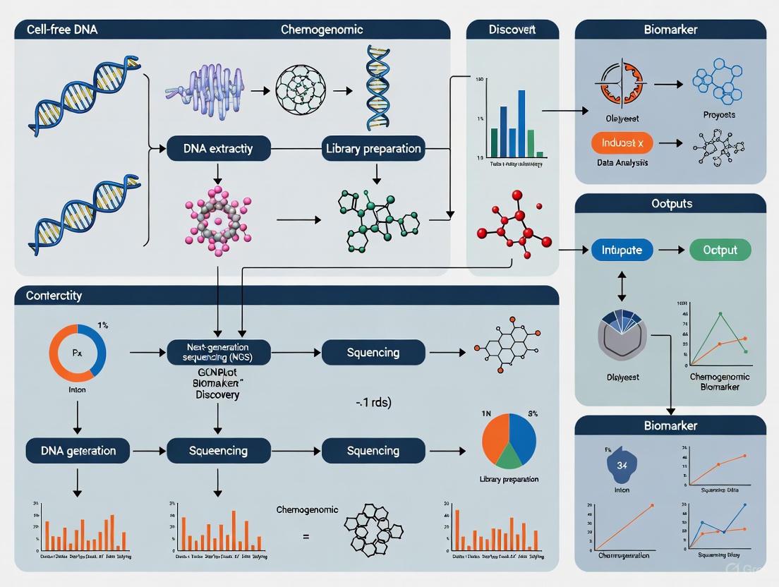
Advanced Cell-Free DNA NGS Workflows: Integrating Chemogenomic Biomarkers for Precision Oncology
This comprehensive review explores the integration of next-generation sequencing (NGS) workflows for cell-free DNA (cfDNA) analysis to unlock chemogenomic biomarkers in precision oncology.
Advanced Cell-Free DNA NGS Workflows: Integrating Chemogenomic Biomarkers for Precision Oncology
Abstract
This comprehensive review explores the integration of next-generation sequencing (NGS) workflows for cell-free DNA (cfDNA) analysis to unlock chemogenomic biomarkers in precision oncology. It covers the fundamental biology of cfDNA release mechanisms and fragmentation patterns, details established and emerging methodological approaches from targeted panels to whole-genome sequencing, and addresses critical troubleshooting and optimization strategies for pre-analytical variables and computational challenges. The article further provides a framework for analytical validation and comparative performance assessment of various cfDNA assays, including tumor-informed and tumor-agnostic methods. Designed for researchers, scientists, and drug development professionals, this resource aims to guide the robust implementation of liquid biopsy workflows to accelerate biomarker discovery and therapeutic monitoring.
The Biology of Cell-Free DNA and Its Role as a Chemogenomic Mirror
The analysis of cell-free DNA (cfDNA) has become a cornerstone of liquid biopsy approaches in clinical oncology and chemogenomic biomarker research. The composition and fragmentation patterns of cfDNA in circulation are direct consequences of its cellular origins and the mechanisms by which it is released. Understanding these release mechanisms—primarily apoptosis, necrosis, and active secretion—is crucial for interpreting cfDNA data in drug development workflows. This protocol details the experimental approaches for characterizing these pathways and their implications for next-generation sequencing (NGS) analyses in biomarker discovery.
Comparative Mechanisms of Cellular DNA Release
The primary pathways of DNA release differ significantly in their regulation, morphological features, and resulting cfDNA characteristics. The table below provides a systematic comparison of these mechanisms:
Table 1: Characteristics of Major cfDNA Release Mechanisms
| Feature | Apoptosis | Necrosis | Active Secretion |
|---|---|---|---|
| Regulation | Programmed, caspase-dependent [1] | Accidental or regulated (necroptosis) [2] [3] | Constitutive or triggered [4] |
| Inducing Stimuli | Developmental cues, DNA damage, cytotoxic drugs [5] | Infection, toxins, physical trauma [2] | Cellular signaling, differentiation [4] |
| Key Molecular Mediators | Caspases, CAD/DFF40, BCL2 family [1] [6] | RIPK1/RIPK3 (necroptosis), membrane rupture [3] | SNARE proteins, porosomes [4] |
| Membrane Integrity | Maintained until late stages; blebbing [2] | Lost; release of intracellular contents [2] [3] | Vesicle-mediated; membrane incorporated [4] |
| Inflammatory Response | Minimal ("silent" removal) [1] | Significant (release of DAMPs) [3] | Variable (depends on cargo) |
| Typical cfDNA Fragment Size | ~167 bp multi-mers (nucleosomal pattern) [6] | Larger, heterogeneous fragments (>1,000 bp) [6] | Larger fragments, often vesicle-protected [6] |
| Immunogenicity | Generally low, can be tolerogenic [3] | High (immunogenic cell death) [3] | Context-dependent |
Experimental Protocols for Investigating cfDNA Release
Protocol: In Vitro cfDNA Release Profiling
Purpose: To quantify and characterize the fragmentation profile of cfDNA released from cultured cells, allowing for the inference of the dominant release mechanism.
Background: A 2024 study profiling 24 human cell lines revealed two distinct cfDNA fragmentation patterns: a "left-skewed" pattern with a peak at ~167 bp (associated with apoptosis) and a "right-skewed" pattern with a peak >1,000 bp (associated with necrosis/vesicular release) [6].
Reagents and Materials:
- Cell lines of interest (e.g., MCF-10A, MCF-7)
- Appropriate cell culture media and supplements
- Serous or similar low-DNA background serum
- DNase/RNase-free tubes and pipette tips
- cfDNA extraction kit (e.g., QIAamp Circulating Nucleic Acid Kit)
- High Sensitivity DNA Analysis Kit (e.g., for Agilent Bioanalyzer or TapeStation)
- Quantitative PCR (qPCR) system or droplet digital PCR (ddPCR)
Procedure:
- Cell Culture and Conditioning:
- Seed cells at a standardized density (e.g., 1x10^6 cells per T-75 flask) in complete media.
- Allow cells to adhere overnight.
- Replace media with fresh media and culture cells without media changes for 1-3 days. Include biological replicates for each time point.
Sample Collection:
- At each time point (e.g., Day 1, 2, 3), carefully collect the conditioned media into a centrifuge tube.
- Centrifuge media at 2,000 x g for 10 minutes to pellet any detached cells or large debris.
- Transfer the supernatant to a new tube and centrifuge at 16,000 x g for 10 minutes to remove smaller particles.
cfDNA Isolation:
- Extract cfDNA from the clarified supernatant using a dedicated cfDNA extraction kit, strictly following the manufacturer's protocol.
- Elute the cfDNA in a small volume (e.g., 20-50 µL) of the provided elution buffer.
cfDNA Quantification and Fragmentomics Analysis:
- Quantify the total yield of cfDNA using a fluorescence-based method (e.g., Qubit dsDNA HS Assay).
- Analyze the fragment size distribution using a High Sensitivity DNA kit on an Agilent Bioanalyzer or similar platform. This will determine if the profile is "left-skewed" (apoptotic) or "right-skewed" (necrotic/vesicular).
Interpretation: A dominant peak at ~167 bp with a laddering pattern is indicative of apoptosis, while a profile enriched for fragments >1,000 bp suggests a significant contribution from necrosis or active vesicular release [6].
Protocol: CRISPR-Based Genetic Screening for cfDNA Regulators (cfCRISPR)
Purpose: To identify genes that functionally regulate the release of cfDNA, providing mechanistic insight into the dominant pathways active in a given cell type.
Background: This novel screening strategy leverages the fact that sgRNA barcodes integrated into a cell's genome are shed proportionally into cfDNA. Knocking out a gene that regulates cfDNA release will alter the sgRNA's representation in the cfDNA pool relative to the cellular genome [6].
Reagents and Materials:
- Lentiviral genome-wide CRISPR/Cas9 sgRNA library
- Target cell line with high cfDNA release (e.g., MCF-10A)
- Polybrene or other transduction enhancers
- Puromycin or other appropriate selection antibiotic
- cfDNA and gDNA extraction kits
- Next-generation sequencing platform
Procedure:
- Library Transduction:
- Transduce the target cell line with the lentiviral sgRNA library at a low multiplicity of infection (MOI ~0.3) to ensure most cells receive a single sgRNA.
- Select transduced cells with puromycin for 5-7 days.
Sample Harvesting:
- Culture the selected cell pool without media changes for 3 days to allow cfDNA accumulation.
- Harvest conditioned media for cfDNA isolation (as in Protocol 3.1).
- In parallel, harvest a portion of the cells for genomic DNA (gDNA) extraction.
Sequencing Library Preparation:
- Amplify the sgRNA barcode regions from both the cfDNA and cellular gDNA samples using PCR with indexing primers.
- Purify the PCR products and quantify the libraries.
High-Throughput Sequencing and Analysis:
- Sequence the cfDNA and gDNA libraries on an NGS platform to a sufficient depth.
- Map the sequenced reads to the sgRNA library reference to count the abundance of each sgRNA in both the cfDNA and gDNA samples.
- For each sgRNA, calculate a "cfDNA release ratio" (e.g., normalized reads in cfDNA / normalized reads in gDNA).
- Compare this ratio to the population average. sgRNAs targeting genes that positively regulate cfDNA release will be depleted in cfDNA, while those targeting negative regulators will be enriched.
Interpretation: Genes involved in apoptotic pathways (e.g., FADD, BCL2L1) are frequently identified as top hits, genetically validating apoptosis as a primary mediator of cfDNA release [6].
Signaling Pathways in DNA Release
The following diagram illustrates the key signaling pathways that lead to DNA release via apoptosis and necroptosis, highlighting points of crosstalk and experimental intervention.
Diagram Title: Signaling Pathways in Programmed and Accidental Cell Death
The Scientist's Toolkit: Research Reagent Solutions
The table below lists key reagents essential for investigating cfDNA release mechanisms in a chemogenomic context.
Table 2: Essential Research Reagents for cfDNA Release Studies
| Reagent / Tool | Function / Application | Example Use Case |
|---|---|---|
| Recombinant TRAIL | Inducer of extrinsic apoptosis [6] | Stimulate caspase-8 mediated apoptosis to increase apoptotic cfDNA yield. |
| Pan-Caspase Inhibitor (e.g., Z-VAD-FMK) | Inhibits executioner caspases [1] | Confirm caspase-dependent cfDNA release; distinguish apoptosis from necroptosis. |
| Necrostatin-1 (Nec-1) | Selective inhibitor of RIPK1-mediated necroptosis [3] | Inhibit regulated necrosis to assess its contribution to total cfDNA pool. |
| Anti-CD27 / Anti-CD38 Antibodies | Cell surface capture of B cells/plasma cells [7] | Isolate specific immune cell populations for cell-type-specific cfDNA analysis. |
| Oligonucleotide-barcoded Antibodies | Link cell surface phenotype to transcriptome (CITE-seq) [7] | Correlate IgG secretion capacity (via SEC-seq) with transcriptional state in single cells. |
| Hydrogel Nanovials (e.g., for SEC-seq) | Platform for accumulating secretions from single cells [7] | Quantify immunoglobulin secretion from single B cells and link to surface markers/transcriptomes. |
| sgRNA Library for cfCRISPR | Genome-wide knockout screening [6] | Identify novel genetic regulators of cfDNA biogenesis and release. |
Application in Chemogenomic Biomarker Research
Integrating an understanding of cfDNA release mechanisms directly enhances NGS workflow design and data interpretation. The fragmentation pattern of cfDNA is not merely a byproduct but an rich source of biological information. For instance, a dominant ~167 bp peak suggests tumor cell death is primarily mediated by apoptosis, potentially in response to a therapeutic agent. In contrast, a shift towards a "right-skewed" profile with larger fragments in serial monitoring could indicate the emergence of treatment resistance via alternative cell death pathways or a change in the tumor microenvironment [6]. Furthermore, leveraging inducers of immunogenic cell death, which can involve specific forms of apoptosis or necrosis, may enhance the release of tumor neoantigens and improve the sensitivity of liquid biopsy assays [3]. The protocols outlined here provide a framework for researchers to deconvolute these signals, thereby refining the use of cfDNA as a dynamic biomarker in drug development.
Characteristic cfDNA Fragmentation Patterns and Nucleosomal Signatures
Cell-free DNA (cfDNA) analysis has emerged as a cornerstone of liquid biopsy, offering a non-invasive window into physiological and pathological processes. The nucleosomal organization of cfDNA imposes characteristic fragmentation patterns that are profoundly influenced by the chromatin landscape of the cell of origin. These patterns provide a rich source of biological information beyond genetic alterations, enabling insights into gene regulation, cell identity, and disease states. Within the context of chemogenomic biomarkers research, understanding these fragmentation signatures is paramount for developing sensitive diagnostic, prognostic, and predictive tools for therapeutic intervention. This document details the fundamental principles, analytical approaches, and practical protocols for investigating cfDNA fragmentation patterns and nucleosomal signatures in cancer and other diseases.
Core Principles of cfDNA Fragmentation
Circulating cfDNA fragments are generated through non-random processes primarily during cellular apoptosis and necrosis. The fragmentation is heavily influenced by the underlying chromatin structure, wherein DNA wrapped around nucleosomes is protected from nuclease digestion, while linker DNA is more susceptible to cleavage. This results in several key characteristics:
- Size Periodicity: cfDNA fragments display a prominent peak at ~166-167 base pairs (bp), corresponding to DNA wrapped around a single nucleosome plus a linker histone (a chromatosome), and show a 10.4-bp periodicity in the 100-160 bp range, reflecting the helical pitch of DNA around the nucleosome core [8].
- Nucleosome Footprints: The in vivo occupancy of nucleosomes in the tissue of origin is imprinted in the cfDNA fragmentation pattern. DNA within open chromatin regions is more susceptible to fragmentation, while nucleosome-bound DNA is protected [9] [8].
- Transcription Factor Footprinting: Very short cfDNA fragments have been found to harbor footprints of transcription factors, revealing the binding sites of these regulatory elements in the cells from which the cfDNA originated [8].
Quantitative Landscape of cfDNA Fragmentation Metrics
Multiple computational metrics have been developed to quantify cfDNA fragmentation patterns. The performance of these metrics varies, and an integrated approach often yields the most robust results. The table below summarizes key fragmentation patterns and their diagnostic performance.
Table 1: Performance of Different cfDNA Fragmentation Metrics in Cancer Detection
| Fragmentation Metric | Description | Category | Reported Performance (AUROC) | Key Findings |
|---|---|---|---|---|
| End Motif (EDM) [9] | Analysis of the frequency of 4-mer sequences at fragment ends | Fragment sequence | 0.943 (Cross-validation) | Highest single diagnostic value in cross-validation; less stable in independent validation |
| Normalized Read Depth [10] | Fragment counts normalized to sequencing depth and region size | Fragment number | 0.943-0.964 (Avg. for cancer type prediction) | Top-performing metric on targeted panels; robust across cohorts |
| Fragment Dispersity Index (FDI) [11] | Integrates distribution of fragment ends with coverage variation | Hybrid (length & coverage) | Robust performance in early cancer diagnosis | Strongly correlates with chromatin accessibility; enables subtyping and prognosis |
| Windowed Protection Score (WPS) [9] [8] | Quantifies nucleosome protection in a sliding window | Hybrid (length & coverage) | Robust predictive capacity | Infers genome-wide nucleosome occupancy; generalizes well in validation |
| Integrated Fragmentation Pattern (IFP) [9] | Ensemble classifier combining 10 fragmentation patterns | Ensemble | Notable improvement over single patterns | Enhances cancer detection and tissue-of-origin determination; improves stability |
Different metrics are suited for various sequencing approaches. A recent study comparing fragmentomics on targeted panels versus whole-genome sequencing found that normalized fragment read depth across all exons provided the best overall performance for predicting cancer types and subtypes on targeted panels, with an average AUROC of 0.943 in one cohort and 0.964 in another [10]. Furthermore, combining multiple fragmentation patterns into an ensemble classifier (e.g., Integrated Fragmentation Pattern) has been shown to yield more stable and powerful performance for cancer detection and tissue-of-origin determination than any single pattern [9].
Experimental Protocols for Fragmentomics Analysis
Protocol: Genome-Wide Nucleosome Profiling using the Windowed Protection Score (WPS)
Principle: The WPS quantifies nucleosome protection by calculating, for a given genomic coordinate, the number of DNA fragments spanning a 120 bp window minus the number of fragments with an endpoint within that window. Protected nucleosomal regions show a high WPS, while nucleosome-depleted regions (e.g., transcription factor binding sites) show a low or negative WPS [8].
Workflow:
Sample Preparation & Sequencing:
- Isolate cfDNA from plasma using a circulating nucleic acid kit (e.g., QIAamp Circulating Nucleic Acid Kit).
- Prepare sequencing libraries without fragmentation or with minimal amplification to preserve native fragment length distributions. Both double-stranded and single-stranded library preparation methods can be used, with the latter offering better recovery of short fragments [8].
- Perform whole-genome sequencing. For nucleosome footprinting, a range of coverages can be effective, from high coverage (e.g., 30x) down to ultra-low-pass (0.1x) [12] [13].
Bioinformatic Processing:
- Align sequencing reads to the reference genome (e.g., hg19/hg38).
- Extract the genomic coordinates of both ends for each aligned fragment.
- Calculate the WPS across the entire genome using a sliding window approach. The formula for a base i is:
WPS(i) = (# of fragments spanning the window [i-60, i+60]) - (# of fragments with an endpoint within [i-60, i+60]). - Call nucleosome positions by identifying local maxima in the WPS profile.
Downstream Analysis:
- Aggregate WPS profiles around genomic features of interest (e.g., transcription start sites, specific transcription factor binding sites).
- Correlate WPS patterns with public epigenetic datasets (e.g., DNase I hypersensitivity, ATAC-seq) to validate inferred chromatin accessibility.
Protocol: Chromatin Accessibility Analysis via Fragmentomics in Open Chromatin Regions
Principle: This protocol leverages the fact that cfDNA within open chromatin regions is more susceptible to fragmentation. It involves calculating various fragmentation metrics specifically within predefined open chromatin regions to enhance signal-to-noise ratio in diagnostic models [9].
Workflow:
Define Open Chromatin Regions:
- Compile a set of open chromatin regions from relevant cell types. This can be sourced from public databases like ENCODE or Roadmap Epigenomics, or from cell-type-specific ATAC-seq or DNase-seq data. Key contributors to cfDNA include B cells, T cells, monocytes, and neutrophils [9].
Feature Calculation:
- From the aligned cfDNA sequencing data, compute multiple fragmentation features within the defined open chromatin regions. Key features include [9]:
- Fragment Length: Mean/median fragment size.
- Fragment Coverage: Number of fragment midpoints.
- End Motif (EDM): Frequency of specific 4-mer sequences at fragment ends.
- Orientation-aware Fragmentation (OCF): Strand-wise sequencing coverage patterns.
- Integrated Fragmentation Score (IFS): A composite score derived from multiple fragmentation features.
- From the aligned cfDNA sequencing data, compute multiple fragmentation features within the defined open chromatin regions. Key features include [9]:
Model Building and Validation:
- Use machine learning (e.g., elastic net, random forest) to train a classification model (cancer vs. healthy) using the computed fragmentation features as input.
- Employ cross-validation and validate the model on independent datasets to ensure generalizability.
- For enhanced stability and performance, integrate all fragmentation patterns into an ensemble classifier [9].
Protocol: Tissue-of-Origin Analysis using Single-Cell Reference Profiles
Principle: This approach correlates cfDNA-inferred nucleosome spacing with gene expression profiles from a comprehensive single-cell RNA sequencing atlas to rank the relative contribution of hundreds of cell types to the plasma cfDNA pool [13].
Workflow:
Nucleosome Signal Extraction:
- Perform whole-genome sequencing of cfDNA (can be ultra-low coverage, e.g., <0.3x).
- Calculate a nucleosome positioning signal, such as the Windowed Protection Score (WPS) or a Fourier Transform intensity at the 196-199 bp wavelength across gene bodies, which correlates with gene expression in the cell of origin [13].
Correlation with Reference Atlas:
- Obtain a single-cell transcriptome reference atlas encompassing a wide range of cell types (e.g., Tabula Sapiens, which includes over 490 cell types).
- For each cell type in the reference, correlate the average gene expression with the cfDNA-derived nucleosome signal (e.g., FFT intensity) across a large set of genes. A strong negative correlation indicates a higher contribution from that cell type.
Deconvolution and Interpretation:
- Rank all cell types in the reference by the strength of their correlation. In healthy individuals, immune cell types (monocytes, lymphocytes) and liver endothelial cells are typically top-ranked [13].
- In disease states (e.g., cancer), look for aberrantly up-ranked cell types that align with the disease biology (e.g., intestinal cells in colorectal cancer, plasma cells in multiple myeloma) [13].
Visualizing Workflows and Signaling Pathways
From Blood Draw to Nucleosome Profile
cfDNA Fragmentation Reflects Chromatin State
The Scientist's Toolkit: Essential Reagents and Materials
Table 2: Key Research Reagent Solutions for cfDNA Fragmentomics
| Item | Function/Application | Example Product/Note |
|---|---|---|
| cfDNA Isolation Kit | Purification of short, low-concentration cfDNA from plasma/serum. | QIAamp Circulating Nucleic Acid Kit (Qiagen). Critical for high yield and integrity. |
| Streck Cell-Free DNA BCT Tubes | Blood collection tubes that stabilize nucleosomal DNA and prevent genomic DNA release from blood cells. | Essential for preserving in vivo fragmentation profiles during sample transport. |
| Library Prep Kit for cfDNA | Construction of sequencing libraries from low-input, short-fragment DNA without bias. | KAPA HyperPrep Kit; NEB NEBNext Ultra II DNA Library Prep Kit. Protocols omitting fragmentation are key. |
| Enzymatic Methylation Conversion Kit | For simultaneous methylation and nucleosome occupancy profiling (cfNOMe). | NEBNext EM-Seq. Preserves fragmentation information better than bisulfite conversion [14]. |
| Targeted Gene Panels | For focused fragmentomics analysis on clinically relevant genes. | Panels from Tempus, Guardant, FoundationOne. Enable analysis on clinically available sequencing data [10]. |
| Bioinformatic Pipelines | For calculating fragmentation metrics (WPS, end motifs, coverage). | Custom scripts; Griffin framework (for GC-bias corrected nucleosome profiling) [12]. |
The analysis of characteristic cfDNA fragmentation patterns and nucleosomal signatures represents a powerful and rapidly advancing frontier in liquid biopsy. The protocols and data outlined herein provide a framework for integrating fragmentomics into chemogenomic biomarker research. By leveraging the rich epigenetic information encoded in the size, distribution, and ends of cfDNA fragments, researchers can gain unprecedented insights into tumor biology, disease heterogeneity, and treatment response, paving the way for more precise non-invasive diagnostics and monitoring.
Circulating tumor DNA (ctDNA) has emerged as a pivotal biomarker in precision oncology, offering a non-invasive window into tumor genomics. This analyte represents a minute fraction of the total cell-free DNA (cfDNA) in circulation, often constituting less than 0.1% in early-stage cancers, set against a background of cfDNA derived from normal cell apoptosis [15] [16]. The analysis of ctDNA within chemogenomic biomarker research provides critical insights for drug development, enabling real-time assessment of tumor dynamics, therapeutic response, and clonal evolution [17] [16]. Next-generation sequencing (NGS) workflows are fundamental to unlocking the potential of this fractional biomarker, yet they present significant technical challenges. This document outlines detailed protocols and applications for ctDNA analysis, framed within the context of cfDNA NGS workflows for advanced chemogenomics research.
ctDNA Applications in Precision Oncology
The clinical utility of ctDNA spans the cancer care continuum, from early detection to monitoring treatment response. Its applications are particularly valuable in providing a comprehensive view of tumor heterogeneity, which is often limited by the spatial constraints of traditional tissue biopsies [16]. The table below summarizes the core applications of ctDNA analysis in solid tumors.
Table 1: Key Applications of ctDNA Analysis in Solid Tumors
| Application | Key Utility | Example Cancer Types | Supporting Evidence |
|---|---|---|---|
| Treatment Response Monitoring | Correlates with tumor burden; predicts radiographic response earlier than imaging [16]. | Non-small cell lung cancer (NSCLC), Colorectal Cancer (CRC), Breast Cancer | A decline in ctDNA levels predicted radiographic response more accurately than follow-up imaging in NSCLC [15]. |
| Minimal Residual Disease (MRD) Detection | Detects molecular relapse post-treatment, often months before clinical or radiographic recurrence [17] [15]. | NSCLC, Colorectal Cancer, Breast Cancer | In breast cancer, SV-based ctDNA assays detected molecular relapse months to years before clinical relapse [15]. |
| Therapy Selection & Genotyping | Identifies actionable genomic alterations (AGAs) to guide targeted therapy [17] [18]. | NSCLC (EGFR, ALK, ROS1, BRAF, etc.) | Plasma-based NGS testing led to higher rates of guideline-recommended treatment (74% vs. 46%) [17]. |
| Resistance Mechanism Monitoring | Detects acquired mutations that confer resistance to targeted therapies, enabling timely treatment modification [15] [16]. | EGFR-mutant NSCLC (e.g., T790M) | In EGFR-mutant NSCLC, monitoring for the T790M resistance mutation allows for a switch to third-generation inhibitors without repeated tissue sampling [15]. |
Experimental Protocols for ctDNA Analysis
A robust ctDNA workflow requires meticulous attention from sample collection through data analysis. The following protocols detail the critical phases.
Pre-Analytical Phase: Sample Collection and Processing
The pre-analytical phase is critical, as variables here significantly impact cfDNA yield, integrity, and the success of downstream applications [19].
- Sample Collection: Collect blood using cell-stabilizing tubes (e.g., Streck, PAXgene). Standard K2EDTA tubes can be used if processing occurs within 1-4 hours of collection [19].
- Plasma Separation: Perform a double centrifugation protocol.
- First centrifugation: 800-1600 × g for 10 minutes at 4°C to separate plasma from cellular components.
- Transfer the supernatant to a fresh tube without disturbing the buffy coat.
- Second centrifugation: 16,000 × g for 10 minutes at 4°C to remove any residual cells.
- Plasma Storage: Immediately aliquot the cleared plasma and store at -80°C to prevent degradation. Avoid freeze-thaw cycles.
- cfDNA Extraction: Use high-sensitivity, magnetic bead-based cfDNA extraction kits. These methods offer high recovery rates, consistency, and are amenable to automation, which is crucial for reproducibility [19]. Validate the extraction system using synthetic cfDNA reference materials spiked into DNA-free plasma to confirm recovery efficiency and specificity.
Analytical Phase: Library Preparation and Sequencing
This phase converts isolated cfDNA into sequence-ready libraries, with specific adaptations for low-input, fragmented material.
Library Preparation:
- Fragmentation: Typically unnecessary as cfDNA is already fragmented (~167 bp). Proceed directly to end-repair and A-tailing [20].
- Adapter Ligation: Use platform-specific adapters (e.g., Illumina P5/P7) with dual-indexing to enable sample multiplexing and reduce index hopping. Incorporate Unique Molecular Identifiers (UMIs) during adapter ligation. UMIs are short random nucleotide sequences that tag individual DNA molecules before amplification, allowing for bioinformatic error correction and distinguishing true low-frequency variants from PCR/sequencing artifacts [16].
- Size Selection: Employ bead-based clean-up to enrich for the mononucleosomal cfDNA fraction (~150-170 bp) and remove longer genomic DNA contaminants and adapter dimers. This step enriches the ctDNA fraction and increases the detection yield of low-frequency variants [15].
- Library Amplification: Use a low-cycle, high-fidelity PCR to amplify the library. Excess cycles can exacerbate duplication rates and bias.
Sequencing:
- Platform: Use an Illumina-based NGS platform for massively parallel sequencing.
- Sequencing Depth: For ctDNA variant detection, especially in MRD settings, ultra-deep sequencing (>50,000x coverage) is mandatory to detect variants with a variant allele frequency (VAF) of <0.1% [15] [21].
- Chemistry: Sequencing-by-synthesis with reversible terminators is the standard [20].
Post-Analytical Phase: Bioinformatic Processing
The bioinformatic pipeline transforms raw sequencing data into actionable results.
- Primary Analysis:
- Base Calling: Convert raw signal data (e.g., Illumina .bcl files) into nucleotide sequences.
- Demultiplexing: Assign reads to individual samples based on their unique dual indices.
- Secondary Analysis:
- Read Trimming & UMI Consensus Building: Trim adapter sequences and group reads by their UMI and alignment coordinates. Generate a consensus sequence for each original DNA molecule to correct for random sequencing errors [16].
- Alignment: Map quality-filtered reads to a reference human genome (e.g., GRCh38).
- Variant Calling: Use specialized algorithms tuned for low-VAF variants. For tumor-informed MRD assays, specifically monitor the mutations identified in the patient's prior tumor tissue sample [17] [21]. The limit of detection for validated assays can reach a VAF of 0.0024% [21].
Table 2: Essential Quality Control Checkpoints in the ctDNA Workflow
| Workflow Stage | QC Parameter | Target Metric | QC Method/Tool |
|---|---|---|---|
| Nucleic Acid Isolation | cfDNA Concentration | >0.1 ng/μL (highly sample-dependent) | Fluorometry (e.g., Qubit, EzCube) [22] |
| cfDNA Integrity | Dominant peak at ~167 bp | TapeStation, Bioanalyzer [19] | |
| Genomic DNA Contamination | Absence of high molecular weight smear (>500 bp) | Electrophoresis [22] | |
| Library Preparation | Library Concentration | Within dynamic range of sequencer | qPCR-based quantification [20] |
| Library Fragment Size | ~200-300 bp (cfDNA + adapters) | TapeStation, Bioanalyzer | |
| Sequencing | Cluster Density | As per platform specification | Sequencing platform output |
| Q30 Score | >80% | Sequencing platform output | |
| Mean Coverage Depth | >50,000x for low-VAF detection | Alignment software (e.g., BWA, GATK) |
The following diagram illustrates the complete end-to-end workflow for ctDNA analysis.
The Scientist's Toolkit: Essential Research Reagents and Materials
Successful ctDNA analysis relies on a suite of specialized reagents and tools. The following table catalogs key solutions for the featured workflows.
Table 3: Essential Research Reagent Solutions for ctDNA Analysis
| Item | Function | Example Types & Notes |
|---|---|---|
| Cell-Stabilizing Blood Collection Tubes | Preserves blood sample integrity by preventing white blood cell lysis and release of genomic DNA, which dilutes ctDNA fraction. | Streck Cell-Free DNA BCT, PAXgene Blood cDNA Tube [19]. |
| Magnetic Bead-Based cfDNA Kits | Isolate and purify cfDNA from plasma with high efficiency and reproducibility; amenable to automation. | Kits from QIAGEN, Circulomics, Norgen Biotek [19]. |
| Reference Standard Materials | Act as process controls for validating extraction efficiency, assay sensitivity, and variant detection accuracy. | Seraseq ctDNA, AcroMetrix ctDNA, nRichDx cfDNA [19]. Contains predefined mutations at specific VAFs. |
| NGS Library Prep Kits (UMI) | Prepare fragmented cfDNA for sequencing while incorporating molecular barcodes for error correction. | Kits from QIAGEN (QIAseq), Bio-Rad, Swift Biosciences [16] [21]. |
| Fluorometers & Spectrophotometers | Precisely quantify low-concentration nucleic acid samples and assess purity. | Combination of EzCube Fluorometer (sensitivity) and EzDrop Spectrophotometer (purity check) is recommended [22]. |
| Targeted NGS Panels | Hybrid-capture or amplicon-based panels for deep sequencing of cancer-associated genes. | Panels covering key NSCLC drivers (EGFR, ALK, ROS1, BRAF, etc.) [17] [18]. |
The journey of analyzing ctDNA—a fractional signal in a vast background of normal cfDNA—demands a rigorously standardized and highly sensitive workflow. From the initial blood draw to the final bioinformatic interpretation, each step must be optimized for the unique challenges posed by this analyte. The protocols and tools outlined here provide a foundation for generating reliable, actionable data in chemogenomic biomarker research. As ctDNA technologies continue to evolve, with advancements in fragmentomics, methylation analysis, and ultrasensitive assays, their integration into standardized NGS workflows will further solidify the role of liquid biopsy in accelerating precision oncology and drug development.
Application Notes
Liquid biopsy has emerged as a transformative tool in oncology research, providing a minimally invasive means to interrogate tumor heterogeneity and dynamics in real-time. By analyzing circulating tumor-derived components, researchers and drug developers can access a comprehensive view of the total tumor burden, overcoming the limitations of traditional tissue biopsies that often fail to capture spatial and temporal heterogeneity [23] [24].
Key Analytical Targets in Liquid Biopsy
The clinical and research utility of liquid biopsy stems from multiple complementary analytes that provide distinct yet overlapping information about tumor biology:
Table 1: Core Liquid Biopsy Biomarkers and Their Research Applications
| Analyte | Key Characteristics | Primary Research Applications | Detection Challenges |
|---|---|---|---|
| Circulating Tumor DNA (ctDNA) | Short DNA fragments (20-50 bp); half-life <2 hours; represents 0.1-1.0% of total cfDNA [25] [26] | Treatment response monitoring; MRD detection; early relapse prediction; identifying resistance mutations [23] [27] | Low abundance in early-stage disease; requires highly sensitive detection methods [28] |
| Cell-Free DNA (cfDNA) | Double-stranded fragments (80-200 bp); baseline concentration 1-10 ng/mL in healthy individuals [26] | Cancer screening; monitoring tumor dynamics; assessing total cellular turnover | Background from hematopoietic system; elevated in various non-malignant conditions [26] |
| Circulating Tumor Cells (CTCs) | Rare cells (1-50 CTCs per 7.5mL blood); metastatic potential; half-life 1-2.5 hours [25] [29] | Studying metastasis mechanisms; drug resistance mechanisms; single-cell analysis | Extreme rarity; requires sophisticated enrichment technologies [25] [29] |
| DNA Methylation Markers | Stable epigenetic modifications; emerge early in tumorigenesis; tissue-specific patterns [28] [29] | Early cancer detection; tissue-of-origin identification; cancer subtyping | Requires bisulfite conversion or enzymatic treatment; complex bioinformatics [28] |
Capturing Tumor Heterogeneity
Liquid biopsy excels at resolving spatial and temporal tumor heterogeneity, which represents a significant challenge for traditional tissue sampling. A 2025 comparative analysis demonstrated that liquid biopsies capture between 33-92% of variants identified across multiple metastatic lesions, with some mutations exclusively detected in liquid biopsy [24]. This comprehensive profiling capability enables researchers to track clonal evolution under therapeutic selective pressure.
Table 2: Performance Characteristics of Liquid Biopsy in Capturing Heterogeneity
| Parameter | Tissue Biopsy | Liquid Biopsy | Research Implications |
|---|---|---|---|
| Spatial Coverage | Single lesion/site [24] | Multiple lesions simultaneously [23] [24] | More representative drug response assessment |
| Temporal Resolution | Limited by invasiveness [25] | Real-time monitoring (serial sampling) [23] [27] | Dynamic tracking of resistance mechanisms |
| Variant Detection | 4-12 mutations per patient (post-mortem tissue) [24] | 4-17 mutations per patient (pre-mortem LBx) [24] | Identification of dominant resistance clones |
| Variant Allele Frequency | 1.5-71.4% (tissue) [24] | 0.2-31.1% (LBx) [24] | Sensitivity to minor subclones with emerging resistance |
Clinical Translation and Validation
The transition of liquid biopsy from research to clinical applications requires rigorous validation. Current research focuses on standardizing pre-analytical variables, improving analytical sensitivity, and demonstrating clinical utility across diverse cancer types. As of 2025, multiple US-registered clinical trials are recruiting patients to validate liquid biopsy applications in immunotherapy monitoring, with 20 trials actively recruiting and 5 not yet recruiting [23].
Experimental Protocols
Protocol: Comprehensive ctDNA NGS Workflow for Chemogenomic Biomarker Discovery
Principle: This protocol describes an end-to-end workflow for isolation, preparation, and sequencing of ctDNA from patient plasma to identify genetic and epigenetic biomarkers relevant to drug response and resistance.
Sample Collection and Processing
Materials:
- K₂EDTA or Streck Cell-Free DNA Blood Collection Tubes
- Refrigerated centrifuge capable of 1600-2500 × g
- Plasma aspiration tools (serological pipettes or automated liquid handlers)
- -80°C freezer for plasma storage
Procedure:
- Blood Collection: Draw 10-20 mL whole blood into appropriate collection tubes. Invert gently 8-10 times.
- Initial Processing: Process within 2-4 hours of collection. Centrifuge at 1600-2500 × g for 10 minutes at 4°C.
- Plasma Separation: Carefully transfer supernatant plasma to sterile tubes without disturbing buffy coat.
- Secondary Centrifugation: Centrifuge plasma at 16,000 × g for 10 minutes at 4°C to remove residual cells.
- Aliquoting and Storage: Transfer cleared plasma to cryovials and store at -80°C until DNA extraction.
Critical Considerations:
- Consistent processing time is essential to prevent leukocyte lysis and background cfDNA increase.
- Avoid freeze-thaw cycles which degrade cfDNA integrity.
- Document time-from-collection-to-processing for quality control.
Cell-Free DNA Extraction and Quantification
Materials:
- Commercial cfDNA extraction kit (e.g., QIAamp Circulating Nucleic Acid Kit)
- Magnetic bead-based purification systems
- Fluorometric quantitation system (Qubit dsDNA HS Assay)
- Fragment analyzer (e.g., Agilent Bioanalyzer, TapeStation)
Procedure:
- Extraction: Extract cfDNA from 1-5 mL plasma according to manufacturer's protocol.
- Elution: Elute in 20-50 μL low-EDTA TE buffer or nuclease-free water.
- Quantification: Measure DNA concentration using fluorometric methods.
- Quality Control: Assess fragment size distribution using microcapillary electrophoresis.
Expected Outcomes:
- Yield: 1-50 ng total cfDNA depending on tumor burden
- Fragment size: predominant peak at ~167 bp with 10-bp periodicity
- DNA Integrity Number (DIN) >7 for high-quality samples
Library Preparation for NGS
Materials:
- Library preparation kit (e.g., Illumina TruSight Oncology ctDNA v2)
- Dual-indexed adapters
- Solid-phase reversible immobilization (SPRI) beads
- Thermal cycler with precise temperature control
Procedure:
- End Repair and A-Tailing: Convert fragment ends to blunt, 5'-phosphorylated ends with 3'-dA overhangs.
- Adapter Ligation: Add dual-indexed adapters with T4 DNA ligase.
- Size Selection: Perform double-sided SPRI bead cleanup (0.5X-0.8X ratios) to enrich for 150-250 bp fragments.
- Library Amplification: Amplify with 8-12 PCR cycles using polymerase with proofreading activity.
- Final Purification: Clean up with 1X SPRI beads and elute in 20-25 μL buffer.
Quality Control Checkpoints:
- Quantify library concentration by qPCR (library quantification kit)
- Confirm library size distribution by fragment analyzer
- Assess adapter dimer formation (<5% of total signal)
Bisulfite Conversion for Methylation Analysis
Materials:
- Bisulfite conversion kit (e.g., EZ DNA Methylation Kit)
- Thermal cycler with heated lid
- Desalting columns or magnetic beads
Procedure:
- Denaturation: Incocate DNA in conversion reagent at 95°C for 30-60 seconds.
- Conversion: Incubate at 50-64°C for 45-90 minutes (time-temperature varies by kit).
- Desalting: Bind converted DNA to silica membrane or magnetic beads.
- Desulfonation: Treat with alkaline desulfonation solution (10-20 minutes).
- Wash and Elute: Wash thoroughly and elute in low-EDTA TE buffer.
Critical Considerations:
- Account for DNA degradation during conversion (30-50% loss expected)
- Include unmethylated and methylated control DNA in each batch
- Process samples within same experiment to minimize batch effects
Target Enrichment and Sequencing
Materials:
- Hybridization capture reagents (e.g., IDT xGen Lockdown Probes)
- Sequence-specific or methyl-binding domain-based enrichment tools
- Thermomixer with accurate temperature control
- Next-generation sequencer (e.g., Illumina NovaSeq X Series)
Procedure:
- Hybridization: Denature libraries at 95°C, then incubate with biotinylated probes at 65°C for 16-24 hours.
- Capture: Bind probe-target hybrids to streptavidin magnetic beads.
- Washing: Perform stringent washes at increasing temperatures (65-72°C) to reduce off-target binding.
- Amplification: Amplify captured libraries with 10-14 PCR cycles.
- Pooling and Normalization: Pool libraries in equimolar ratios based on qPCR quantification.
- Sequencing: Sequence on appropriate platform to achieve >10,000X raw coverage for ctDNA detection.
Sequencing Parameters:
- Minimum coverage: 10,000X raw reads for 0.1% variant detection
- Read length: 2×100 bp or 2×150 bp for sufficient overlap
- Sample multiplexing: 16-96 samples per lane depending on required coverage
Protocol: Single-Cell CTC Analysis for Heterogeneity Studies
Principiple: Isolate and characterize circulating tumor cells at single-cell resolution to understand cellular heterogeneity and identify rare subpopulations with therapeutic relevance.
CTC Enrichment and Isolation
Materials:
- CTC enrichment platform (e.g., CellSearch, microfluidic devices)
- EpCAM or other surface antigen antibodies
- Fluorescence-activated cell sorting (FACS) instrumentation
- Single-cell dispensing system
Procedure:
- Blood Processing: Process 7.5-10 mL blood within 96 hours of collection.
- Immunomagnetic Enrichment: Incubate with antibody-conjugated magnetic beads targeting epithelial markers.
- Magnetic Separation: Place in magnetic field to retain CTCs while removing unbound cells.
- Immunofluorescence Staining: Stain with cytokeratin-FITC, CD45-APC, and DAPI.
- Identification and Sorting: Identify CTCs (CK+/CD45-/DAPI+) and sort single cells into 96- or 384-well plates.
Critical Considerations:
- Account for epithelial-mesenchymal transition (include mesenchymal markers)
- Process matched normal blood as negative control
- Minimize time between sorting and downstream processing
Whole Genome Amplification and Sequencing
Materials:
- Single-cell whole genome amplification kit (e.g., MALBAC, DOP-PCR)
- Multiple displacement amplification reagents
- Library preparation kit for low-input DNA
- Quality control reagents (Bioanalyzer, qPCR)
Procedure:
- Cell Lysis: Lyse single cells in alkaline buffer or with proteinase K.
- DNA Amplification: Perform whole genome amplification according to manufacturer's protocol.
- Quality Assessment: Check amplification success by qPCR of housekeeping genes.
- Library Preparation: Convert amplified DNA to sequencing libraries.
- Sequencing: Sequence at moderate coverage (0.1-0.5X) for copy number variation analysis.
Expected Outcomes:
- 50-80% single-cell amplification success rate
- Identification of subclonal copy number alterations
- Phylogenetic reconstruction of metastatic spread
Visualization Diagrams
Liquid Biopsy Workflow Diagram
Tumor Heterogeneity Capture Diagram
The Scientist's Toolkit: Research Reagent Solutions
Table 3: Essential Reagents and Kits for Liquid Biopsy Research
| Category | Product Examples | Key Features | Application Notes |
|---|---|---|---|
| Blood Collection Tubes | K₂EDTA tubes; Streck Cell-Free DNA BCT; PAXgene Blood cDNA Tubes | Preserves cfDNA profile; inhibits nucleases | Streck tubes allow 3-7 day shipping stability; K₂EDTA requires processing <4 hours [28] |
| cfDNA Extraction Kits | QIAamp Circulating Nucleic Acid Kit; MagMAX Cell-Free DNA Isolation Kit | Optimized for low-concentration samples; high reproducibility | Yields 1-50 ng cfDNA from 1-5 mL plasma; compatible with downstream NGS [29] |
| Library Preparation | Illumina TruSight Oncology ctDNA v2; Swift Accel-NGS Methyl-Seq | Low-input compatibility; unique molecular identifiers | TSO ctDNA v2 covers 600+ cancer genes; UMI error correction enables <0.1% VAF detection [29] |
| Bisulfite Conversion | EZ DNA Methylation Kit; Premium Bisulfite Kit | High conversion efficiency; minimal DNA degradation | 30-50% DNA loss expected; include methylation controls for QC [28] |
| Target Enrichment | IDT xGen Lockdown Probes; Twist Human Methylation Panels | Comprehensive coverage; uniform performance | Hybridization conditions critical for on-target rates; customize panels for specific research [29] |
| CTC Enrichment | CellSearch System; Parsortix Platform; CTC-iChip | FDA-cleared; marker-independent options | CellSearch uses EpCAM enrichment; suitable for epithelial cancers [25] |
| Single-Cell Analysis | 10X Genomics Chromium; SMART-Seq v4; MALBAC kits | Whole transcriptome; low-input sensitivity | Enables heterogeneity studies at single-cell resolution; identifies rare resistant subclones [29] |
Liquid biopsy is a minimally invasive technique that analyzes tumor-derived components from bodily fluids, offering a powerful alternative to traditional tissue biopsies. By capturing a comprehensive picture of tumor heterogeneity and enabling real-time monitoring, liquid biopsy is revolutionizing chemogenomics—the study of how genomic features influence response to pharmacological compounds [23]. The key biomarkers analyzed in liquid biopsies include:
- Circulating Tumor DNA (ctDNA): Fragments of DNA released into the bloodstream by tumor cells through mechanisms such as apoptosis and necrosis [30].
- Circulating Tumor Cells (CTCs): Intact cancer cells shed from primary and metastatic tumors [23].
- Tumor Extracellular Vesicles (EVs): Membrane-bound particles carrying nucleic acids, proteins, and lipids that facilitate cell-cell communication [23].
The integration of these biomarkers with next-generation sequencing (NGS) technologies enables the discovery of chemogenomic biomarkers, which are critical for predicting drug efficacy, understanding resistance mechanisms, and guiding personalized therapy in oncology [23] [31].
Liquid Biopsy Biomarkers and NGS Workflow
Key Biomarker Types and Characteristics
Table 1: Liquid Biopsy Biomarkers in Chemogenomics
| Biomarker Type | Origin & Composition | Primary Clinical Applications | Key Advantages |
|---|---|---|---|
| Circulating Tumor DNA (ctDNA) | Short DNA fragments released via cell death processes (apoptosis, necrosis) [30]. | - Tumor genotyping & mutation profiling- Monitoring treatment response- Minimal Residual Disease (MRD) detection [23] [30]. | - Captures tumor heterogeneity- Highly specific for tumor-associated mutations- Allows for serial monitoring [23]. |
| Circulating Tumor Cells (CTCs) | Whole, viable tumor cells shed into circulation [23]. | - Prognostic assessment- Understanding metastasis mechanisms- Ex vivo drug sensitivity testing [23]. | Provides intact cellular material for functional analyses and culture [23]. |
| Tumor Extracellular Vesicles (EVs) | Membrane-bound vesicles carrying proteins, RNA, and DNA [23]. | - Identifying therapeutic targets- Monitoring drug resistance [23]. | - Protects molecular cargo from degradation- Reflects the state of parental tumor cells [23]. |
Comprehensive NGS Workflow for ctDNA Analysis
The transformation of a blood sample into actionable chemogenomic data involves a multi-stage NGS workflow. Key stages include sample collection, library preparation, sequencing, and bioinformatic analysis, each requiring rigorous optimization to ensure data accuracy and reliability [31] [32].
Diagram 1: Core NGS workflow for liquid biopsy analysis, covering sample collection to data interpretation.
Experimental Protocols
Protocol: Plasma ctDNA Extraction and NGS Library Construction
Objective: To isolate high-quality cell-free DNA (cfDNA) from patient blood plasma and prepare sequencing libraries for the detection of somatic variants and chemogenomic biomarkers.
Materials:
- Blood Collection Tubes: Streck Cell-Free DNA BCT or K2EDTA tubes [32].
- Extraction Kit: QIAamp Circulating Nucleic Acid Kit or similar [32].
- Library Prep Kit: xGen cfDNA & FFPE DNA Library Preparation Kit or similar [32].
- Quantification Tools: Qubit fluorometer and Agilent Bioanalyzer/TapeStation [32].
- Sequencing Platform: Illumina NovaSeq or similar [31].
Procedure:
- Sample Collection and Processing:
- Collect venous blood into preservative tubes. Invert gently 8-10 times.
- Centrifuge at 1,600-2,000 x g for 10-20 minutes at 4°C within 4 hours of collection to separate plasma.
- Carefully transfer the supernatant (plasma) to a fresh tube without disturbing the buffy coat.
- Perform a second, high-speed centrifugation at 16,000 x g for 10 minutes to remove residual cells and debris. Transfer the clarified plasma to a new tube [32].
cfDNA Extraction:
- Follow the manufacturer's protocol for the chosen circulating nucleic acid extraction kit.
- Elute the purified cfDNA in a low-EDTA TE buffer or nuclease-free water. A typical elution volume is 20-50 µL [32].
Quality Control of Extracted cfDNA:
- Quantify the cfDNA using a fluorescence-based method (e.g., Qubit dsDNA HS Assay).
- Assess the fragment size distribution using a high-sensitivity instrument (e.g., Agilent Bioanalyzer 2100). The expected peak should be ~167 bp, characteristic of mononucleosomal DNA [32].
NGS Library Preparation:
- End Repair & A-Tailing: Convert the fragmented DNA ends to blunt, 5'-phosphorylated ends, followed by the addition of a single 'A' nucleotide to the 3' ends.
- Adapter Ligation: Ligate double-stranded DNA adapters with a 3' 'T' overhang to the cfDNA fragments. Include unique molecular identifiers (UMIs) to mitigate PCR amplification bias and enable error correction.
- Library Amplification: Perform limited-cycle PCR (e.g., 8-12 cycles) to enrich for adapter-ligated fragments and add full-length sequencing primers.
- Library Clean-up and Size Selection: Purify the amplified library using solid-phase reversible immobilization (SPRI) beads to remove short fragments and adapter dimers [31] [32].
Final Library QC and Sequencing:
- Quantify the final library concentration by qPCR for accurate molarity.
- Confirm library size and quality (typically ~300-400 bp) using the Bioanalyzer.
- Pool libraries as needed and sequence on the appropriate NGS platform (e.g., Illumina) to a sufficient depth (e.g., >10,000x coverage for low-frequency variants) [31] [32].
Protocol: Targeted Sequencing for Chemogenomic Biomarker Discovery
Objective: To perform deep, targeted sequencing of genes known to harbor alterations that influence drug response, using ctDNA-derived libraries.
Materials:
- Hybridization Capture Kit: xGen Hybridization and Capture Kit or similar.
- Targeted Panels: Commercially available (e.g., Illumina TSO 500 ctDNA) or custom-designed panels covering key cancer genes (e.g., EGFR, KRAS, BRAF, PIK3CA, ALK).
- Sequencing Platform: Illumina series [31].
Procedure:
- Library Enrichment:
- Pool up to 500 ng of pre-made cfDNA libraries from multiple samples.
- Denature the library pool and hybridize with biotinylated probes complementary to the targeted genomic regions for 4-16 hours.
- Capture the probe-bound fragments using streptavidin-coated magnetic beads.
- Wash the beads stringently to remove non-specifically bound DNA.
Post-Capture Amplification and QC:
- Perform a second, limited-cycle PCR to amplify the captured library.
- Purify the final enriched library with SPRI beads.
- Validate enrichment success and quantify the library as described in Section 3.1.
Sequencing and Data Analysis:
Data Analysis and Bioinformatics Pipeline
The computational analysis of NGS data is critical for translating raw sequencing reads into validated chemogenomic insights. The pipeline involves sequential steps of data processing, variant identification, and functional annotation [31] [33].
Diagram 2: Bioinformatics pipeline for identifying and annotating chemogenomic variants from NGS data.
Key Computational Tools for NGS Analysis
Table 2: Essential Bioinformatics Tools for ctDNA NGS Analysis
| Analysis Step | Software/Tool | Primary Function |
|---|---|---|
| Quality Control | FastQC, QualiMap [33] | Assesses sequencing read quality and identifies potential biases. |
| Read Trimming | Trimmomatic, Fastp [33] | Removes low-quality bases and adapter sequences. |
| Sequence Alignment | BWA-MEM, HISAT2, STAR [33] | Maps sequencing reads to a reference genome. |
| Variant Calling | GATK, MuTect2, FreeBayes [33] | Identifies single nucleotide variants (SNVs) and small insertions/deletions (Indels). |
| Variant Annotation | ANNOVAR, Variant Effect Predictor (VEP) [33] | Predicts functional impact of variants (e.g., missense, frameshift) and provides population frequency data. |
| Pathway Analysis | DAVID, Enrichr, GSEA [33] | Identifies overrepresented biological pathways and processes among a set of genes. |
The Scientist's Toolkit: Research Reagent Solutions
Successful implementation of liquid biopsy-based chemogenomics requires a suite of reliable reagents and materials.
Table 3: Essential Research Reagents and Materials
| Item | Function/Description | Example Products/Brands |
|---|---|---|
| Cell-Free DNA Blood Collection Tubes | Preserves blood samples to prevent genomic DNA contamination and cfDNA degradation during transport and storage [32]. | Streck Cell-Free DNA BCT tubes. |
| Nucleic Acid Extraction Kits | Isolate and purify high-integrity cfDNA/ctDNA from plasma samples with high efficiency and low contamination [32]. | QIAamp Circulating Nucleic Acid Kit. |
| NGS Library Preparation Kits | Convert fragmented cfDNA into sequencing-ready libraries via end-repair, A-tailing, adapter ligation, and PCR amplification [32]. | xGen cfDNA & FFPE DNA Library Prep Kit. |
| Targeted Hybridization Capture Panels | Biotinylated probes designed to enrich sequencing libraries for specific genes of interest, allowing for deep sequencing of chemogenomic targets [31]. | Illumina TSO 500 ctDNA, custom panels from IDT. |
| NGS Quantification Kits & Instruments | Accurately measure library concentration and quality prior to sequencing to ensure optimal cluster density and data output [32]. | Qubit dsDNA HS Assay, Agilent High Sensitivity DNA Kit. |
Integrative Chemogenomic Analysis and Pathway Mapping
The final stage involves integrating genomic variant data with drug response knowledge to generate testable hypotheses. This is the core of chemogenomics, where a somatic mutation identified in a liquid biopsy is linked to a potential therapeutic strategy [34] [35].
Diagram 3: The chemogenomic hypothesis generation workflow, linking a detected variant to a potential therapy.
From Biomarker to Therapy: A Clinical Example
The utility of this integrated approach is exemplified by targeting the EGFR L858R mutation in non-small cell lung cancer (NSCLC):
- Biomarker Discovery: Deep sequencing of ctDNA identifies an EGFR L858R activating mutation.
- Knowledge Base Integration: Databases like the FDA's Table of Pharmacogenomic Biomarkers or the Clinical Pharmacogenetics Implementation Consortium (CPIC) guidelines list this mutation as a predictive biomarker for response to EGFR tyrosine kinase inhibitors (TKIs) [34].
- Therapeutic Decision: The patient is treated with a third-generation EGFR TKI (e.g., Osimertinib), which is specifically designed to target this mutation and overcome resistance [34].
- Monitoring: Serial liquid biopsies are used to monitor ctDNA levels, with a decrease indicating treatment response. The emergence of new mutations (e.g., EGFR C797S) in subsequent liquid biopsies can signal the development of resistance, prompting another cycle of chemogenomic analysis and treatment adjustment [23] [30].
This closed-loop workflow demonstrates how liquid biopsy and NGS workflows form a dynamic platform for precision oncology, enabling continuous therapeutic optimization based on the evolving genomic landscape of a patient's cancer.
NGS Workflow Architectures: From Library Prep to Multi-Omic Data Generation
Next-Generation Sequencing (NGS) has revolutionized genomic analysis, offering powerful tools for investigating chemogenomic biomarkers through cell-free DNA (cfDNA) workflows. The analysis of circulating tumor DNA (ctDNA), the tumor-derived fraction of cfDNA, provides a noninvasive method for assessing the molecular landscape of cancer, enabling real-time monitoring of treatment response and identification of resistance mechanisms [36] [37]. For researchers and drug development professionals, selecting the appropriate NGS approach—targeted panels, whole-exome sequencing (WES), or whole-genome sequencing (WGS)—represents a critical decision point that significantly impacts project scope, cost, data volume, and biological insights. Each method offers distinct advantages and limitations, making them suited to different research scenarios within precision oncology and biomarker discovery [38] [39].
Targeted panels focus on sequencing a predefined set of genes known to be associated with specific cancer types or therapeutic responses, providing deep coverage of selected genomic regions [39] [40]. Whole-exome sequencing captures the protein-coding regions of the genome (approximately 2%), where most known disease-causing variants reside [41]. Whole-genome sequencing offers the most comprehensive approach by analyzing the entire genome, including both coding and non-coding regions [42]. The choice between these methodologies must consider multiple factors, including the specific research questions, sample type and quality, required detection sensitivity, bioinformatic capabilities, and budget constraints, particularly when working with the low ctDNA concentrations typical of liquid biopsy samples [36].
Technical Comparison of NGS Methodologies
Core Characteristics and Applications
The three primary NGS approaches differ fundamentally in the genomic regions they interrogate, the data they generate, and their clinical applications, particularly in the context of cfDNA analysis for chemogenomic biomarker research.
Targeted gene panels utilize hybridization-capture or amplicon-based methods to enrich specific genomic regions of interest prior to sequencing [40]. This focused approach enables extremely high sequencing depth (often >500×), which is crucial for detecting low-frequency variants in ctDNA, where tumor-derived DNA can represent a very small fraction of the total cfDNA [36] [39]. Panels are particularly valuable when the patient's phenotype points to a well-characterized group of conditions with known genetic heterogeneity, such as non-small cell lung cancer (NSCLC) where biomarkers like EGFR, ALK, ROS1, and BRAF offer targets for therapeutic intervention [36] [39]. The limited scope reduces data analysis burden and minimizes incidental findings while providing sufficient information for treatment decisions in many clinical scenarios [40].
Whole-exome sequencing (WES) focuses on the exome, which constitutes approximately 1-2% of the human genome (about 30 million base pairs) but harbors an estimated 85% of known disease-causing variants [38] [41]. By sequencing all protein-coding regions, WES provides a balance between comprehensive genomic coverage and practical data management, making it particularly valuable for discovery-oriented research where the genetic basis of disease or treatment response is not fully characterized [38]. However, even the best target enrichment workflows are prone to some degree of target dropout and coverage bias, especially in GC- or AT-rich regions [42]. For cfDNA applications, WES typically achieves moderate coverage (80-150×), which may limit sensitivity for detecting very low-frequency ctDNA variants compared to targeted approaches [39].
Whole-genome sequencing (WGS) provides the most comprehensive genomic analysis by sequencing the entire genome (approximately 3 billion base pairs), including both coding and noncoding regions [41] [42]. This unbiased approach facilitates detection of diverse variant types—including single nucleotide variants (SNVs), insertions/deletions (indels), copy number variations (CNVs), structural variants (SVs), and regulatory element alterations—without prior knowledge of their location [39]. While WGS offers unparalleled opportunities for novel biomarker discovery, it generates substantial data volumes (typically >90 GB per sample) and requires significant computational resources for processing and interpretation [41] [39]. The lower sequencing depth (typically 30-50×) at comparable cost to WES may limit its sensitivity for detecting rare variants in heterogeneous cfDNA samples [39].
Table 1: Comparative Analysis of Targeted Panels, WES, and WGS for cfDNA Research
| Feature | Targeted Panels | Whole Exome Sequencing (WES) | Whole Genome Sequencing (WGS) |
|---|---|---|---|
| Analyzed Region | 50-500 selected genes [39] | All coding exons (~1-2% of genome) [41] [39] | Entire genome (coding + non-coding) [41] [39] |
| Region Size | Tens to thousands of genes [41] | >30 million base pairs [41] | ~3 billion base pairs [41] |
| Average Coverage | 500-1000× [39] | 80-150× [39] | 30-50× [39] |
| Data Volume per Sample | Low (varies with panel size) [39] | 5-10 GB [41] | >90 GB [41] |
| Detection Sensitivity for Low-Frequency Variants | High (ideal for VAF <10%) [39] | Moderate [39] | Lower unless sequenced at high depth [39] |
| Primary Clinical/Research Applications | Conditions with clear phenotype and known genes [39]; Therapy selection [36] | Rare diseases, complex phenotypes [39]; Unexplained hereditary disorders [38] | Unresolved cases, novel biomarker discovery [39] |
| Variant Types Detected | SNPs, InDels, CNV, Fusion [41] | SNPs, InDels, CNV, Fusion [41] | SNPs, InDels, CNV, Fusion, SV [41] |
| Turnaround Time | Fast (e.g., 4 days for validated oncopanel) [40] | Moderate [39] | Slow [39] |
| Cost | Low [39] | Moderate [39] | High [39] |
| Risk of Incidental Findings | Low [39] | Moderate [39] | High [39] |
Advantages and Limitations in cfDNA Research
Each NGS approach presents distinct advantages and limitations when applied to cfDNA analysis for chemogenomic biomarker research. Understanding these trade-offs is essential for selecting the appropriate methodology.
Targeted panels offer several advantages for ctDNA analysis: (1) High sensitivity due to deep sequencing coverage, enabling detection of rare variants with allele frequencies as low as 0.1-0.25% with optimized methods [36]; (2) Cost-effectiveness through focused sequencing resources [39]; (3) Streamlined data analysis with reduced interpretation burden [39]; and (4) Rapid turnaround times, with some validated oncopanels achieving results within 4 days [40]. However, targeted panels have significant limitations: (1) Limited discovery potential as they only detect variants in predefined genes [38]; (2) Inability to detect novel biomarkers outside the panel content [43]; and (3) Rapid obsolescence as new disease-gene associations are identified, with one study noting that 23% of positive WES findings were in genes discovered within the preceding two years [38].
Whole-exome sequencing provides a balanced approach with these advantages: (1) Comprehensive coverage of protein-coding regions without being restricted to known genes [38]; (2) Cost-effective alternative to WGS for focusing on coding regions [42]; and (3) Excellent for hypothesis-generating research where the genetic basis is unclear [39]. The limitations of WES include: (1) Inability to detect functional variants in noncoding regions [38]; (2) Variable coverage uniformity across the exome, potentially missing some variants [42]; (3) Moderate sensitivity for low-frequency variants compared to targeted panels [39]; and (4) Higher interpretation burden than targeted panels due to more variants [39].
Whole-genome sequencing offers the most comprehensive approach with these advantages: (1) Complete genomic characterization including coding, noncoding, and regulatory regions [42]; (2) Superior detection of structural variants, copy number variations, and rearrangements [39]; (3) Hypothesis-free approach enabling novel biomarker discovery [39]; and (4) Future-proof dataset that can be reanalyzed as new genomic insights emerge. The limitations are substantial: (1) Highest cost per sample [39]; (2) Massive data storage and computational requirements [39]; (3) Challenging interpretation of noncoding variants with limited functional annotation [38]; and (4) Lower sensitivity for rare variants at standard coverage depths [39].
Table 2: Performance Metrics for NGS Approaches in Detecting Key Variant Types
| Variant Type | Targeted Panels | WES | WGS |
|---|---|---|---|
| Single Nucleotide Variants (SNVs) | Excellent (high sensitivity at low VAF) [40] | Good [39] | Good [39] |
| Insertions/Deletions (Indels) | Excellent (with optimized panels) [40] | Good [39] | Good [39] |
| Copy Number Variations (CNVs) | Limited [39] | Partial (depends on pipeline) [39] | Excellent [39] |
| Gene Fusions/Rearrangements | Good (for targeted genes) [41] | Moderate [41] | Excellent [41] |
| Structural Variants (SVs) | Limited [39] | Partial [39] | Excellent [39] |
| Noncoding Variants | None (unless specifically targeted) | None | Good [42] |
NGS Workflow for cfDNA Analysis
Standardized Protocol for cfDNA NGS Analysis
The following protocol outlines a comprehensive workflow for NGS analysis of cfDNA samples, with specific considerations for each sequencing approach. This methodology is adapted from validated procedures described in the literature and has been optimized for ctDNA detection sensitivity [36] [20] [40].
Sample Collection and Processing
- Collect peripheral blood (typically 10-20 mL) in cell-stabilizing tubes (e.g., Streck Cell-Free DNA BCT or PAXgene Blood cDNA tubes) to prevent genomic DNA contamination from white blood cell lysis.
- Process samples within 4-6 hours of collection by double centrifugation: first at 1600× g for 10 minutes at 4°C, then transfer plasma to a fresh tube and centrifuge at 16,000× g for 10 minutes at 4°C to remove remaining cellular debris.
- Store plasma at -80°C if not proceeding immediately to DNA extraction.
cfDNA Extraction
- Extract cfDNA from 1-5 mL of plasma using specialized cfDNA extraction kits (e.g., QIAamp Circulating Nucleic Acid Kit, Maxwell RSC ccfDNA Plasma Kit, or similar).
- Quantify cfDNA using fluorometric methods (e.g., Qubit dsDNA HS Assay) rather than UV spectrophotometry, as fluorometry provides more accurate quantification of low-concentration samples.
- Assess cfDNA quality using microfluidic electrophoresis (e.g., Agilent 2100 Bioanalyzer with High Sensitivity DNA chips or TapeStation), expecting a characteristic peak at ~160-170 bp representing mononucleosomal DNA.
- A minimum of 10-50 ng cfDNA is typically required for library preparation, though some optimized workflows can work with lower inputs [40].
Library Preparation
- Convert cfDNA into sequencing libraries using kits specifically designed for low-input and fragmented DNA (e.g., Illumina TruSeq Nano, KAPA HyperPrep, or NEBNext Ultra II DNA Library Prep).
- For targeted panels: Use hybridization capture with biotinylated oligonucleotide probes (e.g., Illumina TruSight Oncology 500, TSO500 ctDNA) or amplicon-based approaches (e.g., AmpliSeq HD) [36] [37] [40].
- For WES: Employ exome capture using platforms such as Agilent SureSelect, Illumina Nextera Rapid Capture, or IDT xGen Exome Research Panel.
- For WGS: Use non-enriched library preparation methods with appropriate fragmentation.
- Incorporate unique molecular identifiers (UMIs) to reduce sequencing artifacts and enable accurate detection of low-frequency variants by accounting for PCR amplification biases and sequencing errors [37].
Target Enrichment (for Panels and WES)
- For hybridization-based approaches: Incubate libraries with biotinylated probes targeting regions of interest, then capture with streptavidin-coated magnetic beads.
- Wash stringently to remove non-specifically bound DNA and elute the enriched targets.
- Amplify the enriched libraries with limited-cycle PCR (typically 8-12 cycles) to generate sufficient material for sequencing.
Sequencing
- Quantify final libraries using qPCR-based methods (e.g., KAPA Library Quantification Kit) for accurate quantification, as this correlates best with cluster density on the flow cell.
- Dilute libraries to appropriate concentrations (typically 1-2 nM) and denature with NaOH immediately before loading.
- Sequence on appropriate NGS platforms (e.g., Illumina NovaSeq 6000, MiSeq, or MGI DNBSEQ-G50RS) with paired-end reads [37] [40].
- For targeted panels: Sequence to high depth (typically >500× mean coverage) to enable detection of low-frequency variants (≤0.5% VAF).
- For WES: Sequence to 80-150× mean coverage.
- For WGS: Sequence to 30-50× mean coverage.
Bioinformatic Analysis
- Demultiplex sequencing data and convert BCL files to FASTQ format.
- Perform quality control assessment using tools such as FastQC.
- Align reads to the reference genome (e.g., GRCh38) using optimized aligners (BWA-MEM, Bowtie 2).
- For UMI-containing libraries: Process reads to group duplicates by their molecular barcodes, correcting for sequencing errors.
- Call variants using specialized tools:
- For panels: Use vendor-specific software (e.g., Sophia DDM) or custom pipelines with MuTect2, VarScan2 for somatic variants [40].
- For WES/WGS: Use GATK best practices for variant calling, including HaplotypeCaller for germline variants and Mutect2 for somatic variants.
- Annotate variants using ANNOVAR, SnpEff, or VEP with appropriate databases (ClinVar, COSMIC, gnomAD, dbSNP).
- Filter variants based on quality metrics, population frequency, and predicted functional impact.
- For cfDNA samples: Apply additional filters for clonal hematopoiesis variants and sequencing artifacts.
Analytical Validation
- Establish limit of detection (LOD) using serially diluted reference standards with known variant allele frequencies (typically 2-5% for WES/WGS, 0.1-1% for targeted panels) [40].
- Assess reproducibility through replicate experiments (inter-run and intra-run precision).
- Validate against orthogonal methods (e.g., digital PCR) for key variants.
Quality Control Metrics
Robust quality control is essential throughout the NGS workflow to ensure reliable results, particularly when working with low-input cfDNA samples.
Pre-sequencing QC Metrics
- cfDNA Quantity: Minimum 10-50 ng for library preparation, though some optimized panels can work with lower inputs [40].
- cfDNA Quality: Fragment size distribution should show peak at ~160-170 bp. Ratio of long (>1000 bp) to short (~160 bp) fragments should be <10% to minimize contamination from cellular genomic DNA.
- Library Concentration: Typically 1-20 nM, quantified by qPCR for accurate measurement.
- Library Size Distribution: Expected size of 200-500 bp including adapters.
Sequencing QC Metrics
- Clustering Density: Optimal range depends on platform (e.g., 170-220 K/mm² for Illumina NovaSeq).
- Q-score: >80% bases with Q30 (0.1% error rate) or higher.
- % Bases ≥ Q30: Should exceed 75% for reliable variant calling.
Post-sequencing QC Metrics
- On-target Rate: Percentage of reads mapping to target regions (>80% for hybrid capture panels, >60% for amplicon-based panels).
- Uniformity of Coverage: >80% of targets covered at ≥0.2× mean coverage.
- Mean Coverage: Varies by application (>500× for targeted panels, 80-150× for WES, 30-50× for WGS) [39].
- Duplicate Rate: <20% for WGS, <50% for hybrid capture, higher rates expected for amplicon-based approaches.
- Insert Size: Should match expected cfDNA fragment distribution.
The Scientist's Toolkit: Essential Reagents and Technologies
Successful implementation of cfDNA NGS workflows requires careful selection of reagents, technologies, and computational tools. The following table summarizes key solutions used in the field.
Table 3: Research Reagent Solutions for cfDNA NGS Workflows
| Category | Product/Technology | Key Features | Application Notes |
|---|---|---|---|
| Blood Collection Tubes | Streck Cell-Free DNA BCTPAXgene Blood cDNA tubes | Preserves blood cells, prevents gDNA releaseStabilizes nucleic acids | Enables extended sample transportMaintains cfDNA profile for days |
| cfDNA Extraction Kits | QIAamp Circulating Nucleic Acid KitMaxwell RSC ccfDNA Plasma KitMagMAX Cell-Free DNA Isolation Kit | Optimized for low-abundance cfDNAAutomated processingHigh recovery from small volumes | Critical for low-VAF variant detectionReduces manual processing timeSuitable for high-throughput labs |
| Library Prep Kits | Illumina TruSeq NanoKAPA HyperPrep KitNEBNext Ultra II DNA Library Prep | Low-input DNA compatibilityUMI incorporationReduced GC bias | Essential for limited cfDNA samplesEnables error correctionImproves coverage uniformity |
| Target Enrichment | Illumina TruSight Oncology 500 ctDNAKAPA HyperCaptureIDT xGen Lockdown Panels | Pan-cancer contentHybridization-based captureCustomizable target content | Detects SNVs, indels, CNVs, fusionsHigh specificity and sensitivityTailored to specific research needs |
| UMI Technologies | TruSight Oncology UMI ReagentsQIAseq UMI technologies | Unique molecular identifiersError correctionBackground noise reduction | Enables detection of variants <0.5% VAFCritical for low-frequency variantsReduces false positives |
| Sequencing Platforms | Illumina NovaSeq 6000MGI DNBSEQ-G50RSIllumina MiSeq | High-throughputCompetitive pricingRapid turnaround | Scalable for large studiesCost-effective for targeted panelsIdeal for validation studies |
| Bioinformatic Tools | Sophia DDMGATK Mutect2BWA-MEMANNOVAR | Machine learning integrationSomatic variant callingRead alignmentVariant annotation | Automated variant classificationGold standard for NGS dataFast and accurate alignmentFunctional interpretation |
Selection Framework and Decision Pathways
Choosing the optimal NGS approach requires systematic consideration of multiple scientific and practical factors. The following decision pathway provides a structured framework for selection based on key project parameters.
Application-Specific Recommendations
Different research scenarios warrant specific NGS approaches based on the biological questions, sample characteristics, and analytical requirements.
Therapy Selection and Resistance Monitoring For clinical applications focused on identifying actionable mutations for therapy selection or detecting resistance mechanisms, targeted panels are typically preferred [36]. Their high sensitivity enables detection of emerging resistance mutations at low variant allele frequencies, which is crucial for timely treatment modifications. Studies have demonstrated that ctDNA NGS testing can better recapitulate NSCLC heterogeneity compared with tissue testing and allows monitoring of therapy response and early identification of resistance mechanisms [36]. The focused nature of panels also facilitates rapid turnaround times (as short as 4 days with optimized workflows), which is often critical in clinical decision-making [40].
Rare Disease Diagnosis and Complex Phenotypes For patients with rare tumors or complex phenotypes without clear genetic etiology, WES provides an optimal balance of comprehensive coverage and practical feasibility [43] [38]. WES can identify pathogenic variants across all protein-coding genes without prior hypothesis about the causative gene, making it particularly valuable for conditions with significant genetic heterogeneity. The American College of Medical Genetics and Genomics (ACMG) recommends both WES and WGS as primary or secondary testing options for patients with rare genetic diseases, congenital abnormalities, developmental delays, or intellectual disabilities [38].
Novel Biomarker Discovery For discovery-oriented research aimed at identifying novel biomarkers, structural variants, or noncoding drivers, WGS offers the most comprehensive approach [43] [39]. The ability to detect variants throughout the genome, including regulatory regions and structural variations, provides unprecedented opportunities for understanding disease mechanisms. However, this approach requires substantial bioinformatic resources and careful consideration of the higher cost and data management challenges [39].
Longitudinal Monitoring and Minimal Residual Disease For tracking tumor evolution over time or detecting minimal residual disease, targeted panels with high sensitivity are typically the method of choice [36] [37]. The ability to repeatedly sample through liquid biopsy and detect very low VAF variants makes targeted approaches ideal for monitoring applications. Highly sensitive techniques like digital droplet PCR (ddPCR) and BEAMing can identify mutations at allelic frequencies as low as 0.01%, but NGS-based approaches provide the advantage of assessing multiple mutations simultaneously [36].
Emerging Trends and Future Directions
The field of cfDNA NGS analysis continues to evolve rapidly, with several emerging trends shaping future research and clinical applications. Multimodal integration of different NGS approaches is increasingly common, with studies combining targeted panels for sensitive variant detection with WES or WGS for broader genomic context [43]. The declining cost of NGS technologies is making comprehensive genomic profiling more accessible, potentially shifting the economic calculus between targeted and comprehensive approaches [44] [38]. Computational advancements in bioinformatics and artificial intelligence are improving variant interpretation, particularly for WGS datasets where noncoding variants remain challenging to interpret [38].
Standardization efforts across laboratories and platforms are critical for ensuring reproducible and comparable results, especially as liquid biopsy approaches move toward clinical implementation [40]. The development of consensus guidelines for analytical validation and clinical interpretation will facilitate broader adoption of cfDNA NGS in precision oncology. Finally, long-read sequencing technologies from PacBio and Oxford Nanopore are emerging as complementary approaches that can overcome some limitations of short-read NGS, particularly for detecting complex structural variants and phasing alleles [44] [39].
As these trends continue, the optimal choice of NGS approach will likely evolve, with increasingly sophisticated decision frameworks that incorporate not only technical considerations but also clinical utility, healthcare economics, and personalized treatment implications. Researchers and clinicians should remain informed about these developments to ensure their NGS strategies leverage the most appropriate and advanced methodologies available.
Within chemogenomic biomarkers research, the analysis of cell-free DNA (cfDNA) via next-generation sequencing (NGS) presents a unique set of challenges, primarily due to the low quantity and fragmented nature of the starting material. The selection of an appropriate library preparation kit and protocol is not merely a preliminary step but a critical determinant of final data quality. Optimal kit selection directly influences the sensitivity and specificity required for detecting rare somatic variants, such as low-allele-fraction mutations, which are central to understanding drug response and resistance. This application note details how strategic choices in library preparation—from input DNA handling to the reduction of sequence artifacts—profoundly impact the reliability and interpretability of downstream data in cfDNA NGS workflows.
The Critical Role of Library Preparation in cfDNA Analysis
The integrity of a chemogenomic biomarker study is established at the very first step: library preparation. For cfDNA applications, this involves converting nanogram or picogram quantities of highly fragmented DNA into a sequencing-ready library. The key challenges in this process include:
- Managing Ultra-Low Inputs: cfDNA samples are often vanishingly small, raising the risk of sample loss during processing. Kits compatible with low input amounts (as little as 10 pg) are essential for successfully capturing this precious genetic material [45].
- Minimizing Sequence Artifacts: Enzymatic fragmentation methods, while advantageous for automation and scalability, can introduce false chimeric reads and hairpin artifacts that convolute the identification of true structural and single nucleotide variants. These artifacts are particularly detrimental to highly sensitive applications like low-allele-fraction variant calling [46].
- Controlling Amplification Bias: The need for PCR amplification in many protocols can introduce GC bias and amplification duplicates, which hinder accurate genome assembly and variant detection. PCR-free methods, or those employing ultra-high-fidelity polymerases, are therefore critical for reducing false positives [45] [46].
The downstream benefits of a well-optimized library preparation protocol are measured through improved variant calling accuracy, more uniform sequence coverage, and enhanced sequencing economy, enabling researchers to derive meaningful biological interpretations from limited cfDNA samples [46].
Quantitative Comparison of Commercially Available Kits
Selecting a library prep kit requires a careful balance of input requirements, hands-on time, and performance characteristics. The following tables summarize key specifications and performance metrics of selected commercially available DNA library preparation kits relevant to cfDNA NGS workflows.
Table 1: Key Specifications of Selected DNA Library Preparation Kits
| Supplier | Kit Name | System Compatibility | Assay Time | Input Quantity | PCR Required? | Key Applications |
|---|---|---|---|---|---|---|
| Illumina | Illumina DNA Prep | Illumina platforms | 3-4 hours | 100-500 ng (Large genomes) | Yes | Amplicon sequencing, WGS [45] |
| Illumina | TruSeq DNA PCR-Free | Illumina platforms | 5 hours | 1 µg | No | Genotyping, WGS [45] |
| Integrated DNA Technologies | xGen ssDNA & Low-Input DNA Library Prep Kit | Illumina instruments | 2 hours | 10 pg – 250 ng | Yes | Sequencing of low-quality/degraded DNA, ssDNA [45] |
| Watchmaker Genomics | DNA Library Prep Kit with Fragmentation | Illumina, Element, Singular | < 90 minutes (PCR-free) | < 1 ng to 500 ng | Optional (PCR-free available) | Somatic mutation calling, WGS, WES [46] |
Table 2: Comparative Performance Metrics for cfDNA Applications
| Performance Metric | xGen ssDNA & Low-Input Kit [45] | Watchmaker DNA Library Prep Kit [46] | Impact on Downstream Data Quality |
|---|---|---|---|
| Reduction in Sequence Artifacts | Information not specified in sources | Up to 90% reduction | Drastically reduces false chimeric reads and false SNVs, improving variant calling accuracy in sensitive assays. |
| Polymerase Error Rate | Information not specified in sources | 40% reduction (vs. standard high-fidelity polymerase) | Minimizes false variant calls, crucial for detecting rare mutations. |
| Adapter-Dimer Formation | Information not specified in sources | Exceedingly small amounts, even with ultra-low input | Maximizes usable sequencing data and improves library complexity. |
| Coverage Uniformity | Information not specified in sources | High uniformity across complex genomes | Reduces the sequencing depth required to cover regions of interest, lowering overall costs. |
Detailed Experimental Protocol for a cfDNA Workflow
The following protocol is adapted from best practices and kit specifications for handling challenging cfDNA samples, with a focus on the Watchmaker DNA Library Prep Kit with Fragmentation due to its documented performance with low inputs [46].
Reagent and Instrument Setup
- Primary Reagent: Watchmaker DNA Library Prep Kit with Fragmentation.
- Supporting Reagents: Watchmaker Full-Length UDI Adapters for sample multiplexing.
- Consumables: Low-bind microcentrifuge tubes and pipette tips to minimize sample loss.
- Equipment: Thermocycler, magnetic separation stand, and a fluorometer for quality control (e.g., Qubit). For automated workflows, a liquid handler such as the PerkinElmer Sciclone G3 NGSx or Beckman Biomek i7 can be used [46].
Step-by-Step Procedure
DNA Quantification and Quality Control:
- Quantify the cfDNA sample using a fluorometric method. Assess fragmentation profile using a Bioanalyzer or TapeStation.
- Critical Step: Accurate quantification is essential for determining the required number of PCR cycles and avoiding over-amplification.
Enzymatic Fragmentation and End-Repair:
- Combine up to 500 ng of cfDNA with the Fragmentation Master Mix.
- Incubate in a thermocycler. Note: The fragmentation is highly tunable; adjust reaction time and temperature to achieve the desired insert size distribution for your application [46].
- Following fragmentation, perform end-repair to generate blunt-ended DNA fragments.
Adapter Ligation:
- Ligate the Watchmaker Full-Length UDI Adapters to the purified, blunt-ended DNA fragments.
- Critical Step: The use of Unique Dual Indexes (UDIs) is mandatory for accurate sample multiplexing and the bioinformatic removal of cross-talk and index-swapping artifacts downstream.
Library Clean-Up and Optional PCR Amplification:
- Purify the adapter-ligated DNA using magnetic beads.
- For low-input cfDNA samples (e.g., < 10 ng), amplify the library using the included Equinox Library Amplification Master Mix for 4-8 cycles. For inputs > 100 ng, proceed with a PCR-free workflow to avoid amplification bias [46].
Final Library QC and Normalization:
- Quantify the final library by fluorometry.
- Assess the library size distribution using a Bioanalyzer. Expect a clean profile with minimal adapter-dimer contamination.
- Normalize libraries to equimolar concentrations for pooling and sequencing.
Workflow Visualization and Decision Pathway
The following diagram outlines the logical decision pathway for selecting an appropriate library preparation strategy for cfDNA NGS, based on sample quality and research objectives.
The Scientist's Toolkit: Essential Research Reagents
Successful execution of a cfDNA NGS experiment relies on a suite of specialized reagents and instruments. The following table details the core components of the toolkit.
Table 3: Essential Research Reagent Solutions for cfDNA NGS
| Item Name | Function/Benefit | Example Use Case in Protocol |
|---|---|---|
| Watchmaker DNA Library Prep Kit with Fragmentation | All-in-one kit for enzymatic fragmentation, end-prep, and ligation. Reduces sequence artifacts by up to 90% [46]. | Core reagent for steps 2-4 of the main protocol. |
| Full-Length Unique Dual Index (UDI) Adapters | Allows multiplexing of hundreds of samples while bioinformatically correcting for index hopping, a major source of false positives [46]. | Used in Step 3: Adapter Ligation. |
| Equinox Library Amplification Master Mix | Ultra-high-fidelity polymerase that reduces error rates by 40%, enhancing accuracy for rare variant detection [46]. | Used in Step 4: Library Clean-Up and Optional PCR Amplification. |
| Magnetic Beads (SPRI) | For size-selective purification of DNA fragments, cleaning up reactions, and removing adapter dimers. | Used in Step 4: Library Clean-Up. |
| Automation Platform (e.g., Biomek i7) | Liquid handling system that standardizes library prep, reduces hands-on time, and minimizes human error for high-throughput applications [46]. | Can be used to automate the entire protocol from fragmentation to PCR setup. |
The analysis of cell-free DNA (cfDNA) and its tumor-derived fraction, circulating tumor DNA (ctDNA), has triggered a significant paradigm shift in diagnostic, prognostic, and predictive outcomes for cancer patients [47]. Liquid biopsy enables real-time monitoring of tumor burden and mutational dynamics, offering a non-invasive window into tumor heterogeneity [47]. However, the accurate detection and quantification of the often minute circulating tumor allele fraction (cTF) within the total cfDNA background remains a paramount challenge, with false-negative results posing a particular risk in clinical decision-making [47].
To address this, the field is moving beyond singular genomic analyses towards multi-modal profiling that integrates distinct molecular features. Cancer arises from the accumulation of multiple genetic and epigenetic changes, and each layer can be exploited for ctDNA quantification [47]. This application note details the synergistic integration of three core data modalities: genomic (somatic mutations and copy number alterations), epigenomic (methylation patterns), and fragmentomic (cfDNA fragmentation patterns) [47]. This multi-omics approach provides a more comprehensive and robust molecular signature of disease, enhancing the sensitivity and specificity of liquid biopsy applications in chemogenomic biomarker research and drug development [48].
Experimental Protocols for Multi-Modal Profiling
A successful multi-modal cfDNA analysis workflow is built upon rigorous pre-analytical steps, specialized library preparation, and dedicated bioinformatic pipelines for each data type.
Pre-Analytical Sample Processing and Quality Control
The accurate estimation of the cTF begins with the proper collection of bodily fluid and efficient isolation of nucleic acids, as pre-analytical variables significantly impact background noise and the probability of detecting a true tumor-derived signal [47].
Critical Protocol Steps:
- Blood Collection and Plasma Isolation: Collect peripheral blood in cell-stabilizing tubes (e.g., Streck, PAXgene). Process within 4-6 hours of draw. Perform a double-centrifugation protocol (e.g., 800-1600 x g for 10 minutes, followed by 10,000-16,000 x g for 10 minutes) to obtain platelet-poor plasma and minimize cellular contamination.
- cfDNA Extraction: Use silica membrane-based or magnetic bead-based commercial kits optimized for low-volume, low-concentration cfDNA extraction from plasma. Elute in a low-EDTA buffer or nuclease-free water.
- Quantitative and Qualitative QC: Accurate quantification is critical. Fluorometric methods (e.g., Qubit dsDNA HS Assay, EzCube Fluorometer) are essential for their sensitivity and specificity for dsDNA, providing accurate concentration measurements for low-yield samples [49] [22]. Spectrophotometry (e.g., EzDrop) should be used in tandem for rapid purity assessment (A260/280 ~1.8-2.0, A260/230 >2.0) to detect protein or solvent contamination [22]. Finally, profile fragment size distribution using a capillary electrophoresis system (e.g., Bioanalyzer, TapeStation) to confirm the characteristic ~166 bp peak and assess the degree of high-molecular-weight genomic DNA contamination [22].
Recommended QC Thresholds:
- Input Material: A minimum of 1-10 ng of cfDNA is recommended for library preparation, with some kits demonstrating robust performance with inputs as low as 0.5 ng [50] [51].
- Purity: A260/280 ratio between 1.8 and 2.0; A260/230 ratio greater than 2.0.
- Fragment Profile: Predominant peak at ~166 bp, indicating high-quality cfDNA.
Library Preparation for Multi-Modal Sequencing
Efficient conversion of limited cfDNA into sequencing-ready libraries is paramount. Specialized kits are designed to retain the short fragments characteristic of cfDNA and minimize bias.
Core Protocol:
- Library Prep Kit Selection: Utilize library preparation kits specifically validated for cfDNA/ctDNA workflows, such as the Twist cfDNA Library Prep Kit, Invitrogen Collibri PS DNA Library Prep Kit, or Watchmaker DNA Library Prep Kit [49] [50] [51]. These kits often incorporate optimized protocols to capture short fragments (~170 bp) and are compatible with low inputs (<1 ng) [49] [50].
- Unique Molecular Indices (UMIs): Incorporate UMIs (also known as molecular barcodes) during library construction. UMIs are short random nucleotide tags attached to each original DNA molecule prior to PCR amplification. This allows for bioinformatic correction of PCR errors and duplicates, significantly improving the sensitivity and accuracy for detecting low-frequency variants (≤0.1% variant allele frequency) [50] [47].
- Target Enrichment (For Targeted Panels): For focused analyses, use hybridization-based capture with panels targeting relevant cancer genes (e.g., FoundationOne Liquid CDx, Guardant360 CDx, Tempus xF). Alternatively, whole-genome sequencing (WGS) can be employed for a hypothesis-free approach, particularly useful for copy number variation (CNV) detection and comprehensive fragmentomics [49] [10].
Data Generation and Analysis Protocols
Genomic Profiling (Somatic Mutations & CNVs)
- Sequencing Method: Targeted sequencing with deep coverage (>10,000x) for somatic variant calling, or WGS at lower coverage (e.g., 30-50x) for CNV analysis [49] [10].
- Bioinformatic Protocol:
- Variant Calling: Align sequences to a reference genome (e.g., GRCh38). For UMI-based libraries, perform consensus read family building to generate error-corrected reads. Call somatic single nucleotide variants (SNVs) and small indels using tools like MuTect2 or VarScan2, with a typical sensitivity threshold for variants down to 0.1% VAF [50].
- CNA Detection: For WGS data, calculate read depth across genomic bins and normalize to a reference set of non-malignant samples. Use tools like ASCAT or ichorCNA to infer tumor purity and ploidy, and identify large-scale copy number alterations [49] [47].
Epigenomic Profiling (Methylation Analysis)
- Sequencing Method: Bisulfite conversion followed by sequencing (WGBS or targeted), or enzymatic conversion methods (e.g., TAPS) coupled with standard library prep [51].
- Bioinformatic Protocol:
- Alignment to a Bisulfite-Converted Genome: Use aligners like Bismark or BWA-meth.
- Methylation Calling: Calculate methylation ratios (number of reads reporting a cytosine divided by total reads at that cytosine) for each CpG site.
- Differential Methylation Analysis: Identify regions with significantly different methylation patterns between tumor and normal cfDNA using tools like MethylKit or dmrseq. Tissue-of-origin analysis can be performed by deconvoluting cfDNA methylation patterns against reference methylomes from different tissues [47].
Fragmentomic Profiling
- Sequencing Method: WGS at low coverage (e.g., 0.5-5x) or targeted sequencing, as fragmentomics patterns can be extracted from standard sequencing data [10].
- Bioinformatic Protocol: Calculate a variety of metrics from the aligned BAM files [10]:
- Fragment Size Distribution: Compute the proportion of fragments in different size bins (e.g., <150 bp, 150-165 bp, etc.), noting the prevalence of shorter fragments in ctDNA.
- Normalized Fragment Read Depth: Count fragments mapping to specific genomic regions (e.g., exons, transcription start sites) and normalize for sequencing depth and region size.
- End Motif Analysis: Analyze the diversity of 4-mer sequences at the fragment ends (End Motif Diversity Score).
- Windowed Protection Score (WPS): Calculate the frequency of DNA fragments spanning and protecting a genomic window of a given size.
The following workflow diagram summarizes the integrated experimental and computational pipeline for multi-modal cfDNA analysis:
Quantitative Performance of Multi-Modal Metrics
Evaluating the performance of individual and combined fragmentomic metrics is crucial for designing effective liquid biopsy assays. Recent research comparing various fragmentomics methods on targeted sequencing panels provides key quantitative insights.
Table 1: Performance Comparison of Fragmentomics Metrics in Cancer Detection via Targeted Sequencing Panels [10]
| Fragmentomics Metric | Average AUROC (UW Cohort) | Average AUROC (GRAIL Cohort) | Key Application Note |
|---|---|---|---|
| Normalized Depth (All Exons) | 0.943 | 0.964 | Top overall performer for distinguishing cancer from non-cancer. |
| Normalized Depth (First Exon, E1) | 0.930 | N/A | Strong performance, but generally outperformed by using all exons. |
| Fragment Size Shannon Entropy | 0.919 | N/A | Measures diversity of fragment sizes; provides independent signal. |
| End Motif Diversity Score (MDS) | 0.888 (for SCLC) | N/A | Top-performing metric for specific cancers like Small Cell Lung Cancer. |
| All Metrics Combined | Varies by cancer type | Varies by cancer type | Can maximize performance for specific cancer type/subtype prediction. |
The performance of these fragmentomics features is maintained even when analysis is restricted to the smaller gene sets found on commercially available targeted panels, though the number of genes covered influences the result.
Table 2: Impact of Commercial Panel Gene-Set Size on Fragmentomics Performance [10]
| Commercial Panel | Number of Genes | Relative Predictive Performance |
|---|---|---|
| FoundationOne Liquid CDx | 309 | Best performance among commercial panels tested |
| Tempus xF | 105 | Intermediate performance |
| Guardant360 CDx | 55 | Lower performance, yet still informative |
The Scientist's Toolkit: Essential Research Reagent Solutions
Selecting the appropriate tools and kits is fundamental to the success of a multi-modal cfDNA workflow. The following table details key solutions referenced in the protocols.
Table 3: Essential Reagents and Kits for Multi-Modal cfDNA Profiling
| Product Category | Example Product | Key Features and Function |
|---|---|---|
| cfDNA Library Prep Kit | Twist cfDNA Library Prep Kit [50] | High conversion rate, robust performance with low input (<1 ng), enables detection of rare variants (≤0.1% VAF). |
| cfDNA Library Prep Kit | Invitrogen Collibri PS DNA Library Prep Kit [49] | Customized protocol to retain short cfDNA fragments (~170 bp); consistent and reproducible for WGS. |
| cfDNA Library Prep Kit | Watchmaker DNA Library Prep Kit [51] | High-complexity libraries from low inputs (500 pg); supports WGS, methylation analysis, and targeted sequencing. |
| Fluorometer for QC | EzCube Fluorometer [22] | High-sensitivity (from 0.01 ng/μL), specific dsDNA quantification; crucial for accurate measurement of low-concentration cfDNA. |
| Spectrophotometer for QC | EzDrop Spectrophotometer [22] | Rapid assessment of sample concentration and purity (A260/280, A260/230); detects contaminants like protein or solvent. |
| UMI Adapters | Twist UMI Adapter System [50] | Unique Molecular Identifiers for error correction and improved variant calling sensitivity in duplex sequencing workflows. |
Integrated Data Analysis and Chemogenomic Applications
The final and most critical step is the integration of genomic, epigenomic, and fragmentomic data to build a powerful predictive model for cancer detection and classification.
Multi-Modal Data Integration Strategies
The fusion of different data modalities can be achieved at different stages of the analysis pipeline, each with distinct advantages [48]:
- Early Integration: Combines raw features from all modalities into a single dataset before model building. This approach can capture all cross-omics interactions but faces the challenge of high dimensionality [48].
- Intermediate Integration: First transforms each data type into a lower-dimensional representation (e.g., using autoencoders) or projects them onto biological networks, then integrates these representations. This balances complexity and information retention [48] [52].
- Late Integration: Builds separate models for each data type (e.g., a classifier based on mutations, another on methylation, and a third on fragmentomics) and combines their predictions at the final stage. This method is robust and handles missing data well but may miss subtle interactions between modalities [48].
Machine learning models, particularly regularized regression (e.g., GLMnet), graph convolutional networks, and similarity network fusion, are then employed on the integrated data to predict cancer phenotypes, subtypes, and treatment responses [48] [10] [52].
Application in Chemogenomic Biomarker Research
In the context of drug development, this multi-modal approach offers several key applications:
- Biomarker Discovery: Identifies complex molecular patterns beyond single mutations that predict response to targeted therapies [48] [53].
- Monitoring Therapy Response: A multi-modal signature can be more sensitive than a single analyte in detecting early molecular changes indicating drug resistance or sensitivity [47].
- Understanding Resistance Mechanisms: By correlating changes in mutation profile, methylation status, and fragmentomics patterns under drug pressure, researchers can infer the activation of alternative signaling pathways or cell-state transitions driving resistance [53].
The following diagram illustrates the conceptual framework for integrating multi-modal data to power chemogenomic insights:
Tumor-Informed vs. Tumor-Agnostic Strategies for Biomarker Discovery
Within the framework of cell-free DNA (cfDNA) next-generation sequencing (NGS) workflows for chemogenomic biomarker research, the selection between tumor-informed and tumor-agnostic strategies represents a critical methodological crossroads. Circulating tumor DNA (ctDNA) has emerged as a transformative, minimally invasive biomarker for detecting minimal residual disease (MRD) and monitoring treatment response in cancer patients [54]. The analytical approaches to ctDNA analysis fall into two principal paradigms: those requiring prior knowledge of the tumor's genetic landscape and those that do not. The tumor-informed approach involves deep sequencing of the patient's tumor tissue to identify patient-specific somatic alterations, which are then tracked in plasma cfDNA [55] [56]. Conversely, tumor-agnostic methods utilize predefined, off-the-shelf panels targeting recurrent mutations or epigenomic patterns across cancer types without requiring initial tumor sequencing [57]. This application note provides a detailed comparative analysis of these competing strategies, presenting structured quantitative data, detailed experimental protocols, and practical implementation guidelines for researchers and drug development professionals engaged in chemogenomic biomarker discovery.
Comparative Performance Analysis
Analytical Sensitivity and Clinical Performance
Table 1: Direct Comparative Performance of Tumor-Informed vs. Tumor-Agnostic Approaches Across Cancer Types
| Cancer Type | Approach | Sensitivity (%) | Specificity (%) | Lead Time to Recurrence (Median) | VAF Detection Limit | Reference |
|---|---|---|---|---|---|---|
| Colorectal Cancer | Tumor-informed | 100 | 87 | 5 months | 0.018% | [54] |
| Colorectal Cancer | Tumor-agnostic (panel) | 67 | 87 | N/A | 0.1% | [54] |
| Colorectal Cancer | Tumor-agnostic (WES) | 86.7-100 | 95 | N/A | N/A | [58] |
| Epithelial Ovarian Cancer | Tumor-informed (WES) | 70.2% concordance | 70.2% concordance | N/A | N/A | [55] |
| Epithelial Ovarian Cancer | Tumor-type informed (methylation) | Superior to tumor-informed | Superior to tumor-informed | N/A | N/A | [55] |
| Early-Stage Breast Cancer | Tumor-agnostic (methylation) | 62.5 | 100 | 152 days | N/A | [59] |
The data reveal consistent advantages for tumor-informed approaches in detecting low VAF mutations, with demonstrated detection limits as low as 0.018% compared to 0.1% for standard tumor-agnostic panels [54]. This enhanced sensitivity is particularly crucial for MRD detection, where ctDNA fractions are exceptionally low. In colorectal cancer, longitudinal monitoring using tumor-informed ctDNA testing achieved 100% sensitivity for recurrence detection, significantly outperforming tumor-agnostic approaches at 67% sensitivity [54]. The tumor-informed approach also demonstrated a 5-month median lead time in predicting disease recurrence ahead of radiological imaging [54].
Whole-exome sequencing (WES) tumor-agnostic approaches show promising sensitivity (86.7-100%) while maintaining high specificity (95%) in colon cancer [58], suggesting that expanded genomic coverage can mitigate some limitations of fixed panels. In epithelial ovarian cancer, a tumor-type informed approach utilizing DNA methylation patterns demonstrated superior performance compared to mutation-based tumor-informed analysis, particularly in monitoring treatment response and detecting MRD [55].
Practical Implementation Considerations
Table 2: Workflow and Practical Implementation Comparison
| Parameter | Tumor-Informed Approach | Tumor-Agnostic Approach |
|---|---|---|
| Tissue Requirement | Mandatory tumor tissue | Optional tumor tissue |
| Assay Development Time | ~4 weeks for custom panel [57] | Immediate use of off-the-shelf panel |
| Handling of Tumor Heterogeneity | Limited to mutations identified in primary tumor | Can detect emerging clones with panel mutations |
| Clonal Hematopoiesis Interference | Low (mutations filtered against tumor profile) [54] | High (requires specialized bioinformatic filtering) [59] |
| Multimodal Analysis Compatibility | Limited to genomic alterations | Compatible with epigenomic features (e.g., methylation) [55] [59] |
| Ideal Application Context | MRD detection in clinical trials | Dynamic therapy monitoring, cancers of unknown primary |
The tumor-informed approach requires mandatory tumor tissue and approximately four weeks for custom panel development, creating potential bottlenecks for rapid clinical implementation [57]. However, this method effectively minimizes false positives from clonal hematopoiesis (CH), as demonstrated in a colorectal cancer study where none of the detected alterations were CH-related [54]. In contrast, tumor-agnostic approaches face significant CH interference, with one breast cancer study noting that the prognostic value of genomic MRD assessment was limited by clonal hematopoiesis of indeterminate potential, including pathogenic mutations in common cancer driver genes [59].
Tumor-agnostic methods excel in situations requiring rapid turnaround and when tumor tissue is unavailable. They also better accommodate multimodal analysis, particularly with epigenomic features like DNA methylation. In early-stage breast cancer, a methylation-based tumor-agnostic approach demonstrated 100% specificity for recurrence detection with a 152-day lead time, outperforming mutation-based tumor-agnostic methods [59].
Experimental Protocols
Tumor-Informed ctDNA Analysis Workflow
Protocol 1: Tumor-Informed ctDNA Analysis for MRD Detection
Sample Collection and Processing
- Tumor Tissue Collection: Obtain surgically-resected tumor tissues, preserve in RNAlater, and store at -80°C until DNA extraction [54].
- Blood Collection: Collect 14mL of peripheral blood in EDTA-2Na or Streck tubes. Process within 30 minutes of collection with initial centrifugation at 2,000×g at 4°C for 10 minutes [54] [60].
- Plasma Separation: Transfer supernatant to fresh tubes and perform second centrifugation at 16,000×g at 4°C for 10 minutes to remove cell debris. Aliquot plasma and store at -80°C [54].
Nucleic Acid Extraction
- Tumor DNA Extraction: Use Allprep DNA Mini Kit (Qiagen) according to manufacturer's protocol. Quantify DNA using Qubit DNA Broad Range assay kit and assess quality via TapeStation Genomic DNA ScreenTape [54].
- cfDNA Extraction: Extract cell-free total nucleic acid using MagMAX Cell-Free Total Nucleic Acid Isolation kit (Applied Biosystems) with input volumes of 4-6mL plasma. Elute in 20-50μL elution buffer [54] [60].
- Quality Assessment: Quantify cfDNA using Qubit DNA HS Assay Kit and assess fragment size distribution using TapeStation High Sensitivity D5000 ScreenTape [54] [60].
Library Preparation and Sequencing
- Tumor Whole Exome Sequencing: Perform WES on tumor DNA and matched peripheral blood cells to identify somatic mutations. Use mechanical shearing to 150bp fragments with 20ng input DNA [55] [58].
- Personalized Panel Design: Select 16-50 tumor-specific somatic mutations (SNVs, indels) based on highest variant allele frequency in tumor tissue [58].
- cfDNA Library Preparation: Use 8.3-20ng cfDNA input with Oncomine Pan-Cancer Cell-Free Assay or similar UMI-based NGS panels. Incorporate unique molecular identifiers during library prep to enable error suppression [54] [56].
- Target Enrichment and Sequencing: Perform hybrid capture-based enrichment using custom-designed probes. Sequence on Ion S5 Prime System using Ion 540/550 chips or Illumina NovaSeq 6000 [54] [55].
Bioinformatic Analysis
- Variant Calling: Align sequences to hg19 reference genome using Torrent Mapping Alignment Program. Perform variant calling with Ion Reporter software (v5.10) with UMI error correction [54].
- MRD Positivity Criteria: Define ctDNA positivity as detection of ≥1 tumor-informed mutation in plasma with VAF above background threshold (typically 0.01-0.05%) [58].
Tumor-Agnostic Methylation-Based ctDNA Detection
Protocol 2: Tumor-Type Informed Methylation-Based ctDNA Detection
Marker Discovery Phase
- Sample Selection: Collect EOC tumor tissues (n=12), matched PBMCs (n=12), and normal ovarian tissues (n=7) [55].
- DNA Extraction and Library Preparation: Extract DNA using DNeasy Blood & Tissue Kit. Prepare libraries with NEBNext Enzymatic Methyl-seq kit with 100ng input DNA [55].
- Targeted Methylation Capture: Perform hybrid capture using Twist Human Methylome Panel. Sequence on Illumina NovaSeq 6000 in paired-end mode (2×100bp) [55].
- Bioinformatic Analysis: Process sequencing reads with Trim Galore (v0.6.6), align with BWAmeth (v0.2.7), and call methylation with MethylDackel (v0.6.0) [55].
- Differential Methylation Analysis: Identify differentially methylated loci (DMLs) using DSS and MethylKit R packages. Apply thresholds of ≥30% methylation difference and FDR <0.001 [55].
- Classifier Training: Train support vector machine classifier using methylation profiles from EOC patients and healthy donors [55].
Clinical Application Phase
- Patient Blood Collection: Collect blood in Streck tubes from EOC patients undergoing chemotherapy and healthy controls [55].
- cfDNA Extraction and Library Preparation: Extract cfDNA from plasma and prepare libraries using enzymatic conversion-based methylation sequencing protocol [55].
- Targeted Methylation Sequencing: Perform targeted sequencing using the validated methylation panel covering identified DMLs [55].
- Classification and Quantification: Apply trained classifier to distinguish cancer from non-cancer samples and quantify ctDNA levels [55].
The Scientist's Toolkit: Essential Research Reagents
Table 3: Key Research Reagents for ctDNA-Based Biomarker Discovery
| Reagent Category | Specific Product | Application Context | Performance Notes |
|---|---|---|---|
| Blood Collection Tubes | Streck Cell-Free DNA BCT | Plasma stabilization for ctDNA studies | Enables room temperature storage for up to 48h [55] [60] |
| cfDNA Extraction Kits | MagMAX Cell-Free Total Nucleic Acid Isolation Kit | High-throughput cfDNA extraction | Compatible with automated systems; high recovery efficiency [54] |
| Methylation Conversion Kits | NEBNext Enzymatic Methyl-seq Kit | Methylation-based ctDNA detection | Avoids bisulfite-induced DNA damage [55] |
| Targeted Capture Panels | Twist Human Methylome Panel | Methylation marker identification | Comprehensive coverage of CpG islands [55] |
| NGS Library Prep | Oncomine Pan-Cancer Cell-Free Assay | Tumor-agnostic mutation detection | Covers 52 genes; UMI incorporation for error correction [54] |
| Reference Materials | Seraseq ctDNA Complete Reference Material | Assay validation and quality control | Contains 25 variants across 16 genes at defined VAFs [60] |
| DNA Quantitation | Qubit DNA HS Assay Kit | Accurate cfDNA quantification | Fluorometric method superior for fragmented DNA [54] [60] |
| Fragment Analysis | Agilent TapeStation High Sensitivity D5000 | cfDNA quality assessment | Confirms mononucleosomal fragment pattern [60] |
The choice between tumor-informed and tumor-agnostic strategies for ctDNA-based biomarker discovery involves careful consideration of analytical requirements, clinical context, and practical constraints. Tumor-informed approaches demonstrate superior sensitivity for MRD detection, with proven capability to predict recurrence months before radiological evidence [54]. Tumor-agnostic strategies offer advantages in turnaround time and tissue independence, with emerging epigenomic approaches showing particular promise for sensitive detection across cancer types [55] [59]. The integration of multimodal features, particularly DNA methylation patterns, represents a promising frontier that may bridge the sensitivity gap while maintaining the practical advantages of tumor-agnostic methodologies. For comprehensive chemogenomic biomarker research, a hybrid approach leveraging the initial sensitivity of tumor-informed analysis with the longitudinal flexibility of tumor-agnostic monitoring may offer the most robust framework for advanced therapeutic development.
The analysis of cell-free DNA (cfDNA) using next-generation sequencing (NGS) has emerged as a transformative approach for discovering chemogenomic biomarkers in cancer and other diseases. CfDNA consists of fragmented DNA molecules released into bodily fluids through various biological processes, including apoptosis, necrosis, and active secretion [61]. These fragments typically display a nucleosomal size pattern, with peaks at approximately 167 base pairs (mononucleosomal), 320 bp (dinucleosomal), and 480 bp (trinucleosomal) [61]. Notably, in pathological conditions like cancer, cfDNA fragments tend to be shorter—typically by 10–20 bp compared to healthy individuals—and this size characteristic has been leveraged as a valuable biomarker [61] [62].
The integration of SNV/indel detection, copy number alteration (CNA) analysis, and methylation profiling within cfDNA NGS workflows provides a comprehensive molecular portrait from a minimally invasive liquid biopsy. This multi-analyte approach is particularly valuable for early cancer detection, treatment monitoring, and assessing tumor heterogeneity. For instance, in pancreatic cancer, integrated models combining fragmentation patterns, copy number alterations, and methylation signatures have demonstrated exceptional diagnostic performance, with area under the curve (AUC) values exceeding 0.99 in distinguishing early-stage patients from healthy controls [62]. This application note details the experimental protocols and analytical techniques for implementing these powerful assays in cfDNA-based biomarker research.
SNV and Indel Detection
Technical Principles and Applications
Single nucleotide variants (SNVs) and insertion/deletion mutations (indels) represent the most frequent forms of somatic variation in cancer genomes. In cfDNA analysis, detecting these mutations enables researchers to identify driver mutations, monitor treatment response, and track clonal evolution. The human exome harbors approximately 85% of disease-causing mutations, making targeted exome sequencing a particularly efficient approach for SNV and indel discovery [63].
The detection of these variants in cfDNA presents unique challenges due to the low fractional abundance of tumor-derived DNA within total cfDNA, which can be less than 1% in early-stage disease. Effective detection requires optimized wet-lab and bioinformatics protocols to distinguish true low-frequency variants from sequencing artifacts and background noise.
Table 1: Key Performance Metrics for SNV/Indel Detection in Targeted NGS Panels
| Parameter | Performance Metric | Experimental Conditions |
|---|---|---|
| Sensitivity | 98.23% for unique variants [40] | Targeted oncopanel (61 genes) |
| Specificity | 99.99% [40] | Targeted oncopanel (61 genes) |
| Limit of Detection | ≥2.9% variant allele frequency (VAF) [40] | DNA input ≥50 ng |
| Coverage Uniformity | >99% [40] | Target region coverage |
| Read Coverage | Median ~2000x deduplicated coverage [64] | SureSeq CLL panel |
Experimental Protocol for SNV and Indel Detection
Library Preparation and Target Enrichment The protocol begins with cfDNA extraction from plasma using specialized kits designed for low-input samples. For hybridization-based capture:
- DNA Fragmentation and Library Prep: Convert cfDNA into sequencing libraries using kits such as the SureSeq NGS Library Preparation kit [64] [65]. This step includes end-repair, A-tailing, and adapter ligation.
- Target Enrichment: Hybridize libraries to biotinylated oligonucleotide probes targeting genes of interest (e.g., the 61-gene oncopanel) [40]. Use the xGen Hybridization and Wash Kit [63] with custom blockers to improve capture efficiency.
- Post-Capture Amplification: Amplify captured libraries with limited PCR cycles (≤14) to maintain library complexity and minimize duplicate reads [63] [40].
- Library Quantification: Accurately quantify final libraries using fluorometric methods (e.g., Qubit) and quality control via fragment analyzers to ensure appropriate size distribution [63].
Sequencing and Data Analysis
- Sequencing Configuration: Utilize paired-end sequencing (2×100 bp to 2×150 bp) on Illumina or MGI platforms to improve mapping accuracy and variant detection [63] [40].
- Bioinformatic Processing:
- Alignment: Map reads to the reference genome using BWA-MEM or Bowtie2 [63].
- Variant Calling: Implement multiple callers (GATK HaplotypeCaller, FreeBayes, Samtools/BCFtools) to enhance detection confidence [63].
- Filtering: Apply minimum read depth (typically >500x) and variant allele frequency thresholds (≥2.9%) to minimize false positives [40].
Figure 1: SNV/Indel Detection Workflow. The process begins with cfDNA extraction and progresses through library preparation, target enrichment, sequencing, and bioinformatic analysis to generate a final clinical report.
Research Reagent Solutions
Table 2: Essential Reagents for SNV/Indel Detection
| Reagent/Library Kit | Function | Key Features |
|---|---|---|
| SureSeq NGS Library Prep Kit [64] [65] | Library construction | Optimized for low-input cfDNA, minimal handling time |
| xGen Exome Hyb Panel v2 [63] | Target enrichment | "Capture-aware" probe design, high on-target rate |
| xGen Universal Blockers TS [63] | Improve capture efficiency | Reduces non-specific binding |
| DNBSEQ-G50RS Sequencer [40] | Sequencing platform | cPAS technology, high SNP/indel detection accuracy |
| Sophia DDM Software [40] | Variant analysis | Machine learning for rapid variant classification |
Copy Number Variation Analysis
Technical Principles and Applications
Copy number alterations (CNAs) represent gross chromosomal changes involving gains or losses of DNA segments larger than 50 base pairs. In cancer, these structural variations can activate oncogenes through amplification or inactivate tumor suppressor genes via deletion. CfDNA CNA analysis provides a non-invasive method for detecting genome-wide copy number changes, offering insights into tumor burden and genomic instability [62].
The read-depth approach for CNA detection in targeted NGS panels relies on normalized coverage comparisons between test samples and reference controls. This method requires exceptional coverage uniformity across targets to distinguish true CNVs from technical artifacts. For instance, the SureSeq CLL CNV panel has demonstrated 100% concordance with microarray data in detecting complex rearrangements ranging from single-gene deletions (e.g., 10 kb covering TP53) to whole-arm somatic deletions, even in samples with tumor content as low as 25% [64].
Table 3: Performance Metrics for CNV Detection in Targeted NGS
| Parameter | Performance Metric | Experimental Conditions |
|---|---|---|
| Concordance with Microarray | 100% [64] [65] | 15 CLL research samples |
| Detection Resolution | 100 kb [66] | CNV-seq for abnormal brain development |
| Tumor Content Sensitivity | As low as 25% [64] | CLL samples with known CNAs |
| Positive Predictive Value | 32.3% in ABD cohort [66] | 130 pediatric samples |
Experimental Protocol for CNV Analysis
Library Preparation and Sequencing for CNV Detection
- DNA Input: Use ≥50 ng of cfDNA or genomic DNA as input material to ensure reliable CNA detection [40].
- Library Preparation: Perform library construction using the SureSeq NGS Library Preparation kit with unique molecular indices to distinguish true biological duplicates from PCR duplicates [64] [65].
- Target Enrichment: Employ custom-designed panels (e.g., CLL CNV - 14 gene panel) with optimized bait placement to ensure uniform coverage across targeted regions, including exonic and intronic areas for breakpoint resolution [64].
- Sequencing: Sequence on Illumina MiSeq or similar platforms using 2×150 bp paired-end reads. Achieve minimum coverage of 100x across target regions, with 10% quantile coverage >250x [40].
Bioinformatic Analysis for CNV Calling
- Read Depth Normalization: Process aligned BAM files to normalize read counts across targets, correcting for GC bias and other technical confounders.
- CNV Calling: Utilize specialized algorithms (e.g., OGT's Interpret software) that employ read-depth analysis with pre-determined parameters to determine copy-number status [65].
- Visualization and Validation: Inspect called CNVs using Integrative Genomics Viewer (IGV) and confirm with orthogonal methods such as array comparative genomic hybridization (aCGH) or multiplex ligation-dependent probe amplification (MLPA) [65].
Figure 2: CNV Analysis Workflow. The bioinformatic pipeline for copy number variant detection begins with sequencing data, progresses through coverage calculation and normalization, and culminates in CNV calling and clinical interpretation according to established guidelines.
Automated CNV Interpretation with NLP
Manual CNV interpretation according to ACMG/ClinGen guidelines is labor-intensive and time-consuming. Natural language processing (NLP)-based software such as CNVisi addresses this challenge by automating the annotation and classification process [67]. These tools integrate multiple databases and apply NLP methods to analyze historical clinical reports, developing knowledge bases for interpretation with reported accuracy of 99.6% compared to genetic experts [67].
The integration of these automated systems into NGS analysis pipelines significantly reduces the manual labor required for CNV interpretation while improving consistency and reproducibility across laboratories. The CNVisi software employs a three-step NLP approach for paragraph segmentation, CNV-paragraph matching, and corpus classification, achieving an overall accuracy of 99.22% in matching CNVs with relevant clinical interpretations [67].
DNA Methylation Profiling
Technical Principles and Applications
DNA methylation is a fundamental epigenetic modification that regulates gene expression without altering the DNA sequence. This modification predominantly occurs at cytosine-phosphate-guanine (CpG) dinucleotide sites and plays critical roles in genomic imprinting, X-chromosome inactivation, embryonic development, and cellular differentiation [68]. In cancer, aberrant methylation patterns—particularly hypermethylation of tumor suppressor gene promoters—serve as valuable biomarkers for early detection and prognosis.
The impact of DNA methylation on gene expression varies by genomic location. Methylation within promoter regions typically suppresses gene expression, while gene body methylation exhibits more complex regulatory functions, influencing splicing processes and maintaining genomic stability [68]. CfDNA methylation patterns reflect the cell types of origin, enabling tissue-of-origin identification in liquid biopsy applications [61] [62].
Comparison of Methylation Detection Methods
Table 4: Performance Comparison of DNA Methylation Detection Methods
| Method | Resolution | DNA Input | Advantages | Limitations |
|---|---|---|---|---|
| Whole-Genome Bisulfite Sequencing (WGBS) [68] | Single-base | ~1 µg | Gold standard, genome-wide coverage | DNA degradation, high cost |
| Enzymatic Methyl-Seq (EM-seq) [68] | Single-base | Low input | Preserves DNA integrity, uniform coverage | Newer method, less established |
| MethylationEPIC Microarray [68] | Pre-defined sites | 500 ng | Cost-effective, standardized processing | Limited to pre-designed sites |
| Oxford Nanopore Technologies [68] | Single-base | ~1 µg | Long reads, direct detection | Higher error rate |
Experimental Protocol for DNA Methylation Analysis
Library Preparation with EM-seq Enzymatic methyl sequencing (EM-seq) offers a robust alternative to bisulfite sequencing that preserves DNA integrity:
- DNA Conversion: Use the TET2 enzyme to convert 5-methylcytosine (5mC) to 5-carboxylcytosine (5caC) while protecting 5hmC with T4 β-glucosyltransferase (T4-BGT) [68].
- Deamination: Treat with APOBEC to selectively deaminate unmodified cytosines while protecting all modified cytosines [68].
- Library Construction: Proceed with standard NGS library preparation including adapter ligation and limited-cycle PCR amplification [69].
- Sequencing: Perform paired-end sequencing (2×150 bp) on Illumina or similar platforms to achieve >30x coverage for genome-wide studies [69].
Bioinformatic Analysis Pipeline
- Quality Control: Process raw reads using FASTQ for quality assessment and adapter trimming [69].
- Alignment: Map bisulfite-converted reads using specialized aligners (Bowtie2, BS-Seeker2) with appropriate reference genomes [69].
- Methylation Calling: Generate CGmap files containing methylation ratios for all cytosine positions [69].
- Differential Analysis: Identify differentially methylated regions (DMRs) using tools such as MethylC-analyzer and HOME, applying appropriate statistical thresholds (e.g., ≥25% methylation difference, FDR <0.05) [69].
- Functional Annotation: Annotate DMRs with genomic features (promoters, enhancers, gene bodies) and integrate with gene expression data where available [69].
Figure 3: DNA Methylation Analysis Workflow. The enzymatic methyl sequencing protocol begins with DNA input, progresses through enzymatic conversion and library preparation, followed by sequencing and bioinformatic analysis for methylation calling and differential methylation analysis.
Integrated Analysis of cfDNA Multi-Omics Features
The true power of cfDNA analysis emerges from integrating multiple molecular features—fragmentomics, copy number alterations, and methylation patterns—into comprehensive diagnostic and prognostic models. For pancreatic cancer detection, a combined model (PCM score) incorporating these multi-omics features demonstrated superior performance (AUC: 0.975) compared to individual feature models (NF: AUC 0.973; motif: AUC 0.858; fragment: AUC 0.968) [62].
This integrated approach leverages the complementary strengths of each analyte: fragmentation patterns reflect nucleosomal positioning and nuclease activity; CNAs indicate genomic instability; and methylation profiles reveal epigenetic reprogramming. The resulting models can distinguish early-stage pancreatic cancer from healthy controls with exceptional accuracy (AUC: 0.994 for stage I/II) and identify CA19-9 negative cancers that would be missed by conventional biomarker testing [62].
For researchers implementing these integrated workflows, careful consideration must be given to sample quality, sequencing depth, and computational infrastructure. Low-pass whole-genome sequencing at ~0.1x coverage effectively captures fragmentation and CNA profiles, while targeted bisulfite or enzymatic methyl sequencing provides cost-effective methylation data for specific genomic regions. The development of automated interpretation pipelines that incorporate machine learning and natural language processing will further enhance the clinical utility of these multi-analyte cfDNA tests [67].
Navigating Pre-Analytical and Computational Challenges in cfDNA NGS
The analysis of cell-free DNA (cfDNA) via next-generation sequencing (NGS) has become a cornerstone of modern chemogenomic biomarkers research, offering a minimally invasive window into disease states and therapeutic responses. However, the reliability of this powerful tool is highly dependent on the integrity of pre-analytical phases, which span from patient blood draw to nucleic acid isolation. It is estimated that pre-analytical errors contribute to 60-70% of all laboratory diagnostic mistakes, highlighting the critical need for standardized procedures in sample management [70] [71]. The primary challenges during this phase include the prevention of genomic DNA (gDNA) contamination from white blood cell lysis, maintenance of cfDNA stability, and control of variables that can compromise downstream analytical sensitivity.
For chemogenomic research—where accurate detection of low-frequency variants is essential for correlating genetic markers with drug response—the integrity of circulating tumor DNA (ctDNA) is paramount. The minor fraction of tumor-derived DNA within total cfDNA can be masked by background wild-type DNA released through in vitro cell lysis, potentially obscuring critical biomarker signals [72] [73]. Pre-analytical factors such as blood collection tube choice, processing delays, centrifugation protocols, and storage conditions significantly influence gDNA contamination, cfDNA yield, fragment distribution, and sequencing library complexity [74] [72] [75]. This document outlines evidence-based protocols and application notes to guide researchers in minimizing pre-analytical variability, thereby enhancing the reproducibility and accuracy of cfDNA NGS workflows in drug development pipelines.
Quantitative Comparison of Pre-Analytical Conditions
Table 1: Performance Comparison of Blood Collection Tubes Over Time at Room Temperature
| Tube Type | Anticoagulant/Stabilizer | Max Recommended Hold Time (RT) | gDNA Contamination Trend | Impact on NGS Library Complexity | Key Considerations |
|---|---|---|---|---|---|
| K₂EDTA | K₂EDTA | ≤24 hours [71] | Severe increase after 24-48 hours [74] | Significant reduction after 7 days [74] | Requires cold storage and rapid processing; unsuitable for shipping |
| Streck cfDNA BCT | Proprietary cell-stabilizing agent | Up to 14 days [74] [71] | Moderate increase after 7-14 days [74] | Minimal impact within 3 days [74] | Formaldehyde-free; enables ambient temperature shipping |
| Roche Cell-Free DNA Collection Tube | Proprietary cell-stabilizing agent | Up to 14 days [74] | Superior control within 14 days [74] | Minimal impact within 3 days [74] | Optimal for preventing white blood cell lysis |
| Heparin Tube | Heparin | Not recommended for cfDNA [76] | Variable; potential polymerase inhibition | Significant impact; atypical fragment patterns [76] | Interferes with PCR and NGS; should be avoided |
Table 2: Effects of Processing Delays and Storage Conditions on cfDNA Quality
| Pre-Analytical Factor | Condition | Effect on cfDNA Concentration | Effect on cfDNA Integrity | Recommended Practice |
|---|---|---|---|---|
| Whole Blood Processing Delay | K₂EDTA at RT (96 hours) | Gradual increase due to gDNA release [72] | Increased high-molecular weight contamination [74] | Process within 24 hours; use stabilized tubes if delay unavoidable |
| Whole Blood Storage Temperature | K₂EDTA at 4°C vs. RT | Lower increase compared to RT [72] | Reduced gDNA release vs. RT [72] | Refrigerate if processing within 24-48 hours |
| Plasma Storage Duration at -80°C | Up to 14 years | Stable yield [75] | Increased gDNA contamination with extended storage [75] | Limit long-term storage; document storage duration |
| Freeze-Thaw Cycles | Multiple cycles | Potential reduction in yield | Increased fragmentation | Aliquot plasma to avoid repeated thawing [71] |
| Centrifugation Protocol | Double centrifugation | Optimal yield with minimal cellular content [72] | Effective removal of residual cells [72] | Initial soft spin (820-1600 × g) followed by high-speed spin (14,000-16,000 × g) |
Detailed Experimental Protocols
Protocol 1: Blood Collection and Processing for cfDNA Analysis
Principle: This protocol aims to obtain high-quality plasma with minimal genomic DNA contamination for cfDNA extraction, suitable for sensitive downstream NGS applications in chemogenomic research.
Materials:
- Blood collection tubes (refer to Table 1 for selection guidance)
- Refrigerated centrifuge capable of 2,000 × g, 16,000 × g
- Low-binding micropipettes and tips
- Polypropylene cryovials for plasma storage
- Personal protective equipment
Procedure:
- Blood Collection: Perform venipuncture using a 21-gauge needle. Draw 8-10 mL of blood directly into the chosen blood collection tube. Invert tubes gently 8-10 times to ensure proper mixing with anticoagulant/stabilizer.
- Transport and Storage: Keep tubes upright at room temperature (18-25°C). If using K₂EDTA tubes, process within 6 hours for optimal results or refrigerate at 4°C for up to 24 hours [72]. Stabilized tubes (Streck, Roche) can be stored at room temperature for up to 3-14 days based on validation studies [74] [71].
- First Centrifugation (Plasma Separation): Within the recommended timeframe, centrifuge blood tubes at 2,000 × g for 10 minutes at 4°C. Use a balanced swing-out rotor to minimize cell disturbance.
- Plasma Transfer: Carefully aspirate the upper plasma layer (approximately 4-4.5 mL from 8 mL blood) without disturbing the buffy coat or red blood cells. Transfer to a clean microcentrifuge or conical tube.
- Second Centrifugation (Cell Debris Removal): Centrifuge the plasma at 16,000 × g for 10 minutes at 4°C to pellet any remaining cells or cellular debris.
- Final Plasma Collection: Transfer the supernatant to fresh cryovials, leaving behind the bottom 0.5 mL to avoid accidental pellet transfer. Aliquot to avoid repeated freeze-thaw cycles.
- Storage: Store plasma at -80°C until cfDNA extraction. For short-term storage (weeks), -20°C is acceptable [71].
Troubleshooting Notes:
- Hemolyzed Samples: Check for pink/red discoloration in plasma. Hemolyzed samples should be noted as they may indicate pre-collection or collection issues.
- High gDNA Contamination: If downstream QC indicates gDNA contamination (e.g., long fragments), verify centrifugation speeds and times, and avoid disturbing the buffy coat during plasma transfer.
Protocol 2: Assessment of gDNA Contamination and cfDNA Integrity
Principle: This quality control protocol evaluates the success of blood collection and processing by quantifying gDNA contamination and assessing cfDNA fragmentation patterns, critical for determining sample suitability for NGS.
Materials:
- Extracted cfDNA samples
- Real-time PCR system and reagents
- Agilent 2100 Bioanalyzer, TapeStation, or Femto Pulse system
- Primers for short (e.g., 90 bp L1PA2) and long (e.g., 400 bp) amplicons
Procedure:
- Quantitative PCR (qPCR) Assessment:
- Design primers for short (≤100 bp) and long (≥400 bp) genomic targets. The short amplicon detects total DNA (cfDNA + gDNA), while the long amplicon primarily detects intact gDNA.
- Perform qPCR reactions using 2-5 μL of extracted cfDNA in triplicate.
- Calculate the ΔCq (Cqlong - Cqshort). A ΔCq < 5 cycles suggests significant gDNA contamination [74].
Fragment Size Distribution Analysis:
- Use a high-sensitivity DNA assay on the Bioanalyzer, TapeStation, or Femto Pulse according to manufacturer's instructions.
- Load 1 μL of extracted cfDNA.
- Analyze the electrophoretogram for the characteristic cfDNA peak at ~160-170 bp and note the presence of high-molecular-weight DNA (>1,000 bp) indicating gDNA contamination [75] [76].
Data Interpretation:
- High-quality cfDNA should show a dominant peak at ~160-170 bp with a laddering pattern corresponding to nucleosomal fragments.
- The ratio of the area under the curve for the 160-170 bp peak to that for >1,000 bp provides a quantitative integrity number. A ratio >5 is generally acceptable for NGS.
Quality Control Criteria:
- Samples with >10% of total DNA fragments >500 bp should be noted for potential gDNA contamination [75].
- For ctDNA analysis, the mutant allele frequency should be interpreted with caution in samples with significant gDNA contamination.
Signaling Pathways and Workflow Visualization
The Scientist's Toolkit: Research Reagent Solutions
Table 3: Essential Materials for cfDNA Pre-Analytical Workflows
| Reagent/Equipment | Function | Specific Examples | Performance Considerations |
|---|---|---|---|
| Cell-Stabilizing Blood Collection Tubes | Preserve blood cells, prevent gDNA release | Streck cfDNA BCT [74], Roche Cell-Free DNA Collection Tube [74] [77], Cell3 Preserver [71] | Enable room temperature transport; maintain cfDNA integrity for up to 14 days [74] |
| cfDNA Extraction Kits | Isolate cfDNA from plasma with high purity and yield | QIAamp Circulating Nucleic Acid Kit [74] [72] [75] | Optimized for low concentration samples; some include carrier RNA to enhance recovery [71] |
| DNA Quantitation Assays | Accurate quantification of low-abundance cfDNA | Qubit dsDNA HS Assay [76], ddPCR [72] | Fluorometric methods preferred over spectrophotometric for accurate low-concentration measurement |
| Fragment Analyzers | Assess cfDNA size distribution and gDNA contamination | Agilent Bioanalyzer, TapeStation, Femto Pulse [76] | Critical QC step; confirms characteristic ~170 bp peak and detects high molecular weight gDNA |
| PCR Reagents | Detect and quantify gDNA contamination | qPCR assays for long vs. short amplicons [74] | ΔCq between long (>400 bp) and short (<100 bp) amplicons indicates gDNA contamination level |
| NGS Library Prep Kits | Prepare sequencing libraries from low-input cfDNA | KAPA HyperPrep, Ligation Sequencing Kits [76] | Optimized for fragmented DNA; maintain molecular complexity of cfDNA |
The generation of reliable cfDNA data for chemogenomic biomarker research hinges on meticulous attention to pre-analytical variables. The selection of appropriate blood collection tubes, adherence to processing timelines, implementation of proper centrifugation protocols, and maintenance of consistent storage conditions collectively determine the success of downstream NGS applications. As evidenced by comparative studies, cell-stabilizing blood collection tubes provide significant advantages for maintaining sample integrity when processing delays are unavoidable, such as in multi-center clinical trials [74] [72]. Furthermore, the implementation of rigorous quality control measures, including fragment size analysis and gDNA contamination assessment, is essential for validating sample suitability prior to resource-intensive NGS workflows.
For the drug development professional, these pre-analytical considerations directly impact the ability to detect low-frequency variants that may serve as critical biomarkers for patient stratification or therapeutic response monitoring. By standardizing and harmonizing these procedures across research institutions and clinical laboratories, the scientific community can improve the reproducibility of cfDNA studies and accelerate the translation of liquid biopsy biomarkers into clinical practice. As the field continues to evolve, ongoing validation of pre-analytical protocols will be necessary to keep pace with technological advancements in sequencing sensitivity and analytical methods.
In the field of chemogenomics, the analysis of cell-free DNA (cfDNA) has emerged as a powerful tool for discovering and monitoring biomarkers relevant to drug response and disease progression. CfDNA refers to short, double-stranded DNA fragments typically ranging from 80-200 base pairs that are released into bodily fluids through cellular processes such as apoptosis, necrosis, and active secretion [78]. The efficient extraction of high-quality cfDNA is a critical first step in next-generation sequencing (NGS) workflows, directly impacting the sensitivity and reliability of downstream analyses for identifying chemogenomic biomarkers.
The choice between magnetic bead-based and silica membrane methods represents a fundamental decision in designing robust cfDNA extraction protocols. While silica membrane methods (column-based) have been widely adopted for their simplicity and cost-effectiveness, magnetic bead-based technologies have gained prominence for their automation compatibility and performance in challenging scenarios [79] [80]. This application note provides a comprehensive comparison of these two methodologies, with specific emphasis on their application in chemogenomic biomarker research using NGS workflows.
Technical Comparison of Extraction Methodologies
Fundamental Principles and Mechanisms
Silica Membrane Technology operates on the principle of DNA adsorption under chaotropic salt conditions, where nucleic acids bind to the silica surface as samples are centrifuged or vacuum-processed through the column. The bound DNA is then washed and subsequently eluted in a low-salt buffer [81]. This method relies on liquid flow through a fixed stationary phase, which can present challenges with viscous samples or those with low nucleic acid concentrations.
Magnetic Bead Technology utilizes silica-coated or functionalized magnetic nanoparticles that bind nucleic acids in the presence of chaotropic salts and alcohol. The magnetic properties allow for particle manipulation through external magnetic fields, enabling liquid phase interactions that increase binding efficiency, particularly for fragmented cfDNA [79] [80]. The dynamic suspension of beads throughout the solution creates a significantly larger binding surface area compared to fixed membranes, enhancing recovery of low-abundance molecules critical for biomarker studies [79].
Comparative Performance Metrics in cfDNA Extraction
Table 1: Comprehensive Performance Comparison of cfDNA Extraction Methods
| Parameter | Magnetic Bead-Based Method | Silica Membrane Method |
|---|---|---|
| Minimum Elution Volume | 10-50 μL [79] | 50-200 μL [79] |
| Processing Time (Manual) | 30-60 minutes [80] | 45-90 minutes (varies by protocol) |
| Automation Compatibility | Excellent (96-well format) [79] | Limited to semi-automated systems |
| Sample Throughput (Automated) | Up to 96 samples per run [79] | Typically 1-12 samples per run |
| Low Concentration Recovery | High efficiency for pg-level DNA [79] | Variable recovery in low-concentration samples [81] |
| Inhibitor Resistance | High (effective removal of PCR inhibitors) [79] | Moderate (susceptible to inhibitor carryover in complex samples) |
| Hands-on Time (Automated) | Minimal (walk-away operation) | Significant (multiple centrifugation steps) |
| Cross-contamination Risk | Low (closed systems) | Moderate (column handling during transfers) |
Table 2: Experimental Recovery Efficiency Comparison from Blood Samples
| Extraction Method | cfDNA Yield (Average Copies/mL) | Relative Efficiency | Application Context |
|---|---|---|---|
| Kit B (Silica Membrane) | 4.24x higher than Kit D [81] | Reference standard | Low concentration samples |
| Kit C (Magnetic Bead) | 1.18x lower than Kit B [81] | Reduced recovery | Standard concentration samples |
| Kit D (Magnetic Bead) | 4.24x lower than Kit B [81] | Significantly reduced | Low concentration samples |
| Optimized Silica Protocol | 3.98x with increased plasma volume [81] | Significantly enhanced | Clinical sample applications |
Recent research directly comparing extraction efficiencies from blood samples revealed that while overall efficiency of several cfDNA extraction kits was similar, silica membrane methods (Kit B) demonstrated superior performance in low-concentration samples, with average DNA yields 4.24-fold and 1.18-fold higher than two magnetic bead-based kits (Kit D and Kit C, respectively) [81]. Furthermore, optimization of the silica membrane protocol through increased plasma input volume and extended elution incubation time significantly enhanced cfDNA recovery, with larger input volumes yielding 2.38 to 3.98 times more cfDNA compared to standard volumes [81].
Detailed Experimental Protocols
Magnetic Bead-Based cfDNA Extraction Protocol
Principle: Magnetic beads functionalized with carboxyl groups or silica coatings bind nucleic acids under high-salt conditions, with separation facilitated by magnetic fields rather than centrifugation [79] [80].
Materials:
- MagMAX Cell-Free DNA Isolation Kit (Thermo Fisher Scientific) or equivalent
- Magnetic separation stand (96-well format compatible)
- Liquid handling system or pipettes
- Proteinase K
- Ethanol (96-100%)
- Elution buffer (TE buffer or nuclease-free water)
Procedure:
- Sample Preparation: Mix 1-10 mL plasma with equal volume of binding buffer containing proteinase K. Incubate at 56°C for 30 minutes to digest proteins and nucleases [80].
- Binding: Add magnetic beads suspended in binding enhancer solution (typically containing polyethylene glycol and high salt concentration). Mix thoroughly by vortexing or pipetting and incubate for 10 minutes at room temperature with continuous agitation [79].
- Capture: Place tube in magnetic stand for 2-5 minutes until solution clears. Carefully remove and discard supernatant without disturbing bead pellet [80].
- Washing:
- First wash: Add 500 μL wash buffer 1 (containing guanidine hydrochloride) to bead pellet. Resuspend thoroughly, capture on magnetic stand, and discard supernatant.
- Second wash: Add 500 μL wash buffer 2 (containing ethanol). Resuspend, capture, and discard supernatant.
- Optional third wash: Repeat with 70% ethanol for enhanced inhibitor removal [80].
- Drying: Air-dry bead pellet for 5-10 minutes to evaporate residual ethanol. Do not overdry as this reduces DNA elution efficiency.
- Elution: Resuspend beads in 20-50 μL elution buffer (TE buffer or nuclease-free water). Incubate at 65°C for 5 minutes to enhance DNA release. Capture beads and transfer eluate containing purified cfDNA to a clean tube [80].
- Quality Assessment: Quantify cfDNA using fluorometric methods (Qubit) and assess fragment size distribution using Bioanalyzer or TapeStation.
Critical Considerations:
- Bead-to-sample ratio must be optimized for different sample types
- Complete removal of ethanol during wash steps is essential for downstream applications
- Elution buffer pH and temperature significantly impact yield [79] [80]
Silica Membrane-Based cfDNA Extraction Protocol
Principle: cfDNA binds to silica membrane in the presence of chaotropic salts under centrifugal force, with impurities removed through washing steps before elution in low-ionic-strength buffer [81].
Materials:
- QIAamp MinElute Virus Spin Kit (QIAGEN) or equivalent
- Microcentrifuge
- Water bath or heating block
- Proteinase K
- Ethanol (96-100%)
- Buffer AL (lysis buffer)
- AW1 and AW2 (wash buffers)
- AE buffer (elution buffer)
Procedure:
- Sample Preparation: Mix 1-5 mL plasma with equal volume of binding buffer and proteinase K (20 μL). Incubate at 56°C for 30 minutes [81].
- Binding: Add 1 mL of buffer AL (containing guanidine hydrochloride) to digested sample. Mix thoroughly by pulse-vortexing. Incubate at 70°C for 10 minutes. Add 1 mL ethanol (96-100%) and mix again by pulse-vortexing [81].
- Column Loading: Apply entire mixture to silica membrane column in 600-700 μL aliquots, centrifuging at 6,000 × g for 1 minute after each addition. Discard flow-through after each spin.
- Washing:
- First wash: Add 500 μL buffer AW1 to column. Centrifuge at 6,000 × g for 1 minute. Discard flow-through.
- Second wash: Add 500 μL buffer AW2 (containing ethanol). Centrifuge at 20,000 × g for 3 minutes. Discard flow-through [81].
- Drying: Centrifuge empty column at full speed for 1 minute to remove residual ethanol.
- Elution: Place column in clean 1.5 mL microcentrifuge tube. Apply 20-100 μL elution buffer (AE) directly to center of membrane. Incubate at room temperature for 3-5 minutes, then centrifuge at 6,000 × g for 1 minute [81].
- Optional Second Elution: For increased yield, repeat elution with fresh buffer and combine eluates.
Optimization Strategies:
- Increasing plasma input volume enhances cfDNA yield (3.98x improvement observed) [81]
- Extending elution incubation time to 10 minutes improves recovery
- Pre-warming elution buffer to 70°C can increase elution efficiency [81]
Workflow Integration and Downstream Compatibility
Integration with NGS Workflows for Chemogenomics
The selection of cfDNA extraction method directly impacts the success of subsequent NGS library preparation and sequencing. Magnetic bead-based systems offer distinct advantages for automated, high-throughput chemogenomic studies where processing numerous samples with minimal variability is essential [79].
For comprehensive genomic analysis, methods like Illumina Complete Long Reads demonstrate how extracted cfDNA can be utilized in advanced sequencing workflows. This approach combines short-read sequencing with long-read information through a unique molecular labeling system, enabling more complete variant detection across complex genomic regions relevant to drug response [82].
Table 3: Downstream Application Compatibility
| Application | Magnetic Bead-Extracted cfDNA | Silica Membrane-Extracted cfDNA |
|---|---|---|
| qPCR/dPCR | Excellent (low inhibitor carryover) | Good (potential inhibitor issues with complex samples) |
| NGS Library Prep | Optimal (fragment size preservation) | Good (dependent on extraction optimization) |
| Methylation Analysis | High quality (minimal degradation) | Variable (potential degradation with prolonged processing) |
| Multiplex Assays | Excellent (automation compatible) | Moderate (manual processing limitations) |
| Low-Frequency Variant Detection | Superior (high recovery efficiency) | Moderate (potential sample loss) |
Specialized Applications in Challenging Sample Types
Different sample matrices present unique challenges for cfDNA extraction. For example, sputum samples require specialized processing with reducing agents like dithiothreitol (DTT) to break down mucins before cfDNA extraction. Studies have demonstrated that optimized digestion protocols can increase DNA yield by 16.4-fold compared to standard methods, significantly improving subsequent NGS performance metrics including library complexity and sequencing uniformity [83].
Similarly, cerebrospinal fluid (CSF) and other low-volume samples often benefit from the enhanced recovery capabilities of magnetic bead methods, particularly when analyzing low-abundance biomarkers for CNS-targeted chemogenomic applications [78].
Decision Framework and Recommendations
Selection Guidelines Based on Research Priorities
The optimal cfDNA extraction method depends on specific research requirements and practical laboratory considerations. The following decision framework provides guidance for method selection:
Choose Magnetic Bead-Based Methods When:
- Processing large sample batches (>50 samples)
- Laboratory infrastructure supports automation (liquid handlers, magnetic separators)
- Sample types contain potential PCR inhibitors (e.g., hemolyzed blood, sputum)
- Downstream applications require very low elution volumes (<50 μL)
- Prioritizing reproducibility and minimal hands-on time
Choose Silica Membrane Methods When:
- Processing small to moderate sample batches (<24 samples)
- Laboratory relies on manual processing methods
- Working with limited budgets (lower reagent costs per sample)
- Processing low-complexity samples (e.g., clear plasma, CSF)
- Method transfer between laboratories with varying equipment
The Researcher's Toolkit: Essential Reagent Solutions
Table 4: Key Research Reagent Solutions for cfDNA Extraction
| Reagent/Category | Function | Example Products |
|---|---|---|
| Magnetic Bead Kits | High-throughput cfDNA isolation | MagMAX Cell-Free DNA Isolation Kit [80] |
| Silica Membrane Kits | Manual cfDNA purification | QIAamp MinElute Virus Spin Kit [81] |
| Sample Collection Tubes | Cell stabilization during storage | Streck Cell-Free DNA BCT [78] |
| Nucleic Acid Stabilizers | Prevent degradation during processing | RNA later, DNA/RNA Shield |
| Digestion Reagents | Complex sample pretreatment | Dithiothreitol (DTT) for sputum [83] |
| Automation Platforms | High-throughput processing | KingFisher systems [80] |
Workflow Visualization
Diagram 1: Comparative cfDNA Extraction Workflow Decision Pathway
Diagram 2: Performance Metrics Comparison Between Extraction Methods
Both magnetic bead-based and silica membrane methods offer distinct advantages for cfDNA extraction in chemogenomic biomarker research. Magnetic bead technology provides superior automation capability, higher throughput, and better performance with challenging samples, making it ideal for large-scale studies requiring consistent, reproducible results. Silica membrane methods remain a cost-effective solution for smaller-scale projects and have demonstrated excellent recovery efficiency, particularly when protocols are optimized for specific sample types.
The selection between these methodologies should be guided by specific research objectives, sample characteristics, available infrastructure, and downstream application requirements. As NGS technologies continue to advance in sensitivity and applications expand in chemogenomics, both extraction methods will maintain important roles in comprehensive cfDNA analysis workflows, with the optimal choice being context-dependent based on the specific needs of each research program.
The analysis of cell-free DNA (cfDNA) for chemogenomic biomarker research presents a paramount challenge: obtaining reliable, complex next-generation sequencing (NGS) data from minute quantities of input material. In applications such as detecting mutated circulating tumor DNA (ctDNA), the target can be present at an allele frequency of 0.5% or lower [84]. The foundational principle is that polymerase chain reaction (PCR) amplification during library construction can generate an unlimited amount of product from limited input but cannot create more information than was present in the original template [85]. The library complexity—defined as the number of unique DNA molecules represented in the library—is therefore directly determined by the input sample and dictates the ultimate sensitivity and accuracy of the assay. When input is reduced, fluctuations in library complexity can lead to technical replicates with vastly different estimates of variant allelic fraction, compromising data integrity for drug development decisions [85]. This application note details the requirements and protocols to overcome these hurdles, ensuring robust NGS workflows for low-abundance cfDNA.
Critical Factors Impacting Library Complexity
Input DNA Quantity and Quality
The quantity and quality of input DNA are the most critical factors determining the achievable complexity of an NGS library. The relationship between input and output is not always linear, and its inconsistency can complicate variant detection [85].
- The Copy Number Limitation: With a nominal 20 nanograms of input cell-free DNA, derived from approximately 2 mL of plasma, the yield is only about 6,000 haploid genomic copies. At a target mutant allele frequency of 0.1%, this equates to just six mutant molecules present in the starting material [84].
- The Conversion Efficiency Problem: The process of converting input DNA molecules into sequenceable library fragments is inefficient. If the workflow has a conversion efficiency of only 20%, the six mutant molecules in the example above would yield, on average, a single sequencing read. distinguishing a real variant from stochastic sequencing noise with a single data point is virtually impossible [84].
- Input Quality Considerations: For cfDNA derived from formalin-fixed, paraffin-embedded (FFPE) samples, DNA is already fragmented. The isolation method must be chosen to obtain sufficient yield and quality. Fluorometric quantification (e.g., Qubit) is recommended over spectrophotometry for precision, and sample amounts higher than the minimum requirements will generally improve library complexity [86] [20].
The Cumulative Impact of Amplification and Errors
The NGS workflow involves multiple amplification steps, each of which can reduce complexity and introduce errors.
- Amplification Reduces Complexity: PCR amplification, whether during the initial target enrichment, library amplification, or clonal amplification on the flow cell, cannot increase the number of unique molecules beyond what was present in the original sample. Over-amplification leads to a higher proportion of duplicate reads (sequences originating from the same original molecule), which does not improve true sequencing depth or sensitivity [85].
- Early Errors Propagate: Amplification artifacts are a major source of bias. An error incorporated by the polymerase early in the first PCR enrichment is carried through subsequent amplifications, resulting in high-quality reads that are incorrect. These errors create noise and false-positive signals, which is particularly damaging when detecting rare variants [84].
- Duplicates and Barcoding: PCR duplicates can inflate coverage metrics without adding new information. Molecular barcoding (or unique molecular identifiers, UMIs) is a critical technique for low-allele-frequency work. This involves labeling individual original molecules with a molecularly-unique string of random bases before amplification, allowing bioinformatic tools to distinguish true unique reads from PCR duplicates [84] [85].
Table 1: Impact of DNA Input on Key NGS Metrics
| NGS Metric | High DNA Input | Low DNA Input (cfDNA context) | Consequence for Low Input |
|---|---|---|---|
| Theoretical Library Complexity | High (millions of unique molecules) | Low (thousands of unique molecules) | Limits the maximum achievable unique coverage depth [84]. |
| Duplicate Read Rate | Low | High | Increased sequencing cost and data inflation without improved sensitivity [85]. |
| Variant Calling Sensitivity | High for common and rare variants | Compromised for rare variants | Reduced ability to detect low allele frequency mutations (e.g., <1%) [84]. |
| Data Reproducibility | High between technical replicates | Low and fluctuating | Vastly different variant allelic fraction estimates between replicates [85]. |
Methodologies for Maximizing Library Complexity
Library Preparation Strategies for Low Input
Choosing an appropriate library construction method is paramount to maximizing the conversion efficiency of precious cfDNA templates.
- Minimizing Sample Loss: Protocols should be streamlined with minimal intermediate purification steps. SeqOnce's RhinoSeq is cited as an example of a "simple additive protocol without intermediate purification steps," which minimizes sample loss and maximizes NGS library complexity [84].
- Enzymatic vs. Mechanical Fragmentation: While mechanical shearing is unbiased, enzymatic fragmentation (or a transposase-based method) requires lower DNA input and enables a more streamlined, automatable workflow, reducing hands-on time and contamination risk [87].
- Transposon-Based "Tagmentation": Illumina's Nextera technology uses a transposase enzyme to simultaneously fragment DNA and ligate adapter sequences in a single-tube reaction known as "tagmentation." This approach circumvents traditional fragmentation, end-repair, and adapter ligation steps, reducing the number of purification steps and overall sample loss [87] [88].
- PCR-Free Protocols: Whenever input amounts allow, PCR-free library preparation is the gold standard for maximizing complexity and minimizing amplification bias and errors. This is particularly relevant for avoiding the biased presentation of AT-rich and GC-rich regions [31].
- Single-Molecule Templates: For applications where quantitative accuracy is paramount, methods that avoid PCR altogether, such as the single-molecule template approach, are recommended to eliminate amplification bias [31].
Quantitative and Qualitative Library QC
Rigorous quality control is non-negotiable for low-input cfDNA libraries. The following methods should be employed:
- Fluorometric Quantification: Use fluorometric assays (e.g., Qubit with dsDNA HS Assay) for precise library quantification, as spectrophotometry (e.g., Nanodrop) can be unreliable [86] [20].
- qPCR for Functional Quantification: Real-time PCR (qPCR) is a common method for quantifying sequencer-ready libraries, as it only amplifies fragments with both adapters correctly ligated, providing a functional assessment [20].
- Assessing Fragment Size Distribution: Gel-based or, more commonly, microfluidic electrophoresis (e.g., Bioanalyzer, TapeStation) is essential for determining the average fragment size and distribution and for detecting adapter dimers, which can consume sequencing capacity [88] [20].
Table 2: Key Reagent Solutions for Low-Input cfDNA NGS
| Research Reagent / Tool | Function | Application in Low-Input Workflows |
|---|---|---|
| Molecular Barcodes (UMIs) | Labels individual DNA molecules before amplification. | Enables bioinformatic removal of PCR duplicates, improving variant calling accuracy at low allele frequencies [84]. |
| Whole Genome Amplification (WGA) | Isothermally amplifies scant DNA input. | Increases template mass from limited samples (e.g., single cells); Phi29 polymerase is preferred for high processivity and low bias [20]. |
| Transposase-Based Kits (e.g., Nextera) | Simultaneously fragments and tags DNA. | Streamlines workflow, reduces hands-on time and sample loss by combining multiple steps [87] [88]. |
| Magnetic Beads | Size selection and purification. | Used for clean-up and size selection to remove adapter dimers and enrich for fragments of the desired size, though gel-based methods may be needed for high-resolution selection [88]. |
| High-Fidelity Polymerase | PCR amplification with low error rate. | Used during library PCR to minimize the introduction of errors during necessary amplification cycles [84]. |
Experimental Protocol for a Low-Input cfDNA NGS Library
This protocol is designed for constructing Illumina-compatible sequencing libraries from low-abundance cfDNA samples (1-10 ng).
Materials and Equipment
- Sample: Purified cfDNA (1-10 ng in 20 μL low TE buffer).
- Kit: A commercial low-input DNA library prep kit (e.g., Illumina Nextera XT DNA Library Prep Kit).
- Reagents: Molecular-barcoded adapters, AMPure XP beads, PCR-grade water.
- Equipment: Thermal cycler, microcentrifuge, magnetic stand, Qubit fluorometer, Bioanalyzer or TapeStation.
Step-by-Step Procedure
Tagmentation Reaction:
- Combine 1-10 ng of cfDNA with the tagmentation enzyme and buffer in a 0.2 mL PCR tube. The total reaction volume should be 20 μL.
- Incubate in a thermal cycler at 55°C for 5-10 minutes. The incubation time can be optimized to achieve the desired fragment size.
- Immediately add a neutralization buffer and mix thoroughly. Incubate at room temperature for 5 minutes.
Adapter Ligation and Sample Indexing PCR:
- To the neutralized tagmentation reaction, add a master mix containing a high-fidelity PCR mix, molecular-barcoded indexing primers (i5 and i7), and nuclease-free water.
- Amplify in a thermal cycler with the following conditions:
- 72°C for 3 minutes
- 98°C for 30 seconds
- 12 cycles of: 98°C for 10 seconds, 60°C for 30 seconds, 72°C for 30 seconds
- 72°C for 5 minutes
- Hold at 4°C.
Library Clean-up and Size Selection:
- Bring the PCR product to a known volume (e.g., 50 μL) with water.
- Add 0.9x volume of AMPure XP beads (45 μL) to the library to remove short fragments and adapter dimers. Mix thoroughly and incubate for 5 minutes.
- Place on a magnetic stand until the supernatant is clear. Transfer the supernatant, which contains the size-selected library, to a new tube.
- Add 0.15x volume of AMPure XP beads (14 μL of the original PCR volume) to the supernatant to capture the desired library fragments. Mix, incubate, and place on the magnet.
- Discard the supernatant and wash the beads twice with 200 μL of 80% ethanol.
- Air-dry the beads and elute the purified library in 20-30 μL of resuspension buffer.
Library QC and Normalization:
- Quantify the final library using a Qubit fluorometer (dsDNA HS Assay).
- Assess size distribution and quality using a High Sensitivity DNA kit on the Bioanalyzer or TapeStation. The expected profile should be a single peak with an average size of 300-500 bp, with no adapter-dimer peak at ~120-150 bp.
- Normalize libraries to 4 nM based on Qubit and Bioanalyzer data, then pool as required for sequencing.
Low-Input cfDNA Library Prep Workflow
Successfully navigating the challenges of low input DNA in cfDNA biomarker research requires a holistic strategy that acknowledges the fundamental limits of PCR and prioritizes the preservation of library complexity. Key takeaways for researchers and drug development professionals include:
- Recognize the Limits: Understand that amplification cannot create new information and that the number of unique molecules in the final library is the ultimate determinant of sensitivity [85].
- Optimize the Workflow: Select library preparation methods that minimize sample loss and hands-on time, such as transposase-based or single-tube protocols [84] [88].
- Employ Molecular Barcodes: For any assay targeting allele frequencies below 5%, the use of UMIs is essential to account for PCR duplicates and polymerase errors, ensuring that variant calls are based on original template molecules [84].
- Monitor True Coverage: Track depth of coverage with unique reads, not just total reads, to guarantee that assay sensitivity and accuracy are maintained in clinical NGS settings [85].
By integrating these principles and protocols, researchers can construct robust, complex NGS libraries from low-abundance cfDNA, thereby unlocking the potential of chemogenomic biomarkers for advanced therapeutic development.
In the context of cell-free DNA (cfDNA) next-generation sequencing (NGS) workflows for chemogenomic biomarkers research, the accurate detection of low-frequency variants presents substantial computational challenges. Circulating tumor DNA (ctDNA) is often highly diluted by cfDNA from non-cancer cells, with variant allele frequencies (VAFs) frequently falling below 1% in early-stage disease or during minimal residual disease monitoring [89] [90]. This biological signal is further obscured by technical noise introduced during library preparation, sequencing, and read alignment [91] [92]. Standard NGS technologies typically report VAFs as low as 0.5% per nucleotide, but reliably observing rarer precursor events requires additional sophistication to measure ultralow-frequency mutations that can be present at frequencies as low as 0.0025% with specialized methods [91] [90]. This application note details structured computational approaches to distinguish true biological signals from background noise in cfDNA sequencing data, enabling more reliable identification of chemogenomic biomarkers for drug development.
The detection of low-frequency variants in cfDNA is complicated by multiple sources of background noise that can generate false positive variant calls. These artifacts originate from different stages of the NGS workflow and must be understood to develop effective computational countermeasures.
Table 1: Sources and characteristics of background noise in cfDNA NGS workflows
| Noise Source | Origin Phase | Impact on Variant Calling | Typical Frequency Range |
|---|---|---|---|
| PCR Errors | Library Preparation | Introduces false SNVs during amplification | ~10⁻³ - 10⁻⁵ per base [91] |
| Sequencing Errors | Sequencing | Base calling inaccuracies | 0.1-1% (nanopore); 0.1-0.5% (Illumina) [93] [91] |
| DNA Damage | Pre-analytical/ Library Prep | Cytosine deamination, oxidation artifacts | Varies with sample quality [91] |
| Ambient Contamination | Wet Lab Processing | Cross-sample contamination, barcode swapping | 3-35% of total counts in scRNA-seq [94] |
| Alignment Artifacts | Data Processing | Mis-mapping, soft-clipping errors | Position-dependent [90] |
The low abundance of tumor-derived DNA against a large background of normal DNA presents the fundamental challenge in ctDNA analysis. VAFs for clinically relevant alterations frequently fall below 1% at early disease stages or after curative-intent treatment, requiring methods with sufficient sensitivity to detect variants at ultralow frequencies (below 1% and as low as 0.05% in clinical practice) [89]. When the input DNA mass is limited—as is common with cfDNA samples—the absolute number of mutant DNA fragments creates a statistical detection barrier. For example, a 10 mL blood draw from a lung cancer patient might yield only ~8000 haploid genome equivalents. If the ctDNA fraction is 0.1%, this provides a mere eight mutant genome equivalents for the entire analysis, making detection statistically improbable [89].
Computational Strategies for Noise Reduction and Variant Calling
UMI-Based Error Correction
Unique Molecular Identifiers (UMIs) represent a powerful approach for distinguishing true biological variants from technical artifacts. UMIs are short random sequences added to each DNA fragment prior to PCR amplification, enabling bioinformatic identification of reads originating from the same original molecule [89] [95]. The underlying principle involves grouping reads sharing the same UMI into "read families" and generating consensus sequences, which effectively suppresses errors occurring during amplification and sequencing [92]. Within a read family, true variants should be present on both strands of a DNA fragment and appear in all members of a read family pair, while sequencing errors and PCR-introduced errors occurring late in amplification typically manifest in only one or a few family members [92].
Practical implementation requires that ~25,000× raw coverage on a targeted panel returns ~4,000× UMI-deduplicated depth, sufficient to call single-nucleotide variants down to ~0.1% VAF for minimal-residual-disease or transplant monitoring [95]. The UMI deduplication yield is approximately 10% under optimal sequencing conditions, meaning variant calling is performed on this much-reduced fraction of deduplicated reads—an important consideration when calculating the number of samples to multiplex in a run [89].
Figure 1: UMI-Based Error Correction Workflow. Unique Molecular Identifiers (UMIs) are ligated to original DNA fragments before amplification, enabling bioinformatic consensus calling to suppress PCR and sequencing errors.
Performance Comparison of Variant Calling Tools
Multiple computational tools have been developed specifically for low-frequency variant detection, employing different statistical approaches to distinguish true variants from background noise.
Table 2: Performance comparison of low-frequency variant calling tools
| Variant Caller | Type | Theoretical LoD | Key Algorithmic Approach | Strengths |
|---|---|---|---|---|
| DeepSNVMiner [92] | UMI-based | 0.025% | Initial variant list + UMI support filtering | High sensitivity (88%) and precision (100%) |
| UMI-VarCal [92] | UMI-based | 0.1% | Poisson statistical test for background errors | Excellent sensitivity (84%) and precision (100%) |
| MAGERI [92] | UMI-based | 0.1% | Beta-binomial modeling of consensus reads | Fast analysis time |
| smCounter2 [92] | UMI-based | 0.5-1% | Beta-binomial distribution modeling | Good for targeted applications |
| LoFreq [92] | Raw-reads | 0.05% | Bernoulli trial with base quality | Does not require UMIs |
| SiNVICT [92] | Raw-reads | 0.5% | Poisson model for SNVs/indels | Suitable for time series analysis |
| outLyzer [92] | Raw-reads | 1% | Thompson Tau background noise test | Best sensitivity in raw-reads category |
| Pisces [92] | Raw-reads | 0.05-1% | Q-score based on Poisson model | Tuned for amplicon sequencing |
Evaluation studies have demonstrated that UMI-based callers generally outperform raw-reads-based callers regarding detection limit and precision. Sequencing depth has almost no effect on the UMI-based callers but significantly influences the raw-reads-based callers [92]. For variants with VAFs below 0.5%, UMI-based methods are strongly recommended, with DeepSNVMiner and UMI-VarCal showing the most consistent performance across various VAF ranges [92].
Coverage and Detection Probability Modeling
Achieving reliable detection of low-frequency variants requires sufficient sequencing depth to ensure statistical confidence. The relationship between variant allele frequency, sequencing depth, and detection probability follows a binomial distribution model, where the probability of detecting a variant supported by at least three unique reads is a function of the depth of coverage and VAF [89].
Table 3: Required coverage depths for variant detection at 99% probability
| Variant Allele Frequency | Required Coverage | Application Context |
|---|---|---|
| 1% | 1,000× | High ctDNA fraction scenarios |
| 0.5% | 2,000× | Typical ctDNA detection limit |
| 0.1% | 10,000× | Early cancer detection |
| 0.05% | 20,000× | Minimal residual disease |
| 0.01% | 100,000× | Ultra-early detection research |
For a variant to be considered as true, it must be supported by at least n individual reads, with the value of n set high enough to avoid reporting false variants due to sequencing errors, yet not too high to avoid missing true variants. While n = 5 works well with DNA extracted from FFPE tissue samples, it should be lowered to n = 3 to achieve the sensitivity needed for ctDNA analysis, as cfDNA is not prone to cytosine deamination [89]. Major commercial therapy selection panels such as Guardant360 CDx or FoundationOne Liquid CDx typically achieve a raw coverage of ~15,000×, which, after deduplication, yields an effective depth of ~2000×—consistent with their reported LoD of ~0.5% [89].
Experimental Protocol: eVIDENCE for Low-Frequency Variant Detection in cfDNA
The eVIDENCE (enhanced Variant IDENtifier for CEll-free DNA) workflow provides a practical approach to identify low-frequency variants and reduce false positive calls from cfDNA sequencing data using molecular barcodes [90].
Sample Preparation and Library Construction
cfDNA Extraction: Extract cfDNA from patient plasma using standardized methods. Mean cfDNA concentration in plasma typically ranges around 76.8 ng/mL, though this varies by cancer type and stage [90].
Library Preparation: Use 10 ng of cfDNA for library preparation with the ThruPLEX Tag-seq kit (Takara Bio) or similar molecular barcoding system. This kit uniquely tags input DNA fragments and constructs NGS libraries with Illumina adapters [90].
Target Capture: Hybridize libraries to a custom capture panel targeting exonic regions and splice sites of cancer-relevant genes (e.g., 79 genes plus TERT promoter region). Other targeted panels can be substituted based on research goals.
Sequencing: Perform sequencing to achieve an average coverage of 6,800×, resulting in approximately 550× average deduplicated sequencing depth [90].
Bioinformatics Processing
Read Alignment: Map sequencing reads to the human reference genome (GRCh38) using standard aligners such as BWA-MEM or STAR.
UMI Processing: Process BAM files using Connor or similar tools designed to handle molecular barcodes. This software combines sequences where the alignment structure and molecular barcodes match, generating a new BAM file with consensus sequences [90].
Sequence End Trimming: Remove UMT and stem sequences and matched base qualities from raw BAM files, as most candidate variants detected from the processed BAM file are located at either end of reads. This step addresses artifacts introduced when artificial sequences are marked "alignment match" instead of "soft-clipping" in the BAM CIGAR field, which can introduce sequence mismatches [90].
UMT Family Generation: From the newly produced BAM files, extract reads covering each position of the candidate variant and their UMT information, grouping them into "UMT families"—groups of reads with the same UMT considered to originate from the same DNA molecule.
Variant Filtering with eVIDENCE
Consensus Thresholding: For each candidate variant, examine base calls within each UMT family. If there are two or more reads that do not support the consensus base call within each UMT family, discard the candidate variant as likely artifact [90].
Validation: Select detected variants in an unbiased manner for experimental validation using orthogonal methods such as digital PCR or independent library preparations.
This method has demonstrated capability to identify variants with VAF of ≥ 0.2% with high specificity, successfully validating all selected variants in unbiased testing [90]. In one application to 27 cfDNA samples from hepatocellular carcinoma patients, eVIDENCE reduced initial variant calls from 36,500 SNVs and 9,300 indels down to 70 SNVs and 7 indels, with 63.6% showing VAF < 1% (0.20-0.98%) [90].
The Scientist's Toolkit: Essential Research Reagents and Computational Tools
Table 4: Essential research reagents and computational tools for low-frequency variant detection
| Category | Item | Specification/Version | Application Purpose |
|---|---|---|---|
| Wet Lab Reagents | ThruPLEX Tag-seq | Takara Bio | Molecular barcoding library prep |
| NEXTFLEX cfDNA-Seq | PerkinElmer | UDI-UMI barcoding library prep | |
| KAPA HyperPrep | Roche | NGS library construction | |
| Target Enrichment Panels | Custom 80-gene | Focused mutation profiling | |
| Computational Tools | DeepSNVMiner | Latest version | UMI-based variant calling |
| UMI-VarCal | Latest version | Low-frequency variant detection | |
| LoFreq | v2.1.5 | Raw-reads variant calling | |
| CellBender | v1.0 | Background noise removal | |
| noisyR | v1.0 | Technical noise filtering | |
| GATK Mutect2 | v4.x | Somatic variant calling | |
| Quality Control | omnomicsQ | Euformatics | Real-time sequencing QC |
| FastQC | v0.11.8 | Read quality assessment | |
| MultiQC | v1.9 | QC report aggregation |
Implementation Framework and Decision Support
Selecting the appropriate computational strategy depends on multiple factors including available sample quantity, required sensitivity, and computational resources. The following decision framework provides guidance for method selection based on experimental goals.
Figure 2: Decision Framework for Method Selection. A structured approach to selecting appropriate computational strategies based on experimental requirements and constraints.
Dynamic Limit of Detection Approach
Rather than applying a fixed LoD across all samples, we recommend implementing a dynamic LoD approach calibrated to sequencing depth and sample quality, thereby enhancing result reliability and confidence in clinical interpretation [89]. This involves:
Coverage-Calibrated Thresholding: Adjust variant calling thresholds based on actual achieved coverage in each genomic region, with higher stringency in poorly covered regions.
Sample-Specific Noise Profiling: Characterize background error patterns for each sample individually rather than applying population-level thresholds.
Quantitative Confidence Scoring: Implement probabilistic variant calling that provides confidence scores for each potential variant rather than binary present/absent calls.
Strategic Bioinformatics Filtering
Implement strategic bioinformatics pipelines with "allowed" and "blocked" lists to enhance accuracy while minimizing false positives [89]. Key filtering criteria include:
- Read Depth and Quality: Minimum depth thresholds (typically 1000× deduplicated reads for 0.1% VAF detection)
- Strand Bias: Exclusion of variants supported predominantly by reads from one direction
- Mapping Quality: Filtering of variants in poorly mapped regions
- Background Polishing: Using tools like noisyR to assess variation in signal distribution and achieve optimal information-consistency across replicates and samples [96]
Managing background noise and low variant allele frequency in cfDNA NGS workflows requires an integrated approach combining wet-lab molecular barcoding techniques with sophisticated computational methods. UMI-based strategies coupled with tools like eVIDENCE provide robust frameworks for detecting variants down to 0.1% VAF and lower, enabling more reliable identification of chemogenomic biomarkers for drug development. As sequencing technologies continue to evolve with approaches like Roche's Sequencing by Expansion (SBX) promising to reduce time from sample to genome from days to hours, computational methods must similarly advance to extract meaningful biological signals from increasingly complex datasets [93]. Through implementation of the protocols and decision frameworks outlined in this application note, researchers can enhance the reliability and reproducibility of low-frequency variant detection in cfDNA analysis, accelerating chemogenomic biomarker discovery and validation.
Standardized Bioinformatics Pipelines for Fragmentomic and Methylation Analysis
The analysis of cell-free DNA (cfDNA) through next-generation sequencing (NGS) has emerged as a cornerstone of modern liquid biopsy applications, enabling non-invasive detection of cancer and other diseases. This application note details standardized bioinformatics pipelines for two pivotal analytical domains in cfDNA research: fragmentomics and methylation analysis. Fragmentomics examines the characteristic size, distribution, and end motifs of cfDNA fragments, while methylation analysis maps epigenetic modifications that regulate gene expression. Both modalities serve as rich sources of chemogenomic biomarkers, providing insights into disease mechanisms, drug response, and resistance patterns. Standardized computational workflows are essential to ensure the reproducibility, robustness, and clinical translatability of these analyses, particularly given the susceptibility of cfDNA to biases introduced by varying library preparation kits and data processing routes [97]. Framed within a broader thesis on cfDNA NGS workflows, this document provides detailed protocols and application guidelines for researchers and drug development professionals.
Fragmentomics Analysis
Background and Significance
Fragmentomics leverages the physical characteristics of cfDNA, which naturally exists as short fragments (~167 bp) in circulation. Circulating tumor DNA (ctDNA) often exhibits distinct fragmentomic features, such as shorter fragment lengths and specific end motifs, compared to cfDNA derived from healthy cells [97]. These patterns are shaped by nucleosomal positioning and nuclease activity in the tumor microenvironment, making them highly informative non-invasive biomarkers for cancer detection, monitoring, and predicting treatment response, including pathological complete response (pCR) in colorectal cancer [98]. The integration of fragmentomic features into machine-learning models has demonstrated high accuracy in distinguishing cancer patients from healthy individuals [98].
Standardized Computational Workflow: TAP and cfDNAPro
A lack of standardized tools for cfDNA-specific analysis poses a significant challenge. To address this, the Trim Align Pipeline (TAP) and cfDNAPro R package provide a unified, cfDNA-optimized framework for data pre-processing, feature extraction, and visualization [97].
The following workflow diagram outlines the primary steps for fragmentomic analysis:
Key Experimental Steps and Protocols:
- Library Preparation and Sequencing: Extract cfDNA from plasma using standardized kits (e.g., QIAsymphony DSP Circulating DNA Kit). Prepare libraries—accounting for kit-specific biases—and sequence using paired-end Illumina platforms [97].
- Quality Control and Trimming: Perform initial quality assessment of raw FASTQ files using FastQC. Conduct adapter trimming and quality-based read trimming using Trim Galore (a wrapper for Cutadapt and FastQC) to remove low-quality bases and adapter sequences [99].
- cfDNA-Optimized Alignment: Process sequencing data through the Trim Align Pipeline (TAP), a Nextflow pipeline designed for library-specific trimming and cfDNA-optimized alignment. This step generates BAM files, which should be down-sampped to a standardized coverage (e.g., 1x) for comparative analysis [97].
- Fragmentomic Feature Extraction with cfDNAPro: Use the cfDNAPro R package to extract quantitative fragmentomic features from BAM files. Key features include [97]:
- Fragment Size Distribution: Calculate the frequency of fragments per length.
- End Motif Analysis: Determine the frequency of 4-nucleotide sequences at the fragment ends.
- Genomic Region Coverage: Analyze coverage patterns around Transcription Start Sites (TSS) and Transcription Factor Binding Sites (TFBS).
- Cross-Feature Analysis: Integrate features, such as comparing length profiles of fragments with and without single nucleotide variations (SNVs).
- Data Integration and Machine Learning: Input extracted features into machine learning models (e.g., logistic regression, random forests) to build classifiers for cancer detection or treatment response prediction [98].
Critical Performance Metrics and Kit Biases
Different library preparation kits can introduce significant variations in fragmentomic feature quantification. A systematic evaluation of nine library kits revealed notable biases [97]. The table below summarizes key descriptive metrics influenced by kit selection:
Table 1: Impact of Library Preparation Kits on Fragmentomic Metrics
| Library Kit | Median Mitochondrial Reads (%) | Unmapped Reads | Mismatched Nucleotides | Notable Characteristics |
|---|---|---|---|---|
| Watchmaker | 0.03% (4.4x higher) | Low | Medium | Elevated mitochondrial reads [97] |
| ThruPLEX Tag-Seq | Low | Higher | More | More mismatched nucleotides [97] |
| SureSelect XT HS2 | Low | Higher | Fewer | Dual molecular barcodes [97] |
| NEBNext Ultra II | Low | Medium | More | More mismatched nucleotides [97] |
| PlasmaSeq | Low | Medium | Fewer | Fewer mismatches [97] |
Methylation Analysis
Background and Significance
DNA methylation involves the addition of a methyl group to cytosine, typically at CpG dinucleotides. In cancer, global hypomethylation and promoter-specific hypermethylation of tumor suppressor genes are common early events. These stable, cancer-specific epigenetic patterns are ideal biomarkers for liquid biopsies [28]. Methylation also influences cfDNA fragmentation, as nucleosomes protect methylated DNA from nuclease degradation, leading to its relative enrichment in circulation [28].
Standardized Computational Workflow
Enzymatic Methyl Sequencing (EM-seq) offers a robust alternative to bisulfite conversion, preserving DNA integrity and improving library complexity [100]. The TwistMethNext pipeline provides an end-to-end Nextflow-based solution for methylation analysis [99].
The foundational workflow for methylation analysis is depicted below:
Key Experimental Steps and Protocols:
- Library Preparation with Enzymatic Conversion: Use the Twist NGS Methylation Detection System and NEBNext EM-seq Kit for enzymatic conversion of unmethylated cytosines. This method is less damaging to DNA than bisulfite treatment and detects 15% more CpG sites, resulting in higher-quality libraries [100].
- Quality Control and Trimming: Assess raw sequencing data quality with FastQC and perform trimming with Trim Galore to remove adapters and low-quality bases [99] [69].
- Bisulfite Read Alignment: Align processed reads to a bisulfite-converted reference genome using aligners such as Bismark or bwa-meth. The pipeline supports both pre-built genome indexes and on-the-fly indexing [99] [69].
- Post-Alignment Processing and Deduplication: Sort and index aligned BAM files using Samtools. Remove PCR duplicates to avoid overestimating methylation levels [99].
- Methylation Calling and Differential Analysis: Extract methylation calls for individual CpG sites. For differential methylation analysis, use R-based packages like methylKit or edgeR to identify Differentially Methylated Regions (DMRs) between sample groups (e.g., cancer vs. healthy) [99] [69].
- Comprehensive Reporting and Visualization: Generate a unified quality control report integrating results from FastQC, Trim Galore, Bismark, and Qualimap using MultiQC. Create visualizations such as volcano plots and perform functional enrichment analysis on DMRs using tools like clusterProfiler [99].
Performance of Targeted Methylation Sequencing
Targeted panels, such as the Twist Human Methylome Panel, enhance sensitivity and cost-effectiveness for analyzing limited cfDNA input. The Twist system demonstrates high sensitivity in detecting Differentially Methylated Regions (DMRs) across a wide range of methylation levels [100].
Table 2: Analytical Performance of a Targeted Methylation Sequencing Workflow
| Performance Metric | Result / Characteristic | Implication for cfDNA Analysis |
|---|---|---|
| CpG Detection | 15% more CpGs than bisulfite conversion [100] | Improved coverage and biomarker discovery potential |
| Input DNA | Compatible with low and challenging inputs [100] | Ideal for limited cfDNA samples from liquid biopsies |
| Hybridization Time | < 4 hours [100] | Faster turnaround time for clinical assays |
| DMR Detection | High sensitivity for both hypo- and hypermethylated regions [100] | Robust cancer signal detection |
The Scientist's Toolkit: Essential Research Reagents and Materials
Successful implementation of fragmentomic and methylation workflows relies on specific, high-quality reagents and computational tools.
Table 3: Essential Research Reagent Solutions and Computational Tools
| Category | Product / Tool | Function and Application |
|---|---|---|
| Library Prep (Methylation) | NEBNext EM-seq Kit [100] | Enzymatic conversion of unmethylated cytosines; preserves DNA integrity. |
| Target Enrichment | Twist Custom Methylation Panels [100] | Hybrid-capture probes for targeted sequencing of methylated regions. |
| Performance Enhancer | Twist Methylation Enhancer [100] | Proprietary blocker that reduces off-target capture in methylation workflows. |
| cfDNA Extraction | QIAsymphony DSP Circulating DNA Kit [97] | Automated extraction of high-quality cfDNA from plasma. |
| Blood Collection | Streck Cell-Free DNA BCT Tubes [101] | Preserves blood samples, prevents background DNA release from white blood cells. |
| Fragmentomics Analysis | cfDNAPro R Package [97] | Extracts fragment size, end motifs, and genomic coverage from BAM files. |
| Methylation Analysis | TwistMethNext Pipeline [99] | End-to-end Nextflow pipeline for quality control, alignment, and DMR analysis. |
Standardized bioinformatics pipelines are fundamental for generating robust, reproducible, and clinically actionable data from cfDNA NGS workflows. The TAP/cfDNAPro framework for fragmentomics and the TwistMethNext pipeline for methylation analysis provide comprehensive, user-friendly solutions that control for technical variability and enable the integration of multi-modal data. As the field advances towards multi-omic AI models for cancer detection and monitoring, adherence to such standardized protocols will be critical for validating chemogenomic biomarkers, accelerating drug development, and ultimately translating liquid biopsy research into routine clinical practice.
Assay Validation and Benchmarking: Ensuring Clinical-Grade Performance
Within the framework of chemogenomic biomarker research, the establishment of robust, analytically validated cell-free DNA (cfDNA) next-generation sequencing (NGS) workflows is a critical prerequisite for generating reliable and actionable data. Analytical validation provides the foundational evidence that a test consistently and accurately measures the intended biomarkers, ensuring that subsequent research findings and clinical interpretations are trustworthy [102]. For cfDNA-based applications—ranging from early cancer detection to therapy monitoring—this process formally characterizes key performance metrics including sensitivity, specificity, accuracy, and reproducibility [60] [103]. The inherent challenges of cfDNA analysis, such as its low abundance in plasma, its highly fragmented nature, and the presence of non-tumor-derived DNA, make rigorous validation not merely a formality but an essential component of any credible research protocol [61]. This document outlines the core principles, quantitative benchmarks, and detailed experimental protocols necessary to establish analytical validity for cfDNA NGS workflows, with a specific focus on their application in chemogenomic biomarker discovery and development.
Core Performance Metrics and Quantitative Benchmarks
The analytical performance of a cfDNA NGS assay is quantitatively described by several key metrics. These metrics are typically established using commercially available reference standards and contrived samples to ensure consistency and allow for inter-laboratory comparisons.
Sensitivity refers to the lowest value of an analyte that an assay can reliably detect. In cfDNA analysis, this is most often expressed as the Limit of Detection (LOD), defined as the lowest variant allele frequency (VAF) or tumor fraction at which a variant can be detected with ≥95% probability [103]. Specificity is the ability of an assay to correctly not detect an analyte when it is absent, measured by the Limit of Blank (LOB) [103]. Reproducibility and Precision describe the assay's consistency across different runs, days, operators, and instruments, often reported as Positive/Negative Percent Agreement [102].
The tables below summarize expected performance benchmarks for different variant types, as established in recent analytical validation studies.
Table 1: Analytical Sensitivity (Limit of Detection) for Key Variant Classes in cfDNA Testing
| Variant Type | 95% LOD | Context and Assay |
|---|---|---|
| SNV/Indel | 0.15 % VAF | Tumor-naive CGP assay (Northstar Select) [103] |
| SNV/Indel | Median 1.25% VAF (Panel-wide) | Comprehensive Genomic Profiling (Labcorp Plasma Complete) [102] |
| Copy Number Amplification (CNA) | 2.11 copies | Tumor-naive CGP assay (Northstar Select) [103] |
| CNA | 1.72-fold change | Comprehensive Genomic Profiling (Labcorp Plasma Complete) [102] |
| Gene Fusion | 0.30% Tumor Fraction | Tumor-naive CGP assay (Northstar Select) [103] |
| Translocation | 0.48% fusion read fraction | Comprehensive Genomic Profiling (Labcorp Plasma Complete) [102] |
| Microsatellite Instability (MSI-H) | 0.07% Tumor Fraction | Tumor-naive CGP assay (Northstar Select) [103] |
| Microsatellite Instability (MSI-H) | 0.47% sequence mutation VAF | Comprehensive Genomic Profiling (Labcorp Plasma Complete) [102] |
Table 2: Analytical Specificity and Precision Benchmarks for cfDNA Assays
| Performance Metric | Variant Type | Benchmark Performance | Context and Assay |
|---|---|---|---|
| Analytical Specificity | SNV/Indel | >99.9999% [103] | Tumor-naive CGP assay (Northstar Select) |
| Analytical Specificity | SNV/Indel | 99.9999% [102] | Comprehensive Genomic Profiling (Labcorp Plasma Complete) |
| Average Positive Agreement (Precision) | Sequence Mutations | 94.9% [102] | Comprehensive Genomic Profiling (Labcorp Plasma Complete) |
| Average Negative Agreement (Precision) | Sequence Mutations | 99.9% [102] | Comprehensive Genomic Profiling (Labcorp Plasma Complete) |
| Precision/Reproducibility | CNAs, Translocations, MSI-H | 100% Positive and Negative Agreement [102] | Comprehensive Genomic Profiling (Labcorp Plasma Complete) |
Experimental Protocols for Analytical Validation
A standardized and controlled experimental workflow is mandatory for generating validation data that is both meaningful and defensible. The following sections detail protocols for critical stages of the validation process.
Protocol 1: Pre-Analytical cfDNA Extraction and Quality Control
Principle: Efficient and reproducible recovery of high-quality, high-integrity cfDNA from plasma is the most critical pre-analytical factor. This protocol validates a magnetic bead-based extraction system, which is favored for its scalability, automation compatibility, and high recovery of fragmented DNA [60].
Materials:
- K2-EDTA or Streck Cell-Free DNA Blood Collection Tubes.
- High-speed centrifuge capable of 16,000 × g.
- Magnetic bead-based cfDNA extraction kit (e.g., from manufacturers like Anchor Molecular, nRichDx, or ThermoFisher).
- Agilent TapeStation or Bioanalyzer with High Sensitivity D1000 assay.
- DNA-free plasma matrix (e.g., Zeptometrix).
- Quantitative PCR (qPCR) system.
Procedure:
- Sample Collection and Processing: Draw blood into approved collection tubes. Centrifuge at 1,600 × g for 10 minutes at room temperature to separate plasma. Transfer the supernatant to a new tube and perform a second centrifugation at 16,000 × g for 10 minutes to remove residual cells. Aliquot and store plasma at -80°C if not used immediately.
- Extraction Efficiency and Linearity:
- Spike a commercially available cfDNA reference standard (e.g., nRichDx, Seraseq ctDNA complete) at known concentrations (e.g., from 10 ng to 200 ng) into a constant volume (e.g., 2 mL) of DNA-free plasma [60].
- Extract cfDNA from these contrived samples and a range of plasma volumes (e.g., 0.5 mL, 1 mL, 2 mL, 4 mL, 6 mL) spiked with a fixed concentration of cfDNA standard.
- Elute all samples in a constant volume of low-EDTA TE buffer or nuclease-free water.
- Quantification and Qualification:
- Quantify the recovered cfDNA using a fluorometric method (e.g., Qubit).
- Assess fragment size distribution and the presence of genomic DNA contamination using the TapeStation/Bioanalyzer. A successful extraction shows a dominant peak at ~167 bp (mononucleosomal) and minimal signal above 1000 bp [60].
- Downstream Analytical Recovery:
- Use droplet digital PCR (ddPCR) or qPCR to target a specific mutation (e.g., KRAS p.G12V) present in the reference standard. Calculate the percentage recovery of the spiked mutant allele [60].
Validation Endpoints:
- Extraction yield (ng cfDNA per mL plasma).
- Fragment size profile (peak at ~167 bp).
- % Recovery of spiked mutant alleles via ddPCR/qPCR.
- Absence of high molecular weight genomic DNA.
Protocol 2: Determining Limit of Detection (LOD) and Sensitivity
Principle: The LOD is determined by testing multiple replicates of reference materials harboring variants at known, low VAFs. A logistic regression model is then fitted to the data to find the VAF at which 95% of the replicates test positive [103].
Materials:
- Multi-analyte ctDNA reference standards with certified VAFs across a relevant range (e.g., 0.06% to 1.0%). Examples include AcroMetrix ctDNA plasma controls or Seraseq ctDNA complete reference materials [60] [103].
- Fully validated NGS library preparation and sequencing workflow.
- Bioinformatic pipeline for variant calling.
Procedure:
- Sample Preparation: Select a reference standard panel that includes a range of VAFs below the assay's expected LOD (e.g., 0.06%, 0.1%) and above it (e.g., 0.5%, 1.0%). Include a negative control (0% VAF).
- Replicate Testing: For each VAF level, process a minimum of 20-60 replicates through the entire NGS workflow, from extraction to sequencing. The number of replicates must be sufficient for statistical power.
- Data Analysis:
- For each variant in the reference standard at each VAF level, record the proportion of replicates in which the variant was correctly called.
- Fit a probit or logistic regression model to the binary detection data (positive/negative) versus the log of the VAF.
- From the fitted model, calculate the VAF at which the probability of detection reaches 95%. This is the LOD95 for that variant class.
Validation Endpoints:
- LOD95 for SNVs/Indels (reported as % VAF).
- LOD95 for CNAs (reported as copy number or fold-change).
- LOD95 for Fusions and MSI (reported as tumor fraction).
Protocol 3: Assessing Reproducibility and Precision
Principle: Reproducibility is assessed by measuring the concordance of results when the same sample is tested across multiple variables, including different days, different operators, and different instrument lots [102].
Materials:
- Well-characterized contrived samples or pooled patient-derived cfDNA with known positive and negative variants.
- The full suite of equipment and reagents for the NGS workflow.
Procedure:
- Experimental Design: Prepare a set of 3-5 samples, including positive samples (with variants near the LOD and at higher VAFs) and negative samples.
- Inter-Run Precision: Process these samples in triplicate (or more) across three different days by two different operators.
- Intra-Run Precision: Process these samples in multiple replicates within a single sequencing run.
- Data Analysis:
- For qualitative variants, calculate the Positive Percent Agreement (PPA) and Negative Percent Agreement (NPA) across all comparisons.
PPA = [Number of True Positives / (Number of True Positives + Number of False Negatives)] * 100NPA = [Number of True Negatives / (Number of True Negatives + Number of False Positives)] * 100- Report the average PPA and NPA for sequence mutations, CNAs, fusions, and MSI status [102].
Validation Endpoints:
- Average Positive Agreement (APA) and Average Negative Agreement (ANA) for all variant types.
The Scientist's Toolkit: Essential Research Reagents and Materials
Table 3: Key Reagents and Materials for cfDNA Analytical Validation
| Item | Function in Validation | Example Products / Specifications |
|---|---|---|
| ctDNA/cfDNA Reference Standards | Provides a truth set for determining LOD, accuracy, and precision. Contains predefined variants at specific VAFs. | Seraseq ctDNA Complete, AcroMetrix ctDNA Plasma Control, nRichDx cfDNA Standard [60]. |
| DNA-Free Plasma Matrix | Serves as a negative control and a diluent for creating contrived samples with specific cfDNA concentrations. | Commercial human plasma, certified to be free of endogenous DNA [60]. |
| Magnetic Bead-Based cfDNA Kits | For automated, high-recovery extraction of cfDNA; minimizes gDNA contamination and maximizes yield. | Kits from manufacturers such as Anchor Molecular, Thermo Fisher, or QIAGEN [60]. |
| Fragment Analyzer | Critical quality control instrument for verifying cfDNA fragment size distribution and assessing gDNA contamination. | Agilent TapeStation, Bioanalyzer; must use High Sensitivity assays [60]. |
| Digital PCR (dPCR) System | Orthogonal method for absolute quantification of specific variants; used to confirm NGS results and calculate recovery rates. | Droplet Digital PCR (ddPCR) from Bio-Rad or similar platforms [103]. |
Biological Pathways and Workflow Visualization
The analytical validation process is designed to accurately detect biomarkers that originate from specific biological pathways. In cancer, tumor-derived cfDNA (ctDNA) is released into the bloodstream primarily through processes such as apoptosis, necrosis, and active secretion [61]. The fragmentation pattern of cfDNA is not random but is shaped by nucleosomal positioning and nuclease activity (e.g., DNase1, DNase1L3, DFFB), resulting in a characteristic peak at ~167 base pairs [61]. Genomic features such as copy number alterations, fusions, and microsatellite instability reflect underlying tumorigenic pathways, including dysregulated hedgehog, VEGF, MAPK, TGF-β, and Wnt signaling, which are often enriched in cancers like pancreatic ductal adenocarcinoma [62]. The following diagram illustrates the complete experimental workflow for establishing analytical validation, from sample origin to final performance metrics.
The validation process is intrinsically linked to the biological characteristics of cfDNA. The following diagram details the molecular processes that govern cfDNA formation and fragmentation, which directly influence the analytical targets and performance of NGS assays.
Cell-free DNA (cfDNA) analysis has emerged as a cornerstone of liquid biopsy, enabling non-invasive access to tumor-derived genetic and epigenetic information. For researchers and drug development professionals, selecting the appropriate cfDNA assay is paramount, as the choice directly impacts the sensitivity, specificity, and breadth of detectable chemogenomic biomarkers. This application note provides a comparative analysis of four core cfDNA analytical domains—Single Nucleotide Variants (SNVs), Copy Number Variations (CNVs), Methylation, and Fragmentomics—by summarizing recent performance data and detailing standardized protocols to guide robust assay implementation in preclinical and clinical research.
Comparative Performance of cfDNA Assays
The analytical performance of an assay is a critical determinant of its suitability for specific research applications, such as therapy response monitoring or minimal residual disease detection. The table below synthesizes key performance metrics from recent studies for the four primary cfDNA assay types.
Table 1: Comparative Performance Metrics of Core cfDNA Assay Types
| Assay Type | Representative Technology | Detection Sensitivity | Key Strengths | Reported Detection Rate | Primary Application |
|---|---|---|---|---|---|
| SNV Detection | Targeted NGS Panels (e.g., Oncomine Breast cfDNA) | VAF ~0.1% - 0.25% [104] [36] | High sensitivity for known, predefined mutations; excellent for therapy selection [104] [36]. | 12.5% (3/24) in early breast cancer [105]. | Identifying specific somatic mutations for targeted therapy. |
| CNV Detection | Shallow Whole Genome Sequencing (sWGS) | Varies with tumor fraction; can detect aneuploidy [105]. | Genome-wide, untargeted approach; cost-effective at low coverage [105] [36]. | 7.7% (3/40) in early breast cancer [105]. | Detecting chromosomal amplifications/deletions and genome-wide aneuploidy. |
| Methylation Profiling | Genome-wide Sequencing (e.g., MeD-Seq, WMS) | High sensitivity for early-stage cancer [106]. | Early tumorigenesis marker; enables tissue of origin identification [105] [107] [106]. | 57.5% (23/40) in early breast cancer [105]. | Early cancer detection, tumor subtyping, and disease monitoring. |
| Fragmentomics | Whole-Genome Sequencing (eGS / WMS) | Complements other methods; enhances multi-modal models [107] [106]. | Tumor-agnostic; provides rich epigenetic information beyond sequence [107] [106]. | Often combined with other methods for performance boost [106]. | Inferring nucleosome positioning and chromatin organization. |
The data reveals a critical trade-off. Targeted SNV assays offer high sensitivity for specific mutations but require prior knowledge of targets and showed a low detection rate in early-stage breast cancer [105] [104]. In contrast, untargeted approaches like methylation profiling demonstrated a significantly higher detection rate in the same patient cohort (57.5%), underscoring its utility as an early-event marker [105]. CNV analysis via sWGS, while cost-effective, showed the lowest detection rate in this study, suggesting it may be less sensitive for very early disease [105]. The integration of multiple modalities, such as combining methylation with fragmentomics and CNV, has been shown to dramatically enhance overall sensitivity for cancer detection [106].
Experimental Protocols
Standardized protocols are essential for generating reproducible and reliable cfDNA data. The following sections detail the core methodologies for the assays discussed.
Targeted SNV Detection using an NGS Panel
This protocol describes the process for using a targeted NGS panel, such as the Oncomine Breast cfDNA panel, to identify single nucleotide variants in plasma-derived cfDNA [105].
- cfDNA Input: 10 ng.
- Library Preparation: Use an amplicon-based NGS library prepared according to the manufacturer's instructions. The panel typically contains amplicons covering hotspots in genes like AKT1, ERBB2, ESR1, and PIK3CA [105].
- Sequencing: Sequence to a high median read depth (e.g., 20,000x) to ensure sufficient coverage for variant calling [105].
- Data Analysis:
- Alignment: Map sequencing reads to a reference genome (e.g., hg38).
- Variant Calling: Identify somatic variants using a calibrated variant caller.
- Filtering: Apply a variant allele frequency (VAF) threshold based on the assay's limit of detection (LOD). A variant is considered positive if its VAF, based on unique fragments, is above the LOD [105].
Tumor-Agnostic CNV Detection via mFAST-SeqS
The mFAST-SeqS method is a low-cost, rapid technique for detecting genome-wide copy number alterations without prior tumor tissue information [105].
- cfDNA Input: 1 ng.
- Library Preparation:
- Amplification: Amplify LINE-1 repetitive sequences distributed throughout the genome using a single primer pair.
- Pooling: Equimolarly pool the resulting libraries [105].
- Sequencing: Sequence on a platform like the Illumina MiSeq to generate at least 90,000 single-end 150 bp reads per sample [105].
- Data Analysis:
- Normalization: Normalize read counts per chromosome arm to the total library size.
- Z-score Calculation: Calculate a Z-score per chromosome arm relative to a set of healthy female control samples (e.g., n=18).
- Aneuploidy Score: Square the Z-scores and sum them into a genome-wide aneuploidy score. A sample is considered aneuploid (ctDNA positive) if the genome-wide aneuploidy score is ≥ 5 [105].
Genome-Wide Methylation Analysis using MeD-Seq
MeD-Seq is a bisulfite-free method for profiling genome-wide methylation patterns by digesting cfDNA with a methylation-sensitive restriction enzyme [105].
- cfDNA Input: 10 ng.
- Enzymatic Digestion: Digest cfDNA with LpnPI, which cleaves DNA at specific motifs, yielding 32 bp fragments around methylated CpG sites.
- Library Preparation:
- Ligation: Ligate the digested fragments to dual-indexed adaptors.
- Multiplexing: Multiplex the libraries [105].
- Sequencing: Sequence the libraries. Samples may first be sequenced to ~2 million reads, with continued sequencing to ~20 million reads if further depth is required [105].
- Data Analysis: Analyze the sequence data to identify differentially methylated regions (DMRs) or calculate metrics like the Methylated Fragment Ratio (MFR) in genomic windows for downstream machine learning classification [106].
Multi-Modal Analysis: THEMIS Workflow
The THEMIS approach is an advanced, integrated workflow that simultaneously extracts methylation, fragmentation, copy number, and end-motif information from a single cfDNA sample using whole-methylome sequencing (WMS) [106].
- cfDNA Input: Derived from 4 mL of plasma.
- Library Preparation: Adapt an enzyme-based (TET2/APOBEC) bisulfite-free method for whole-methylome sequencing (WMS) to minimize DNA damage. Spike-in unmethylated lambda DNA to estimate conversion efficiency [106].
- Sequencing: Perform low-pass paired-end sequencing (e.g., ~60 million properly paired reads, ~2x genome coverage) [106].
- Multi-Modal Feature Extraction:
- Methylation (MFR): Divide the genome into 1 Mb windows and calculate the ratio of fully methylated fragments in each [106].
- Fragmentation (FSI): Divide the genome into 5 Mb windows and calculate the ratio of short (100-166 bp) to long (169-240 bp) fragments [106].
- Copy Number (CAFF): Size-select fragments (<151 bp and >220 bp) to enhance tumor-derived signal and quantify copy number changes of chromosome arms [106].
- End Motif (FEM): Quantify the frequencies of 256 possible 4-mer sequences at the 5' end of cfDNA fragments [106].
- Data Integration: Construct an ensemble machine learning classifier (e.g., using SVM and logistic regression models) that integrates the four feature modalities to generate a final cancer detection score [106].
Diagram 1: THEMIS multi-modal cfDNA analysis workflow.
The Scientist's Toolkit: Essential Research Reagents
A successful cfDNA workflow relies on high-quality, standardized reagents and controls at every step. The following table lists key materials and their critical functions in the analytical process.
Table 2: Essential Reagents for Robust cfDNA Analysis
| Reagent / Material | Function / Application | Example Use-Case |
|---|---|---|
| Cell-Free DNA BCT Tubes | Stabilizes blood cells to prevent gDNA contamination during shipment/storage [60]. | Clinical sample collection and transport. |
| Magnetic Bead-Based cfDNA Kits | High-throughput, automated extraction of high-quality, short-fragment cfDNA [60]. | Standardized cfDNA purification from plasma. |
| Reference Standards (Seraseq, nRichDx) | Synthetic cfDNA with known variant VAFs for assay validation, QC, and spike-in recovery experiments [60]. | Determining LOD, accuracy, and precision. |
| Qubit Fluorometer & dsDNA HS Assay | Fluorometric quantification of cfDNA concentration, superior for low-concentration samples [105] [60]. | Accurate measurement of cfDNA yield post-extraction. |
| Agilent TapeStation | Microfluidic electrophoresis for assessing cfDNA fragment size distribution and profile integrity [60]. | QC to confirm mononucleosomal peak (~167 bp). |
| TET2/APOBEC Enzymes | Key components of bisulfite-free methylation sequencing, enabling multi-modal analysis [106]. | Whole-methylome sequencing (WMS) library prep. |
The comparative data and protocols presented herein underscore that there is no single "best" cfDNA assay; rather, the choice is dictated by the specific research question. Targeted SNV panels are ideal for monitoring known therapy-resistant mutations, while untargeted methylation and multi-modal assays offer superior sensitivity for early detection and tumor-agnostic applications. The ongoing integration of multiple analytical dimensions—SNVs, CNVs, methylation, and fragmentomics—into unified workflows, as demonstrated by the THEMIS approach, represents the future of cfDNA analysis. This powerful strategy maximizes the informational yield from each precious plasma sample, paving the way for more sensitive, accurate, and clinically actionable insights in cancer research and drug development.
Utilizing Reference Materials and Controls for Robust Quality Assurance
The analysis of cell-free DNA (cfDNA) via Next-Generation Sequencing (NGS) has emerged as a cornerstone of liquid biopsy in cancer research and diagnostic biomarker development. This minimally invasive approach provides invaluable molecular insights for malignancies such as pancreatic cancer and esophageal cancer [62] [108]. However, the intrinsic characteristics of cfDNA—including its low concentration, high fragmentation, and the presence of trace amounts of circulating tumor DNA (ctDNA) against a background of wild-type DNA—pose significant technical challenges. A robust Quality Assurance (QA) framework, anchored by appropriate reference materials and controls, is therefore not merely beneficial but essential for generating reliable, reproducible, and clinically translatable data.
Quality assurance is critical to every laboratory, as incorrect results can lead to erroneous conclusions, misdirected research, and potentially, significant health risks [109]. In the context of cfDNA-based chemogenomic biomarker research, a rigorous QA system ensures that the subtle biological signals of interest (e.g., somatic mutations, epigenetic modifications, fragmentation profiles) can be confidently distinguished from technical artifacts and background noise.
The Role of Reference Materials and Controls
Definitions and Purpose
Reference materials are substances with one or more specific, defined characteristics that serve as comparative values for analyses [109]. In cfDNA research, this could be a sample with a precisely defined variant allele frequency (VAF) for a specific mutation. Certified Reference Materials (CRMs) represent the highest standard, produced by accredited institutions and accompanied by a certificate detailing validated methods, measurement uncertainty, and traceability [109]. According to ISO/IEC 17025, accredited laboratories are often required to use CRMs.
These materials are indispensable for multiple aspects of the research workflow, including [109]:
- Calibration of instruments and assays.
- Validation of new methods and protocols.
- Process control and quality assurance in daily laboratory routine.
- Performance testing to ensure continued analytical accuracy.
Application in cfDNA NGS Workflows
The application of reference materials is vividly illustrated in targeted NGS workflows for cfDNA. For instance, one study utilized a commercially available cfDNA standard (Seraseq ctDNA Complete Reference Material) to validate a hybridization-based capture workflow. Researchers demonstrated that using 25-50 ng of this reference material allowed for the confident detection of single nucleotide variants (SNVs) and indels at a VAF as low as 0.5% [110]. This capability is paramount for detecting rare mutant alleles in a background of wild-type cfDNA, a common scenario in early-stage cancer detection.
Without such well-characterized materials, it would be impossible to establish a baseline for assay performance, define limits of detection, or compare results across different laboratories or over time.
Experimental Protocols for QA in cfDNA Biomarker Studies
Protocol: Validating a Targeted cfDNA NGS Workflow Using Reference Materials
This protocol is adapted from a study that evaluated a hybridization-based NGS workflow for cfDNA analysis [110].
1. Sample Preparation:
- Obtain a certified cfDNA reference material with known mutations and a defined VAF (e.g., 1%, 0.5%).
- Extract cfDNA from patient plasma samples using a validated extraction kit. A typical input is 200 μL of plasma [108].
- Use 10-50 ng of the reference material or patient cfDNA for library construction [110].
2. Library Preparation and Enrichment:
- Construct sequencing libraries using a method suitable for low-input and fragmented DNA, such as single-stranded library preparation (e.g., SALP-seq) [108] or a modified dual-indexed, UMI-enabled workflow [110].
- Perform hybridization-based target enrichment using a custom panel targeting genes of interest (e.g., a 40-gene, 64 kb panel) [110].
3. Sequencing:
- Sequence the enriched libraries on an Illumina platform (e.g., NextSeq 500/550) to a minimum depth of 11-12 million reads. To achieve a lower limit of detection, aim for higher coverages (e.g., 29-41 million reads) [110].
4. Bioinformatic Analysis:
- Align sequencing reads to the reference genome (e.g., hg19).
- Process Unique Molecular Identifiers (UMIs) to eliminate PCR duplicates and reduce background noise.
- Call variants (SNVs, indels) and filter them based on quality scores and UMI support.
- For fragmentation and nucleosome footprint analysis, calculate fragment size distributions and map cleavage patterns around transcription start sites [62] [108].
5. Quality Assessment and Data Interpretation:
- Assay Performance: Compare the detected VAFs in the reference material to the expected values. The assay is considered validated if all expected variants at the stated VAF (e.g., 14 SNVs and 5 indels at 0.5% VAF) are consistently detected [110].
- Sample Analysis: Apply the validated bioinformatic pipeline to patient cfDNA samples. The QA metrics established using the reference material provide confidence in the somatic mutations identified in the research samples.
Protocol: Implementing a Multi-Feature cfDNA Quality Control for Cancer Detection
This protocol is based on a large-scale study for pancreatic cancer detection that integrated multiple cfDNA features [62].
1. Cohort and Sample QA:
- Enroll a large, multi-center cohort (e.g., 975 individuals) including cancer patients, individuals with benign conditions, and healthy controls.
- Randomly distribute participants into a Training cohort, a Testing cohort, and at least one independent External Validation cohort to mitigate overfitting.
2. Multi-Omic cfDNA Feature Extraction:
- Subject plasma cfDNA from all participants to low-pass whole-genome sequencing.
- Extract the following four classes of features from the sequencing data:
- Copy Number Alterations (CNA): Identify genomic regions with aberrant copy numbers.
- Fragmentation Profiles: Calculate the median fragment size and other size distribution metrics.
- End Motif Signatures: Analyze the sequences at the ends of cfDNA fragments.
- Nucleosome Footprint (NF) Signatures: Map patterns of genomic protection and cleavage.
3. Model Construction and Internal QA:
- In the Training cohort, use a feature selection algorithm (e.g., LASSO) to identify the most predictive features from the four classes.
- Construct a combined diagnostic model (e.g., a "PCM score") by integrating the selected CNA, fragment, motif, and NF signatures.
- The performance of the combined model must be superior to models built on individual features alone (e.g., AUC of 0.975 for the combined model vs. 0.858-0.973 for individual features) [62].
4. Rigorous Validation as the Ultimate QA:
- Validate the model's performance in the held-out Testing cohort and independent External Validation cohorts.
- The model must maintain high accuracy in clinically challenging scenarios, such as distinguishing early-stage (I/II) pancreatic cancer from healthy controls (AUC ≈ 0.994) and identifying CA19-9 negative cancers [62].
The following diagram illustrates the logical relationship and workflow of this multi-feature approach:
Data Presentation and Analysis
The integration of diverse cfDNA features and the use of reference materials generate complex, multi-dimensional data. Presenting this data clearly is crucial for interpretation and decision-making.
Table 1: Performance of an Integrated cfDNA Model vs. Individual Features and Traditional Biomarkers in Pancreatic Cancer Detection [62]
| Model / Analyte | Cohort | Area Under Curve (AUC) | 95% Confidence Interval |
|---|---|---|---|
| PCM Score (Combined) | Training | 0.975 | 0.961 - 0.988 |
| Nucleosome Footprint (NF) | Training | 0.973 | 0.959 - 0.986 |
| Fragmentation Profile | Training | 0.968 | 0.952 - 0.983 |
| End Motif Signature | Training | 0.858 | 0.823 - 0.894 |
| PCM Score (Combined) | Testing | 0.979 | 0.961 - 0.998 |
| PCM Score (Combined) | External Validation 1 | 0.992 | 0.983 - 1.000 |
| PCM Score (Combined) | Early-Stage (I/II) vs. HC | 0.994 | 0.989 - 0.999 |
| PCM Score (Combined) | CA19-9 Negative vs. HC | 0.990 | 0.977 - 1.000 |
| CA19-9 | Pancreatic Cancer vs. PBT | 0.819 | 0.755 - 0.883 |
Table 2: Performance Metrics of a Targeted cfDNA NGS Workflow Using Reference Material [110]
| Input cfDNA | Variant Allele Frequency (VAF) | SNVs Detected | Indels Detected | Mean Target Coverage | Sequencing Reads |
|---|---|---|---|---|---|
| 10 ng | 1.0% | 14 / 14 | 5 / 5 | ~4,200x | 43 M |
| 25 ng | 0.5% | 14 / 14 | 5 / 5 | ~5,400x | 29 M |
| 50 ng | 0.5% | 14 / 14 | 5 / 5 | ~7,700x | 41 M |
The Scientist's Toolkit: Essential Research Reagents
A successful cfDNA NGS workflow for biomarker research relies on a suite of essential reagents and materials, each serving a critical function in the QA process.
Table 3: Essential Research Reagent Solutions for cfDNA NGS Workflows
| Reagent / Material | Function / Purpose | Example from Literature |
|---|---|---|
| Certified cfDNA Reference Material | To validate assay sensitivity, specificity, and limit of detection; for routine process control. | Seraseq ctDNA Complete Reference Material with known VAFs used to confirm detection of SNVs/indels down to 0.5% VAF [110]. |
| Targeted Hybridization Capture Panel | To enrich for genomic regions of interest (e.g., cancer-associated genes) prior to sequencing, enabling focused analysis. | A 64 kb custom SureSeq myPanel targeting 213 exons in 40 genes [110]. |
| UMI-Adapters for Library Prep | To tag individual DNA molecules uniquely, allowing bioinformatic correction of PCR errors and duplication, greatly improving variant calling accuracy. | A modified NGS workflow utilizing unique dual indexing (UDI) and UMIs [110]. Single-strand adaptors with barcodes in SALP-seq [108]. |
| Bioinformatic Analysis Software | For read alignment, UMI deduplication, variant calling, and advanced analyses (fragmentation, nucleosome footprint, CNAs). | OGT's Interpret NGS Software; Bowtie2 for alignment; samtools/bedtools for data processing; randomForest in R for feature selection [110] [108] [62]. |
Signaling Pathways and Biological Context
The biological validity of cfDNA features is often rooted in their connection to fundamental cancer pathways. Analysis of nucleosome footprint data from cfDNA can reveal open chromatin regions associated with active genes involved in key oncogenic signaling pathways.
For example, KEGG pathway analysis of differentially represented nucleosome footprints in pancreatic cancer cfDNA identified enrichment in several critical cancer-related pathways, including the Hedgehog, VEGF, MAPK, TGF-β, and Wnt signaling pathways [62]. This connection provides a biological rationale for the diagnostic and prognostic power of cfDNA features, moving beyond correlation toward mechanistic understanding.
Blood-based liquid biopsy is increasingly utilized in the clinical care of patients with cancer, and the fraction of tumor-derived DNA in circulation (tumor fraction; TFx) has demonstrated clinical validity across multiple cancer types [111] [112]. The accurate quantification of TFx is critical for interpreting liquid biopsy results, as it informs whether a negative result is a true negative or due to insufficient tumor DNA shedding [113] [114]. Shallow whole-genome sequencing (sWGS) of cell-free DNA (cfDNA) presents a highly cost-effective method to determine TFx from a single blood sample without prior knowledge of tumor-specific mutations [111]. This Application Note details the validation of the sWGS approach coupled with the ichorCNA computational pipeline, facilitating its broad application in clinical cancer care and chemogenomic biomarker research [111] [115].
Performance Characteristics and Validation Data
Rigorous validation demonstrates that sWGS for TFx determination is a sensitive, precise, and reproducible method suitable for clinical application. Key performance metrics from a comprehensive validation study are summarized in the table below.
Table 1: Analytical Validation Results for sWGS TFx Assay
| Performance Characteristic | Result | Experimental Details |
|---|---|---|
| Sensitivity (Lower Limit of Detection) | 97.2% to 100% | Detection of TFx of 3% at 1× and 0.1× mean sequencing depth, respectively [111] |
| Precision (Repeatability) | >95% agreement | TFx agreement across replicates of the same specimen [111] [112] |
| Precision (Reproducibility) | >95% agreement | TFx agreement for duplicate samples processed in different batches and on distinct sequencing instruments (HiSeqX and NovaSeq) with no observable differences [111] |
| Minimum cfDNA Input | 5 ng | Minimum acceptable input; 20 ng is the preferred input quantity [111] |
| Pre-analytical Factor (Tube Type) | Comparable results | EDTA or Streck tubes yield comparable TFx estimates if processed within 8 hours of a single venipuncture [111] |
The clinical utility of TFx extends beyond mere quantification. In metastatic solid tumors, TFx can guide the interpretation of negative liquid biopsy results and inform subsequent testing strategies [114]. Furthermore, in metastatic breast cancer, sWGS data can be leveraged to identify complex biological features, such as DNA-based subtypes and a genomic signature tracking retinoblastoma loss-of-heterozygosity, which are significantly associated with poor response and survival following endocrine therapy and CDK4/6 inhibitor treatment [116].
Table 2: Clinical Utility of TFx in Interpreting Liquid Biopsy Results
| Context | Finding | Clinical Implication |
|---|---|---|
| Liquid Biopsy & Tissue Concordance | Positive Percent Agreement (PPA) and Negative Predictive Value (NPV) between liquid and tissue biopsies for driver alterations increased to 98% and 97%, respectively, in samples with ctDNA TF ≥1% [113]. | A negative liquid biopsy result with a TF ≥1% is a highly reliable "informative negative," reducing the need for confirmatory tissue testing [113]. |
| Negative Liquid Biopsy Follow-up | Among lung cancer patients with a negative liquid biopsy and subsequent tissue testing, 37% had a driver alteration found in tissue; all these patients had a ctDNA TF <1% [113]. | A negative liquid biopsy result with a TF <1% is an "indeterminate negative" and should be prioritized for reflex tissue testing [113]. |
| Assay Triaging | For cfDNA samples with no mutations detected by a targeted panel (cf-IMPACT) and low TFx, a more sensitive assay (MSK-ACCESS) revealed somatic mutations in 14/29 (48%) of cases [114]. | TFx measurement can guide the choice of subsequent, more appropriate sequencing assays to maximize mutation detection [114]. |
Experimental Protocols
Sample Collection, Processing, and cfDNA Extraction
Blood Collection:
- Collect venous blood into Streck cell-free DNA BCT tubes or EDTA tubes [111] [114]. If using EDTA tubes, process plasma within 8 hours of venipuncture to ensure comparable performance to Streck tubes [111].
Plasma Processing:
- Process samples through standard density gradient centrifugation within 4 hours of collection [111].
- Subject plasma to an additional high-speed centrifugation step at 18,000–19,000 × g for 10 minutes to remove any remaining cellular debris [111] [114].
- Aliquot and store the resulting cell-free plasma supernatant at –80°C until DNA extraction [114].
cfDNA Extraction:
- Extract cfDNA from 4–6 mL of plasma using the QIAsymphony DSP Virus/Pathogen Midi Kit on a QIAsymphony SP liquid handling system (QIAGEN) or similar [111] [114].
- Quantify the extracted cfDNA. Assess quality and fragment size distribution using automated electrophoresis (e.g., Fragment Analyzer with High Sensitivity genomic DNA Analysis Kit) [114].
Library Preparation and Shallow Whole-Genome Sequencing
Library Preparation:
- Use 5 ng to 50 ng of cfDNA as input, with 20 ng being the preferred starting quantity [111].
- Prepare sequencing libraries using a ligation-based method. For targeted approaches, the Illumina Cell-Free DNA Prep with Enrichment kit can be used, which incorporates Unique Molecular Identifiers (UMIs) to reduce errors [117].
Sequencing:
- Perform shallow WGS on platforms such as the Illumina HiSeqX or NovaSeq [111].
- Sequence libraries to a low, genome-wide mean coverage of 0.1x to 1.0x [111] [116]. This typically requires sequencing to approximately 10 million reads per sample [114].
- Use 150 bp paired-end sequencing runs [111].
Computational Analysis with ichorCNA
Data Processing:
- The ichorCNA pipeline is used to quantify TFx from the sWGS data [111].
- The pipeline assesses read coverage and normalizes read counts for GC content and mappability using the
HMMcopytool [111]. - A hidden Markov model (HMM) is then employed to simultaneously predict segments of somatic copy number alterations (SCNAs) and estimate TFx, while accounting for potential subclonality and tumor ploidy [111].
- It is recommended to use a panel of normal (PON) reference, generated from sequencing cfDNA from healthy donors (e.g., 20 independent donors), to create a noise model and improve specificity [111].
Figure 1: Workflow for TFx determination from blood sample collection to computational analysis.
Integrated Workflow for TFx-Guided Profiling
The quantification of TFx is not an endpoint but a critical decision point in a comprehensive liquid biopsy profiling strategy. The following diagram and description outline a TFx-guided workflow for optimizing genomic analysis.
Figure 2: Decision workflow for integrating TFx into liquid biopsy result interpretation and assay selection.
- Initial Targeted Profiling & TFx Quantification: The process begins with cfDNA extraction and simultaneous analysis using a targeted NGS panel and sWGS for TFx estimation [113] [114].
- Mutation Detection Check: If the targeted panel detects actionable mutations, the results are acted upon. If no mutations are found, the TFx value is used to guide next steps [114].
- TFx Thresholding (≥1%): A TFx ≥1% indicates that the tumor is adequately shedding DNA and the liquid biopsy has successfully sampled it. A negative result in this context is a high-confidence "informative negative," suggesting the absence of targetable alterations in the panel's genes. This supports proceeding with clinical action, such as initiating non-targeted therapy [113].
- Low TFx Reflex Testing: A negative result with TFx <1% is an "indeterminate negative" due to the high risk of false negatives. In this case, the sample should be reflexed to a more sensitive, deeper sequencing assay (e.g., an ultra-deep UMI-based panel) to attempt mutation detection despite the low tumor DNA content [113] [114].
- High TFx Reflex to Broader Assay: In a research context, a high TFx sample with a negative targeted result may be triaged to a broader assay like whole-exome sequencing (WES) to discover alterations in genes not covered by the initial panel, characterize mutational signatures, or calculate tumor mutational burden [114].
The Scientist's Toolkit
Table 3: Essential Research Reagent Solutions for sWGS TFx Workflow
| Item | Function/Description | Example Products/Formats |
|---|---|---|
| Blood Collection Tubes | Preserves cell-free DNA and prevents genomic DNA contamination from white blood cell lysis. | Streck Cell-Free DNA BCT tubes; EDTA tubes (with sub-8-hour processing) [111] [114] |
| cfDNA Extraction Kit | Automated, high-recovery isolation of short-fragment cfDNA from plasma. | QIAsymphony DSP Virus/Pathogen Midi Kit (QIAGEN) [111] [114] |
| Library Prep Kit | Prepares sequencing libraries from low-input, low-quality cfDNA. Can include UMIs for error correction. | Illumina Cell-Free DNA Prep with Enrichment; KAPA Hyper Prep Kit [117] [114] |
| Sequencing Platform | High-throughput sequencer for generating 150 bp paired-end reads at shallow genome-wide coverage. | Illumina NovaSeq, HiSeqX, NextSeq 2000 Systems [111] [117] |
| Computational Pipeline | The core bioinformatics tool for estimating TFx and SCNAs from low-coverage sWGS data. | ichorCNA [111] [116] |
| Panel of Normal (PON) Reference | A set of cfDNA sequences from healthy donors used to model technical noise and improve specificity. | Generated in-house from 20+ healthy donor cfDNA samples sequenced with the same sWGS protocol [111] |
Within cancer drug development and chemogenomic biomarker research, the accurate identification of actionable mutations is paramount for guiding targeted therapies. While traditional tumor tissue biopsy has long been the gold standard, it is invasive, may not always be feasible, and fails to capture the dynamic genomic landscape of tumors under therapeutic pressure. The analysis of cell-free DNA (cfDNA) from liquid biopsies presents a minimally invasive alternative, enabling real-time tumor genotyping and serial monitoring. This application note details the methodologies and concordance data from recent studies comparing next-generation sequencing (NGS) of cfDNA against tissue-based NGS for detecting clinically relevant mutations. The protocols and data herein are designed to provide researchers and drug development professionals with a framework for implementing robust cfDNA NGS workflows in a preclinical and clinical research setting.
Key Concordance Findings Across Major Cancers
Recent large-scale studies provide critical quantitative data on the performance of cfDNA-based assays compared to tissue sequencing. The table below summarizes key concordance metrics and detection rates across different cancer types.
Table 1: Summary of cfDNA vs. Tissue Biopsy Concordance Studies
| Cancer Type | Study Description | Tissue NGS Detection Rate | cfDNA NGS Detection Rate | Overall Concordance | Key Insights |
|---|---|---|---|---|---|
| Advanced NSCLC [118] | 232 patients; F1CDx (tissue) vs. F1L/F1LCDx (plasma) | 36.2% (Tier I/II actionable) | 34% (Tier I/II actionable) | High actionable rate comparability | Actionability rates between tissue and liquid biopsy are highly comparable. |
| Advanced NSCLC [119] | 59 patients; SOC tissue genotyping vs. ctDNA-NGS | - | - | 71.2% (for small variants) | A ctDNA-first testing strategy could increase molecular diagnostic yield. |
| Advanced NSCLC [120] | 132 patients; tissue NGS vs. UltraSEEK ctDNA | - | 82% (for specific mutations) | 82% (Mutation Concordance) | ctDNA identified therapeutically relevant mutations at a comparable rate. |
| Lung Adenocarcinoma [121] | 100 patients; tissue NGS vs. plasma NGS | 74 relevant mutations (94.8% sensitivity) | 41 relevant mutations (52.6% sensitivity) | Significantly higher tissue sensitivity | Tissue-NGS detected significantly more alterations; negative plasma results may require tissue confirmation. |
| Pediatric CNS Tumors [122] | 56 patients; CSF liquid biopsy via lcWGS | - | 45% (CSF), 3% (serum) (via CNV profiling) | CSF is a superior source for CNS malignancies | Demonstrated the clinical utility of CSF liquid biopsy for diagnosis and monitoring. |
The data indicates that concordance is influenced by cancer type, disease burden, and the biological source of cfDNA (e.g., plasma vs. cerebrospinal fluid). In advanced NSCLC, cfDNA assays demonstrate high concordance for actionable mutations, supporting their use in clinical decision-making [118] [120]. However, the generally higher sensitivity of tissue-based NGS underscores that cfDNA assays are best positioned as a complementary or alternative tool for when tissue is unavailable or insufficient [121] [119].
Detailed Experimental Protocols
This section provides a detailed methodology for conducting a concordance study, from sample collection to data analysis, with a focus on a hybrid-capture based cfDNA NGS workflow.
Sample Collection and Processing
Proper pre-analytical sample handling is critical for preserving the integrity of low-abundance cfDNA.
- Blood Collection: Draw blood into cell-stabilizing collection tubes (e.g., Roche Cell-Free DNA BCTs or Streck cfDNA BCTs). Stabilization is crucial to prevent leukocyte lysis and the consequent release of genomic DNA, which dilutes the tumor-derived fraction [119] [120].
- Plasma Isolation: Process samples within 48 hours of collection [120]. Centrifuge tubes at 1,600 × g for 10 minutes to separate plasma from cellular components. Carefully transfer the supernatant and perform a second centrifugation at 16,000 × g for 10 minutes to remove any remaining cells or debris [119] [123]. Aliquot the purified plasma and store at -80°C to avoid freeze-thaw cycles.
- cfDNA Extraction: Use manual or automated silica-column/bead-based kits specifically designed for cfDNA (e.g., QIAamp Circulating Nucleic Acid Kit) [124] [119] [120]. These kits efficiently isolate the short, fragmented cfDNA. Extract cfDNA from 2-4 mL of plasma and elute in a small volume (e.g., 50-60 µL) of a low-EDTA buffer or nuclease-free water to maximize concentration for downstream steps [120]. Including an exogenous control during extraction is recommended to monitor efficiency [123].
Library Preparation and Target Enrichment
This protocol is based on a hybridization-capture approach suitable for low-input cfDNA.
- Library Preparation: Use a commercial library prep kit designed for cfDNA (e.g., Twist Library Preparation Kit). The workflow involves:
- End Repair & A-Tailing: Convert fragmented dsDNA into blunt-ended, 5'-phosphorylated fragments, followed by the addition of an 'A' base to the 3' end.
- Adapter Ligation: Ligate Unique Molecular Identifier (UMI)-containing adapters to the fragments. UMIs are short, random nucleotide sequences that tag each original DNA molecule, allowing for bioinformatic correction of PCR errors and sequencing artifacts [110] [119].
- Library Amplification: Perform a limited-cycle PCR to amplify the adapter-ligated library. The number of cycles should be minimized based on input to reduce amplification bias [110].
- Target Enrichment: For a focused panel of cancer-related genes, use a hybridization-based capture approach.
- Hybridization: Incubate the prepared library with a custom biotinylated probe panel (e.g., covering 40-324 genes relevant to the cancer type, such as the SureSeq myPanel or a similar custom design) [110] [118].
- Capture & Wash: Bind the probe-library hybrids to streptavidin-coated magnetic beads, and perform stringent washes to remove non-specifically bound DNA.
- Post-Capture PCR: Amplify the enriched library with a second, limited-cycle PCR to generate sufficient material for sequencing [110].
Sequencing and Bioinformatic Analysis
- Sequencing: Sequence the final libraries on an Illumina platform (e.g., NovaSeq 6000) to achieve high sequencing depth. For cfDNA applications, a minimum median deduplicated depth of 4,000x is recommended to confidently detect low-frequency variants [119].
- Bioinformatic Processing:
- Demultiplexing: Assign sequenced reads to respective samples based on their unique dual indexes (UDIs).
- Read Alignment: Map quality-filtered reads to the human reference genome (e.g., Hg19/GRCh37).
- UMI Consensus Building: Group reads originating from the same original DNA fragment using their UMIs and generate a consensus sequence to correct for errors.
- Variant Calling: Call somatic variants (SNVs, indels) using a validated variant caller (e.g., GATK Mutect2). Apply filters based on supporting UMI counts, strand bias, and population frequency databases (e.g., gnomAD) [119].
- Actionability Assessment: Annotate variants and classify them based on clinical actionability (e.g., using ESCAT tiers) [118].
The following workflow diagram illustrates the key steps in the cfDNA NGS process:
Figure 1: cfDNA NGS Workflow for Actionable Mutations.
The Scientist's Toolkit: Essential Research Reagents & Solutions
The following table catalogues critical reagents and kits used in the featured studies for establishing a reliable cfDNA NGS workflow.
Table 2: Key Research Reagent Solutions for cfDNA NGS Workflows
| Product Name | Type/Function | Key Features & Applications |
|---|---|---|
| QIAamp Circulating Nucleic Acid Kit [124] [119] [120] | cfDNA Purification | Silica-membrane technology for high-quality cfDNA isolation from plasma/serum; considered a community gold standard. |
| Roche Cell-Free DNA BCTs [119] [120] | Blood Collection Tubes | Cell-free DNA blood collection tubes with preservatives to prevent white blood cell lysis for up to 48 hours. |
| Twist Library Preparation Kit [119] | NGS Library Prep | Kit for preparing sequencing-ready libraries from low-input cfDNA, compatible with hybridization capture. |
| SureSeq myPanel Custom Panel [110] | Hybridization Capture Probes | Customizable panels of biotinylated probes for targeted enrichment of specific gene sets (e.g., 40 genes, 213 exons). |
| FoundationOne Liquid CDx (F1LCDx) [118] | Commercial ctDNA Assay | Comprehensive NGS-based in vitro diagnostic test analyzing 324 genes from plasma for actionable alterations. |
| UltraSEEK Lung Panel v2 [120] | Targeted ctDNA Assay | A targeted, non-NGS panel for detecting 78 SNVs/indels in key lung cancer genes (e.g., EGFR, KRAS, BRAF) from plasma. |
| chemagic cfDNA Kits [123] | Automated cfDNA Extraction | Magnetic bead-based chemistry for automated, high-throughput cfDNA purification on chemagic instruments. |
Critical Considerations for Research & Development
When integrating cfDNA analysis into chemogenomics research, several factors are crucial for success:
- Pre-analytical Variability: The entire workflow, from blood draw to plasma freezing, must be standardized to minimize gDNA contamination and cfDNA degradation. The use of stabilized blood collection tubes and defined centrifugation protocols is non-negotiable for reproducible results [123].
- Assay Sensitivity and LOD: Define the limit of detection (LOD) for your assay, particularly for low variant allele frequency (VAF) mutations. The use of UMIs is essential for achieving reliable detection of variants at VAFs of 0.5% or lower, which is often required for monitoring minimal residual disease or emerging resistance [110].
- Tumor Heterogeneity and Evolution: A key advantage of cfDNA is its ability to capture the spatial and temporal heterogeneity of tumors. Serial liquid biopsies can reveal clonal evolution and the emergence of resistance mechanisms under treatment pressure, providing invaluable insights for drug development [122].
- Biological vs. Analytical Challenges: Discordance between tissue and plasma can be due to analytical false negatives (low ctDNA shed, low assay sensitivity) or biological truths (spatially distinct tumors not releasing DNA into the bloodstream). Techniques like increasing sequencing depth or analyzing CSF for CNS malignancies can mitigate some of these challenges [122] [121].
The following diagram outlines the logical decision-making process for method selection in biomarker discovery:
Figure 2: Decision Framework for cfDNA vs. Tissue Biopsy in Biomarker Research.
Liquid biopsy analysis of cfDNA represents a transformative tool in the landscape of cancer research and drug development. The concordance data and detailed protocols provided herein demonstrate that cfDNA NGS is a robust and reliable method for detecting actionable mutations, particularly in advanced cancers with significant ctDNA shed. While tissue biopsy remains the benchmark for comprehensive molecular profiling, cfDNA analysis offers an unparalleled advantage for longitudinal studies, assessing tumor heterogeneity, and profiling cases where tissue is inaccessible. By implementing the standardized workflows and critical considerations outlined in this document, researchers can confidently leverage cfDNA technologies to accelerate the discovery and validation of novel chemogenomic biomarkers.
Conclusion
The integration of robust cfDNA NGS workflows is fundamentally advancing the field of chemogenomics by providing a dynamic, non-invasive tool for biomarker discovery and therapeutic monitoring. The journey from understanding basic cfDNA biology to implementing validated, multi-modal assays requires careful navigation of methodological choices and pre-analytical variables. Standardized workflows for extraction, sequencing, and computational analysis are paramount for generating reliable, clinically actionable data. Future directions will be shaped by the adoption of long-read sequencing technologies for integrated multi-omics, the development of sophisticated multi-modal AI models, and the continued expansion of liquid biopsy into early cancer detection and minimal residual disease monitoring. Ultimately, these advancements promise to deepen our understanding of drug response mechanisms and solidify the role of cfDNA in personalized oncology.
References
- 1. Apoptosis, Pyroptosis, and Necrosis: Mechanistic Description of Dead and Dying Eukaryotic Cells - PMC [https://pmc.ncbi.nlm.nih.gov/articles/PMC1087413/]
- 2. Necrosis Vs. Apoptosis: Processes, Necrotic Cell Death ... [https://www.akadeum.com/blog/necrosis-vs-apoptosis-processes-necrotic-cell-death-apoptosis-steps/?srsltid=AfmBOoqWYObbzVa7pkR5dlykZoyx0yxWabTfmLzjYoOrl5WTIlfcIfqs]
- 3. Immunogenicity of necrotic cell death - PMC [https://pmc.ncbi.nlm.nih.gov/articles/PMC6309818/]
- 4. Exocytosis [https://en.wikipedia.org/wiki/Exocytosis]
- 5. New Insights into the Link Between DNA Damage and Apoptosis [https://pmc.ncbi.nlm.nih.gov/articles/PMC3717195/]
- 6. An in vitro CRISPR screen of cell-free DNA identifies ... [https://www.nature.com/articles/s42003-024-06129-1]
- 7. SEC-seq: association of molecular signatures with ... [https://www.nature.com/articles/s41467-023-39367-8]
- 8. Cell-free DNA comprises an in vivo nucleosome footprint ... [https://pmc.ncbi.nlm.nih.gov/articles/PMC4715266/]
- 9. Systematically Evaluating Cell‐Free DNA Fragmentation ... [https://pmc.ncbi.nlm.nih.gov/articles/PMC11321639/]
- 10. Analysis of cfDNA fragmentomics metrics and commercial ... [https://www.nature.com/articles/s41467-025-64153-z]
- 11. Fragment dispersity index analysis of cfDNA ... [https://www.sciencedirect.com/science/article/pii/S2667237525001195]
- 12. A framework for clinical cancer subtyping from nucleosome ... [https://www.nature.com/articles/s41467-022-35076-w]
- 13. Cell type signatures in cell-free DNA fragmentation profiles ... [https://www.nature.com/articles/s41467-024-46435-0]
- 14. cfNOMe — A single assay for comprehensive epigenetic ... [https://genomemedicine.biomedcentral.com/articles/10.1186/s13073-020-00750-5]
- 15. Monitoring and Assessment of Circulating Tumor DNA in ... [https://pmc.ncbi.nlm.nih.gov/articles/PMC12550271/]
- 16. Circulating tumor DNA to monitor treatment response in ... [https://www.nature.com/articles/s41698-025-00876-y]
- 17. Circulating Tumor DNA as a Prognostic and Predictive ... [https://pmc.ncbi.nlm.nih.gov/articles/PMC12564818/]
- 18. New update to the guidelines on testing predictive biomarkers ... [https://pmc.ncbi.nlm.nih.gov/articles/PMC10119050/]
- 19. Standardized Workflow and Analytical Validation of Cell ... [https://pmc.ncbi.nlm.nih.gov/articles/PMC12293894/]
- 20. Next-Generation Sequencing Illumina Workflow–4 Key Steps [https://www.thermofisher.com/us/en/home/life-science/cloning/cloning-learning-center/invitrogen-school-of-molecular-biology/next-generation-sequencing/illumina-workflow.html]
- 21. NGS-defined measurable residual disease (MRD) after ... [https://www.nature.com/articles/s41408-023-00833-7]
- 22. Critical Considerations for cfDNA Analysis in NGS ... [https://www.blue-raybio.com/en/blog/Quantification-and-Quality-Control-Critical-Considerations-for-cfDNA-Analysis-in-NGS-Applications]
- 23. Liquid biopsy – a narrative review with an update on current ... [https://pmc.ncbi.nlm.nih.gov/articles/PMC12333414/]
- 24. Dissecting Tumor Heterogeneity by Liquid Biopsy—A ... [https://www.mdpi.com/1422-0067/26/15/7614]
- 25. Liquid biopsy in cancer: current status, challenges and ... [https://www.nature.com/articles/s41392-024-02021-w]
- 26. The Potential of cfDNA as Biomarker - PubMed Central - NIH [https://pmc.ncbi.nlm.nih.gov/articles/PMC11906534/]
- 27. Liquid biopsy- A pivotal test to help navigate clinical ... [https://www.sciencedirect.com/science/article/pii/S2950195425000396]
- 28. a roadmap for DNA methylation biomarkers in liquid biopsies [https://www.nature.com/articles/s41388-025-03624-5]
- 29. Cancer Liquid Biopsy Research | NGS to detect tumor ... [https://www.illumina.com/areas-of-interest/cancer/research/applications/liquid-biopsy-research.html]
- 30. Cell-Free DNA: Features and Attributes Shaping the ... - PubMed [https://pubmed.ncbi.nlm.nih.gov/40237938/]
- 31. An NGS Workflow Blueprint for DNA Sequencing Data and Its ... [https://pmc.ncbi.nlm.nih.gov/articles/PMC4827795/]
- 32. Key Strategies for Optimizing NGS Workflows [https://www.technologynetworks.com/genomics/articles/key-strategies-for-optimizing-ngs-workflows-395232]
- 33. Computational Tools for Next-Generation Sequencing ... [https://dromicslabs.com/computational-tools-for-next-generation-sequencing-analysis-a-comprehensive-guide/]
- 34. Pharmacogenomics Biomarker Discovery and Validation ... [https://pmc.ncbi.nlm.nih.gov/articles/PMC7877857/]
- 35. Biomarker discovery and validation: Bridging research and ... [https://www.abcam.com/en-us/knowledge-center/immunology-and-infectious-disease/biomarker-discovery-and-validation]
- 36. Next Generation Sequencing-Based Profiling of Cell Free ... [https://pmc.ncbi.nlm.nih.gov/articles/PMC7766403/]
- 37. ctDNA Sequencing | NGS for ctDNA analysis [https://www.illumina.com/areas-of-interest/cancer/clinical-cancer-research/somatic-mutations/ctdna-sequencing.html]
- 38. Panels, WES, or WGS: Which is Best for Rare Disease ... [https://3billion.io/blog/which-is-the-best-ngs-approach-for-rare-disease-diagnosis-panels-wes-or-wgs]
- 39. NGS Approaches in Clinical Diagnostics: From Workflow to ... [https://pmc.ncbi.nlm.nih.gov/articles/PMC12524640/]
- 40. A targeted next-generation sequencing panel for ... [https://www.nature.com/articles/s41598-025-08039-6]
- 41. WGS vs. WES vs. Targeted Sequencing Panels [https://www.cd-genomics.com/resource-wgs-vs-wes-vs-targeted-panels.html]
- 42. WES vs WGS vs Custom Panels [https://sequencing.roche.com/us/en/article-listing/wes-wgs-custom.html]
- 43. Translational and clinical comparison of whole genome ... [https://www.nature.com/articles/s41698-024-00788-3]
- 44. Next-Generation Sequencing Technology: Current Trends ... [https://pmc.ncbi.nlm.nih.gov/articles/PMC10376292/]
- 45. Which NGS DNA Library Prep Kit should you choose? [https://frontlinegenomics.com/which-ngs-dna-library-prep-kit-should-you-choose/]
- 46. Watchmaker DNA Library Prep Kits with Fragmentation [https://www.watchmakergenomics.com/dna-ngs-solutions/dna-prep-kit.html?tab=DATA&anchor=fig4]
- 47. Genomics and Epigenomics Approaches for the ... [https://www.mdpi.com/1422-0067/26/22/10982]
- 48. Integrating Multi-Omics for Precision Medicine: 2025 Guide [https://lifebit.ai/blog/integrating-multi-modal-genomic-and-multi-omics-data-for-precision-medicine/]
- 49. NGS Masters: Whole-Genome Sequencing of cfDNA and ... [https://www.thermofisher.com/tr/en/home/life-science/sequencing/next-generation-sequencing/ngs-library-preparation-illumina-systems/master-ngs-collibri-library-prep/whole-genome-sequencing-cfdna-ctdna.html]
- 50. Twist cfDNA Library Preparation Kit [https://www.twistbioscience.com/products/ngs/library-preparation/cfDNA-library-prep-kit]
- 51. Watchmaker DNA Library Prep Kits [https://www.watchmakergenomics.com/dna-ngs-solutions/dna-lpk.html]
- 52. Harnessing AI in Multi-Modal Omics Data Integration [https://pmc.ncbi.nlm.nih.gov/articles/PMC11972123/]
- 53. Optimizing Biomarker Detection Using Multi-Modal Cancer ... [https://www.insideprecisionmedicine.com/topics/precision-medicine/optimizing-biomarker-detection-using-multi-modal-cancer-genomic-profiling/]
- 54. Tumor-informed or tumor-agnostic circulating tumor DNA ... [https://pmc.ncbi.nlm.nih.gov/articles/PMC9909342/]
- 55. Improved tumor-type informed compared to tumor-informed ... [https://jeccr.biomedcentral.com/articles/10.1186/s13046-025-03433-4]
- 56. NGS-Based Tumor-Informed Analysis of Circulating ... [https://pubmed.ncbi.nlm.nih.gov/37410235/]
- 57. Tumor Informed vs Agnostic MRD [https://canaryonco.com/blog/tumor-informwed-vs-agnostic-mrd/]
- 58. Whole-exome tumor-agnostic ctDNA analysis enhances ... [https://www.nature.com/articles/s43018-025-00960-z]
- 59. Comprehensive tumor-agnostic evaluation of genomic and ... [https://www.sciencedirect.com/science/article/pii/S205970292501155X]
- 60. Standardized Workflow and Analytical Validation of Cell ... [https://www.mdpi.com/2073-4409/14/14/1062]
- 61. Cell-Free DNA: Features and Attributes Shaping the Next ... [https://pmc.ncbi.nlm.nih.gov/articles/PMC12062165/]
- 62. Cell-free DNA testing for the detection and prognosis ... [https://www.nature.com/articles/s41467-025-61890-z]
- 63. Improve SNV and Indel Detection in Exome NGS Assay | IDT [https://www.idtdna.com/page/support-and-education/decoded-plus/how-to-improve-snv-and-indel-detection-in-an-exome-ngs-assay/]
- 64. Copy number alterations, SNVs and indels in CLL using NGS [https://www.ogt.com/resources/ngs-resources-and-support/ngs-resources/concurrent-detection-of-somatic-copy-number-alterations-and-gene-variants-snvindels-in-cll-samples-using-a-targeted-ngs-panel/]
- 65. An approach for determination of CNV using short-read NGS [https://www.ogt.com/ca/resources/ngs-resources-and-support/ngs-resources/an-approach-for-determination-of-copy-number-variation-using-short-read-next-generation-sequencing/]
- 66. Clinical application of CNV-seq for diagnosing abnormal ... [https://www.nature.com/articles/s41598-025-18350-x]
- 67. Application and clinical utility assessment of natural language ... [https://translational-medicine.biomedcentral.com/articles/10.1186/s12967-025-07063-4]
- 68. Comparison of current methods for genome-wide DNA ... [https://epigeneticsandchromatin.biomedcentral.com/articles/10.1186/s13072-025-00616-3]
- 69. Computational Workflow for Genome-Wide DNA ... [https://pubmed.ncbi.nlm.nih.gov/41230550/]
- 70. Pre-analytical Practices in the Molecular Diagnostic Tests ... [https://pmc.ncbi.nlm.nih.gov/articles/PMC7691716/]
- 71. What You Need To Consider with Cell-Free DNA Testing [https://nonacus.com/blog-the-art-of-maximising-cell-free-dna-yield-from-plasma-2/]
- 72. Effects of Collection and Processing Procedures on Plasma ... [https://pmc.ncbi.nlm.nih.gov/articles/PMC6197164/]
- 73. Harmonizing cell-free DNA Collection and Processing ... [https://pmc.ncbi.nlm.nih.gov/articles/PMC7334102/]
- 74. Performance comparison of blood collection tubes as liquid ... [https://pmc.ncbi.nlm.nih.gov/articles/PMC6818589/]
- 75. Impact of Long-Term Plasma Storage on Cell-Free DNA ... [https://pmc.ncbi.nlm.nih.gov/articles/PMC12292636/]
- 76. Comparative analysis of cfDNA collection tubes for ... [https://nanoporetech.com/document/requirements/cfDNA-collect-tubes]
- 77. Comparison of Blood Collection Tubes from Three Different ... [https://www.sciencedirect.com/science/article/pii/S1525157817302040]
- 78. 无细胞DNA 作为生物标志物的潜力:神经退行性疾病的机遇与挑战 [https://www.ebiotrade.com/newsf/2025-3/20250316034451286.htm]
- 79. 磁珠法核酸提取试剂盒:96 孔高通量,精准捕获每1pg 核酸 [https://health.baidu.com/m/detail/ar_9341973023137853556]
- 80. 磁珠法核酸提取的原理、步骤及注意事项 [https://www.thermofisher.cn/cn/en/home/life-science/dna-rna-purification-analysis/viral-rna-dna-purification/magnetic-nucleic-acid-extraction-solution.html]
- 81. 血液样本中DNA与RNA提取效率的研究 [https://xuebao.shsmu.edu.cn/article/2025/1674-8115/1674-8115-2025-45-4-476.shtml]
- 82. 人类全基因组测序的Illumina Complete Long Reads软件 ... [https://www.illumina.com.cn/science/genomics-research/articles/complete-long-read-software-analysis.html]
- 83. CN109762809A - 痰液标本中cfDNA的提取方法 [https://patents.google.com/patent/CN109762809A/zh]
- 84. Understanding NGS Library Complexity: Sources of ... [https://seqonce.com/blog/understanding-ngs-library-complexity-sources-of-sequencing-errors-limits-of-detection/]
- 85. Impact of Reducing DNA Input on Next-Generation ... [https://www.sciencedirect.com/science/article/pii/S1525157820300428]
- 86. NGS Library Construction | DNA Technologies Core - UC Davis [https://dnatech.ucdavis.edu/ngs-library-construction]
- 87. Preparation of DNA Sequencing Libraries for Illumina ... [https://www.thermofisher.com/us/en/home/life-science/cloning/cloning-learning-center/invitrogen-school-of-molecular-biology/next-generation-sequencing/dna-sequencing-preparation-illumina.html]
- 88. Library construction for next-generation sequencing [https://pmc.ncbi.nlm.nih.gov/articles/PMC4351865/]
- 89. Real-World Technical Hurdles of ctDNA NGS Analysis [https://pmc.ncbi.nlm.nih.gov/articles/PMC12564474/]
- 90. eVIDENCE: a practical variant filtering for low-frequency ... [https://www.nature.com/articles/s41598-019-51459-4]
- 91. Next-Generation Sequencing Methodologies To Detect ... [https://pmc.ncbi.nlm.nih.gov/articles/PMC10843083/]
- 92. Evaluating the performance of low-frequency variant ... [https://www.nature.com/articles/s41598-023-47135-3]
- 93. Roche unveils a new class of next-generation sequencing ... [https://www.roche.com/media/releases/med-cor-2025-02-20]
- 94. The effect of background noise and its removal on the analysis ... [https://genomebiology.biomedcentral.com/articles/10.1186/s13059-023-02978-x]
- 95. Cell-free DNA: dynamic regulation and NGS strategies for ... [https://www.revvity.co.jp/blog/cell-free-dna-dynamic-regulation-and-ngs-strategies-ultra-low-frequency-variant-detection]
- 96. noisyR: enhancing biological signal in sequencing ... [https://pmc.ncbi.nlm.nih.gov/articles/PMC8373073/]
- 97. A standardized framework for robust fragmentomic feature ... [https://genomebiology.biomedcentral.com/articles/10.1186/s13059-025-03607-5]
- 98. Cell-free DNA fragmentomics as a non-invasive tool for ... [https://pmc.ncbi.nlm.nih.gov/articles/PMC12629946/]
- 99. TwistMethNext [https://www.protocols.io/view/twistmethnext-d7n39mgn.html]
- 100. NGS Methylation Detection System [https://www.twistbioscience.com/products/ngs/library-preparation/methylation-detection-system]
- 101. Cell-Free DNA, Tumor Molecular Concordance, and Clinical ... [https://pmc.ncbi.nlm.nih.gov/articles/PMC12489365/]
- 102. Analytical Validation of the Labcorp Plasma Complete Test, ... [https://pubmed.ncbi.nlm.nih.gov/39818317/]
- 103. Validation of a liquid biopsy assay with increased sensitivity ... [https://pmc.ncbi.nlm.nih.gov/articles/PMC12447423/]
- 104. Comparative Performance of a Targeted Next-Generation ... [https://pubmed.ncbi.nlm.nih.gov/41072730/]
- 105. A comparative study of four cell-free DNA assays for ... [https://pmc.ncbi.nlm.nih.gov/articles/PMC12217890/]
- 106. Multimodal analysis of cell-free DNA whole-methylome ... [https://www.nature.com/articles/s41467-023-41774-w]
- 107. Analysis of the clinical application value of cfDNA ... [https://www.sciencedirect.com/science/article/pii/S0888754325000503]
- 108. Finding new cancer epigenetic and genetic biomarkers from ... [https://pmc.ncbi.nlm.nih.gov/articles/PMC7387736/]
- 109. Quality assurance in the lab: Why reference material is important [https://food.r-biopharm.com/news/quality-assurance-reference-material/]
- 110. Hybridization-based NGS workflow for use with cell-free DNA [https://www.ogt.com/ca/resources/ngs-resources-and-support/ngs-resources/development-of-a-targeted-hybridization-based-ngs-workflow-for-use-with-cell-free-dna/]
- 111. Assay Validation of Cell-Free DNA Shallow Whole ... [https://pmc.ncbi.nlm.nih.gov/articles/PMC11090203/]
- 112. Assay Validation of Cell-Free DNA Shallow Whole-Genome ... [https://pubmed.ncbi.nlm.nih.gov/38490303/]
- 113. Measurement of ctDNA Tumor Fraction Identifies ... [https://pmc.ncbi.nlm.nih.gov/articles/PMC11145175/]
- 114. Tumor fraction-guided cell-free DNA profiling in metastatic ... [https://genomemedicine.biomedcentral.com/articles/10.1186/s13073-021-00898-8]
- 115. Assay Validation of Cell-Free DNA Shallow Whole ... [https://www.broadinstitute.org/publications/broad1348051]
- 116. Circulating tumor DNA reveals complex biological features ... [https://www.nature.com/articles/s41467-023-36801-9]
- 117. Illumina Cell-Free DNA Prep with Enrichment [https://www.illumina.com/products/by-type/sequencing-kits/library-prep-kits/cell-free-dna-prep-enrichment.html]
- 118. Tissue- and liquid-biopsy based NGS profiling in advanced ... [https://pmc.ncbi.nlm.nih.gov/articles/PMC11263796/]
- 119. Clinical utility of liquid biopsy next-generation sequencing ... [https://www.nature.com/articles/s41598-025-13667-z]
- 120. Detection of actionable mutations in circulating tumor DNA ... [https://pmc.ncbi.nlm.nih.gov/articles/PMC12120126/]
- 121. Comparison of solid tissue sequencing and liquid biopsy ... [https://www.sciencedirect.com/science/article/pii/S0893395222003775]
- 122. Ultra-low-input cell-free DNA sequencing for tumor ... [https://pubmed.ncbi.nlm.nih.gov/40579709/]
- 123. Tips on establishing a reliable cell free DNA (cfDNA) ... [https://www.revvity.com/blog/tips-establishing-reliable-cell-free-dna-cfdna-workflow-plasma-samples]
- 124. cfDNA Isolation Methods and Protocols [https://www.qiagen.com/us/knowledge-and-support/knowledge-hub/bench-guide/cell-free-dna-guide/handling-cfdna/cfdna-isolation-methods-and-protocols]