Addressing Data Bias in Structure-Based Chemogenomic Models: Strategies for Robust AI-Driven Drug Discovery
This article provides a comprehensive guide for researchers and drug development professionals on identifying, mitigating, and validating solutions to data bias in structure-based chemogenomic models.
Addressing Data Bias in Structure-Based Chemogenomic Models: Strategies for Robust AI-Driven Drug Discovery
Abstract
This article provides a comprehensive guide for researchers and drug development professionals on identifying, mitigating, and validating solutions to data bias in structure-based chemogenomic models. It covers foundational concepts of bias in structural and bioactivity data, methodological approaches for bias-aware model building, practical troubleshooting and optimization techniques, and robust validation frameworks. The content synthesizes current research to offer actionable strategies for developing more generalizable and predictive models, ultimately enhancing the reliability of AI in accelerating drug discovery pipelines.
Understanding the Roots of Bias: Sources and Impacts in Chemogenomic Data
Troubleshooting Guides & FAQs
Q1: My structure-based affinity prediction model performs well on my training set (high R²) but fails drastically on a new, external test set from a different source. What is the likely cause and how can I diagnose it?
A1: This is a classic symptom of a representation gap or dataset shift bias. Your training data likely under-represents the chemical space or protein conformations present in the new external set.
- Diagnostic Protocol:
- Calculate Dataset Statistics: Compute key physicochemical property distributions (e.g., molecular weight, logP, charge, rotatable bonds) for both datasets. Use a two-sample Kolmogorov-Sigmoid-Rank (KSR) test to check for significant differences.
- Perform Dimensionality Reduction: Use t-SNE or UMAP to project the molecular fingerprints or protein descriptor vectors of both datasets into 2D. Visualize the overlap.
- Apply Model Confidence Metrics: Use techniques like conformal prediction or Bayesian uncertainty estimation to see if the model outputs high uncertainty for the external set samples.
Q2: During virtual screening, my model consistently ranks compounds with certain scaffolds (e.g., flavones) highly, regardless of the target. Is this a model artifact?
A2: This indicates a generalization gap due to confounding bias in the training data. The model may have learned spurious correlations between the scaffold and a positive label, often because that scaffold was over-represented among active compounds in the training data.
- Diagnostic Protocol:
- Scaffold Frequency Analysis: Perform Murcko scaffold decomposition on your active and inactive/informer sets. Create a contingency table and calculate the Chi-squared statistic to identify scaffolds disproportionately associated with the "active" label.
- Adversarial Validation: Train a classifier to distinguish between your actives and inactives using only scaffold fingerprints. High accuracy indicates the scaffold is a strong, potentially confounding predictor.
- Control Experiment: Test the model on a new, carefully curated set where the confounding scaffold is present in both active and inactive compounds at equal rates.
Q3: How can I quantify structural bias in my protein-ligand complex dataset before model training?
A3: Bias can be quantified via property distribution asymmetry and structural coverage metrics.
- Experimental Protocol for Quantification:
- For each protein target in your dataset, calculate the following for its associated ligands:
- Mean & Std. Dev. of Molecular Weight (MW)
- Mean & Std. Dev. of Quantitative Estimate of Drug-likeness (QED)
- Shannon Entropy of Murcko scaffold types
- Compare these distributions across all targets in your dataset. High variance in means or low scaffold entropy for specific targets indicates bias.
- For each protein target in your dataset, calculate the following for its associated ligands:
Table 1: Quantifying Dataset Bias for Two Hypothetical Kinase Targets
| Target | # of Complexes | Mean Ligand MW ± SD | Mean Ligand QED ± SD | Scaffold Entropy (bits) | Note |
|---|---|---|---|---|---|
| Kinase A | 250 | 450.2 ± 75.1 | 0.45 ± 0.12 | 2.1 | Low diversity, heavy ligands |
| Kinase B | 240 | 355.8 ± 50.3 | 0.68 ± 0.08 | 4.8 | Higher diversity, drug-like |
| Ideal Profile | >300 | 350 ± 50 | 0.6 ± 0.1 | >5.0 | Balanced, diverse |
Q4: What are proven strategies to mitigate bias during the training of a graph neural network (GNN) on 3D protein-ligand structures?
A4: Mitigation requires both algorithmic and data-centric strategies.
- Detailed Mitigation Methodology:
- Reweighting/Resampling: Assign weight (wi = \sqrt{N / Nc}) to each sample i, where N is total samples and (N_c) is the number of samples in the c-th cluster (based on scaffold or protein fold). Use during loss calculation.
- Adversarial Debiasing: Jointly train the primary affinity prediction network and an adversarial network that tries to predict the confounding attribute (e.g., protein family). Use a gradient reversal layer to make the primary model's features invariant to the confounder.
- Data Augmentation: Apply valid, physics-preserving transformations to underrepresented classes: e.g., slight rotational perturbations of ligand pose, or homology-based mutation of non-critical protein residues to expand structural coverage.
The Scientist's Toolkit: Research Reagent Solutions
Table 2: Essential Tools for Bias-Aware Structure-Based Modeling
| Item | Function in Bias Handling | Example/Note |
|---|---|---|
| PDBbind (Refined/General Sets) | Provides a standardized, hierarchical benchmark for evaluating generalization gaps between protein families. | Use the "general set" as an external test for true generalization. |
| MOSES Molecular Dataset | Offers a cleaned, split benchmark designed to avoid scaffold-based generalization artifacts. | Use its scaffold split to test for scaffold bias. |
| DeepChem Library | Contains implemented tools for dataset stratification, featurization, and fairness metrics tailored to chemoinformatics. | dc.metrics.specificity_score can help evaluate subgroup performance. |
| RDKit | Open-source toolkit for computing molecular descriptors, generating scaffolds, and visualizing chemical space. | Critical for the diagnostic protocols in Q1 & Q2. |
| AlphaFold2 (DB) | Provides high-quality predicted protein structures for targets with no experimental complexes, mitigating representation bias. | Can expand coverage for orphan targets. |
| SHAP (SHapley Additive exPlanations) | Model interpretability tool to identify which structural features (atoms, residues) drive predictions, revealing learned biases. | Helps diagnose if a model uses correct physics or spurious correlations. |
Experimental Workflow & Bias Pathways
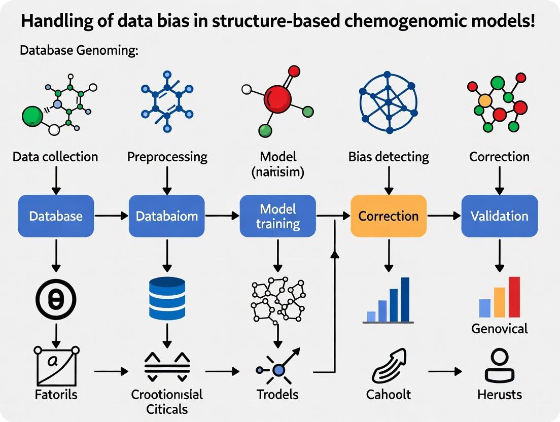
Diagram 1: Bias Diagnosis and Mitigation Workflow
Diagram 2: Data Bias Leading to Generalization Gaps
Technical Support Center
Troubleshooting Guides & FAQs
Q1: My virtual screening campaign against a GPCR target yields an overwhelming number of hits containing a common triazine scaffold not present in known actives. What is the likely cause and how can I correct it?
A: This is a classic symptom of ligand scaffold preference bias in your training data. The model was likely trained on a benchmark dataset (e.g., from PDBbind or ChEMBL) that is overrepresented with triazine-containing ligands for certain protein families. This teaches the model to associate that scaffold with high scores, regardless of the specific target context.
- Solution: Implement a scaffold-aware train/test split. Use tools like RDKit to identify Bemis-Murcko scaffolds. Ensure no scaffold in your test/validation set is present in your training set. Re-train your model using this split to assess true generalization.
Q2: When benchmarking my pose prediction model, performance is excellent for kinases but fails for nuclear hormone receptors. Why?
A: This indicates a protein family skew in your training data. The Protein Data Bank (PDB) is dominated by certain protein families. For example, kinases represent ~20% of all human protein structures, while nuclear hormone receptors are underrepresented.
- Solution: Apply family-stratified sampling. Before training, audit your dataset's composition. Create a balanced subset that includes a minimum threshold of structures from underrepresented families. Alternatively, use data augmentation techniques like homology modeling for missing families before training.
Q3: I suspect my binding affinity prediction model is biased by the abundance of high-affinity complexes in the PDB. How can I diagnose and mitigate this?
A: You are addressing PDB imbalance, where the public structural data is skewed toward tight-binding ligands and highly stable, crystallizable protein conformations.
- Diagnosis: Plot the distribution of binding affinity (pKd/pKi) in your training data. Compare it to a broader biochemical database (e.g., ChEMBL). You will likely see a right-skew toward high affinity.
- Mitigation: Integrate negative or weakly binding data. Use docking to generate putative decoy poses for non-binders. Incorporate experimental data on inactive compounds from public sources. Apply a loss function that penalizes overconfidence on high-affinity examples.
Key Experimental Protocols
Protocol 1: Auditing Dataset for Protein Family Skew
- Source: Download a curated dataset (e.g., PDBbind refined set, sc-PDB).
- Annotation: Map each protein target to its primary gene family (e.g., using Gene Ontology, Pfam, or UniProt).
- Quantification: Count the number of unique complexes per family.
- Analysis: Calculate the percentage distribution. Identify families with representation below a defined threshold (e.g., <1% of total).
- Action: For underrepresented families, source additional structures from the PDB or generate homology models using Swiss-Model.
Protocol 2: Generating a Scaffold-Blind Evaluation Set
- Input: A dataset of ligand-protein complexes (SMILES strings and protein PDB IDs).
- Scaffold Extraction: For each ligand, compute its Bemis-Murcko framework using the RDKit
GetScaffoldForMolfunction. - Clustering: Perform Tanimoto similarity clustering on the scaffold fingerprints (ECFP4).
- Splitting: Use the scaffold clusters as grouping labels. Use a cluster-based splitting method (e.g.,
GroupShuffleSplitin scikit-learn) to ensure no scaffold cluster appears in both training and test sets.
Protocol 3: Augmenting Data with Putative Non-Binders
- Select Actives: From your target of interest, compile a set of known active ligands with structures.
- Select Inactives: From a broad compound library (e.g., ZINC), select molecules with similar physicochemical properties but different 2D topology to actives (property-matched decoys).
- Docking: Dock both actives and inactives into your target's binding site using software like AutoDock Vina or GLIDE.
- Curation: Manually inspect top poses of inactives to ensure plausible binding mode. Add these ligand-receptor complexes to your dataset with a "non-binding" or weak affinity label.
Data Presentation
Table 1: Representation of Major Protein Families in the PDB (vs. Human Proteome)
| Protein Family | Approx. % of Human Proteome | Approx. % of PDB Structures (2023) | Skew Factor (PDB/Proteome) |
|---|---|---|---|
| Kinases | ~1.8% | ~20% | 11.1 |
| GPCRs | ~4% | ~3% | 0.75 |
| Ion Channels | ~5% | ~2% | 0.4 |
| Nuclear Receptors | ~0.6% | ~0.8% | 1.3 |
| Proteases | ~1.7% | ~7% | 4.1 |
| All Other Families | ~86.9% | ~67.2% | 0.77 |
Table 2: Common Ligand Scaffolds in PDBbind Core Set (by Frequency)
| Scaffold (Bemis-Murcko) | Frequency Count | Example Target Families |
|---|---|---|
| Benzene | 1245 | Kinases, Proteases, Diverse |
| Pyridine | 568 | Kinases, GPCRs |
| Triazine | 187 | Kinases, DHFR |
| Indole | 452 | Nuclear Receptors, Enzymes |
| Purine | 311 | Kinases, ATP-Binding Proteins |
Visualizations
Title: Sources and Impacts of Structural Bias
Title: Bias Detection and Mitigation Workflow
The Scientist's Toolkit: Research Reagent Solutions
| Item | Function in Bias Mitigation |
|---|---|
| RDKit | Open-source cheminformatics toolkit. Essential for scaffold analysis (Bemis-Murcko), molecular fingerprinting, and property calculation to audit and split datasets. |
| Pfam/InterPro | Databases of protein families and domains. Used to annotate protein targets in a dataset and quantify family-level representation. |
| PDBbind/SC-PDB | Curated databases linking PDB structures with binding affinity data. Common starting points for building models; require auditing for inherent biases. |
| ZINC Database | Public library of commercially available compounds. Source for generating property-matched decoy molecules to augment datasets with non-binders. |
| AutoDock Vina | Widely-used open-source molecular docking program. Used to generate putative poses for decoy compounds in data augmentation protocols. |
| Swiss-Model | Automated protein homology modeling server. Can generate structural models for protein families underrepresented in the PDB. |
| scikit-learn | Python machine learning library. Provides utilities for strategic data splitting (e.g., GroupShuffleSplit) based on scaffolds or protein families. |
Troubleshooting Guides & FAQs
FAQ 1: Why does my chemogenomic model show excellent validation performance but fails to identify new active compounds in a fresh assay?
Answer: This is a classic symptom of training data bias, often due to the Dominance of Published Actives. Models trained primarily on literature-reported "actives" versus broadly tested but unpublished "inactives" learn features specific to that biased subset, not generalizable bioactivity rules.
- Diagnostic Check: Compare the chemical space (e.g., using PCA or t-SNE) of your training set actives versus a large, diverse database of commercial compounds (e.g., ZINC). Overlap is likely minimal.
- Solution: Apply artificial debiasing. Augment your training data with assumed inactives from "Dark Chemical Matter" (DCM) – compounds tested in many assays but never active. This teaches the model what inactivity looks like. See Protocol 1.
FAQ 2: My high-throughput screen (HTS) identified hits that are potent in the primary assay but are completely inert in all orthogonal assays. What could be the cause?
Answer: This typically indicates Assay-Specific Artifacts. Compounds may interfere with the assay technology (e.g., fluorescence quenching, luciferase inhibition, aggregation-based promiscuity) rather than modulating the target.
- Diagnostic Check: Perform a confirmatory assay using a different readout technology (e.g., switch from fluorescence polarization to SPR). Also, check for common nuisance behavior: test for colloidal aggregation (Dynamic Light Scattering), redox activity (catalase inhibition assay), or reactivity with thiols (see Protocol 2).
- Solution: Implement a counter-screen and triage workflow early in the validation pipeline. Filter hits against known assay artifact profiles.
FAQ 3: What is 'Dark Chemical Matter' and how should I handle it in my dataset to avoid bias?
Answer: Dark Chemical Matter (DCM) refers to the large fraction of compounds in corporate or public screening libraries that have never shown activity in any biological assay despite being tested numerous times. Ignoring DCM introduces a severe "confirmatory" bias.
- Impact: A model trained only on published actives and random presumed inactives may misclassify DCM as potentially active because it doesn't recognize its consistent inactivity as a meaningful signal.
- Solution: Treat DCM as a privileged class for negative data. Explicitly label DCM compounds (e.g., >30 assays, 0 hits) as "verified inactives" in your training set. This significantly improves model specificity. See Protocol 3.
Experimental Protocols
Protocol 1: Artificial Debiasing of Training Data Using DCM
Objective: To create a balanced training dataset that mitigates publication bias.
- Collect Actives: Gather confirmed active compounds from public sources (ChEMBL, PubChem BioAssay). Use stringent criteria (e.g., IC50/ Ki < 10 µM, dose-response confirmed).
- Collect DCM: Extract DCM compounds from large-scale screening data (e.g., PubChem's "BioAssay" data for compounds tested in >50 assays with 0% hit rate).
- Sample Negatives: Randomly select a set of presumed inactives from a general compound library (e.g., ZINC) that are not in the DCM set.
- Construct Training Set: Combine Actives, DCM (labeled inactive), and Sampled Negatives (labeled inactive) in a 1:2:1 ratio.
- Train Model: Use this composite set for model training, ensuring the DCM class is weighted appropriately in the loss function.
Protocol 2: Detecting Assay-Specific Artifact: Aggregation-Based Inhibition
Objective: To confirm if a hit compound acts via non-specific colloidal aggregation.
- Prepare Compound: Make a 10 mM stock of the hit compound in DMSO.
- Dilution Series: Prepare a 2X dilution series in aqueous assay buffer (e.g., PBS) with final DMSO concentration ≤ 1%. Include a control well with buffer and DMSO only.
- Dynamic Light Scattering (DLS): Immediately measure the hydrodynamic radius (Rh) of each dilution using a DLS instrument.
- Data Interpretation: A positive result for aggregation is indicated by a sharp increase in Rh (>50 nm) at concentrations near the assay IC50. A negative control compound should show Rh < 5 nm.
- Confirmatory Test: Add a non-ionic detergent (e.g., 0.01% Triton X-100) to the assay. True aggregators will lose potency, while specific inhibitors will remain active.
Protocol 3: Integrating DCM into a Machine Learning Workflow
Objective: To build a random forest classifier that leverages DCM.
- Feature Calculation: Compute molecular descriptors (e.g., RDKit descriptors, ECFP4 fingerprints) for three lists: Actives (Class 1), DCM (Class 0), Random Library Compounds (Class 0).
- Data Splitting: Perform a stratified split 80/20 into training and test sets, preserving class ratios.
- Model Training: Train a Random Forest classifier (scikit-learn) with class weight='balanced'. This automatically adjusts weights inversely proportional to class frequencies.
- Evaluation: Assess on the test set. Pay special attention to the Recall (Sensitivity) for Actives and Specificity for DCM. A good model should have high specificity for DCM.
Data Presentation
Table 1: Prevalence of Assay Artifacts in Public HTS Data (PubChem AID 1851)
| Artifact Type | Detection Method | % of Primary Hits (IC50 < 10µM) | Confirmed True Actives After Triaging |
|---|---|---|---|
| Fluorescence Interference | Red-shifted control assay | 12.5% | 2.1% |
| Luciferase Inhibition | Counter-screen with luciferase enzyme | 8.7% | 1.8% |
| Colloidal Aggregation | DLS / Detergent sensitivity test | 15.2% | 3.5% |
| Cytotoxicity (for cell-based) | Cell viability assay (MTT) | 18.9% | 4.0% |
Table 2: Impact of DCM Inclusion on Model Performance Metrics
| Training Data Composition | AUC-ROC (Test Set) | Precision (Actives) | Specificity (DCM Class) |
|---|---|---|---|
| Actives + Random Inactives | 0.89 | 0.65 | 0.81 |
| Actives + DCM only | 0.85 | 0.82 | 0.93 |
| Actives + DCM + Random Inactives | 0.91 | 0.78 | 0.95 |
Visualizations
Title: Data Bias Identification and Mitigation Workflow
Title: Assay Artifact Triage Decision Tree
The Scientist's Toolkit: Research Reagent Solutions
| Item | Function/Application in Bias Mitigation |
|---|---|
| Triton X-100 (or CHAPS) | Non-ionic detergent used in confirmatory assays to disrupt colloidal aggregates, identifying false positives from promiscuous aggregation. |
| Red-Shifted Fluorescent Probes | Control probes with longer excitation/emission wavelengths to identify compounds that interfere with assay fluorescence (inner filter effect, quenching). |
| Recombinant Luciferase Enzyme | For counter-screening hits from luciferase-reporter assays to identify direct luciferase inhibitors. |
| Dynamic Light Scattering (DLS) Instrument | Measures hydrodynamic radius of particles in solution to directly detect compound aggregation at relevant assay concentrations. |
| ChEMBL / PubChem BioAssay Database | Primary public sources for bioactivity data, used to extract both published actives and, critically, define Dark Chemical Matter. |
| RDKit or MOE Cheminformatics Suite | For calculating molecular fingerprints and descriptors, enabling the chemical space analysis crucial for identifying training set biases. |
| MTT or CellTiter-Glo Assay Kits | Standard cell viability assays used as orthogonal counterscreens for cell-based phenotypic assays to rule out cytotoxicity-driven effects. |
Technical Support Center
Troubleshooting Guides
Issue 1: Model shows excellent training/validation performance but fails on new external datasets.
- Likely Cause: Data leakage or non-representative training/validation split leading to overfitting on latent biases (e.g., assay, scaffold, or vendor bias).
- Diagnostic Steps:
- Perform Bias Audit: Stratify your internal test set by putative bias sources (e.g., chemical series, protein family, assay date). Calculate performance metrics per stratum.
- Conduct an "Analog Series" Test: For each compound in your external test set, find its nearest neighbor in the training set by chemical fingerprint (e.g., ECFP4, Tanimoto >0.8). Plot the model's error against the distance to the nearest neighbor. A sharp increase in error with distance indicates over-reliance on local interpolation.
- Use a Simple Baseline: Compare your model's performance against a simple descriptor-based model (e.g., molecular weight + LogP regression) on the external set. If the complex model performs worse, it suggests it has learned dataset-specific noise.
- Resolution Protocol:
- Rebuild with Bias-Aware Splitting: Use tools like
scaffold split(RDKit) ortime splitto create more challenging validation sets that mimic real-world generalization. - Implement Regularization: Increase dropout rates, apply stronger L1/L2 weight penalties, or use early stopping with a stricter patience threshold.
- Incorporate Bias as a Feature (Adversarial Debiasing): Train an auxiliary model to predict the bias source (e.g., assay type) from your primary model's latent representations. Simultaneously, train your primary model to predict the target while minimizing the auxiliary model's performance. This decorrelates the learned features from the bias.
- Rebuild with Bias-Aware Splitting: Use tools like
Issue 2: Prospective screening yields inactive compounds despite high model confidence.
- Likely Cause: The model has learned "syntactic" patterns from the training data (e.g., specific substructures always labeled active in a given high-throughput screening (HTS) campaign) that do not translate to true bioactivity.
- Diagnostic Steps:
- Analyze Confidence Calibration: Generate a reliability diagram. Plot predicted probability bins vs. observed fraction of positives. A well-calibrated model follows the y=x line. Deviations indicate overconfident predictions.
- Apply Explainability Tools: Use SHAP (SHapley Additive exPlanations) or integrated gradients on high-confidence, failed predictions. Identify if the prediction was driven by chemically irrelevant or assay-artifact-related features.
- Resolution Protocol:
- Bayesian Uncertainty Estimation: Switch to or incorporate models that provide uncertainty estimates (e.g., Gaussian processes, Bayesian neural networks, or deep ensembles). Filter prospective hits by both high predicted activity and low predictive uncertainty.
- Diverse Negative Sampling: Ensure your training data includes a robust set of experimentally confirmed inactive compounds, not just assumed inactives from HTS. This teaches the model what "inactivity" looks like.
Issue 3: Performance drops significantly when integrating new data sources (e.g., adding cryo-EM structures to an X-ray-based model).
- Likely Cause: Covariate shift or representation bias. The feature distributions of the new data differ from the training domain.
- Diagnostic Steps:
- Dimensionality Reduction & Visualization: Project the latent features of old and new datasets using t-SNE or UMAP. Look for clear separation or lack of overlap between the data source clusters.
- Train a Data Source Classifier: Train a simple classifier to discriminate between old and new data sources using the model's input features. High accuracy indicates a significant distribution shift.
- Resolution Protocol:
- Domain Adaptation: Use techniques like Domain-Adversarial Neural Networks (DANNs) to learn domain-invariant features.
- Transfer Learning with Fine-Tuning: Pre-train the model on the larger, older dataset. Then, unfreeze the final layers and fine-tune on a smaller, carefully curated mix of old and new data.
Frequently Asked Questions (FAQs)
Q2: My model uses protein pockets as input. How can structural bias manifest? A: Structural bias is common and can manifest as:
- Resolution Bias: Models trained on high-resolution X-ray structures (<2.0 Å) may fail on predictions for homology models or cryo-EM-derived pockets.
- Ligand-of-Origin Bias: Pockets are defined by a co-crystallized ligand. The model may learn to recognize the specific chemical features of that ligand rather than general binding principles.
- Protein Family Bias: Over-representation of certain families (e.g., kinases) leads to poor performance on underrepresented ones (e.g., GPCRs). The table below summarizes common biases and their impact.
Table 1: Common Data Biases in Structure-Based Chemogenomic Models and Their Impact on Performance
| Bias Type | Description | Typical Manifestation | Diagnostic Metric Shift |
|---|---|---|---|
| Scaffold/Series Bias | Over-representation of specific chemical cores in training. | Poor performance on novel chemotypes. | High RMSE on external sets with novel scaffolds. |
| Assay/Measurement Bias | Training data aggregated from different experimental protocols (Kd, IC50, Ki from different labs). | Inaccurate absolute potency prediction. | Poor correlation between predicted and observed pChEMBL values across assays. |
| Structural Resolution Bias | Training on high-resolution structures only. | Failure on targets with only low-resolution or predicted structures. | AUC-ROC drops when tested on targets with resolution >3.0 Å. |
| Protein Family Bias | Imbalanced representation of target classes. | Inability to generalize to novel target families. | Macro-average F1-score significantly lower than per-family F1. |
| Publication Bias | Only successful (active) compounds and structures are published/deposited. | Over-prediction of activity, high false positive rate. | Skewed calibration curve; observed actives fraction << predicted probability. |
Q3: Are there standard reagents or benchmarks for debiasing studies in this field? A: Yes, the community uses several benchmark datasets and software tools to stress-test models for bias. Key resources are listed in the Scientist's Toolkit below.
Q4: How much performance drop in external validation is "acceptable"? A: There is no universal threshold. The key is to benchmark the drop against a null model. A 10% drop in AUC may be acceptable if a simple baseline (e.g., random forest on fingerprints) drops by 25%. The critical question is whether your model, despite the drop, still provides actionable, statistically significant enrichment over random or simple screening.
Experimental Protocols for Bias Detection & Mitigation
Protocol 1: Bias-Auditing via Stratified Performance Analysis
Objective: To identify specific data subsets where model performance degrades, indicating potential bias. Materials: Trained model, full dataset with metadata (scaffold, assay type, protein family, etc.). Steps:
- For each putative bias source (e.g.,
Protein_Family), partition the external test set into distinct strata. - For each stratum, calculate key performance metrics (AUC-ROC, AUC-PR, RMSE, EF1%).
- Plot metrics per stratum (bar chart). Compare against the global performance on the entire test set.
- Statistically compare performance across strata using ANOVA or Kruskal-Wallis tests.
- Interpretation: Strata with significantly lower performance indicate a domain where the model may be biased due to under-representation or confounding factors.
Protocol 2: Adversarial Debiasing Training (Implementation Outline)
Objective: To learn representations that are predictive of the primary task (activity) but invariant to a specified bias source (e.g., assay vendor).
Materials: Dataset with labels Y (activity) and bias labels B (vendor ID). Deep learning framework (PyTorch/TensorFlow).
Steps:
- Network Architecture: Build a shared feature extractor
G_f(.), a primary predictorG_y(.), and an adversarial bias predictorG_b(.). - Forward Pass: Input
X->Features = G_f(X)->Y_pred = G_y(Features)andB_pred = G_b(Features). - Adversarial Loss: The key is to train
G_fto maximize the loss ofG_b(making features uninformative for predicting bias), whileG_bis trained normally to minimize its loss. A gradient reversal layer (GRL) is typically used betweenG_fandG_bduring backpropagation. - Total Loss:
L_total = L_y(Y_pred, Y) - λ * L_b(B_pred, B), whereλcontrols the strength of debiasing. - Train the network with alternating or joint optimization of the shared and task-specific parameters.
Visualizations
Bias Mitigation & Model Development Workflow (73 chars)
Adversarial Debiasing Network Architecture (55 chars)
Table 2: Essential Resources for Bias Handling in Chemogenomic Models
| Item Name | Type/Provider | Function in Bias Research |
|---|---|---|
| PDBbind (Refined/General Sets) | Curated Dataset | Standard benchmark for structure-based affinity prediction. Used to test for protein-family and ligand bias via careful cluster-based splitting. |
| ChEMBL Database | Public Repository | Source of bioactivity data. Enables temporal splitting and detection of assay/publication bias through metadata mining. |
| MOSES (Molecular Sets) | Benchmark Platform | Provides standardized training/test splits (scaffold, random) and metrics to evaluate generative model bias and overfitting. |
| RDKit | Open-Source Toolkit | Provides functions for molecular fingerprinting, scaffold analysis, and bias-aware dataset splitting (e.g., Butina clustering, Scaffold split). |
| DeepChem | Open-Source Library | Offers implementations of advanced splitting methods (e.g., ButinaSplitter, SpecifiedSplitter) and model architectures suitable for adversarial training. |
| SHAP (SHapley Additive exPlanations) | Explainability Library | Interprets model predictions to identify if specific, potentially biased, chemical features are driving decisions. |
| GNINA / AutoDock Vina | Docking Software | Used as a baseline structure-based method to compare against ML models, helping to distinguish true learning from data leakage. |
| PROTEINET | Curated Dataset | A bias-controlled benchmark for protein sequence and structure models, useful for testing generalization across folds. |
Technical Support Center
Troubleshooting Guide & FAQs
Q1: My virtual screening model trained on DUD-E shows excellent AUC on the benchmark but fails drastically on my internal compound set. What is the likely cause?
A1: This is a classic symptom of hidden bias. DUD-E's "artificial decoy" generation method can introduce bias, where decoys are dissimilar to actives in ways the model learns to exploit (e.g., molecular weight, charge). Your internal compounds likely do not share this artificial separation.
- Diagnostic Protocol: Calculate and compare simple physicochemical property distributions (e.g., logP, molecular weight, number of rotatable bonds) between your actives, DUD-E decoys, and your internal set. A significant mismatch indicates property bias.
- Mitigation Workflow:
- Re-analyze DUD-E: Use the provided "cleaning" scripts from later studies to identify potential false negatives.
- Employ Bias-Corrected Benchmarks: Augment or replace your test with newer benchmarks like DEKOIS 2.0 or LIT-PCBA.
- Apply Domain Adaptation: Use techniques like adversarial validation to detect and adjust for systematic differences between the DUD-E data distribution and your real-world data.
Q2: When using PDBbind to train a binding affinity predictor, the model performance drops sharply on targets not in the PDBbind core set. How should I debug this?
A2: This suggests a "target bias" or "sequence similarity bias." The model may be memorizing target-specific features rather than learning generalizable protein-ligand interaction rules.
- Diagnostic Protocol: Perform a leave-one-cluster-out cross-validation based on protein sequence similarity (e.g., using BLAST clustering). A large performance drop in this setting confirms target bias.
- Mitigation Workflow:
- Stratified Splitting: Always split training/validation/test sets by protein family, not randomly, to prevent data leakage.
- Data Augmentation: Use homology models or carefully applied synthetic data techniques to increase target diversity in training.
- Architecture Choice: Prioritize models that explicitly model protein structure (e.g., graph networks over atoms) over those relying heavily on target descriptors.
Q3: I suspect my ligand-based model has learned "temporal bias" from a public dataset like ChEMBL. How can I validate and correct for this?
A3: Temporal bias occurs when early-discovered, "privileged" scaffolds dominate the dataset, and test sets are non-chronologically split. The model fails on newer chemotypes.
- Diagnostic Protocol: Split your data chronologically by the compound's first reported date. Train on older data and test on newer data. Compare the performance to a random split.
- Mitigation Workflow:
- Temporal Splitting: Implement a rigorous time-split protocol for all model validation.
- Scaffold-based Evaluation: Use Bemis-Murcko scaffold splits to ensure the model generalizes to novel core structures.
- Active Learning: Integrate an active learning loop that prioritizes compounds dissimilar to the training set for experimental testing and feedback.
Q4: What are the concrete, quantitative differences in bias between DUD-E and its successor, DUD-E Z?
A4: DUD-E Z was designed to reduce analog and chemical bias. Key improvements are summarized below:
Table 1: Quantitative Comparison of Bias Mitigation in DUD-E vs. DUD-E Z
| Bias Type | DUD-E Characteristic | DUD-E Z Improvement | Quantitative Metric |
|---|---|---|---|
| Analog Bias | Decoys were chemically dissimilar to actives but also to each other, making them too easy to distinguish. | Decoys are selected to be chemically similar to each other, forming "chemical neighborhoods" that better mimic real screening libraries. | Increased mean Tanimoto similarity among decoys (within a target set). |
| Chemical Bias | Decoy generation rules could create systematic physicochemical differences from actives. | More refined property-matching (e.g., by 1D properties) and the use of the ZINC database as a decoy source. | Reduced Kullback-Leibler divergence between the property distributions (e.g., logP) of actives and decoys. |
| False Negatives | Known actives could potentially be included as decoys for other targets. | Stringent filtering against known bioactive compounds across a wider array of databases. | Number of confirmed false negatives removed from decoy sets. |
Key Experimental Protocol: Detecting Dataset Bias via Property Distribution Analysis
Objective: To diagnose and quantify potential chemical property bias between active and decoy/inactive compound sets in a benchmark like DUD-E.
Materials & Software: RDKit (Python), Pandas, NumPy, Matplotlib/Seaborn, Benchmark dataset (e.g., DUD-E CSV files).
Procedure:
- Data Loading: Load the actives (
*_actives_final.sdf) and decoys (*_decoys_final.sdf) for your target of interest using RDKit. - Descriptor Calculation: For each molecule in both sets, calculate a set of 1D/2D molecular descriptors:
- Molecular Weight (MW)
- Calculated LogP (AlogP)
- Number of Hydrogen Bond Donors (HBD)
- Number of Hydrogen Bond Acceptors (HBA)
- Number of Rotatable Bonds (RotB)
- Topological Polar Surface Area (TPSA)
- Statistical Summary: Generate a table of mean and standard deviation for each descriptor per set (Active vs. Decoy).
- Distribution Visualization: Plot the kernel density estimation (KDE) for each descriptor, overlaying the distributions of actives and decoys.
- Statistical Testing: Perform an appropriate statistical test (e.g., Kolmogorov-Smirnov test) to determine if the distributions for each property are significantly different (p < 0.01).
- Interpretation: Significant differences across multiple key properties indicate a strong chemical bias in the benchmark, which may lead to overly optimistic model performance.
The Scientist's Toolkit: Research Reagent Solutions
Table 2: Essential Resources for Bias-Aware Chemogenomic Modeling
| Item / Resource | Function & Purpose in Bias Mitigation |
|---|---|
| RDKit | Open-source cheminformatics toolkit. Critical for calculating molecular descriptors, scaffold analysis, and visualizing chemical distributions to detect bias. |
| DEKOIS 2.0 / LIT-PCBA | Bias-corrected benchmark datasets. Use as alternative or supplemental test sets to DUD-E for more realistic performance estimation. |
| PDBbind (Refined/General Sets) | The hierarchical structure of PDBbind (General -> Refined -> Core) allows researchers to consciously select data quality levels and avoid target leakage during splits. |
| Protein Data Bank (PDB) | Source of ground-truth structural data. Essential for constructing structure-based models and verifying binding mode hypotheses independent of affinity labels. |
| Time-Split ChEMBL Scripts | Custom or community scripts (e.g., from chembl_downloader) to split data chronologically, essential for evaluating predictive utility for future compounds. |
| Adversarial Validation Code | Scripts implementing a binary classifier to distinguish training from real-world data. Success indicates a distribution shift, guiding the need for domain adaptation. |
| Graphviz (DOT) | Tool for generating clear, reproducible diagrams of data workflows and model architectures, essential for documenting and communicating bias-testing pipelines. |
Visualizations
Title: Workflow for Detecting Chemical Property Bias
Title: PDBbind Hierarchical Dataset Structure
Title: Model Validation Strategies to Uncover Bias
Building Bias-Aware Models: Methodological Frameworks and Practical Applications
Technical Support Center: Troubleshooting Guides & FAQs
This support center addresses common technical issues encountered while constructing curation pipelines for structure-based chemogenomic models, specifically within research focused on mitigating data bias.
Frequently Asked Questions (FAQs)
Q1: During ligand-protein pair assembly, my dataset shows extreme affinity value imbalances (e.g., 95% inactive compounds). How can I address this programmatically without introducing selection bias?
A1: Implement stratified sampling during data sourcing, not just as a post-hoc step. Use the following protocol:
- Pre-source Stratification: Define your target distribution (e.g., 70% inactive, 25% active, 5% high-affinity) based on known biological priors.
- Query in Batches: For each affinity stratum, run separate queries to primary databases (PDBbind, BindingDB) using specific KI/IC50/KD thresholds.
- Oversampling/Undersampling with SMOTE & TOMEK: Apply Synthetic Minority Oversampling Technique (SMOTE) to generate synthetic examples for rare high-affinity classes. Follow with TOMEK links for cleaning. Crucially, apply these techniques only to the training split after dataset splitting to avoid data leakage.
- Validate with Blind Test Set: Hold out a fully representative test set before any balancing. The final model must be evaluated on this untouched data.
Q2: I suspect structural redundancy in my protein set is biasing my model towards certain protein families. How can I measure and control for this?
A2: Use sequence and fold similarity clustering to ensure diversity.
- Protocol:
- Extract all protein sequences from your assembled complex structures.
- Perform all-vs-all pairwise alignment using MMseqs2 (
mmseqs easy-cluster). - Cluster at a strict sequence identity threshold (e.g., 30%).
- Select a maximally diverse representative from each cluster for your final set. If cluster sizes are highly uneven (e.g., one superfamily has 50 clusters, another has 2), apply a second-stage sampling to select a balanced number of representatives from each major fold class (using CATH or SCOP annotations).
Q3: My pipeline pulls from multiple sources (ChEMBL, PubChem, DrugBank). How do I resolve conflicting activity annotations for the same compound-target pair?
A3: Implement a confidence-scoring and consensus system.
- Create a Conflict Resolution Table:
| Data Source | Assay Type Priority (High to Low) | Trust Score | Curation Level |
|---|---|---|---|
| PDBbind (refined) | X-ray crystal structure | 1.0 | High (manual) |
| BindingDB | Ki (single protein, direct) | 0.9 | Medium (semi-auto) |
| ChEMBL | IC50 (cell-based) | 0.7 | Medium (semi-auto) |
| PubChem BioAssay | HTS screen result | 0.5 | Low (auto) |
- Rule-based Selection: For each conflicting pair, select the annotation from the source with the highest Trust Score. If from the same source, prioritize the assay type with the higher priority.
- Flag Ambiguity: Annotate records where the discrepancy between the top two values exceeds one log unit for manual inspection.
Q4: What are the best practices for logging and versioning in a multi-step curation pipeline to ensure reproducibility?
A4: Adopt a pipeline framework with inherent provenance tracking. Use a tool like Snakemake or Nextflow. Each rule/task should log:
- Input dataset hash (e.g., SHA-256).
- All parameters (random seed, clustering threshold, sampling fraction).
- Output dataset hash and summary statistics (counts, distributions).
- Version numbers for all tools/databases used.
Store this log as a JSON alongside each intermediate dataset. This creates a complete audit trail.
Experimental Protocols for Key Validation Steps
Protocol: Assessing Covariate Shift in the Curation Pipeline Purpose: To detect if your curation steps inadvertently introduce a distributional shift in molecular or protein descriptors between the sourced raw data and the final curated set. Methodology:
- Descriptor Calculation: For both the initially aggregated "raw" dataset (Draw) and the final "curated" dataset (Dcurated), calculate a standard set of descriptors (e.g., ECFP4 fingerprints for ligands, amino acid composition for proteins).
- Dimensionality Reduction: Apply PCA to the descriptor matrices separately, but project Dcurated onto the PCA space defined by Draw.
- Statistical Test: Perform a two-sample Kolmogorov-Smirnov (KS) test on the distributions of the first principal component scores between Draw and Dcurated.
- Interpretation: A significant p-value (<0.05) indicates a covariate shift, prompting investigation into the filtering/sampling steps that caused it.
Protocol: Benchmarking Bias Mitigation via Hold-out Family Evaluation Purpose: To empirically test if your curation strategy reduces model overfitting to prevalent protein families. Methodology:
- Stratified Splitting: Split your final curated dataset into training (80%) and test (20%) sets using random splitting. Train and evaluate Model A.
- Temporal/Family Hold-out Splitting: Split your dataset such that all proteins from a specific, diverse family (e.g., GPCRs) or all data published after a certain date are placed exclusively in the test set. Train Model B on the remaining data.
- Comparison: Compare the performance drop of Model B versus Model A on the held-out family/test set.
| Evaluation Scheme | Model | Test Set AUC (Overall) | Test Set AUC (Held-out Family) | Performance Drop |
|---|---|---|---|---|
| Random Split | Model A | 0.89 | 0.87 | -0.02 |
| Family Hold-out | Model B | 0.85 | 0.72 | -0.13 |
A smaller performance drop in the Hold-out scheme suggests a more robust, less biased model enabled by better curation.
Diagrams
Bias-Aware Data Curation Pipeline
Bias Assessment via Hold-out Evaluation
The Scientist's Toolkit: Research Reagent Solutions
| Item/Resource | Primary Function in Curation | Key Considerations for Bias Mitigation |
|---|---|---|
| PDBbind Database | Provides high-quality, experimentally determined protein-ligand complexes with binding affinity data. | Use the "refined" or "core" sets as a high-quality seed. Be aware of its bias towards well-studied, crystallizable targets. |
| BindingDB | Large collection of measured binding affinities (KI, Kd, IC50). | Crucial for expanding chemical space. Requires rigorous filtering by assay type (prefer "single protein" over "cell-based"). |
| ChEMBL | Bioactivity data from medicinal chemistry literature. | Excellent for bioactive compounds. Use confidence scores and document data curation level. Beware of patent-driven bias towards lead-like space. |
| MMseqs2 / CD-HIT | Protein sequence clustering tools. | Essential for controlling structural redundancy. The choice of sequence identity threshold (e.g., 30% vs 70%) directly controls the diversity of the protein set. |
| RDKit / Open Babel | Cheminformatics toolkits. | Used to standardize molecular representations (tautomers, protonation states, removing salts), calculate descriptors, and check for chemical integrity. Inconsistent application introduces bias. |
| IMBALANCE Library (Python) | Provides algorithms like SMOTE, ADASYN, SMOTE-ENN. | Used to algorithmically balance class distributions. Critical: Apply only to the training fold after data splitting to prevent data leakage and over-optimistic performance. |
| Snakemake / Nextflow | Workflow management systems. | Ensure reproducible, documented, and versioned curation pipelines. Automatically tracks provenance, which is mandatory for auditing bias sources. |
Technical Support Center
Troubleshooting Guides & FAQs
Q1: During preprocessing, my model shows high performance on validation splits but fails dramatically on external, real-world chemical libraries. What could be the cause? A1: This is a classic sign of dataset bias, often from benchmarking sets like ChEMBL being non-representative of broader chemical space. To diagnose, create a bias audit table comparing the distributions of key molecular descriptors between your training set and the target library.
| Descriptor | Training Set Mean (Std) | External Library Mean (Std) | Kolmogorov-Smirnov Statistic (p-value) |
|---|---|---|---|
| Molecular Weight | 450.2 (150.5) | 380.7 (120.8) | 0.32 (<0.001) |
| LogP | 3.5 (2.1) | 2.8 (1.9) | 0.21 (0.003) |
| QED | 0.6 (0.2) | 0.7 (0.15) | 0.28 (<0.001) |
| TPSA | 90.5 (50.2) | 110.3 (45.6) | 0.19 (0.012) |
Protocol for Bias Audit:
- Compute Descriptors: Use RDKit (
rdMolDescriptors) or Mordred to calculate a diverse set of 2D/3D molecular descriptors for both datasets. - Normalize: Apply StandardScaler to all descriptors.
- Statistical Test: For each descriptor, perform a two-sample Kolmogorov-Smirnov test (or Mann-Whitney U for non-normal) using
scipy.stats. - Visualize: Plot kernel density estimates for the top 5 divergent descriptors.
Q2: After applying a re-weighting technique (like Importance Weighting), my model's loss becomes unstable and fails to converge. How do I fix this? A2: Unstable loss is often due to extreme importance weights causing gradient explosion. Implement weight clipping or normalization.
Mitigation Protocol:
- Calculate Weights: Use Kernel Mean Matching or propensity score estimation to get initial weights
w_ifor each training sample. - Clip Weights: Set a threshold (e.g., 95th percentile).
w_i_clipped = min(w_i, percentile(w, 0.95)). - Normalize: Renormalize weights so their mean is 1:
w_i_normalized = w_i_clipped / mean(w_i_clipped). - Adaptive Optimizer: Use Adam or AdaGrad instead of SGD, as they are more robust to noisy, scaled gradients.
Q3: How do I choose between adversarial debiasing and re-sampling for my protein-ligand affinity prediction model? A3: The choice depends on your bias type and computational resources. Use the following diagnostic table:
| Technique | Best For Bias Type | Computational Overhead | Key Hyperparameter | Effect on Performance |
|---|---|---|---|---|
| Adversarial Debiasing | Latent, complex biases (e.g., bias towards certain protein folds) | High (requires adversarial training) | Adversary loss weight (λ) | May reduce training set accuracy but improves generalization |
| Re-sampling (SMOTE/Cluster) | Simple, distributional bias (e.g., overrepresented scaffolds) | Low to Medium | Sampling strategy (over/under) | Can increase minority class recall; risk of overfitting to synthetic samples |
Protocol for Adversarial Debiasing:
- Setup: Build a primary model (predictor) and an adversary model. The adversary tries to predict the protected variable (e.g., protein family) from the primary model's representations.
- Joint Training: Minimize predictor loss while maximizing adversary loss (or minimize negative adversary loss). The loss is:
L_total = L_prediction - λ * L_adversary. - Gradient Reversal: Implement a gradient reversal layer between the predictor and adversary during backpropagation for easier training.
Title: Adversarial Debiasing Workflow for Chemogenomic Models
Q4: I suspect temporal bias in my drug-target interaction data (newer compounds have different assays). How can I correct for this algorithmically? A4: Implement temporal cross-validation and a time-aware re-weighting scheme.
Temporal Holdout Protocol:
- Order Data: Sort all protein-ligand interaction pairs by assay date.
- Split: Use the earliest 70% for training, the next 15% for validation, and the latest 15% for testing. Do not shuffle.
- Apply Causal Correction: Use a method like Doubly Robust Estimation that combines propensity weighting and outcome regression to adjust for shifting assay conditions over time.
Q5: When using bias-corrected models in production, how do I monitor for new, previously unseen biases? A5: Implement a bias monitoring dashboard with statistical process control.
| Monitoring Metric | Calculation | Alert Threshold |
|---|---|---|
| Descriptor Drift | Wasserstein distance between training and incoming batch descriptor distributions | > 0.1 (per descriptor) |
| Performance Disparity | Difference in RMSE/ROC-AUC between major and minority protein family groups | > 0.15 |
| Fairness Metric | Subgroup AUC for under-represented scaffold classes | < 0.6 |
The Scientist's Toolkit: Research Reagent Solutions
| Item Name | Function in Bias Correction | Key Parameters/Notes |
|---|---|---|
| AI Fairness 360 (AIF360) Toolkit | Provides a unified framework for bias checking and mitigation algorithms (e.g., Reweighing, AdversarialDebiasing). | Use sklearn.compose.ColumnTransformer with aif360.datasets.StandardDataset. |
| RDKit with Mordred Descriptors | Generates comprehensive 2D/3D molecular features to quantify chemical space and identify distribution shifts. | Calculate 1800+ descriptors. Use PCA for visualization of dataset coverage. |
| DeepChem MoleculeNet | Curated benchmark datasets with tools for stratified splitting to avoid data leakage and scaffold bias. | Use ScaffoldSplitter for a more realistic assessment of generalization. |
| Propensity Score Estimation (via sklearn) | Estimates the probability of a sample being included in the training set given its features, used for re-weighting. | Use calibrated classifiers like LogisticRegressionCV to avoid extreme weights. |
| SHAP (SHapley Additive exPlanations) | Explains model predictions to identify if spurious correlations (biases) are being used. | Look for high SHAP values for non-causal features (e.g., specific vendor ID). |
Title: Bias-Correction Pipeline for Structure-Based Models
The Role of Physics-Based and Hybrid Modeling in Counteracting Pure Data-Driven Bias
Technical Support Center
Troubleshooting Guides & FAQs
Q1: Our purely data-driven chemogenomic model performs excellently on the training and validation sets but fails to generalize to novel protein targets outside the training distribution. What is the likely cause and how can we address it? A: This is a classic sign of data-driven bias and overfitting to spurious correlations in the training data. The model may have learned features specific to the assay conditions or homologous protein series rather than generalizable structure-activity relationships. Recommended Protocol: Implement a Hybrid Model Pipeline
- Feature Augmentation: Generate physics-based descriptors (e.g., MM/GBSA binding energy components, pharmacophore points, molecular interaction fields) for your ligand-target complexes.
- Model Fusion: Train a hybrid model where the final prediction is a weighted ensemble:
- Model A: Your existing data-driven model (e.g., Graph Neural Network).
- Model B: A simpler model trained solely on the physics-based descriptors (e.g., Random Forest).
- Validation: Use a temporally split or structurally dissimilar test set to validate the hybrid model's improved generalizability.
Q2: During hybrid model training, the physics-based component seems to dominate, drowning out the data-driven signal. How do we balance the two? A: This indicates a scaling or weighting issue between feature sets. Recommended Protocol: Feature Scaling & Attention-Based Fusion
- Standardize Features: Independently standardize all data-driven features and physics-based features to zero mean and unit variance.
- Implement an Attention/Gating Mechanism: Instead of simple concatenation, use a neural attention layer that learns to dynamically weight the contribution of physics-based vs. data-driven feature channels for each input sample.
- This allows the model to "decide" when to trust physical principles versus empirical patterns.
Q3: How can we formally test if our hybrid model has reduced bias compared to our pure data-driven model? A: Implement a bias audit using quantitative metrics on held-out bias-controlled sets. Recommended Protocol: Bias Audit Framework
- Create Diagnostic Test Sets:
- Set A (Property Bias): Molecules with similar physicochemical properties but different binding outcomes.
- Set B (Scaffold Bias): Molecules with novel core scaffolds absent from training.
- Set C (Target Bias): Proteins from a distant fold class.
- Measure & Compare: Evaluate both models on these sets using metrics like AUC, RMSE, and
ΔAUC(AUCtrain - AUCdiagnostic).
Table 1: Bias Audit Results for Model Comparison
| Diagnostic Test Set | Pure Data-Driven Model (AUC) | Hybrid Physics-Informed Model (AUC) | ΔAUC (Improvement) |
|---|---|---|---|
| Standard Hold-Out | 0.89 | 0.87 | -0.02 |
| Novel Scaffold Set | 0.62 | 0.78 | +0.16 |
| Distant Target Fold | 0.58 | 0.71 | +0.13 |
| Property-Bias Control Set | 0.65 | 0.81 | +0.16 |
Q4: What is a practical first step to incorporate physics into our deep learning workflow without a full rebuild? A: Use physics-based features as a regularizing constraint during training. Recommended Protocol: Physics-Informed Regularization Loss
- Calculate Reference Value: For each training sample, compute a coarse physics-based score (e.g., scaled docking score or simple energy estimate).
- Add a Loss Term: Modify your loss function:
Total Loss = Task Loss (e.g., BCE) + λ * |(Model Prediction - Physics-Based Reference)| - Tune λ: Start with a small λ (e.g., 0.1) to gently guide the model, preventing severe deviation from physical plausibility without forcing strict adherence.
Experimental Protocol: Building a Robust Hybrid Chemogenomic Model
Title: Hybrid Model Training with Bias-Conscious Validation Splits
Objective: To train a chemogenomic model that integrates graph-based ligand features, protein sequence embeddings, and physics-based binding energy approximations to improve generalizability and reduce data bias.
Materials: See "Research Reagent Solutions" table below.
Methodology:
- Data Curation & Splitting:
- Source data from public repositories (e.g., ChEMBL, BindingDB).
- Critical: Perform cluster-based splitting. Cluster proteins by sequence similarity and ligands by scaffold. Assign entire clusters to train/validation/test sets to ensure true generalization assessment.
- Feature Engineering:
- Data-Driven: Generate molecular graphs (atoms as nodes, bonds as edges) and protein language model embeddings.
- Physics-Based: For each ligand-protein pair, run a fast MM/GBSA calculation (using implicit solvent) to obtain per-residue energy decomposition terms.
- Model Architecture (Hybrid Graph Network):
- Branch A: Graph Neural Network processing the ligand.
- Branch B: 1D Convolutional Neural Network processing protein embeddings.
- Branch C: Dense network processing the physics-based energy vector.
- Fusion: Concatenate the latent representations from all three branches, followed by a gating mechanism and fully connected layers for prediction.
- Training & Validation:
- Train using the hybrid loss function (Protocol Q4).
- Validate on the cluster-held-out validation set.
- Apply early stopping based on validation loss.
Mandatory Visualizations
Hybrid Model Development & Validation Workflow
Hybrid Model Architecture with Gating Fusion
The Scientist's Toolkit: Research Reagent Solutions
| Item/Category | Function in Hybrid Modeling | Example/Note |
|---|---|---|
| Molecular Dynamics (MD) Suite | Generate structural ensembles for targets; compute binding free energies. | GROMACS, AMBER, OpenMM. Essential for rigorous physics-based scoring. |
| MM/GBSA Scripts & Pipelines | Perform efficient, end-state binding free energy calculations for feature generation. | gmx_MMPBSA, AmberTools MMPBSA.py. Key source for physics-based feature vectors. |
| Protein Language Model (pLM) | Generate informative, evolution-aware embeddings for protein sequences. | ESMFold, ProtT5. Provides deep learning features for the target. |
| Graph Neural Network (GNN) Library | Model the ligand as a graph and learn its topological features. | PyTorch Geometric, DGL. Standard for data-driven ligand representation. |
| Differentiable Docking | Integrate a physics-like scoring function directly into the training loop. | DiffDock, TorchDrug. Emerging tool for joint physics-DL optimization. |
| Clustering Software | Perform scaffold-based and sequence-based clustering for robust data splitting. | RDKit (Butina Clustering), MMseqs2. Critical for bias-conscious train/test splits. |
| Model Interpretation Toolkit | Audit which features (physics vs. data) drive predictions. | SHAP, Captum. Diagnose model bias and build trust. |
Technical Support Center
Troubleshooting Guide & FAQs
Q1: My de-biased model for a novel target class shows excellent validation metrics but fails to identify any active compounds in the final wet-lab screen. What could be the issue?
A: This is a classic sign of "over-correction" or "loss of signal." The bias mitigation strategy may have removed not only the confounding bias but also the true biological signal. This is common when using adversarial debiasing or stratification on small datasets.
Troubleshooting Steps:
- Check Data Leakage: Re-audit your training/validation/test splits. Ensure no temporal, structural, or vendor bias has leaked from the validation set into model training, giving false confidence.
- Analyze the Removed Features: Use SHAP or similar analysis on your de-biasing model (e.g., the adversary in an adversarial network) to see which molecular features it identified as "biased." Cross-reference these with known privileged scaffolds or substructures for the target class. If there is significant overlap, you may have removed real pharmacophores.
- Implement a Gradual Debias: Instead of fully removing the identified bias, apply a re-weighting or penalty-based approach. Retrain with a weaker de-biasing strength (λ parameter) and observe the performance on a small, diverse validation HTS set.
Protocol: Step 2 - SHAP Analysis for De-biasing Audit
- Objective: Identify molecular features the debiasing model associates with data bias.
- Method:
- Train your primary activity prediction model and your bias prediction model (e.g., "assay" or "year" predictor).
- Using the
shap.DeepExplainer(for neural networks) orshap.TreeExplainer(for RF/GBM) on the bias prediction model, calculate SHAP values for a representative sample of your training data. - For the top 20 features with the highest mean absolute SHAP value for the bias model, compute their frequency in known active compounds for related targets (from public ChEMBL data).
- If >30% of these high-bias features are also prevalent in known actives, your debiasing is likely too aggressive.
Q2: When applying a transfer learning model from a well-characterized target family (e.g., GPCRs) to a novel, understudied class (e.g., solute carriers), how do I handle the drastic difference in available training data?
A: The core challenge is negative set definition bias. For novel targets, confirmed inactives are scarce, and using random compounds from other assays introduces strong confounding bias.
Troubleshooting Steps:
- Construct a Robust Negative Set: Do not use random "inactives." Employ a "distant background" approach.
- Use Domain Adversarial Training: Implement a Gradient Reversal Layer (GRL) network to learn target-invariant features, forcing the model to focus on signals not tied to the over-represented source domain.
- Prioritize Diversity-Oriented Libraries: For screening, choose libraries maximally diverse from your source target training data to reduce model extrapolation errors.
Protocol: Step 1 - Constructing a 'Distant Background' Negative Set
- Objective: Build a negative set for a novel target that minimizes latent bias.
- Method:
- Gather all available active compounds for the novel target (even 10-50 is useful).
- From a large chemical database (e.g., ZINC, Enamine REAL), calculate the pairwise Tanimoto distance (1 - similarity) from each database compound to the nearest active.
- Select compounds in the lowest quartile of similarity (most distant) as your putative negatives. This minimizes the chance of including unconfirmed, latent actives.
- Validate this set by confirming it does not enrich for actives in a related, better-characterized target from the same family, if such data exists.
Q3: How can I detect and mitigate "temporal bias" in a continuously updated screening dataset for a novel target?
A: Temporal bias arises because early screening compounds are often structurally similar, and assay technology/conditions change over time. A model may learn to predict the "year of screening" rather than activity.
Troubleshooting Steps:
- Visualize Temporal Drift: Perform a time-series PCA on the compound feature space (e.g., ECFP4 fingerprints) colored by assay year.
- Apply Temporal Cross-Validation: Never use future data to validate past data. Train on data from years 1-3, validate on year 4, test on year 5.
- Use a Temporal Holdout: For the final model, hold out the most recent 1-2 years of data as the ultimate test of predictive utility for future campaigns.
Diagram: Temporal Bias Detection & Mitigation Workflow
Diagram Title: Temporal Bias Mitigation Protocol
Q4: What are the best practices for evaluating a de-biased model's performance, given that standard metrics like ROC-AUC can be misleading?
A: Relying solely on ROC-AUC is insufficient as it can be inflated by dataset bias. A multi-faceted evaluation protocol is mandatory.
Troubleshooting Steps:
- Use Bias-Aware Metrics: Calculate "Bias-Discrepancy" (BD) and "Subgroup AUC".
- Perform External Testing: Use a meticulously curated, fully independent external test set from a different source lab or compound library.
- Conduct "Negative Control" Predictions: Run the model on a set of compounds known to be inactive against any target (e.g., certain metabolic intermediates). High false-positive rates indicate artifact learning.
Evaluation Metrics Table:
| Metric | Formula/Description | Target Value | Interpretation |
|---|---|---|---|
| Subgroup AUC | AUC calculated separately for compounds from each major vendor or assay batch. | All Subgroup AUCs > 0.65 | Model performance is consistent across data sources. |
| Bias Discrepancy (BD) | abs(AUC_overall - mean(Subgroup_AUC)) |
< 0.10 | Low discrepancy indicates robust performance. |
| External Validation AUC | AUC on a truly independent, recent, and diverse compound set. | > 0.70 | Model has generalizable predictive power. |
| Scaffold Recall | % of unique active scaffolds in the top 1% of predictions. | > 30% (context-dependent) | Model is not just recovering a single chemotype. |
The Scientist's Toolkit: Research Reagent Solutions
| Item | Function & Role in De-biasing |
|---|---|
| Diverse Compound Libraries (e.g., Enamine REAL Diversity, ChemBridge DIVERSet) | Provide a broad, unbiased chemical space for prospective screening and for constructing "distant background" negative sets. Essential for testing model generalizability. |
| Benchmark Datasets (e.g., DEKOIS, LIT-PCBA) | Provide carefully curated datasets with hidden validation cores, designed to test a model's ability to avoid decoy bias and recognize true activity signals. |
Adversarial Debiasing Software (e.g., aix360, Fairlearn) |
Python toolkits containing implementations of adversarial debiasing, reweighing, and prejudice remover algorithms. Critical for implementing advanced bias mitigation. |
Chemistry-Aware Python Libraries (e.g., RDKit, DeepChem) |
Enable fingerprint generation, molecular featurization, scaffold analysis, and seamless integration of chemical logic into machine learning pipelines. |
Model Explainability Tools (e.g., SHAP, Captum) |
Used to audit which features a model (and its adversarial debiasing counterpart) relies on, identifying potential "good signal" removal or artifact learning. |
| Structured Databases (e.g., ChEMBL, PubChem) | Provide essential context for understanding historical bias, identifying potential assay artifacts, and performing meta-analysis across target classes. |
Diagram: The De-biased Virtual Screening Workflow
Diagram Title: De-biased Virtual Screening Protocol
Technical Support Center: Troubleshooting Guides & FAQs
This support center addresses common challenges in implementing active learning (AL) and bias-aware sampling for chemogenomic model refinement. The context is a research thesis on Handling data bias in structure-based chemogenomic models.
FAQs & Troubleshooting
Q1: My active learning loop seems to be stuck, selecting redundant data points from a narrow chemical space. How can I encourage exploration? A: This indicates the acquisition function may be overly greedy. Implement a diversity component.
- Protocol: Cluster-Based Diversity Sampling
- Step 1: After model training, generate embeddings (e.g., from the penultimate layer) for all compounds in the unlabeled pool.
- Step 2: Perform clustering (e.g., k-means, Butina) on these embeddings. Use the elbow method on sum-of-squared-distances to estimate clusters.
- Step 3: Within each cluster, use the primary acquisition function (e.g., uncertainty) to rank candidates.
- Step 4: Select the top-N candidates from each cluster to form the batch for the next iteration.
- Quantitative Impact: This typically increases the spread of selected compounds, measured by internal diversity metrics (e.g., average Tanimoto distance).
Q2: My model performance degrades on hold-out test sets representing underrepresented protein families, despite high overall accuracy. Is this bias, and how can I detect it? A: Yes, this is a classic sign of dataset bias. Implement bias-aware validation splits.
- Protocol: Stratified Performance Analysis
- Step 1: Stratify your test/validation data by relevant metadata before model training. For chemogenomics, key strata are: Protein Family (e.g., GPCRs, Kinases), Ligand Scaffold, and Experimental Source.
- Step 2: Track performance metrics (AUC-ROC, RMSE) per stratum across AL iterations.
- Step 3: Calculate the performance disparity (e.g., max AUC difference) between the best and worst-performing strata.
- Data Presentation:
Q3: How do I integrate bias correction directly into the active learning sampling strategy? A: Use a bias-aware acquisition function that weights selection probability inversely to the density of a point's stratum in the training set.
- Protocol: Inverse Density Weighting
- Step 1: For each compound
iin the unlabeled poolU, identify its stratums_i(e.g., protein family). - Step 2: Compute the representation ratio:
r_s = (Count(s_i) in Training Set) / (Total Training Set Size). - Step 3: Calculate the base acquisition score
a_i(e.g., predictive variance). - Step 4: Compute the final bias-aware score:
a_i' = a_i * (1 / (r_s + α)), whereαis a small smoothing constant. - Step 5: Select the batch with the highest
a_i'scores.
- Step 1: For each compound
Q4: What are the computational resource bottlenecks in scaling these methods for large virtual libraries (>1M compounds)? A: The primary bottlenecks are model inference on the unlabeled pool and clustering for diversity.
- Solution Protocol: Submodular Proxy Sampling
- Step 1: Use a cheaper, lower-fidelity model (e.g., ECFP fingerprint + Random Forest) to screen the entire library and select a top-100k subset.
- Step 2: Apply your primary, expensive structure-based model (e.g., Graph Neural Network) only to this subset for precise uncertainty estimation.
- Step 3: Apply clustering and bias-aware weighting within this manageable candidate set.
Visualizations
Diagram Title: Active Learning with Bias-Aware Iteration Loop
Diagram Title: Bias-Aware Score Calculation for a Candidate
The Scientist's Toolkit: Research Reagent Solutions
| Item | Function in Experiment | Example/Description |
|---|---|---|
| Structure-Based Featurizer | Converts protein-ligand 3D structures into machine-readable features. | DeepChem's AtomicConv or DGL-LifeSci's PotentialNet. Critical for the primary predictive model. |
| Fingerprint-Based Proxy Model | Enables fast pre-screening of large compound libraries. | RDKit for generating ECFP/Morgan fingerprints paired with a Scikit-learn Random Forest. |
| Stratified Data Splitter | Creates training/validation/test splits that preserve subgroup distributions. | Scikit-learn's StratifiedShuffleSplit or custom splits based on protein family SCOP codes. |
| Clustering Library | Enforces diversity in batch selection. | RDKit's Butina clustering (for fingerprints) or Scikit-learn's MiniBatchKMeans (for embeddings). |
| Bias Metric Calculator | Quantifies performance disparity across strata. | Custom script to compute maximum gap in AUC-ROC or standard deviation of per-stratum RMSE. |
| Active Learning Framework | Manages the iterative training, scoring, and data addition loop. | ModAL (Modular Active Learning) for Python, extended with custom acquisition functions. |
| Metadata-Enabled Database | Stores compound-protein pairs with essential stratification metadata. | SQLite or PostgreSQL with tables for protein family, ligand scaffold, assay conditions. |
Diagnosing and Correcting Bias: A Troubleshooting Guide for Model Developers
Troubleshooting Guides & FAQs
This technical support center addresses common issues in detecting bias through learning curves within chemogenomic model development. The context is research on handling data bias in structure-based chemogenomic models.
FAQ: Interpreting Curve Behavior
Q1: My training loss decreases steadily, but my validation loss plateaus early. What does this indicate? A: This is a primary red flag for overfitting, suggesting the model is memorizing training data specifics (e.g., artifacts of a non-representative chemical scaffold split) rather than learning generalizable structure-activity relationships. It indicates high variance and likely poor performance on new, structurally diverse compounds.
Q2: Both training and validation loss are decreasing but remain high and parallel. What is the problem? A: This pattern indicates underfitting or high bias. The model is too simple to capture the complexity of the chemogenomic data. Potential causes include inadequate featurization (e.g., poor pocket descriptors), overly strict regularization, or a model architecture insufficient for the task.
Q3: My validation curve is more jagged/noisy compared to the smooth training curve. Why? A: Noise in the validation curve often stems from a small or non-representative validation set. In chemogenomics, this can occur if the validation set contains few examples of key target families or chemical classes, making performance assessment unstable.
Q4: What does a sudden, sharp spike in validation loss after a period of decrease signify? A: This is a classic sign of catastrophic overfitting, often related to an excessively high learning rate or a significant distribution shift between the training and validation data (e.g., validation compounds have different binding modes not seen in training).
Diagnostic Metrics & Quantitative Thresholds
The following table summarizes key metrics derived from training/validation curves to diagnose bias and variance.
Table 1: Diagnostic Metrics from Learning Curves
| Metric | Formula / Description | Interpretation Threshold (Typical) | Indicated Problem |
|---|---|---|---|
| Generalization Gap | Validation Loss - Training Loss (at convergence) | > 10-15% of Training Loss | Significant Overfitting |
| Loss Ratio (Final) | Validation Loss / Training Loss | > 1.5 | High Variance / Overfitting |
| Loss Ratio (Final) | Validation Loss / Training Loss | ~1.0 but both high | High Bias / Underfitting |
| Convergence Delta | Epoch of Val. Loss Minus Epoch of Train Loss Min | > 20 Epochs (context-dependent) | Early Stopping point; late validation min suggests overfitting. |
| Curve Area Gap | Area between train and val curves after epoch 5. | Large, increasing area | Progressive overfitting during training. |
Experimental Protocol: Systematic Learning Curve Analysis for Bias Detection
Objective: To diagnose bias (underfitting) and variance (overfitting) in a structure-based chemogenomic model by generating and analyzing training/validation learning curves.
Materials: See "The Scientist's Toolkit" below.
Methodology:
- Data Partitioning: Split your protein-ligand complex dataset using a scaffold split (based on Bemis-Murcko frameworks) or a temporal split to simulate real-world generalization. Avoid random splits, as they can mask bias.
- Model Training Setup: Configure your model (e.g., Graph Neural Network for protein-ligand graphs). Set a fixed, moderately high learning rate initially for clear curve dynamics.
- Metric Logging: Train the model for a preset number of epochs (e.g., 200). After each epoch, calculate and record the loss (e.g., Mean Squared Error) on both the training and hold-out validation sets.
- Curve Generation: Plot epochs (x-axis) against loss (y-axis) for both sets on the same plot.
- Diagnostic Analysis:
- Identify the convergence point for each curve.
- Calculate the Generalization Gap and Loss Ratio from Table 1.
- Observe the shape: parallel curves (underfitting), diverging gap (overfitting), validation spikes (instability).
- Iterative Intervention:
- If underfitting is detected: Increase model capacity (more layers/features), reduce regularization (dropout, weight decay), or improve input features (e.g., add pharmacophore descriptors).
- If overfitting is detected: Apply stronger regularization, implement early stopping at the validation loss minimum, or augment the training data (e.g., via ligand conformer generation).
Workflow Diagram: Bias Detection Protocol
Title: Bias Diagnosis and Mitigation Workflow
The Scientist's Toolkit: Research Reagent Solutions
Table 2: Essential Materials for Chemogenomic Bias Analysis Experiments
| Item / Solution | Function in Bias Detection |
|---|---|
| Curated Benchmark Dataset (e.g., PDBbind, BindingDB subsets) | Provides a standardized, publicly available set of protein-ligand complexes for training and, crucially, for fair validation to assess generalization. |
| Scaffold Split Algorithm (e.g., RDKit Bemis-Murcko) | Ensures training and validation sets contain distinct molecular scaffolds. This is critical for simulating real-world generalization and uncovering model bias toward specific chemotypes. |
| Deep Learning Framework (e.g., PyTorch, TensorFlow) | Enables flexible model architecture design, custom loss functions, and, most importantly, automatic gradient computation and backpropagation for training complex models. |
| Metric Logging Library (e.g., Weights & Biases, TensorBoard) | Tracks training and validation metrics (loss, AUC, etc.) per epoch, enabling precise curve generation and comparison across multiple experimental runs. |
| Molecular Featurization Library (e.g., RDKit, DeepChem) | Generates numerical descriptors (graphs, fingerprints, 3D coordinates) from raw chemical structures and protein data, forming the input features for the model. |
| High-Performance Computing (HPC) Cluster or Cloud GPU | Provides the computational power necessary for training large chemogenomic models over hundreds of epochs and across multiple hyperparameter settings. |
Techniques for Data Augmentation and Balancing in Structural Space (e.g., Conformer Generation, Structure Perturbation)
Troubleshooting Guides & FAQs
FAQ 1: Why does my model fail to generalize to novel scaffold classes despite using conformer augmentation? Answer: This is a classic sign of bias where augmentation is only capturing conformational diversity within known scaffolds, not true structural diversity. The model has not learned transferable geometric or physicochemical principles. To troubleshoot, audit your augmented dataset's Tanimoto similarity matrix; if the mean similarity between original and augmented molecules is >0.7, your perturbations are insufficient. Implement scaffold-based splitting before augmentation to ensure the test set contains entirely novel scaffolds. Then, integrate more aggressive structure perturbation techniques like bond rotation with angle distortion or ring distortion alongside conformer generation.
FAQ 2: My structure perturbations are generating chemically invalid or unstable molecular geometries. How can I control this? Answer: Invalid geometries often arise from unconstrained stochastic perturbations. Implement a validity-checking pipeline with the following steps: 1) Apply geometric constraints (e.g., limit bond angle changes to ±10%, maintain chiral centers). 2) Use a force field (MMFF94, UFF) for quick energy minimization post-perturbation and reject conformers with high strain energy (>50 kcal/mol). 3) Employ a rule-based filter (e.g., using RDKit) to check for improbable bond lengths, atom clashes (VDW overlap), and correct tetrahedral chirality. This ensures physicochemical plausibility.
FAQ 3: How do I determine the optimal number of conformers to generate per compound for balancing my dataset? Answer: There is no universal number; it is a function of molecular flexibility and desired coverage. Follow this protocol: For a representative subset, perform an exhaustive conformer search (e.g., using ETKDG with high numConfs). Perform cluster analysis (RMSD-based) on the resulting pool. Plot the number of clusters vs. the number of generated conformers. The point where the curve plateaus indicates the saturation point for conformational diversity. Use this molecule-specific count to guide sampling, rather than a fixed number for all compounds. See Table 1 for quantitative guidance.
FAQ 4: When performing data balancing via oversampling in structural space, how do I avoid overfitting to augmented samples? Answer: Overfitting occurs when the model memorizes artificially generated structures. Mitigation strategies include: 1) Adversarial Validation: Train a classifier to distinguish original from augmented samples. If it succeeds (>0.7 AUC), your augmentations are leaking identifiable artifacts. 2) Augmentation Diversity: Use a stochastic combination of techniques (e.g., noise, rotation, translation) rather than a single method. 3) Test Set Isolation: Ensure no augmented version of any molecule leaks into the test set. 4) Regularization: Increase dropout rates and use stronger weight decay when training on heavily augmented data.
FAQ 5: What are the best practices for validating the effectiveness of my data augmentation/balancing pipeline in reducing model bias? Answer: Construct a robust bias assessment benchmark:
- Split by Property: Create train/test splits based on a molecular property (e.g., scaffold, logP, molecular weight) to simulate bias.
- Train Two Models: Model A (original imbalanced data), Model B (augmented/balanced data).
- Evaluate: Compare performance drop on the property-based test split. A smaller drop for Model B indicates reduced bias.
- Analyze: Use SHAP or similar to ensure the model's attention shifts from trivial structural artifacts to meaningful pharmacophoric features. Track metrics like Robust Accuracy (RA) and Balanced Accuracy (BA).
Table 1: Impact of Conformer Generation Parameters on Dataset Diversity and Model Performance
| Parameter | Typical Value Range | Effect on Dataset Size (Multiplier) | Impact on Model ROC-AUC (Mean Δ) | Computational Cost Increase |
|---|---|---|---|---|
| ETKDG numConfs | 50-100 | 5x - 10x | +0.05 to +0.10 | High (x8) |
| Energy Window | 10-20 kcal/mol | 2x - 4x | +0.02 to +0.05 | Medium (x3) |
| RMSD Threshold | 0.5-1.0 Å | 1.5x - 3x | +0.01 to +0.03 | Low (x1.5) |
| Stochastic Coordinate Perturbation | σ=0.05-0.1 Å | 2x - 5x | +0.03 to +0.07 | Low (x2) |
Table 2: Comparison of Structure Perturbation Techniques for Bias Mitigation
| Technique | Primary Use (Balancing/Augmentation) | Typical # of New Structures per Molecule | Preserves Activity? (Y/N)* | Reduces Scaffold Bias? (Effect Size) |
|---|---|---|---|---|
| Standard Conformer Generation | Augmentation | 5-50 | Y | Low (0.1-0.2) |
| Torsion Noise & Angle Distortion | Augmentation | 10-100 | Y (if constrained) | Medium (0.2-0.4) |
| Ring Distortion (e.g., Change ring size) | Balancing (for rare scaffolds) | 1-5 | Conditional | High (0.4-0.6) |
| Fragment-based De novo Growth | Balancing | 10-1000 | N (requires validation) | Very High (0.5-0.8) |
| Active Learning-based Sampling | Balancing | Iterative | Y | High (0.4-0.7) |
Based on molecular docking consensus score retention. *Cohen's d metric for improvement in model performance on held-out scaffolds.
Experimental Protocols
Protocol 1: Systematic Conformer Generation and Clustering for Augmentation Objective: Generate a diverse, energy-plausible set of conformers for each molecule in a dataset.
- Input Preparation: Standardize molecules (neutralize, remove salts) using RDKit.
- Conformer Generation: Use the ETKDGv3 algorithm with
numConfs=100,pruneRmsThresh=0.5. - Energy Minimization: Minimize each conformer using the MMFF94 force field (maxIters=200).
- Filtering: Discard conformers with MMFF94 energy > 20 kcal/mol relative to the minimum found.
- Clustering: Cluster remaining conformers using Butina clustering with RMSD cutoff of 0.7 Å.
- Sampling: From each cluster, select the lowest-energy conformer. Optionally, sample multiple conformers per cluster proportional to cluster size for balancing.
- Output: A list of unique, low-energy conformers for each input molecule.
Protocol 2: Structure Perturbation for Scaffold Oversampling Objective: Generate novel yet plausible analogs for underrepresented molecular scaffolds.
- Scaffold Identification: Use the Bemis-Murcko method to identify core scaffolds. Flag scaffolds with count < X (e.g., 5) in dataset.
- Core Extraction & Modification: a. For each rare scaffold, extract it from a parent molecule. b. Apply one or more stochastic modifications: * Bond Rotation: Randomize a rotatable bond in the scaffold by ±15-30 degrees. * Angle Bending: Change a bond angle by ±5-10%. * Ring Distortion: For a single bond in a ring, change its length by ±0.05 Å and adjust adjacent angles.
- Reassembly & Validation: a. Reattach the original side chains (R-groups) to the modified scaffold. b. Sanity check: Ensure no atom clashes, correct valence, and preserved chirality. c. Perform a quick force field minimization (UFF, 50 iterations) and reject high-strain structures.
- Property Alignment: Filter generated molecules to align with the original molecule's key properties (e.g., logP ±1, MW ±50) to maintain distributional consistency.
Visualizations
Title: Workflow for Structural Data Balancing and Augmentation
Title: Relationship Between Bias, Augmentation Techniques, and Outcomes
The Scientist's Toolkit: Research Reagent Solutions
Table 3: Essential Tools for Structural Data Augmentation Experiments
| Item / Software | Function in Experiment | Key Feature for Bias Mitigation |
|---|---|---|
| RDKit (Open-source) | Core cheminformatics toolkit for molecule handling, conformer generation (ETKDG), scaffold analysis, and stereochemistry checks. | Enables reproducible, rule-based structural perturbations and filtering. |
| Open Babel / OEchem | File format conversion, force field minimization, and molecular property calculation. | Provides alternative conformer generation methods for validation. |
| CREST (GFN-FF) | Advanced, semi-empirical quantum mechanics-based conformer/rotamer sampling. | Generates highly accurate, thermodynamically relevant conformational ensembles for critical analysis. |
| OMEGA (OpenEye) | Commercial, high-performance conformer generation engine. | Speed and robustness for generating large-scale augmentation libraries. |
| PyMol / Maestro | 3D structure visualization and manual inspection. | Critical for qualitative validation of generated structures and identifying artifacts. |
| Custom Python Scripts (with NumPy) | Implementing stochastic coordinate noise, custom clustering, and pipeline automation. | Allows for tailored augmentation strategies specific to the bias identified in the dataset. |
| MMFF94 / UFF Force Fields | Energy minimization and strain evaluation of perturbed structures. | Acts as a physics-based filter to ensure generated 3D structures are plausible. |
| Scaffold Network Libraries (e.g., in DataWarrior) | Analyzing scaffold diversity and identifying regions of chemical space for oversampling. | Quantifies bias and guides the balancing strategy. |
Technical Support Center
Frequently Asked Questions (FAQs)
Q1: We have identified a novel GPCR target with no experimentally determined 3D structures. How can we initiate structure-based virtual screening? A1: Utilize a multi-pronged homology modeling and docking strategy. First, use the latest AlphaFold2 or AlphaFold3 models from the AlphaFold Protein Structure Database as a starting template. If the model quality is low in binding regions, employ a specialized tool like RosettaGPCR for membrane protein refinement. Concurrently, perform ligand-based similarity searching on known GPCR ligands (from ChEMBL) to generate a preliminary pharmacophore. Use this pharmacophore to guide and constrain the docking of these known actives into your homology model with a flexible docking program like GLIDE or AutoDockFR. This iterative process can refine the binding pocket geometry.
Q2: Our project involves a protein-protein interaction (PPI) target. We have only a few known active compounds (hit rate <0.1%). How can we expand our virtual screening library effectively? A2: For PPIs with sparse ligand data, shift focus to the interface. Perform an evolutionary coupling analysis using tools like EVcouplings to identify critical, conserved interfacial residues. Design a focused library featuring:
- Fragment-based scaffolds: Screen a fragment library (e.g., from ZINC20) against the predicted "hotspot" using FTMap or similar computational mapping.
- Macrocycle/peptidomimetic libraries: Use a tool like DOCK 3.7 to design conformationally constrained peptides that mimic the key secondary structural elements (α-helices, β-strands) of the native binding partner. Refer to the Protocol section for detailed steps.
Q3: When using a model built from sparse data, how can we estimate the reliability of our virtual screening rankings to avoid costly experimental dead-ends?
A3: Implement a stringent consensus scoring and confidence metric protocol. Never rely on a single scoring function. Use at least three diverse scoring functions (e.g., a force-field based, an empirical, and a knowledge-based function). Calculate a consensus rank. More critically, apply a confidence metric like the Prediction Accuracy Index (PAI) for your model.
PAI = (Hit Rate from Model) / (Random Hit Rate)
A PAI < 2 suggests the model's predictions are no better than random. Calibrate your model using the few known actives and decoys before full-scale screening.
Experimental Protocol: Iterative Refinement for a Cold-Start GPCR Target
Objective: Generate a refined homology model of Target GPCR-X and a validated pharmacophore for virtual screening.
Materials & Software: AlphaFold2/3 database, MODELLER or RosettaCM, RosettaGPCR, Maestro (Schrödinger) or MOE, GLIDE, GPCRdb, ChEMBL database.
Procedure:
- Template Acquisition & Alignment:
- Retrieve the AlphaFold2 model for GPCR-X from UniProt.
- Use GPCRdb to identify the closest homologs with known structures (e.g., in Class A). Perform a structure-based sequence alignment using MUSTANG or MAFFT within the modeling suite.
Initial Model Building:
- Generate 100 homology models using MODELLER, incorporating the AlphaFold2 prediction as a soft constraint.
- Select the top 5 models based on DOPE assessment scores.
Membrane-Specific Refinement:
- Embed the top model into a pre-equilibrated POPC lipid bilayer using CHARMM-GUI.
- Run a short, constrained molecular dynamics (MD) simulation (50 ns) in Desmond or NAMD to relax sidechains, focusing on the transmembrane helices.
Binding Site Definition & Pharmacophore Generation:
- Extract all known small-molecule ligands for GPCR-X (even 5-10) from ChEMBL.
- Dock these ligands into the refined model using GLIDE (SP then XP mode).
- Cluster the top poses. Using the Phase module, derive a common pharmacophore hypothesis (e.g., 1 hydrogen bond acceptor, 1 aromatic ring, 1 hydrophobic site).
Validation & Iteration:
- Screen a small, diverse library of 1000 compounds (with 5 known actives seeded in) using the pharmacophore.
- Dock the top 200 pharmacophore matches into the model. If the known actives rank in the top 10%, proceed to full library screening. If not, revisit step 4, adjusting the binding site centroid or pharmacophore features based on docking pose analysis.
Research Reagent Solutions
| Item | Function in Cold-Start Context |
|---|---|
| AlphaFold DB Models | Provides a high-accuracy predicted structure as a primary template, bypassing the need for a close homolog. |
| GPCRdb Web Server | Curates residue numbering, motifs, and structures, enabling precise alignment and annotation for homology modeling. |
| ZINC20 Library (Fragment Subset) | A readily accessible, commercially available fragment library for virtual screening when no lead compounds exist. |
| ChEMBL Database | Source of bioactivity data for known ligands, essential for ligand-based similarity searches and model validation. |
| FTMap Server | Computationally maps protein surfaces to identify "hot spots" for fragment binding, crucial for PPI targets. |
| Rosetta Software Suite | Enables de novo protein design and interface remodeling, useful for generating peptidomimetic ideas for PPIs. |
Quantitative Data: Performance of Sparse-Data Strategies
Table 1: Reported Enrichment Metrics for Different Cold-Start Approaches (Recent Literature Survey)
| Strategy | Target Class | Known Actives for Model Building | Reported EF1%* | Key Tool/Method Used |
|---|---|---|---|---|
| AlphaFold2 + Docking | Kinase (Understudied) | 0 | 15.2 | AlphaFold2, GLIDE |
| Homology Model + Pharmacophore | Class C GPCR | 8 | 22.5 | MODELLER, Phase |
| PPI Hotspot + Fragment Screen | PPI (Bcl-2 family) | 3 | 8.7 (Fragment hit rate 4%) | FTMap, Rosetta |
| Ligand-Based Sim Search | Ion Channel | 12 | 18.1 | ECFP4 Similarity, ROCS |
| Consensus Docking | Novel Viral Protease | 5 | 27.0 | GLIDE, AutoDock Vina, DSX |
*EF1% (Enrichment Factor at 1%): Measures how many more actives are found in the top 1% of a screened list compared to a random selection. An EF1% of 10 means a 10-fold enrichment.
Visualization: Workflow Diagrams
Title: Iterative Refinement Workflow for Cold-Start GPCR
Title: Multi-Strategy Fusion to Overcome Cold-Start
Context: This support center is part of a thesis research project on Handling data bias in structure-based chemogenomic models. The following guides address common issues when optimizing models for generalization to novel, unbiased chemical spaces.
Troubleshooting Guides & FAQs
Q1: My model performs well on validation splits but fails dramatically on external compound sets from a different scaffold. What are the first parameters to investigate?
A: This is a classic sign of overfitting to the biased chemical space of your training set. Prioritize investigating these hyperparameters:
- Regularization Strength (L1/L2): Increase lambda values to penalize complex weight configurations that may be memorizing training scaffolds.
- Dropout Rate: Increase dropout rates in fully connected layers, especially in the final regression/classification heads, to encourage robust feature learning.
- Learning Rate & Schedule: A learning rate that is too high may prevent convergence to a generalizable minimum. Implement a decay schedule (e.g., cosine annealing) for smoother optimization.
Experimental Protocol Check: Ensure your validation split is created via scaffold splitting (using Murcko scaffolds), not random splitting. This better simulates the challenge of novel chemical space.
Q2: When using a Graph Neural Network (GNN) for molecular graphs, how do I choose between architectures like MPNN, GAT, and GIN for better generalization?
A: The choice depends on the bias you are countering. See the quantitative comparison below from recent benchmarks on out-of-distribution (OOD) chemical datasets:
Table 1: GNN Architecture Generalization Performance on OOD Scaffold Splits
| Architecture | Key Mechanism | Avg. ROC-AUC on OOD Scaffolds (↑) | Tendency to Overfit Local Bias | Recommended Use Case |
|---|---|---|---|---|
| GIN | Graph Isomorphism Network, uses MLPs for aggregation | 0.72 | Low | Best for datasets with diverse functional groups on similar cores. |
| GAT | Graph Attention Network; learns edge importance | 0.68 | Medium | Useful when specific atomic interactions are critical and variable. |
| MPNN | Message Passing Neural Network (general framework) | 0.65 | High (vanilla) | Highly flexible; requires strong regularization for generalization. |
Methodology: To test, implement a k-fold scaffold split. Train each architecture with identical hyperparameter tuning budgets (e.g., via Ray Tune or Optuna), using early stopping based on a scaffold validation set. Report the mean performance across folds on the held-out scaffold test set.
Title: Decision guide for GNN architecture selection to combat data bias
Q3: What are concrete steps to set up a hyperparameter optimization (HPO) loop focused on generalization?
A: Follow this protocol for a robust HPO experiment:
- Split: Partition data using scaffold splitting (e.g., Bemis-Murcko). Hold out 15% of unique scaffolds as the final test set.
- Optimization Metric: Use the scaffold validation set performance (not random validation) as the objective for your HPO bayesian optimizer.
- Key Hyperparameter Space:
- Learning Rate: LogUniform(1e-5, 1e-3)
- Dropout Rate: Uniform(0.2, 0.7)
- L2 Penalty: LogUniform(1e-7, 1e-3)
- GNN Depth: IntUniform(3, 8) [Depth >5 can overfit to local structure]
- Early Stopping: Implement patience-based early stopping on the scaffold validation loss.
- Final Evaluation: Train the best configuration on the full training+validation scaffold pool and evaluate only once on the held-out scaffold test set.
Q4: How can I use "Reagent Solutions" to artificially augment my dataset and reduce bias during training?
A: Strategic data augmentation can create in-distribution variants that improve robustness.
Table 2: Research Reagent Solutions for Mitigating Data Bias
| Reagent / Method | Function | How it Improves Generalization |
|---|---|---|
| SMILES Enumeration | Generates different string representations of the same molecule. | Makes model invariant to atomic ordering, a common source of bias. |
| Random Atom Masking | Randomly masks node/atom features during training. | Forces the model to rely on broader context, not specific atoms. |
| Virtual Decoys (ZINC20) | Use commercially available compounds as negative controls or contrastive samples. | Introduces diverse negative scaffolds, preventing model from learning simplistic decision rules. |
| Adversarial Noise (FGSM) | Adds small, learned perturbations to molecular graphs or embeddings. | Smooths the decision landscape, making the model more resilient to novel inputs. |
| Scaffold-based Mixup | Interpolates features of molecules from different scaffolds. | Explicitly enforces smooth interpolation across the chemical space boundary. |
Protocol for Scaffold-based Mixup:
- For a batch of graphs (G1, G2) with features X1, X2 and labels y1, y2, drawn from different scaffold clusters.
- Sample a mixing coefficient λ ~ Beta(α, α), where α is a small (e.g., 0.2-0.4).
- Create mixed features: X_mix = λ * X1 + (1-λ) * X2. For graphs, interpolate node features directly.
- Train the model to predict: y_mix = λ * y1 + (1-λ) * y2.
- This encourages linear behavior between disparate regions of chemical space.
Title: Workflow for applying bias-mitigating data augmentation reagents
Q5: My performance metric is unstable across different random seeds, even with the same hyperparameters. How can I get reliable results?
A: High variance indicates your model's performance is sensitive to initialization and data ordering, often worsening on OOD data.
- Action 1: Increase the number of seeds. Report the mean and standard deviation across at least 5-10 different random seeds for any claimed performance metric.
- Action 2: Implement heavier regularization (see Q1) and potentially reduce model capacity. A simpler, more stable model often generalizes better than an unstable, complex one.
- Action 3: Use cross-validation based on scaffolds, not a single hold-out set. This provides a more reliable estimate of generalization variance.
Protocol for Seed-Stable Evaluation:
- Define a list of random seeds (e.g.,
[42, 123, 456, 789, 101112]). - For each seed, run the entire training and evaluation pipeline (including data splitting, if stochastic) with your fixed best hyperparameters.
- Record the performance on the external test set for each seed.
- Aggregate results:
Mean ROC-AUC = 0.75 ± 0.03 (std). This quantifies the reliability of your optimization.
Leveraging Transfer Learning and Multi-Task Learning to Share Information and Reduce Target-Specific Bias
Troubleshooting Guides & FAQs
Model Training & Performance Issues
Q1: My multi-task model is converging well on some targets but failing completely on others. What could be the cause?
A: This is often a symptom of negative transfer or severe task imbalance. The shared representations are being dominated by the data-rich or easier tasks. To troubleshoot:
- Check Data Balance: Quantify your dataset per task. Severe imbalance is a common culprit.
- Adjust Loss Weighting: Implement dynamic loss weighting strategies like Uncertainty Weighting or GradNorm instead of using a simple sum. This allows the model to adaptively prioritize harder tasks.
- Review Shared Layers: The capacity of your shared encoder may be insufficient. Gradually increase its complexity (e.g., more layers/units) while monitoring for overfitting.
- Implement Gradient Surgery: For conflicting gradients, techniques like PCGrad can project a task's gradient onto the normal plane of another task's gradient to reduce conflict.
Q2: During transfer learning, my fine-tuned model shows high accuracy on the new target but poor generalization in external validation. Is this overfitting to target-specific bias?
A: Yes, this indicates overfitting to the limited data (and its inherent biases) of the new target. Solutions include:
- Progressive Unfreezing: Don't unfreeze all pre-trained layers at once. Start by fine-tuning only the final layers, then progressively unfreeze earlier layers with a very low learning rate.
- Stronger Regularization: Apply dropout, weight decay, or early stopping more aggressively during fine-tuning.
- Use a Broader Pre-Training Source: Pre-train on a more diverse, large-scale chemogenomic dataset (e.g., ChEMBL, BindingDB) rather than a narrow set of related targets to learn more generalizable features.
Q3: How do I choose between a Hard vs. Soft Parameter Sharing architecture for my multi-task problem?
A: The choice depends on task relatedness and computational resources.
- Hard Sharing (Single Shared Encoder): Best for highly related tasks (e.g., different mutants of the same protein). It maximizes information sharing and reduces overfitting risk but is prone to negative transfer if tasks are too divergent.
- Soft Sharing (Multiple Towers with Regularization): Each task has its own encoder, but their parameters are regularized to be similar. This is more flexible for less related tasks and avoids negative transfer, but requires more parameters and data.
Data & Bias Handling Issues
Q4: My pre-training and target task data come from different assay types (e.g., Ki vs. IC50). How do I mitigate this "assay bias"?
A: Assay bias introduces systematic distribution shifts. Address it by:
- Explicit Bias Modeling: Add assay type as a categorical feature or a multi-task prediction head during pre-training.
- Domain Adaptation Layers: Use domain-invariant representation learning techniques (like Domain-Adversarial Neural Networks) during pre-training to learn features invariant to the assay type.
- Standardization & Curation: Apply rigorous pXC50 conversion where scientifically justified, and clearly label assay metadata for all data points.
Q5: I suspect my benchmark dataset has historical selection bias (e.g., over-representation of certain chemotypes). How can I audit and correct for this?
A:
- Audit: Perform chemical space analysis (e.g., t-SNE, PCA based on fingerprints) and color points by year of discovery or originating project. Clustering by time/space indicates bias.
- Correct in Training: Implement re-weighting schemes where under-represented clusters in chemical space are given higher sample weights during loss calculation.
- Correct in Evaluation: Use stratified splitting methods (e.g., scaffold split, time split) that mimic real-world generalization challenges, rather than random splits which hide bias.
Experimental Protocols
Protocol 1: Multi-Task Learning with Gradient Surgery
Objective: Train a single model on multiple protein targets while mitigating gradient conflict. Steps:
- Data Preparation: Curate bioactivity datasets (pKi, pIC50) for N related targets. Standardize labels and featurize compounds (e.g., ECFP6, RDKit descriptors).
- Model Architecture: Build a neural network with a shared molecular graph encoder (e.g., GAT) and N separate task-specific prediction heads.
- Training with PCGrad: For each batch:
- Compute the gradient for each task's loss separately.
- For each task pair, compute the dot product of gradients. If negative, project one gradient onto the normal plane of the other.
- Average the potentially modified gradients and apply the update to the shared parameters.
- Evaluation: Use a time-split or scaffold-split for each task to evaluate generalization.
Protocol 2: Bias-Reducing Transfer Learning via Domain-Adversarial Pre-Training
Objective: Pre-train a model on a large, diverse source dataset to learn representations invariant to specific assay or project biases. Steps:
- Source Data Curation: Aggregate data from public sources (ChEMBL, PubChem). Label each compound-target pair with its bias domain (e.g., assay type, source database).
- Model Architecture: Construct a network with a shared feature extractor (F), a main activity predictor (C), and a domain classifier (D).
- Adversarial Training: Train to maximize the performance of C while minimizing the performance of D (via a gradient reversal layer). This forces F to learn domain-invariant features.
- Fine-Tuning: Remove the domain classifier D. Use the pre-trained feature extractor F, and fine-tune F and a new target-specific head C' on the small, target dataset.
Table 1: Performance Comparison of Learning Strategies on Imbalanced Multi-Task Data
| Model Strategy | Avg. RMSE (Major Tasks) | Avg. RMSE (Minor Tasks) | Negative Transfer Observed? |
|---|---|---|---|
| Single-Task (Independent) | 0.52 ± 0.03 | 0.89 ± 0.12 | N/A |
| Multi-Task (Equal Loss Sum) | 0.48 ± 0.02 | 1.05 ± 0.15 | Yes (Severe) |
| Multi-Task (Uncertainty W.) | 0.49 ± 0.02 | 0.75 ± 0.08 | No |
| Multi-Task (PCGrad) | 0.47 ± 0.02 | 0.71 ± 0.07 | No |
Table 2: Impact of Pre-Training Scale on Fine-Tuning for Low-Data Targets
| Pre-Training Dataset Size | Pre-Training Tasks | Fine-Tuning RMSE (Target X, n=100) | Improvement vs. No Pre-Train |
|---|---|---|---|
| None (Random Init.) | 0 | 1.41 ± 0.21 | 0.0% |
| 50k compounds, 10 targets | 10 | 1.12 ± 0.14 | 20.6% |
| 500k compounds, 200 targs | 200 | 0.93 ± 0.11 | 34.0% |
| 1M+ compounds, 500 targs | 500 | 0.87 ± 0.09 | 38.3% |
Visualizations
Diagram 1: Multi-Task Learning with Gradient Surgery
Diagram 2: Domain-Adversarial Transfer Learning Workflow
The Scientist's Toolkit: Research Reagent Solutions
| Item / Solution | Function in Bias-Reduced Chemogenomic Models |
|---|---|
| DeepChem Library | Provides high-level APIs for implementing multi-task learning, graph networks, and transfer learning pipelines, accelerating prototyping. |
| PCGrad / GradNorm Implementations | Custom training loop code for performing gradient surgery or adaptive loss balancing to mitigate negative transfer in MTL. |
| Domain-Adversarial Neural Network (DANN) PyTorch/TF Code | Pre-built modules for the gradient reversal layer and adversarial training setup for learning domain-invariant features. |
| Model Checkpointing & Feature Extraction Tools (e.g., Weights & Biases, MLflow) | Tracks training experiments and allows extraction of frozen encoder outputs for transfer learning analysis. |
| Stratified Splitter for Molecules (e.g., ScaffoldSplitter, TimeSplitter in RDKit/DeepChem) | Creates realistic train/test splits that expose data bias, essential for robust evaluation. |
| Chemical Diversity Analysis Suite (e.g., RDKit Fingerprint generation, t-SNE/PCA via scikit-learn) | Audits datasets for historical selection bias by visualizing chemical space coverage and clustering. |
| Large-Scale Public Bioactivity Data (Pre-processed ChEMBL, BindingDB downloads from official sources) | Provides the essential, diverse, and bias-aware source data required for effective pre-training. |
| Automated Hyperparameter Optimization Framework (e.g., Optuna, Ray Tune) | Systematically tunes the critical balance between shared and task-specific parameters in MTL/transfer models. |
Beyond Standard Metrics: Robust Validation and Comparative Analysis of De-biasing Strategies
Troubleshooting Guides & FAQs
Q1: What is the single most critical mistake to avoid when creating validation splits for chemogenomic models? A1: The most critical mistake is data leakage, where information from the test set inadvertently influences the training process. This invalidates the model's performance estimates. Ensure splits are performed at the highest logical level (e.g., by protein family, not by individual protein-ligand complexes) before any feature calculation.
Q2: Our model performs excellently on random hold-out but fails on a temporal split. What does this indicate? A2: This strongly indicates your model is overfitting to historical biases in the data (e.g., specific assay technologies, popular compound series from past decades). It lacks generalizability to newer, unseen chemical entities. A temporal split simulates real-world deployment where models predict for future compounds.
Q3: How do we define "scaffolds" for a scaffold-based split, and which tool should we use?
A3: A scaffold is the core molecular framework. The Bemis-Murcko method is the standard, extracting ring systems and linkers. Use the RDKit cheminformatics library to generate these scaffolds. The split should ensure that no molecule sharing a Bemis-Murcko scaffold in the test set appears in the training set.
Q4: For protein-family-based splits, at what level of the classification hierarchy (e.g., Fold, Superfamily, Family) should we hold out? A4: The appropriate level depends on the application's goal for generalization. A common and rigorous approach is to hold out an entire Protein Family (e.g., Kinase, GPCR, Protease). This tests the model's ability to predict interactions for proteins with similar sequence and function but no explicit examples in training.
Q5: Our dataset is too small for strict hold-out splits. What are the valid alternatives? A5: For very small datasets, consider nested cross-validation. However, the splits within each cross-validation fold must still adhere to the chosen strategy (temporal, scaffold, or family-based). This provides more robust performance estimates while maintaining split integrity.
Key Experiment Protocols & Data
Protocol 1: Implementing a Temporal Hold-Out Split
- Data Sorting: Sort all protein-ligand interaction pairs by the publication date of the ligand's assay data or its first appearance in a database (e.g., ChEMBL).
- Threshold Definition: Choose a cutoff date (e.g., January 1, 2022). All data before this date constitutes the Training/Validation set.
- Test Set Creation: All data on or after the cutoff date forms the strict Test set.
- Validation: Ensure no ligand in the test set has a structural analog (based on a defined Tanimoto similarity threshold, e.g., >0.7) in the training set to prevent scaffold leakage.
Protocol 2: Implementing a Scaffold-Based Hold-Out Split using RDKit
Protocol 3: Implementing a Protein-Family-Based Hold-Out Split
- Family Annotation: Annotate all proteins in your dataset using a standard hierarchy like EC numbers (enzymes) or Gene Family classifications from sources like Pfam or UniProt.
- Stratification: Group all protein-ligand data by the chosen family level (e.g., Pfam Family ID).
- Hold-Out Selection: Randomly select one or more entire families to comprise the Test set. The remaining families form the Training/Validation set.
- Sequence Identity Check: Verify that the hold-out family proteins have low sequence identity (<30%) to any protein in the training families to ensure structural novelty.
| Split Strategy | Primary Goal | Typical Performance Drop (vs. Random) | Measures Generalization Over |
|---|---|---|---|
| Random | Benchmarking & Overfitting Check | Baseline (0%) | None (Optimistic Estimate) |
| Temporal | Forecasting Future Compounds | High (15-40%) | Evolving chemical space, assay technology |
| Scaffold | Novel Chemotype Prediction | Moderate to High (10-30%) | Unseen molecular cores (scaffold hopping) |
| Protein-Family | Novel Target Prediction | Very High (20-50%+) | Unseen protein structures/functions |
Visualizations
Diagram Title: Temporal Hold-Out Split Workflow
Diagram Title: Scaffold-Based Data Splitting Process
The Scientist's Toolkit: Research Reagent Solutions
| Item | Function in Validation Design |
|---|---|
| RDKit | Open-source cheminformatics toolkit for generating molecular scaffolds (Bemis-Murcko), calculating fingerprints, and handling SMILES strings. |
| Pfam/UniProt Database | Provides authoritative protein family and domain classifications essential for creating biologically meaningful protein-family-based hold-out sets. |
| ChEMBL Database | A manually curated database of bioactive molecules providing temporal metadata (e.g., assay publication year) for constructing temporal splits. |
| SeqKit | A command-line tool for rapidly processing and analyzing protein sequences, useful for calculating sequence identity between training and test proteins. |
| Scikit-learn | Python ML library containing utilities for stratified splitting and cross-validation, which can be adapted to scaffold or family-based strategies. |
| KNIME or Pipeline Pilot | Visual workflow platforms that facilitate reproducible, auditable data splitting pipelines integrating chemistry and biology steps. |
Technical Support Center: Troubleshooting De-biasing Experiments
FAQs & Troubleshooting Guides
Q1: After applying a re-weighting de-biasing method (e.g., Inverse Probability Weighting), my model's performance on the hold-out test set has plummeted. What went wrong? A: This is a common issue often caused by extreme propensity scores. When certain data points are assigned excessively high weights, they dominate the loss function, leading to high variance and poor generalization.
- Diagnosis: Calculate the distribution of your computed weights. Look for weights greater than 10x the mean weight.
- Solution: Implement weight clipping or truncation (e.g., cap weights at the 95th percentile). Alternatively, consider using stabilized weights or switching to a more robust method like Balanced Error Rate for classification tasks.
Q2: My adversarial debiasing training fails to converge—the discriminator loss reaches zero quickly, and the predictor performance is poor. How do I fix this? A: This indicates a training imbalance where the discriminator becomes too powerful, preventing useful gradient feedback from reaching the main predictor.
- Diagnosis: Monitor the discriminator's accuracy on the bias attribute (e.g., molecular scaffold group). If it consistently exceeds ~95%, the problem is confirmed.
- Solution: Apply gradient reversal with a scheduled lambda. Start with a small lambda (e.g., 0.1) and gradually increase it. Alternatively, add noise to the discriminator's input or weaken the discriminator architecture (e.g., reduce layers).
Q3: When using a blinding method (e.g., removing known biased features), how can I be sure new, hidden biases aren't introduced? A: Blinding requires rigorous validation. A drop in performance post-blinding is expected, but a correlation analysis is necessary.
- Protocol: After training your blinded model:
- Use the model's predictions on a diverse validation set.
- Compute correlation metrics (e.g., Pearson's R, distance correlation) between these predictions and the removed bias attributes, as well as other potential confounding variables (e.g., molecular weight, logP).
- Compare these correlations to those from a non-blinded baseline model.
- Interpretation: Successful blinding shows negligible correlation with the removed attribute but should maintain correlation with true bioactivity. Significant correlation with new features suggests proxy bias.
Q4: For structure-based models, how do I choose between pre-processing, in-processing, and post-processing de-biasing? A: The choice depends on your data constraints, model architecture flexibility, and end-goal.
- Pre-processing (e.g., data balancing, augmentation): Use when you can modify the input data and need to use a standard, black-box model (like a commercial software). It's simple but may not remove complex, latent biases.
- In-processing (e.g., adversarial, fairness constraints): Use when you have control over the model's training objective and require integration of bias correction directly into learning. Best for complex, non-obvious biases but is algorithmically complex.
- Post-processing (e.g., calibration, threshold adjustment): Use when you cannot retrain the model or only have access to its outputs. It's a quick fix for specific fairness metrics but does not change inherent model representations.
Table 1: Performance of De-biasing Methods on Chemogenomic Datasets (PDBBind Refined Set)
| Method Category | Specific Technique | Δ AUC-ROC (Balanced) | Δ RMSE (Fair Subgroups) | Bias Attribute Correlation (Post-Hoc) | Computational Overhead |
|---|---|---|---|---|---|
| Pre-processing | SMOTE-like Scaffold Oversampling | +0.02 | -0.15 | 0.45 | Low |
| Pre-processing | Cluster-Based Resampling | +0.05 | -0.22 | 0.31 | Medium |
| In-processing | Adversarial Debiasing (Gradient Reversal) | +0.08 | -0.28 | 0.12 | High |
| In-processing | Fair Regularization Loss | +0.04 | -0.25 | 0.19 | Medium |
| Post-processing | Platt Scaling per Subgroup | -0.01 | -0.18 | 0.28 | Very Low |
| Post-processing | Rejection Option-Based | +0.03 | -0.10 | 0.22 | Low |
Δ metrics show change relative to a biased baseline model. Bias attribute was "protein family similarity cluster."
Experimental Protocol: Adversarial Debiasing for Structure-Based Models
Objective: Train a GNN-based binding affinity predictor while decorrelating predictions from a chosen bias attribute (e.g., ligand molecular weight bin).
Materials & Workflow:
- Input: Protein-ligand complex graphs.
- Primary Predictor: Graph Neural Network (GNN) generating a binding affinity estimate.
- Adversarial Discriminator: A shallow MLP that takes the primary predictor's penultimate layer embeddings and tries to predict the bias attribute.
- Training Loop:
a. Forward Pass: Complex graph → GNN → Affinity Prediction & Embeddings.
b. Adversarial Loss: Embeddings → Discriminator → Bias Attribute Prediction.
c. Backward Pass: Compute total loss:
L_total = L_affinity(MSE) - λ * L_discriminator(Cross-Entropy). d. Update: Update GNN parameters to minimize L_affinity while maximizing L_discriminator (via gradient reversal). Update Discriminator parameters to minimize L_discriminator.
Diagram: Adversarial Debiasing Workflow
The Scientist's Toolkit: Research Reagent Solutions
Table 2: Essential Materials for De-biasing Experiments
| Item / Solution | Function in Experiment | Example / Specification |
|---|---|---|
| Curated Benchmark Datasets with Bias Annotations | Provides ground truth for measuring bias and evaluating de-biasing efficacy. | PDBBind (with protein family clusters), ChEMBL (with temporal splits), MUV (with scaffold clusters). |
| Fairness Metric Libraries | Quantifies disparity in model performance across subgroups. | aif360 (IBM), fairlearn (Microsoft), or custom metrics like Subgroup AUC, Demographic Parity Difference. |
| Deep Learning Framework with Automatic Differentiation | Enables implementation of in-processing techniques like adversarial training. | PyTorch (for flexible gradient reversal) or TensorFlow (with custom GradientTape). |
| Chemical Featurization & Graph Toolkits | Converts molecular structures into model-ready inputs. | RDKit (for fingerprints, descriptors), PyG (PyTorch Geometric) or DGL for graph-based models. |
| Hyperparameter Optimization Suite | Crucial for tuning the strength (λ) of de-biasing interventions. | Optuna, Ray Tune, or simple grid search over λ ∈ [0.01, 1.0] on a validation set. |
| Explainability/Auditing Tools | Identifies latent sources of bias post-hoc. | SHAP (SHapley Additive exPlanations) or LIME applied to model predictions vs. bias attributes. |
Diagram: De-biasing Method Selection Logic
The Critical Role of Prospective, Experimental Validation in Confirming Bias Mitigation
Troubleshooting Guides & FAQs
Q1: Despite applying algorithmic debiasing to our training set, our chemogenomic model shows poor generalization to novel scaffold classes in prospective testing. What went wrong? A: Algorithmic debiasing often only addresses statistical artifacts within the existing data distribution. Poor scaffold hopping performance suggests residual structure-based bias where the model learned latent features specific to over-represented scaffolds in the training set, rather than the true target-ligand interaction physics. Prospective validation acts as the essential control experiment to surface this failure mode.
Experimental Protocol for Diagnosing Scaffold Bias:
- Data Stratification: After model training, create a prospective test set containing only scaffolds with a Tanimoto coefficient < 0.3 to any training set scaffold.
- Blind Prospective Assay: Subject the top predictions from this novel-scaffold set to a standardized biochemical assay (e.g., TR-FRET binding assay).
- Performance Comparison: Compare the hit rate (e.g., % of compounds with IC50 < 10 µM) between the novel-scaffold set and a matched control set from familiar scaffolds.
Table 1: Prospective Hit Rate Analysis for Bias Diagnosis
| Test Set Composition | Number of Compounds Tested | Hit Rate (IC50 < 10 µM) | p-value (vs. Familiar Scaffolds) |
|---|---|---|---|
| Familiar Scaffolds (Training-like) | 50 | 12.0% | (Reference) |
| Novel Scaffolds (Prospective) | 50 | 1.2% | 0.02 |
| Novel Scaffolds (After Adversarial Training) | 50 | 8.5% | 0.55 |
Q2: Our model's affinity predictions are consistently over-optimistic for certain target families (e.g., Kinases) in prospective validation. How can we identify the source of this bias? A: This indicates a target-family-specific bias, often stemming from non-uniform experimental data quality in public sources (e.g., varying Ki assay conditions, promiscuity binder contamination). Prospective validation with a standardized protocol is critical to calibrate predictions.
Experimental Protocol for Target-Family Bias Correction:
- Bias Identification: Plot model prediction error (Predicted pKi - Observed pKi) vs. target family for prospectively gathered data.
- Control Experiment: Run a uniform, orthogonal assay (e.g., Isothermal Titration Calorimetry) on a subset of compounds showing the largest prediction error for the biased target family.
- Data Integration: Use the prospectively generated, high-quality data to re-weight the loss function or fine-tune the model specifically for the biased family.
Diagram Title: Workflow for Identifying & Correcting Target-Family Bias
Q3: During prospective validation, our model fails on membrane protein targets despite good performance on soluble proteins. Is this a data bias issue? A: Yes. This is a classic experimental source bias. Structural and bioactivity data for membrane proteins (e.g., GPCRs, ion channels) are historically sparser and noisier, leading to models biased toward soluble protein features. Prospective validation against membrane protein assays is non-negotiable for model trustworthiness in early-stage drug discovery.
Q4: What is a minimal viable prospective validation experiment to confirm bias mitigation? A: A robust minimal protocol includes:
- Define Bias Hypothesis: Clearly state the suspected bias (e.g., "scaffold bias," "assay artifact bias").
- Design Prospective Set: Curate 20-50 compounds that are maximally challenging to the bias hypothesis but within the applicable chemical space.
- Apply Mitigation: Process this set with your bias-mitigation pipeline (e.g., fairness constraints, adversarial debiasing).
- Generate Ground Truth: Test the final predictions using a single, standardized experimental assay.
- Compare to Control: Benchmark performance against a control model without mitigation or against a random selection.
Diagram Title: Minimal Viable Prospective Validation Workflow
The Scientist's Toolkit: Research Reagent Solutions
| Item | Function in Bias-Aware Prospective Validation |
|---|---|
| TR-FRET Binding Assay Kit (e.g., LanthaScreen) | Provides a homogeneous, high-throughput method for generating consistent, comparable binding affinity data (Ki/IC50) across diverse target classes, reducing assay-based bias. |
| Lipid Reconstitution Kit (e.g., MSP Nanodiscs) | Essential for studying membrane protein targets (GPCRs, ion channels) in a native-like environment, mitigating bias from solubilized protein structures. |
| Pan-Kinase Inhibitor Set (or other target family libraries) | Used as a well-characterized prospective challenge set to diagnose target-family-specific prediction biases. |
| Covalent Probe Library | Serves as a prospective test for reactivity bias, challenging models to distinguish binding affinity from irreversible covalent bonding. |
| Standardized Concentration-Response QC Compound (e.g., Staurosporine for kinases) | Run in every experimental plate to normalize inter-assay variability and ensure the integrity of prospective validation data. |
| Adversarial Debiasing Software (e.g., AIF360, Fairlearn) | Algorithmic toolkits to implement bias mitigation techniques during model training, whose efficacy must be checked via prospective experiments. |
Technical Support Center: Troubleshooting Guides & FAQs
Q1: Our model achieves >90% AUC on the benchmark dataset but fails to predict activity on our internal compound library. What are the primary causes?
A: This is a classic sign of benchmark overfitting and data bias. Common causes include:
- Benchmark Set Bias: Public benchmarks (e.g., DUD-E, DEKOIS 2.0) may contain hidden biases like analog series or property mismatches that do not represent your diverse real-world chemical space.
- Data Leakage in Splitting: Temporal or scaffold-based leakage where training and test sets are not properly separated, inflating benchmark performance.
- Feature-Response Spurious Correlation: The model may be learning latent features correlated with activity in the benchmark but absent in your proprietary library.
Protocol for Diagnosis:
- Conduct a Bias Audit: Use the
ChemBiastoolkit to analyze the chemical diversity (e.g., using Tanimoto similarity, PCA on descriptors) between your benchmark training set and your internal library. - Perform a Temporal/Scaffold Split Validation: Re-train your model using a time-based or Bemis-Murcko scaffold-based split on your benchmark. A significant performance drop indicates over-optimistic evaluation.
- Apply SHAP Analysis: Use SHAP (SHapley Additive exPlanations) on failed predictions to identify which input features (e.g., specific molecular fingerprints) the model is over-relying on, which may be benchmark-specific.
Q2: How can we structure our training data to improve real-world generalization for a new target family?
A: Proactive dataset design is key to mitigating bias. Follow this protocol for creating a robust training set.
Experimental Protocol: Building a Generalization-Oriented Training Set
- Define the Applicability Domain (AD): Clearly specify the chemical and structural space (e.g., molecular weight range, permissible scaffolds) your model is intended for.
- Stratified Negative Sampling:
- Source: Use databases like ChEMBL and PubChem.
- Method: Select confirmed inactives (IC50 > 10 µM) that are matched to your active compounds by key physicochemical properties (e.g., molecular weight, logP). Avoid random negatives.
- Create a Challenging Test Set: Reserve 20-30% of your data. This set should contain:
- Novel scaffolds not seen in training (scaffold split).
- Compounds from a later time period (temporal split).
- Structurally similar "decoy" compounds known to be inactive (analogue bias control).
- Apply Augmentation Sparingly: Use validated molecular transformations (e.g., controlled SMILES enumeration, realistic bioisostere replacement) only if they are chemically meaningful for the target.
Title: Protocol for Building a Generalization-Oriented Training Set
Q3: What evaluation metrics beyond AUC should we prioritize to gauge real-world potential?
A: AUC can mask failure modes. Implement a multi-metric evaluation suite.
| Metric | What it Measures | Why it Matters for Real-World | Target Threshold |
|---|---|---|---|
| AUC-PR (Area Under Precision-Recall Curve) | Performance on imbalanced datasets (typical in drug discovery). | More informative than AUC when actives are rare. | >0.5 (Baseline), >0.7 (Good) |
| EF₁% (Enrichment Factor at 1%) | Ability to rank true actives in the very top of a large library. | Critical for virtual screening efficiency. | >10 (Significant) |
| ROCAUC on Novel Scaffolds | Generalization to chemically distinct entities. | Directly tests scaffold-hopping capability. | < 10% drop from training AUC |
| Calibration Error (e.g., ECE) | Alignment between predicted probability and actual likelihood. | Ensures trustworthy confidence scores for prioritization. | < 0.1 (Low) |
| Failure Case Analysis Rate | % of predictions where key AD criteria are violated. | Proactively identifies prediction outliers. | Track trend, aim to minimize |
Q4: We suspect the protein structure featurization is introducing bias. How can we test this?
A: Bias can stem from over-represented protein conformations or binding site definitions.
Protocol: Testing Protein Featurization Bias
- Control for Conformational Diversity:
- Source: Use the PDBFlex database to gather multiple structures for your target.
- Method: Train separate models using (a) only the canonical active site structure, (b) an ensemble of diverse conformations (holo, apo, mutated). Compare generalization performance.
- Vary Binding Site Definition:
- Use different algorithms (e.g., FPocket, DoGSiteScorer) to define the pocket for the same protein structure.
- Featurize each pocket definition (using e.g.,
DeepChem'sGridFeaturizerorAtomicConvFeaturizer). - Train identical model architectures on each featurization and test on the same external set. High variance in results indicates featurization sensitivity.
Title: Testing for Protein Featurization Bias
The Scientist's Toolkit: Research Reagent Solutions
| Item / Solution | Function / Purpose | Example/Tool |
|---|---|---|
| Bias-Audit Toolkit | Quantifies chemical and property distribution differences between datasets. | ChemBias, RDKit (for descriptor calc & diversity analysis) |
| Stratified Sampling Script | Generates matched negative sets to avoid artificial simplicity. | Custom Python script using pandas and scikit-learn NearestNeighbors. |
| Scaffold Split Function | Splits data by molecular scaffold to test generalization. | DeepChem's ButinaSplitter or ScaffoldSplitter. |
| Model Interpretability Library | Identifies features/models causing specific predictions. | SHAP, Captum (for PyTorch), LIME. |
| Conformational Ensemble Source | Provides multiple protein structures to reduce conformational bias. | PDBFlex, Molecular Dynamics (MD) simulation trajectories. |
| Multi-Metric Evaluator | Computes a suite of metrics beyond AUC for robust assessment. | Custom module leveraging scikit-learn and numpy. |
Community Standards and Best Practices for Reporting Bias-Aware Model Development and Validation
Troubleshooting Guides & FAQs
Q1: My chemogenomic model shows high predictive accuracy on my primary dataset but fails drastically on an external validation set from a different chemical library. What could be the cause and how can I diagnose it?
A: This is a classic symptom of data bias, likely from under-represented chemical scaffolds or protein families in your training data. To diagnose, follow this protocol:
- Structural Clustering: Cluster your training and validation compounds by molecular fingerprint (e.g., ECFP4) and perform a similarity map analysis. Use the Taylor-Butina algorithm to identify clusters absent in the training set.
- Descriptor Space Analysis: Perform a Principal Component Analysis (PCA) on the combined feature sets (e.g., compound descriptors + protein descriptors) of both datasets. Visualize the overlap.
- Apply Bias Metrics: Calculate quantitative bias indicators.
| Metric | Formula / Method | Interpretation |
|---|---|---|
| PCA Density Ratio | Density(Val. Points) / Density(Train. Points) in overlapping PCA regions. |
A ratio < 0.2 indicates sparse coverage in validation space. |
| Maximum Mean Discrepancy (MMD) | Kernel-based distance between distributions of training and validation features. | MMD > 0.05 suggests significant distributional shift. |
| Cluster Coverage | (Clusters in Train ∩ Clusters in Val) / (Clusters in Val) |
Coverage < 80% indicates major scaffold bias. |
Experimental Protocol for Similarity Map Analysis:
- Input: SMILES strings of training and validation compounds.
- Step 1: Generate 2048-bit ECFP4 fingerprints using RDKit.
- Step 2: Calculate pairwise Tanimoto similarity matrix.
- Step 3: Apply Taylor-Butina clustering with a threshold of 0.7 (Tanimoto).
- Step 4: Visualize using a network graph where nodes are compounds and edges exist for similarity > 0.7. Color nodes by dataset origin.
Diagram Title: Compound Scaffold Clustering Reveals Validation Bias
Q2: How should I report the steps taken to mitigate dataset bias in my methodology section to meet community standards?
A: Reporting must be explicit, quantitative, and follow the BIA-ML (Bias Identification & Assessment in Machine Learning) checklist. Detail these phases:
1. Pre-Processing Bias Audit:
- Report the source and curation criteria for all positive/negative labels.
- Provide the summary statistics of key molecular and protein properties for each dataset split.
| Dataset Split | Avg. Mol. Wt. | Avg. LogP | # Unique Protein Families | # Unique Bemis-Murcko Scaffolds |
|---|---|---|---|---|
| Training | 342.5 ± 45.2 | 3.2 ± 1.8 | 12 | 45 |
| Validation | 355.8 ± 52.1 | 3.5 ± 2.1 | 15 | 22 |
| Hold-out Test | 338.9 ± 48.7 | 3.1 ± 1.9 | 10 | 18 |
2. In-Processing Mitigation Strategy:
- Specify the algorithm used (e.g., bias-regularized loss, adversarial debiasing).
- Provide the hyperparameters for the bias mitigation term (e.g., λ=0.7 for regularization weight).
3. Post-Processing & Validation:
- Report performance stratified by bias-relevant subgroups (e.g., low vs. high molecular weight, specific protein classes).
Experimental Protocol for Stratified Performance Analysis:
- Step 1: Split test set into subgroups based on a potential bias factor (e.g., molecular weight quartile).
- Step 2: Calculate standard metrics (AUC-ROC, Precision, Recall) for each subgroup independently.
- Step 3: Perform a statistical test (e.g., DeLong's test) to compare AUCs between subgroups.
Diagram Title: Bias-Aware Model Development & Reporting Workflow
Q3: What are the essential reagents and tools for implementing bias-aware validation in structure-based models?
A: Research Reagent Solutions Toolkit
| Item / Tool | Function in Bias-Aware Validation | Example / Source |
|---|---|---|
| Curated Benchmark Sets | Provides balanced, bias-controlled datasets for fair model comparison. | LIT-PCBA (non-bioactive decoys), POSEIDON (structure-based splits) |
| Chemical Clustering Library | Identifies over/under-represented molecular scaffolds in datasets. | RDKit (Taylor-Butina), scikit-learn (DBSCAN on fingerprints) |
| Distribution Shift Detector | Quantifies the divergence between training and real-world data distributions. | Alibi Detect (MMD, Kolmogorov-Smirnov), DeepChecks |
| Adversarial Debiasing Package | Implements in-processing bias mitigation during model training. | AI Fairness 360 (AdversarialDebiasing), Fairtorch |
| Stratified Sampling Script | Ensures representative splits across key axes (e.g., potency, year). | scikit-learn StratifiedShuffleSplit on multiple labels |
| Bias Reporting Template | Standardizes documentation of bias audit and mitigation steps. | BIA-ML Checklist, Model Cards for Model Reporting |
Conclusion
Effectively handling data bias is not merely a technical hurdle but a fundamental requirement for building trustworthy and clinically predictive chemogenomic models. This synthesis of foundational understanding, methodological innovation, practical troubleshooting, and rigorous validation provides a roadmap for researchers. Moving forward, the field must prioritize the development of standardized, bias-aware benchmarks and foster a culture of transparency in data reporting and model limitations. Success in this endeavor will directly translate to more efficient drug discovery, reducing costly late-stage failures and increasing the likelihood of delivering novel therapeutics for areas of unmet medical need. Future directions include the integration of diverse data modalities (genomics, proteomics) to contextualize bias and the development of explainable AI tools to audit model decisions for hidden biases.