Ortholog Identification for Target Validation: A Cross-Species Guide for Drug Discovery
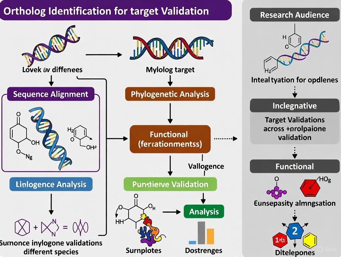
This article provides a comprehensive guide for researchers and drug development professionals on leveraging ortholog identification to validate therapeutic targets across species.
Ortholog Identification for Target Validation: A Cross-Species Guide for Drug Discovery
Abstract
This article provides a comprehensive guide for researchers and drug development professionals on leveraging ortholog identification to validate therapeutic targets across species. It covers the foundational principles of orthology and its critical role in establishing disease relevance and predicting essential genes. The content details state-of-the-art computational methods and databases, addresses common challenges and optimization strategies for complex gene families and big data, and outlines rigorous validation frameworks to assess prediction accuracy and functional conservation. By synthesizing current methodologies and applications, this resource aims to enhance the efficiency and success rate of preclinical target validation in biomedical research.
The Critical Link: Why Orthologs are Fundamental to Target Validation
Defining Orthologs, Paralogs, and the Ortholog Conjecture for Functional Prediction
In the field of comparative genomics and cross-species target validation, accurately identifying evolutionary relationships between genes is fundamental. The terms orthologs and paralogs describe different types of homologous genes—genes related by descent from a common ancestral sequence [1] [2]. Understanding this distinction is critical for predicting gene function in newly sequenced genomes and for selecting appropriate targets for drug development research across species.
Homology refers to biological features, including genes and their products, that are descended from a feature present in a common ancestor. All homologous genes share evolutionary ancestry, but they can be separated through different evolutionary events [1]. The proper classification of these homologous relationships forms the basis for reliable functional annotation transfer, a process essential for leveraging model organism research to understand human disease mechanisms and identify therapeutic targets.
Defining Orthologs and Paralogs
Conceptual Definitions
- Orthologs: Genes in different species that evolved from a common ancestral gene by speciation events [1] [2]. Orthologs typically retain the same function during evolution [3].
- Paralogs: Genes related by gene duplication events within a genome [1] [2]. Paralogs often evolve new functions, though they may retain similar functions, especially if the duplication event was recent [3].
The diagram below illustrates the evolutionary relationships between orthologs and paralogs:
Figure 1: Evolutionary relationships showing orthologs and paralogs. Orthologs (blue) arise from speciation events, while paralogs (red) arise from gene duplication events. All genes shown are homologs, sharing common ancestry [1].
Refined Classification: Inparalogs and Outparalogs
Paralogs can be further categorized based on the timing of duplication events relative to speciation:
- Inparalogs: Paralogs that arise from duplication events after a speciation event of interest. These are co-orthologs to a single-copy gene in another species [4] [5].
- Outparalogs: Paralogs that arise from duplication events before a speciation event. These are not considered orthologs according to standard definitions [4].
This distinction is particularly important for functional prediction, as inparalogs are more likely to retain similar functions compared to outparalogs, which have had more evolutionary time to diverge.
The Ortholog Conjecture and Modern Evidence
The Traditional Ortholog Conjecture
The ortholog conjecture is a fundamental hypothesis in comparative genomics that proposes orthologous genes are more likely to retain similar functions than paralogous genes [5]. This conjecture has guided computational gene function prediction for decades, with the assumption that orthologs evolve functions more slowly than paralogs [6]. This principle has been embedded in many functional annotation pipelines, where orthologs are preferentially used to transfer functional annotations from well-studied model organisms to newly sequenced genomes.
Contemporary Evidence Challenging the Conjecture
Recent large-scale studies using experimental functional data have challenged the validity of the ortholog conjecture:
Table 1: Key Studies Testing the Ortholog Conjecture
| Study | Data Analyzed | Key Findings | Implications |
|---|---|---|---|
| Nehrt et al. (2011) [5] | Experimentally derived functions of >8,900 human and mouse genes | Paralogs were often better predictors of function than orthologs; same-species paralogs most functionally similar | Challenges fundamental assumption in function prediction |
| Stamboulian et al. (2020) [6] | Experimental annotations from >40,000 proteins across 80,000 publications | Strong evidence against ortholog conjecture in function prediction context; paralogs provide valuable functional information | Supports using all available homolog data regardless of type |
These findings demonstrate that the relationship between evolutionary history and functional conservation is more complex than traditionally assumed. Paralogs—particularly those within the same species—can provide equal or superior functional information compared to orthologs [5]. This has significant implications for target validation strategies, suggesting researchers should consider both orthologs and paralogs when predicting gene function.
Ortholog Identification Methods and Databases
Computational Methodologies
Several computational approaches have been developed to identify orthologous relationships:
- Pairwise Reciprocal Best Hits: Identifies pairs of genes in two species that are each other's best match in the other species [7]. This method forms the basis for many ortholog databases but can be misled by complex gene families.
- Phylogenetic Tree-Based Methods: Uses phylogenetic trees to infer orthology by comparing gene trees with species trees [4]. While more accurate, these methods are computationally intensive.
- Synteny-Based Approaches: Leverages conserved gene order and genomic context to identify orthologs [4]. Particularly useful for detecting orthology in complex genomic regions.
- Graph-Based Clustering (OrthoMCL): Applies Markov Cluster algorithm to similarity graphs to group orthologs and recent paralogs across multiple taxa [7]. Effective for handling eukaryotic genomes with multiple paralogs.
The OrthoMCL workflow exemplifies a robust approach for eukaryotic ortholog identification:
Figure 2: OrthoMCL workflow for identifying ortholog groups across multiple eukaryotic species [7].
Table 2: Comparison of Major Ortholog Databases
| Database | Methodology | Coverage | Strengths | Limitations |
|---|---|---|---|---|
| Clusters of Orthologous Groups (COG) [4] | Reciprocal best hits across multiple species | Originally prokaryotic-focused, now includes some eukaryotes | Well-established, manually curated | Limited eukaryotic coverage |
| OrthoMCL [7] | Graph-based clustering using MCL algorithm | Multiple eukaryotic species | Handles "recent" paralogs effectively | Computationally intensive for large datasets |
| Ensembl Compara [4] | Synteny-enhanced reciprocal best hits | Wide range of vertebrate species | Incorporates genomic context | Complex to navigate |
| InParanoid [8] | Pairwise ortholog clustering with inparalog inclusion | Focused on pairwise species comparisons | Accurate for two-species comparisons | Limited to pairwise comparisons |
| Gene-Oriented Ortholog Database (GOOD) [8] | Genomic location-based clustering of isoforms | Mammalian species | Handles alternative splicing effectively | Limited species coverage |
Specialized Tools for Particular Applications
Recent developments have produced specialized ortholog identification tools tailored to specific research communities:
- AlgaeOrtho: A user-friendly tool built upon SonicParanoid and PhycoCosm database specifically designed for identifying orthologs across diverse algal species [9]. This tool generates ortholog tables, similarity heatmaps, and phylogenetic trees to facilitate target identification for bioengineering applications.
- SonicParanoid: A fast, accurate command-line tool for identifying orthologs across multiple species, particularly useful for large-scale comparative genomics studies [9].
Experimental Protocols for Ortholog Identification and Validation
Protocol: Reference Gene Identification for Cross-Species Expression Studies
Objective: Identify reliable reference genes for cross-species transcriptional profiling, as demonstrated in studies of Anopheles Hyrcanus Group mosquitoes [10].
Materials and Reagents:
- Biological Material*: Samples from multiple related species at various developmental stages
- RNA Extraction: TRI reagent or equivalent
- cDNA Synthesis: DNase I, SuperScript IV reverse transcriptase or equivalent
- qPCR: SYBR Green master mix, species-specific primers
- Analysis Software: geNorm, BestKeeper, NormFinder, or RefFinder
Procedure:
- Sample Collection: Collect specimens from multiple developmental stages for each species (e.g., larval, pupal, adult stages) [10].
- RNA Extraction and Quality Control: Extract total RNA using standard methods, treat with DNase I to remove genomic DNA contamination.
- Candidate Gene Selection: Select potential reference genes based on previous studies and housekeeping gene functions (e.g., ribosomal proteins, actin, GAPDH, elongation factors) [10].
- Primer Design and Validation: Design primers in conserved regions across species, test primer efficiency using standard curves with serial cDNA dilutions.
- qPCR Analysis: Perform quantitative PCR for all candidate genes across all samples and species.
- Stability Analysis: Analyze expression stability using multiple algorithms (geNorm, BestKeeper, NormFinder, RefFinder) [10].
- Validation: Select genes with highest stability rankings for use in cross-species comparisons.
Expected Results: Identification of pan-species reference genes with stable expression across developmental stages and species, enabling valid cross-species transcriptional comparisons.
Protocol: OrthoMCL-Based Ortholog Group Identification
Objective: Identify groups of orthologous genes across multiple eukaryotic genomes using OrthoMCL methodology [7].
Materials and Software:
- Computational Resources: High-performance computing cluster recommended for large datasets
- Input Data: Protein sequences in FASTA format for all species of interest
- Software: BLASTP, OrthoMCL pipeline, Markov Cluster algorithm (MCL)
- Database Resources: Optional integration with GUS (Genomic Unified Schema) for data storage
Procedure:
- Data Preparation: Download or compile complete protein sequences for all genomes of interest in FASTA format.
- All-against-all BLAST: Perform BLASTP comparisons of all proteins against all other proteins with an E-value cutoff of 1e-5 [7].
- Ortholog Pair Identification: Identify reciprocal best hits between pairs of species as putative orthologs.
- Paralog Identification: Identify "recent" paralogs as sequences within the same genome that are reciprocally more similar to each other than to any sequence from another species.
- Similarity Graph Construction: Construct a similarity graph where nodes represent proteins and weighted edges represent BLAST similarity scores, normalized to account for systematic biases.
- MCL Clustering: Apply Markov Cluster algorithm with appropriate inflation parameter (typically 1.5-3.0) to identify ortholog groups [7].
- Result Interpretation: Analyze clusters containing sequences from at least two species as final ortholog groups.
Expected Results: Clusters of orthologous proteins and recent paralogs across the analyzed genomes, suitable for functional annotation, evolutionary analysis, and target identification.
Research Reagent Solutions for Ortholog Studies
Table 3: Essential Research Reagents for Ortholog Identification and Validation
| Reagent/Tool Category | Specific Examples | Function in Ortholog Research |
|---|---|---|
| Sequence Databases | PhycoCosm (JGI), Ensembl, NCBI RefSeq | Source of protein and nucleotide sequences for ortholog analysis |
| Ortholog Identification Software | OrthoMCL, OrthoFinder, InParanoid, SonicParanoid | Computational detection of orthologous relationships |
| Multiple Sequence Alignment Tools | ClustalW, MAFFT, MUSCLE | Align orthologous sequences for phylogenetic analysis |
| Phylogenetic Analysis Packages | IQ-TREE, RAxML, PhyML | Construct phylogenetic trees to validate orthology |
| Gene Expression Analysis | qPCR reagents, RNA extraction kits, reverse transcriptases | Experimental validation of ortholog function through expression studies |
| Functional Annotation Resources | Gene Ontology (GO) database, KEGG pathways | Functional comparison of orthologs and paralogs |
| Synteny Visualization Tools | Genomicus, UCSC Genome Browser, Ensembl Compara | Visualize conserved gene order to support orthology predictions |
Applications in Target Validation and Drug Discovery
The accurate identification of orthologs plays a critical role in target validation across species, particularly in drug discovery research. Key applications include:
- Model Organism Translation: Understanding orthologous relationships enables researchers to translate findings from model organisms (e.g., mice, zebrafish, Drosophila) to human biological processes and disease mechanisms [5].
- Drug Target Conservation: Assessing the conservation of potential drug targets across species helps evaluate the relevance of animal models for specific therapeutic areas.
- Toxicology Prediction: Identifying orthologs of human drug metabolism enzymes in preclinical models improves prediction of compound metabolism and potential toxicity.
- Functional Compensation: Recognizing paralogous relationships helps identify potential functional compensation mechanisms that might affect drug efficacy or cause side effects.
The traditional reliance on the ortholog conjecture for functional prediction is being replaced by more nuanced approaches that incorporate both orthologs and paralogs, particularly same-species paralogs that show strong functional conservation [5]. This expanded view provides a more comprehensive framework for target validation across species.
The distinction between orthologs and paralogs remains fundamental to comparative genomics and cross-species target validation, though the traditional ortholog conjecture requires refinement in light of contemporary evidence. Current research indicates that both orthologs and paralogs provide valuable functional information, with same-species paralogs often being strong predictors of gene function. Researchers engaged in target validation across species should employ robust ortholog identification methods such as OrthoMCL while considering functional information from both orthologs and paralogs. The integration of computational predictions with experimental validation, particularly through cross-species expression studies, provides the most reliable approach for translating findings across species boundaries in drug development research.
The Role of Target Product Profiles (TPPs) in Defining Validation Requirements
In translational research, particularly in drug development and comparative genomics, Target Product Profiles (TPPs) serve as critical strategic documents that align development activities with predefined commercial and regulatory goals. A TPP outlines the desired characteristics of a final product—such as a therapeutic, vaccine, or diagnostic—to address an unmet clinical need [11] [12]. When integrated with ortholog identification, a methodology for finding equivalent genes across species, TPPs provide a powerful framework for defining and validating therapeutic targets in non-human model organisms, thereby de-risking and accelerating the development pipeline [13].
This integration is vital for cross-species research, where understanding the function of a gene in a model organism relies on the confirmed equivalence of its ortholog to the human target. This document details the application of TPPs to establish rigorous validation requirements for ortholog-based research, providing structured protocols for researchers and development professionals.
The Strategic Foundation of Target Product Profiles
A Target Product Profile is a strategic planning tool that summarizes the key attributes of an intended product. Originally championed by regulatory authorities, its primary purpose is to guide development by ensuring that every research and development activity is aligned with the goals of the final product, as described in its label [14]. A well-constructed TPP provides a clearly articulated set of goals that help focus and guide development activities to reach the desired commercial outcome [11].
Core Components of a TPP
A comprehensive TPP is structured around the same sections that will appear in the final drug label or product specification sheet [14]. It typically defines both minimum acceptable and ideal or "stretch" targets for each attribute. Failure to meet the "essential" parameters will often mean termination of product development, while meeting the "ideal" profile significantly increases the product's value [11] [14].
Table 1: Core Components of a Target Product Profile for a Therapeutic Candidate
| Drug Label Section | TPP Attribute / Target | Minimum Acceptable | Ideal Target |
|---|---|---|---|
| Indications & Usage | Therapeutic indication & patient population | Treatment of adults with moderate-to-severe Disease X | First-line treatment for all disease severities in adults & pediatrics |
| Dosage & Administration | Dosing regimen & route | Oral, twice daily, with titration | Oral, once daily, no titration needed |
| Dosage Forms & Strengths | Formulation | Immediate-release tablet | Multiple strengths for flexible dosing |
| Contraindications | Absolute contraindications | Hypersensitivity to active ingredient | None |
| Warnings/Precautions | Major safety risks | Monitoring for hepatotoxicity required | No black-box warning |
| Adverse Reactions | Tolerability profile | Comparable to standard of care | Superior to standard of care |
| Clinical Studies | Efficacy endpoints & outcomes | Non-inferiority on primary endpoint vs. standard of care | Superiority on primary and key secondary endpoints |
| How Supplied/Storage | Shelf life & storage | 24 months at 2-8°C | 36 months at room temperature |
TPPs as a Framework for Defining Validation Requirements
The TPP moves from a strategic document to an operational tool by defining the specific evidence required to confirm that a candidate meets its predefined targets. This is especially critical when relying on model organisms for early-stage validation, where the biological relevance must be firmly established.
Linking TPP Attributes to Ortholog Validation
For a TPP attribute like "Efficacy Endpoints," the underlying requirement is a confirmed biological pathway conserved between humans and the model organism used for preclinical testing. The TPP drives the need to identify and validate the correct ortholog in the research model.
Table 2: Translating TPP Attributes into Ortholog Validation Requirements
| TPP Attribute | Downstream Validation Requirement | Ortholog-Based Research Question |
|---|---|---|
| Clinical Efficacy (e.g., >80% point estimate) [15] | Robust, predictive in vivo efficacy model | Does the model organism's ortholog recapitulate the human protein's function in the disease-relevant pathway? |
| Safety Profile (Differentiation from standard of care) [11] | Understanding of conserved off-target biology | Are the binding sites or interactive partners of the target protein conserved in the model organism? |
| Target Population (e.g., pediatrics) [15] | Validation in multiple physiological contexts | Is the ortholog expressed and functional similarly across developmental stages in the model? |
| Onset/Duration of Protection (e.g., within 2 weeks, for 3 years) [15] | Pharmacodynamic biomarker development | Can the ortholog's activity be reliably measured and linked to the functional outcome in the model system? |
Application Note: Integrating TPPs and Ortholog Identification for Target Validation
Experimental Objective
To establish a robust, TPP-informed workflow for identifying and validating orthologs of a human disease gene in a model organism, ensuring the model is fit-for-purpose in evaluating a candidate therapeutic's efficacy and safety.
Experimental Workflow
The following diagram illustrates the integrated workflow for using TPPs to guide ortholog identification and validation.
Protocol 1: Defining TPP-Derived Validation Criteria
- Draft the TPP: Assemble a cross-functional team (discovery, clinical, regulatory) to draft a TPP for the candidate therapeutic. The TPP should be structured around future labeling concepts [14].
- Identify Critical Attributes: Pinpoint the TPP attributes that are dependent on the biological function of the target. These typically include efficacy endpoints, safety profile, and target population [11] [15].
- Define Evidence Requirements: For each critical attribute, define the specific biological evidence needed. For example:
- For Efficacy: "Demonstrate that modulating the ortholog in the model organism produces the predicted physiological effect aligned with the human mechanism of action."
- For Safety: "Evaluate the sequence and structural conservation of off-target binding sites in the model organism versus human."
Protocol 2: Ortholog Identification and Confirmation
Objective: To accurately identify the ortholog of the human target gene in the chosen model organism using established bioinformatics tools.
- Sequence Collection: Obtain the protein sequence of the human target gene. Download the complete proteome (all protein sequences) for the model organism(s) of interest from a reliable database such as PhycoCosm, Ensembl, or NCBI [16].
- Ortholog Inference: Use an orthology inference tool to identify putative orthologs.
- Recommended Tools: OrthoFinder (for high accuracy and phylogenetic analysis) [17] or SonicParanoid (for speed and user-friendly downstream processing) [16].
- Input: The human protein sequence and the model organism proteome file(s).
- Command (OrthoFinder example):
orthofinder -f ./proteome_files -t 8, where-tspecifies the number of CPU threads.
- Ortholog Confirmation: The output of these tools (e.g., Orthogroups.tsv from OrthoFinder) will list groups of orthologous genes. Identify the orthogroup containing your human protein and extract the putative ortholog from the model organism.
- Phylogenetic Validation (Optional but Recommended): For critical targets, perform a phylogenetic analysis to confirm the orthology relationship visually and exclude paralogs (genes related by duplication within a species) [17]. Tools like AlgaeOrtho can automatically generate sequence similarity heatmaps and unrooted phylogenetic trees for this purpose [16].
Protocol 3: Experimental Validation of Functional Equivalence
Objective: To experimentally verify that the identified ortholog performs the same biological function as the human target.
- Expression Pattern Analysis:
- Use techniques like RT-qPCR or RNA-Seq to analyze the spatial and temporal expression pattern of the ortholog in the model organism. The expression should be consistent with the expected role in the disease-relevant pathway or tissue.
- Functional Rescue/Complementation Assay:
- In vitro: Transfer the model organism ortholog into a human cell line where the native human gene has been knocked down. Assess if the ortholog can rescue the lost cellular function.
- In vivo: Knock out the ortholog in the model organism and observe the phenotype. Subsequently, introduce the human gene to see if it can rescue the phenotype, providing strong evidence of functional conservation.
- Biochemical Assays:
- Characterize the ortholog's biochemical activity (e.g., kinase activity, receptor binding affinity) and compare it to the human protein. Use assays relevant to the TPP's efficacy mechanism.
- Pharmacological Profiling:
- Test the lead therapeutic candidate (or tool compound) on the model organism ortholog to confirm that the interaction (e.g., binding, inhibition) is conserved. The pharmacodynamic response should mirror what is anticipated in humans.
The Scientist's Toolkit: Research Reagent Solutions
Table 3: Essential Materials and Tools for TPP-Guided Ortholog Research
| Research Reagent / Tool | Function / Application | Example(s) |
|---|---|---|
| Orthology Inference Software | Identifies groups of orthologous genes from protein sequences across multiple species. | OrthoFinder [17], SonicParanoid [16], OrthoMCL [13] |
| Proteome Databases | Provides the complete set of protein sequences for a species, required for ortholog searches. | PhycoCosm (algae) [16], Ensembl, NCBI, UniProt |
| Multiple Sequence Alignment Tool | Aligns orthologous sequences to assess conservation and infer phylogeny. | Clustal Omega (used in AlgaeOrtho pipeline) [16], MAFFT |
| Target Enrichment Probe Set | Custom probes to capture orthologous loci for phylogenomics or functional genomics. | Orthoptera-specific OR-TE probe set [18] |
| Model Organism Databases | Curated genomic and functional data for specific model organisms (e.g., yeast, mouse, zebrafish). | SGD, MGI, ZFIN |
The integration of Target Product Profiles with rigorous ortholog identification and validation creates a traceable and defensible bridge from early-stage discovery to clinical application. By using the TPP to define the "what" and "why" of validation, and ortholog research to address the "how," research teams can make informed decisions on model organism selection, derisk preclinical development, and increase the likelihood that their final product will successfully address an unmet clinical need. This structured approach ensures that resources are invested in the most predictive models and assays, ultimately accelerating the translation of basic research into effective therapies.
Linking Target Essentiality to Drug Efficacy in Pathogens and Disease Models
In the field of pharmaceutical innovation, the identification of novel drug targets is a critical and challenging step in the development process. Essential genes, defined as those vital for cell or organism survival, have emerged as highly promising candidates for therapeutic targets in disease treatment [19]. These genes encode critical cellular functions and regulate core biological processes, making them paramount in assessing new drug targets [19]. The systematic identification of essential genes provides a powerful strategy for uncovering potential therapeutic targets, thereby accelerating new drug development across various diseases, including infectious pathogens and cancer [19].
This framework is particularly powerful when applied within a cross-species research paradigm, where ortholog identification enables the validation of targets from model organisms to humans. Understanding the characteristics of essential genes—including their conditional nature, evolutionary conservation, and network properties—provides crucial insights for rational drug design, helping to improve efficacy while anticipating potential side effects [20] [19].
Key Concepts and Definitions
Essential Genes and Their Characteristics
Essential genes are no longer perceived as a binary or static concept. Contemporary research reveals that gene essentiality is often conditional, dependent on specific genetic backgrounds and biochemical environments [19]. These genes typically exhibit high evolutionary conservation across species, indicating their fundamental biological importance, and demonstrate evolvability, where non-essential genes can acquire essential functions through evolutionary processes [19].
From a drug development perspective, essential genes represent particularly valuable targets because their disruption or modulation directly impacts pathogen survival or disease progression. In many pathogens, essential genes account for only 5-10% of the genetic complement but represent targets for the majority of antibiotics [19].
Network Properties Influencing Drug Effects
The position and role of drug targets within biological networks significantly influence therapeutic outcomes and side effect profiles. Key network properties include:
- Essentiality: Drugs targeting essential genes (those encoding critical cellular functions) demonstrate higher efficacy but may also cause more side effects [20] [19].
- Centrality: Targets with high degree (number of direct protein interactions) and betweenness (position in shortest paths) within protein interactome networks are associated with increased side effects [20].
- Interaction Interfaces: Single-interface targets (binding different partners at same interfaces) cause more adverse effects than multi-interface targets when disrupted [20].
Table 1: Network Properties of Drug Targets and Their Implications
| Network Property | Definition | Impact on Drug Effects |
|---|---|---|
| Target Essentiality | Gene required for organism survival [19] | Determines drug efficacy; primary driver of side effects [20] |
| Degree Centrality | Number of direct interaction partners [20] | Higher degree correlates with more side effects [20] |
| Betweenness Centrality | Number of shortest paths going through the target [20] | Higher betweenness correlates with more side effects [20] |
| Interface Sharing | Proportion of partners binding at same interface [20] | Single-interface targets cause more side effects when disrupted [20] |
Experimental Protocols for Essential Gene Identification
CRISPR-Cas9 Functional Genomics Screening
CRISPR-Cas9 screening enables genome-wide identification of essential genes through targeted gene knockouts. The protocol below applies to both pathogen and mammalian systems:
- Library Design: Design and clone guide RNA (gRNA) libraries targeting all protein-coding genes in the target organism.
- Viral Transduction: Transduce cells with lentiviral vectors at low MOI (0.3-0.5) to ensure single gRNA integration.
- Selection Pressure: Apply appropriate selection (e.g., antibiotics for integrated vectors) for 48-72 hours.
- Population Sampling: Harvest cell samples at multiple time points (e.g., days 0, 7, 14, 21) to monitor population dynamics.
- gRNA Quantification: Extract genomic DNA and sequence gRNA regions to determine relative abundance.
- Essentiality Scoring: Calculate gene essentiality scores based on gRNA depletion using specialized algorithms (MAGeCK, CERES).
Critical Considerations: Include non-targeting control gRNAs; use sufficient biological replicates (n≥3); optimize infection efficiency for each cell type; confirm Cas9 activity before screening.
Transposon Mutagenesis (Tn-seq) for Bacterial Pathogens
Transposon sequencing identifies essential genes in bacterial pathogens through random insertion mutagenesis:
- Transposon Delivery: Introduce mariner-based transposon into pathogen via conjugation or electroporation.
- Mutant Library Generation: Grow library to high coverage (≥50,000 unique mutants) under permissive conditions.
- Selection Passage: Passage library through relevant conditions (e.g., antibiotic exposure, host infection models).
- Genomic DNA Extraction: Harvest genomic DNA from input and output populations.
- Library Preparation: Fragment DNA, enrich transposon-chromosome junctions, and prepare sequencing libraries.
- Sequence Analysis: Map insertion sites and identify genomic regions with significant insertion depletion.
This method successfully identified pyrC, tpiA, and purH as essential genes and potential antibiotic targets in Pseudomonas aeruginosa [19].
Cross-Species Ortholog Validation Protocol
Validating essential gene conservation across species requires specialized approaches:
- Ortholog Identification: Use reciprocal BLAST with threshold E-value < 1e-10 and alignment coverage > 70%.
- Reference Gene Selection: Identify stable reference genes for normalization in cross-species qPCR (see Table 2).
- Expression Profiling: Measure target gene expression across multiple species and developmental stages.
- Functional Complementation: Test whether orthologs can rescue essential gene function in knockout models.
Table 2: Reference Genes for Cross-Species Expression Studies
| Biological Context | Recommended Reference Genes | Stability Measure | Application Scope |
|---|---|---|---|
| Larval Stage (Mosquito) | RPL8, RPL13a [10] | High stability across 6 species | Cross-species comparison at larval stage |
| Adult Stage (Mosquito) | RPL32, RPS17 [10] | Stable across all adult stages | Cross-species adult comparison |
| Multiple Stages (An. belenrae) | RPS17 [10] | Most stable across stages | Intra-species normalization |
| Multiple Stages (An. kleini) | RPS7, RPL8 [10] | Most stable across stages | Intra-species normalization |
Data Analysis and Visualization Workflows
Network Analysis of Target Essentiality
Network Analysis Workflow for Drug Target Identification
Cross-Species Ortholog Validation Pipeline
Cross-Species Ortholog Validation Pipeline
The Scientist's Toolkit: Research Reagent Solutions
Table 3: Essential Research Reagents and Platforms for Target Essentiality Studies
| Reagent/Platform | Function | Application Context |
|---|---|---|
| CRISPR-Cas9 Libraries | Genome-wide gene knockout screening | Identification of essential genes in mammalian and pathogen systems [19] |
| Mariner Transposon Systems | Random insertion mutagenesis | Essential gene identification in bacterial pathogens (Tn-seq) [19] |
| Network Analysis Tools (Gephi) | Network visualization and metric calculation | Analysis of target centrality, degree, and betweenness [20] [21] |
| qPCR Reference Genes (RPS17, RPL8) | Expression normalization in cross-species studies | Stable reference for comparing gene expression across related species [10] |
| Protein Interaction Databases | Source of protein-protein interaction data | Construction of interactome networks for target characterization [20] |
| Ortholog Prediction Tools | Identification of conserved genes across species | Cross-species target validation and essentiality transfer [10] |
Case Studies and Applications
Essential Targets inPseudomonas aeruginosa
A comprehensive transposon mutagenesis study identified three essential genes—pyrC, tpiA, and purH—as promising antibiotic targets in P. aeruginosa [19]. These genes encode functions in pyrimidine biosynthesis (pyrC), glycolysis (tpiA), and purine metabolism (purH). Follow-up validation confirmed that inhibitors targeting these essential pathways exhibited potent bactericidal activity against multidrug-resistant clinical isolates, demonstrating the power of systematic essential gene identification for antibiotic development.
Network Analysis for Side Effect Prediction
A systematic analysis of 4,199 side effects associated with 996 drugs revealed that drugs causing more side effects are characterized by high degree and betweenness of their targets in the human protein interactome network [20]. This finding provides a network-based framework for predicting potential side effects during drug development, emphasizing that both essentiality and centrality of drug targets are key factors contributing to side effects and should be incorporated into rational drug design [20].
Cross-Species Reference Genes in Mosquito Vectors
A recent study identified reliable reference genes for cross-species transcriptional profiling across six Anopheles mosquito species [10]. The research demonstrated that RPL8 and RPL13a showed the most stable expression at larval stages, while RPL32 and RPS17 exhibited stability across adult stages [10]. These reference genes enable accurate comparison of gene expression across closely related pathogen vectors, facilitating research into species-specific differences in vector competence and insecticide susceptibility.
Utilizing Evolutionary Conservation to Infer Gene Function and Disease Relevance
Evolutionary conservation analysis provides a powerful framework for identifying functionally important genes and regions across species. The fundamental premise is that genomic elements under purifying selection due to their critical biological roles will be retained throughout evolution. For researchers in drug discovery and target validation, this approach enables prioritization of candidate genes with higher potential clinical relevance and lower likelihood of redundancy.
Comparative genomics studies have demonstrated that human disease genes are highly conserved in model organisms, with 99.5% of human disease genes having orthologs in rodent genomes [22]. This remarkable conservation enables researchers to utilize model organisms for functional studies, though with important caveats regarding specific disease mechanisms. The distribution of conservation varies significantly across biological systems—genes associated with neurological and developmental disorders typically exhibit slower evolutionary rates, while those involved in immune and hematological systems evolve more rapidly [22]. This variation has direct implications for selecting appropriate model systems for specific disease contexts.
Recent methodological advances now allow researchers to move beyond simple sequence alignment to identify functionally conserved elements, even when sequences have significantly diverged. These approaches are particularly valuable for studying non-coding regulatory elements, which often show rapid sequence turnover while maintaining functional conservation [23].
Quantitative Frameworks for Conservation Analysis
Key Metrics and Their Applications
Table 1: Evolutionary Conservation Metrics and Their Applications in Target Validation
| Metric | Calculation Method | Biological Interpretation | Target Validation Application |
|---|---|---|---|
| dN/dS Ratio (KA/KS) | Ratio of non-synonymous to synonymous substitution rates [22] | Values <1 indicate purifying selection; >1 indicate positive selection | Identify genes under functional constraint across species [22] |
| Taxonomy-Based Measures (VST/STP) | Incorporates taxonomic distance between species with matching variants [24] | Variants shared with distant taxa are more likely deleterious in humans | Improved prediction of pathogenic missense variants [24] |
| Ornstein-Uhlenbeck Process | Models expression evolution with parameters for drift (σ) and selection (α) [25] | Quantifies stabilizing selection on gene expression levels | Identify genes with constrained expression patterns across tissues [25] |
| Sequence Conservation Score | Percentage identity from multiple sequence alignments | Estimates degree of sequence constraint | Filter for highly conserved genes as candidate essential genes [26] |
| Indirect Positional Conservation | Synteny-based mapping using IPP algorithm [23] | Identifies functional conservation despite sequence divergence | Discover conserved non-coding regulatory elements [23] |
Performance Comparison of Conservation Metrics
Table 2: Predictive Performance of Conservation Methods for Pathogenic Variants
| Method | Underlying Principle | AUC Value | Advantages | Limitations |
|---|---|---|---|---|
| LIST | Taxonomy-distance exploitation [24] | 0.888 | Superior performance for deleterious variants in non-abundant domains | Requires carefully curated multiple sequence alignments |
| PhyloP | Phylogenetic p-values [24] | 0.820 | Models nucleotide substitution rates | Lower precision across variant types |
| SIFT | Sequence homology-based [24] | 0.818 | Predicts effect on protein function | Limited to coding variants |
| PROVEAN | Alignment-based conservation [24] | 0.816 | Handles indels and single residues | Performance drops with shallow alignments |
| SiPhy | Phylogeny-based conservation [24] | 0.810 | Models context-dependent evolution | Computationally intensive |
Experimental Protocols
Protocol 1: Identification of Evolutionarily Constrained Genes for Target Prioritization
Purpose: To systematically identify and prioritize evolutionarily constrained genes as high-value targets for therapeutic development.
Materials:
- Genomic sequences from multiple vertebrate species
- Computing infrastructure for large-scale comparative analyses
- Quality-controlled variant datasets (e.g., gnomAD, ClinVar)
Procedure:
Ortholog Identification
- Identify 1:1 orthologs across target species using reciprocal best BLAST hits or specialized tools like Ensembl Compara
- Filter for genes with orthologs across minimum 10 species spanning appropriate evolutionary distances
Evolutionary Rate Calculation
- Perform multiple sequence alignment for each ortholog group using MAFFT or Clustal Omega
- Calculate dN/dS ratios using codeml from PAML package or similar tools
- Classify genes as evolutionarily constrained (dN/dS < 0.5)
Conservation Metric Integration
Functional Validation Prioritization
- Prioritize genes with strong conservation signals (composite score > 0.8) and disease association
- Exclude genes with known redundancy through paralog analysis
- Select top candidates for experimental validation in model systems
Troubleshooting:
- For genes with limited phylogenetic coverage, consider using the IPP algorithm to infer positional conservation [23]
- Address alignment uncertainties by using multiple alignment methods and comparing results
- For rapidly evolving gene families, focus on conserved domains rather than full-length proteins
Protocol 2: Experimental Validation of Conserved Non-Coding Elements
Purpose: To functionally validate conserved non-coding regulatory elements identified through comparative genomics.
Materials:
- Embryonic tissue from model organisms (mouse, chicken)
- Chromatin profiling reagents (ATAC-seq, ChIP-seq kits)
- Reporter constructs for enhancer assays
- In vivo electroporation or transgenic system
Procedure:
Identification of Non-Coding Conservation
Functional Testing
- Clone candidate elements into reporter vectors (e.g., luciferase, GFP)
- Test enhancer activity in cell-based systems
- Validate in vivo using model organisms at equivalent developmental stages [23]
Disease Variant Assessment
- Identify human variants within conserved non-coding elements
- Test impact of variants on regulatory activity
- Correlate with disease phenotypes and expression quantitative trait loci (eQTLs)
Diagram 1: Workflow for Identification of Indirectly Conserved Regulatory Elements
The Scientist's Toolkit: Research Reagent Solutions
Table 3: Essential Research Resources for Evolutionary Conservation Studies
| Resource Category | Specific Tools/Databases | Primary Function | Application Context |
|---|---|---|---|
| Multiple Sequence Alignment | MAFFT, Clustal Omega, MUSCLE | Generate protein/nucleotide alignments | Calculation of evolutionary rates [22] |
| Evolutionary Rate Analysis | PAML (codeml), HYPHY, GERP++ | Calculate dN/dS and conservation scores | Quantifying selective pressure [22] |
| Variant Pathogenicity Prediction | LIST, SIFT, PolyPhen-2, CADD | Predict functional impact of variants | Prioritizing disease-associated variants [24] |
| Expression Evolution Analysis | OU model implementation (R/BioConductor) | Model expression level evolution | Identify constrained expression patterns [25] |
| Synteny-Based Mapping | Interspecies Point Projection (IPP) | Identify positionally conserved elements | Discovery of conserved regulatory elements [23] |
| Genomic Data Integration | Ensembl, UCSC Genome Browser | Visualize conservation across species | Contextualize conservation patterns [22] |
Advanced Analytical Approaches
Modeling Expression Evolution
Gene expression levels evolve under distinct selective pressures that can be quantified using Ornstein-Uhlenbeck (OU) processes [25]. This framework models expression evolution through two key parameters: σ (drift rate) and α (strength of stabilizing selection toward an optimal expression level θ). The application of OU models to RNA-seq data across 17 mammalian species revealed that most genes evolve under stabilizing selection, with expression differences between species saturating at larger evolutionary distances [25].
Diagram 2: Ornstein-Uhlenbeck Model of Expression Evolution
Taxonomic Distance Integration
Traditional conservation measures treat all species equally, but newer approaches exploit taxonomic distances to improve predictive power. The LIST method incorporates two taxonomy-aware measures: Variant Shared Taxa (VST), which quantifies the taxonomic distance to species sharing a variant of interest, and Shared Taxa Profile (STP), which captures position-specific variability across the taxonomy tree [24]. These measures significantly improve identification of deleterious variants, particularly in protein regions like intrinsically disordered domains that are poorly captured by conventional methods.
Applications in Disease Gene Discovery
Evolutionary conservation analyses have revealed systematic differences between disease gene categories. Genes associated with neurological disorders show significantly slower evolutionary rates compared to immune system genes [22]. This pattern makes neurological disease genes particularly amenable to study in model organisms, though important exceptions exist, such as trinucleotide repeat expansion disorders where rodent orthologs contain substantially fewer repeats [22].
For infectious disease applications, conservation analysis in bacterial pathogens like Pseudomonas aeruginosa has identified essential genes as promising antibiotic targets [26]. These studies demonstrate that essential and highly expressed genes in bacteria evolve at lower rates, enabling target prioritization based on evolutionary conservation [26].
Regulatory element conservation presents special challenges, as sequence conservation dramatically decreases with evolutionary distance—only ~10% of enhancers show sequence conservation between mouse and chicken [23]. However, synteny-based approaches like IPP can identify 5-fold more conserved enhancers through positional conservation, enabling functional studies of non-coding regions relevant to disease [23].
From Sequence to Function: Methods and Tools for Ortholog Identification
Orthology, describing genes that originated from a common ancestor through speciation events, is a cornerstone of comparative genomics and functional genetics [27] [28]. Accurate ortholog identification is particularly critical for target validation in cross-species research, where understanding gene function and essentiality in a model organism can inform drug target prioritization in a pathogen or human [29]. The two predominant computational approaches for inferring these relationships are graph-based and tree-based methods, each with distinct theoretical foundations and practical implications for researchers. This application note provides a detailed comparison of these methodologies, supported by experimental protocols and quantitative benchmarks, to guide their application in target validation pipelines.
Orthology Fundamentals and Their Application to Target Validation
Defining Orthology and Paralogy
According to Fitch's classical definition, orthologs are homologous genes diverged by a speciation event, while paralogs are diverged by a gene duplication event [28] [30]. This distinction is biologically significant as orthologs often, though not always, retain equivalent biological functions across species—a concept known as the "ortholog conjecture" [31] [28]. This functional conservation makes ortholog identification indispensable for transferring functional annotations from well-characterized model organisms to less-studied species, a common requirement in biomedical and agricultural research.
The Critical Role of Orthology in Target Validation
In pharmaceutical and parasitology research, orthology inference enables the prediction of essential genes in non-model pathogens by leveraging functional data from model organisms. Studies have demonstrated that evolutionary conservation and the presence of essential orthologues in diverse eukaryotes are strong predictors of gene essentiality [29]. Furthermore, genes absent from the host genome but present and essential in the pathogen represent promising candidates for selective drug targets with minimized host toxicity [29]. Quantitative analyses show that combining orthology with essentiality data can yield up to a five-fold enrichment in essential gene identification compared to random selection [29].
Table 1: Key Orthology Concepts in Target Validation
| Concept | Description | Application to Target Validation |
|---|---|---|
| Orthologs | Genes diverged via speciation [28] | Primary candidates for functional annotation transfer |
| Paralogs | Genes diverged via gene duplication [28] | May indicate functional divergence; potential for redundancy |
| Hierarchical Orthologous Groups (HOGs) | Nested sets of orthologs defined at different taxonomic levels [30] | Enables precise identification of duplication events and functional conservation depth |
| Essentiality Prediction | Leveraging essential orthologs across species to predict gene indispensability [29] | Prioritizes high-value targets likely required for pathogen survival |
Orthology Inference Methodologies
Graph-Based Methods
Graph-based approaches infer orthology directly from pairwise sequence comparisons, bypassing the need for explicit gene and species trees [27]. These methods typically construct an orthology graph where vertices represent genes and edges connect pairs estimated to be orthologs [27]. Popular implementations include OrthoMCL, ProteinOrtho, and OMA [27] [32].
A key theoretical insight is that under a tree-like evolutionary model, true orthology graphs must be cographs—graphs that can be generated through a series of join and disjoint union operations and do not contain induced paths of four vertices (P~4~) [27]. This structural property enables error correction in inferred graphs by editing them to the closest cograph [27]. For complex evolutionary scenarios involving hybridization or horizontal gene transfer, level-1 networks (networks with relatively few hybrid vertices per "block") provide a more flexible explanatory framework, with characterizations showing that level-1 explainable orthology graphs are precisely those in which every primitive subgraph is a near-cograph (a graph where removing a single vertex results in a cograph) [27].
Graph 1: Graph-based orthology inference workflow. The process begins with sequence comparison and progresses through graph construction and clustering to final orthologous groups, with potential error correction if the graph violates the cograph property.
Tree-Based Methods
Tree-based methods require gene trees and species trees as input and infer orthology through reconciliation—mapping the gene tree onto the species tree to determine whether each divergence event represents a speciation (orthology) or duplication (paralogy) [27] [30]. This approach, implemented in tools like OrthoFinder and EnsemblCompara, uses event-based maximum parsimony, assigning costs to evolutionary events (duplication, speciation, transfer) to find the most plausible reconciliation [27] [30].
The Hierarchical Orthologous Groups (HOGs) framework represents a powerful extension of tree-based methods, organizing genes into nested sets defined at different taxonomic levels [30]. HOGs can be conceptualized as clades within a reconciled gene tree, with each HOG corresponding to genes descended from a single ancestral gene at a specific taxonomic level [30]. This hierarchical organization enables researchers to pinpoint exactly when in evolutionary history duplications occurred, providing critical context for understanding functional conservation and divergence.
Graph 2: Tree-based orthology inference workflow. This approach requires both gene trees and a species tree, with reconciliation identifying speciation and duplication events to define hierarchical orthologous groups.
Quantitative Comparison of Methodologies
Table 2: Methodological Comparison: Graph-Based vs. Tree-Based Approaches
| Characteristic | Graph-Based Methods | Tree-Based Methods |
|---|---|---|
| Theoretical Basis | Orthology graphs and cograph theory [27] | Gene tree-species tree reconciliation [27] [30] |
| Primary Input | Pairwise sequence similarities [27] | Gene trees and species tree [27] |
| Computational Efficiency | Generally more efficient; suitable for large datasets [27] | Computationally challenging; can be impractical for genome-wide data [27] |
| Handling Complex Evolution | Can be extended to networks (e.g., level-1 networks) [27] | Requires complex reconciliation models for transfers/hybridization |
| Output Resolution | Flat or moderately hierarchical orthologous groups | Explicit hierarchical orthologous groups (HOGs) [30] |
| Key Strengths | Speed, scalability, robustness to incomplete data | Detailed evolutionary history, precise duplication timing |
| Common Tools | OrthoMCL, ProteinOrtho, OMA [27] [32] | OrthoFinder, EnsemblCompara, PANTHER [27] [30] |
Recent benchmarking using the Orthobench resource, which contains 70 expert-curated reference orthogroups (RefOGs), revealed that methodological improvements have significantly enhanced orthology inference accuracy. A phylogenetic reassessment of Orthobench RefOGs revised 44% (31/70) of the groups, with 24 requiring major revision affecting phylogenetic extent [33]. This highlights the importance of using updated benchmarks when evaluating method performance.
Table 3: Orthology Benchmark Performance of Selected Methods
| Method | Approach | Precision (SwissTree) | Recall (SwissTree) | Scalability |
|---|---|---|---|---|
| FastOMA | Graph-based with hierarchical resolution [32] | 0.955 [32] | 0.69 [32] | Linear scaling; processes 2,086 genomes in 24h [32] |
| OMA | Graph-based with hierarchical resolution [32] | High (comparable) [32] | Moderate [32] | Quadratic scaling; processes 50 genomes in 24h [32] |
| OrthoFinder | Tree-based [33] | Benchmarked [33] | Benchmarked [33] | Quadratic scaling [32] |
| Panther | Tree-based [32] | High [32] | High recall [32] | Not specified |
Experimental Protocols
Protocol 1: Graph-Based Orthology Inference with FastOMA
FastOMA represents a modern, scalable graph-based method that maintains high accuracy while achieving linear scalability in the number of input genomes [32].
Research Reagent Solutions
- Input Proteomes: FASTA files of protein sequences for all species of interest
- OMAmer: Alignment-free k-mer-based tool for sequence placement [32]
- Linclust: Highly scalable clustering tool from MMseqs package [32]
- Species Tree: NCBI taxonomy or user-provided reference phylogeny [32]
Step-by-Step Procedure
- Gene Family Inference (RootHOGs):
- Map input proteins to reference Hierarchical Orthologous Groups (HOGs) using OMAmer
- Group proteins mapped to the same reference HOG into query rootHOGs
- For sequences without recognizable homologs in the reference database, perform additional clustering using Linclust to form new rootHOGs
Orthology Inference:
- For each query rootHOG, infer the nested structure of HOGs through a bottom-up traversal of the species tree
- At each taxonomic level, combine HOGs from child levels based on evolutionary relationships
- Resolve the full hierarchical structure of orthologous groups across all taxonomic levels
Output Generation:
- Generate HOGs at all taxonomic levels for downstream analysis
- Export orthology relationships in standardized formats
Applications in Target Validation
- Rapid identification of conserved orthologs across multiple pathogen species
- Detection of pathogen-specific genes absent from host genomes by examining rootHOG membership
- Essentiality prediction based on conservation patterns across diverse eukaryotes
Protocol 2: Tree-Based Orthology Inference with Hierarchical Orthologous Groups (HOGs)
The HOG framework provides a phylogenetic approach to orthology inference, enabling precise determination of duplication events and their evolutionary timing [30].
Research Reagent Solutions
- Multiple Sequence Alignment Tool: MAFFT L-INS-i algorithm [33]
- Alignment Trimming: TrimAL [33]
- Phylogenetic Inference: IQ-TREE with model selection [33]
- Species Tree: Known taxonomy or inferred species phylogeny
Step-by-Step Procedure
- Gene Tree Reconstruction:
- Perform multiple sequence alignment using MAFFT L-INS-i algorithm
- Trim alignment with TrimAL to remove poorly aligned regions
- Infer gene tree using IQ-TREE with best-fitting model of sequence evolution
- Assess statistical support using appropriate methods (e.g., bootstrapping)
Tree Reconciliation:
- Reconcile gene trees with the species tree using maximum parsimony or probabilistic methods
- Label internal nodes as speciation or duplication events
- Map gene duplication and loss events onto the species tree
HOG Extraction:
- Define HOGs at each taxonomic level corresponding to clades rooted at speciation nodes
- Organize HOGs in a nested hierarchy reflecting the species phylogeny
- Annotate each HOG with its taxonomic level and evolutionary history
Applications in Target Validation
- Precise identification of lineage-specific gene duplications that may indicate functional specialization
- Reconstruction of ancestral gene content to understand gene family evolution in pathogens
- Differentiation between ancient conserved orthologs and recently diverged paralogs for functional annotation
Application to Target Validation: A Case Study in Parasitic Nematodes
The utility of orthology inference for target prediction is exemplified by research on blood-feeding strongylid nematodes like Haemonchus contortus and Ancylostoma caninum [29]. These parasites cause significant agricultural and human health burdens, yet functional genomic tools for directly testing gene essentiality are limited.
Researchers constructed a database integrating essentiality information from four model eukaryotes (C. elegans, D. melanogaster, M. musculus, and S. cerevisiae) with orthology mappings from OrthoMCL [29]. Analysis revealed that evolutionary conservation and the presence of essential orthologues are each strong predictors of essentiality, with absence of paralogues further increasing the probability of essentiality [29].
By applying quantitative orthology and essentiality criteria, researchers achieved a five-fold enrichment in essential gene identification compared to random selection [29]. This approach enabled prioritization of potential drug targets from among the ~20,000 genes in these parasites, demonstrating the power of orthology inference to guide target validation in non-model organisms where direct genetic manipulation is challenging.
Both graph-based and tree-based orthology inference methods offer distinct advantages for target validation research. Graph-based methods provide computational efficiency and scalability for large multi-genome analyses, while tree-based approaches offer detailed evolutionary histories and precise duplication timing. The emerging generation of tools like FastOMA combines the scalability of graph-based methods with the hierarchical resolution of tree-based approaches [32], while frameworks like Hierarchical Orthologous Groups (HOGs) provide a structured way to represent complex evolutionary relationships [30].
For researchers engaged in cross-species target validation, orthology inference remains an indispensable tool for predicting gene essentiality, identifying pathogen-specific targets, and transferring functional annotations. Method selection should be guided by research goals: graph-based methods for large-scale screening and tree-based approaches for detailed evolutionary analysis of candidate targets. As genomic data continue to expand, ongoing methodological innovations will further enhance our ability to accurately infer orthology relationships and apply these insights to validate therapeutic targets across the tree of life.
In biomedical and pharmaceutical research, the identification of molecular targets across species is a foundational step for understanding disease mechanisms and developing new therapeutics. The principle that orthologous genes—genes in different species that evolved from a common ancestral gene by speciation—largely retain their ancestral function provides a powerful framework for extrapolating functional knowledge from model organisms to humans, or for understanding pathogen biology [34] [30]. This approach is particularly critical in target validation, where researchers assess the potential of a biological molecule to be a drug target. Accurate ortholog identification helps establish biologically relevant model systems, predicts potential side effects due to off-target interactions, and informs on the translatability of preclinical findings.
However, evolutionary processes such as gene duplication and loss create complex gene families, making simple pairwise gene comparisons insufficient. Hierarchical Orthologous Groups (HOGs) offer a refined solution by systematically organizing genes across multiple taxonomic levels, providing a structured view of gene evolution and enabling more precise functional inference [30]. This article details four key resources—OrthoDB, OMA, OrthoFinder, and BUSCO—that empower researchers to navigate this complexity, providing protocols for their effective application in target validation workflows.
The field offers several complementary resources for orthology inference, each with distinct strengths in methodology, taxonomic scope, and output. The table below summarizes the key features of OrthoDB, OMA, OrthoFinder, and BUSCO for direct comparison.
Table 1: Key Features of Ortholog Identification Resources
| Resource | Primary Function | Key Methodological Approach | Taxonomic Scope (as of 2024) | Key Outputs |
|---|---|---|---|---|
| OrthoDB | Integrated resource of pre-computed orthologs with functional annotations | Hierarchical orthology delineation using OrthoLoger software; aggregates functional data [34] [35] | 5,827 Eukaryotes; 17,551 Bacteria; 607 Archaea [34] | Hierarchical Orthologous Groups (OGs), functional descriptors, evolutionary annotations, BUSCO datasets |
| OMA (Orthologous MAtrix) | Database and method for inferring orthologs | Graph-based inference of orthologs and paralogs, focusing on pairs and groups [34] [35] | 713 Eukaryotes; 1,965 Bacteria; 173 Archaea (2024) [34] | Pairwise orthologs, OMA Groups (HOGs), Gene Ontology annotations |
| OrthoFinder | Software for de novo inference of orthologs from user-provided proteomes | Phylogenetic methodology; infers rooted gene trees and orthogroups [30] | N/A (Software for user-defined species sets) | Orthogroups, gene trees, gene duplication events, phylogenetic analysis |
| BUSCO | Tool for assessing genome/assembly completeness using universal orthologs | Mapping user sequences to benchmark sets of universal single-copy orthologs [34] | Wide coverage of Eukaryotes and Prokaryotes via OrthoDB-derived sets [34] | Completeness scores (% of complete, duplicated, fragmented, missing BUSCOs) |
Table 2: Data Content and Access for Orthology Resources
| Resource | Functional Annotations | Evolutionary Annotations | Data Access & APIs |
|---|---|---|---|
| OrthoDB | Gene Ontology, InterPro domains, KEGG pathways, EC numbers, textual descriptions [34] [35] | Evolutionary rate, phyletic profile (universality/duplicability), sibling groups [34] | Web interface, REST API, SPARQL/RDF, Python/R API, bulk download [34] |
| OMA | Gene Ontology, functional annotations | Inference of orthologs and paralogs, HOGs | OMA Browser, REST API, bulk download [35] |
| OrthoFinder | N/A (can be added post-analysis) | Gene trees, duplication events, species tree inference | Command-line tool, output files (TSV, Newick) |
| BUSCO | N/A | Implied by universal single-copy ortholog presence | Command-line tool, pre-computed sets for major lineages |
OrthoDB: Application Protocol for Target Assessment
OrthoDB is a comprehensive resource that provides pre-computed orthologous groups with integrated functional and evolutionary annotations, making it highly efficient for initial target assessment.
Workflow for Target Characterization
Step-by-Step Protocol
Query the Database
- Navigate to the OrthoDB website (https://www.orthodb.org).
- Use the search bar with a gene identifier (e.g., human gene symbol, UniProt ID) or genome assembly accession.
- Filter your search using the "get gene" or "search orthologs" selectors to refine results [34].
Retrieve and Analyze Orthologous Groups (OGs)
- OrthoDB presents a list of OGs related to your query. Select the appropriate OG at the taxonomic level relevant to your research (e.g., Vertebrata for comparison across vertebrates, or a narrower clade for more precise functional inference) [34] [30].
- In the OG detail view, examine the Sankey diagram to navigate the hierarchical relationship of OGs across taxonomic levels [34].
- Download protein or newly added coding DNA sequences (CDS) for the entire OG using the "View fasta" links for subsequent analysis [34].
Leverage Functional and Evolutionary Annotations for Target Assessment
- Functional Descriptors: Review the aggregated functional summary, which consolidates data from UniProt, Gene Ontology (GO), InterPro domains, KEGG pathways, and enzyme codes. This provides a concise overview of molecular function and biological role [34] [35].
- Evolutionary Traits: Critically evaluate the evolutionary descriptors unique to OrthoDB:
- Phyletic Profile: Assess the "universality" (proportion of species with orthologs) and "duplicability" (proportion of multi-copy orthologs). A universal gene may indicate a core biological function, while lineage-restriction might suggest species-specific adaptations. High duplicability could signal gene family expansions relevant to functional redundancy or specialization [34].
- Evolutionary Rate: The relative degree of sequence conservation can indicate selective pressure; rapidly evolving genes might be involved in host-pathogen interactions or other adaptive processes [34].
Access Data Programmatically (For Advanced Users)
- For large-scale analyses, use the REST API or the new Python and R Bioconductor API packages to programmatically access OrthoDB data and integrate it into custom analysis pipelines [34].
OrthoFinder: Protocol for De Novo Orthology Inference
OrthoFinder is the tool of choice when working with novel genomes or a specific set of proteomes not fully covered by pre-computed databases.
Workflow for Phylogenomic Analysis
Step-by-Step Protocol
Input Preparation
- Gather proteome files (in FASTA format) for all species of interest. Ensure consistent sequence annotation is helpful for downstream interpretation.
- Place all proteome files in a single directory.
Running OrthoFinder
- Install OrthoFinder (e.g., via Conda:
conda install -c bioconda orthofinder). - Run a basic analysis:
orthofinder -f /path/to/proteome_directory -t <number_of_threads>. - For large datasets, consider using the
-M msaoption for more accurate gene tree inference, though this increases computational time.
- Install OrthoFinder (e.g., via Conda:
Analysis of Results for Target Validation
- Orthogroups: The primary output is "Orthogroups.tsv". This file lists all genes belonging to each orthogroup (a HOG at the level of the entire species set). Identify the orthogroup containing your target gene.
- Gene Trees: Examine the rooted gene trees for your orthogroup of interest (located in the "ResolvedGeneTrees" folder). This allows visual confirmation of orthology/paralogy relationships and helps identify potential species-specific duplications that might complicate experimental validation [30].
- Gene Duplication Events: The "GeneDuplicationEvents" output identifies which nodes in the gene trees represent duplications. This is critical for understanding whether a gene pair are true orthologs or out-paralogs, which is vital for selecting the correct counterpart in a model organism [30].
BUSCO: Protocol for Quality Control in Genomics
BUSCO assessments are a critical first step to ensure the reliability of genomic data used for ortholog identification and downstream target validation.
Workflow for Genome Assessment
Step-by-Step Protocol
Dataset Selection and Tool Execution
- Select the appropriate BUSCO lineage dataset (e.g.,
mammalia_odb10for mammals,eukaryota_odb10for broad eukaryotic analysis) that matches the taxonomy of your sample. - Run BUSCO on your genome assembly or annotated gene set. Example command for a transcriptome:
busco -i transcriptome.fa -l eukaryota_odb10 -m transcriptome -o busco_result
- Select the appropriate BUSCO lineage dataset (e.g.,
Interpretation for Target Validation Context
- Analyze the summary output and
full_table.tsv:- High "Complete" BUSCOs (>90-95% for well-assembled genomes): Indicates high gene space completeness, increasing confidence that an absent ortholog represents a true biological loss rather than an assembly artifact.
- Low "Fragmented" and "Missing" BUSCOs: Minimizes false negatives in ortholog searches.
- Low "Duplicated" BUSCOs: A high duplication rate in a haploid genome might indicate assembly issues, such as haplotypic duplication, which could artificially inflate gene copies and mislead orthology calls [34].
- Analyze the summary output and
Table 3: Key Research Reagents and Computational Tools for Orthology Research
| Resource / Tool | Type | Primary Function in Orthology Workflow |
|---|---|---|
| OrthoDB | Database | One-stop resource for pre-computed hierarchical orthologs with integrated functional and evolutionary annotations [34]. |
| OrthoFinder | Software | State-of-the-art tool for de novo inference of orthologs, gene trees, and gene duplication events from custom proteome sets [30]. |
| BUSCO | Tool & Dataset | Provides benchmark sets of universal single-copy orthologs and a tool to assess the completeness of genomic data [34]. |
| OrthoLoger | Software | The underlying method used by OrthoDB for orthology delineation; also available as a standalone tool or Conda package for mapping new genomes to OrthoDB groups [34] [35]. |
| AlgaeOrtho | Tool / Workflow | Example of a domain-specific (algal) pipeline built upon SonicParanoid for identifying and visualizing orthologs, demonstrating a tailored application [16]. |
| LEMOrtho | Benchmarking Framework | A Live Evaluation of Methods for Orthologs delineation, useful for comparing and selecting the best orthology inference method for a specific project [35]. |
OrthoDB, OMA, OrthoFinder, and BUSCO form a powerful ecosystem of resources for accurate ortholog identification. OrthoDB offers a comprehensive starting point with its rich annotations, while OrthoFinder provides flexibility for custom datasets. BUSCO acts as an essential gatekeeper for data quality. By applying the protocols outlined herein, researchers can robustly leverage evolutionary relationships to validate therapeutic targets across species, thereby strengthening the foundation of translational biomedical research.
The escalating challenge of anthelmintic resistance in parasitic nematodes poses a significant threat to global health and food security, creating an urgent need for novel therapeutic targets [36] [37]. Traditional approaches to anthelmintic discovery have been protracted, expensive, and technically demanding, often relying on whole-organism phenotypic screening without prior knowledge of molecular targets [38] [39]. The integration of orthology identification with machine learning (ML) prediction frameworks now enables systematic prioritization of essential genes in parasitic nematodes, dramatically accelerating the early discovery pipeline [38] [40]. This workflow establishes a robust protocol for cross-species target prediction by leveraging the extensive functional genomic data available for model organisms like Caenorhabditis elegans and translating these insights to medically and agriculturally important parasites through orthology relationships [36] [38].
Table 1: Key Definitions in Orthology-Based Target Discovery
| Term | Definition | Application in Workflow |
|---|---|---|
| Orthologs | Genes in different species that evolved from a common ancestral gene by speciation [28] | Central bridge for functional annotation transfer |
| Essential Genes | Genes critical for organism survival, whose inhibition causes lethality or significant fitness loss [38] | Primary candidates for anthelmintic targeting |
| Chokepoint Reactions | Metabolic reactions that consume a unique substrate or produce a unique product [41] | Prioritization filter for metabolic targets |
| Target Deconvolution | Process of identifying the molecular target of a bioactive compound [36] | Experimental validation of predicted targets |
Orthology-Based Machine Learning Prediction Pipeline
Feature Selection and Model Training
The foundation of accurate essential gene prediction lies in curating informative features with proven predictive power across species. Based on successful applications in Dirofilaria immitis, Brugia malayi, and Onchocerca volvulus, 26 features have been identified as strong predictors of gene essentiality [38] [40]. The most informative predictors include OrthoFinder_species (identifying ortholog groups across species), exon count, and subcellular localization predictors (nucleus and cytoplasm) [38]. These features are derived from genomic, transcriptomic, and proteomic data sources, enabling multi-faceted assessment of gene criticality.
For model training, multiple machine learning algorithms should be evaluated, including Gradient Boosting Machines (GBM), Generalized Linear Models (GLM), Neural Networks (NN), Random Forests (RF), Support Vector Machines (SVM), and Extreme Gradient Boosting (XGB) [38]. In comparative studies, GBM and XGB typically achieve the highest performance, with ROC-AUC values exceeding 0.93 for C. elegans and approximately 0.9 for Drosophila melanogaster when trained on 90% of the data [38]. The model training process requires careful cross-validation and performance assessment using both ROC-AUC and Precision-Recall AUC (PR-AUC) metrics to ensure robust predictions.
Cross-Species Prediction Implementation
The practical implementation of cross-species prediction involves a structured workflow that transfers essentiality annotations from well-characterized model organisms to poorly studied parasitic nematodes. This process begins with comprehensive data collection from reference databases, followed by orthology inference, feature engineering, and finally ML-based prediction with priority ranking [38] [40].
Table 2: Machine Learning Performance for Essential Gene Prediction
| Model Algorithm | C. elegans ROC-AUC | D. melanogaster ROC-AUC | Recommended Use Case |
|---|---|---|---|
| Gradient Boosting (GBM) | ~0.93 [38] | ~0.9 [38] | Primary prediction model |
| XGBoost (XGB) | ~0.93 [38] | ~0.9 [38] | High-dimensional data |
| Random Forest (RF) | ~0.91 [38] | ~0.9 [38] | Feature importance analysis |
| Neural Network (NN) | >0.87 [38] | >0.8 [38] | Complex non-linear relationships |
Figure 1: Machine Learning Workflow for Cross-Species Essential Gene Prediction
Experimental Validation and Target Deconvolution
Proteomic Approaches for Target Identification
Following computational prediction, experimental validation is crucial for confirming essential gene function and anthelmintic potential. Stability-based proteomic methods have emerged as powerful tools for direct target identification, requiring no compound modification (label-free) and applicable to both lysed cells and live parasites [36]. Thermal Proteome Profiling (TPP) has been successfully applied to parasitic nematodes including Haemonchus contortus, identifying protein targets for anthelmintic candidates like UMW-868, ABX464, and UMW-9729 [36]. In these studies, TPP revealed significant stabilization of specific H. contortus proteins (HCON014287 and HCON011565 for UMW-868; HCON_00074590 for ABX464) upon compound binding, providing direct evidence of drug-target interactions [36].
The cellular thermal shift assay (CETSA) provides a complementary approach to TPP, typically using Western blot or targeted proteomics to validate known or suspected interactions [36]. For researchers without access to advanced mass spectrometry facilities, drug affinity responsive target stability (DARTS) offers a more accessible alternative that measures protein stability upon drug binding via proteolysis sensitivity [36]. Although DARTS has been primarily applied to C. elegans mitochondrial targets to date, the principle is transferable to parasitic systems with protocol optimization [36].
Functional Validation Using Genetic Approaches
Genetic validation provides critical confirmation of gene essentiality through direct manipulation of target genes. While RNA interference (RNAi) has been efficiently implemented in C. elegans, its application in parasitic nematodes remains challenging due to variable RNAi uptake efficiency across species [36]. CRISPR/Cas9 gene editing offers a highly specific alternative for functional validation, enabling precise knockout or modification of predicted essential genes [36]. The development of robust CRISPR/Cas9 systems for parasitic nematodes represents a current technical frontier, with successful implementations emerging for key species [36].
Resistance mutation mapping provides an additional genetic validation approach, identifying drug targets by analyzing resistance-associated mutations in parasite populations [36]. This method is particularly powerful when combined with chemical mutagenesis screens, offering an unbiased pathway for discovering essential drug targets without prior mechanistic assumptions [36].
Table 3: Experimental Methods for Target Validation
| Method | Principle | Advantages | Throughput |
|---|---|---|---|
| Thermal Proteome Profiling (TPP) | Measures drug-induced thermal stability of proteins [36] | Label-free, proteome-wide, detects direct/indirect interactions | Medium |
| Cellular Thermal Shift Assay (CETSA) | Monitors thermal stability of specific proteins [36] | Targeted validation, lower equipment requirements | Low-Medium |
| Drug Affinity Responsive Target Stability (DARTS) | Measures protease resistance upon drug binding [36] | No compound modification, simple setup | Medium |
| CRISPR/Cas9 | Gene editing to knock out or modify target genes [36] | Highly specific, enables functional validation | Low |
| Resistance Mutation Mapping | Identifies targets via resistance-conferring mutations [36] | Unbiased, powerful for essential pathway identification | Low |
The Scientist's Toolkit: Research Reagent Solutions
Successful implementation of this workflow requires access to specialized reagents and computational resources. The following toolkit summarizes essential materials and their applications in orthology-based anthelmintic discovery.
Table 4: Essential Research Reagents and Resources
| Reagent/Resource | Function | Application Example |
|---|---|---|
| OrthoFinder | Identifies ortholog groups across multiple species [16] | Inferring orthologs between C. elegans and parasitic nematodes |
| SonicParanoid | Fast, accurate command-line orthology inference [16] | Processing proteome data for AlgaeOrtho tool |
| PhycoCosm Database | Hosts multi-omics data for diverse species [16] | Accessing genomic and proteomic data for non-model parasites |
| AlgaeOrtho Tool | Processes ortholog results with visualization [16] | Identifying orthologs of proteins of interest across species |
| Thermal Shift Assay Kits | Measure protein thermal stability [36] | Implementing CETSA for target validation |
| Protease Kits for DARTS | Provide optimized proteolysis conditions [36] | Conducting DARTS experiments without mass spectrometry |
| CRISPR/Cas9 Systems | Enable targeted gene editing [36] | Functional validation of predicted essential genes |
Integrated Workflow and Pathway Analysis
Chokepoint Analysis for Metabolic Target Prioritization
Complementary to ML-based essential gene prediction, chokepoint analysis of metabolic pathways provides a systematic approach for identifying critical enzymatic reactions that represent promising intervention points [41]. A "chokepoint reaction" is defined as a reaction that either consumes a unique substrate or produces a unique product within a metabolic network [41]. Inhibition of chokepoint enzymes causes either toxic accumulation of substrates or critical deficiency of products, leading to parasite death or severe fitness costs.
This approach has been successfully applied across 10 nematode species, identifying both common chokepoints (CommNem) present in all species and parasite-specific chokepoints (ParaNem) [41]. The practical application of this method led to the discovery of Perhexiline, a compound showing efficacy against both C. elegans and parasitic nematodes including Haemonchus contortus and Onchocerca lienalis [41]. Mode-of-action studies confirmed that Perhexiline affects the fatty acid oxidation pathway, reducing oxygen consumption rates in C. elegans and demonstrating the utility of chokepoint-based discovery [41].
Figure 2: Chokepoint Analysis Workflow for Metabolic Target Identification
Integrated Validation Framework
A robust validation framework combines computational predictions with orthogonal experimental approaches to build confidence in proposed anthelmintic targets. This integrated strategy should include transcriptional profiling across developmental stages to confirm expression in relevant parasite tissues and life stages [38] [40]. High-priority essential genes typically show strong transcriptional activity across development and enrichment in pathways related to ribosome biogenesis, translation, RNA processing, and signaling [38].
Functional validation should employ multiple complementary methods, including pharmacological inhibition (where compounds are available), genetic approaches (RNAi or CRISPR where feasible), and phenotypic assessment of parasite viability, development, and reproduction [36] [39]. For compounds identified through screening, target deconvolution using the proteomic methods described in Section 3.1 provides critical mechanistic insights [36]. The final validation step should assess specificity by confirming minimal activity against host orthologs, leveraging the orthology frameworks established early in the workflow [41] [28].
This comprehensive workflow from orthology inference through experimental validation establishes a systematic, reproducible pipeline for anthelmintic target discovery. By integrating computational predictions with orthogonal experimental validation, researchers can prioritize the most promising targets for further drug development, ultimately addressing the critical need for novel anthelmintics in the face of rising drug resistance.
Leveraging Hierarchical Orthologous Groups (HOGs) for Evolutionary-Aware Analysis
In the field of comparative genomics, Hierarchical Orthologous Groups (HOGs) provide a powerful framework for understanding gene evolution across multiple taxonomic levels. HOGs represent sets of genes that have descended from a single common ancestor within a specific taxonomic range of interest [42]. Unlike traditional flat orthologous groups, HOGs offer a taxonomically-nested structure that captures evolutionary relationships at different phylogenetic depths, making them particularly valuable for target validation studies in cross-species research [30]. This hierarchical organization allows researchers to precisely trace gene duplication events and functional diversification across evolutionary timescales, providing critical insights for drug target identification and validation.
The fundamental principle behind HOGs is their ability to represent evolutionary histories through a structured framework that aligns with species phylogeny. A HOG defined at a deep taxonomic level (e.g., vertebrates) encompasses all descendant genes, while HOGs at more recent levels (e.g., mammals) represent finer subdivisions, effectively creating nested subfamilies that reflect evolutionary relationships [43]. This multi-resolution perspective enables researchers to select the appropriate evolutionary context for their specific validation needs, whether studying conserved core biological processes or lineage-specific adaptations [30].
HOGs in Comparative Genomics and Target Validation
Key Conceptual Frameworks
HOGs can be conceptualized through several complementary perspectives, each offering unique insights for target validation research:
Groups of Extant Orthologs and Paralogs: A HOG represents a set of homologous genes found in extant species that have all descended from a single ancestral gene at a specified taxonomic level [30]. This perspective helps researchers distinguish between orthologs (resulting from speciation) and paralogs (resulting from gene duplication), a critical distinction when extrapulating functional data across species [44].
Clades on Reconciled Gene Trees: From a phylogenetic perspective, HOGs correspond to clades within reconciled gene trees where internal nodes are labeled as speciation or duplication events [30]. For example, a HOG at the Mammalia level corresponds to a clade of genes where all extant species are mammals, providing a evolutionarily coherent group for functional analysis.
Gene Families and Subfamilies: HOGs provide a structured way to define gene families and subfamilies, with deeper taxonomic levels representing entire gene families and more recent levels corresponding to functionally distinct subfamilies [30]. This hierarchical organization is particularly valuable for understanding functional conservation and divergence in drug target candidates.
Ancestral Gene Proxies: At any taxonomic level, a HOG serves as a proxy for an ancestral gene, with the nested structure of HOGs allowing researchers to trace these ancestral genes across evolutionary time [30]. This perspective facilitates the reconstruction of ancestral gene functions and evolutionary trajectories.
Advantages over Traditional Orthology Methods
Traditional orthology inference methods often fail to capture the evolutionary complexity necessary for robust target validation across species. HOGs address several key limitations:
Temporal Resolution of Duplication Events: Unlike flat orthologous groups, HOGs explicitly capture when duplication events occurred relative to speciation events, making it possible to distinguish orthologs from in-paralogs [30]. This temporal resolution is essential for understanding whether functional differences between genes reflect genuine biological differences or are artifacts of分析方法.
Handling of Complex Gene Families: For large, duplication-rich gene families (e.g., G-protein coupled receptors or protein kinases), HOGs provide a structured framework to navigate complex evolutionary relationships that often confound traditional methods [30]. This capability is particularly valuable when studying gene families that are frequently targeted by pharmaceuticals.
Evolutionary Context for Functional Annotation: The hierarchical structure of HOGs provides built-in evolutionary context for functional annotations, allowing researchers to distinguish between conserved core functions and lineage-specific innovations [30]. This context helps prioritize targets with desired evolutionary conservation profiles.
Table 1: Comparison of Orthology Inference Approaches for Target Validation
| Feature | Pairwise Orthology | Flat Orthologous Groups | HOGs |
|---|---|---|---|
| Evolutionary Depth | Single level | Single level | Multiple hierarchical levels |
| Duplication Handling | Limited | Coarse-grained | Explicit timing relative to speciation |
| Paralog Discrimination | Poor | Moderate | Excellent |
| Ancestral State Inference | Not supported | Not supported | Directly supported |
| Cross-Species Extrapolation | Limited context | Limited context | Evolutionarily aware context |
Computational Protocols for HOG Inference
Graph-Based HOG Inference with GETHOGs
The GETHOGs (Graph-based Efficient Technique for Hierarchical Orthologous Groups) algorithm provides a robust approach for inferring HOGs directly from pairwise orthology information, without requiring computationally expensive gene tree inference or gene/species tree reconciliation [42]. The protocol consists of the following key steps:
Orthology Graph Construction: The process begins with constructing an orthology graph where each node represents a gene, and edges represent inferred pairwise orthologous relationships between genes [43]. This graph serves as the foundational data structure for all subsequent analyses.
Connected Component Identification: The algorithm identifies connected components within the orthology graph, with each component representing a putative gene family composed of genes descended from a common ancestral gene [43]. These components form the candidate set for hierarchical decomposition.
Taxonomy-Aware Decomposition: Each connected component is decomposed into HOGs at different taxonomic levels using the species phylogeny as a guide [42]. The algorithm traverses the species tree from leaves to root, defining HOGs at each internal node that represent sets of genes descending from a single ancestral gene at that taxonomic level.
Stringency Parameter Application: GETHOGs implements several extensions with stringency parameters to handle imperfect input data, allowing researchers to balance sensitivity and specificity based on their specific requirements [42]. These parameters help manage challenges such as incomplete genomes, annotation errors, and evolutionary rate variations.
The following workflow diagram illustrates the GETHOGs pipeline for inferring Hierarchical Orthologous Groups from genomic data:
Hybrid Approaches for Primary and Secondary Ortholog Detection
The HyPPO (Hybrid Prediction of Paralogs and Orthologs) framework combines similarity-based and phylogeny-based approaches to distinguish between primary orthologs (not affected by accelerated mutation rates after duplication) and secondary orthologs (affected by post-duplication divergence) [44]. This distinction is particularly valuable for target validation, as primary orthologs typically maintain similar functions, while secondary orthologs may exhibit functional divergence:
Primary Ortholog Identification: HyPPO uses exact graph partitioning techniques to identify primary orthologs, which must form cliques in the orthology graph [44]. This mathematical property provides a formal justification for the clustering steps performed by many orthology prediction methods and ensures robust group identification.
Species Tree Construction: The framework constructs a species tree from the identified orthology clusters, providing an evolutionary framework for subsequent analyses [44]. This species tree serves as a reference for determining evolutionary relationships and identifying appropriate model organisms for specific target validation questions.
Secondary Ortholog Inference: Using the constructed species tree, HyPPO identifies secondary orthologs by analyzing evolutionary paths that include duplication events followed by accelerated mutation rates [44]. This capability allows researchers to identify cases where orthologous relationships exist but functional conservation may be compromised.
Integration with P4-Free Graphs: The method leverages the theoretical foundation of P4-free orthology graphs, which have a special tree representation interpretable as a gene tree [44]. This mathematical framework ensures evolutionary consistency in the inferred relationships.
Table 2: Performance Comparison of Orthology Inference Methods
| Method | Accuracy | Precision | Recall | Cluster Score |
|---|---|---|---|---|
| HyPPO | 0.940 | 0.911 | 0.875 | 0.915 |
| HyPPO + Species Tree | 0.949 | 0.924 | 0.905 | 0.915 |
| OMA-GETHOGs | 0.877 | 0.940 | 0.699 | 0.831 |
| OrthoMCL | 0.812 | 0.845 | 0.496 | 0.690 |
Experimental Applications and Workflows
Target Conservation Analysis Protocol
A critical application of HOGs in pharmaceutical research is assessing the evolutionary conservation of potential drug targets across species. The following protocol provides a standardized workflow for target conservation analysis:
Step 1: HOG Selection and Extraction: Identify and extract relevant HOGs containing the target of interest from databases such as OMA, EggNOG, or OrthoDB [30] [43]. The selection should be based on the taxonomic scope most relevant to the research question (e.g., mammals for targets being developed for human diseases).
Step 2: Evolutionary Conservation Scoring: Calculate conservation metrics for the target across species of interest. Key metrics include taxonomic breadth (number of species containing the target), sequence conservation (percentage identity across species), and gene loss rate (frequency of loss events in the phylogenetic tree) [30].
Step 3: Functional Domain Analysis: Annotate functional domains within the target protein and assess their conservation patterns across the HOG. Identify domains with universal conservation versus those with lineage-specific variations that might impact function or drug binding [30].
Step 4: Paralog Discrimination: Identify and characterize paralogs within the HOG that arose from gene duplication events. Assess potential functional redundancy or neofunctionalization that could impact target specificity and off-target effects [44] [30].
Step 5: Binding Site Evolution: For targets with known or predicted binding sites, analyze the evolutionary conservation of residues critical for small molecule binding or protein-protein interactions. Identify species-specific variations that could impact compound efficacy or toxicity [30].
The following diagram illustrates the logical relationships in evolutionary-aware target validation using HOGs:
Cross-Species Functional Annotation Transfer
HOGs provide an evolutionarily rigorous framework for transferring functional annotations from well-characterized model organisms to less-studied species, a common requirement in target validation pipelines:
Evolutionarily Informed Annotation: Transfer functional annotations between genes within the same HOG, with confidence scores based on evolutionary distance and conservation metrics [30]. This approach minimizes erroneous annotation transfers that can occur with pairwise methods when paralogs are present.
Lineage-Specific Function Prediction: Identify potential functional innovations by detecting rapidly evolving branches or lineage-specific conserved amino acid changes within HOGs [30]. These evolutionary signatures can indicate functional adaptations relevant to species-specific drug responses.
Experimental Design Optimization: Use HOG structure to select appropriate model organisms for functional validation studies based on evolutionary proximity to humans and experimental tractability [30]. The hierarchical nature of HOGs enables rational selection of multiple model systems representing different evolutionary distances.
Successful implementation of HOG-based analysis requires leveraging specialized computational resources and databases. The following table details essential research reagents and resources for evolutionary-aware target validation studies:
Table 3: Essential Research Reagent Solutions for HOG-Based Analysis
| Resource/Reagent | Type | Primary Function | Access Information |
|---|---|---|---|
| OMA Browser | Database | HOG visualization and querying | http://omabrowser.org [45] [43] |
| GETHOGs Algorithm | Software | Graph-based HOG inference | Part of OMA standalone package [42] |
| HyPPO Framework | Software | Primary/secondary ortholog detection | https://github.com/manuellafond/HyPPO [44] |
| EggNOG Database | Database | Precomputed HOGs with functional annotations | http://eggnog.embl.de [30] |
| OrthoDB | Database | Hierarchical orthology across multiple taxa | https://www.orthodb.org [30] |
| Species Tree Resources | Data | Reference phylogenies for HOG construction | Various sources (e.g., NCBI Taxonomy) [44] |
Hierarchical Orthologous Groups represent a paradigm shift in orthology inference that directly addresses the needs of evolutionarily-aware target validation research. By explicitly capturing the taxonomic context of gene relationships, HOGs enable researchers to make informed decisions about target conservation, model organism selection, and functional extrapolation across species. The structured frameworks and protocols outlined in this application note provide a foundation for implementing HOG-based analyses in pharmaceutical research and development workflows, ultimately enhancing the efficiency and success rates of cross-species target validation efforts.
Integrating Synteny with OrthoRefine to Improve Ortholog-Paralog Discrimination
In the context of target validation across species, accurately identifying true orthologs—genes diverged by speciation events—is paramount. Orthologs, more than paralogs, tend to retain conserved biological functions, making their correct discrimination critical for extrapolating findings from model organisms to humans in drug development research [46] [47]. Standard ortholog prediction tools, while powerful, can misclassify recent paralogs as orthologs, especially in the face of incomplete genome data or gene loss, potentially leading to misleading conclusions in functional genomics [47].
The integration of synteny—the conservation of gene order across genomes—provides a powerful, independent line of evidence to refine ortholog calls. OrthoRefine is a standalone tool that automates this synteny-based refinement of pre-computed ortholog groups, such as those generated by OrthoFinder, offering a significant enhancement in ortholog-paralog discrimination specifically valuable for target validation studies [46].
Key Principles and Quantitative Evidence
The OrthoRefine Algorithm: A Syntenic "Look-Around Window"
OrthoRefine operates on a straightforward but effective principle. For each gene within an orthologous group (OG) previously identified by a tool like OrthoFinder, the algorithm examines its genomic neighborhood using a "look-around window" [46].
- Workflow: The process involves centering a window of a user-defined size on a candidate gene in one genome and counting how many genes within that window have their orthologs (belonging to the same OG) in the corresponding genomic region of the other genome.
- Synteny Ratio Calculation: A synteny ratio ((s_r)) is calculated by dividing the number of these matching gene pairs by the total window size ((w)). A gene pair is considered syntenic if this ratio exceeds a defined cutoff (default = 0.5) [46].
- Output - Syntenic Ortholog Groups (SOGs): Following pairwise comparisons between all genes from different genomes in the original OG, subsets of genes linked by synteny are classified as SOGs. This process effectively splits a single OG containing paralogs into multiple SOGs or refines it into a single, high-confidence SOG by removing non-syntenic paralogs [46].
Performance and Optimization Data
OrthoRefine has been rigorously tested on both bacterial and eukaryotic datasets. The table below summarizes key performance metrics and the impact of window size on ortholog refinement.
Table 1: OrthoRefine Performance and Parameter Optimization
| Metric / Parameter | Reported Finding | Biological Context / Implication |
|---|---|---|
| Paralog Elimination | Efficiently removes paralogs from OrthoFinder's Hierarchical Orthogroups (HOGs) [46]. | Increases the specificity of ortholog datasets, crucial for functional inference. |
| Typical Runtime | A few minutes for ~10 bacterial genomes on a standard desktop PC [46]. | Accessible for researchers without high-performance computing resources. |
| Optimal Window Size (General) | Smaller windows (e.g., 8 genes) [46]. | Suitable for datasets of closely related genomes where local synteny blocks are well-conserved. |
| Optimal Window Size (Distantly Related) | Larger windows (e.g., 30 genes) [46]. | Better for datasets with less conserved gene order, capturing broader syntenic regions. |
| Validation with Phylogenetics | Improved ortholog identification supported by phylogenetic analysis [46]. | Confirms that synteny-based refinement produces more evolutionarily accurate groupings. |
| Comparative Study Findings | Use of synteny resulted in more reliably identified orthologs and paralogs compared to conventional methods [48]. | Reinforces the utility of synteny as a method for generating high-confidence ortholog sets for downstream analysis. |
Application Notes & Protocols
This section provides a detailed, step-by-step protocol for employing OrthoRefine to refine ortholog calls, framed within a target validation workflow where distinguishing the true human ortholog of a pre-clinical drug target is essential.
Prerequisites and Input Data Preparation
- Genome Annotation Files: For each genome in your analysis, obtain a feature table file in the RefSeq format used by NCBI. These files contain the necessary genomic coordinates for each gene. For de novo assemblies, generate these annotations using NCBI's Prokaryotic Genome Annotation Pipeline (PGAP) or a similar tool [46].
- Initial Ortholog Identification: Run OrthoFinder with your protein sequence files (e.g.,
.faaformat) from all species of interest. Use default parameters or adjust as needed for your project. OrthoFinder will produce a directory of results, including the fileOrthogroups/Orthogroups.tsv(or HOGs) which is required for the next step [46]. - Input List File: Create a simple text file (e.g.,
genomes_list.txt) where each line contains the RefSeq assembly accession for each genome used in the OrthoFinder analysis, matching the order of the input FASTA files [46].
Step-by-Step OrthoRefine Protocol
Table 2: Essential Research Reagents and Computational Tools
| Item / Software | Function / Role in the Protocol | Source / Note |
|---|---|---|
| OrthoFinder | Generates initial clustering of homologous genes into orthogroups. | https://github.com/davidemms/OrthoFinder [46] |
| OrthoRefine | Refines initial orthogroups using synteny to discriminate orthologs from paralogs. | Standalone tool; requires OrthoFinder output and genome annotations [46] |
| NCBI Feature Table Files | Provides genomic coordinates of genes, essential for synteny analysis. | From RefSeq assembly or generated via annotation pipelines [46] |
| Genome List File | A text file linking OrthoFinder input to specific genome assemblies. | User-generated; critical for correct execution. |
The following diagram illustrates the complete experimental workflow, from data preparation to the final output of high-confidence syntenic ortholog groups.
Software Execution: Run OrthoRefine from the command line, specifying the necessary inputs:
-og: Path to the Orthogroups.tsv file from OrthoFinder.-g: Path to your genome list text file.-a: Path to the directory containing all RefSeq annotation files.-o: Desired output directory for OrthoRefine results.
Parameter Optimization:
- Window Size (
-w): The primary parameter to optimize. Begin with the default or a small window (e.g.,-w 8) for closely related species. For distantly related species, test larger windows (e.g.,-w 20to-w 30) [46]. - Synteny Ratio Cutoff (
-s): The minimum ratio of matching genes to window size (default 0.5). A higher cutoff increases stringency.
- Window Size (
Output Interpretation: OrthoRefine generates a new set of orthogroups—the SOGs. Analyze the
*_syntenic_orthogroups.tsvfile. Compare it to the original OrthoFinder output to identify which putative orthologs were removed as non-syntenic paralogs and which were retained as high-confidence syntenic orthologs.
A Diagram of the Core OrthoRefine Algorithm
The following diagram details the core synteny detection logic within OrthoRefine, showing how the look-around window is applied to discriminate between syntenic orthologs and non-syntenic paralogs.
Integrating synteny analysis via OrthoRefine provides a robust, automated method for enhancing ortholog-paralog discrimination. By adding a layer of genomic context, it significantly improves the specificity of ortholog identification over standard sequence-based methods alone. For research aimed at target validation across species, this tool is invaluable for pinpointing the most reliable human ortholog of a pre-clinical target, thereby de-risking drug discovery and increasing the confidence with which biological insights can be translated across species.
Navigating Challenges: Optimizing Orthology Predictions in Complex Scenarios
The field of comparative genomics is undergoing a seismic shift driven by ambitious, large-scale sequencing initiatives such as the Earth BioGenome Project, which aims to sequence the genomes of 1.5 million eukaryotic species [32]. This explosion of data presents an unprecedented opportunity to understand the evolutionary origins and genetic innovations underlying biological processes, with significant implications for target validation in cross-species research. A fundamental and critical step in this comparative analysis is the accurate identification of orthologs—genes in different species that originated from a single gene in their last common ancestor [49]. Orthologs often retain conserved biological functions over evolutionary time, making them cornerstone candidates for validating therapeutic targets and extrapolating findings from model organisms to humans [50].
However, traditional orthology inference methods face acute scalability issues. Methods that rely on all-against-all sequence comparisons scale poorly, becoming computationally prohibitive with thousands of genomes. For instance, processing just over 2,000 genomes using the established Orthologous Matrix (OMA) algorithm required more than 10 million CPU hours [32]. This creates a significant bottleneck, forcing researchers to analyze vast genomic datasets in a piecemeal fashion. Addressing this "big data" challenge requires a new generation of algorithms designed for linear scalability and high performance without sacrificing accuracy. FastOMA represents one such solution, enabling rapid and precise orthology inference at the scale of the entire tree of life [32] [50].
FastOMA: A Scalable Solution for Orthology Inference
Algorithmic Innovation and Workflow
FastOMA is a complete, ground-up rewrite of the OMA algorithm, engineered specifically for linear scalability in the number of input genomes [32]. Its core innovation lies in avoiding unnecessary all-against-all sequence comparisons by leveraging existing knowledge of the sequence universe and a highly efficient parallel computing approach. The algorithm consists of two main steps, as illustrated in the workflow below.
Step 1: Gene Family Inference. FastOMA first maps input protein sequences to coarse-grained gene families, known as root-level Hierarchical Orthologous Groups (rootHOGs), using OMAmer, a rapid, alignment-free k-mer-based tool [32] [51]. This step leverages the evolutionary information stored in the OMA reference database to efficiently group homologous sequences. Proteins that cannot be mapped to existing families (e.g., novel genes) are subsequently clustered using Linclust, a highly scalable clustering tool from the MMseqs2 package [32] [51]. By grouping sequences into families upfront, FastOMA eliminates the computational burden of comparing evolutionarily unrelated genes across the entire dataset.
Step 2: Hierarchical Orthologous Group (HOG) Inference. For each rootHOG, FastOMA reconstructs the nested evolutionary history of the gene family by performing a bottom-up traversal of the provided species tree [32]. Starting from the genes of extant species (the leaves), the algorithm identifies sets of genes that form a HOG at each ancestral node, meaning they descended from a single gene in that ancestor [49]. This results in a hierarchical structure where HOGs defined at recent clades are nested within larger HOGs defined at older, deeper clades, providing a comprehensive and phylogenetically aware map of gene relationships [32] [49].
Key Features for Robust Performance
FastOMA incorporates several features that make it particularly robust for handling complex, real-world genomic data:
- Taxonomy-Guided Subsampling: The algorithm uses a known species phylogeny (e.g., from NCBI Taxonomy) to guide and reduce the number of sequence comparisons, contributing significantly to its speed. Performance can be further improved with a more resolved species tree [32].
- Handling Gene Model Imperfections: It is designed to manage multiple protein isoforms from alternative splicing, automatically selecting the most evolutionarily conserved isoform. It can also accommodate fragmented gene models, leading to more accurate inferences [32].
- Flexible Input Tree: While defaulting to the NCBI taxonomy, users can supply any rooted species tree in Newick format. The tree does not need to be fully resolved (binary) or have branch lengths, making it accessible for non-specialists [51].
Validation and Performance Benchmarks
Accuracy and Scalability
FastOMA was rigorously benchmarked by the Quest for Orthologs (QfO) consortium, demonstrating that its design for speed does not compromise accuracy [32] [50]. The table below summarizes its performance against other state-of-the-art methods on key benchmark categories.
Table 1: Performance Benchmark of FastOMA on Quest for Orthologs (QfO) Reference Sets
| Benchmark Category | Metric | FastOMA Performance | Comparative Context |
|---|---|---|---|
| SwissTree (Reference Gene Phylogenies) | Precision | 0.955 | Outperforms other methods [32] |
| Recall | ~0.69 | In line with most methods; lower than Panther/OrthoFinder [32] | |
| Generalized Species Tree (Eukaryota level) | Topological Error (Normalized Robinson-Foulds) | 0.225 | Among the lowest errors [32] |
A key achievement of FastOMA is its linear scaling behavior with an increasing number of genomes. This is a fundamental improvement over other fast methods like OrthoFinder and SonicParanoid, which exhibit quadratic time complexity [32]. This linear scaling is what makes genome-scale analysis practical: FastOMA successfully inferred orthology among all 2,086 eukaryotic UniProt reference proteomes in under 24 hours using 300 CPU cores—a task the original OMA algorithm could only perform for about 50 genomes in the same time frame [32].
Protocol: Inference of Orthologs with FastOMA for Cross-Species Target Analysis
This protocol details the procedure for inferring Hierarchical Orthologous Groups (HOGs) from a set of proteomes using FastOMA. The resulting HOGs are essential for identifying conserved, single-copy orthologs for phylogenetic analysis or for tracing the evolutionary history of a drug target gene family across species.
Research Reagent Solutions
Table 2: Essential Materials and Reagents for Orthology Inference with FastOMA
| Item | Function/Description | Source/Reference |
|---|---|---|
| Input Proteomes | Protein sequences for each species of interest in FASTA format. File names (e.g., human.fa) serve as species identifiers. |
User-provided (e.g., from Ensembl, NCBI) |
| Rooted Species Tree | A phylogenetic tree of the input species in Newick format. Guides HOG inference and does not require branch lengths. | User-provided or from resources like NCBI Taxonomy via the ete3 ncbiquery tool [51] |
| OMAmer Database | A reference database of HOGs for k-mer-based sequence placement. | OMA Browser (automatically downloaded; default is LUCA database) [51] |
| FastOMA Software | The core scalable orthology inference pipeline, implemented as a Nextflow workflow. | GitHub: DessimozLab/FastOMA [51] |
Step-by-Step Procedure
Input Data Preparation
- Proteomes: Place the protein sequence file for each species in FASTA format (
.faextension) in a dedicated directory (e.g.,proteome/). Ensure sequence headers do not contain special characters like||[51]. - Species Tree: Prepare a rooted species tree in Newick format. The leaf names must match the proteome file names (without the
.faextension). Internal nodes can be labeled or left unlabeled.
- Proteomes: Place the protein sequence file for each species in FASTA format (
Software Installation and Deployment FastOMA is best run using Nextflow with a containerization tool like Docker or Singularity, which manages all dependencies automatically. The following command pulls the pipeline and runs it with the Docker profile.
- Alternative Profiles: On high-performance computing clusters, use
-profile singularity(if Docker is unavailable) or-profile slurm_singularityto run with the Singularity container engine and the Slurm workload manager [51].
- Alternative Profiles: On high-performance computing clusters, use
Output Interpretation The primary output of FastOMA is an OrthoXML file containing the full hierarchical structure of the HOGs. This file can be loaded into tools like PyHAM to visualize gene families and extract orthologs at specific taxonomic levels [51]. FastOMA also generates supplementary files, including:
- TSV files listing root-level HOGs.
- FASTA files for each root-level HOG.
- A list of marker genes (one gene per species maximum).
Application Notes for Target Validation
- Identifying Conserved Targets: To validate a drug target, first locate its corresponding HOG. A target that is single-copy and conserved across a wide range of species (including model organisms and humans) within its HOG may indicate a fundamental biological role and strengthen its candidacy [49].
- Understanding Gene Family History: The hierarchical structure of HOGs allows researchers to pinpoint the evolutionary origin of a gene family and trace subsequent duplication and loss events. This is crucial for understanding paralogy—a major confounder in target validation—and for selecting appropriate model organisms that possess true orthologs of the human target [49] [50].
- Analysis of Massive Datasets: FastOMA's scalability enables the inclusion of hundreds or thousands of species in the analysis. This provides a comprehensive view of a target's evolutionary history and conservation pattern across the tree of life, increasing the statistical power and confidence in cross-species validation studies [32].
The burgeoning field of genomics demands tools that can keep pace with its data output. FastOMA meets this challenge by providing a scalable, accurate, and robust solution for orthology inference. Its ability to process thousands of genomes in a practical timeframe, while providing the rich, hierarchical data structure of HOGs, makes it an invaluable asset for modern comparative genomics. For researchers engaged in target validation across species, FastOMA offers a powerful and efficient means to identify conserved orthologs, delineate complex gene families, and ultimately, build a more rigorous and evolutionarily-informed foundation for translational drug development research.
The accurate identification of orthologs—genes in different species that evolved from a common ancestral gene—is a cornerstone of comparative genomics and translational biology. It enables the validation of therapeutic targets across model organisms and is crucial for understanding gene function and disease mechanisms. However, this process is significantly complicated by two fundamental biological phenomena: the existence of multi-domain proteins and the prevalence of alternative splicing. Multi-domain proteins, which constitute a majority of proteins in eukaryotes, combine multiple structural and functional units, creating complex evolutionary histories where domains can be individually lost, gained, or rearranged [52]. Concurrently, alternative splicing allows a single gene to produce multiple transcript isoforms, dramatically expanding the functional proteome and often in a species- or tissue-specific manner [53]. This application note details integrated experimental and computational protocols designed to resolve these complexities, providing a robust framework for ortholog identification and effective cross-species target validation within drug discovery pipelines.
Quantitative Foundations: Key Data for Cross-Species Studies
Recent large-scale studies provide essential quantitative benchmarks for understanding the prevalence and impact of multi-domain proteins and alternative splicing. The following tables summarize critical data that should inform the design and interpretation of ortholog identification studies.
Table 1: Prevalence and Modeling of Multi-Domain Proteins
| Aspect | Quantitative Finding | Implication for Ortholog Identification |
|---|---|---|
| Proteome Prevalence | ~80% of eukaryotic proteins are multi-domain [52]. | Orthology assessment must occur at the domain level, not the whole-gene level, to avoid erroneous assignments. |
| Structure Prediction Performance | D-I-TASSER successfully folded 73% of full-chain protein sequences in the human proteome [52]. | High-accuracy structural models enable domain boundary identification and functional residue mapping for distant homologs. |
| Impact of Pathogenic Variants | 60% of pathogenic missense variants reduce protein stability; contribution varies by domain family and disease type (recessive vs. dominant) [54]. | Functional orthology requires conservation of stability and key functional residues, not just overall sequence similarity. |
Table 2: Alternative Splicing (AS) as a Regulatory Layer
| Aspect | Quantitative Finding | Implication for Ortholog Identification |
|---|---|---|
| Association with Lifespan | 731 conserved AS events (37% of those analyzed) were significantly associated with maximum lifespan (MLS) across mammals [55]. | Splicing regulation of a gene can be under strong evolutionary selection, indicating its functional importance. |
| Tissue Specificity | The brain contains twice as many tissue-specific MLS-associated AS events as peripheral tissues [55]. | Ortholog function should be validated in the relevant tissue context, as splicing is highly tissue-specific. |
| Disease Link | An estimated 10-30% of disease-causing variants affect splicing [53]. | Identifying orthologs requires confirming the conservation of exonic/intronic sequences that govern correct splicing. |
| Functional Insights | In early embryonic development, many genes show sex-dependent differences primarily at the level of alternative splicing and isoform switching, rather than overall gene expression [56]. | Functional equivalence between species may hinge on the conservation of specific isoforms, not just the gene's presence. |
Experimental Protocols for Resolution and Validation
Protocol A: Deep-Learning Assisted Multi-Domain Protein Ortholog Identification
This protocol leverages advanced protein structure prediction to accurately define domains and identify true orthologs based on structural and functional conservation.
I. Experimental Workflow
The following diagram outlines the integrated computational and experimental workflow for resolving multi-domain protein orthology.
II. Detailed Methodology
- Input and Deep Multiple Sequence Alignment (MSA): Begin with the query protein sequence. Use the D-I-TASSER pipeline to iteratively search genomic and metagenomic databases (e.g., UniRef, Metaclust) to construct deep MSAs. The pipeline includes a rapid deep-learning-guided process to select the optimal MSA for subsequent steps [52].
- Spatial Restraint Prediction: Generate multiple spatial restraints using integrated deep learning tools.
- Threading, Domain Partition, and Assembly:
- The LOMETS3 meta-server performs multiple threading alignments to identify template fragments from the PDB [52].
- A dedicated domain partition module iteratively identifies potential domain boundaries. This module creates domain-level MSAs, threading alignments, and spatial restraints.
- Perform Replica-Exchange Monte Carlo (REMC) simulations to assemble the full-length model. The simulation is guided by a hybrid force field that combines the deep learning-derived restraints with a knowledge-based physical force field, optimizing both domain structures and inter-domain orientations [52] [57].
- Ortholog Identification and Validation:
- Ortholog Identification: Use the resolved domain architecture and high-confidence structural models to search for orthologs in target species. Employ structure-based alignment tools (e.g., TM-align) on individual domains. An ortholog should demonstrate high structural similarity (TM-score > 0.5 for domains) and conserved functional residues.
- Functional Validation: Cross-reference identified orthologs with large-scale experimental datasets like the "Human Domainome 1" [54]. This resource provides abundance measurements for over 500,000 missense variants across 522 human domains. Check if residues critical for stability or function (e.g., active sites, binding interfaces) are conserved in the ortholog, as mutations at these sites often have severe phenotypic consequences (e.g., 60% of pathogenic variants reduce stability [54]).
Protocol B: Population-Scale Alternative Splicing Orthology Mapping
This protocol uses long-read sequencing to accurately define full-length transcript isoforms and identify genetically regulated splicing events conserved across species, which are high-value therapeutic targets.
I. Experimental Workflow
The diagram below illustrates the process for mapping and validating splicing orthology from sample collection to functional insight.
II. Detailed Methodology
- Sample Preparation and Sequencing:
- Collect relevant tissues from a population of individuals across the species of interest (e.g., from human cohorts or model organisms). The IsoIBD project and Project JAGUAR are exemplars of this approach, sequencing RNA from hundreds of samples [53].
- Perform long-read RNA sequencing using platforms like Pacific Biosciences (PacBio) Sequel II/Revio or Oxford Nanopore Technologies (ONT). Long-read technology is critical as it sequences entire RNA molecules in a single pass, providing unambiguous isoform information and overcoming the assembly ambiguities of short-read sequencing [53].
- Transcriptome Assembly and Splicing Analysis:
- Map raw sequencing reads to the appropriate reference genome (e.g., GRCh38 for human) using aligners like HISAT2 [56].
- Assemble transcripts and quantify their abundance in TPM (Transcripts Per Kilobase Million) using StringTie [56].
- For single-cell data, identify differential alternative splicing events between conditions (e.g., disease vs. healthy, different species) using BRIE2 (Bayesian Regression for Isoform Estimation), which calculates a Bayes factor to assess significance [56].
- Mapping Splicing Quantitative Trait Loci (sQTLs):
- Combine genotype data (from WGS or SNP arrays) with the alternative splicing data (e.g., Percent Spliced In - PSI - values).
- Perform sQTL mapping to identify genetic variants that significantly correlate with changes in splicing patterns. Events with stronger coordination by RNA-binding protein (RBP) motifs are more likely to be genetically programmed and conserved [55].
- Identifying Conserved Splicing Orthology:
- Orthology at the splicing level is not simply the presence of the same gene but the conservation of specific, functionally relevant regulated splicing events. Focus on splicing events in genes that are also orthologs at the DNA level and whose splicing patterns are:
- Functional Validation in Model Organisms:
- Use CRISPR/Cas9 in a model organism like zebrafish to edit the genomic region orthologous to the human sQTL.
- F0 Crispants (injected embryos) can be generated within days to rapidly test for phenotypic consequences related to the disease, such as behavioral changes (neuroscience), cardiac function (cardiovascular), or tumor development (oncology) [58].
- For precise modeling, use knock-in or base editing to introduce the specific human patient-derived variant into the zebrafish ortholog and assess its impact on splicing and phenotype [58].
The Scientist's Toolkit: Essential Research Reagents and Solutions
Table 3: Key Reagents for Resolving Complex Gene Histories
| Research Reagent / Solution | Function in Protocol | Specific Examples & Notes |
|---|---|---|
| D-I-TASSER Suite | Integrated pipeline for predicting single and multi-domain protein structures by combining deep learning and physical simulations. | Freely available for academic use [52]. Outperforms AlphaFold2/3 on difficult and multi-domain targets in benchmark tests [52] [57]. |
| Human Domainome 1 Dataset | Large-scale reference of variant effects on protein stability. Used to validate the functional impact of residues in orthologs. | Contains abundance measurements for 563,534 missense variants across 522 domains [54]. Critical for benchmarking and interpreting variants of unknown significance. |
| Pacific Biosciences (PacBio) Sequel II/Revio System | Long-read sequencer for full-length RNA transcriptome analysis. Enables unambiguous isoform identification without assembly. | Used in projects like IsoIBD and JAGUAR to build population-scale maps of alternative splicing [53]. Overcomes limitations of short-read sequencing for splicing analysis. |
| BRIE2 Software | Bayesian statistical tool for detecting differential alternative splicing events from single-cell RNA-seq data. | Specifically designed for scRNA-seq data. Outputs a Bayes factor to rank significant splicing changes [56]. |
| CRISPR/Cas9 Gene Editing System | Rapid functional validation of candidate orthologs and disease-associated variants in model organisms like zebrafish. | F0 Crispants allow for high-throughput phenotypic screening in days. Base editing enables precise introduction of single-nucleotide variants [58]. |
| SCENIC Computational Tool | Infers gene regulatory networks from single-cell RNA-seq data, identifying key transcription factors and regulons active in specific cell states. | Helps contextualize ortholog function within conserved regulatory networks during processes like embryonic development [56]. |
In the critical field of ortholog identification for cross-species target validation, the integrity of protein-coding gene annotations is paramount. Gene prediction errors and the misclassification of pseudogenes represent two significant sources of false positives that can compromise research validity. Computational gene prediction, especially in non-model eukaryotes, often produces incomplete or incorrect sequences due to complex exon-intron structures and technical limitations [59]. Concurrently, pseudogenes—genomic sequences resembling functional genes but typically lacking coding potential—can be mistakenly annotated as genuine protein-coding genes, further confounding orthology assignment [60] [61]. These inaccuracies propagate through databases and can lead to erroneous conclusions in downstream functional analyses and drug target selection. This application note provides a structured framework to identify, quantify, and mitigate these pitfalls through standardized quality control protocols and analytical workflows.
Quantitative Assessment of the Problem
Prevalence of Gene Prediction Errors
Recent large-scale analyses reveal that gene prediction inaccuracies affect a substantial proportion of publicly available proteomes. The following table summarizes key findings from a study of primate proteomes, which detected numerous sequence errors when compared to a high-quality human reference [59].
Table 1: Gene Prediction Errors in Primate Proteomes
| Organism | Total Proteins Analyzed | Proteins with Errors | Total Errors Detected | Most Common Error Type |
|---|---|---|---|---|
| Chimpanzee (Pan troglodytes) | 19,010 | ~47% | 4,932 | Internal Deletion |
| Gorilla (Gorilla gorilla) | 18,540 | ~47% | 7,787 | Internal Deletion |
| Macaque (Macaca mulatta) | 18,327 | ~47% | 6,621 | Internal Deletion |
| Orangutan (Pongo abelii) | 17,814 | ~47% | 11,166 | Internal Deletion |
| Gibbon (Nomascus leucogenys) | 17,478 | ~47% | 13,806 | Internal Deletion |
| Marmoset (Callithrix jacchus) | 17,110 | ~47% | 6,233 | Internal Deletion |
The study identified three primary error categories: internal deletions (missed exons or fragments), internal insertions (false exons), and mismatched segments where part of the correct sequence is replaced by an erroneous one [59]. These errors primarily stem from undetermined genome regions, sequencing or assembly issues, and limitations in the statistical models used to represent gene structures [59].
Pseudogene Classification and Functional Potential
Pseudogenes are classified based on their origin, which informs strategies for their identification. The functional potential of some pseudogenes adds complexity to their annotation.
Table 2: Pseudogene Classification and Features
| Pseudogene Type | Formation Mechanism | Genomic Features | Potential for Function |
|---|---|---|---|
| Unitary | Spontaneous mutations in a functional gene disable it. | No functional counterpart in the genome. | Low; typically non-functional. |
| Duplicated | Gene duplication followed by disabling mutation. | Retains intron-exon structure of parent gene. | Variable; may acquire new regulatory roles. |
| Processed (Retrotransposed) | Reverse transcription of mRNA and genomic re-integration. | Lacks introns, often has poly-A tracts. | Higher; often transcribed and can regulate parent gene. |
Although many pseudogenes are neutral "genomic fossils," a subset is transcribed and can regulate their protein-coding counterparts, for example by acting as microRNA decoys [60] [61]. This evidence of function complicates the binary classification of sequences as purely functional or non-functional.
Experimental Protocols for Quality Control
Protocol 1: Identifying Gene Prediction Errors with OMArk
Purpose: To assess the quality of a gene repertoire annotation for a given species by identifying erroneous gene inferences, including fragmented and mispredicted genes [62].
Workflow Overview:
Procedure:
- Input Preparation: Obtain the proteome of interest in FASTA format.
- Software Setup: Install OMArk, a software package designed for gene annotation quality control [62].
- Execution: Run OMArk, which leverages the OMA (Orthologous MAtrix) database as a reference of known protein sequences and their evolutionary relationships [62].
- Analysis: OMArk compares the input proteome against the OMA database and categorizes each protein based on its consistency with the expected evolutionary history of the species.
- Output Interpretation: Review the OMArk report to identify:
- Fragmented proteins: Proteins that are subsets of a single protein in closely related species.
- Mispredicted proteins: Proteins that overlap multiple proteins in reference species, suggesting a gene fusion or mis-annotation.
- Proteins from contaminant species.
Protocol 2: Discriminating Pseudogenes from Functional Genes
Purpose: To differentiate true protein-coding genes from pseudogenes, minimizing false positive assignments in ortholog datasets.
Workflow Overview:
Procedure:
- Sequence Analysis for Disabling Mutations:
- Evolutionary Constraint Analysis:
- Calculate the ratio of non-synonymous to synonymous substitution rates (Ka/Ks) by comparing the candidate sequence to its ortholog in a related species.
- A Ka/Ks ratio close to 1 suggests neutral evolution, a hallmark of pseudogenes. Functional genes typically show Ka/Ks significantly less than 1 due to purifying selection [60] [61].
- Cross-Species Conservation:
- Assess the sequence conservation of the candidate across multiple species. A lack of sequence conservation, especially in otherwise conserved genomic regions, can indicate a pseudogene.
- Experimental Validation (if possible):
- Use RNA-seq or other transcriptomic data to check for evidence of expression. While some pseudogenes are transcribed, the absence of expression can support pseudogene classification.
- For critical targets, consider experimental validation of protein expression.
The Scientist's Toolkit: Research Reagent Solutions
The following table details key bioinformatic tools and databases essential for implementing the protocols described in this note.
Table 3: Essential Reagents and Resources for Quality Control
| Item Name | Type | Function/Application | Key Features |
|---|---|---|---|
| OMArk | Software Package | Evaluates protein-coding gene annotation quality [62]. | Uses evolutionary relationships from OMA database; identifies fragmented and mispredicted genes. |
| OMA Database | Protein Family Database | Reference database of orthologous proteins [62]. | Provides curated evolutionary histories for protein families. |
| ESTScan | Software Tool | Detects coding regions and compensates for sequencing errors [63]. | Useful for analyzing sequences with high error rates; can be retrained for specific genomes. |
| BLASTP | Alignment Algorithm | Identifies orthologous relationships between proteins across species [59]. | Core tool for comparative analysis to identify anomalies in protein sequences. |
| Ka/Ks Calculator | Computational Script | Calculates evolutionary selection pressure on a gene. | Helps discriminate pseudogenes (Ka/Ks ~1) from functional genes (Ka/Ks <1). |
Systematic quality control is not an optional step but a foundational requirement for robust ortholog identification and successful target validation across species. By quantitatively assessing gene annotation errors and rigorously filtering pseudogenes using the standardized protocols and tools outlined herein, researchers can significantly reduce false positives. This proactive approach ensures that downstream functional analyses and drug discovery efforts are built upon a reliable genomic foundation, ultimately increasing the translational potential of cross-species research.
Selecting the Right Model Organism Using Ortholog Conservation Analysis
Selecting an appropriate model organism is a foundational step in biomedical research, particularly for studies aimed at understanding human disease mechanisms and validating therapeutic targets. The core hypothesis underlying comparative biology—that molecular pathways are conserved across species—relies entirely on the accurate identification of orthologs, genes descended from a common ancestor through speciation events [64]. The systematic selection of model organisms based on ortholog conservation analysis provides a powerful framework for improving the translational relevance of preclinical research. This approach moves beyond traditional selection criteria to offer an evidence-based methodology that directly assesses the molecular similarity between a candidate organism and humans for specific biological processes under investigation.
The limitations of relying solely on established "supermodel organisms" have become increasingly apparent. Research findings from these traditional models often fail to generalize to humans, contributing to the alarming attrition rates in drug development, where only 8% of basic research successfully translates to clinical applications and 95% of drug candidates fail during clinical development [65]. These shortcomings frequently stem from undetected functional divergence between orthologs, where genes with conserved sequences may nonetheless have acquired different biological roles in different species [64]. By implementing rigorous ortholog conservation analysis, researchers can make informed decisions about model organism selection that maximize biological relevance while minimizing resource expenditure on inappropriate models.
Theoretical Foundation: Orthologs and Their Functional Conservation
Defining Orthology and Functional Equivalence
Orthologs are evolutionarily related genes that diverged through speciation events and are mutually the closest related sequences in different species [64]. They are traditionally considered ideal candidates for identifying functionally equivalent genes across taxa, forming the basis for transferring gene function information from model to non-model organisms [64]. This principle, often referred to as the ortholog conjecture, suggests that orthologs are more likely to retain ancestral function compared to paralogs (genes related through duplication events) [64].
However, a nuanced understanding reveals that not all orthologs maintain identical functions across species. Contemporary research indicates that orthologs can functionally diversify and contribute to varying phenotypes in different species [64]. For example, when essential yeast genes were replaced with their human orthologs, only 40% of cases produced viable cells, demonstrating that functional conservation is not universal even for essential genes [64]. This finding underscores the importance of treating functional equivalence of orthologs as a null hypothesis that must be critically tested rather than automatically assumed [64].
Assessing Functional Divergence Between Orthologs
The functional equivalence of orthologs should be evaluated through multiple lines of evidence examining both changes in biochemical activity and alterations in functional context. A gene's biochemical activity refers to its causal effects, such as the ability of the encoded protein to bind specific molecules or catalyze biochemical reactions [64]. The functional context encompasses the overarching processes in which a gene's biochemical activity is embedded, such as metabolic pathways or multi-protein complexes [64].
Table 1: Levels of Evidence for Assessing Functional Conservation of Orthologs
| Evidence Level | Assessment Method | Information Provided |
|---|---|---|
| Sequence Conservation | Protein sequence alignment, identity scores | Basic similarity at amino acid level |
| Protein Architecture | Protein feature comparisons, domain organization | Conservation of functional domains and motifs |
| Molecular Interactions | Protein-protein interaction networks, genetic interactions | Conservation of functional context and pathways |
| Expression Patterns | Single-cell RNA-seq, spatial transcriptomics | Conservation of cellular and tissue context |
| Phenotypic Rescue | Experimental complementation assays | Functional equivalence in vivo |
Systematic assessment of ortholog conservation requires integrating several lines of evidence, including comparisons of protein feature architectures, predicted 3D structures, and network of molecular interactions [64]. Databases such as the Gene Ontology (GO), KEGG pathway maps, and protein interaction databases (e.g., STRING) provide valuable resources for evaluating the conservation of functional context [64].
Quantitative Analysis of Ortholog Conservation Across Species
Proteome-Wide Conservation of Aging-Related Genes
Comprehensive analysis of ortholog conservation reveals significant variation across model organisms and biological processes. A recent study analyzing the exploration degree of popular model organisms utilizing annotations from the UniProtKB knowledge base examined the overlap between human aging genes and genomes of 30 model organisms [66]. The research focused on understanding the genomic and post-genomic data of various organisms in relation to aging as a model for studying molecular mechanisms underlying pathological processes and physiological states [66].
Table 2: Ortholog Conservation of Human Aging Genes Across Model Organisms
| Organism | Taxon ID | Number of Genes | Percentage of Annotated Genes | Aging Gene Orthologs |
|---|---|---|---|---|
| Homo sapiens (Human) | 9606 | 19,846 | 103%* | 2,227 (reference) |
| Mus musculus (Mouse) | 10,090 | 21,700 | 82% | High conservation |
| Drosophila melanogaster (Fruit fly) | 7227 | 13,986 | 27% | Moderate conservation |
| Caenorhabditis elegans (Nematode) | 6239 | 19,985 | 22% | Moderate conservation |
| Saccharomyces cerevisiae (Yeast) | 559,292 | 6,600 | 101%* | Limited conservation |
| Danio rerio (Zebrafish) | 7955 | 30,153 | 11% | High conservation for vertebrates |
| Rattus norvegicus (Rat) | 10,116 | 24,964 | 32% | Very high conservation |
| Gallus gallus (Chicken) | 9031 | 17,077 | 13% | Moderate conservation |
| Xenopus laevis (Frog) | 8355 | 108,155 | 3.2% | Moderate conservation |
| Heterocephalus glaber (Naked mole-rat) | 10,181 | 23,320 | 0.03% | High conservation with unique adaptations |
*Percentages exceeding 100% indicate redundant annotation compared to Ensembl [66]
The findings indicate that genomic and post-genomic data for more primitive species, such as bacteria and fungi, are more comprehensively characterized compared to other organisms, attributed to their experimental accessibility and simplicity [66]. Additionally, the genomes of the most studied model organisms allow for detailed analysis of specific processes like aging, revealing a greater number of orthologous genes related to the process under investigation [66].
Limitations of Sequence Conservation for Regulatory Elements
The conservation of regulatory elements presents particular challenges for ortholog identification. A recent study profiling the regulatory genome in mouse and chicken embryonic hearts found that most cis-regulatory elements (CREs) lack sequence conservation, especially at larger evolutionary distances [23]. Fewer than 50% of promoters and only approximately 10% of enhancers showed sequence conservation between mouse and chicken [23]. This finding demonstrates that relying solely on sequence alignability significantly underestimates the true extent of functional conservation in regulatory regions.
To address this limitation, researchers developed the Interspecies Point Projection (IPP) algorithm, a synteny-based approach designed to identify orthologous positions in two genomes independent of sequence divergence [23]. This method increased the identification of putatively conserved CREs more than fivefold for enhancers (from 7.4% using sequence alignment alone to 42% with IPP) and more than threefold for promoters (from 18.9% to 65%) in the mouse-chicken comparison [23]. This demonstrates the critical importance of incorporating syntenic conservation alongside sequence conservation when evaluating functional equivalence between species.
Ortholog Identification and Analysis Methodologies
Computational Workflow for Ortholog Identification
Figure 1: Computational Workflow for High-Confidence Ortholog Identification
The NCBI Orthologs pipeline exemplifies a robust computational approach that integrates multiple data types for high-precision ortholog assignments [67]. This method processes genomes individually, ensuring scalability, and combines protein similarity, nucleotide alignment, and microsynteny information [67]. The pipeline employs a decision tree that evaluates candidate homologous gene pairs against competing pairs using multiple metrics simultaneously, as true orthologs typically outperform paralogous relationships across these metrics collectively [67].
The key computational steps include:
- Protein Sequence Analysis: Protein sequences from query and subject genomes are compared using DIAMOND or BLASTP in an all-versus-all manner. A modified Jaccard index normalizes the alignment score to account for variations in protein lengths [67].
- Nucleotide-Level Conservation: For homologous gene pairs, annotated exons (including untranslated regions) are concatenated and extended with flanking exonic sequences. These sequences are aligned using discontiguous-megablast, with conservation scored using a modified Jaccard index [67].
- Microsynteny Assessment: Microsynteny is evaluated by scoring the number of homologous gene pairs within a 20-locus window (10 adjacent loci on either side of the gene pair under consideration) [67].
- Ortholog Call Validation: Orthologs are identified by examining all homologous gene pairs using an algorithm that relies on the computed metrics, with particular emphasis on microsynteny conservation when available [67].
Experimental Validation of Ortholog Function
Figure 2: Experimental Workflow for Functional Validation of Orthologs
Computational predictions of orthology require experimental validation, particularly for studies where functional conservation is critical. A powerful approach combines single-cell transcriptomics with functional genetic screening in experimentally tractable model organisms [68] [58].
The experimental validation workflow includes:
Identification of Orthologous Cell Types: Single-cell RNA sequencing (scRNA-seq) enables the identification of orthologous cell types across species. Researchers have developed semi-automated computational pipelines combining classification and marker-based cluster annotation to identify orthologous cell types across primates [68]. This approach is crucial as it strengthens confidence in cell type assignments across species.
Functional Assessment via Genetic Manipulation: CRISPR/Cas9-based gene editing enables rapid inactivation of candidate genes in model organisms like zebrafish embryos within days, generating F0 Crispant models that can be screened for disease-relevant phenotypes without establishing stable mutant lines [58]. This allows rapid assessment of whether a gene is causally involved in a disease process, moving beyond correlation to functional validation.
Evaluation of Marker Gene Transferability: Comparative transcriptomics reveals that the transferability of marker genes decreases as the evolutionary distance between species increases [68]. This highlights the importance of experimentally verifying that orthologs not only share sequence similarity but also maintain similar expression patterns and cellular contexts.
In Vivo Functional Rescue Assays: For critical orthologs, functional conservation can be tested through experimental complementation assays, where the human gene is expressed in the model organism knockout background to assess whether it can rescue the mutant phenotype [64].
The Scientist's Toolkit: Research Reagent Solutions
Table 3: Essential Research Reagents and Resources for Ortholog Analysis
| Resource Category | Specific Tools | Application and Function |
|---|---|---|
| Orthology Databases | NCBI Orthologs, Ensembl Compara, OrthoDB, PANTHER | Provide pre-computed orthology relationships across multiple species |
| Genome Browsers | UCSC Genome Browser, Ensembl Genome Browser | Visualize genomic context, synteny, and conservation |
| Gene Function Annotation | Gene Ontology (GO), KEGG Pathways, Reactome | Assess functional conservation beyond sequence similarity |
| Genetic Manipulation Tools | CRISPR/Cas9 systems, Morpholinos (zebrafish) | Experimental validation of gene function in model organisms |
| Single-Cell Technologies | 10x Genomics, Single-cell RNA-seq protocols | Identify orthologous cell types and compare expression patterns |
| Protein Interaction Resources | STRING database, BioPlex, HuRI | Evaluate conservation of molecular interaction networks |
| Synteny Analysis Tools | Interspecies Point Projection (IPP) algorithm, Cactus alignments | Identify conserved genomic regions beyond sequence similarity |
Protocol: Systematic Model Organism Selection Using Ortholog Conservation
Define Biological Process and Target Gene Set
- Process Characterization: Clearly delineate the biological process or pathway of interest, noting key molecular functions, cellular components, and biological processes from Gene Ontology terms.
- Curate Target Gene Set: Compile a comprehensive set of human genes involved in the process using databases such as GO, KEGG, Reactome, or domain-specific resources (e.g., Human Ageing Genomic Resources for aging studies).
- Prioritize Critical Nodes: Identify genes within the pathway that represent critical regulatory nodes, rate-limiting steps, or known causal factors for relevant pathologies.
Identify Candidate Model Organisms
- Literature Survey: Identify organisms traditionally used to study the biological process, noting any documented limitations or advantages.
- Practical Considerations: Evaluate organisms based on practical research constraints including generation time, ease of genetic manipulation, cost of maintenance, and availability of genetic tools.
- Portfolio Diversification: Consider a portfolio approach that includes both established model organisms and non-traditional species with specific biological advantages [65].
Analyze Ortholog Conservation
- Database Mining: Query orthology databases (NCBI Orthologs, Ensembl Compara) to identify orthologs of the target gene set in candidate organisms.
- Conservation Metrics: Calculate percentage conservation of the target gene set across candidates, noting patterns of absence in specific pathway components.
- Synteny Assessment: For critical genes, examine microsynteny conservation using genome browsers to confirm orthology relationships, particularly for gene-dense regions or paralogous gene families.
- Regulatory Conservation: Evaluate conservation of non-coding regulatory elements using methods like IPP when available, especially for processes where transcriptional regulation is crucial [23].
Assess Functional Context Conservation
- Pathway Completeness: Verify that all critical components of the pathway are present and that absence of specific orthologs doesn't indicate non-functional pathway conservation.
- Network Analysis: Examine protein-protein interaction conservation for key pathway components using resources like STRING.
- Expression Pattern Comparison: Consult expression atlases and single-cell RNA-seq data when available to assess whether orthologs show similar tissue and cellular expression patterns.
Experimental Validation
- Pilot Functional Assays: Perform small-scale experiments to test conservation of pathway function using chemical inhibitors or activators with known mechanisms where possible.
- Genetic Validation: Use CRISPR/Cas9 or other gene perturbation technologies to validate the function of key orthologs in the candidate model organism [58].
- Rescue Experiments: For critical orthologs, test whether human genes can rescue genetic perturbation phenotypes in the model organism to confirm functional conservation.
Decision Matrix and Model Selection
- Scoring System: Develop a weighted scoring system that incorporates ortholog conservation percentage, functional validation results, and practical considerations.
- Portfolio Approach: Consider using multiple model organisms to address different aspects of the biological question, leveraging the particular strengths of each system.
- Document Rationale: Clearly document the ortholog conservation analysis and decision process to support experimental design and interpretation of results.
Application Case Study: Aging Research
The selection of model organisms for aging research demonstrates the practical application of ortholog conservation analysis. Research has shown that the most studied model organisms enable detailed analysis of the aging process, revealing a greater number of orthologous genes related to aging [66]. However, the number of orthologous aging genes varies significantly across species.
Mouse models (Mus musculus) show high conservation of aging-related genes and are extensively characterized, making them valuable for studying conserved aspects of mammalian aging [66]. Nevertheless, their relatively short lifespan (2-3 years) and substantial maintenance costs present limitations for large-scale longevity studies.
Naked mole-rats (Heterocephalus glaber) have emerged as valuable non-traditional models due to their exceptional longevity and resistance to age-related diseases, despite having only 0.03% of their genes annotated in UniProtKB [66]. This highlights that sometimes unique biological phenotypes may outweigh comprehensive genomic annotation in model selection.
Zebrafish (Danio rerio) offer a compelling combination of genetic tractability, cellular visualization capabilities, and conservation of vertebrate aging pathways, despite having only 11% of their genes annotated in UniProtKB [66] [58]. Their use in target validation is particularly valuable for high-throughput chemical screening in the context of aging [58].
Invertebrate models including the fruit fly (Drosophila melanogaster) and nematode (Caenorhabditis elegans) provide powerful systems for genetic screening of aging pathways with lower cost and shorter generation times, though they show more limited conservation of human aging genes [66]. The comprehensive annotation of their genomes (27% and 22% respectively) facilitates ortholog analysis and experimental design [66].
This case study illustrates how ortholog conservation analysis provides a systematic framework for selecting appropriate aging models based on specific research questions, balancing genetic conservation with practical experimental considerations.
The Impact of Incomplete Lineage Sorting and Horizontal Gene Transfer
In the field of cross-species research, particularly in target validation for drug development, the accurate identification of orthologs—genes in different species that evolved from a common ancestral gene by speciation—is paramount. Two evolutionary phenomena, Incomplete Lineage Sorting (ILS) and Horizontal Gene Transfer (HGT), present significant challenges to this process. ILS occurs when ancestral genetic polymorphisms persist during rapid speciation events, leading to incongruences between gene trees and the species tree [69]. HGT, the non-genealogical transfer of genetic material between organisms, introduces foreign genes that can confuse orthology assignments [70] [28]. This Application Note details protocols for identifying and accounting for ILS and HGT in ortholog identification pipelines, ensuring robust cross-species target validation.
Background and Quantitative Significance
The prevalence of ILS and HGT across diverse lineages underscores their importance in genomic studies. The table below summarizes key quantitative findings from recent research.
Table 1: Documented Prevalence and Impact of ILS and HGT
| Evolutionary Phenomenon | Taxonomic Group | Genomic Prevalence | Functional Impact | Citation |
|---|---|---|---|---|
| Incomplete Lineage Sorting (ILS) | Marsupials | >31% of the genome (Dromiciops) | Affected complex morphological traits (hemiplasy) | [69] |
| ILS | Hominids | >30% of the human genome | Affected craniofacial and appendicular skeletal traits | [71] |
| ILS | Pancrustacea (e.g., crustaceans, insects) | Pervasive conflicting signals at deep splits | Contributed to unresolved phylogeny of Allotriocarida | [72] |
| ILS & Introgression | Liliaceae tribe Tulipeae (Tulipa) | Pervasive, preventing unambiguous evolutionary history | Confounded relationships among Amana, Erythronium, and Tulipa | [73] |
| Horizontal Gene Transfer (HGT) | Plants (general) | Hundreds of events discovered | Adaptation and functional diversification (e.g., stress tolerance, pathogen resistance) | [70] |
| Plant-to-Plant HGT | Parasitic Plants (Orobanchaceae, Convolvulaceae) | >600 cases; >42% involve parasitic plants and hosts | Contributed to metabolic capacity and parasitic ability | [70] |
| Plant-to-Plant HGT | Grasses (Poaceae) | >95% of non-parasitic plant HGT events | Enhanced adaptation and stress tolerance | [70] |
| Plant-Prokaryote HGT | Various Plants (e.g., ferns, wheat, barley) | Multiple documented events | Confers insect resistance, drought tolerance, and pathogen resistance | [70] |
Experimental Protocols for Detection and Analysis
Protocol 1: Phylogenomic Workflow for Detecting Horizontal Gene Transfer
This protocol is designed to identify foreign genes within a genome and assess their potential functional impact.
I. Materials and Reagents
- Genomic/Transcriptomic Data: High-quality sequenced genomes and/or transcriptomes for the target species and a broad panel of potential donor and outgroup species.
- Computational Hardware: High-performance computing cluster with substantial memory and multi-core processing capabilities.
- Software Tools:
II. Procedure
- Gene Prediction and Annotation: Identify all protein-coding genes in the target genome using ab initio and evidence-based annotation tools.
- Initial Similarity Screening: Perform an all-vs-all sequence similarity search (e.g., using DIAMOND) between the target proteome and a comprehensive non-redundant protein database. Flag genes with best hits to phylogenetically distant taxa.
- Gene Tree Construction: a. For each candidate HGT gene, collect homologous sequences from the target species, putative donor group, and a wide range of other taxa, including close relatives and outgroups. b. Generate a robust multiple sequence alignment. c. Construct a gene tree using maximum likelihood or Bayesian methods.
- Species Tree Reconciliation: a. Compare the gene tree to a trusted species tree. b. Identify strong topological conflicts where the gene from the target species is nested within a clade of distantly related species with high statistical support (e.g., bootstrap >90%). c. Use statistical tests like the Approximately Unbiased (AU) test to reject the null hypothesis that the gene tree is congruent with the species tree.
- Compositional Analysis: Check that the candidate HGT gene does not have an atypical nucleotide or codon composition compared to the rest of the genome, which could suggest a sequencing or assembly artifact.
- Functional Impact Assessment: Annotate the function of the putative horizontally acquired gene using domain databases (e.g., Pfam [28]) and perform Gene Ontology (GO) term enrichment analysis to hypothesize its potential role in adaptation.
III. Visualization The logical workflow for HGT detection is outlined below.
Protocol 2: Assessing Incomplete Lineage Sorting with Coalescent Methods
This protocol uses multi-species coalescent models to quantify the impact of ILS and distinguish it from other sources of gene tree discordance like hybridization.
I. Materials and Reagents
- Genomic Data: Whole genome or transcriptome data for all ingroup and outgroup species. A large number of nuclear orthologous genes (e.g., >1,000) is required for robust analysis [73].
- Computational Hardware: High-performance computing cluster, as coalescent analyses are computationally intensive.
- Software Tools:
- Orthology Inference: OrthoFinder, HaMStR.
- Species Tree Inference (Coalescent): ASTRAL-III, MP-EST.
- Gene Tree Discordance Analysis: PhyParts, IQ-TREE for sCF/sDF calculation.
- Introgression Test: D-statistics (ABBA-BABA test), QuIBL [73].
- Phylogenetic Network Inference: PhyloNet, SplitsTree.
II. Procedure
- Ortholog Identification: Identify sets of single-copy orthologs across all study species using a tool like OrthoFinder, which accounts for gene duplications.
- Gene Tree Estimation: For each orthologous locus, generate a high-quality multiple sequence alignment and infer an individual gene tree.
- Species Tree Inference: Reconstruct the species tree using a coalescent-based method (e.g., ASTRAL) that models ILS. This method estimates the dominant species history from the distribution of gene trees.
- Quantify Gene Tree Discordance: a. Map all individual gene trees onto the species tree. b. Calculate metrics like gene concordance factors (gCF) and site concordance factors (sCF) to measure the percentage of genes and alignment sites supporting a given branch in the species tree [73]. c. Calculate site discordance factors (sDF1/sDF2) to quantify support for alternative topologies.
- Test for Introgression: Apply D-statistics to test for significant gene flow between lineages that could mimic ILS signals. Use QuIBL to quantify the relative contributions of ILS and introgression to discordance [73].
- Polytomy Test: Perform statistical tests (e.g., likelihood ratio test) to determine if a node is best represented as a hard polytomy, indicative of a true rapid radiation where ILS is extreme [73].
III. Visualization The workflow for ILS assessment and its distinction from introgression is shown below.
The Scientist's Toolkit: Research Reagent Solutions
Table 2: Essential Resources for Orthology Research Accounting for ILS and HGT
| Resource Name | Type | Primary Function | Relevance to ILS/HGT |
|---|---|---|---|
| ASTRAL-III | Software | Multi-species coalescent-based species tree inference. | Infers the dominant species tree from thousands of gene trees while modeling ILS. Critical for Protocol 2. |
| OrthoFinder | Software | Scalable and accurate orthogroup inference across genomes. | Provides the foundational sets of orthologous genes for phylogenomic analysis in both protocols. |
| InParanoid DB | Database | Database of orthologs including domain-level orthology. | Aids in detecting complex HGT events where only a protein domain may have been transferred [28]. |
| DIAMOND | Software | High-speed sequence similarity search tool. | Accelerates the initial screening for potential HGT candidates by rapidly comparing proteomes against large databases [28]. |
| D-Statistics (ABBA-BABA) | Algorithm | Test for gene flow/introgression between taxa. | Distinguishes phylogenetic discordance caused by introgression from that caused by ILS in Protocol 2 [73]. |
| Pfam Database | Database | Extensive collection of protein families and domains. | Annotates functional domains in putative horizontally acquired genes to assess potential adaptive value [28]. |
Application to Target Validation: A Case Study Scenario
Consider a scenario where a research team identifies a promising drug target, Gene X, in a model organism (e.g., mouse). To validate its relevance in humans, they must correctly identify the true human ortholog.
- The Problem: Standard BLAST search reveals two human genes, X-α and X-β, with high sequence similarity to the mouse Gene X. A simple phylogenetic analysis shows a confusing topology.
- Analysis with Present Protocols:
- The team runs Protocol 1 (HGT Detection) and rules out that one of the genes was acquired via HGT, which would make it unsuitable for cross-species physiological comparison.
- They then run Protocol 2 (ILS Assessment). They reconstruct a species tree for mouse, human, and several other primates and mammals. They find that for this gene family, the gene tree containing X-β is congruent with the species tree, confirming it as the true ortholog.
- However, the gene tree for X-α is discordant. D-statistics find no evidence of introgression. Coalescent analysis reveals that the phylogenetic confusion is due to ILS stemming from an ancient rapid radiation in mammals. X-α is therefore an out-paralog, a gene duplicated before the human-mouse speciation event.
- Conclusion and Action: The team correctly identifies X-β as the one-to-one ortholog for functional validation in human cell lines. Investigating X-α without this understanding could have led to misleading results due to potential functional divergence after duplication.
Concluding Remarks
ILS and HGT are not mere evolutionary curiosities; they are pervasive forces that shape genomes and confound straightforward orthology assignments. Ignoring them in cross-species research introduces significant risk, potentially leading to the validation of incorrect targets. The protocols and tools detailed herein provide a robust framework for researchers to detect and account for these complex evolutionary events. Integrating these phylogenomic workflows into standard orthology identification pipelines is essential for improving the accuracy and success rate of target validation in drug development.
Ensuring Accuracy: Benchmarking and Functionally Validating Orthologs
The accurate identification of orthologs—genes in different species that evolved from a common ancestral gene by speciation—is a cornerstone of comparative genomics and is critical for target validation in cross-species research [13]. The Quest for Orthologs (QfO) consortium is a joint effort to address the challenges of orthology prediction by establishing community standards, providing standardized reference datasets, and maintaining a public benchmark service [74]. This framework allows for the fair and unbiased comparison of orthology inference methods, which is essential for researchers and drug development professionals who rely on these predictions to transfer functional and genetic information from model organisms to humans [74] [75].
The QfO consortium maintains a set of core resources designed to standardize orthology inference and evaluation.
QfO Reference Proteomes
A fundamental standard is the QfO Reference Proteomes dataset, a predefined set of canonical protein sequences that serves as a common input for orthology prediction methods [75]. The 2022 version includes 78 species (48 Eukaryotes, 23 Bacteria, and 7 Archaea), representing 1,383,730 protein sequences in total [75]. This dataset is designed to be taxonomically representative while remaining computationally manageable. The proteomes are continuously updated through a synchronized effort with underlying databases like UniProtKB, Ensembl, and RefSeq to incorporate improved genome assemblies and annotations [75].
Orthology Benchmark Service
The QfO orthology benchmark service (https://orthology.benchmarkservice.org) hosts a wide range of standardized benchmarks to evaluate the performance of orthology inference methods [75] [76]. The service gathers ortholog predictions from different methods and tests them against the same set of benchmarks and reference proteomes, providing an objective performance assessment [75]. As of 2022, the service contained public predictions from 18 distinct orthology assignment methods [75].
Quantitative Benchmarking and Method Performance
A meta-analysis of public ortholog predictions reveals the landscape of method performance and relationships.
Table 1: Selected Orthology Inference Methods in the QfO Benchmark (2022)
| Method Name | Type / Description | Key Characteristics |
|---|---|---|
| OMA Groups [77] | Graph-based (cliques) | Groups of genes in which all pairs are orthologs; high specificity. |
| OMA Pairs [77] | Graph-based (pairs) | High-confidence pairs of orthologous genes based on evolutionary distances. |
| OrthoFinder [77] | Graph-based (phylogenetic) | Uses phylogenetics for orthogroup inference; widely used. |
| InParanoid [77] | Graph-based (pairwise) | Identifies orthologs while differentiating inparalogs and outparalogs. |
| PANTHER (all) [77] | Tree-based | Phylogenetic tree-based classification; returns all orthologs. |
| FastOMA [77] | Graph-based | Scalable software package for orthology inference. |
| Domainoid+ [77] | Domain-based | Infers orthologs on a domain level using Pfam domains. |
| BBH (SW alignments) [77] | Pairwise (Reciprocal Best Hit) | Classic method using Smith-Waterman pairwise alignments. |
| Hieranoid 2 [77] | Hierarchical | Performs pairwise orthology analysis at each node in a guide tree. |
| MetaPhOrs [77] | Phylogeny-based | Repository of orthologs/paralogs from public phylogenetic trees. |
Feature Architecture Similarity: A New Orthology Benchmark
A significant recent development is the introduction of the Feature Architecture Similarity (FAS) benchmark [75]. This benchmark assesses whether predicted orthologs conserve their protein feature architecture, which includes domains, transmembrane regions, and disordered regions. The underlying hypothesis is that orthologous proteins, due to functional conservation, tend to maintain similar architectures [75].
- Benchmark Implementation: The FAS method decorates protein sequences with features and compares the resulting multi-dimensional architectures between predicted ortholog pairs. The similarity scores range from 0 (no shared features) to 1 (matching architectures) [75].
- Key Findings: The FAS benchmark revealed a strong positive correlation (Pearson’s correlation coefficient: 0.98) between the number of methods supporting an ortholog pair and its average FAS score. Ortholog pairs unanimously supported by all 18 methods had a mean FAS score >0.9, while pairs supported by only one or two methods had scores <0.7 [75].
- Method Performance Insights: The benchmark showed that methods differ in their tolerance for feature architecture variation. For instance, while OMA Groups showed the highest average FAS score, it had the lowest recall. In contrast, OMA Hierarchical Orthologous Groups (HOGs) produced many more orthology relations but with a substantially lower average FAS score, indicating the inclusion of sequences with divergent architectures, potentially due to paralogs [75].
Experimental Protocols for Orthology Benchmarking
The following section details the standard protocols for using QfO resources.
Protocol: Submitting Predictions to the QfO Benchmark Service
This protocol allows method developers to evaluate their orthology inference tools against community standards.
- Input Data Preparation: Download the standardized set of 78 reference proteomes from the QfO website [75].
- Orthology Inference: Run the orthology prediction method on the reference proteomes. The tool must generate ortholog predictions for the entire set.
- Formatting Predictions: Format the results according to QfO specifications. The service requires predictions to be formatted in a standardized way for fair comparison.
- Submission: Upload the formatted prediction file to the orthology benchmark service web server (https://orthology.benchmarkservice.org).
- Analysis: The service automatically runs the predictions through a suite of benchmarks. The developer receives a report detailing their method's performance across the different benchmarks, including the new FAS benchmark.
Protocol: Conducting Feature Architecture Similarity Analysis
This protocol outlines the steps for the FAS benchmark, which can also be applied to custom ortholog sets [75].
- Feature Annotation: Decorate all protein sequences in the analysis set with relevant features. This includes:
- Protein domains from databases like Pfam and SMART [75].
- Signal peptides and transmembrane domain predictions.
- Low-complexity regions and other structural features.
- Ortholog Pair Selection: Input a set of predicted ortholog pairs for evaluation.
- Architecture Comparison: For each ortholog pair (Proteins A and B):
- Calculate the similarity score using Protein A's architecture as the reference.
- Calculate the similarity score using Protein B's architecture as the reference.
- Score Aggregation: Compute the average bi-directional FAS score for the ortholog pair.
- Benchmarking: Assess the overall performance by calculating the mean FAS score across all predicted ortholog pairs for a given method. A higher mean score indicates better conservation of feature architectures among predictions.
Workflow for the QfO Benchmark Service
The Scientist's Toolkit: Essential Research Reagents
Table 2: Key Resources for Orthology Analysis and Target Validation
| Resource / Reagent | Type | Function in Research |
|---|---|---|
| QfO Reference Proteomes [75] | Standardized Dataset | Provides a common set of high-quality protein sequences from 78 species for standardized orthology inference. |
| Orthology Benchmark Service [75] [76] | Web Service | Allows for standardized benchmarking of orthology prediction methods to guide tool selection. |
| Cactus Multiple Sequence Alignments [78] | Genomic Alignment | Provides reference-free whole-genome alignments for hundreds of species, enabling accurate mapping of regulatory elements. |
| HALPER Tool [78] | Software Tool | Constructs contiguous putative orthologs of regulatory elements from the fragmented outputs of the halLiftover tool. |
| OrthoSelect Pipeline [79] | Software Pipeline | Automates the construction of phylogenomic data sets from EST sequences, including orthology assignment and alignment. |
The Quest for Orthologs consortium provides an critical framework for the community through its reference proteomes, benchmark service, and community standards. The introduction of benchmarks like Feature Architecture Similarity represents an advance in assessing the functional relevance of predicted orthologs. For researchers engaged in target validation across species, leveraging these resources ensures that orthology predictions—the foundation for knowledge transfer from model organisms to humans—are accurate, reliable, and fit-for-purpose.
Cross-species complementation assays are a cornerstone of functional genomics, enabling researchers to validate gene function and identify therapeutic targets by leveraging the power of model organisms like yeast. These assays test whether a human gene can replace the function of its ortholog in a yeast mutant, thereby rescuing a specific growth or morphological defect. A successful complementation provides strong evidence of functional conservation across vast evolutionary distances and establishes a platform for downstream applications, including drug screening and functional characterization of human disease genes [80] [81]. This protocol details the application of human-to-yeast complementation within the broader context of ortholog identification for target validation, providing a framework for researchers to build and utilize "humanized yeast" models.
Key Concepts and Workflow
The core principle of a cross-species complementation assay is the functional replacement of a yeast gene with its human counterpart. An ortholog is a gene evolved from a common ancestral gene in different species, and its ability to complement indicates retained essential function. The typical workflow involves identifying a candidate yeast gene and its human ortholog, creating a yeast deletion strain, introducing the human gene, and assaying for phenotypic rescue [80] [82].
The diagram below illustrates the logical decision-making process for a cross-species complementation assay.
Ortholog Identification and Tool Selection
The first critical step is the accurate identification of human orthologs for your yeast gene of interest. Several bioinformatics tools are available, each with specific strengths.
Table 1: Bioinformatics Tools for Ortholog Identification
| Tool Name | Algorithm/Base | Primary Function | Key Feature | Application in Complementation |
|---|---|---|---|---|
| AlgaeOrtho [16] | SonicParanoid, PhycoCosm DB | Processes ortholog groups for visualization. | User-friendly interface; generates heatmaps and phylogenetic trees. | Ideal for researchers with limited bioinformatics experience. |
| InParanoid [81] | Graph-based algorithm from BLAST | Recognizes ortholog groups between species. | Used in systematic studies to curate human orthologs for yeast genes. | Suitable for building a curated list of candidate genes. |
| EggNOG [81] | Hidden Markov Models (HMMs) | Estimates orthology groups. | Exhaustive protein clustering. | Useful for broad ortholog identification across multiple species. |
| OrthoFinder [16] | N/A | Compares two or more entire genomes. | Genome-wide orthogroup inference. | Best for comprehensive, multi-species comparative genomics. |
Experimental Protocol
Stage 1: Strain and Plasmid Preparation
Key Research Reagent Solutions:
- Yeast Strains: Common backgrounds include M3 and BY4741 [82]. The choice of strain is critical, as auxotrophic markers (e.g., MET15 in BY4741) can influence the assay outcome by affecting cellular redox state [82].
- Cloning System: The Gateway system is widely used. Human ORFs are sourced from collections like the ORFeome and transferred into yeast destination vectors (e.g., pAG416-GPD-ccdB) via LR recombination [81].
- Expression Vectors: Plasmids such as pAG416-GPD (CEN/ARS, URA3) or pYX212 (2µ, URA3) allow for constitutive expression of the human gene under the GPD or TPII promoters, respectively [82] [81].
Procedure:
- Curate Orthologs: Using tools from Table 1, identify human ortholog(s) for your yeast gene (e.g., hFEN1 for yRAD27) [80].
- Obtain Human Gene: Source the human open reading frame (ORF) from a validated cDNA library such as the ORFeome [81].
- Clone into Vector: Perform a Gateway LR reaction to shuttle the human ORF into a suitable yeast expression vector. Verify the final plasmid sequence by Sanger sequencing to ensure no errors are present [81].
- Prepare Yeast Mutant: Obtain or generate a haploid yeast strain where the target gene (e.g., RAD27) is deleted. For non-essential genes, the deletion may confer sensitivity to specific chemicals or a chromosome instability (CIN) phenotype [80].
Stage 2: Transformation and Phenotypic Assay
Procedure:
- Transform Yeast: Introduce the expression plasmid containing the human gene into the competent yeast deletion mutant using standard transformation protocols (e.g., lithium acetate method).
- Plate and Grow: Plate the transformed cells on selective medium (e.g., lacking uracil) and incubate under permissive conditions (e.g., 25-30°C with glucose) to select for transformants.
- Assay for Complementation: Replica-plate or streak positive transformants onto plates containing restrictive conditions. These are conditions where the yeast mutant fails to grow but the wild-type strain grows normally. This can include:
- Quantify Rescue: Monitor growth over 2-5 days. A successful complementation is indicated by restored growth under restrictive conditions, comparable to a wild-type positive control.
The following workflow summarizes the key experimental stages from ortholog identification to final validation.
Data Analysis and Interpretation
Successful assays generate quantitative and qualitative data. The table below summarizes potential outcomes from a hypothetical screen targeting human genes involved in chromosome instability (CIN) [80].
Table 2: Exemplar Data from a Cross-Species Complementation Screen
| Human Gene Expressed | Yeast Mutant Background | Assay Condition (Restrictive) | Growth Phenotype (Rescue) | Secondary Phenotype (e.g., CIN) | Interpretation |
|---|---|---|---|---|---|
| hFEN1 | yrad27Δ | Chemical X, 30°C | Strong Rescue | CIN Rescued | Full Functional Complementer |
| hVDAC1 | por1Δ | Glycerol, 37°C | Strong Rescue | N/D | Full Functional Complementer [82] |
| hVDAC3 | por1Δ | Glycerol, 37°C | No Rescue | N/D | Non-Functional Complementer [82] |
| hVDAC3 (Cys-less) | por1Δ | Glycerol, 37°C | Partial Rescue | N/D | Function Depends on Redox State [82] |
| Gene Y | yxyzΔ | High Temperature | Partial Rescue | CIN Not Rescued | Partial Function / Off-Target Effect |
Key Considerations for Interpretation:
- Positive Result: Restored growth under restrictive conditions strongly suggests the human protein can perform the essential function of the missing yeast protein.
- Negative Result: A lack of growth does not automatically mean the genes are not orthologs. Consider if the human protein is expressed and localized correctly in yeast, or if species-specific interaction partners are required.
- Secondary Assays: Always corroborate growth data with secondary assays specific to the gene's function, such as microscopy for cell morphology [81], or specialized assays for chromosome instability [80]. This helps distinguish between true functional complementation and mere bypass suppression.
Applications in Target Validation and Drug Discovery
Humanized yeast models created via successful complementation are powerful platforms for target validation and inhibitor screening.
- Validation of Drug Targets: A humanized yeast strain can be used to test species-specific inhibitors. For example, the HU-based PTPD compound was validated as a specific inhibitor of hFEN1 in a yrad27Δ strain expressing the human gene, while another compound, NSC-13755, was shown to have off-target effects [80].
- Functional Divergence in Gene Families: Systematic humanization of gene families, such as the cytoskeleton, can discern which human paralogs have retained ancestral core functions and which have diverged. This identifies the most relevant targets for therapeutic intervention [81].
- Integration with Advanced Technologies: Combining humanized yeast with CRISPR screening can systematically identify genetic interactions and modifiers of the human gene's function, uncovering novel pathways and potential combination therapies [83].
In the field of cross-species drug target validation, accurately inferring evolutionary relationships is paramount. Orthologs, genes separated by speciation events, often retain conserved molecular functions, making them crucial for extrapolating biological knowledge from model organisms to humans [84] [17]. The central challenge in phylogenomics lies in reconciling evolutionary histories inferred from different genes, a problem addressed by two primary paradigms: Taxonomic Congruence (TC) and Total Evidence (TE) [85]. Taxonomic Congruence involves inferring separate gene trees and deriving a consensus, whereas Total Evidence combines all genetic data into a single concatenated alignment for a simultaneous analysis [85]. This protocol provides a detailed framework for assessing taxonomic congruence in phylogenomic trees, a critical step for ensuring the reliability of ortholog identification in translational research.
Ortholog Identification and Alignment
Identifying Single-Copy Orthologs
The first step involves identifying a robust set of single-copy orthologs (SCOs) across the species of interest. SCOs minimize complications from paralogy and are the preferred markers for species tree inference.
- Software Selection: OrthoFinder is a highly accurate and user-friendly tool for phylogenetic orthology inference from genomic-scale protein sequence data [17]. It automates the process from sequence input to the identification of orthogroups, gene trees, and the rooted species tree.
- Procedure:
- Input: Prepare protein sequence files in FASTA format for all species under analysis.
- Run OrthoFinder: Execute OrthoFinder with default parameters. The algorithm performs an all-vs-all sequence search, infers orthogroups, constructs gene trees for each orthogroup, and calculates the rooted species tree [17].
- Output Parsing: From the OrthoFinder results, extract orthogroups that contain exactly one gene per species. These are your candidate SCOs. OrthoFinder provides statistics on orthogroups and gene duplication events, aiding in this selection [17].
- Refinement with Synteny (Optional): For closely related species, ortholog identification can be refined using synteny (conservation of gene order). The tool OrthoRefine uses a "look-around window" approach to identify and eliminate non-syntenic paralogs from orthologous groups initially identified by OrthoFinder, thereby increasing specificity [46].
Multiple Sequence Alignment and Cleaning
For each identified SCO, perform a multiple sequence alignment (MSA).
- Alignment Software: Use alignment tools like MAFFT or T-COFFEE [84].
- Coding Sequence Alignment: For nucleotide alignments of coding sequences (CDS), first translate sequences to peptides, align the peptide sequences, and then map the alignment back to the corresponding CDS. This approach accounts for codon structure and is more accurate than direct nucleotide alignment [84].
- Alignment Trimming: Trim poorly aligned regions and gaps using tools like Gblocks to reduce noise and potential artifacts in subsequent phylogenetic analysis.
Table 1: Key Software for Ortholog Identification and Alignment
| Software/Tool | Primary Function | Key Features | Application Context |
|---|---|---|---|
| OrthoFinder [17] | Phylogenetic orthology inference | Infers orthogroups, gene trees, rooted species tree; high accuracy | Genome-wide ortholog identification across multiple species |
| OrthoRefine [46] | Synteny-based ortholog refinement | Uses gene order to eliminate paralogs; improves specificity | Enhancing ortholog sets for closely related genomes |
| BUSCO [86] | Assessment of ortholog completeness | Benchmarks universal single-copy orthologs | Evaluating assembly quality and selecting predefined orthologs |
| T-COFFEE [84] | Multiple sequence alignment | Accurate protein sequence alignment | Creating reliable alignments for phylogenetic inference |
Phylogenetic Inference and Congruence Assessment
Gene Tree and Species Tree Inference
- Gene Tree Construction: Infer a phylogenetic tree for each aligned SCO. Use maximum likelihood or Bayesian methods implemented in software such as IQ-TREE or MrBayes. For each gene, select the best-fitting substitution model using tools like ModelTest [84] [86].
- Species Tree Inference: Two primary strategies are used, corresponding to the TC and TE paradigms:
- Total Evidence (TE): Concatenate all aligned SCOs into a "supermatrix" and infer a single species tree from this combined dataset [85].
- Taxonomic Congruence (TC): Use a coalescent-based method (e.g., ASTRAL) to infer the species tree from the set of individual gene trees, accounting for incomplete lineage sorting [85] [86].
Quantifying Taxonomic Congruence
Assess the agreement between the inferred evolutionary relationships and a reference taxonomy (e.g., NCBI taxonomy) or between individual gene trees and the species tree.
- Metric for Taxonomic Concordance: Measure the percentage of clades in the inferred tree that are monophyletic with respect to established taxonomic groups [86]. Higher percentages indicate greater congruence.
- Gene Tree Discordance Analysis: Calculate a metric like Gene Tree Concordance Factor, which is the percentage of decisive gene trees that contain a given branch from the species tree. This quantifies the support for species tree nodes from individual genes.
- Topological Comparison: Use the Robinson-Foulds distance or other tree distance metrics to quantify differences between the gene trees and the species tree, or between trees inferred under different methods [85].
Table 2: Quantitative Comparison of Taxonomic Congruence (TC) vs. Total Evidence (TE) Methods
| Analysis Feature | Taxonomic Congruence (TC) | Total Evidence (TE) | Key Findings from Literature |
|---|---|---|---|
| Robustness to Missing Data | More sensitive to incomplete gene data per species | Less sensitive; uses all available characters | TE methods are more robust when dealing with incomplete genomic datasets [85] |
| Handling Incomplete Lineage Sorting | Explicitly models it via coalescent framework | Does not model it; can be misled by it | TC with coalescent methods is superior in rapid radiations [85] |
| Phylogenetic Informativeness | Depends on the resolution of individual gene trees | Combines signal; often produces higher node support | For BUSCO genes, higher-rate sites in TE analyses can produce more congruent phylogenies [86] |
| Computational Scalability | Can be computationally intensive with many genes | Concatenation is generally faster | TE is often preferred for very large datasets due to scalability [85] |
Visualization and Interpretation
Effective visualization is critical for interpreting complex phylogenetic trees and their associated data.
- Software Selection: The R package
ggtreeis a powerful and flexible tool for visualizing phylogenetic trees and associated data [87]. It integrates seamlessly with other R data analysis workflows. For web-based, interactive sharing, PhyloScape is a recently developed platform that supports multiple annotation formats and plug-ins [88]. - Annotation: Annotate trees with taxonomic metadata (e.g., species, family), support values (e.g., bootstrap), and evolutionary rates to identify patterns and conflicts [87] [89].
- Layouts: Use different tree layouts (rectangular, circular, fan, unrooted) to best display the data and highlight specific evolutionary relationships [87] [90].
The following workflow diagram summarizes the core protocol for assessing taxonomic congruence.
The Scientist's Toolkit: Essential Research Reagents and Materials
Table 3: Essential Computational Tools and Resources for Phylogenomic Analysis
| Item Name | Function/Application | Specific Use Case in Protocol |
|---|---|---|
| OrthoFinder [17] | Phylogenetic orthology inference | Core tool for identifying orthologs and paralogs from raw protein sequences. |
| BUSCO Datasets [86] | Benchmarking universal single-copy orthologs | Pre-defined sets of orthologs for assessing assembly quality and as phylogenetic markers. |
| ModelTest | DNA/Protein substitution model selection | Selecting the best evolutionary model for each gene alignment prior to tree inference. |
| IQ-TREE | Maximum likelihood phylogenetic inference | Software for constructing individual gene trees and the concatenated species tree. |
| ASTRAL | Coalescent-based species tree inference | Inferring the species tree from a set of gene trees (Taxonomic Congruence approach). |
| ggtree [87] | Phylogenetic tree visualization | Annotating and publishing high-quality tree figures; integrating tree and associated data. |
| PhyloScape [88] | Web-based tree visualization | Creating interactive, shareable tree visualizations for online publication and exploration. |
| OrthoRefine [46] | Synteny-based ortholog refinement | Post-processing OrthoFinder results to remove non-syntenic paralogs for closely related species. |
Concluding Remarks
This protocol outlines a comprehensive workflow for conducting a comparative analysis of taxonomic congruence in phylogenomic studies. The choice between Total Evidence and Taxonomic Congruence is not always straightforward; empirical studies suggest that TE methods can be more robust and produce phylogenies with higher taxonomic congruence, particularly when using conserved, single-copy orthologs [85] [86]. However, TC methods are crucial for detecting and accounting for underlying gene tree heterogeneity. Therefore, applying both approaches provides a more complete picture of evolutionary history, which is essential for making reliable inferences in ortholog-based target validation across species.
Using Curated Gene Sets (CUSCOs) to Improve Specificity in Assembly Assessment
Within genomics, accurate genome assembly assessment is a critical, foundational step for downstream research, including target validation in cross-species studies. The presence of universal single-copy orthologs has become the standard metric for quantifying assembly completeness. However, the standard method, Benchmarking Universal Single-Copy Orthologs (BUSCO), can produce false positives due to undetected, pervasive ancestral gene loss events, leading to misrepresentation of true assembly quality [91]. This deficiency is particularly critical in evolutionary and pharmacological research, where accurate ortholog identification across species is paramount. To overcome this, a novel approach using a Curated set of BUSCOs (CUSCOs) has been developed, which filters orthologs to provide up to 6.99% fewer false positives compared to the standard BUSCO search [91]. This application note details the methodology and protocols for implementing CUSCOs to achieve more precise genome assembly assessments.
Background & Rationale
The Limitations of Standard Universal Ortholog Assessments
Universal single-copy orthologs are the most conserved components of genomes and are routinely used for studying evolutionary histories and assessing new assemblies [91]. However, traditional tools and databases do not fully incorporate the varying evolutionary histories and taxonomic biases present in available genomic data. Research analyzing 11,098 genomes across plants, fungi, and animals revealed that 215 taxonomic groups significantly deviate from their respective lineages in terms of standard BUSCO completeness, with 169 groups showing elevated duplicated orthologs often stemming from ancestral whole-genome duplication events [91]. These variations lead to systematic inaccuracies where standard BUSCO analyses misidentify genes, with a mean lineage-wise misidentification rate of 2.25% to 13.33% under default parameters [91]. This noise directly impacts the reliability of ortholog sets used for cross-species target validation.
The CUSCO Advantage
The CUSCO framework addresses these limitations by applying a rigorous filtering process to the standard BUSCO sets. This curation is informed by a comprehensive analysis of public genomic data, which allows for the identification and removal of ortholog groups that are prone to lineage-specific losses or duplications. The result is a more specific and evolutionarily informed gene set that provides a more accurate measure of assembly completeness, reducing false-positive classifications [91]. The implementation of this method is supported by the phyca software toolkit, which reconstructs consistent phylogenies and offers more precise assembly assessments [91].
Protocol: Implementing CUSCO for Assembly Assessment
This protocol outlines the steps for utilizing CUSCOs to assess genome assembly completeness, from data preparation to final interpretation.
Pre-assessment: Data Collection and CUSCO Selection
Objective: To gather the necessary genomic data and select the appropriate CUSCO lineage set.
- Obtain Genome Assembly: Acquire the genome assembly file to be assessed in FASTA format.
- Select CUSCO Lineage: Choose the most specific CUSCO lineage set relevant to your species from the available 10 major eukaryotic lineages: Viridiplantae, Liliopsida, Eudicots, Chlorophyta, Fungi, Ascomycota, Basidiomycota, Metazoa, Arthropoda, and Vertebrata [91]. The CUSCO sets are derived from the standard BUSCO libraries but have been subsequently curated. Researchers must download these sets from the dedicated database [91].
Assessment Execution via thephycaToolkit
Objective: To run the CUSCO analysis on the target assembly.
The primary tool for executing a CUSCO assessment is the phyca software toolkit [91]. The general command structure is as follows:
Parameters and Options:
-i --input: (Required) Path to the input genome assembly file in FASTA format.-l --lineage: (Required) Path to the directory of the selected CUSCO lineage dataset.-o --output: (Required) Name of the output directory for results.-m --mode: (Required) Mode of operation. Usegenomefor assembled genomes.-c --cpu: (Optional) Number of CPU threads to use (default: 1).--long: (Optional) Flag to perform full optimization for gene finding training (recommended for higher accuracy).
Post-assessment: Syntenic Analysis for Closely Related Assemblies
Objective: To perform higher-resolution comparisons between closely related assemblies. For robust comparisons of closely related assemblies, a syntenic BUSCO metric is recommended as it offers higher contrast and better resolution than standard gene searches [91].
- Run CUSCO on Multiple Assemblies: Execute the CUSCO assessment as described in Section 3.2 on all assemblies intended for comparison.
- Extract BUSCO Coordinates: From the output of each run, extract the genomic coordinates of the complete BUSCO genes from the
full_table_*.tsvfile. - Perform Synteny Analysis: Use the synteny tools within the
phycatoolkit to compare the physical order and orientation of these BUSCO genes across the different assemblies. This analysis identifies conserved syntenic blocks and structural variations.
Interpretation of Results
Objective: To accurately interpret the output files and derive meaningful conclusions about assembly quality.
- Summary File (
short_summary_*.txt): This file provides the key metrics in BUSCO notation. The most important metrics are:- Complete (C): The percentage of CUSCO genes found as single-copy in the assembly. This is the primary metric for completeness.
- Complete & Duplicated (D): The percentage of CUSCO genes found in more than one copy. A high value may indicate a polyploid genome or assembly artifacts.
- Fragmented (F): The percentage of CUSCO genes only partially recovered.
- Missing (M): The percentage of CUSCO genes not found in the assembly.
- Full Results Table (
full_table_*.tsv): This tab-separated file provides detailed information for every CUSCO gene, including its status (Complete, Fragmented, Missing), genomic coordinates, and score. This is essential for deep-dive investigations. - Synteny Output: The syntenic analysis will generate files and visualizations highlighting conserved and disrupted regions, providing evidence for assembly correctness at a chromosomal level.
The following workflow diagram illustrates the key stages of the CUSCO assessment process.
Quantitative Comparison: CUSCO vs. BUSCO
The core advantage of the CUSCO method is its higher specificity. The table below summarizes a quantitative comparison based on a large-scale study of eukaryotic genomes.
Table 1: Performance comparison of CUSCO versus standard BUSCO in assembly assessment.
| Metric | CUSCO | Standard BUSCO | Improvement | Notes |
|---|---|---|---|---|
| False Positive Rate | Reduced | Baseline | Up to 6.99% fewer false positives [91] | Key metric for specificity |
| Lineage-wise Gene Misidentification | Mitigated | 2.25% to 13.33% (mean) [91] | Significant reduction | Due to filtering of pervasive gene losses |
| *Taxonomic Concordance in Phylogenies | High | Variable | Produces more congruent phylogenies [91] | *Indirect benefit of using curated orthologs |
| Handling of Ancestral WGD | Accounted for | Detects but does not filter | Better interpretation of elevated duplications [91] | WGD: Whole Genome Duplication |
The Scientist's Toolkit: Research Reagent Solutions
The following table lists the essential software and data resources required to implement the CUSCO assembly assessment protocol.
Table 2: Key research reagents and software solutions for CUSCO-based assembly assessment.
| Item Name | Type | Function & Application in Protocol | Source / Reference |
|---|---|---|---|
| CUSCO Lineage Sets | Curated Data | Filtered sets of universal orthologs for specific lineages (e.g., Vertebrata, Arthropoda) used as the reference for assessment. | Public database [91] |
phyca Software Toolkit |
Software | The core analysis suite that runs the CUSCO assessment, performs phylogeny reconstruction, and syntenic comparisons. | GitHub [91] |
| NCBI Genome Data | Data Source | A public repository of genomic assemblies used for the initial curation of CUSCOs and for comparative purposes. | https://www.ncbi.nlm.nih.gov/genome [91] |
| BUSCO Baseline Sets | Data | The original ortholog sets from OrthoDB that serve as the basis for CUSCO curation. | http://busco.ezlab.org [92] |
The development of new therapeutics for Neglected Tropical Diseases (NTDs) remains an urgent global health challenge, with these diseases affecting over a billion people worldwide, primarily in underserved populations [93]. A significant bottleneck in the drug discovery pipeline is the high attrition rate of potential drug targets, often discovered through in vitro screening against readily available but non-human pathogens [93]. This case study outlines a structured approach to applying evolutionary orthology to de-risk target selection before committing substantial resources to preclinical development. By leveraging the principle that orthologs—genes separated by a speciation event—are most likely to retain conserved function from a common ancestor, researchers can make more informed decisions about a target's relevance to human disease [28].
Orthology analysis provides a powerful framework for target validation by establishing a bridge between experimentally tractable model organisms and human pathophysiology. This is particularly critical for NTDs, where research funding is limited and the imperative for efficient resource allocation is high [93]. The protocols herein are designed for researchers and drug development professionals aiming to integrate computational and experimental biology for more robust and predictive target assessment.
Table 1: Essential databases and tools for orthology-based target assessment. This table summarizes key computational resources for identifying orthologs and retrieving associated functional data, which forms the foundation for the target de-risking workflow.
| Resource Name | Type | Primary Function | Key Features / Data Provided |
|---|---|---|---|
| InParanoidDB [28] | Database | Domain-level ortholog inference | Explicitly contains domain-level orthologs; enables comparison of evolutionary relationships for different protein regions. |
| Quest for Orthologs (QfO) Consortium [28] | Consortium / Benchmarking | Method standardization and benchmarking | Provides reference proteomes, benchmark datasets, and standardized file formats to improve interoperability and reproducibility of orthology predictions. |
| Pfam Database [28] | Database | Protein family and domain classification | Provides domain definitions crucial for analyzing multidomain proteins and understanding complex evolutionary histories involving domain rearrangements. |
| COG/MBGD [28] | Database | Orthology groups (Prokaryotes) | Incorporates domain-level concepts for orthology classification in prokaryotic systems, which is relevant for many NTD pathogens. |
| DIAMOND [28] | Software Tool | Sequence comparison | A high-throughput tool for orthology analysis across large sets of complete proteomes, offering significantly reduced runtime compared to BLAST. |
Orthology Analysis Workflow for Target Prioritization
The following protocol provides a step-by-step methodology for leveraging orthology in the evaluation of putative drug targets for neglected diseases.
Protocol: Computational Identification and Assessment of Orthologs
I. Define the Target and Species of Interest
- Input: A candidate gene/protein from a pathogen (e.g., Trypanosoma brucei) or a human host factor identified via screening.
- Action: Clearly define the set of species for comparison. This typically includes:
- The source organism of the initial target.
- Model organisms used for preliminary experimental validation (e.g., Mus musculus).
- The ultimate target species for therapeutic intervention (e.g., Homo sapiens).
- Other relevant pathogens or outgroup species for evolutionary context.
II. Identify Orthologs Using Specialized Resources
- Action: Query multiple orthology databases (see Table 1) to identify putative one-to-one orthologs and co-orthologs. The QfO consortium provides a portal to various methods and resources that adhere to community standards [28].
- Critical Consideration: For multidomain proteins, consult domain-level resources like InParanoidDB to check for discordant domain evolution, where different domains within the same protein may have distinct evolutionary trajectories [28]. This can reveal potential functional divergence not apparent from full-length sequence analysis.
III. Reconcile Gene Trees with Species Trees
- Action: For high-confidence shortlists, perform a phylogenetic reconstruction.
- Generate a multiple sequence alignment of the candidate protein and its putative homologs.
- Construct a gene tree.
- Reconcile the gene tree with a known species tree to infer orthology and paralogy relationships definitively. This step helps distinguish true orthologs from in-paralogs and out-paralogs, which is critical for accurate functional inference [28].
IV. Integrate Functional and Structural Data
- Action: Annotate the identified orthologs with available functional data (e.g., Gene Ontology terms, expression profiles) and structural information (e.g., conserved active sites, protein domains from Pfam).
- Goal: Assess the degree of functional conservation among orthologs. A high degree of conservation in critical functional domains increases confidence in the relevance of model organism studies.
The following diagram illustrates the logical flow of this computational analysis, from target definition to final prioritization.
Protocol: Experimental Validation of Ortholog Function
I. Express Orthologs in a Standardized System
- Objective: To compare the function of orthologs from different species under identical conditions, minimizing confounding variables.
- Method: Clone the coding sequences of the orthologs identified in the computational protocol (e.g., from the human pathogen, mouse, and human) into identical expression vectors. Express and purify the proteins from a standardized system like E. coli or HEK293 cells.
II. Perform Functional Assays
- Objective: To quantitatively assess the biochemical activity of each ortholog.
- Method: Develop a target-specific activity assay (e.g., an enzymatic assay for a kinase, a binding assay for a receptor). Test all purified orthologs in this assay under the same conditions.
- Data Analysis: Compare kinetic parameters (e.g., Km, Vmax) or binding affinity (Kd) across orthologs. High conservation of functional parameters strengthens the case for translational relevance.
III. Conduct Cross-Species Complementation Assays
- Objective: To test if an ortholog from one species can functionally replace the ortholog in another species in vivo.
- Method: In a genetically tractable model organism (e.g., yeast, C. elegans), knock down or knock out the endogenous ortholog of the target. Then, introduce the human or pathogen ortholog and assay for rescue of the phenotype.
- Interpretation: Successful complementation is strong evidence of deep functional conservation, significantly de-risking the target.
Application to a Hypothetical NTD Target
To illustrate the practical application of this workflow, consider a hypothetical project focused on the kinase CRK12 from Trypanosoma brucei, the causative agent of Human African Trypanosomiasis, a neglected disease [93].
Table 2: Orthology-driven assessment of a hypothetical kinase target (TbCRK12). This table demonstrates how data from the computational and experimental protocols can be synthesized for a go/no-go decision on a specific target.
| Analysis Criterion | Finding for TbCRK12 | De-risking Implication |
|---|---|---|
| One-to-One Ortholog in Human? | Yes, identified via tree reconciliation. | Risk: High. Potential for off-target toxicity in humans. Requires careful screening of inhibitor selectivity. |
| Essentiality in Pathogen | Confirmed via gene knockout studies in T. brucei [93]. | Validation: Strong. Target is critical for pathogen survival, a key prerequisite. |
| Functional Conservation | Active site residues 95% identical; in vitro kinase activity similar. | Validation: Strong. Suggests inhibitors developed against TbCRK12 are likely to be effective. |
| Cross-Species Complementation | Human ortholog cannot complement TbCRK12 knockout in trypanosomes. | De-risking Opportunity: High. Suggests functional divergence that could be exploited for selective inhibitor design. |
| Overall Assessment | - | High-Priority Target. Despite human ortholog, significant functional divergence offers a potential window for selective inhibition. |
The data summarized in Table 2 would lead a project team to prioritize TbCRK12 for high-throughput screening. The subsequent inhibitor discovery campaign would be designed with a strong emphasis on early selectivity profiling against the human ortholog.
The following workflow maps the path from a potential target to a de-risked candidate, integrating the key decision points from the analysis above.
The Scientist's Toolkit: Research Reagent Solutions
Table 3: Essential research reagents for orthology-focused experimental protocols. This list provides key materials for conducting the functional assays described in the validation protocol.
| Reagent / Material | Function in Orthology Studies |
|---|---|
| Heterologous Expression Systems (e.g., E. coli, Baculovirus, HEK293 cells) | For the production and purification of recombinant ortholog proteins from different species under standardized conditions for in vitro assays. |
| Activity Assay Kits (e.g., kinase activity, protease activity) | To provide standardized, reproducible methods for quantitatively comparing the biochemical function of different orthologs. |
| Cloning Vectors with compatible promoters for model organisms | Essential for performing cross-species complementation assays by expressing one ortholog in the cellular context of another. |
| CRISPR-Cas9 Systems for model organisms | To knock out the endogenous ortholog gene in model systems, creating the null background required for complementation assays. |
| Selective Inhibitors (if available for target class) | Useful as control compounds in functional assays to verify the activity of expressed orthologs and test for conserved inhibitor sensitivity. |
Integrating orthology analysis into the earliest stages of drug target selection for neglected diseases creates a more rigorous and predictive framework for decision-making. The structured workflow presented here—combining robust computational phylogenetics with targeted experimental validation—helps prioritize targets with the highest likelihood of translational success while flagging potential pitfalls like off-target toxicity early in the process. As genomic data continues to expand and orthology prediction methods are enhanced by artificial intelligence, this evolutionary approach will become an indispensable component of a modern, de-risked drug discovery pipeline for NTDs [28].
Conclusion
Ortholog identification has evolved from a basic bioinformatics task into a sophisticated, indispensable component of target validation that directly impacts the success of drug discovery pipelines. By integrating foundational evolutionary principles with robust methodological frameworks, researchers can confidently prioritize targets with a higher probability of essentiality and translational relevance. The future of the field lies in overcoming scalability challenges with next-generation algorithms like FastOMA, embracing AI and structural data, and refining functional validation through community-driven benchmarking efforts such as the Quest for Orthologs consortium. A rigorous, orthology-informed approach to target validation will continue to de-risk preclinical development, ultimately accelerating the delivery of new therapies for human disease.
References
- 1. Homology: Orthologs and Paralogs [https://www.nlm.nih.gov/ncbi/workshops/2023-08_BLAST_evol/ortho_para.html]
- 2. Orthologs and paralogs - Metagenomics wiki [https://www.metagenomics.wiki/pdf/definition/orthologs]
- 3. 7.13C: Homologs, Orthologs, and Paralogs [https://bio.libretexts.org/Bookshelves/Microbiology/Microbiology_(Boundless)/07%3A_Microbial_Genetics/7.13%3A_Bioinformatics/7.13C%3A_Homologs_Orthologs_and_Paralogs]
- 4. Overview and comparison of ortholog databases [https://www.sciencedirect.com/science/article/abs/pii/S1740674906000266]
- 5. Testing the Ortholog Conjecture with Comparative Functional ... [https://journals.plos.org/ploscompbiol/article?id=10.1371/journal.pcbi.1002073]
- 6. the value of orthologs and paralogs in function prediction [https://pubmed.ncbi.nlm.nih.gov/32657391/]
- 7. OrthoMCL: Identification of Ortholog Groups for Eukaryotic ... [https://pmc.ncbi.nlm.nih.gov/articles/PMC403725/]
- 8. Gene-oriented ortholog database: a functional comparison ... [https://pmc.ncbi.nlm.nih.gov/articles/PMC2860896/]
- 9. AlgaeOrtho, a bioinformatics tool for processing ortholog ... [https://pmc.ncbi.nlm.nih.gov/articles/PMC11913701/]
- 10. Validation of reliable reference genes for comparison ... [https://www.nature.com/articles/s41598-024-84361-9]
- 11. Constructing a Target Product Profile – Industry's Perspective [https://www.biocurate.com/resources/constructing-a-target-product-profile-industrys-perspective/]
- 12. Target Product Profiles for medical tests: a systematic review ... [https://bmcmedicine.biomedcentral.com/articles/10.1186/s12916-020-01582-1]
- 13. Unlocking Orthologs in Functional Genomics [https://www.numberanalytics.com/blog/ultimate-guide-to-orthologs-in-functional-genomics]
- 14. Writing a Target Product Profile (TPP) for Drug Development [https://intuitionlabs.ai/articles/target-product-profile-guide]
- 15. BARDA Target Product Profiles (TPP) [https://medicalcountermeasures.gov/barda/tpp]
- 16. Frontiers | AlgaeOrtho, a bioinformatics tool for processing ortholog ... [https://www.frontiersin.org/journals/microbiology/articles/10.3389/fmicb.2025.1541898/full]
- 17. OrthoFinder: phylogenetic orthology inference for comparative genomics [https://genomebiology.biomedcentral.com/articles/10.1186/s13059-019-1832-y]
- 18. Orthoptera- specific enrichment (OR-TE) probes resolve... target [https://www.nature.com/articles/s41598-024-72622-6?error=cookies_not_supported&code=94f26c8b-d300-49ca-94e9-ff0ff3c29494]
- 19. The identification of essential cellular genes is critical for ... [https://www.sciencedirect.com/science/article/abs/pii/S1359644624003404]
- 20. Target Essentiality and Centrality Characterize Drug Side Effects [https://pmc.ncbi.nlm.nih.gov/articles/PMC3708859/]
- 21. Best Network Visualization Tools in 2025 [https://infranodus.com/docs/network-visualization-software]
- 22. Evolutionary conservation and selection of human disease ... [https://pmc.ncbi.nlm.nih.gov/articles/PMC463309/]
- 23. Conservation of regulatory elements with highly diverged ... [https://www.nature.com/articles/s41588-025-02202-5]
- 24. Improved measures for evolutionary conservation that ... [https://www.nature.com/articles/s41467-019-09583-2]
- 25. A quantitative framework for characterizing the evolutionary ... [https://pmc.ncbi.nlm.nih.gov/articles/PMC6314168/]
- 26. Evolutionary conservation of essential and highly expressed ... [https://bmcgenomics.biomedcentral.com/articles/10.1186/1471-2164-11-234]
- 27. Orthology and near-cographs in the context of phylogenetic ... [https://pmc.ncbi.nlm.nih.gov/articles/PMC12490074/]
- 28. Quest for Orthologs in the era of Data Deluge and AI [https://link.springer.com/article/10.1007/s00239-025-10272-6]
- 29. Drug target prediction and prioritization: using orthology to ... [https://bmcgenomics.biomedcentral.com/articles/10.1186/1471-2164-11-222]
- 30. Reconstructing Evolutionary Histories with Hierarchical ... [https://link.springer.com/article/10.1007/s00239-025-10277-1]
- 31. Perspectives on Orthology During the Quest for Orthologs [https://link.springer.com/article/10.1007/s00239-025-10290-4]
- 32. Orthology inference at scale with FastOMA [https://www.nature.com/articles/s41592-024-02552-8]
- 33. Benchmarking Orthogroup Inference Accuracy [https://pmc.ncbi.nlm.nih.gov/articles/PMC7738749/]
- 34. OrthoDB and BUSCO update: annotation of orthologs with ... [https://pmc.ncbi.nlm.nih.gov/articles/PMC11701741/]
- 35. OrthoDB v11: annotation of orthologs in the widest sampling of ... [https://pmc.ncbi.nlm.nih.gov/articles/PMC9825584/]
- 36. Advances in Anthelmintic Target Identification - PMC [https://pmc.ncbi.nlm.nih.gov/articles/PMC12028019/]
- 37. Anthelmintic drug discovery: target identification, screening ... [https://pmc.ncbi.nlm.nih.gov/articles/PMC7277699/]
- 38. Genome-Wide Inference of Essential Genes in Dirofilaria ... [https://www.mdpi.com/1422-0067/26/20/9923]
- 39. High-content approaches to anthelmintic drug screening [https://pmc.ncbi.nlm.nih.gov/articles/PMC8364491/]
- 40. Inference of essential genes in Brugia malayi and ... [https://www.sciencedirect.com/science/article/pii/S2001037024002575]
- 41. Discovery of Anthelmintic Drug Targets and Drugs Using ... [https://journals.plos.org/plospathogens/article?id=10.1371/journal.ppat.1003505]
- 42. Inferring hierarchical orthologous groups from ... [https://pubmed.ncbi.nlm.nih.gov/23342000/]
- 43. Types of orthologous groupings [https://omabrowser.org/oma/homologs/]
- 44. Accurate prediction of orthologs in the presence of ... [https://pmc.ncbi.nlm.nih.gov/articles/PMC6022570/]
- 45. What are Hierarchical Orthologous Groups (HOGs)? [https://lab.dessimoz.org/blog/2016/12/08/what-hogs-are]
- 46. OrthoRefine: automated enhancement of prior ortholog ... [https://bmcbioinformatics.biomedcentral.com/articles/10.1186/s12859-024-05786-7]
- 47. Improving the specificity of high-throughput ortholog prediction [https://pmc.ncbi.nlm.nih.gov/articles/PMC1524997/]
- 48. Synteny Identifies Reliable Orthologs for Phylogenomics ... [https://research.wur.nl/en/publications/synteny-identifies-reliable-orthologs-for-phylogenomics-and-compa]
- 49. Identifying orthologs with OMA: A primer - PMC [https://pmc.ncbi.nlm.nih.gov/articles/PMC7014581/]
- 50. FastOMA: a fast and accurate orthology inference tool [https://lab.dessimoz.org/blog/2025/01/02/fastoma]
- 51. GitHub - DessimozLab/ FastOMA : FastOMA is a scalable software... [https://github.com/DessimozLab/FastOMA]
- 52. Deep-learning-based single-domain and multidomain ... [https://www.nature.com/articles/s41587-025-02654-4]
- 53. Five questions on alternative splicing with Omar El Garwany [https://sangerinstitute.blog/2025/08/14/five-questions-on-alternative-splicing-with-omar-el-garwany/]
- 54. Site-saturation mutagenesis of 500 human protein domains [https://www.nature.com/articles/s41586-024-08370-4]
- 55. The implications of alternative splicing regulation for ... [https://www.nature.com/articles/s41467-025-65339-1]
- 56. Single‐Cell Analysis of Alternative Splicing and Gene ... [https://pmc.ncbi.nlm.nih.gov/articles/PMC12239682/]
- 57. Deep-learning-based single-domain and multidomain protein ... [https://pubmed.ncbi.nlm.nih.gov/40410405/]
- 58. Target Validation with Zebrafish in Drug Discovery [https://www.zeclinics.com/blog/target-validation-drug-discovery-zebrafish]
- 59. Understanding the causes of errors in eukaryotic protein ... [https://pmc.ncbi.nlm.nih.gov/articles/PMC7656754/]
- 60. Pseudogenes: Pseudo-functional or key regulators in ... [https://pmc.ncbi.nlm.nih.gov/articles/PMC3078729/]
- 61. Review Pseudogenes: Pseudo or Real Functional Elements? [https://www.sciencedirect.com/science/article/abs/pii/S1673852713000568]
- 62. OMArk, a tool for gene annotation quality control, reveals ... [https://www.nature.com/articles/s41587-024-02155-w]
- 63. The effect of sequencing errors on metagenomic gene prediction [https://bmcgenomics.biomedcentral.com/articles/10.1186/1471-2164-10-520]
- 64. A Multi-level Perspective on the Evolution of Orthologs and ... [https://link.springer.com/article/10.1007/s00239-025-10276-2]
- 65. Leveraging evolution to identify novel organismal models ... [https://research.arcadiascience.com/pub/result-evolutionary-organismal-selection/release/4/]
- 66. Model Organisms in Aging Research: Evolution of ... [https://pmc.ncbi.nlm.nih.gov/articles/PMC11765380/]
- 67. NCBI Orthologs: Public Resource and Scalable Method for ... [https://link.springer.com/article/10.1007/s00239-025-10268-2]
- 68. Identification and comparison of orthologous cell types ... [https://elifesciences.org/reviewed-preprints/105398]
- 69. Incomplete lineage sorting and phenotypic evolution in ... [https://www.sciencedirect.com/science/article/pii/S0092867422003440]
- 70. Mechanisms, detection, and impact of horizontal gene transfer ... [https://pmc.ncbi.nlm.nih.gov/articles/PMC12448838/]
- 71. Phenotypic signatures of incomplete lineage sorting in ... [https://pubmed.ncbi.nlm.nih.gov/41266166/]
- 72. Incomplete lineage sorting and long-branch attraction ... [https://www.frontiersin.org/journals/ecology-and-evolution/articles/10.3389/fevo.2024.1243221/full]
- 73. Incomplete lineage sorting and introgression among genera ... [https://bmcbiol.biomedcentral.com/articles/10.1186/s12915-025-02204-z]
- 74. Quest For Orthologs: Home [https://questfororthologs.org/]
- 75. New developments for the Quest for Orthologs benchmark ... [https://pmc.ncbi.nlm.nih.gov/articles/PMC11632614/]
- 76. New developments for the Quest for Orthologs benchmark ... [https://pubmed.ncbi.nlm.nih.gov/39664814]
- 77. Public Projects 2022 - Ortholog Benchmarking [https://orthology.benchmarkservice.org/proxy/projects/2022/]
- 78. HALPER facilitates the identification of regulatory element orthologs ... [https://pmc.ncbi.nlm.nih.gov/articles/PMC7520040/]
- 79. OrthoSelect: a protocol for selecting orthologous groups in... [https://bmcbioinformatics.biomedcentral.com/articles/10.1186/1471-2105-10-219]
- 80. Cross-Species Complementation of Nonessential Yeast ... [https://pubmed.ncbi.nlm.nih.gov/31937519/]
- 81. Systematic Humanization of the Yeast Cytoskeleton Discerns ... [https://pmc.ncbi.nlm.nih.gov/articles/PMC7404242/]
- 82. Developing a Novel and Optimized Yeast Model for Human ... [https://www.mdpi.com/1422-0067/25/23/13010]
- 83. CRISPR screening redefines therapeutic target ... [https://www.sciencedirect.com/science/article/pii/S2095177925001741]
- 84. Combining Bioinformatics and Phylogenetics to Identify ... [https://pmc.ncbi.nlm.nih.gov/articles/PMC1667096/]
- 85. Total evidence or taxonomic congruence? A comparison of ... [https://pubmed.ncbi.nlm.nih.gov/33155874/]
- 86. Universal orthologs infer deep phylogenies and improve ... [https://pmc.ncbi.nlm.nih.gov/articles/PMC12302877/]
- 87. Chapter 4 Phylogenetic Tree Visualization [https://yulab-smu.top/treedata-book/chapter4.html]
- 88. PhyloScape: interactive and scalable visualization platform for ... [https://bmcbioinformatics.biomedcentral.com/articles/10.1186/s12859-025-06203-3]
- 89. BIRCH Tutorial - Visualization of Phylogenetic Trees [https://home.cc.umanitoba.ca/~psgendb/doc/BIRCH/tutorials/bioLegato/phylogeny/trees/trees.html]
- 90. A reference guide for tree analysis and visualization - PMC [https://pmc.ncbi.nlm.nih.gov/articles/PMC2844399/]
- 91. Universal orthologs infer deep phylogenies and improve genome ... [https://bmcbiol.biomedcentral.com/articles/10.1186/s12915-025-02328-2]
- 92. GitHub - WenchaoLin/BUSCO-Mod: Modified Version of BUSCO... [https://github.com/WenchaoLin/BUSCO-Mod]
- 93. Novel Drug Targets and Drug Candidates for Neglected ... [https://www.mdpi.com/journal/pharmaceuticals/special_issues/1D391J9Z2L]