Optimizing Nucleic Acid Extraction from Chemical-Treated Samples: A Complete Guide for Reliable Biomolecular Analysis
This article provides a comprehensive guide for researchers and drug development professionals facing the unique challenges of nucleic acid extraction from chemically treated samples.
Optimizing Nucleic Acid Extraction from Chemical-Treated Samples: A Complete Guide for Reliable Biomolecular Analysis
Abstract
This article provides a comprehensive guide for researchers and drug development professionals facing the unique challenges of nucleic acid extraction from chemically treated samples. It covers the foundational principles of how common chemical treatments affect nucleic acid integrity and yield, explores optimized and novel extraction methodologies, details systematic troubleshooting for common pitfalls like inhibitors and degradation, and outlines rigorous validation and comparative analysis techniques. By synthesizing current research and protocols, this resource aims to empower scientists to obtain high-quality, amplifiable DNA and RNA from complex sample matrices, thereby ensuring success in downstream molecular applications such as PCR, sequencing, and diagnostic assays.
Understanding the Challenge: How Chemical Treatments Impact Nucleic Acid Integrity and Yield
Common Chemical Treatments and Their Effects on Nucleic Acids
This guide addresses frequently asked questions for researchers optimizing nucleic acid extraction from chemically treated samples, a critical step in drug development and diagnostic research.
▎FAQs on Chemical Treatments and Nucleic Acid Integrity
How do common anticoagulants in blood samples affect downstream PCR analysis?
Anticoagulants are essential for preserving blood samples but can become significant PCR inhibitors if not properly managed.
- EDTA (Ethylenediaminetetraacetic acid): Acts as a chelating agent by binding magnesium ions (
Mg²⁺), which are essential cofactors for DNA polymerase. This binding effectively halts the polymerase reaction [1]. - Heparin: Inhibits PCR by binding directly to DNA polymerase, preventing the enzyme from interacting with the DNA template [1].
Troubleshooting Guide:
- Dilution: Diluting the DNA extract reduces the concentration of the anticoagulant to a level where it no longer inhibits the polymerase [1].
- Purification: Using silica-based columns or magnetic beads can effectively separate nucleic acids from these inhibitors during extraction [1].
- Enzyme Selection: Employing inhibitor-tolerant DNA polymerases engineered for resistance to such compounds can improve amplification success [1].
What is the impact of humic substances from soil or plant samples on DNA analysis?
Humic substances are a major challenge when extracting nucleic acids from environmental samples like soil or decomposed material.
- Inhibition Mechanism: These large, heterogeneous molecules can interfere with the PCR process through multiple mechanisms, including direct interaction with the DNA polymerase and fluorescence quenching of the fluorophores used in qPCR, dPCR, and MPS analysis [1].
- Differential Effects: Digital PCR (dPCR) has been shown to be less affected than quantitative PCR (qPCR) for quantification in the presence of inhibitors like humic acid. This is because dPCR relies on end-point measurement rather than amplification kinetics, making quantification more robust [1].
Troubleshooting Guide:
- Enhanced Purification: Specific kits designed for environmental samples containing additives that precipitate or bind humic acids are recommended.
- Direct PCR Kits: Using specialized "direct PCR" kits with inhibitor-tolerant polymerases can bypass the need for extensive purification, saving time and avoiding DNA loss, especially from samples with high DNA content [1].
How does the use of EDTA in bone demineralization protocols interfere with later steps?
Bone demineralization is a necessary but delicate step for DNA extraction, and EDTA is the most common agent used.
- Primary Function: EDTA chelates calcium ions to soften and dissolve the mineral matrix of the bone, making intracellular DNA accessible [2].
- Secondary Inhibition: The same chelating property becomes a problem if residual EDTA carries over into the PCR, as it will sequester the required
Mg²⁺[2] [1].
Optimized Protocol:
- Balance Chemical and Mechanical Lysis: Combine EDTA treatment with efficient mechanical homogenization (e.g., using a bead ruptor with ceramic or steel beads) to reduce incubation time and the required EDTA volume [2].
- Thorough Washing: After demineralization, pellet the bone powder and wash multiple times with a suitable buffer (e.g., TE buffer or nuclease-free water) to remove residual EDTA before proceeding with protein digestion and DNA purification.
- Post-Extraction Purification: A final purification step using a spin-column protocol is crucial to eliminate any remaining traces of EDTA.
What are the key strategies to protect RNA from degradation during and after extraction?
RNA is inherently less stable than DNA due to its single-stranded structure and susceptibility to ribonucleases (RNases). The table below summarizes the primary mechanisms of RNA degradation and corresponding stabilization strategies.
Table: RNA Degradation Mechanisms and Stabilization Strategies
| Degradation Mechanism | Description | Stabilization Strategy |
|---|---|---|
| Hydrolysis | Water molecules break the RNA backbone, accelerated at elevated temperatures and non-optimal pH [3]. | Store RNA in buffered solutions at slightly acidic pH (e.g., pH 5-6) and frozen (-80°C). Avoid repeated freeze-thaw cycles [3]. |
| Enzymatic Breakdown (RNases) | Ubiquitous RNases rapidly degrade RNA [2]. | Use RNase-free reagents and consumables. Include RNase inhibitors (e.g., RNasin) in reactions. Use denaturing agents during lysis [2]. |
| Oxidation | Reactive oxygen species modify nucleotide bases, leading to strand breaks [2]. | Add antioxidants to storage buffers. Store samples in oxygen-free environments or at -80°C [2]. |
| Deadenylation | The CCR4-NOT complex shortens the 3' poly-A tail, marking the mRNA for decay [4]. | Emerging research uses peptide inhibitors to block the CCR4-NOT complex, stabilizing the poly-A tail and extending mRNA lifespan [4]. |
Experimental Protocol: Evaluating RNA Stability in Solution
- Objective: To determine the impact of buffering species and pH on the integrity of your RNA sample.
- Materials: Purified RNA, different buffering solutions (e.g., Sodium Acetate, Tris-HCl, Citrate), pH meter.
- Method:
- Aliquot the same amount of RNA into different buffering solutions at various pH levels (e.g., pH 5.0, 6.0, 7.0, 8.0).
- Incubate the samples at a controlled temperature (e.g., 4°C and 37°C) for a set period.
- Analyze RNA integrity using an Agilent Bioanalyzer or gel electrophoresis. Metrics like the RNA Integrity Number (RIN) can be used.
- Expected Outcome: RNA integrity is generally better preserved in slightly acidic buffers (e.g., Sodium Acetate at pH 5.2) compared to neutral or basic buffers over time [3].
▎Research Reagent Solutions
Table: Essential Reagents for Nucleic Acid Research on Chemically Treated Samples
| Reagent / Tool | Primary Function | Key Consideration |
|---|---|---|
| Inhibitor-Tolerant DNA Polymerases | Amplifies DNA in the presence of common inhibitors like humic acid, heparin, or heme [1]. | Essential for "direct PCR" from complex samples, avoiding purification-related DNA loss. |
| Mechanical Homogenizer (e.g., Bead Ruptor) | Physically breaks down tough tissues (bone, plant) [2]. | Reduces reliance on harsh chemical lysing agents. Optimize speed/bead type to minimize DNA shearing. |
| Silica-Membrane / Magnetic Bead Kits | Purifies nucleic acids by binding them in high-salt buffers; elution in low-salt or water [1]. | Effectively removes many PCR inhibitors (salts, proteins, organics). Critical for post-EDTA cleanup. |
| RNase Inhibitors | Non-competitively binds to RNases, inactivating them [2]. | Crucial for all RNA work. Should be added to cell lysates and storage buffers. |
| Antioxidants (e.g., DTT) | Protects nucleic acids from oxidative damage by scavenging reactive oxygen species [2]. | Useful for long-term storage of nucleic acids, especially for precious or archival samples. |
| CCR4-NOT Inhibitor Peptides | Protects mRNA poly-A tail from deadenylation, thereby increasing its intracellular half-life [4]. | An emerging tool for mRNA-based therapeutics and research to enhance protein expression levels. |
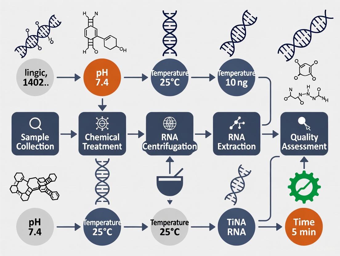
▎Experimental Workflow for Nucleic Acid Optimization
The following diagram summarizes the logical workflow for troubleshooting nucleic acid extraction and analysis from chemically challenging samples.
Frequently Asked Questions (FAQs)
1. What are the most common sources of nucleic acid degradation in a laboratory setting, and how can I prevent them? Nucleic acid degradation primarily occurs through four mechanisms: oxidation, hydrolysis, enzymatic breakdown, and physical shearing. Oxidation is triggered by exposure to heat or UV radiation, while hydrolysis occurs when water molecules break the DNA backbone, leading to depurination. Enzymatic breakdown is caused by nucleases present in biological samples, and physical shearing results from overly aggressive mechanical disruption during homogenization. Prevention strategies include adding antioxidants to samples, storing samples at -80°C in dry conditions, using nuclease inhibitors like EDTA during extraction, and optimizing homogenization parameters to balance effective disruption with DNA preservation [2].
2. My downstream applications (e.g., PCR, sequencing) are failing due to inhibitors. Which contaminants are most commonly co-precipitated with nucleic acids, and how are they removed? Common co-precipitating inhibitors include polysaccharides, polyphenols, proteins, and salts. Complex samples like soil and stool contain huge amounts of diverse interfering components that inhibit enzymatic reactions. Effective removal strategies involve using specialized lytic reagents containing phosphates and mild chaotropic agents to solubilize nucleic acids while minimizing degradation. Furthermore, novel combinations of protein-precipitating agents and tri- or tetra-valent salts can precipitate and remove these contaminants. For plant materials high in polysaccharides and polyphenols, modifying lysis buffers with CTAB or adding reducing agents like β-mercaptoethanol is effective [5] [6].
3. How can I improve my nucleic acid yield from a difficult, chemically-treated sample? Optimizing the binding conditions during solid-phase extraction is crucial for improving yield. Using a binding buffer at a lower pH (e.g., pH 4.1 instead of 8.6) reduces the negative charge on silica beads, minimizing electrostatic repulsion with negatively charged DNA and significantly enhancing binding efficiency. Furthermore, the mode of bead mixing is critical; a "tip-based" method (repeatedly aspirating and dispensing the binding mix) exposes beads to the sample more rapidly than orbital shaking, reducing the binding time from 5 minutes to 1 minute for the same efficiency and dramatically improving yield, especially for higher input amounts [7].
Troubleshooting Guides
Low Yield and Purity
| Symptom | Possible Cause | Solution |
|---|---|---|
| Low DNA/RNA yield | Inefficient cell lysis | - Use a combination of chemical (e.g., CTAB, optimized lytic reagents) and mechanical methods (e.g., bead beating).- Optimize incubation time and temperature for lysis [2] [5] [6]. |
| Suboptimal binding to purification matrix | - Lower the pH of the binding buffer (e.g., to ~4.1) [7].- Use a more efficient mixing method like tip-based mixing instead of orbital shaking [7].- Increase the amount of silica beads for higher input samples [7]. | |
| Nucleic acid degradation | - Ensure samples are flash-frozen and stored at -80°C.- Include nuclease inhibitors (e.g., EDTA, RNase inhibitors) in the lysis buffer [2] [6]. | |
| Low purity (inhibitors present) | Co-precipitation of polysaccharides/polyphenols | - Modify lysis buffer composition (e.g., add CTAB for polysaccharides, PVP or β-mercaptoethanol for polyphenols) [6].- Use purification methods that employ protein-precipitating agents and multivalent salts [5]. |
Degradation and Integrity Issues
| Symptom | Possible Cause | Solution |
|---|---|---|
| Smeared or absent bands on gel | Physical shearing during homogenization | - Use a homogenizer that allows precise control over speed, cycle duration, and temperature [2].- For sensitive samples, use specialized bead types (e.g., ceramic) and fine-tune processing parameters [2]. |
| Chemical degradation (oxidation/hydrolysis) | - Control temperature during processing to avoid excessive heat buildup [2].- Use buffered solutions to maintain a stable pH and avoid hydrolytic damage [2]. | |
| RNA is degraded | RNase contamination | - Maintain an RNase-free environment and use RNase-inhibiting reagents [6].- Process certain plant tissues, like young leaves, with shorter lysis times to minimize exposure [6]. |
Workflow Optimization Diagram
The following diagram illustrates the optimized workflow for nucleic acid extraction, integrating key steps to overcome degradation and inhibitor challenges.
Research Reagent Solutions
The following table details key reagents and materials used in optimized nucleic acid extraction protocols, along with their specific functions in addressing core challenges.
| Reagent/Material | Function & Rationale |
|---|---|
| CTAB (Cetyltrimethylammonium bromide) | A cationic detergent in lysis buffers that effectively separates polysaccharides and other contaminants from nucleic acids, crucial for plant and other complex samples [6]. |
| EDTA (Ethylenediaminetetraacetic acid) | A chelating agent that inactivates metal-dependent DNases and RNases, preventing enzymatic degradation of nucleic acids during extraction [2]. |
| Silica-coated Magnetic Beads | A solid matrix for solid-phase extraction. Binding is significantly enhanced by using a low-pH buffer and efficient "tip-based" mixing, leading to higher yields and faster processing times [7]. |
| Chaotropic Salts (e.g., Guanidine) | Denature proteins and inactivate nucleases and viruses in the sample. They also facilitate the binding of nucleic acids to silica surfaces in solid-phase extraction methods [7]. |
| Polyvinylpyrrolidone (PVP) | Binds to and helps remove polyphenols from plant extracts, preventing their oxidation and subsequent co-precipitation with nucleic acids, which can inhibit downstream applications [6]. |
| β-mercaptoethanol | A reducing agent added to lysis buffers to prevent the oxidation of phenolic compounds in plant samples, thereby improving the purity and yield of the extracted nucleic acids [6]. |
| Optimized Lytic Reagent | A composition containing phosphates and mild chaotropic agents designed to effectively solubilize nucleic acids from complex samples like soil and stool without significant degradation, while also aiding in inhibitor removal [5]. |
The Triple Protection Strategy is a simplified, semi-unified protocol for extracting DNA and RNA from diverse prokaryotic and eukaryotic sources. This approach ensures nucleic acids are safeguarded during the critical lysis step by a specific chemical environment created by Ethylenediaminetetraacetic acid (EDTA), Sodium Dodecyl Sulfate (SDS), and Sodium Chloride (NaCl) [8]. Research indicates that this combined environment is improper for RNase to render DNA free of RNA, and even for DNase to degrade the DNA, thereby preserving the integrity of the target nucleic acids from the moment of cell disruption [8]. This method is recognized for its effectiveness, reduced use of toxic materials, and the high quality of the resulting nucleic acids, making them suitable for sensitive downstream applications like PCR and RT-PCR [8].
Core Functions of the Triple Protection Components
The table below details the specific protective role of each component in the lysis buffer.
Table 1: Core Functions of the Triple Protection Reagents
| Reagent | Primary Function in Lysis | Mechanism of Action |
|---|---|---|
| EDTA (Chelating Agent) | Inhibits metalloenzymes (e.g., DNases, RNases) [8] [2] | Chelates (binds) divalent metal ions (Mg²⁺, Mn²⁺), which are essential cofactors for many nucleases [8]. |
| SDS (Detergent) | Disrupts cellular membranes and denatures proteins [9] | Solubilizes lipid bilayers and binds to proteins, causing them to unfold and inactivating enzymes including nucleases [9]. |
| NaCl (Salt) | Aids in protein precipitation and maintains ionic strength [8] | Neutralizes charges on proteins, facilitating their precipitation and removal during the phase separation step, thereby protecting nucleic acids [8]. |
Troubleshooting Common Lysis Buffer and Extraction Issues
This section addresses specific challenges researchers might encounter when working with the triple protection strategy or nucleic acid extraction in general.
FAQ 1: I suspect my extracted DNA is degraded. What are the common causes related to the lysis step?
DNA degradation can occur through several mechanisms. Inefficient lysis or protection can lead to enzymatic breakdown, while physical methods can cause shearing [2].
- Enzymatic Degradation: Incomplete inhibition of DNases is a primary cause. Ensure your lysis buffer contains fresh and adequate EDTA to chelate all metal ions. Also, verify that SDS is present at the correct concentration to denature nucleases effectively [8] [2].
- Oxidation/Hydrolysis: Exposure to environmental stressors like heat or reactive oxygen species can damage DNA. Store samples and reagents appropriately and consider the use of antioxidants [2].
- Mechanical Shearing: Overly aggressive mechanical homogenization can fragment DNA. For tough samples, balance effective disruption with DNA preservation by optimizing homogenization speed and time, and consider using specialized instruments like a bead-based homogenizer [2].
FAQ 2: My nucleic acid yield is low. How can I optimize the lysis efficiency?
Low yield often points to incomplete cell disruption or inefficient recovery of nucleic acids.
- Incomplete Lysis: The lysis buffer alone may not be sufficient for some robust sample types, like bacterial cells or tissues [9] [10]. For such challenging samples, combine chemical lysis with mechanical methods.
- Bacterial Cultures: Use lysozyme in the lysis buffer followed by freeze-thaw cycles and/or brief sonication [10].
- Tissues/Plant Matter: Use grinding in liquid nitrogen or employ a high-throughput bead homogenizer (e.g., Bead Ruptor Elite, PreOmics BeatBox) to achieve complete homogenization [8] [2] [11].
- Protein Contamination: If proteins are not effectively separated, they can coprecipitate with nucleic acids or interfere with binding. Ensure the neutral saturated salt solution (NaCl) is added correctly and the sample is mixed gently but thoroughly to precipitate proteins during the phase separation step [8].
FAQ 3: How does the protocol differ for RNA extraction to ensure RNase inhibition?
While the triple protection strategy creates a suboptimal environment for RNases, RNA requires even more stringent handling.
- DNase Treatment: After RNA is isolated and precipitated, any contaminating DNA is degraded by adding an RNase-free DNase [8].
- Critical Reagents: Use DEPC-treated water to dissolve the purified RNA pellet, which inactivates any RNases that may be present in the water [8].
- Lysis Buffer Variation: The protocol uses an acidic saturated salt solution during the phase separation step for RNA, which helps to partition RNA into the aqueous phase while leaving DNA and proteins in the interphase/organic phase [8].
Advanced Optimization and Alternative Protocols
For particularly challenging samples (e.g., bone, formalin-fixed tissues, or soil), the basic protocol may require optimization.
- Challenging Samples: Hard, mineralized tissues like bone require a combinatorial approach. This involves chemical demineralization with EDTA alongside powerful mechanical homogenization to physically break through the matrix. It is critical to balance EDTA concentration, as it is a known PCR inhibitor at high levels [2].
- Alternative Solid-Phase Methods: Magnetic nanoparticle (MNP)-based isolation is a modern, efficient alternative to traditional organic extraction. Methods using nanoparticles like NiFe₂O₄ or amine-functionalized variants offer a cost-effective, automatable, and toxic-reagent-free path to high-quality DNA [12]. These methods are particularly suited for high-throughput applications and can be optimized with specific binding and elution buffers (e.g., Tris-HCl, Phosphate Buffer) [12].
Table 2: Comparison of Nucleic Acid Isolation Technologies
| Method | Mechanism | Key Advantages | Suitable Downstream Applications |
|---|---|---|---|
| Triple Protection (Organic/Salt-Precipitation) | Chemical cell lysis followed by salt-induced protein precipitation and alcohol precipitation of nucleic acids [8]. | Effective for diverse sample types; semi-unified for DNA/RNA; cost-effective [8]. | PCR, RT-PCR, cloning [8]. |
| Magnetic Bead-Based | Nucleic acids bind to surface-coated paramagnetic beads in high-salt buffer and are released in low-salt or water [12] [13]. | Amenable to automation; high purity; no centrifugation; no toxic reagents [12] [13]. | NGS, PCR, qPCR, sequencing [12] [13]. |
| Silica Column-Based | Nucleic acids bind to a silica membrane in high-salt buffer and are eluted in low-salt buffer or water [13]. | Well-established; consistent results; good for multiple sample types. | NGS, PCR, qPCR, cloning [13]. |
| Rapid Extraction Buffers | Simple lysis and inactivation of nucleases using specialized buffer solutions [13]. | Extremely fast (minutes); simple protocol; high-throughput [13]. | End-point PCR, genotyping [13]. |
Experimental Protocol: Hepatic DNA Extraction from Mouse
This is a detailed methodology for extracting DNA from mouse liver, as outlined in the primary source [8].
Materials and Reagents:
- Lysis Buffer: 1X STE (50 mM NaCl, 50 mM Tris-HCl, 100 mM EDTA; pH 8.0)
- 10% SDS solution
- Proteinase K
- Neutral saturated salt solution (NaCl)
- 100% and 70% Ethanol
- RNase A
- Tris-EDTA (TE) Buffer or DD water
Procedure:
- Homogenization: Take 1g of liver tissue, cut it into pieces, and grind it using a mortar and pestle in 3 ml of lysis buffer containing 900 µl of 10% SDS. Transfer the emulsion to a micro-centrifuge tube. Add 100 µg of Proteinase K per ml of emulsion and incubate for 1 hour at 50°C [8].
- Phase Separation: Add 350 µl of neutral saturated salt solution (NaCl) per ml of emulsion. Cap the tube and shake gently by hand for 15 seconds. Incubate at room temperature for 10 minutes. Centrifuge at 590 × g for 15 minutes at room temperature. The DNA will be in the clear aqueous phase (upper layer) [8].
- DNA Precipitation: Transfer the aqueous phase to a new tube. Mix with two volumes of room-temperature absolute ethanol. Invert the tube several times until DNA precipitates (becomes visible as a stringy or cloudy mass) [8].
- DNA Wash: Remove the supernatant. Wash the DNA pellet once with 1 ml of 75% ethanol. Centrifuge at 9500 × g for 5 minutes to reprecipitate the DNA. Carefully discard the supernatant [8].
- DNA Dissolving: Air-dry the pellet for 5 minutes. Dissolve the DNA in an appropriate volume of DD water or TE buffer. Quantify the DNA and store at -20°C [8].
- Removal of RNA: To the dissolved DNA, add 50 µg per ml of RNase A and incubate for 1 hour at 37°C to remove any residual RNA contamination [8].
The Scientist's Toolkit: Essential Research Reagent Solutions
Table 3: Key Reagents for Nucleic Acid Extraction and Analysis
| Reagent / Tool | Function | Application Notes |
|---|---|---|
| EDTA | Nuclease inhibition via metal ion chelation [8] [2]. | Critical component of the "triple protection"; concentration must be sufficient to inactivate all cellular nucleases. |
| SDS | Membrane disruption and protein denaturation [8] [9]. | Works synergistically with EDTA and Proteinase K to dismantle cellular structures and inactivate enzymes. |
| NaCl (Saturated Solution) | Protein precipitation and charge neutralization [8]. | The use of neutral vs. acidic saturated salt solution is a key differentiator between DNA and RNA protocols [8]. |
| Proteinase K | Broad-spectrum protease digestion of cellular proteins [8]. | Essential for degrading nucleases and other proteins; incubation at 50-60°C enhances its activity. |
| RNase A / DNase I | Removal of unwanted nucleic acids [8]. | Added after initial extraction and precipitation to obtain pure DNA or RNA, free of contamination [8]. |
| Magnetic Nanoparticles (e.g., NiFe₂O₄) | Solid-phase nucleic acid binding for purification [12]. | Enable rapid, automatable, and toxic-reagent-free isolation of high-quality DNA and RNA for sensitive applications like NGS [12]. |
Visualizing the Triple Protection Workflow and Mechanisms
The following diagrams illustrate the logical workflow of the extraction process and the protective mechanism of the lysis buffer components.
Diagram 1: DNA Extraction Workflow. This flowchart outlines the key steps in the triple protection protocol for DNA extraction, from homogenization to the final pure product.
Diagram 2: Triple Protection Mechanism. This diagram shows how the components of the lysis buffer work in concert to neutralize key threats to nucleic acid integrity during cell lysis.
Core Concepts: The Four Fundamental Steps
The purification of nucleic acids is a critical first step in molecular biology. The process can be broken down into four essential stages, each with a specific goal, to ensure the yield and purity required for downstream applications [8].
- 1. Lysis: This initial step involves disrupting the cellular structure to create a lysate and release the nucleic acids into solution. Methods can be physical (e.g., grinding, bead beating), chemical (e.g., detergents, chaotropic salts), or enzymatic (e.g., Proteinase K, lysozyme) [14] [8].
- 2. Dehydration and Protein Denaturation: Cellular proteins are dehydrated and precipitated out of the solution. This is often achieved using high-concentration salt solutions, which cause proteins to fall out of solution, protecting the nucleic acids from nucleases [14] [8].
- 3. Separation: The soluble nucleic acid is separated from the precipitated cellular proteins and other insoluble debris. This is commonly accomplished by centrifugation, filtration, or bead-based methods [14] [8].
- 4. Precipitation: The nucleic acids are forced out of the solution, typically by adding alcohol (e.g., ethanol or isopropanol). The insoluble nucleic acid pellet is then collected via centrifugation, washed, and dissolved in an aqueous buffer like Tris-EDTA or nuclease-free water [14] [8].
This workflow can be visualized as follows:
Troubleshooting Guide for Common Nucleic Acid Purification Issues
This guide addresses common challenges encountered during nucleic acid purification, specifically framed within the context of working with chemically treated samples, which can introduce unique complications.
Table 1: Troubleshooting Common Purification Problems
| Problem | Possible Cause | Solution |
|---|---|---|
| Low Yield | Incomplete lysis due to robust cell walls in chemically treated samples [15] [16]. | Optimize lysis by combining mechanical disruption (bead beating) with enzymatic digestion (Proteinase K) [14] [15]. |
| Column overload or clogging from excessive biomass or indigestible fibers [15]. | Reduce the input material to the recommended amount. For fibrous tissues, centrifuge the lysate to remove debris before binding [15]. | |
| Inefficient binding of nucleic acids to the purification matrix [16]. | Ensure the binding buffer is fresh and at the correct pH. Verify that ethanol was added to the binding buffer as specified [17]. | |
| DNA Degradation | Nuclease activity in DNase-rich tissues (e.g., liver, spleen) exacerbated by chemical treatment [15]. | Flash-freeze samples in liquid nitrogen and store at -80°C. Keep samples on ice during preparation and use nuclease inhibitors [15]. |
| Improper sample storage or overly large tissue pieces [15]. | Cut tissue into the smallest possible pieces and store samples properly with stabilizers like RNAlater [15]. | |
| Protein Contamination | Incomplete digestion of proteins, especially in fixed or stabilized samples [15]. | Extend Proteinase K digestion time (30 min to 3 hours) after the tissue appears dissolved. Ensure the lysis buffer is appropriate [15]. |
| Membrane clogged with tissue fibers or protein complexes [15]. | Centrifuge the lysate at maximum speed for 3 minutes to pellet fibers before loading onto the column [15]. | |
| Salt Contamination | Carryover of guanidine salts from the binding or wash buffers [15] [18]. | Perform additional wash steps with 70-80% ethanol. Ensure wash buffer is completely removed before elution [17] [18]. |
| Splashing of the lysate/buffer mixture into the column cap or upper area [15]. | Pipette carefully onto the center of the membrane, avoid transferring foam, and close caps gently [15]. | |
| RNA Contamination in DNA Prep | Co-purification of RNA, leading to inflated DNA quantification [14]. | Add RNase A (e.g., to the elution buffer) during or after the purification process to digest RNA [14] [8]. |
| gDNA Contamination in RNA Prep | Insufficient shearing of genomic DNA during homogenization [18]. | Use a high-velocity bead beater or polytron for homogenization. Include a DNase digestion step, either on-column or in-solution [18]. |
The logical flow for diagnosing and resolving these common issues is summarized below:
Special Considerations for Chemically-Treated and FFPE Samples
Research involving formalin-fixed, paraffin-embedded (FFPE) or other chemically treated samples presents specific challenges. The fixation process creates crosslinks between proteins, nucleic acids, and other biomolecules, which must be reversed for successful extraction [19].
- Optimized Lysis: Standard lysis is insufficient. A robust proteolytic digestion using Proteinase K under defined temperatures is crucial to break crosslinks and free nucleic acids [19].
- Severe Conditions for DNA: Due to its higher stability, DNA isolation from FFPE samples often requires more severe conditions, such as longer incubation times and higher temperatures, which can serendipitously help remove more crosslinks [19].
- Handling Fragmented Nucleic Acids: Both RNA and DNA from FFPE samples will be fragmented. Downstream assays must be designed accordingly, typically targeting shorter amplicons (e.g., 60-70 bp) for successful amplification [19].
Table 2: Comparison of Two Common FFPE Nucleic Acid Extraction Methodologies
| Feature | RecoverAll Total Nucleic Acid Isolation Kit | MagMAX FFPE DNA/RNA Ultra Kit |
|---|---|---|
| Deparaffinization | Required (uses xylene or substitute and ethanol) [19]. | Not required; direct incubation in proteolytic solution [19]. |
| Digestion Format | Performed in a tube with buffer and protease [19]. | Performed in a 96-well plate with buffer, protease, and a wax-penetrating additive [19]. |
| Isolation Method | Filter-based spin column (glass-fiber filter) [19]. | Bead-based (magnetic beads) [19]. |
| Throughput | Lower (e.g., 40 reactions per kit) [19]. | Higher, amenable to automation (e.g., 96 reactions per kit) [19]. |
| Best For | Lower throughput, manual processing [19]. | High-throughput labs, automated platforms like KingFisher [19]. |
Optimization and Advanced Techniques
Magnetic Bead-Based Extraction
Magnetic beads-based nucleic acid extraction is widely used in automated systems and can be optimized for efficiency.
- Speed Variation: Using a single, slow mixing speed can result in lower RNA extraction efficiency. Implementing varied speeds (slow, moderate, fast) during the mixing process significantly increases positivity rates and lowers Ct values in downstream RT-PCR, as it enhances the capture and mixing processes [20].
- Heating Step: For some magnetic beads-based kits, skipping the heating step during lysis and elution does not significantly reduce extraction quality, simplifying the protocol [20].
Heat-Based Extraction
The heat-shock method is a simple and effective alternative, especially in resource-limited settings.
- Methodology: Heating samples at 90-95°C for 5 minutes disrupts cells to release nucleic acids [20].
- Separation without Centrifugation: While centrifugation gives the best results, cell debris can also be separated by allowing samples to cool or simply settling at room temperature, making this a feasible method in laboratories without centrifugation facilities [20].
Frequently Asked Questions (FAQs)
Q1: My nucleic acid yields are consistently low, but my samples are fresh. What is the most likely cause? The most common cause of low yields is incomplete lysis or homogenization [16] [18]. Ensure your lysis method is appropriate for your sample type. For tough tissues, use a combination of physical disruption (e.g., grinding in liquid nitrogen) and enzymatic digestion. Also, verify that the binding conditions are correct, including the freshness and concentration of the ethanol used [17].
Q2: How can I tell if my DNA sample is contaminated with salt, and how do I fix it? Salt contamination is indicated by a low A260/A230 ratio (<2.0) in spectrophotometric measurements [15] [18]. To resolve this, ensure wash buffers are prepared correctly and perform an additional wash step with 70-80% ethanol. Be careful during pipetting to avoid splashing buffer into the column cap, as this is a common source of salt carryover [15].
Q3: I am working with pancreas tissue, and my DNA is always degraded. What specific steps should I take? Organs like the pancreas, liver, and intestine are rich in nucleases. To prevent degradation:
- Flash-freeze the tissue immediately after collection in liquid nitrogen and store at -80°C.
- During processing, keep the tissue frozen on ice.
- Cut the tissue into the smallest possible pieces before adding it to the lysis buffer.
- Ensure your lysis buffer contains chaotropic salts (e.g., guanidine) to inactivate nucleases immediately upon contact [14] [15].
Q4: For downstream applications like long-range PCR, what is the best practice for eluting DNA? For applications requiring high-molecular-weight DNA, elution in a slightly basic buffer (e.g., 10 mM Tris-HCl, pH 8-9) is superior to water. DNA is more stable at a basic pH and will dissolve faster and more completely in a buffer. Allow the elution buffer to stand on the silica membrane for a few minutes before centrifugation to maximize recovery [17].
The Scientist's Toolkit: Essential Reagent Solutions
Table 3: Key Reagents for Nucleic Acid Extraction and Their Functions
| Reagent | Function |
|---|---|
| Chaotropic Salts(e.g., Guanidine HCl, Guanidine thiocyanate) | Denature proteins and nucleases; disrupt hydrogen bonding in water to facilitate nucleic acid binding to silica [14] [17]. |
| Proteinase K | A broad-spectrum serine protease that digests proteins and aids in lysing tough tissues; works optimally under denaturing conditions [14] [17]. |
| EDTA (Ethylenediaminetetraacetic acid) | Chelates divalent cations (Mg²⁺, Ca²⁺) that are cofactors for nucleases, thereby protecting nucleic acids from degradation [21] [8]. |
| SDS (Sodium Dodecyl Sulfate) | An ionic detergent that solubilizes cellular membranes and denatures proteins, aiding in cell lysis [14] [8]. |
| RNase A | An enzyme that degrades RNA; used to remove RNA contamination from DNA preparations [14] [8]. |
| DNase I (RNase-free) | An enzyme that degrades DNA; used to remove genomic DNA contamination from RNA preparations [8] [18]. |
| Beta-Mercaptoethanol (BME) | A reducing agent that helps inactivate RNases by breaking disulfide bonds, particularly critical for stabilizing RNA during extraction [18]. |
| Silica Matrix / Magnetic Beads | The solid phase that selectively binds nucleic acids in the presence of chaotropic salts and high concentrations of ethanol, allowing for purification from other cellular components [14] [17]. |
Optimized Protocols and Novel Methods for Challenging Sample Types
Research Reagent Solutions
The following table details key reagents and materials essential for implementing the semi-unified nucleic acid extraction protocol, along with their primary functions. [21]
| Reagent/Material | Function in the Protocol |
|---|---|
| EDTA (Ethylenediaminetetraacetic acid) | Chelates divalent cations (Mg²⁺), which are essential cofactors for DNase and RNase enzymes, protecting nucleic acids from degradation. |
| SDS (Sodium Dodecyl Sulfate) | A detergent that disrupts cellular membranes (lysis) and denatures proteins, facilitating the release of nucleic acids. |
| NaCl (Sodium Chloride) | Provides the appropriate ionic strength to shield the negative charges on the phosphate backbone of nucleic acids, preventing aggregation and aiding in stability. |
| RNase A | An enzyme added after DNA extraction to degrade any residual RNA contamination, yielding pure DNA. |
| DNase I | An enzyme used during RNA isolation to degrade DNA contaminants, resulting in high-quality RNA. |
| Magnetic Nanoparticles (e.g., NiFe₂O₄) | A modern, cost-effective solid phase for binding and purifying nucleic acids from complex matrices, reducing the use of toxic reagents. [22] |
| Lysis Buffer | A mixture typically containing EDTA, SDS, and NaCl, creating the "triple protection" environment for nucleic acids during cell disruption. |
| Elution Buffer | A low-salt solution (e.g., Tris-EDTA buffer or nuclease-free water) used to release purified nucleic acids from silica columns or magnetic beads. |
Troubleshooting Guide: Common Issues and Solutions
This section addresses specific challenges you might encounter when using semi-unified protocols, especially with chemically-treated samples.
FAQ 1: My nucleic acid yield is consistently low. What could be the cause?
- A: Low yield can stem from several points in the protocol. The table below outlines common causes and recommended solutions. [23]
| Problem Cause | Solution |
|---|---|
| Inadequate Lysis | Solution: Optimize lysis conditions. For tough cell walls (e.g., gram-positive bacteria, plant cells), incorporate mechanical disruption (bead beating) or extended enzymatic digestion (lysozyme, proteinase K) in addition to the standard SDS-based chemical lysis. [21] [23] |
| Carryover of Inhibitors | Solution: Ensure thorough washing steps. If using spin columns, do not reduce wash buffer volumes. For magnetic bead-based protocols, ensure complete resuspension of beads during washes. Adding an extra wash step can be beneficial for complex samples like blood or soil. [23] |
| Inefficient Binding | Solution: Verify the pH and composition of the binding buffer. Ensure the sample is adequately mixed with the buffer. If using magnetic nanoparticles, optimize the incubation time and mixing frequency to maximize contact. [22] [23] |
FAQ 2: My extracted nucleic acids are degraded. How can I prevent this?
- A: Degradation is primarily caused by nucleases. The semi-unified protocol's "triple protection" lysis environment (EDTA, SDS, NaCl) is designed to inhibit these enzymes. [21] For additional protection:
- Work quickly and on ice whenever possible.
- Use nuclease-free tubes and tips.
- For RNA-specific work, use dedicated RNase-free reagents and consider adding a specific RNase inhibitor.
- Store extracts at -80°C (especially for RNA) and avoid repeated freeze-thaw cycles. [23]
FAQ 3: My downstream applications (PCR, RT-PCR) are failing due to contaminated nucleic acids. What should I do?
- A: Contamination can be from co-purified biomolecules or cross-sample contamination.
- DNA contamination in RNA preps (and vice-versa): This is addressed directly in the semi-unified protocol. Always add DNase I during RNA isolation and RNase A after DNA extraction to obtain pure nucleic acids. [21]
- Protein or Salt Carryover: Perform the recommended wash steps thoroughly and ensure the final eluate does not contain ethanol from wash buffers.
- Cross-Contamination: Use aerosol-resistant pipette tips and change gloves frequently. For high-throughput labs, automated nucleic acid extraction systems can significantly reduce this risk. [23]
FAQ 4: How can I adapt this protocol for samples treated with harsh chemicals?
- A: Chemical treatments can cross-link or fragment nucleic acids.
- Extended Lysis: Increase lysis incubation times and consider using higher concentrations of proteinase K to reverse formaldehyde cross-links.
- Additional Purification: If inhibitors from the chemical treatment persist, perform a post-extraction purification using magnetic bead-based clean-up protocols, which are effective for removing a wide range of contaminants. [22]
- Quality Control: Always assess the integrity of extracted nucleic acids from chemically treated samples via gel electrophoresis or a bioanalyzer before proceeding to expensive downstream applications. [23]
Experimental Protocol: Core Methodology
This section provides the detailed, step-by-step methodology for the semi-unified nucleic acid extraction protocol.
Lysis under "Triple Protection"
- Principle: The combined action of EDTA, SDS, and NaCl creates an environment that physically disrupts cells and chemically inactivates nucleases, preserving nucleic acid integrity. [21]
- Procedure:
- Resuspend cell pellet or tissue sample in 500 µL of lysis buffer (e.g., 50 mM EDTA, 1% SDS, 100 mM NaCl, pH 8.0).
- For prokaryotic cells with robust walls, add 10 µL of lysozyme (10 mg/mL) and incubate at 37°C for 15-30 minutes.
- Add 10 µL of proteinase K (20 mg/mL) and incubate at 55-60°C for 30-60 minutes or until the solution clears. Vortex intermittently.
- Add 200 µL of 5 M NaCl to further precipitate proteins and polysaccharides. Mix thoroughly.
Separation and Purification
- Principle: Nucleic acids are separated from cellular debris, proteins, and other contaminants.
- Procedure (Organic Extraction Method):
- Add an equal volume of phenol:chloroform:isoamyl alcohol (25:24:1) to the lysate. Vortex vigorously.
- Centrifuge at >12,000 × g for 5 minutes at 4°C.
- Carefully transfer the upper aqueous phase (containing nucleic acids) to a new tube.
- Add an equal volume of chloroform to remove residual phenol. Vortex and centrifuge as before. Transfer the aqueous phase to a new tube.
- Procedure (Solid-Phase Method - Column or Magnetic Beads):
- Mix the lysate with a binding buffer (e.g., containing guanidinium thiocyanate) that promotes nucleic acid binding to a silica surface.
- Transfer the mixture to a spin column or add functionalized magnetic nanoparticles. [22]
- Centrifuge the column or use a magnet to capture the beads. Discard the flow-through.
Washing
- Principle: Remove salts, solvents, and other impurities while the nucleic acids remain bound.
- Procedure:
- Wash the silica membrane or magnetic beads twice with 700 µL of a wash buffer (typically ethanol-based).
- Centrifuge or use a magnet to ensure all wash buffer is removed. A final "dry" spin is recommended for columns to evaporate residual ethanol.
Elution
- Principle: Release pure nucleic acids from the solid phase into an aqueous buffer.
- Procedure:
- Add 50-100 µL of elution buffer (TE buffer or nuclease-free water) to the center of the silica membrane or to the dried magnetic beads.
- Incubate at room temperature for 2-5 minutes to allow for complete rehydration and elution.
- Centrifuge (for columns) or use a magnet (for beads) to collect the purified nucleic acid eluate.
Post-Extraction Treatment for Purity
- For DNA purification: Add 2 µL of RNase A (10 mg/mL) to the eluted DNA. Incubate at 37°C for 15 minutes. [21]
- For RNA purification: Add 2 µL of DNase I (1 U/µL) to the eluted RNA. Incubate at 37°C for 15 minutes, then inactivate the enzyme (e.g., with EDTA or heat). [21]
Protocol Workflow Visualization
The following diagram illustrates the logical workflow and decision points within the semi-unified nucleic acid extraction protocol.
Advanced Method: Magnetic Nanoparticle Workflow
For a modern, cost-effective, and less toxic approach, magnetic nanoparticles can be integrated into the purification step. The following diagram details this advanced workflow. [22]
This guide provides targeted troubleshooting for researchers optimizing nucleic acid extraction from chemically-treated samples. The methods detailed below—mechanical, enzymatic, and chaotropic salt-based lysis—are critical for achieving high yields of pure, intact nucleic acids for sensitive downstream applications in drug development and molecular diagnostics.
Lysis Method Selection Guide
Choosing the appropriate lysis technique is paramount for sample integrity. The following workflow diagram outlines the key decision process for selecting an optimal method based on your sample type and research goals.
Troubleshooting Common Lysis Problems
FAQ: Why is my nucleic acid yield low after mechanical lysis?
Potential Causes and Solutions:
- Incomplete Cell Disruption: Ensure your method matches the cell wall toughness. Bacterial spores and plant tissues require more aggressive techniques like bead beating.
- Nucleic Acid Degradation during Lysis: Mechanical methods can generate heat. Always use cooling intervals during sonication or bead beating, and include nuclease inhibitors in your buffer [24] [25].
- Inefficient Binding to Purification Matrix: After lysis, the released nucleic acids must bind to silica columns or beads. Verify that the correct concentration of chaotropic salts and ethanol is used, as this is critical for efficient binding [17].
FAQ: My protein recovery is low or the protein is inactive after enzymatic lysis. What went wrong?
Potential Causes and Solutions:
- Enzyme Incompatibility: The lytic enzyme must match the cell wall composition. Use lysozyme for bacterial peptidoglycan, lyticase for fungal glucans, or cellulase for plant cells [24].
- Suboptimal Reaction Conditions: Enzymatic lysis is sensitive to buffer conditions (pH, temperature, osmotic stability). For protoplast formation, the lysis buffer must be isotonic, often achieved by adding sugars like sorbitol or mannitol, to prevent premature rupture [26].
- Co-purifying Nucleic Acids Increasing Viscosity: The released chromosomal DNA can create a viscous lysate that traps proteins, reducing yield. Add a nuclease like Benzonase to the lysis buffer to digest nucleic acids and reduce viscosity [25].
FAQ: How do I choose between urea and guanidinium salts for chaotropic lysis?
Guanidinium salts (e.g., GdmCl, GdmSCN) and urea are potent denaturants but have different strengths. The table below summarizes their distinct properties to guide your selection.
Table 1: Comparison of Chaotropic Salts for Lysis and Denaturation
| Chaotropic Salt | Common Concentrations | Mechanism of Action | Preferred For | Considerations |
|---|---|---|---|---|
| Guanidinium HCl (GdmCl) | 4-6 M [27] | Competes for hydrogen bonds; disrupts hydrophobic interactions; stacks with aromatic groups [27]. | Preferentially denatures and disrupts proteins rich in alpha-helices [27]. | Highly effective for inactivating nucleases during nucleic acid extraction [17]. |
| Guanidinium Thiocyanate (GdmSCN) | 2-4 M | Similar to GdmCl, but the thiocyanate anion is highly chaotropic, enhancing potency [28]. | Rapid and complete denaturation of DNA and proteins; a key component in RNA stabilization reagents (e.g., TRIzol) [29]. | The most potent denaturant among guanidinium salts; can fully denature DNA origami structures at 2 M and 50°C [28]. |
| Urea | 6-9 M [27] | Disrupts the hydrogen-bonding network of water, leading to solvation of hydrophobic residues; can also form direct hydrogen bonds [27]. | Preferentially denatures and disrupts proteins rich in beta-sheets [27]. | Can form cyanate ions at high temperatures, which can carbamylate proteins. Use fresh solutions. |
Essential Research Reagent Solutions
A properly formulated lysis buffer is more than just a denaturant. The table below lists key components and their functions for effective and reliable cell lysis.
Table 2: Key Components of a Lysis Buffer and Their Functions
| Reagent Category | Example Components | Primary Function | Application Notes |
|---|---|---|---|
| Buffering Agents | Tris-HCl, HEPES, Phosphate buffers | Maintain stable pH during lysis, crucial for biomolecule stability [26] [30]. | Avoid amine-based buffers (e.g., Tris) in cross-linking reactions [26]. |
| Chaotropic Salts | Guanidine HCl, Guanidine Thiocyanate, Urea | Denature proteins, inactivate nucleases, and facilitate nucleic acid binding to silica [17] [27]. | Concentration determines denaturing strength; GdmSCN is most potent [28]. |
| Detergents | SDS (ionic), Triton X-100 (non-ionic), CHAPS (zwitterionic) | Solubilize lipid membranes and proteins [24] [30]. | Ionic detergents (SDS) fully denature; non-ionic are milder and preserve protein function [30]. |
| Reducing Agents | Dithiothreitol (DTT), β-mercaptoethanol | Break disulfide bonds in proteins, aiding denaturation and preventing aggregation [26] [30]. | Add fresh before use as they oxidize in solution. |
| Protease Inhibitors | PMSF, EDTA, Protease Inhibitor Cocktails | Prevent proteolytic degradation of target proteins [26] [30]. | PMSF targets serine proteases; EDTA inhibits metalloproteases [26]. |
| Nucleases | Benzonase, DNase I, RNase A | Digest unwanted nucleic acids to reduce lysate viscosity and prevent co-purification [25]. | Essential for streamlining protein purification and improving chromatography [25]. |
| Osmotic Stabilizers | Sucrose, Sorbitol, Mannitol | Maintain osmotic balance to protect organelles or create protoplasts during gentle lysis [26]. | Critical for enzymatic lysis of yeast/fungi (1M sorbitol) and plant cells (mannitol) [26]. |
Optimized Protocol: Combined Mechanical and Chaotropic Lysis for Soil DNA
This protocol, adapted from a comparative study, is effective for difficult samples like chemically-treated soils or sediments [31].
Principle: Brief, low-speed bead milling provides physical disruption, while a buffered SDS-chloroform mixture ensures chemical lysis. This combination maximizes DNA yield and minimizes shearing.
Procedure:
- Sample Preparation: Lyophilize 100 mg of soil or sediment and grind to a fine powder with a mortar and pestle [31].
- Bead Mill Homogenization: Transfer the powder to a tube with lysis beads. Add 1-2 mL of pre-cooled Phosphate-Tris Lysis Buffer (pH 8.0) containing:
- 100 mM Sodium Phosphate
- 100 mM Tris-HCl
- 2% (w/v) SDS
- 100 mM NaCl Homogenize at low speed for 30-120 seconds. Monitor temperature to avoid overheating [31].
- Chemical Lysis and Extraction: Add an equal volume of chloroform to the homogenate. Mix thoroughly and centrifuge at 10,000 x g for 15 minutes at 4°C to separate the phases [31].
- DNA Recovery: Carefully transfer the upper aqueous phase to a new tube. The DNA can be purified using a commercial silica column kit or by Sephadex G-200 spin column chromatography to remove PCR-inhibiting humic substances [31].
Solid-Phase Extraction (SPE) is a foundational sample preparation technique critical for purifying and concentrating analytes from complex matrices. In modern nucleic acid research, particularly for chemically-treated samples, optimizing SPE is essential for obtaining high-quality DNA and RNA for downstream applications like sequencing and PCR. The technique has evolved from traditional silica membranes to advanced magnetic bead-based methods, each with distinct advantages and operational challenges. This technical support center addresses the most common experimental hurdles researchers face, providing targeted troubleshooting and methodological guidance to enhance extraction efficiency, reproducibility, and nucleic acid recovery from challenging sample types.
Troubleshooting Common Solid-Phase Extraction Issues
Frequently Asked Questions (FAQs) and Solutions
Q1: What is the most common cause of low analyte recovery in SPE, and how can I fix it? Low recovery often stems from improper sorbent choice, inadequate elution conditions, or the cartridge drying out. Ensure the sorbent chemistry matches your analyte's polarity and charge state. For elution, increase solvent strength or volume; for ionizable analytes, adjust pH to neutralize the charge. Always prevent sorbent beds from drying before sample loading by re-conditioning if necessary [32] [33].
Q2: My flow rate is inconsistent. What should I check? Flow rate variations are frequently caused by particulate clogging, high sample viscosity, or uneven sorbent packing. Always filter or centrifuge samples to remove particulates. For viscous samples, dilute with a weak, matrix-compatible solvent. Using a controlled vacuum manifold or pump can standardize flow rates across samples [32].
Q3: How do I know if I've exceeded my SPE cartridge's capacity? Sorbent overload leads to analyte breakthrough and loss. As a general rule, silica-based sorbents have a capacity of ~5% of their mass, while polymeric sorbents can hold up to ~15%. For example, a 100 mg C18 cartridge can bind approximately 5 mg of analyte. If you suspect overload, reduce the sample load or use a cartridge with higher capacity [32].
Q4: Why is my cleanup insufficient, with many co-extracted impurities? This typically indicates a suboptimal purification strategy or wash solvent strength. For targeted analysis, choose a mode that retains your analyte and selectively washes out impurities. Re-optimize your wash conditions—small changes in organic percentage or pH can significantly improve selectivity. Using a more selective sorbent (e.g., ion-exchange) can also enhance cleanup [32].
Q5: Can I reuse magnetic beads? Reuse is only acceptable when cross-sample contamination is not a concern, such as in repeated purifications of the same sample. For most applications, especially with different biological samples, use fresh beads to prevent carryover contamination and ensure consistent binding capacity [34].
Troubleshooting Guide Table
The following table summarizes common SPE problems, their causes, and recommended solutions for efficient problem-solving.
Table 1: Solid-Phase Extraction Troubleshooting Guide
| Problem | Primary Causes | Recommended Solutions |
|---|---|---|
| Low Recovery [32] [33] | - Incorrect sorbent choice- Weak elution solvent- Insufficient elution volume- Column dried out | - Match sorbent chemistry to analyte (RP, Ion-Exchange, Normal-Phase)- Increase organic % in eluent or adjust its pH- Increase elution volume; use multiple fractions- Keep sorbent wet; re-condition if dried |
| Poor Reproducibility [32] | - Variable flow rates during loading- Column bed dried out- Wash solvent too strong | - Use a manifold or pump to control flow rate (<5 mL/min)- Ensure proper conditioning/equilibration before loading- Weaken wash solvent strength; avoid prolonged soaking |
| Slow Flow Rate [32] [33] | - Particulate clogging- High sample viscosity- Overly dense sorbent packing | - Filter or centrifuge sample pre-loading; use a prefilter- Dilute sample with weak solvent to reduce viscosity- Apply gentle positive pressure if not clogged |
| Incomplete Cleanup [32] | - Incorrect purification strategy- Poorly chosen wash solvents | - Retain analyte and wash impurities, not vice-versa- Re-optimize wash solvent composition, pH, and ionic strength |
| Magnetic Beads Not Pelleting [35] | - Solution too viscous- Bead aggregation | - Increase separation time on magnet (2-5 mins)- Add Tween 20 (~0.05%) or DNase I to lysate |
Optimized Experimental Protocols
Optimized Two-Phase SPE for Urinary Nucleic Acid Adducts
Background: This protocol is designed for the untargeted analysis of the urinary nucleic acid adductome, which requires maximal recovery of a wide spectrum of chemically diverse adducts. Single-phase SPE is often insufficient for this purpose [36].
Methodology:
- SPE Sorbent Combination: The optimized combination uses ENV+ cartridges coupled with Phenyl (PHE) cartridges. This two-phase system provides superior retention for a cocktail of 20 different nucleic acid adduct standards compared to any single sorbent [36].
- Sample Preparation: Urine samples are spiked with a cocktail of isotopically labeled internal standards (e.g., at a final concentration of 1 µg/mL) to monitor recovery and aid in identification [36].
- Conditioning and Loading: Condition the combined sorbents as per manufacturer recommendations. Load the urine sample at a controlled, slow flow rate to ensure optimal binding.
- Washing and Elution: Wash with a solvent that removes matrix interferences without eluting the target adducts. Elute with a solvent of sufficient strength and volume to disrupt analyte-sorbent interactions completely.
- Analysis: The eluate is analyzed using untargeted high-resolution mass spectrometry. Data processing with software like FeatureHunter 1.3 can identify approximately 500 distinct adduct features in mouse and human urine samples, demonstrating the method's effectiveness [36].
Acid-Activated Bentonite (ASAB) for Pathogenic Samples
Background: This innovative SPE method uses modified clay to overcome limitations of traditional silica-based kits, such as suboptimal recovery and chaotropic ion inhibition. It is highly versatile for DNA, RNA, and miRNA from various samples [37].
Methodology:
- Matrix Fabrication:
- Acid Activation: Treat raw bentonite with sulfuric acid. This process increases its surface area by 2.2 times, creating more binding sites [37].
- Amino-Functionalization: Modify the sulfuric acid-activated bentonite (SAB) with 3-aminopropyl(diethoxy)methylsilane (APDMS) to create ASAB [37].
- Cross-linking: Use a homobifunctional imidoester (HI) reagent to create reversible cross-links between the amine groups on ASAB and the nucleic acids, enabling pH-dependent binding and elution [37].
- Extraction Protocol:
- Binding: Mix the sample with the ASAB-HI complex at a pH that facilitates binding. The system can handle large volumes (up to 50 mL) to enrich trace pathogens [37].
- Washing: Wash away contaminants with an appropriate buffer.
- Elution: Elute the purified nucleic acids using an elution buffer at a specific pH that breaks the reversible cross-links [37].
- Performance: This method demonstrated a 3.95-fold increase in DNA recovery from human urine and plasma and a 6.3-fold improvement in unstable viral RNA isolation from clinical swabs compared to a commercial silica-based SPE kit [37].
Magnetic Beads Optimization for Nucleic Acids
Background: Magnetic beads are widely used for high-throughput nucleic acid purification. Their performance depends on proper handling and buffer optimization.
Methodology:
- Bead Handling:
- Resuspension: Always vortex magnetic beads thoroughly before use to redisperse settled particles [34].
- Storage: Store beads at 2-8°C or room temperature per instructions. Do not freeze, as freezing can crack the beads and destroy their magnetic and binding properties [34].
- Pelleting: If beads do not pellet, increase separation time on the magnet to 2-5 minutes. For viscous solutions or aggregates, add Tween 20 (up to 0.1%) or DNase I to the lysate [35].
- Binding Capacity Optimization: The binding capacity is length-dependent. For large fragments (>2 kb), expect reduced capacity due to steric hindrance.
- Elution: For efficient elution, resuspend the bead-nucleic acid complex completely in a low-ionic-strength buffer (e.g., water or Tris-HCl) and incubate at 65°C for >5 minutes. Using pre-heated elution buffer can improve yield [35].
The Scientist's Toolkit: Essential Research Reagents
Table 2: Key Reagent Solutions for Nucleic Acid SPE
| Reagent/Material | Function in SPE Optimization |
|---|---|
| ENV+ & PHE Sorbents [36] | A two-phase SPE combination that provides broad retention for diverse nucleic acid adducts in untargeted adductomics. |
| Acid-Activated Bentonite (ASAB) [37] | A high-surface-area SPE matrix for efficient, versatile extraction of DNA, RNA, and miRNA from various sample types. |
| Homobifunctional Imidoester (HI) [37] | A cross-linker for pH-dependent, reversible binding of nucleic acids to amine-functionalized surfaces like ASAB. |
| Isotopically Labeled Standards [36] | Internal standards used to quantitatively monitor analyte recovery and identify endogenous adducts in mass spectrometry. |
| Magnetic Beads (Streptavidin) [35] | Paramagnetic particles with a streptavidin coating for highly specific capture of biotinylated nucleic acids or other molecules. |
| Tween 20 [35] | A non-ionic detergent added to buffers to reduce nonspecific binding and bead aggregation, improving purity and recovery. |
Workflow and Pathway Diagrams
Two-Phase SPE Optimization Workflow
ASAB-SPE Nucleic Acid Extraction Pathway
Magnetic Bead Purification Logic
In nucleic acid research, the purity of your final extract is paramount. Contaminating genomic DNA (gDNA) in RNA preps, or RNA in DNA samples, can lead to inaccurate quantification, failed reactions, and misleading results in sensitive downstream applications like qPCR, sequencing, and genotyping. This guide provides targeted strategies for researchers to diagnose, troubleshoot, and resolve these common contamination challenges, ensuring the integrity of your nucleic acid extracts from complex or chemically-treated samples.
Frequently Asked Questions (FAQs)
1. Why is my RNA still contaminated with genomic DNA even after using a standard isolation kit? Virtually no RNA isolation method consistently produces DNA-free RNA without a specific DNase digestion step. Contamination occurs because the physical and chemical processes that lyse cells to release RNA also release genomic DNA, which can co-purify due to its similar properties. This is true for both phenol-based (e.g., TRIzol) and silica column-based methods [38] [18]. The solution is to incorporate a dedicated DNase treatment into your protocol.
2. How can I quickly check my RNA sample for DNA contamination? You can use several methods to detect DNA contamination:
- Agarose Gel Electrophoresis: Visualize the RNA. DNA contamination will appear as a high molecular weight smear or band above the 28S ribosomal RNA band [39] [40].
- "Minus-RT" Control: In an RT-PCR experiment, include a control that contains all components except the reverse transcriptase. If a PCR product is generated, it was amplified from contaminating DNA, not your RNA [38].
- Spectrophotometry: While a 260/280 ratio below ~1.8 may suggest protein contamination, UV absorbance is not a reliable method for detecting gDNA contamination on its own [39] [40].
3. What is the most effective method for removing DNA from my RNA sample? The most effective and reliable method is treatment with DNase I, followed by complete inactivation or removal of the enzyme. The key is using an optimized system that ensures complete DNA digestion without harming your RNA or leaving behind active DNase that can degrade your downstream reaction components [38]. On-column DNase treatment during purification is a highly effective and convenient approach [40].
4. How do I remove RNA contamination from a DNA sample? The standard method is to treat the DNA sample with RNase. A recommended protocol is to add RNase I (which works in standard buffers like TE) to your DNA sample and incubate at 30°C for 20 minutes. Following digestion, the RNase can be removed using magnetic bead cleanups (e.g., AMPure XP) or spin columns. Do not heat-inactivate the RNase, as heating can denature your DNA and introduce biases in subsequent library prep steps [39].
5. My RNA has good concentration but performs poorly in RT-PCR. What could be wrong? This is often caused by carryover of salts or inhibitors from the extraction process. A low 260/230 ratio in spectrophotometry indicates guanidine salt or organic compound carryover, which can inhibit enzymatic reactions. The solution is to perform additional wash steps with 70-80% ethanol during a column-based cleanup or to re-precipitate the RNA [18] [41]. Also, ensure that any DNase used has been properly inactivated or removed [38].
Troubleshooting Guides
Table 1: Troubleshooting RNA Isolation and DNase Treatment
| Problem | Possible Cause | Solution |
|---|---|---|
| DNA Contamination | No DNase step in protocol; inefficient homogenization | Incorporate an on-column or in-solution DNase treatment [18] [40]. Improve mechanical homogenization to shear gDNA [2] [18]. |
| Low RNA Yield | Incomplete cell lysis; RNA degradation | Optimize lysis with mechanical disruption (bead beating) and/or enzymatic treatment (proteinase K) [40]. Stabilize samples immediately upon collection using lysis buffer or DNA/RNA Shield [40]. |
| Poor RNA Purity (Low 260/230) | Carryover of guanidine salts or other inhibitors | Add extra wash steps with 70-80% ethanol during column purification [18] [41]. Ensure the eluate is clear of any wash buffer before elution. |
| DNase I is Inefficient | Suboptimal reaction conditions; presence of inhibitors | Use an optimized DNase digestion buffer [38]. Ensure the RNA sample is free of chelating agents like EDTA, which can inhibit DNase activity. |
| RNA Degradation Post-DNase | Harsh DNase inactivation method | Avoid heat inactivation in the presence of divalent cations (Mg²⁺, Ca²⁺) which catalyze RNA hydrolysis [38]. Use a specialized DNase Removal Reagent for gentle and effective inactivation [38]. |
Table 2: Troubleshooting DNA Isolation and RNase Treatment
| Problem | Possible Cause | Solution |
|---|---|---|
| RNA Contamination | No RNase step in protocol | Treat DNA sample with RNase I (e.g., 1 µl per sample, incubate at 30°C for 20 min) [39]. |
| Incomplete RNase Removal | Use of heat inactivation | Do NOT heat-inactivate RNase I. Remove the enzyme using a magnetic bead cleanup or spin column after digestion is complete [39]. |
| DNA Degradation after RNase Treatment | RNase contamination in final sample | Always purify the DNA sample after RNase treatment using bead-based methods or spin columns to remove the enzyme [39]. |
Experimental Protocols
Protocol 1: On-Column DNase I Treatment for RNA Purification
This protocol is integrated into many commercial RNA kits and is highly effective for removing gDNA contamination during extraction [40].
- Sample Lysis: Lyse your sample (cells, tissue, etc.) in an appropriate lysis buffer. Ensure complete homogenization.
- Bind RNA: Apply the lysate to the RNA purification column and centrifuge. The RNA binds to the silica membrane, while many contaminants pass through.
- DNase I Digestion: Prepare a master mix of DNase I and the provided digestion buffer. Pipet this mix directly onto the center of the silica membrane.
- Incubate: Incubate the column at room temperature for 15-20 minutes. This allows the DNase I to digest any bound genomic DNA.
- Wash and Elute: Proceed with the standard wash steps to remove the DNase and other impurities. Elute your DNA-free RNA in nuclease-free water.
Protocol 2: In-Solution DNase Treatment and Removal with a Specialized Reagent
This protocol is for treating RNA that has already been purified but still shows DNA contamination [38].
- Set Up Reaction: Combine your RNA sample with 10X DNase I Reaction Buffer and RNase-free DNase I.
- Incubate: Incubate at 37°C for 15-30 minutes.
- Remove DNase: Add a specialized DNase Removal Reagent directly to the reaction mix. Flick the tube to mix and incubate at room temperature for 2 minutes.
- Centrifuge: Centrifuge the tube for 1 minute. The DNase Removal Reagent, with the bound DNase and cations, will form a pellet.
- Recover RNA: Carefully transfer the supernatant, which contains your purified, DNA-free RNA, to a new tube. It is now ready for downstream applications.
Research Reagent Solutions
Table 3: Essential Reagents for Nucleic Acid Decontamination
| Reagent | Function | Key Characteristics |
|---|---|---|
| RNase-free DNase I | Digests contaminating genomic DNA in RNA samples. | Must be certified RNase-free; provided with an optimized reaction buffer for maximum activity [38]. |
| DNase Removal Reagent | Inactivates and removes DNase I after digestion. | Enables simple, room-temperature inactivation without hazardous phenol or heat-induced RNA damage [38]. |
| RNase I | Digests contaminating RNA in DNA samples. | Active in common buffers like TE; does not require a specific reaction buffer [39]. |
| DNA/RNA Shield | Sample Stabilization Reagent | Inactivates nucleases upon contact, stabilizing nucleic acids in samples at room temperature for transport and storage [40]. |
| Magnetic Beads (e.g., AMPure XP) | Post-reaction cleanup. | Used to remove enzymes like RNase I after digestion, ensuring they do not interfere with downstream steps [39]. |
| Beta-Mercaptoethanol (BME) | RNase Inactivation | Added to lysis buffer to inactivate RNases and stabilize RNA during extraction from tough samples [18]. |
Workflow and Pathway Diagrams
Strategic DNase Treatment Workflow for RNA Purification
RNA Removal Protocol from DNA Samples
Troubleshooting Guides and FAQs for On-Chip Nucleic Acid Extraction
Frequently Asked Questions
1. My on-chip extraction yields low amounts of nucleic acid. What could be the cause? Low yield is often due to inefficient cell lysis or incomplete elution. For complex samples like saliva, ensure your lysis buffer is optimized. The POC-Pure method, for instance, uses a custom buffer with guanidine HCl and proteinase K for effective RNase inactivation and viral lysis in salivary samples [42]. Also, verify that the binding conditions (e.g., chaotropic salt concentration) are correct for your specific chip's silica membrane [42] [14].
2. I am getting inhibitors in my final eluate that affect downstream LAMP. How can I improve purity? Inhibitors often carry over from the sample matrix if washing steps are inefficient. Ensure wash buffers contain appropriate alcohols and salts to remove contaminants like proteins and saccharides without eluting the nucleic acid [14]. For salivary samples, the incorporation of a three-way valve actuator in the microfluidic chip can help optimize these wash steps [42].
3. My microfluidic chip frequently gets clogged, especially with complex samples. Clogging is typically caused by cellular debris or particulates in the lysate. Implement a pre-clearing step by centrifugation or filtration before loading the lysate onto the chip [14]. Furthermore, using xurographic and laser-cut chips designed with appropriate channel dimensions can mitigate this issue [42].
4. The heating within my cartridge is inconsistent during thermal lysis or elution. Inconsistent heating is a common design challenge. Ensure a lack of air gaps between the heating blocks in the reader and the well in the cartridge. The use of a soft, thermally conductive interface material can improve heat transfer [43]. Optimizing the thickness of the heat spreader is also crucial; a thinner spreader (e.g., 0.25mm) can reduce the time to reach a stable temperature by over 65% compared to a 4mm spreader [43].
Troubleshooting Common Experimental Issues
Issue: High Background or Low Signal in Downstream Detection
- Potential Cause: Excessive shearing of DNA or RNA during extraction, leading to fragments that are too small.
- Solution: Optimize mechanical homogenization parameters. If using a bead-based method, control the speed, cycle duration, and bead type to efficiently lyse cells while minimizing mechanical stress on the nucleic acids [2].
Issue: DNA Degradation During the Extraction Process
- Potential Cause: Incomplete inactivation of nucleases (DNases, RNases), especially in biofluids like saliva.
- Solution: Formulate your extraction buffer to include effective nuclease inhibitors. The POC-Pure method addresses this by optimizing conditions for RNase inactivation using reducing agents and guanidine HCl [42]. Ensure fresh buffers are used, as contaminated buffers can also cause issues [44].
Performance Data and Protocol Comparison
The table below summarizes key performance metrics from recent studies on on-chip and magnetic nanoparticle-based nucleic acid extraction methods, providing a benchmark for your experiments.
Table 1: Quantitative Performance of Emerging Extraction Methods
| Extraction Method | Sample Input Volume | Limit of Detection (LoD) | Reported Cost for 96 preps | Key Application |
|---|---|---|---|---|
| On-Chip (POC-Pure) [42] | 200 µL | DNA: <0.25 copies/µLRNA: <0.5 copies/µL | Not specified | Point-of-care salivary diagnostics |
| MNP-based (Tris-HCl Protocol) [12] | Not specified | Not specified | ~17.76 EUR | Bacterial plasmid & genomic DNA |
| Commercial Column-based Kit [12] | Not specified | Not specified | ~1283.96 EUR | General purpose |
| Traditional (Phenol-Chloroform) [12] | Not specified | Not specified | ~35.08 EUR | General purpose |
Table 2: Technical Comparison of Nucleic Acid Extraction Chemistries
| Chemistry | Binding Mechanism | Key Advantage | Key Disadvantage | Suitability for POC |
|---|---|---|---|---|
| Silica-based [14] [45] | Binding to silica under high-salt chaotropic conditions | Well-established, high purity | Requires multiple steps for binding/washing | Good (can be adapted to chips) |
| Magnetic Beads [12] [45] | Nucleic acids bind to coated magnetic particles | Amenable to automation, no centrifugation | Requires magnet, potential for bead aggregation | Excellent |
| Cellulose-based [14] | Binding to cellulose in high salt and alcohols | High concentration eluates | Less common, protocols may be less optimized | Moderate |
| Phenol-Chloroform [45] | Solubility partitioning in organic phases | Effective for difficult samples | Uses toxic reagents, labor-intensive | Poor |
Detailed Experimental Protocols
Protocol 1: Rapid On-Chip Nucleic Acid Extraction for Saliva
This protocol is adapted from the POC-Pure method for microfluidic chips [42].
- RNase Inactivation and Lysis: Mix 200 µL of salivary sample with the custom POC-Pure lysis buffer. The buffer contains guanidine HCl (a chaotropic salt), reducing agents, and proteinase K. Incubate at an optimized temperature to inactivate nucleases and lyse viral particles.
- Loading and Binding: Transfer the lysate to the inlet reservoir of the microfluidic chip. Activate the fluidic system (e.g., via integrated pumps, centrifugal force, or vacuum) to move the lysate across a silica membrane. The chaotropic conditions facilitate the binding of nucleic acids to the silica surface.
- Washing: Using the chip's fluidic architecture, pass wash buffers containing salt and ethanol through the membrane. This step removes proteins, inhibitors, and other contaminants. A three-way valve actuator can be used to precisely control buffer flow.
- Elution: Introduce a low-ionic-strength elution buffer (e.g., nuclease-free water or TE buffer) to the membrane. Purified nucleic acids are released into a small volume eluate, which is collected from the output reservoir for downstream analysis.
Protocol 2: MNP-Based DNA Isolation from Bacterial Cultures
This protocol is adapted from cost-effective methods using magnetic nanoparticles [12].
- Synthesis of MNPs: Synthesize Nickel Ferrite (NiFe2O4) nanoparticles using an ultrasonic polyol method. Transition metal nitrate salts and iron nitrate are dissolved in polyethylene glycol (PEG) and sonicated. The resulting nanoparticles are heated to 300°C for 3 hours to form a magnetizable spinel structure [12].
- Cell Lysis: Resuspend the bacterial pellet in a lysis buffer. This can be a traditional lysis buffer or an optimized buffer like Tris-HCl with NaCl.
- Binding to MNPs: Add the synthesized NiFe2O4 magnetic nanoparticles to the cleared lysate. Incubate to allow DNA to adsorb onto the surface of the MNPs.
- Magnetic Separation: Place the tube on a magnetic stand to aggregate the MNP-DNA complexes. Carefully discard the supernatant containing contaminants.
- Washing and Elution: Wash the pellet with an ethanol-based wash buffer while it is retained by the magnet. Elute the pure DNA in water or Tris-EDTA buffer after removing the magnetic field.
The Scientist's Toolkit: Research Reagent Solutions
Table 3: Essential Reagents and Materials for On-Chip NA Extraction
| Item | Function/Description | Example Use Case |
|---|---|---|
| Chaotropic Salts (e.g., Guanidine HCl) [42] [14] | Disrupts cells, inactivates nucleases, and enables nucleic acid binding to silica matrices. | Key component in the POC-Pure lysis and binding buffer [42]. |
| Silica Membranes/Beads [42] [14] | Solid-phase matrix that binds nucleic acids under high-salt conditions and releases them under low-salt conditions. | The core of the on-chip extraction column; also used in magnetic silica particles [42] [45]. |
| Proteinase K [42] | Broad-spectrum serine protease that digests nucleases and other contaminants. | Used in the initial lysis step to degrade proteins and increase nucleic acid yield and purity [42]. |
| Magnetic Nanoparticles (MNPs) [12] | Particles (e.g., NiFe2O4) that bind nucleic acids, allowing for separation using a magnet, eliminating the need for centrifugation. | Used in a cost-effective protocol for isolating plasmid and genomic DNA from bacteria [12]. |
| Elution Buffer (e.g., TE Buffer, Nuclease-free Water) [14] | A low-ionic-strength solution (e.g., 10mM Tris-HCl, pH 8.0) that releases purified nucleic acids from the binding matrix. | Final step in both on-chip and MNP protocols to elute DNA/RNA for downstream applications [42] [14]. |
Workflow and System Diagrams
On-Chip Nucleic Acid Extraction Workflow
Integrated POC Diagnostic System
Solving Common Problems: A Troubleshooting Guide for Low Yield, Degradation, and Inhibition
In nucleic acid extraction, yield is fundamentally determined by the efficiency of two initial steps: cell lysis and nucleic acid binding. For research involving chemically-treated samples, which may have altered cell integrity or unique contaminant profiles, these steps are particularly prone to failure. Incomplete lysis fails to release the full complement of nucleic acids, while inefficient binding prevents what is released from being captured, leading to consistently low yields that compromise downstream analyses [17] [46] [47]. This guide addresses the specific challenges of optimizing these critical steps to ensure reliable and robust results from complex sample types.
Troubleshooting Guide: Key Questions and Solutions
How can I confirm that incomplete lysis is causing my low yield?
Incomplete lysis is a primary cause of low nucleic acid yield. Specific indicators and diagnostic steps can confirm this issue.
Indicators of Incomplete Lysis: Lower-than-expected nucleic acid yield is the most direct sign [17] [46]. Visually, you may observe pieces of undisrupted tissue or debris in the homogenate, indicating lost material [18]. A poor A260/230 ratio can also suggest incomplete solubilization of the sample, though this can also indicate other issues like residual salt [17].
Distinguishing from Other Issues: To distinguish incomplete lysis from problems like contamination or degradation, analyze your sample on a gel. Incomplete lysis typically results in low yield but the nucleic acids that are extracted are intact. If degradation were the primary issue, you would see a smeared gel pattern [18]. Contamination, on the other hand, often manifests as unexpected bands or peaks in controls [48].
Confirmatory Test: A simple test is to re-extract the pellet or flow-through from your initial extraction. The presence of additional, substantial nucleic acid in this second round confirms that your primary lysis was inefficient [17].
What factors lead to inefficient binding of nucleic acids to silica columns or magnetic beads?
Inefficient binding can occur even after successful lysis, causing nucleic acids to be lost in the flow-through.
Incorrect Salt and Ethanol Conditions: Binding to silica matrices is highly dependent on the presence of chaotropic salts (e.g., guanidine HCl) and a specific concentration of ethanol [17]. Too little ethanol can impede binding and proper washing, while too much can cause the co-precipitation of degraded biomaterials that interfere with accurate quantification [17]. The use of old or low-quality ethanol stocks that have absorbed water can skew the actual working concentration, leading to binding failure [17].
Overwhelmed Binding Capacity: Exceeding the binding capacity of the column or beads is a common error [47]. This can happen if too much starting material is used, leading to inefficient binding and carryover of contaminants like proteins [46] [18].
Fragment Length Dependence: For magnetic bead-based systems, binding capacity can be fragment length-dependent due to steric hindrance. Larger DNA or RNA fragments (greater than 2 kb) may require specialized kits or optimized binding conditions, such as longer incubation times, for efficient capture [35].
Incorrect pH: Maintaining the correct pH is essential for binding. A high pH and low salt concentration help maintain the negative charge on both nucleic acids and some types of magnetic beads, promoting correct binding interactions and reducing nonspecific background [35].
What optimized protocols can improve lysis and binding efficiency?
Optimizing your protocol for specific sample types can dramatically improve yield.
Enhanced Lysis Protocols: For tough materials like tissues or soil, a combination of mechanical and chemical lysis is often necessary.
- Mechanical Disruption: Use a rotor-stator homogenizer for animal tissues (5-90 seconds) or a bead mill with appropriate bead sizes (e.g., 0.1 mm for bacteria, 0.5 mm for yeast, 3–7 mm for animal/plant tissues) [49].
- Enzymatic Digestion: Incorporate enzymes like Proteinase K (which works best in denaturing conditions) or lysozyme (for bacteria, used before denaturants) to digest structural proteins and cell walls [17] [50]. For difficult samples, an incubation of 1–3 hours with Proteinase K can support thorough digestion [50].
- Lysis Buffer Additives: For RNA extraction, adding beta-mercaptoethanol (e.g., 10 µl of 14.3 M BME per 1 ml of lysis buffer) can inactivate RNases and stabilize the sample [18].
Optimized Binding Protocols:
- Ensure Proper Reagent Quality: Always use fresh, high-quality, molecular-grade ethanol (100%, 200 proof) for preparing binding and wash buffers [17].
- Optimize Incubation: For binding large DNA fragments to magnetic beads, increase the incubation time, even up to overnight, to maximize capture [35].
- Salt Concentration: For biotinylated nucleic acid capture on streptavidin beads, optimize salt concentration. For fragments up to 1 kb, a final concentration of 1 M NaCl is often optimal [35].
- Multiple Extractions: For complex samples like soil, performing multiple successive extractions on the same sample and pooling the results can significantly increase yield and reduce community analysis bias, as different microbial populations may be lysed in different cycles [51].
Frequently Asked Questions (FAQs)
Q1: My RNA yield is low, but the RNA is intact. What should I focus on? A1: Focus squarely on your homogenization method. Ensure it is thorough and provides good shearing of the genomic DNA to release RNA from all cells. Any visible pieces of tissue in your homogenate represent lost RNA. Also, ensure you are using an elution volume large enough to fully rehydrate and release the RNA from the silica membrane [18].
Q2: How does the sample type influence the choice of lysis method? A2: The sample type is the primary determinant for the lysis method.
- Cell Cultures & Easy-to-Lyse Cells: Chemical lysis with detergents and chaotropes is often sufficient [14].
- Bacteria & Yeast: Require enzymatic (e.g., lysozyme) or vigorous mechanical lysis (bead beating) to break down tough cell walls [49] [14].
- Animal Tissues: Typically require mechanical homogenization (e.g., rotor-stator, bead mill) combined with chemical lysis [49].
- Plant & Fungal Samples: Often need the most rigorous methods, such as grinding under liquid nitrogen with a mortar and pestle followed by bead beating [49].
Q3: What are the specific considerations for lysing chemically-treated samples? A3: Chemically-treated samples may have cross-linked proteins or altered cell wall integrity. This can necessitate:
- Longer Incubation Times: Extended incubation with Proteinase K can help reverse cross-linking and digest hardened proteins [50].
- Stronger Detergents: The use of powerful ionic detergents like SDS may be required [14].
- Enhanced Mechanical Force: Combining chemical lysis with more aggressive mechanical disruption, such as longer bead-beating times or the use of a GenoGrinder, can be necessary to break down fortified structures [51].
Q4: After optimizing my protocol, I still have low yield. What is my next step? A4: Systematically investigate the issue.
- Check Sample Integrity: Ensure your starting material is fresh and has not been degraded by improper storage or repeated freeze-thaw cycles [46] [48].
- Verify Equipment: Confirm that your homogenizer is functioning correctly and that the correct probe or bead size is being used.
- Perform a Pilot Experiment: Run a small-scale test with a control sample of known yield to isolate the problem to either your sample or your protocol [48].
- Troubleshoot the Elution: Low yield can be due to inefficient elution. Ensure all wash buffers are removed, and try eluting with a pre-heated (65°C) buffer, allowing it to stand on the membrane for several minutes before centrifugation [35].
Experimental Protocols for Yield Improvement
Protocol 1: Sequential Extraction for Comprehensive Lysis
This protocol, adapted from a soil DNA study, is designed to maximize yield from difficult or heterogeneous samples by performing multiple extractions on the same sample material [51].
Application: Ideal for samples where comprehensive community analysis is critical or when working with soils, feces, or other complex matrices.
Workflow:
- Initial Extraction: Perform the standard extraction protocol on your sample (e.g., 250 mg of soil or tissue homogenate) through the initial lysis and centrifugation steps. Collect the supernatant (Extract 1).
- Successive Extractions: To the remaining pellet, add fresh aliquots of lysis solution and bead solution (if applicable).
- Repeat Lysis: Repeat the lysis and centrifugation steps. Collect the new supernatant (Extract 2).
- Pool Extracts: Repeat steps 2-3 for a total of 3-6 extractions. Pool the supernatants (Extract 1, 2, 3...) before proceeding to the binding, washing, and elution steps common to all extracts.
Logical Workflow Diagram:
Protocol 2: Optimized Binding for Large DNA Fragments
This protocol provides specific conditions for efficiently binding high molecular weight DNA to magnetic beads, where steric hindrance can reduce yields [35].
Application: Essential for purifying large DNA fragments (>2 kb) using magnetic bead-based systems, such as for long-read sequencing applications.
Workflow:
- Prepare Lysate: Create a cleared lysate using your standard method, avoiding excessive shearing if large DNA is desired.
- Adjust Binding Conditions: To the lysate, add NaCl to a final concentration of 1 M. Ensure the sample is mixed thoroughly.
- Incubate for Binding: Add the magnetic beads and incubate the mixture for a prolonged period. For fragments over 2 kb, incubate with mixing for up to 15 minutes at room temperature for smaller fragments, or overnight for very large fragments.
- Separate and Wash: Place the tube on a magnetic stand until the solution clears. Carefully remove and discard the supernatant.
- Wash Beads: Wash the beads with a wash buffer containing a high salt concentration (e.g., 1 M NaCl) and ethanol to maintain binding while removing contaminants.
- Elute: Elute the DNA in a low-ionic-strength buffer like TE or nuclease-free water. For better elution efficiency, resuspend the beads completely and incubate at 65°C for >5 minutes before magnetic separation [35].
The following tables summarize key quantitative factors that influence lysis and binding efficiency.
Table 1: Lysis Optimization Parameters by Sample Type
| Sample Type | Recommended Mechanical Method | Key Chemical/Enzymatic Additives | Typical Incubation Time |
|---|---|---|---|
| Animal Tissue | Rotor-stator homogenizer (5-90 sec) [49] | Proteinase K, Detergents (SDS) [17] [14] | 1-3 hours (Proteinase K) [50] |
| Bacteria | Bead beating with 0.1 mm glass beads [49] | Lysozyme, Chaotropic salts [17] [14] | 15-60 min (Lysozyme, pre-denaturant) [17] |
| Yeast | Bead beating with 0.5 mm glass beads [49] | Zymolase, Chaotropic salts [14] | 30-60 min (Zymolase) |
| Plant | Mortar & pestle (liquid N₂), then bead mill with steel beads [49] | Detergents, Chaotropic salts, BME (for RNA) [18] | Varies; grind until powdered |
| Whole Blood | Chemical lysis (primary), vortexing [48] | Proteinase K, Detergents, EDTA [48] | 10-60 min (Proteinase K) [48] |
Table 2: Binding and Wash Buffer Optimization
| Parameter | Silica Column/Membrane [17] | Magnetic Beads (General) [35] | Magnetic Beads (Large Fragments >2kb) [35] |
|---|---|---|---|
| Optimal Binding Salt | High chaotrope (e.g., guanidine HCl) | Varies by kit; often high salt | 1 M NaCl (final concentration) |
| Optimal Ethanol % | Kit-specific; critical for selectivity | Kit-specific | Kit-specific |
| Optimal Binding Time | During centrifugation (minutes) | 5-15 minutes | Up to overnight incubation |
| Optimal Wash Salt | Low chaotrope, then ethanol | Wash buffers with salt/ethanol | Wash buffers with salt/ethanol |
| Optimal Elution Buffer | 10 mM Tris-HCl (pH 8-9) or water [17] | Water or low-ionic-strength buffer [35] | Pre-heated (65°C) low-ionic-strength buffer [35] |
The Scientist's Toolkit: Key Reagents and Materials
Table 3: Essential Reagents for Optimizing Lysis and Binding
| Reagent/Material | Function | Application Notes |
|---|---|---|
| Chaotropic Salts (e.g., Guanidine HCl, Guanidine thiocyanate) | Destabilize proteins (inactivate nucleases) and enable nucleic acid binding to silica [17]. | Critical for both lysis and binding steps in silica-based methods. |
| Proteinase K | A broad-spectrum serine protease that digests proteins and nucleases [17]. | Works best under denaturing conditions; incubate at 56°C for 1-3 hours [50]. |
| Lysozyme | Enzymatically breaks down bacterial cell walls [17]. | Must be used before the addition of denaturing salts or detergents [17]. |
| Beta-Mercaptoethanol (BME) | A reducing agent that inactivates RNases by breaking disulfide bonds [18]. | Essential for RNA extraction from RNase-rich tissues; add to lysis buffer. |
| Silica-coated Magnetic Beads | A solid phase for nucleic acid binding in the presence of chaotropes and alcohol [14]. | Allow for automation; "mobile solid phase" improves wash efficiency. |
| DNase I (RNase-free) | Digests contaminating genomic DNA during RNA purification [18]. | Can be used on-column or in-solution after elution. |
| Molecular Grade Ethanol | Used in binding and wash buffers to facilitate nucleic acid adsorption to silica and to remove salts [17]. | Must be high-quality (100%, 200 proof); old stocks can absorb water and reduce effective concentration [17]. |
In the context of optimizing nucleic acid extraction from chemical-treated samples, controlling nuclease activity and physical shearing is paramount for obtaining high-quality, intact DNA and RNA. Nucleases are ubiquitous, resilient enzymes that can rapidly degrade nucleic acids, compromising downstream applications such as sequencing, PCR, and drug development. This guide provides targeted troubleshooting and FAQs to help researchers identify, prevent, and remediate nuclease contamination and handling-related degradation in their experiments.
Troubleshooting Guide: Common Problems and Solutions
The table below summarizes frequent issues related to nuclease degradation and harsh handling, their common causes, and proven solutions.
| Problem | Common Causes | Recommended Solutions |
|---|---|---|
| Low DNA/RNA Yield | • Incomplete cell lysis [52]• Sample thawing, allowing nuclease activity [53]• Column overloading with DNA [53]• Inefficient binding to purification matrix [52] | • Optimize lysis conditions (buffer, time, mechanical methods) [52].• Keep frozen samples on ice; add lysis buffers directly to frozen samples [53].• Do not exceed recommended input material [53].• Ensure proper mixing of sample and binding buffer; use correct salt concentration [52]. |
| DNA/RNA Degradation | • High nuclease content in tissues (e.g., pancreas, liver) [53]• Improper sample storage (long-term at 4°C or -20°C) [53]• Introduction of nucleases from contaminated surfaces or through handling [54] [55] | • Flash-freeze tissues in liquid nitrogen; store at -80°C [53].• Use nuclease-free reagents and consumables [55].• Wear gloves, use dedicated workspaces, and clean surfaces with decontamination reagents [55] [56]. |
| Protein Contamination | • Incomplete digestion of sample [53]• Clogged purification membrane with tissue fibers [53] | • Extend Proteinase K digestion time [53].• Centrifuge lysate to remove insoluble fibers before purification [53]. |
| Salt Contamination | • Carryover of chaotropic salts (e.g., guanidine thiocyanate) from binding buffer [53] | • Avoid touching the upper column area during pipetting [53].• Close column caps gently to avoid splashing [53].• Perform additional wash steps with ethanol-containing buffers [53]. |
| Physical DNA Shearing | • Harsh pipetting or vortexing [56]• Centrifugation at high speeds [56] | • Use gentle pipetting techniques [56].• Use wide-bore pipette tips for large DNA molecules [56]. |
Essential Experimental Protocols
Protocol for Routine Nuclease Monitoring in the Lab
Regular monitoring is critical for maintaining a nuclease-free environment, especially when working with sensitive, chemical-treated samples [54].
Methodology:
- Test Item Selection: Select key equipment, consumables, and reagents. This includes blanks from automated extraction runs, elution buffers, pipette tips, centrifuges tubes, and water sources [54].
- Sample Collection: Collect 350 µl of each liquid test item. For surfaces, use a swab rinsed with a suitable buffer [54].
- Fluorometric Assay: Use cleavable, fluorescent DNA and RNA substrates.
- Prepare reactions in duplicate for both RNase and DNase assays (80 µl per well).
- Incubate the reactions according to the kit specifications.
- Measure fluorescence to quantify nuclease activity.
- Action Thresholds: Elevated nuclease levels are defined as ≥2.90 x 10⁻⁹ U for RNase and ≥1.67 x 10⁻³ U for DNase. Any result at or above these levels warrants immediate remedial action, such as re-cleaning equipment or discarding contaminated reagents [54].
This monitoring regimen helps fulfill obligations under quality standards like ISO 17025 and proactively identifies contamination sources before they impact sample quality [54].
Protocol for Decontaminating Surfaces and Equipment
RNases are notoriously resilient and require specific methods for inactivation [55].
Decontamination Methods:
- Chemical Treatment: Use commercial RNase decontamination solutions that work on contact. For solutions, diethyl pyrocarbonate (DEPC) treatment can be used, but it must be removed by heat treatment before use [55].
- Autoclaving: Use high-pressure saturated steam at 121°C for 20 minutes. Note that some RNases can regain partial activity upon cooling [55].
- Baking: Bake glassware and metalware at high temperatures (e.g., 450°F or ~230°C for 6-8 hours) to inactivate RNases [55].
- Gamma Irradiation: This method can cause structural changes to RNase A, reducing its enzymatic activity. It is a known sterilization method for medical and laboratory equipment [55].
The Scientist's Toolkit: Key Research Reagent Solutions
The following table details essential materials for preventing nuclease degradation and ensuring successful nucleic acid extraction.
| Item | Function & Explanation |
|---|---|
| Proteinase K | An enzyme used to digest proteins and nucleases during cell lysis, preventing them from degrading nucleic acids [53]. |
| RNase A & DNase I | Enzymes used selectively to remove unwanted RNA from DNA samples, and unwanted DNA from RNA samples, respectively [54] [39]. |
| Nuclease Inhibitors | Chemical or biological compounds added to RNA-based products to bind and render contaminating RNases inactive [55]. |
| Chaotropic Salts (e.g., Guanidine HCl) | Components of lysis and binding buffers that disrupt cells, inactivate nucleases, and enable nucleic acid binding to silica matrices [14]. |
| EDTA (in TE Buffer) | A chelating agent that binds magnesium ions, which are essential cofactors for many nucleases, thereby inhibiting their activity during DNA storage [56]. |
| Certified Nuclease-Free Consumables | Tips, tubes, and containers that are manufactured and certified to be free of detectable nuclease activity, preventing introduction of contaminants [55]. |
| RNase Decontamination Solutions | Reagents used to quickly and effectively eliminate RNase activity from laboratory surfaces, glassware, and equipment [55]. |
Visual Guide: Nuclease Monitoring and Prevention Strategy
This diagram illustrates the core workflow for implementing a proactive nuclease monitoring system in your laboratory.
Frequently Asked Questions (FAQs)
Q1: My tissue lysate appears turbid after Proteinase K digestion. What does this mean, and how does it affect my DNA? A turbid lysate often indicates the presence of indigestible protein fibers, common in fibrous tissues (muscle, skin) or brain tissue. These fibers can clog the silica membrane of purification columns, leading to reduced yield and protein contamination. Solution: Centrifuge the lysate at maximum speed for 3 minutes to pellet the fibers before transferring the cleared supernatant to the column [53].
Q2: I suspect my DNA sample is contaminated with RNA. How can I remove it, and how will I know if it's successful? For DNA samples, an RNase digestion step should be included in the isolation protocol. If RNA contamination persists, add RNase I (e.g., 1 µl) to your sample and incubate at 30°C for 20 minutes. The RNase can later be removed using bead-based cleanup kits. Success can be checked by agarose gel electrophoresis; RNA contamination appears as a low molecular weight smear (50-200 bp) below the genomic DNA band [39].
Q3: My DNA extraction blank shows detectable nuclease activity. What is the likely source? Nuclease activity in a blank—which contains only buffer processed through the full extraction—points to contamination from the laboratory equipment itself. The most likely sources are automated liquid handler components (like probe sleeves or tip reservoirs) or contaminated reagents. This finding underscores the need for a robust cleaning protocol for automated systems and using certified nuclease-free reagents [54].
Q4: What are the best practices for long-term storage of purified DNA to prevent degradation? For long-term storage, DNA should be stored at -20°C or -80°C in a buffered solution like TE buffer (10 mM Tris-HCl, 1 mM EDTA, pH 8.0). The EDTA chelates divalent cations to inhibit nuclease activity. Avoid using frost-free freezers, as the automatic thaw cycles can degrade DNA. For frequent use, make aliquots to avoid repeated freeze-thaw cycles, which can shear DNA and reduce quality [56].
The efficacy of any polymerase chain reaction (PCR) is fundamentally dependent on the quality of the extracted nucleic acid template. The presence of PCR inhibitors—a heterogeneous class of substances co-purified during extraction—can lead to reduced sensitivity, inaccurate quantification, or complete amplification failure [57]. For researchers working with chemically-treated samples or complex biological matrices, these inhibitors pose a significant bottleneck. The most common and challenging inhibitors include polysaccharides, polyphenols, and salt carryover from extraction buffers [58] [57]. This guide details the mechanisms of these inhibitors and provides evidence-based, optimized protocols for their removal, ensuring successful downstream applications like quantitative PCR, sequencing, and genotyping within the critical context of nucleic acid extraction optimization.
Troubleshooting Common PCR Inhibitors
This section provides a targeted guide for diagnosing and resolving the most frequent PCR inhibition issues. The table below summarizes the core problems, their observable effects, and the recommended solutions.
Table 1: Troubleshooting Common PCR Inhibitors
| Inhibitor Category | Specific Inhibitors | Observable Effects on Sample/Assay | Recommended Removal Strategies |
|---|---|---|---|
| Plant Metabolites | Polysaccharides, Polyphenols (e.g., from plants, soil) | Sticky, brownish DNA; low A260/A230 ratio; failed or inefficient PCR [58]. | • High Salt Buffer (1.4 M NaCl) to prevent polysaccharide solubility [58].• Polyvinylpyrrolidone (PVP) to bind and remove polyphenols [58] [59].• CTAB-SDS Sequential Extraction for recalcitrant plant tissues [60]. |
| Salt Carryover | Chaotropic salts (Guanidine), EDTA, LiCl | Low A260/A230 ratio; inhibited polymerase activity (EDTA chelates Mg²⁺) [7] [57]. | • Increased/optimized wash steps (e.g., 3 washes instead of 2) with ethanol-containing buffers [59].• Ensure complete drying of purification matrix post-wash to evaporate residual ethanol [57].• Dilution of the final eluate to reduce salt concentration [57]. |
| Other Common Inhibitors | Humic Acids (soil), Hematin (blood), Melanin | PCR failure even with adequate DNA concentration; affected samples range from forensic to environmental [57]. | • Specialized Silica Beads/Kits designed for inhibitor removal [59].• Chemical Slurry Treatment, a novel method effective against humic acid, hematin, and melanin [61].• Additives like BSA in the PCR mix to bind inhibitors [57]. |
Detailed Experimental Protocols for Inhibitor Removal
Optimized Protocol for Polysaccharide and Polyphenol-Rich Plant Tissues
This protocol, adapted from a highly efficient method for recalcitrant plants like Betula pendula and Vitis vinifera, uses high salt and PVP to obtain inhibitor-free nucleic acids [58].
Materials and Reagents:
- Buffer 1: 200 mM Tris-HCl, 1.4 M NaCl, 0.5% (v/v) Triton X-100, 3% (w/v) CTAB, 0.1% (w/v) PVP (add before use) [58].
- Buffer 2 (for DNA): 50 mM Tris-HCl, 2 M guanidine thiocyanate, 0.2% (v/v) mercaptoethanol (add before use), 0.2 mg/ml Proteinase K (add before use) [58].
- Chloroform-isoamyl alcohol (24:1, v/v)
- 2 M Sodium acetate (pH 4 for RNA)
- 4 M NaCl (for DNA) or 2 M LiCl (for RNA)
- Isopropanol and 75% (v/v) ethanol
Workflow:
- Lysis: Homogenize 50 mg of fresh leaf tissue in 400 µl of Buffer 1. Vortex for 20 seconds and incubate at 60°C for 30 minutes [58].
- Deproteinization: Add 400 µl of chloroform-isoamyl alcohol, shake vigorously for 2 minutes, and centrifuge at 10,000 rpm for 15 minutes [58].
- Supernatant Transfer: Transfer 300 µl of the upper aqueous phase to a new tube.
- Inhibitor Removal & Digestion:
- For DNA: Add 150 µl (1/2 volume) of Buffer 2. Incubate at 40°C for 15 minutes. Add 150 µl (1/2 volume) of 4 M NaCl, shake, and place on ice for 5 minutes [58].
- For RNA: Add 150 µl (1/2 volume) of Buffer 2. Incubate at 40°C for 15 minutes. Add 150 µl (1/2 volume) of 2 M LiCl and keep on ice for 10 minutes [58].
- Precipitation: Add 2 volumes of cold isopropanol (for DNA) or isopropanol followed by storage at -20°C for 1 hour (for RNA). Centrifuge at 8,000-12,000 rpm for 15 minutes to pellet the nucleic acids [58].
- Wash and Elution: Wash the pellet gently with 75% ethanol, dry, and dissolve in TE buffer (DNA) or DEPC-treated water (RNA). A final incubation at 70°C for 10 minutes helps dissolve the nucleic acids [58].
Sequential Detergent Protocol for High-Quality Genomic DNA
A 2024 study demonstrated that sequential extraction using a cationic detergent (CTAB) followed by an anionic detergent (SDS) is the key to obtaining high-quality, impurity-free DNA from diverse, difficult plant species [60].
Key Finding: Extraction of ethanol-precipitated DNA from a standard CTAB protocol using an SDS buffer yielded DNA with excellent A260/280 (1.75–1.85) and A260/230 (>2.0) ratios. The reverse procedure (SDS followed by CTAB) or a double CTAB procedure did not yield high-quality DNA [60].
Workflow:
- Perform a standard CTAB-based DNA extraction protocol, including an isopropanol or ethanol precipitation step [60].
- Re-dissolve the resulting nucleic acid pellet in an SDS-based extraction buffer.
- Continue with the standard steps of the SDS protocol, including deproteinization and a final precipitation.
- The resulting DNA is of high purity and suitable for demanding applications like next-generation sequencing [60].
Optimizing Magnetic Bead-Based Extraction to Minimize Inhibition
Magnetic bead-based methods are prevalent but require optimization to maximize yield and purity while minimizing inhibitor carryover.
Key Optimizations:
- Speed Variation: A 2023 study found that using varied speeds (slow, moderate, and fast modes) for magnetic rod movement during mixing significantly increased RNA extraction efficiency and positivity rates for SARS-CoV-2 detection compared to a single, slow speed mode [20].
- Bead Mixing Mode: A 2025 study introduced a "tip-based" mixing method, where the binding mix is aspirated and dispensed repeatedly. This method achieved ~85% DNA binding within 1 minute, significantly faster and more efficient than orbital shaking [7].
- Binding pH: The same study confirmed that a lower pH (4.1) of the lysis binding buffer (LBB) favors DNA binding to silica beads, achieving 98.2% binding within 10 minutes compared to 84.3% at pH 8.6 [7].
The Scientist's Toolkit: Essential Reagents for Inhibitor Removal
Table 2: Research Reagent Solutions for PCR Inhibitor Removal
| Reagent | Function in Inhibitor Removal | Example Application |
|---|---|---|
| Polyvinylpyrrolidone (PVP) | Binds to and co-precipitates polyphenols, preventing them from interfering with polymerases [58]. | Added to the initial lysis buffer for plant tissues high in polyphenols [58] [59]. |
| Cetyltrimethylammonium bromide (CTAB) | A cationic detergent effective in precipitating polysaccharides and denaturing proteins, often used in plant DNA extraction [60]. | Used as the primary lysis buffer component for difficult plant samples [58] [60]. |
| Beta-Mercaptoethanol | A strong reducing agent that helps denature proteins and disrupt disulfide bonds in polysaccharides, aiding in their removal [58] [59]. | Added to the lysis or wash buffers to help remove complex polysaccharides [59]. |
| Guanidine Thiocyanate | A potent chaotropic salt that denatures proteins and nucleases, facilitates binding to silica, and helps inactivate nucleases and viruses [58] [7]. | A key component of lysis buffers in both column-based and magnetic beads-based kits [7]. |
| Proteinase K | A broad-spectrum serine protease that degrades proteins and helps inactivate nucleases [58] [59]. | Used in lysis to digest contaminating proteins and cellular enzymes [58]. |
| BSA (Bovine Serum Albumin) | An amplification facilitator that binds to inhibitors like phenolics, humic acids, and tannic acid, neutralizing their effect in the PCR tube [57]. | Added directly to the PCR master mix when trace inhibitors are suspected in the DNA eluate [57]. |
| Inhibitor Removal Slurry | A novel chemical slurry packed in a spin-column that binds a wide range of inhibitors (humic acid, hematin, melanin) [61]. | Used in a quick centrifugation step after initial DNA purification to remove stubborn contaminants [61]. |
Workflow and Pathway Diagrams
PCR Inhibition Mechanisms and Countermeasures
This diagram visualizes the key pathways through which common inhibitors disrupt PCR and the corresponding strategies to mitigate them.
Optimized Nucleic Acid Extraction Workflow
This diagram outlines a generalized, optimized workflow for nucleic acid extraction that incorporates key steps for effective inhibitor removal.
Frequently Asked Questions (FAQs)
Q1: My DNA extract from a plant sample is sticky and brown. What is the cause, and how can I fix it? The sticky, brown appearance is a classic sign of contamination by polysaccharides and polyphenols [58]. To fix this, repeat the extraction using a protocol that includes a high-salt concentration (e.g., 1.4 M NaCl) in the lysis buffer to prevent polysaccharide solubilization and add PVP (e.g., 0.1-2%) to bind and remove polyphenols [58] [59].
Q2: I have followed a kit protocol, but my PCR is still inhibited. What are my options? If inhibition persists after a standard kit-based purification, consider these steps:
- Re-purify: Use a dedicated PCR inhibitor removal product, such as a chemical slurry spin-column, which can remove >96% of various inhibitors in a single centrifugation step [61].
- Dilute: Dilute your DNA template. This dilutes the inhibitors to a sub-inhibitory concentration, though it also dilutes the target DNA [57].
- Use a Robust Enzyme: Switch to a DNA polymerase known for high inhibitor tolerance [57].
- Add Facilitators: Include additives like BSA (e.g., 0.1-0.5 µg/µL) in your PCR mix, which can bind to and neutralize a range of inhibitors [57].
Q3: My nucleic acid has a low A260/A230 ratio. What does this indicate, and how can I improve it? A low A260/A230 ratio indicates contamination with chaotropic salts (e.g., from guanidine-based lysis buffers) or carbohydrates [59] [57]. To improve this ratio, you can increase the number of wash steps with the provided wash buffer (e.g., perform 3 washes instead of 2) and ensure the purification matrix is completely dry before elution to evaporate all residual ethanol [59] [57].
Q4: How does the magnetic bead mixing method affect extraction efficiency? The mixing method is crucial. A recent study showed that a "tip-based" mixing method, where the lysate-bead mixture is aspirated and dispensed, achieved ~85% DNA binding in 1 minute, far more efficient than standard orbital shaking [7]. Furthermore, varying the speed of magnetic beads during mixing increases extraction efficiency and positivity rates compared to using a single speed [20].
Technical Troubleshooting Guides
Low Nucleic Acid Yield
Problem: The quantity of nucleic acid recovered after extraction is lower than expected.
| Possible Cause | Recommended Solution |
|---|---|
| Suboptimal Elution Buffer pH | Use Tris-HCl buffer (pH 8-9) for DNA elution, as DNA is more stable and dissolves faster at slightly basic pH [17]. |
| Low Elution Volume | Optimize the elution buffer volume for your starting material. For spin columns, consider a higher volume or a second elution step [52]. |
| Inefficient Elution from Beads | For magnetic beads, ensure proper incubation at the recommended temperature and time. Repeating the elution step can maximize yield [52]. |
| Residual Ethanol Contamination | Perform a dry spin of spin columns after washing to remove residual ethanol, which prevents proper hydration and elution of nucleic acids [17]. |
| Over-drying Magnetic Beads | Avoid over-drying magnetic beads, as this causes clumping and reduces binding capacity. Leave beads slightly damp after washing [52]. |
Poor Nucleic Acid Purity
Problem: The extracted nucleic acid is contaminated, inhibiting downstream applications.
| Possible Cause | Recommended Solution |
|---|---|
| Residual Chaotropic Salts | Ensure thorough washing with ethanol-based buffers before elution. An extra wash step may be necessary for salt removal [17]. |
| Carryover of Inhibitors | Use specialized elution buffers for challenging samples. For example, a Tween20-based buffer has shown high virus recovery efficiency from passive samplers [62]. |
| Protein Contamination | Optimize protein removal during earlier lysis and wash steps using proteinase K treatment or additional purification steps [52]. |
Frequently Asked Questions (FAQs)
Q1: What is the ideal buffer for eluting DNA, and why?
For DNA elution, 10 mM Tris buffer at pH 8-9 is highly recommended. DNA is more stable at a slightly basic pH and will dissolve faster in a buffer than in water. Using water (which often has a slightly acidic pH) can result in poor rehydration of high molecular weight DNA and lower final yields [17].
Q2: How does elution buffer temperature impact the yield of nucleic acids?
Heating the elution buffer can significantly improve yield. Warming the buffer to 60-70°C helps reduce viscosity and promotes better flow through the column or more efficient release from magnetic beads, leading to higher nucleic acid recovery [52]. For RNA, however, care must be taken to avoid high temperatures that could degrade the RNA.
Q3: My nucleic acid yield is low, but my lysis was efficient. What elution-related factors should I check?
- Volume: Ensure the elution volume is appropriate for the amount of starting material. A volume that is too low will not efficiently recover all the bound nucleic acid [52].
- Technique: Allow the elution buffer to stand on the spin column membrane or with the magnetic beads for several minutes (e.g., 5 minutes) before centrifugation. This incubation time is critical for the buffer to fully rehydrate and dissolve the nucleic acids [17] [39].
- Multiple Elutions: For maximum recovery, especially with low-concentration samples, perform two consecutive elutions [39].
Q4: How can I tell if my low yield is due to poor elution versus inefficient binding?
A practical method is to save the flow-through from the initial binding step and the first elution step. You can then attempt to precipitate the nucleic acids from these solutions. If you find a significant amount of your target in the binding flow-through, the issue is binding. If it is in the first elution flow-through, the issue is elution efficiency [17].
Research data on key elution parameters are summarized in the table below for easy comparison.
Table 1: Quantitative Effects of Elution Parameters on Nucleic Acid Recovery
| Parameter | Condition Tested | Performance Outcome | Source / Method |
|---|---|---|---|
| Buffer pH | Lysis Binding Buffer (LBB) at pH 4.1 | ~98.2% of input DNA bound to beads [7] | Magnetic silica bead-based extraction (SHIFT-SP) |
| Lysis Binding Buffer (LBB) at pH 8.6 | Maximum 84.3% of input DNA bound to beads [7] | Magnetic silica bead-based extraction (SHIFT-SP) | |
| Elution Buffer | Tween20-based buffer with Promega Wizard Kit | Highest recovery efficiency for SARS-CoV-2 and MS2 from passive samplers [62] | GAC-based passive sampling in freshwater |
| Bead-based Elution | Optimized "tip-based" method with 30-50µL beads | Achieved 92-96% binding efficiency for high-input DNA [7] | SHIFT-SP method |
Experimental Protocols
Optimized Elution Protocol for Magnetic Silica Beads (SHIFT-SP)
This protocol is adapted from the high-yield SHIFT-SP method [7].
Key Materials:
- Magnetic Silica Beads
- Low-pH Lysis/Binding Buffer (LBB) (e.g., pH ~4.1)
- Elution Buffer (e.g., Tris-HCl, pH 8-9)
- Thermal shaker or water bath
Methodology:
- Binding: After sample lysis in a low-pH LBB, mix with magnetic silica beads using a rapid "tip-based" method (repeated aspiration and dispensing) for 1-2 minutes to maximize binding efficiency [7].
- Washing: Separate the beads on a magnet and discard the supernatant. Wash the beads with a wash buffer (e.g., ethanol-based) to remove impurities and salts.
- Elution: Remove all traces of wash buffer. Add an appropriate volume of pre-heated (60-70°C) elution buffer (e.g., Tris-HCl, pH 8-9).
- Incubation: Incubate the bead-buffer mixture at an elevated temperature (e.g., 62°C) for an optimized duration to facilitate the release of nucleic acids from the beads.
- Collection: Separate the beads on a magnet and carefully transfer the supernatant containing the eluted nucleic acids to a fresh tube.
Protocol: Evaluating Elution Buffers for Viral Recovery from Passive Samplers
This protocol is derived from research on maximizing viral nucleic acid yield from passive samplers [62].
Key Materials:
- Granular Activated Carbon (GAC) passive samplers
- Candidate Elution Buffers (e.g., Tween20-based buffer, other commercial buffers)
- Total Nucleic Acid (TNA) Extraction Kits (e.g., Promega Wizard Enviro TNA Kit)
Methodology:
- Elution: Process the GAC passive sampler by adding the test elution buffer. The Tween20-based buffer demonstrated superior performance in field applications [62].
- Extraction: Subject the eluate to a total nucleic acid extraction using a commercial kit, following the manufacturer's instructions. The Promega Wizard kit was identified as particularly effective in this workflow [62].
- Quantification: Use quantitative PCR (qPCR) to measure the recovery efficiency of target viruses (e.g., SARS-CoV-2, bacteriophage MS2) from the eluted and extracted nucleic acids.
Workflow and Relationship Diagrams
The Scientist's Toolkit
Table 2: Essential Reagents for Optimized Elution
| Item | Function in Elution |
|---|---|
| Tris-HCl Buffer (pH 8-9) | The standard buffer for DNA elution; its slightly basic pH promotes DNA stability and efficient rehydration from silica matrices [17]. |
| Nuclease-free Water | Suitable for RNA elution, as RNA dissolves readily in water. It is critical that the water is RNase-free to prevent degradation [17]. |
| Tween20-based Buffer | A specialized elution buffer effective for recovering viral nucleic acids from complex matrices like granular activated carbon (GAC) passive samplers [62]. |
| Magnetic Silica Beads | A solid-phase matrix for nucleic acid binding and purification. Elution efficiency is highly dependent on buffer conditions and temperature [7] [12] [52]. |
| Silica Spin Columns | A solid-phase matrix embedded in a column format. Efficient elution requires proper buffer, volume, and incubation time [17]. |
Cross-Contamination Prevention and Proper Storage Conditions
FAQs and Troubleshooting Guides
Frequently Asked Questions
Q1: What are the most common sources of cross-contamination in nucleic acid extraction? The most common sources include aerosol generation during sample mixing, improper handling leading to sample-to-sample contact, contaminated reagents, and carryover from previous PCR amplification products. Environmental contaminants like airborne particles and improper storage of samples can also introduce foreign nucleic acids [63] [64] [65].
Q2: How can I prevent contamination of my samples from myself or the environment? Always wear gloves and change them regularly. Use dedicated lab coats, employ physical barriers, and work in a clean, designated space. Using filter pipette tips and ensuring all equipment is regularly decontaminated, for example with UV light or 10% bleach, is crucial [63] [64].
Q3: My extracted DNA appears degraded. What could have gone wrong during storage? Degradation often occurs due to improper sample storage prior to extraction. Tissues stored for long periods at 4°C or -20°C without stabilization will show degradation. This is especially critical for nuclease-rich tissues like liver, pancreas, and kidney, which must be flash-frozen in liquid nitrogen and stored at -80°C [66] [67].
Q4: My DNA yield from blood is low. What are the potential causes related to sample handling? Low yield from blood can be caused by several factors: the blood sample being too old (fresh whole blood should not be older than a week), incomplete cell lysis, or thawing of frozen blood samples which allows DNase activity to degrade DNA. Always add Proteinase K and lysis buffer directly to frozen blood samples [66] [68].
Troubleshooting Common Problems
| Problem | Potential Cause | Solution |
|---|---|---|
| Low DNA Yield | Incomplete cell or tissue lysis [65]. | Optimize lysis protocol; use mechanical disruption (homogenization) or extend lysis incubation time [65]. |
| Column overload or clogged membrane (common in fibrous tissues or blood with high hemoglobin) [66]. | Reduce input material; for turbid lysates, centrifuge to remove fibers/protein precipitates before binding [66]. | |
| Frozen cell pellet thawed improperly [66]. | Thaw cell pellets slowly on ice and resuspend gently with cold PBS [66]. | |
| DNA Degradation | Sample not stored properly prior to extraction [66] [67]. | Flash-freeze tissues in liquid nitrogen or dry ice and store at -80°C. Use stabilizing reagents like RNAlater for longer storage at 4°C/-20°C [66] [67]. |
| High nuclease content in tissues (e.g., liver, pancreas) [66]. | Treat tissues with extreme care, keep frozen and on ice during preparation, and use recommended amounts of Proteinase K [66]. | |
| Protein Contamination | Incomplete digestion of the sample, particularly fibrous tissues [66]. | Cut tissue into small pieces, extend lysis time by 30 min to 3 hours, and centrifuge lysate to remove indigestible fibers [66]. |
| Salt Contamination | Carryover of guanidine salt from binding buffer during spin-column use [66]. | Avoid touching the upper column area with the pipette tip, avoid transferring foam, and close caps gently. Invert columns with wash buffer as per protocol [66]. |
| RNA Contamination | Too much input material or insufficient lysis time in DNA extraction [66]. | Do not exceed recommended input amounts. Tissue samples benefit from extended lysis time after dissolution for more efficient RNase A digestion [66]. |
Sample Preservation and Storage Conditions
Proper sample storage is a critical pre-analytical step that directly impacts the quality, integrity, and yield of extracted nucleic acids. The table below summarizes optimal storage conditions for various sample types to prevent degradation and preserve nucleic acid integrity for research on chemical-treated samples.
Table: Guidelines for Sample Storage Prior to Nucleic Acid Extraction
| Sample Type | Short-Term Storage | Long-Term Storage | Special Considerations |
|---|---|---|---|
| Whole Blood | 2-8°C for a few days with an anticoagulant (EDTA is preferred over heparin) [67] [68]. | -20°C or -80°C for a few weeks. For long-term storage, prepare blood nuclei [67]. | Avoid heparin as it can inhibit PCR. For frozen blood, add lysis buffer and enzymes directly to the frozen sample to prevent DNase activity upon thawing [66] [68]. |
| Animal & Human Tissues | Stabilizing reagents (e.g., RNAlater) at 4°C or -20°C [66] [67]. | Flash-freeze in liquid nitrogen or dry ice and store at -80°C [66] [67]. Lysed tissue can be stored in lysis buffer at ambient temperature for months [67]. | Cut tissue into the smallest possible pieces before freezing to ensure rapid lysis and prevent nuclease degradation from within large tissue chunks [66]. |
| Formalin-Fixed Paraffin-Embedded (FFPE) Tissue | --- | Store at room temperature. This is a long-term storage method [67]. | Use neutral-buffered formalin. Fixation should not exceed 24 hours to avoid overfixation and DNA cross-linking/modification, which impairs nucleic acid quality [67]. |
| Cell Cultures | --- | Centrifuge to pellet cells, remove supernatant, and store pellet at -20°C or -80°C. Alternatively, prepare and store cell nuclei at -20°C [67]. | |
| Plant Tissue | 4°C for up to 24 hours in a closed container to prevent dehydration [67]. | -80°C. For room temperature storage, completely desiccate using silica gel or a dehydrator in less than 24 hours. Store dried samples in the dark in a desiccator [67]. | Large samples can be stored in a plastic bag with a wet paper towel for a short time [67]. |
| Biological Fluids (e.g., plasma, urine) | 2-8°C for several hours [67]. | -20°C or -80°C [67]. |
Experimental Protocol: Comparing Preservation Methods for Shipping A study on earthworm tissue compared preservation methods for long-distance shipping [69].
- Methods: Tissue pieces were subjected to three preservation methods: Freezing (-20°C), Ethanol (75%), and Freeze-drying. Samples were shipped from the USA to Austria.
- DNA Extraction: Two methods were used: a silica-based kit (peqGOLD) and Chelex 100.
- Evaluation: PCR amplification success for DNA fragments of different lengths was assessed.
- Conclusion: Freeze-drying was the recommended preservation method, especially when paired with a silica-based extraction method. It eliminates the risk of thawing during transport, requires no special packaging, and allows for room-temperature storage. The DNA extraction method significantly influenced amplification success and should be considered when choosing a preservation method [69].
Workflow and System Diagrams
Cross-Contamination Prevention Workflow
The following diagram illustrates a logical workflow for preventing contamination, integrating key steps from sample collection to post-amplification analysis.
The Scientist's Toolkit: Key Research Reagent Solutions
Table: Essential Materials for Contamination Prevention and Storage
| Item | Function/Benefit |
|---|---|
| EDTA | An anticoagulant for blood samples; preferred over heparin as it does not inhibit PCR [68]. |
| DNA/RNA Stabilizing Reagents (e.g., RNAlater) | Preserve nucleic acid integrity in tissues and cells at 4°C or -20°C, preventing degradation during storage or shipping [66] [67]. |
| Proteinase K | An essential enzyme for digesting proteins and disrupting cellular structures during lysis, crucial for efficient nucleic acid release [66]. |
| Chaotropic Salts (e.g., Guanidine Thiocyanate) | Component of lysis/binding buffers; denature proteins and enable nucleic acid binding to silica surfaces in spin columns or magnetic beads [66] [70]. |
| Silica Spin Columns/Magnetic Beads | Solid-phase matrices that bind nucleic acids in the presence of chaotropic salts, allowing for purification and concentration while removing contaminants and inhibitors [65] [71] [70]. |
| HEPA Filtration & Negative Pressure Systems | Features in automated extraction instruments that capture airborne particles and ensure unfiltered air is not released, reducing environmental contamination [63]. |
| Chelex Resin | A chelating resin used in a fast, cheap extraction method; binds metal ions and removes PCR inhibitors, useful for forensic-type samples [70] [69]. |
Ensuring Success: Quality Control, Method Validation, and Comparative Analysis
Troubleshooting Guides
Nucleic Acid Purity and Yield Issues
| Problem | Possible Cause | Solution |
|---|---|---|
| Low Nucleic Acid Yield | Incomplete cell lysis or tissue homogenization [18] [72]. | Optimize lysis protocol; use mechanical disruption or enzymatic digestion appropriate for the sample type [72]. |
| Overloading of silica spin columns, especially with DNA-rich tissues [73]. | Reduce the amount of input material to the recommended level [73]. | |
| Inefficient elution from the solid phase (e.g., silica membrane) [72]. | Use the recommended elution buffer volume and ensure proper incubation; elute with a larger volume [18]. | |
| Abnormal A260/A280 Ratio | Protein contamination (ratio < 1.8) [18]. | Clean up the sample with another round of purification; ensure complete homogenization and use less starting material [18]. |
| Carryover of organic compounds like phenol [18]. | Perform additional washing steps with 70-80% ethanol; use ethanol precipitation to desalt the sample [18]. | |
| Abnormal A260/A230 Ratio | Carryover of guanidine salts or other buffer components [73] [18]. | Perform additional wash steps with 70-80% ethanol; avoid pipetting foam or touching the upper column area during transfers [73] [18]. |
| Contamination from other biological inhibitors (e.g., humic acids) [18]. | Re-purify the sample using specialized inhibitor removal technologies [18]. | |
| DNA Degradation | Activity of endogenous nucleases, common in tissues like pancreas, liver, or intestine [73]. | Flash-freeze samples in liquid nitrogen; store at -80°C; keep samples on ice during preparation [73]. |
| Improper sample storage or use of degraded starting material [73]. | Ensure proper storage conditions; use fresh samples where possible [73]. | |
| RNA Degradation | RNase activity during sample collection or processing [18]. | Homogenize samples quickly in lysis buffer containing beta-mercaptoethanol (BME); use RNase-free reagents and consumables [18]. |
| Allowance of samples to thaw during processing [18]. | Keep frozen samples on ice until homogenization in lysis buffer [18]. | |
| gDNA Contamination in RNA | Inefficient shearing of genomic DNA during homogenization [18]. | Use a homogenization method that sufficiently breaks down genomic DNA, such as a high-speed bead beater [18]. |
| Insufficient removal of DNA during extraction [18]. | Treat the RNA sample with a DNase enzyme, followed by removal of the enzyme [18]. |
Gel Electrophoresis Issues
| Problem | Possible Cause | Solution |
|---|---|---|
| Fuzzy or Irregular Bands | Gel melted due to voltage being too high [74]. | Run the gel at a lower voltage for a longer period [74] [75]. |
| Poor Separation of Bands | Agarose percentage not optimal for the fragment size range [75]. | Use a higher % agarose gel to resolve smaller fragments; use a lower % gel to separate larger fragments [75]. |
| Weak or No Signal | Insufficient staining of DNA/RNA [75]. | Ensure ethidium bromide or alternative stain was added to the gel and/or running buffer; post-stain the gel if necessary [75]. |
| Smeared DNA/RNA Bands | DNA or RNA is degraded [18]. | Check sample integrity and prevent nuclease activity during extraction [73] [18]. |
| Overloading of the gel well [75]. | Load less DNA into the well [75]. |
Frequently Asked Questions (FAQs)
1. My nucleic acid yield is low, but my sample was processed correctly. What could be wrong? Low yields from otherwise properly processed samples can often be traced back to the starting material. The tissue may have been stored improperly or for too long, leading to degradation. For DNA-rich tissues like spleen, liver, or kidney, using more than the recommended amount can overload the column, creating tangled DNA that cannot be eluted, thereby paradoxically reducing your yield [73]. Ensure accurate quantification of starting material and adhere to protocol recommendations.
2. My RNA has a good A260/A280 ratio but fails in downstream applications like reverse transcription. Why? A good A260/A280 ratio indicates the sample is free of significant protein contamination. However, failure in downstream reactions often points to the presence of carried-over inhibitors, such as guanidine salts from the lysis buffer, which are not always evident from the spectrophotometry ratios. These salts can inactivate enzymes. An additional ethanol wash or an ethanol precipitation step can help desalt the sample and restore compatibility with enzymatic reactions [18].
3. What does a high A260/A230 ratio (>2.5) indicate, and should I be concerned? While low A260/A230 ratios indicate contamination, a ratio significantly higher than the expected range (e.g., ~2.0-2.2) can occur. For instance, slight variations in the concentration of EDTA in the elution buffer can strongly influence absorbance at 230 nm, leading to a higher-than-usual ratio. At New England Biolabs, such elevated ratios have been consistent with highly pure samples and do not negatively impact downstream applications [73].
4. How can I improve the resolution and crispness of bands on my agarose gel? Several factors can improve band resolution:
- Voltage and Time: Run the gel at a lower voltage for a longer period [75].
- Gel Percentage: Adjust the agarose percentage to better match the size of your DNA fragments [75].
- Loading Amount: Load less DNA into the well [75].
- Well Shape: Using a wider and thinner gel comb can also create sharper bands [75].
5. My tissue lysate appears turbid after Proteinase K digestion. What is this, and how does it affect my DNA? Turbidity in lysates from fibrous tissues (muscle, heart, skin) or brain tissue is often due to the release of small, indigestible protein fibers. These fibers can clog the silica membrane of your spin column, reducing DNA yield and causing protein contamination in your final eluate. To solve this, follow the protocol's instruction to centrifuge the lysate at maximum speed for several minutes to pellet these fibers before transferring the cleared supernatant to the column [73].
Quantitative Data and Specifications
Table 1: Comparison of Nucleic Acid Quantification Methods
| Method | Principle | Measures | Sample Volume | Sensitivity | Key Considerations |
|---|---|---|---|---|---|
| UV Spectrophotometry | Absorption of UV light by nucleic acids (A260) [76]. | Concentration, Purity (A260/280, A260/230) | 1-5 µL | 2-50 ng/µL (DNA) | Cannot distinguish between DNA and RNA; sensitive to contaminants [18]. |
| Fluorometry | Fluorescence emission from dyes binding specifically to DNA or RNA [76]. | Concentration | 1-10 µL | < 0.5 ng/µL (DNA) | Highly specific; unaffected by contaminants like salts or free nucleotides. |
Table 2: Interpreting Spectrophotometric Ratios for Nucleic Acid Purity
| Ratio | Ideal Value | Low Value Interpretation | High Value Interpretation |
|---|---|---|---|
| A260/A280 | ~1.8 (DNA), ~2.0 (RNA) | Protein contamination (e.g., phenol) [18]. | N/A |
| A260/A230 | 2.0 - 2.2 | Contamination by salts, carbohydrates, or guanidine [73] [18]. | May indicate slight variations in EDTA concentration; often not problematic [73]. |
Experimental Protocols
Protocol 1: Assessing DNA Concentration and Purity via Spectrophotometry
This protocol outlines the steps for using a UV spectrophotometer to determine the concentration and purity of a DNA sample.
Key Research Reagent Solutions:
- TE Buffer (10 mM Tris-HCl, 1 mM EDTA, pH 8.0): A common elution and dilution buffer. The Tris stabilizes DNA and the EDTA chelates Mg2+ to inhibit DNases.
- DNase-/RNase-Free Water: Pure water used to elute or dilute samples to prevent nucleic acid degradation.
Methodology:
- Blank the Instrument: Use the same solution that your DNA is dissolved in (e.g., TE buffer or nuclease-free water) to blank the spectrophotometer.
- Measure the Sample: Pipette 1-2 µL of the blanking solution onto the measurement pedestal. Take an absorbance measurement to ensure the blank is clean. Then, wipe away the blank and pipette 1-2 µL of your DNA sample for measurement.
- Record Data: Record the absorbance values at 230 nm, 260 nm, and 280 nm. The instrument software will typically calculate the concentration and ratios automatically.
- Interpret Results:
- Concentration: Calculate based on A260: 1 A260 unit = 50 µg/mL for double-stranded DNA.
- Purity: Check the A260/A280 and A260/A230 ratios against the ideal values listed in Table 2.
Protocol 2: Visualizing DNA Integrity via Agarose Gel Electrophoresis
This protocol describes how to prepare and run a standard agarose gel to check the integrity of extracted DNA [75].
Key Research Reagent Solutions:
- Agarose: A polysaccharide derived from seaweed that forms a porous gel matrix for separating DNA fragments by size.
- TAE Buffer (Tris-Acetate-EDTA): A common running buffer that conducts electricity and maintains a stable pH during electrophoresis.
- DNA Loading Dye: Contains a dense agent (e.g., glycerol) to help samples sink into wells and visible dyes (e.g., bromophenol blue) to track migration.
- Ethidium Bromide (EtBr) or Alternative DNA Stain: A fluorescent dye that intercalates between DNA bases, allowing visualization under UV light. (Caution: EtBr is a mutagen; handle with appropriate personal protective equipment [75].)
- DNA Ladder: A mixture of DNA fragments of known sizes, essential for estimating the size of unknown DNA bands.
Methodology:
- Prepare the Gel:
- Mix agarose powder with 1x TAE buffer to the desired percentage (e.g., 1% w/v) in a microwavable flask [75].
- Heat the mixture in a microwave until the agarose is completely dissolved, swirling occasionally [75].
- Cool the agarose solution to approximately 50°C, then add DNA stain if desired [75].
- Pour the gel into a casting tray with a well comb in place and allow it to solidify completely [75].
Prepare and Load Samples:
Run and Visualize the Gel:
- Connect the chamber to a power supply and run the gel at 80-150 V until the dye front has migrated 75-80% down the gel [75].
- Turn off the power, remove the gel, and visualize the DNA bands using a UV transilluminator or a blue light system [75]. Intact genomic DNA should appear as a single, high-molecular-weight band with minimal smearing towards the lower sizes.
Workflow and Process Diagrams
Nucleic Acid Analysis Workflow
Troubleshooting Guide: PCR Amplification
This section addresses common challenges researchers face when performing PCR amplification following nucleic acid extraction.
Frequently Asked Questions
Q: My PCR reaction yields no visible product after gel electrophoresis. What are the primary causes?
A reaction that yields no product can result from issues with template quality, reagent integrity, or thermal cycling parameters [77] [78].
- Template DNA Issues: The template may be degraded, contain inhibitors, or be present in insufficient quantity [77] [79]. Visually evaluate template integrity via agarose gel electrophoresis; intact genomic DNA should appear as a high-molecular-weight band. Re-purify the template if necessary to remove inhibitors like phenol or salts [77].
- Primer and Annealing Temperature: Poor primer design, low primer concentration, or an incorrect annealing temperature are common culprits [78] [80]. Recalculate primer melting temperatures (Tm) and optimize the annealing temperature using a gradient thermal cycler. The optimal annealing temperature is typically 3–5°C below the calculated Tm of the primers [77].
- Reagent Omission or Enzyme Inactivity: Ensure all reaction components, especially the DNA polymerase, are active and added correctly. Set up a positive control with a known template and primer set to verify enzyme functionality [80].
Q: I observe multiple non-specific bands or a smear in my PCR product. How can I improve specificity?
Non-specific amplification is often due to suboptimal reaction conditions that allow primers to bind to non-target sequences [77] [78].
- Optimize Annealing Conditions: Increase the annealing temperature in 1-2°C increments to enhance stringency [77]. Shorten the annealing time to minimize binding to non-specific sequences [77].
- Use a Hot-Start DNA Polymerase: Hot-start enzymes remain inactive until a high-temperature activation step, preventing non-specific primer extension during reaction setup [77] [78].
- Adjust Mg²⁺ Concentration: Excess Mg²⁺ can reduce specificity and fidelity [77] [78]. Optimize the Mg²⁺ concentration in 0.2–1.0 mM increments [78].
- Review Primer Design: Ensure primers are specific to the target and do not form hairpins or primer-dimers [77] [80]. Avoid primers with complementary sequences at their 3' ends.
Q: My PCR product sequence shows errors or mutations. What factors affect fidelity?
Low fidelity can compromise downstream applications like cloning and sequencing [77].
- Polymerase Choice: Use a high-fidelity DNA polymerase with proofreading (3'→5' exonuclease) activity [78].
- dNTP Concentration and Quality: Use balanced, equimolar concentrations of all four dNTPs. Unbalanced nucleotide concentrations increase the error rate [77]. Prepare fresh dNTP mixes if necessary [78].
- Cycle Number: Reduce the number of PCR cycles. A high number of cycles can increase the incorporation of mismatched nucleotides [77].
- Mg²⁺ Concentration: Excessive Mg²⁺ concentration can favor nucleotide misincorporation. Review and optimize Mg²⁺ levels for your specific reaction [77].
Evaluating Nucleic Acid Quality for Downstream Applications
The success of downstream applications depends heavily on the quality and quantity of the extracted nucleic acids. The following table summarizes the requirements for common applications.
Table 1: Nucleic Acid Purity and Quantity Guidelines for Downstream Applications
| Application | Recommended DNA Quantity | Purity (A260/A280) | Key Quality Considerations |
|---|---|---|---|
| PCR Amplification | 5–50 ng (human genomic) [79] | ~1.8 [81] | Absence of PCR inhibitors (e.g., hemoglobin, heparin, detergents) [79]. |
| Genotyping & qPCR | 10–50 ng (per reaction) [79] | ~1.8 [81] | High integrity and reproducibility. Degradation or contamination can lead to false results [79]. |
| Cloning | 0.5–10 μg (for restriction digest) [79] | ~1.8 [81] | For large insert libraries, high molecular weight DNA is critical. Silica-based methods may not yield fragments >50 kb [79]. |
| Sequencing (NGS) | Varies by platform | 1.8–2.0 [81] | High integrity is crucial for accurate sequence assembly. Fluorometric quantification (e.g., PicoGreen) is often preferred [81]. |
Key Quality Control Methods:
- Spectrophotometry (A260/A280): A ratio of ~1.8 is generally accepted as pure for DNA. Significantly lower ratios may indicate protein or phenol contamination [81].
- Agarose Gel Electrophoresis: Assesses DNA integrity. Intact genomic DNA appears as a single, high-molecular-weight band. A smeared appearance indicates degradation [81].
- Fluorometric Analysis (e.g., PicoGreen): Provides a more sensitive and accurate quantification of double-stranded DNA concentration and can help estimate integrity, as the measurement is affected by fragmentation [81].
Experimental Protocol: Validating Extracted DNA via PCR
This protocol is designed to validate the functionality of nucleic acids extracted from chemically treated samples through the amplification of target genes.
Materials & Reagents
Table 2: Research Reagent Solutions for PCR Validation
| Reagent / Material | Function / Description |
|---|---|
| Extracted DNA Template | The nucleic acid sample whose quality and functionality are being evaluated. |
| Sequence-Specific Primers | Oligonucleotides designed to flank the target sequence of interest. |
| High-Fidelity DNA Polymerase | Enzyme that catalyzes DNA synthesis; high-fidelity versions reduce error rates. |
| dNTP Mix | Equimolar solution of deoxynucleotides (dATP, dCTP, dGTP, dTTP), the building blocks for new DNA strands. |
| PCR Buffer (with Mg²⁺) | Provides optimal chemical conditions (pH, salts) for polymerase activity. Mg²⁺ is a essential cofactor. |
| Sterile Nuclease-Free Water | Solvent to bring the reaction to its final volume, free of contaminating nucleases. |
Procedure
- Primer Design: Design primers specific to your target gene with the following characteristics [80]:
- Length: 15–30 nucleotides.
- GC content: 40–60%.
- Melting temperatures (Tm) between 52–58°C, with less than 5°C difference between the primer pair.
- Avoid self-complementarity and long di-nucleotide repeats.
- Reaction Setup: Prepare a 50 μL reaction mixture on ice in a thin-walled PCR tube as outlined below [80]. For multiple samples, prepare a Master Mix to minimize pipetting errors and ensure consistency.
- Component | Volume (μL) | Final Concentration
- Sterile Nuclease-Free Water | Q.S. to 50 μL | -
- 10X PCR Buffer | 5 | 1X
- dNTP Mix (10 mM total) | 1 | 200 μM (each)
- Forward Primer (20 μM) | 1 | 0.4 μM
- Reverse Primer (20 μM) | 1 | 0.4 μM
- DNA Template (variable) | 1–5 | 1–100 ng
- DNA Polymerase (e.g., Taq) | 0.5 | 1.25 U
- Total Volume | 50 | -
- Thermal Cycling: Place the tubes in a thermal cycler and run a program such as:
- Initial Denaturation: 95°C for 2–5 minutes.
- Amplification (25–35 cycles):
- Denature: 95°C for 20–30 seconds.
- Anneal: Tm of primers -5°C for 20–30 seconds (optimize with a gradient).
- Extend: 72°C for 1 minute per kb of amplicon length.
- Final Extension: 72°C for 5–10 minutes.
- Hold: 4°C ∞.
- Product Analysis: Analyze the PCR products by agarose gel electrophoresis. A single, sharp band of the expected size indicates successful amplification and validates the quality of the extracted DNA.
Workflow Diagram: PCR Validation of Extracted DNA
Optimizing Extraction for Downstream Functionality
The nucleic acid extraction method must be selected and optimized with the final application in mind.
Choosing the Right Extraction Method
- Solid-Phase Extraction (Silica Columns/Magnetic Beads): This is the most common method. It is robust and provides a good balance of yield, purity, and speed [7]. Magnetic beads are particularly amenable to automation for high-throughput workflows [20].
- Heat-Based Extraction: A rapid, low-cost method suitable for scenarios with limited laboratory facilities or when a fast turnaround is critical [20]. It can be effective for PCR-based detection, though purity may be lower than with solid-phase methods.
Key Optimization Parameters for Magnetic Bead-Based Extraction
Recent research highlights factors that significantly impact the efficiency of high-yield magnetic bead-based methods like SHIFT-SP [7]:
- Binding Buffer pH: A lower pH (e.g., ~4.1) reduces the negative charge on silica beads, minimizing electrostatic repulsion with negatively charged DNA and significantly improving binding efficiency [7].
- Bead Mixing Dynamics: Aggressive mixing, such as a pipette "tip-based" method where the binding mix is repeatedly aspirated and dispensed, rapidly exposes beads to the sample. This can achieve ~85% DNA binding in 1 minute, compared to ~61% with standard orbital shaking [7].
- Elution Conditions: The pH and temperature of the elution buffer are critical for achieving a high-yield eluate. Using a pre-warmed, slightly basic elution buffer can enhance the release of nucleic acids from the silica matrix [7].
Decision Pathway for Extraction Method Selection
The efficiency of nucleic acid extraction is a critical determinant of success in downstream molecular applications, ranging from diagnostic PCR to next-generation sequencing. This process is particularly challenging when working with chemically-treated or otherwise compromised samples, where the integrity and yield of DNA can be substantially reduced. The scientific community primarily utilizes two approaches: conventional methods, often based on organic extraction, and modern commercial kits, which typically employ spin-column or magnetic bead technologies [12] [17]. Within the context of optimizing protocols for difficult samples, this guide provides a technical support framework to help researchers navigate the selection, execution, and troubleshooting of these methods to achieve reliable, high-quality results for drug development and clinical research.
Core Principles and Methodologies
Fundamental Mechanisms of Nucleic Acid Binding
Most commercial kits operate on the principle of silica-based binding. Under high-salt, chaotropic conditions, the negative charges on the phosphate backbone of nucleic acids are neutralized, allowing them to bind selectively to a silica membrane or magnetic beads. Contaminants are removed through wash steps, and pure nucleic acids are eluted in a low-salt buffer or water [17]. The core steps are:
- Lysis: Cells are lysed using buffers containing chaotropic salts (e.g., guanidine HCl) that denature proteins and nucleases, and detergents to solubilize membranes. Enzymes like Proteinase K may be added for more robust samples [17].
- Binding: In the presence of chaotropic salts and ethanol, nucleic acids bind to the silica matrix [17].
- Washing: Impurities are removed with wash buffers, often containing ethanol to remove residual salts [17].
- Elution: Pure nucleic acids are released in a low-ionic-strength solution like Tris buffer or water [17].
Workflow Selection for Chemical-Treated Samples
The following decision tree outlines a strategic workflow for selecting and optimizing a nucleic acid extraction method for challenging samples.
Comparative Performance Data
Quantitative Comparison of Extraction Methods
The table below summarizes key performance and cost metrics for different types of extraction methods, drawing on comparative studies.
Table 1: Comparison of Nucleic Acid Extraction Method Efficiencies
| Method / Kit Name | Typical Cost per Sample (EUR) | Key Advantages | Reported Performance in Studies |
|---|---|---|---|
| Traditional Organic (Phenol-Chloroform) | ~0.37 [12] | Can handle difficult samples; cost-effective for large volumes. | Can yield high concentration but may have co-purified inhibitors; uses toxic reagents [12]. |
| MNP-based Protocol (in-house) | ~0.19 [12] | Low cost; avoids toxic reagents; suitable for automation [12]. | High quality/quantity DNA isolated from bacterial cells; cost-effective [12]. |
| DNeasy Blood & Tissue (QIAGEN) | 4.48 [82] | High efficiency for small sample volumes. | Significantly higher total & bacterial DNA yield from single paper points vs. other kits [82]. |
| NucleoSpin Tissue Mini (MACHEREY-NAGEL) | 3.48 [82] | Balanced cost and processing time. | Lower DNA yield compared to DNeasy kit in a pilot study [82]. |
| ZymoBIOMICS DNA Miniprep (ZYMO RESEARCH) | 6.51 [82] | Includes mechanical lysis for tough cells. | Lower DNA yield compared to DNeasy kit in a pilot study [82]. |
| FADE Method (Forensic aDNA-based) | N/A | Optimized for highly degraded DNA in bones/teeth. | Improved STR peak heights by 30-45% in heat-treated samples vs. standard methods [83]. |
The Researcher's Toolkit: Essential Reagents and Materials
Table 2: Key Research Reagent Solutions and Their Functions
| Reagent / Material | Function in Extraction | Considerations for Challenging Samples |
|---|---|---|
| Chaotropic Salts (e.g., Guanidine HCl/thiocyanate) | Denature proteins, inactivate nucleases, and enable nucleic acid binding to silica [17]. | Critical for disrupting tough structures and protecting nucleic acids from degradation. |
| Proteinase K | Broad-spectrum serine protease that digests proteins and nucleases [84] [17]. | Amount and incubation time may need increase for fibrous tissues or fixed samples [84]. |
| EDTA (Ethylenediaminetetraacetic acid) | Chelating agent that binds metal ions, inhibiting nuclease activity [2]. | Essential for nuclease-rich tissues (e.g., liver, spleen). Also used for bone demineralization [2]. |
| Silica-Magnetic Beads | Solid phase for binding nucleic acids in the presence of chaotropic salts; separated via magnet [12]. | Facilitates automation and high-throughput processing. Reduces need for centrifugation [12]. |
| Elution Buffer (10 mM Tris, pH 8-9) | Hydrates and releases DNA from the silica matrix [17]. | Preferred over water for more complete elution of high molecular weight DNA. |
| Binding Buffer with Ethanol | Creates conditions for nucleic acids to adsorb to silica surfaces [17]. | Fresh, high-quality ethanol is crucial; old stocks can lead to low yields [46] [17]. |
Troubleshooting Guide and FAQs
Troubleshooting Common Extraction Problems
Problem: Low or No Yield
- Cause & Solution 1: Incomplete Lysis. This is a common cause. Ensure sufficient homogenization (e.g., using bead beating for tough tissues) and confirm you are using the correct lysis buffer for your sample type. Increase incubation time with Proteinase K for fibrous tissues [84] [46].
- Cause & Solution 2: Incorrect Binding Conditions. Ensure binding buffers are fresh and prepared correctly, especially the ethanol concentration. Low-quality or hydrated ethanol will skew the working concentration and impair binding [17].
- Cause & Solution 3: Overloaded Column or Beads. Using more sample than the kit's binding capacity allows will result in lost material. Reduce the input amount, particularly for DNA-rich tissues like spleen or liver [84].
- Cause & Solution 4: Inefficient Elution. Using too little eluent, or eluting with water when the DNA is high molecular weight, can reduce yield. Use an adequate volume of pre-warmed Tris buffer (pH 8-9) and let it incubate on the membrane for 1-2 minutes before centrifugation [17].
Problem: DNA Degradation
- Cause & Solution 1: Sample Condition and Handling. Use fresh samples whenever possible. Flash-freeze tissues in liquid nitrogen and store at -80°C. Avoid repeated freeze-thaw cycles. For nuclease-rich tissues (e.g., pancreas, liver), keep samples frozen on ice during preparation [84] [2] [46].
- Cause & Solution 2: Lysis Issues. If tissue pieces are too large, nucleases can degrade DNA before lysis occurs. Cut tissue into the smallest possible pieces or use grinding with liquid nitrogen [84].
- Cause & Solution 3: Environmental Factors. If the extraction process takes too long or is performed at high ambient temperatures, degradation can occur. Work quickly and keep samples on ice when possible [46].
Problem: Low Purity (e.g., Low A260/A230 or A260/A280 Ratios)
- Cause & Solution 1: Residual Salt. Inadequate washing will leave chaotropic salts in the preparation, leading to low A260/230 ratios. Ensure wash buffers are prepared correctly and consider an extra wash step. A final "dry spin" of an empty column helps remove residual ethanol from the wash buffer [17].
- Cause & Solution 2: Protein Contamination. This can be caused by incomplete digestion or overloading. Ensure sufficient Proteinase K digestion time and do not exceed the recommended sample input. For fibrous tissues, centrifuge the lysate to remove indigestible fibers before binding [84].
- Cause & Solution 3: Co-purified Impurities. For environmental or soil-rich samples, humic acids can co-purify. Use specialized kits or pre-treatment methods designed to remove these inhibitors [85] [17].
Frequently Asked Questions (FAQs)
Q1: How can I improve DNA extraction from a chemically-treated or fixed sample?
A1: Chemically treated samples (e.g., formalin-fixed tissues) present challenges like cross-linking and fragmentation. Optimization strategies include:
- Extended Lysis: Increase the incubation time with Proteinase K (e.g., overnight) to reverse cross-links.
- Increased Lysis Temperature: Raising the lysis temperature to 56°C or higher can improve efficiency, though this must be balanced against potential DNA fragmentation [83].
- Specialized Kits/Protocols: Employ methods derived from ancient DNA (aDNA) or forensic techniques, such as the FADE method, which are explicitly designed to recover short, damaged DNA fragments [83].
Q2: My sample volume is very small (e.g., a single biopsy or paper point). How can I maximize yield?
A2: For low-biomass samples, kit selection and technique are crucial.
- Kit Selection: Use kits known for high efficiency with small volumes. A recent pilot study found the QIAGEN DNeasy Blood and Tissue Kit yielded significantly more DNA from single paper points than other tested kits [82].
- Carrier RNA: For very low concentrations, some protocols use carrier RNA during lysis to improve precipitation and binding efficiency.
- Reduced Elution Volume: Elute into a smaller volume to increase the final concentration, but ensure it is sufficient to cover the membrane for complete hydration.
Q3: What are the advantages of magnetic bead-based extraction over spin-columns?
A3: Both methods use the same silica-binding chemistry, but differ in format:
- Automation: Magnetic bead methods are more easily adapted to high-throughput, automated liquid handling systems, reducing hands-on time and contamination risk [12].
- No Centrifugation: Beads are separated by a magnet, eliminating the need for centrifugation steps [12].
- Scalability: Bead-based methods can be more easily scaled up for large-volume samples.
- Spin-columns, however, remain a simple, reliable, and cost-effective choice for most manual laboratory workflows [83].
Q4: Why is my extracted DNA not working in downstream PCR, even though the spectrophotometer shows good concentration and purity?
A4: Spectrophotometry can be misleading. The most common reasons are:
- Carry-over Inhibitors: Residual guanidine salts, ethanol, or other compounds from the extraction process can inhibit PCR. Ensure proper washing and drying of the spin column/magnetic beads. Using a fluorometer (e.g., Qubit) for quantification is more accurate for PCR, as it measures only dsDNA and is less affected by contaminants [82].
- Co-purified Inhibitors: Samples like soil, blood, or plants may contain inherent PCR inhibitors (e.g., humic acid, hemoglobin, polyphenols) that are not fully removed by standard kits. Consider using inhibitor removal kits or diluting the DNA template.
Troubleshooting Guide
This guide addresses common challenges encountered when extracting DNA from processed foods and plant materials rich in secondary metabolites, providing targeted solutions for researchers.
Table 1: Troubleshooting Common DNA Extraction Issues
| Problem | Possible Cause | Recommended Solution |
|---|---|---|
| Low DNA Yield | Polysaccharide or polyphenol co-precipitation, trapping DNA [86]. | Add a pre-lysis wash with a buffer designed to remove metabolites. Increase the volume of wash buffers after binding. |
| Incomplete cell lysis due to tough plant or processed food matrices [14]. | Combine physical methods (bead beating [14] [31]) with chemical (CTAB, SDS [14]) and enzymatic (Proteinase K [87]) lysis. | |
| DNA bound to ionic polysaccharides, preventing elution. | Increase the salt concentration (e.g., NaCl) in the lysis and/or binding buffer to disrupt these interactions [21] [31]. | |
| DNA Degradation | Endogenous nucleases activated during sample processing [87]. | Flash-freeze samples in liquid nitrogen and grind them while frozen [87]. Perform all steps on ice or at 4°C using pre-chilled buffers. |
| High levels of secondary metabolites (e.g., phenolics) that damage DNA. | Include chelating agents (EDTA [21]) and antioxidants (e.g., PVP, β-mercaptoethanol) in the lysis buffer to inactivate nucleases and sequester phenolics. | |
| Sample was not stored properly or is too old [87]. | For long-term storage, flash-freeze tissue and store at -80°C. Use stabilizing reagents for shorter-term storage at 4°C or -20°C [87]. | |
| Poor Purity (Low A260/A280) | Contamination by proteins or phenolic compounds [86]. | Increase the number of wash steps with a salt/ethanol solution [14]. Use a second precipitation or a silica-based clean-up step to remove contaminants [86]. |
| Polysaccharide contamination. | Use a high-concentration salt precipitation or column-based purification to selectively remove polysaccharides. | |
| Inhibition in Downstream PCR | Co-purification of PCR inhibitors such as polyphenols, polysaccharides, or salts [31]. | Further purify DNA using Sephadex G-200 spin columns [31] or silica-based clean-up kits [14]. Dilute the DNA template to reduce inhibitor concentration. |
| Carryover of guanidine salts from binding buffers [87]. | Ensure wash buffers contain ethanol and are thoroughly removed. Pipette carefully to avoid transferring foam or liquid that contacts the upper column area [87]. | |
| Incomplete Tissue Digestion | Tissue pieces are too large [87]. | Cut starting material into the smallest possible pieces or use cryogenic grinding with liquid nitrogen before lysis [87]. |
| The lysis buffer is ineffective against the specific matrix. | Optimize the lysis buffer composition (e.g., using CTAB for plants) and extend the lysis incubation time, optionally with agitation [87]. |
Frequently Asked Questions (FAQs)
Q1: What are the fundamental steps in any DNA extraction protocol? The DNA purification process consists of five basic steps, consistent across most chemistries [14]:
- Creation of Lysate: Disrupting the cellular structure to release nucleic acids using physical, chemical, and/or enzymatic methods.
- Clearing of Lysate: Separating soluble DNA from cell debris and insoluble material via centrifugation, filtration, or bead-based methods.
- Binding: Binding the DNA of interest to a purification matrix (e.g., silica, cellulose) under specific buffer conditions.
- Washing: Removing proteins, metabolites, and other contaminants with alcohol-based wash buffers.
- Elution: Releasing the purified DNA from the matrix into an aqueous, low-ionic-strength buffer like TE or nuclease-free water.
Q2: My plant DNA extract is brown, suggesting phenol contamination. How can I improve purity? Brown discoloration indicates co-extracted polyphenols that can oxidize and covalently bind to DNA, inhibiting enzymes. To address this [86]:
- Lysis Buffer Additives: Increase the concentration of polyvinylpyrrolidone (PVP) or β-mercaptoethanol in your lysis buffer. These compounds bind to and neutralize polyphenols.
- Post-Extraction Purification: After initial extraction, perform a second precipitation or use a silica-based clean-up column [14]. For stubborn contamination, Sephadex G-200 gel filtration has proven effective at removing PCR-inhibiting substances from crude extracts [31].
Q3: How can I optimize my protocol for a high-throughput workflow? For processing many samples, magnetic bead-based chemistry is highly amenable to automation [14]. Key optimization parameters from recent studies include:
- Binding Mixing Mode: A "tip-based" method, where the binding mix is repeatedly aspirated and dispensed, was far more efficient and faster than orbital shaking, achieving ~85% DNA binding in 1 minute [7].
- Speed Variation: Using varied speeds (slow, moderate, fast) during mixing with magnetic beads significantly increases nucleic acid capture efficiency compared to a single speed mode [20].
Q4: My DNA pellet is difficult to resuspend. What can I do? Over-drying the DNA pellet is a common cause of resuspension problems [86].
- Prevention: Limit the air-drying time after the ethanol wash step to no more than 5 minutes. Avoid using vacuum suction devices, as they almost always cause over-drying.
- Solution: Add your elution buffer (e.g., TE or 8 mM NaOH) before the pellet is completely dry. Incubate the solution at 37°C or 45°C and pipet periodically until the DNA is fully dissolved [86].
Protocol 1: Optimized Magnetic Bead-Based Extraction (SHIFT-SP)
This protocol, adapted from a 2025 study, is designed for speed and high yield, making it suitable for automation and high-throughput needs [7].
Workflow Diagram:
Key Steps:
- Lysis: Prepare a cleared lysate from your sample using appropriate mechanical (bead beating) and chemical (SDS) methods [14] [31].
- Binding: Combine the lysate with a low-pH (4.1) Lysis Binding Buffer (LBB) containing guanidine thiocyanate and 30-50 µL of magnetic silica beads. Use a "tip-based" mixing method (repeated aspiration and dispensing) for 1-2 minutes at 62°C to maximize binding efficiency [7].
- Washing: Capture the beads with a magnet and wash twice with a salt/ethanol solution to remove contaminants [14].
- Elution: Add a low-ionic-strength elution buffer (e.g., TE or water) and incubate at 65°C for 2 minutes with mixing to release the DNA [7].
Protocol 2: Consolidated Protocol for Complex Matrices
This protocol synthesizes effective elements from multiple established methods for challenging samples like soils and plants [21] [31].
Workflow Diagram:
Key Steps:
- Physical Disruption: Flash-freeze the sample with liquid nitrogen and grind it to a fine powder using a mortar and pestle [87] [31].
- Chemical & Enzymatic Lysis: Incubate the powder in a pre-warmed lysis buffer (e.g., Phosphate-Tris buffer, SDS, NaCl, EDTA [31]) with the addition of Proteinase K [87] and RNase A [21]. Include PVP to bind polyphenols. Incubate at 60°C with agitation for 30 minutes to 3 hours until fully lysed [87].
- Debris Removal: Centrifuge the lysate at maximum speed for 3-5 minutes to pellet insoluble debris, fibers, and precipitated metabolites [87] [31].
- DNA Purification: Transfer the supernatant to a fresh tube. Purify the DNA using a method suited to your downstream application:
Table 2: Comparison of Nucleic Acid Extraction Method Efficiencies
| Method Type | Key Parameter | Performance / Yield | Total Time | Key Advantage |
|---|---|---|---|---|
| Magnetic Bead-Based (SHIFT-SP) [7] | Binding pH (4.1 vs 8.6) | ~98% binding (pH 4.1) vs ~84% (pH 8.6) | 6-7 minutes | Extreme speed and high yield; automation compatible. |
| Mixing Mode (Tip-based vs Orbital) | ~85% binding in 1 min (Tip) vs ~61% (Orbital) | |||
| Heat-Shock Method [20] | With Centrifugation | Lowest Ct value (best) | ~10-15 min | Simplicity; no specialized equipment needed. |
| Without Centrifugation | Acceptable Ct value (positive result) | |||
| Traditional Column-Based [7] | Not Specified | ~50% of SHIFT-SP DNA yield | ~25 minutes | Widely accessible technology. |
| Bead Mill Homogenization (Soil) [31] | Speed/Duration (Low speed, 30-120s) | Optimized for high MW DNA (16-20 kb) | Varies | Effective for tough, complex matrices. |
The Scientist's Toolkit: Essential Research Reagents
Table 3: Key Reagents for Nucleic Acid Extraction and Their Functions
| Reagent | Function in the Protocol |
|---|---|
| Guanidine Salts (e.g., GITC) | Chaotropic agent. Disrupts cells, inactivates nucleases, and enables nucleic acid binding to silica matrices [14]. |
| Sodium Dodecyl Sulfate (SDS) | Ionic detergent. Solubilizes lipids and denatures proteins during cell lysis [14] [31]. |
| Cetyltrimethylammonium bromide (CTAB) | Detergent. Effective in precipitating and removing polysaccharides and polysaccharide-protein complexes, especially from plant tissues. |
| Proteinase K | Broad-spectrum serine protease. Digests and inactivates nucleases and other proteins, protecting nucleic acids during isolation [87]. |
| Polyvinylpyrrolidone (PVP) | Binds to and removes polyphenolic compounds that can co-purify with DNA and inhibit downstream enzymes. |
| Ethylenediaminetetraacetic acid (EDTA) | Chelating agent. Binds magnesium and calcium ions, which are cofactors for DNases, thereby protecting DNA from degradation [21]. |
| β-Mercaptoethanol | Reducing agent. Helps to denature proteins and prevent oxidation of phenolic compounds into darker, inhibitory substances. |
| Silica Matrices (Columns/Magnetic Beads) | Solid phase. Bind nucleic acids in the presence of high salt, allowing for efficient washing and subsequent elution [7] [14]. |
| RNase A | Ribonuclease. Degrades contaminating RNA in the sample to obtain pure DNA, often added during or after lysis [21]. |
| Sephadex G-200 | Gel filtration resin. Used in spin columns to effectively separate PCR inhibitors (e.g., humic acids) from DNA in crude extracts [31]. |
Establishing Benchmarks for High-Quality Nucleic Acids in Clinical and Research Settings
Troubleshooting Guide: Common Nucleic Acid Extraction Issues
This guide addresses common challenges encountered when extracting nucleic acids, particularly from chemically treated or challenging samples.
Problem: Low DNA/RNA Yield
| PROBLEM | CAUSE | SOLUTION |
|---|---|---|
| Low Yield | Inadequate cell lysis due to large tissue pieces or incorrect enzyme use [88]. | Optimize lysis protocol: cut tissue into small pieces, use appropriate mechanical homogenization, and ensure enzymes are added and mixed correctly before adding lysis buffer [88] [2]. |
| Column overload or clogging, especially from DNA-rich tissues or fibrous materials [88]. | Do not exceed recommended input amounts. For fibrous tissues, centrifuge lysate to remove indigestible fibers before column binding [88]. | |
| Improper sample storage leading to degradation [88]. | Flash-freeze samples in liquid nitrogen and store at -80°C. Use stabilizing reagents for shorter-term storage [88] [2]. |
Problem: DNA/RNA Degradation
| PROBLEM | CAUSE | SOLUTION |
|---|---|---|
| Degradation | Nuclease activity in DNase/RNase-rich tissues (e.g., liver, pancreas) or old blood samples [88]. | Process samples quickly on ice. For frozen blood, add lysis buffer and enzymes directly to the frozen sample to inhibit nuclease activity during thawing [88]. |
| Sample not stored properly [88]. | Shock-freeze tissue samples with liquid nitrogen or dry ice and store at -80°C. Avoid repeated freeze-thaw cycles [88] [89]. | |
| Overly aggressive mechanical disruption causing shearing [2]. | Use homogenizers that allow precise control over speed and cycle duration (e.g., Bead Ruptor Elite). Optimize settings to balance effective lysis with DNA preservation [2]. |
Problem: Contamination
| PROBLEM | CAUSE | SOLUTION |
|---|---|---|
| Salt Contamination | Carryover of guanidine salts from binding buffer [88]. | Avoid pipetting lysate onto the upper column area or transferring foam. Close caps gently to avoid splashing. Perform additional wash steps if needed [88]. |
| Protein Contamination | Incomplete tissue digestion or clogged membrane with protein fibers [88]. | Extend lysis incubation time. Centrifuge lysate to remove fibers before column binding, especially for fibrous tissues [88]. |
| RNA Contamination | Insufficient RNase A activity in viscous lysates [88]. | Do not exceed input material recommendations. Extend lysis time to improve RNase A efficiency [88]. |
| Cross-Contamination | Manual handling errors between samples [89]. | Use sterile techniques, fresh pipette tips, and a unidirectional workflow. Consider automated systems with disposable cartridges [89]. |
Frequently Asked Questions (FAQs)
Q1: What are the critical quality control benchmarks for DNA before sequencing? The key benchmarks are [90]:
- Mass/Quantity: Use a fluorometer (e.g., Qubit) for accurate DNA quantification, as it is specific for DNA and not affected by residual RNA or contaminants [90].
- Purity: Use a spectrophotometer (e.g., NanoDrop). Ideal purity ratios are OD 260/280 ~1.8 and OD 260/230 of 2.0-2.2. Significantly lower ratios indicate contamination by protein/phenol or salts, respectively [90].
- Size/Integrity: Use gel electrophoresis (e.g., pulsed-field for long fragments >10 kb) or specialized instruments (e.g., Agilent Bioanalyzer/Femto Pulse). Verify that the fragment size distribution matches your application requirements [90].
Q2: How can I optimize extraction from tough, fibrous tissues like skin or muscle? Fibrous tissues require a combined approach [88] [91]:
- Physical Disruption: Cut the tissue into the smallest possible pieces or use grinding with liquid nitrogen [88].
- Optimized Digestion: Use specialized lysis buffers and ensure adequate Proteinase K digestion. For some tissues, extending the lysis time by 30 minutes to 3 hours after the tissue dissolves can improve yield and purity [88].
- Post-Lysis Cleanup: Centrifuge the lysate at maximum speed for several minutes to remove indigestible protein fibers that can clog the purification membrane [88].
Q3: Our lab frequently gets low yields from low-input samples like liquid biopsies. What can we do? Focus on maximizing recovery through high-sensitivity protocols [92]:
- Protocol Selection: Use extraction kits specifically validated for low-input and challenging samples, such as those designed for circulating tumor DNA [92].
- Minimize Losses: Choose methods with efficient nucleic acid binding and elution. Optimize elution conditions, such as using a pre-warmed elution buffer and allowing sufficient incubation time [89].
- Avoid Inhibitors: Ensure thorough washing steps to remove contaminants that can inhibit downstream reactions [89].
Q4: What is the best way to store extracted nucleic acids for long-term use?
- DNA: Store at -20°C or -80°C in TE buffer or appropriate storage buffers [89] [90].
- RNA: Is more labile and generally requires storage at -80°C [89]. For both, avoid repeated freeze-thaw cycles, which accelerate degradation. Aliquot the nucleic acids for single-use whenever possible [89].
Experimental Protocol: Quality Control for Nucleic Acids
Following established quality control (QC) protocols is non-negotiable for generating reliable data. This protocol outlines the standard methods for assessing DNA quality and quantity [90].
Objective
To accurately determine the concentration, purity, and integrity of DNA samples after extraction to ensure they are suitable for downstream applications like sequencing or PCR.
Materials and Equipment
- Purified DNA sample
- Qubit Fluorometer and Qubit dsDNA BR Assay Kit (Thermo Fisher Scientific)
- NanoDrop 2000 Spectrophotometer (Thermo Fisher Scientific) or equivalent
- Agilent 2100 Bioanalyzer with the appropriate DNA Kit (e.g., DNA 12000) or equipment for gel electrophoresis
- TE buffer or nuclease-free water
Step-by-Step Procedure
Part A: Quantification of DNA Mass
- Prepare Qubit Working Solution: Dilute the Qubit dsDNA BR reagent 1:200 in Qubit buffer as per the kit instructions.
- Prepare Standards: Pipette 190 µL of working solution into each of two Qubit assay tubes. Add 10 µL of standard #1 to one tube and 10 µL of standard #2 to the other. Mix by vortexing.
- Prepare Sample Tubes: Pipette 199 µL of working solution into new assay tubes. Add 1 µL of your DNA sample to each tube. Mix by vortexing.
- Measure: Incubate all tubes for 2 minutes at room temperature. Read the samples on the Qubit fluorometer and record the concentration.
Part B: Assessment of DNA Purity
- Initialize NanoDrop: Clean the sensor pedestal with a lint-free wipe and apply 1-2 µL of the blank solution (your elution buffer, e.g., TE buffer).
- Blank Measurement: Perform a blank measurement.
- Measure Sample: Wipe the pedestal clean and apply 1-2 µL of your DNA sample. Measure the sample and record the 260/280 and 260/230 ratios.
Part C: Assessment of DNA Size and Integrity
- For samples with fragments <10 kb: Use the Agilent 2100 Bioanalyzer according to the manufacturer's protocol for the appropriate DNA kit. This provides an electrophoretogram and gel-like image.
- For samples with fragments >10 kb: Use pulsed-field gel electrophoresis (PFGE) or the Agilent Femto Pulse system. PFGE involves embedding DNA in agarose plugs and running a specialized gel with alternating electric fields to separate large fragments.
Key Considerations
- The Qubit assay is highly specific for double-stranded DNA and is recommended over spectrophotometry for accurate quantification, as it is less affected by contaminants or RNA [90].
- A NanoDrop 260/280 ratio lower than 1.8 suggests protein contamination, while a low 260/230 ratio indicates contamination by salts or organic compounds [90].
- Homogeneity is critical for accurate quantification of high molecular weight DNA. If the sample is viscous, dilute it further in TE buffer and mix by gentle inversion—do not vortex, as this will cause shearing [90].
| PRODUCT / RESOURCE | FUNCTION & APPLICATION |
|---|---|
| Silica Spin Columns / Magnetic Beads | Solid phase for binding nucleic acids, allowing for separation from contaminants and inhibitors during purification [89]. |
| Proteinase K | Broad-spectrum serine protease essential for digesting proteins and inactivating nucleases during cell lysis [88]. |
| RNase A / DNase | Enzymes used to remove unwanted RNA (e.g., during DNA extraction) or DNA (e.g., during RNA extraction) from the sample [88]. |
| Guanidine Thiocyanate (GTC) | A chaotropic salt in binding buffers that denatures proteins, facilitates nucleic acid binding to silica, and inactivates nucleases [88]. |
| Qubit Fluorometer & Assay Kits | Instrument and reagents for highly accurate, DNA-specific quantification, unaffected by common contaminants [90]. |
| NanoDrop Spectrophotometer | Instrument for rapid assessment of nucleic acid concentration and purity (via 260/280 and 260/230 ratios) [90]. |
| Agilent 2100 Bioanalyzer | Microfluidics-based system for evaluating the size distribution and integrity of DNA or RNA fragments [90]. |
| Mechanical Homogenizer (e.g., Bead Ruptor) | Instrument for the physical disruption of tough samples (tissue, bacteria, bone) to ensure effective lysis and high nucleic acid yield [2]. |
Workflow Visualization
Diagram 1: Nucleic Acid Quality Assurance Pathway
Diagram 2: Troubleshooting Extraction Problems
Conclusion
Optimizing nucleic acid extraction from chemically treated samples is a critical, multi-faceted process that hinges on understanding sample-specific challenges, applying robust and tailored methodologies, and implementing rigorous quality control. Success is achieved not by a single universal protocol, but by a strategic approach that combines foundational protective strategies—like the use of EDTA and chaotropic salts—with meticulous troubleshooting to overcome inhibitors and prevent degradation. The future of this field points toward greater integration, with the development of rapid, point-of-care extraction systems and streamlined protocols that maintain high sensitivity and specificity. By adopting these optimized practices, researchers in drug development and biomedical science can significantly enhance the reliability of their molecular data, accelerating discoveries and improving diagnostic outcomes.
References
- 1. PCR inhibition in qPCR, dPCR and MPS—mechanisms and ... [https://pmc.ncbi.nlm.nih.gov/articles/PMC7072044/]
- 2. How to Work with Challenging or Limited Genomic ... [https://blog.omni-inc.com/blog/how-to-work-with-challenging-or-limited-genomic-samples-updated-for-2025]
- 3. Factors Affecting Stability of RNA – Temperature, Length, ... [https://www.sciencedirect.com/science/article/pii/S0022354923004987]
- 4. Active substance prevents mRNA degradation [https://www.mpg.de/23584435/1016-moph-don-t-kill-the-messenger-rna-151445-x]
- 5. Nucleic acid isolation and inhibitor removal from complex ... [https://patents.google.com/patent/US20230159911A1/en]
- 6. Accelerating Nucleic Acid Extraction In Agriculture [https://blog.omni-inc.com/blog/accelerating-nucleic-acid-extraction-in-agriculture]
- 7. A rapid and high-yield method for nucleic acid extraction [https://www.nature.com/articles/s41598-025-95226-0]
- 8. Nucleic acid protocols: Extraction and optimization - PMC [https://pmc.ncbi.nlm.nih.gov/articles/PMC5361071/]
- 9. Lysis Buffer - an overview [https://www.sciencedirect.com/topics/agricultural-and-biological-sciences/lysis-buffer]
- 10. high-throughput mechanical cell lysis [https://instruct-eric.org/forums/topic/51]
- 11. FAQ | Frequently Asked Questions [https://www.preomics.com/faq]
- 12. Investigation and optimization of DNA isolation efficiency ... [https://pmc.ncbi.nlm.nih.gov/articles/PMC12274735/]
- 13. Nucleic acid extraction and purification reagents [https://www.biosearchtech.com/products/nucleic-acid-sample-preparation/nucleic-acid-extraction-and-purification-reagents]
- 14. DNA Purification | DNA Extraction Methods [https://www.promega.com/resources/guides/nucleic-acid-analysis/dna-purification/]
- 15. Troubleshooting Guide for Genomic DNA Extraction & ... [https://www.neb.com/en-us/tools-and-resources/troubleshooting-guides/troubleshooting-guide-for-t3010?srsltid=AfmBOoqmE4xBDo4KsEsxw9vkZVEtFvEzMPdeojE6eswOaNAE3BCGSNWU]
- 16. 10 Common Mistakes in Nucleic Acid Extraction and How ... [https://www.4esci.com/10-common-mistakes-in-nucleic-acid-extraction-and-how-to-avoid-them.html?srsltid=AfmBOopso6ZIuXDMHeXhLSC6kp8juSbXkBUJDYEHbcK3T6CO_OS0aPMT]
- 17. A complete guide to how nucleic acid extraction kits work [https://bitesizebio.com/13516/how-dna-extraction-rna-miniprep-kits-work/]
- 18. Troubleshooting RNA Isolation [https://bitesizebio.com/2345/troubleshooting-rna-isolation/]
- 19. Extraction of Nucleic Acids from FFPE Samples [https://www.thermofisher.com/us/en/home/references/ambion-tech-support/rna-isolation/general-articles/extraction-of-nucleic-acids-from-ffpe-samples.html]
- 20. Optimization of nucleic acid extraction methods for rapid ... [https://pmc.ncbi.nlm.nih.gov/articles/PMC10785147/]
- 21. Nucleic acid protocols: Extraction and optimization [https://www.sciencedirect.com/science/article/pii/S2215017X16300431]
- 22. Investigation and optimization of DNA isolation efficiency ... [https://www.sciencedirect.com/science/article/pii/S2215017X25000311]
- 23. 10 Common Mistakes in Nucleic Acid Extraction and How ... [https://www.4esci.com/10-common-mistakes-in-nucleic-acid-extraction-and-how-to-avoid-them.html?srsltid=AfmBOoqS6bQnQqrnCrxsBodRzK5qH7YcakhH64xjlUHYEk-AZBELbupG]
- 24. Introduction to cell lysis for cellular analysis [https://www.abcam.com/en-us/knowledge-center/cell-biology/cell-lysis]
- 25. The Impact of Nucleic Acid Contamination in Protein ... [https://bitesizebio.com/86307/nucleic-acid-contamination-protein-purification/]
- 26. Composition of a Lysis Buffer for Biological Samples [https://opsdiagnostics.com/notes/lysisbuffers.html]
- 27. A study of urea and GnHCl induced protein denaturation [https://www.sciencedirect.com/science/article/abs/pii/S1386142516306710]
- 28. Time-Dependent DNA Origami Denaturation by ... [https://pmc.ncbi.nlm.nih.gov/articles/PMC9368935/]
- 29. RNA Extraction from Tissue and Cells [https://www.thermofisher.com/us/en/home/life-science/dna-rna-purification-analysis/rna-extraction/rna-sample-extraction.html]
- 30. Lysis buffer [https://en.wikipedia.org/wiki/Lysis_buffer]
- 31. Evaluation and Optimization of DNA Extraction ... [https://pmc.ncbi.nlm.nih.gov/articles/PMC91634/]
- 32. Common Problems in Solid-Phase Extraction: A Practical ... [https://www.welch-us.com/blogs/knowleage-base/common-problems-in-solid-phase-extraction-a-practical-troubleshooting-guide]
- 33. Chromatography Troubleshooting Guides-Solid Phase ... [https://www.thermofisher.com/us/en/home/industrial/chromatography/chromatography-learning-center/chromatography-consumables-resources/chromatography-troubleshooting-guides/chromatography-troubleshooting-guides-spe.html]
- 34. SPRI beads/Magnetic Beads FAQs [https://biodynami.com/spri-beads-magnetic-beads-faq/?srsltid=AfmBOoqG3CXgJuQDxaUMEZuGMS8JPfLBalvNh_A5VeIE4IlGmoZPDdXk]
- 35. Dynabeads Nucleic Acid Purification Support ... [https://www.thermofisher.com/us/en/home/technical-resources/technical-reference-library/nucleic-acid-purification-analysis-support-center/dynabeads-nucleic-acid-purification-support/dynabeads-nucleic-acid-purification-support-troubleshooting.html]
- 36. Development of an Optimized Two-Step Solid-Phase ... [https://pmc.ncbi.nlm.nih.gov/articles/PMC12024844/]
- 37. Acid-activated bentonite for solid-phase nucleic ... [https://www.sciencedirect.com/science/article/abs/pii/S0003267025003228]
- 38. DNase Treatment & Removal Reagents [https://www.thermofisher.com/us/en/home/references/ambion-tech-support/rna-isolation/tech-notes/a-new-method-to-remove-dna.html]
- 39. How should I purify my samples? How should I remove DNA ... [https://dnatech.ucdavis.edu/faqs/how-should-i-purify-my-samples-how-should-i-remove-dna-or-rna-contamination]
- 40. Tips & Tricks For RNA Isolation [https://www.zymoresearch.com/blogs/blog/tips-tricks-for-rna-isolation?srsltid=AfmBOooHNy-GbBlTYbU1Yva_CXJcoUWjFJ0PrzUx8-vANeCoJ9nGa-4I]
- 41. Troubleshooting Guide for Total RNA Extraction & Purification [https://www.neb.com/en-us/tools-and-resources/troubleshooting-guides/troubleshooting-guide-for-total-rna-extraction-and-purification?srsltid=AfmBOorBZYgIFmxyWi-0s5wKLUTcs3gpLprs6TLXc1hPjUEmcI6yN293]
- 42. - Rapid nucleic acid on for chip - extraction - point salivary... of care [https://pubs.rsc.org/en/content/articlelanding/2025/ay/d4ay02201g]
- 43. Addressing point-of-care diagnostic device challenges [https://flex.com/resources/addressing-engineering-challenges-in-point-of-care-diagnostic-device/]
- 44. ChIP Troubleshooting Guide [https://www.bosterbio.com/frequently-asked-questions/chip-troubleshooting-guide?srsltid=AfmBOooUi84Q9z7qgCAo9mumgaIpziWXwbkpbOqPG_Ul_FIzWnK6qZ1Q]
- 45. What are the different types of nucleic acid extraction? [https://frontlinegenomics.com/what-are-the-different-types-of-nucleic-acid-extraction/]
- 46. Nucleic Acid Extraction Troubleshooting [https://www.dldevelop.com/support/Nucleic%20Acid%20Extraction%20Troubleshooting.html]
- 47. 10 Common Mistakes in Nucleic Acid Extraction and How ... [https://www.4esci.com/10-common-mistakes-in-nucleic-acid-extraction-and-how-to-avoid-them.html?srsltid=AfmBOopb78Tk4_a5Xv4_TfVrMGbO7HYlmymbahwJofzY-TgGKqBfmd47]
- 48. Troubleshooting DNA Extraction from Blood [https://www.mpbio.com/troubleshooting-dna-extraction-from-blood?srsltid=AfmBOoq7InnRi3WPfihRzUcMdRDI8AjTqXkDlX_sUyIJgqx4-ccVoxFY]
- 49. Cell lysis techniques and homogenization for RNA extraction [https://www.qiagen.com/us/knowledge-and-support/knowledge-hub/bench-guide/rna/rna-purification/cell-lysis-and-homogenization-for-rna-extraction]
- 50. Optimizing Sample Preparation for DNA Extraction [https://dpxtechnologies.com/optimizing-sample-preparation-for-dna-extraction/]
- 51. Assessment of Bias Associated with Incomplete Extraction ... [https://pmc.ncbi.nlm.nih.gov/articles/PMC2725469/]
- 52. Maximizing Yield from Nucleic Acid Extraction/Purification: A Troubleshooting Guide - IMCS [https://imcstips.com/how-to-maximize-yield-from-nucleic-acid-extraction-or-purification/]
- 53. Troubleshooting Guide for Genomic DNA Extraction & Purification (NEB #T3010) | NEB [https://www.neb.com/en-us/tools-and-resources/troubleshooting-guides/troubleshooting-guide-for-t3010]
- 54. Implementing routine monitoring for nuclease ... [https://pmc.ncbi.nlm.nih.gov/articles/PMC10828063/]
- 55. RNase Contamination – Challenges & Solutions [https://www.susupport.com/blogs/knowledge/rnase-contamination-challenges-solutions]
- 56. Terminology of Molecular Biology for DNA handling [https://www.genscript.com/biology-glossary/17630/-dna-handling]
- 57. Inhibit inhibition: PCR inhibition and how to prevent it [https://www.bioecho.com/blog/inhibit-inhibition-pcr-inhibition-and-how-to-prevent-it]
- 58. An efficient protocol for isolation of inhibitor-free nucleic ... [https://pmc.ncbi.nlm.nih.gov/articles/PMC4752943/]
- 59. Mag-Bind® Plant DNA DS 96 Kit [https://omegabiotek.com/product/plant-extraction-mag-bind-plant-dna-ds-96/]
- 60. Cationic and anionic detergent buffers in sequence yield ... [https://www.sciencedirect.com/science/article/pii/S0003269723003378]
- 61. A New Quick & Easy Method for PCR Inhibitor Removal [https://www.ishinews.com/a-new-quick-easy-method-for-pcr-inhibitor-removal/]
- 62. Maximizing viral nucleic acid yield from passive samplers [https://www.sciencedirect.com/science/article/pii/S0048969724079919]
- 63. Blog/How to Reduce Chances of Lab Contamination During ... [https://www.medtl.net/news/blog/how-to-reduce-chances-of-lab-contamination-during-dna-and-rna-extraction.html]
- 64. How to Avoid DNA Contamination: 10 Essential Tips [https://bento.bio/resources/bento-lab-advice/10-essential-tips-to-avoid-dna-contamination-during-extraction-and-amplification/]
- 65. 10 Common Mistakes in Nucleic Acid Extraction and How ... [https://www.4esci.com/10-common-mistakes-in-nucleic-acid-extraction-and-how-to-avoid-them.html?srsltid=AfmBOopqwp7l5YR1ka8ccTVkCOhWvAVQPvwTWqoXt02ptljPFs7SGnlk]
- 66. Troubleshooting Guide for Genomic DNA Extraction & ... [https://www.neb.com/en-us/tools-and-resources/troubleshooting-guides/troubleshooting-guide-for-t3010?srsltid=AfmBOoqzslfsZ1BK4riMBvywv4JqPrudjLCol1BYLLsbdrRkQMGgoOjv]
- 67. Sample storage prior to extraction of genomic DNA T [https://www.qiagen.com/us/knowledge-and-support/knowledge-hub/bench-guide/dna/handling-dna/sample-storage-prior-to-extraction-of-genomic-dna]
- 68. Troubleshooting DNA Extraction from Blood [https://www.mpbio.com/troubleshooting-dna-extraction-from-blood?srsltid=AfmBOoo-Sz3dc1NjIKeILgMPPWmorUQ1vASjKaF0KeCYy0CGV8gMJmS4]
- 69. Storage and shipping of tissue samples for DNA analyses [https://pmc.ncbi.nlm.nih.gov/articles/PMC4461180/]
- 70. Nucleic Acid Extraction Methods [https://www.biochain.com/blog/nucleic-acid-extraction-methods/]
- 71. Nucleic Acid Extraction Techniques - PMC [https://pmc.ncbi.nlm.nih.gov/articles/PMC7122149/]
- 72. 10 Common Mistakes in Nucleic Acid Extraction and How ... [https://www.4esci.com/10-common-mistakes-in-nucleic-acid-extraction-and-how-to-avoid-them.html?srsltid=AfmBOorJKFNPjNoSBXnl-G8loFPwZo_kYGBHkM4B3FiLvW6I-McGuxLl]
- 73. Troubleshooting Guide for Genomic DNA Extraction & ... [https://www.neb.com/en-us/tools-and-resources/troubleshooting-guides/troubleshooting-guide-for-t3010?srsltid=AfmBOormUXjYnpHaB0j5zURHFgTYIcpVDSqARJSAlNE5DlPIEPy5MFbT]
- 74. Gel Electrophoresis Steps [https://azurebiosystems.com/blog/gel-electrophoresis-steps/]
- 75. Protocols Agarose Gel Electrophoresis [https://www.addgene.org/protocols/gel-electrophoresis/]
- 76. Fluorescence Fundamentals | Thermo Fisher Scientific - US [https://www.thermofisher.com/us/en/home/references/molecular-probes-the-handbook/introduction-to-fluorescence-techniques.html]
- 77. PCR Troubleshooting Guide | Thermo Fisher Scientific - US [https://www.thermofisher.com/us/en/home/life-science/cloning/cloning-learning-center/invitrogen-school-of-molecular-biology/pcr-education/pcr-reagents-enzymes/pcr-troubleshooting.html]
- 78. PCR Troubleshooting Guide [https://www.neb.com/en-us/tools-and-resources/troubleshooting-guides/pcr-troubleshooting-guide?srsltid=AfmBOoq6bABVohP_dQyMrkqg3WBC6kaX5GAYdCtP-J8YQKqUmGFw2C8I]
- 79. Downstream Applications after Genomic DNA Purification [https://www.sigmaaldrich.com/US/en/technical-documents/technical-article/genomics/dna-and-rna-purification/downstream-applications-after-genomic-dna-purification?srsltid=AfmBOopfibNrvNUOs_SN470vXukXl3H48_MxSmpNihUG0IfxFPFKzQ-3]
- 80. Polymerase Chain Reaction: Basic Protocol Plus ... [https://pmc.ncbi.nlm.nih.gov/articles/PMC4846334/]
- 81. DNA Source Selection for Downstream Applications Based ... [https://pmc.ncbi.nlm.nih.gov/articles/PMC4991598/]
- 82. Comparative pilot study of three commercial kits for ... [https://pmc.ncbi.nlm.nih.gov/articles/PMC12372513/]
- 83. Development and evaluation of an optimized Ancient DNA ... [https://www.sciencedirect.com/science/article/abs/pii/S1872497325001516]
- 84. Troubleshooting Guide for Genomic DNA Extraction & ... [https://www.neb.com/en/tools-and-resources/troubleshooting-guides/troubleshooting-guide-for-t3010?srsltid=AfmBOooOroVz7xspcYDmlU2NFDOFQzTkIYcFJOehal22pT3v7NNl0P8L]
- 85. Review of the Efficiency of Ten Different Commercial Kits ... [https://www.forensicscijournal.com/journals/jfsr/jfsr-aid1045.php]
- 86. Genomic DNA Purification Support—Troubleshooting [https://www.thermofisher.com/us/en/home/technical-resources/technical-reference-library/nucleic-acid-purification-analysis-support-center/genomic-dna-purification-support/genomic-dna-purification-support-troubleshooting.html]
- 87. Troubleshooting Guide for Genomic DNA Extraction & ... [https://www.neb.com/en-us/tools-and-resources/troubleshooting-guides/troubleshooting-guide-for-t3010?srsltid=AfmBOoo1jrjbE4qB_pkROpn-osNxn0S4MHgvudrhsWVIGZfi1lx0XUeA]
- 88. Troubleshooting Guide for Genomic DNA Extraction & ... [https://www.neb.com/en-us/tools-and-resources/troubleshooting-guides/troubleshooting-guide-for-t3010?srsltid=AfmBOop8wK72XE8_wAhYV-dJjt4zHg-V9njjRTCljZ8TR0JhY5fhJx-4]
- 89. 10 Common Mistakes in Nucleic Acid Extraction and How ... [https://www.4esci.com/10-common-mistakes-in-nucleic-acid-extraction-and-how-to-avoid-them.html?srsltid=AfmBOoro2fiAA-OS24obcbeIucM2urvODGmOuxi0lY776QgtFGUA5oyy]
- 90. Input DNA/RNA QC [https://nanoporetech.com/document/input-dna-rna-qc]
- 91. Nucleic Acid Extraction in Clinical Research [https://www.reprocell.com/blog/biopta/nucleic-acid-extraction-in-clinical-research]
- 92. The Critical Role of Nucleic Acid Extraction, Sensitivity, and ... [https://mustafalab.com/index.php/2025/05/26/the-critical-role-of-nucleic-acid-extraction-sensitivity-and-quality-assurance-in-molecular-diagnostics-and-research/]