NGS vs Microarray for Chemical Perturbation Profiling: A 2025 Strategic Guide
Choosing between Next-Generation Sequencing (NGS) and microarrays for chemical perturbation studies is a critical decision that impacts data quality, cost, and biological insights.
NGS vs Microarray for Chemical Perturbation Profiling: A 2025 Strategic Guide
Abstract
Choosing between Next-Generation Sequencing (NGS) and microarrays for chemical perturbation studies is a critical decision that impacts data quality, cost, and biological insights. This article provides a comprehensive comparison for researchers and drug development professionals, covering foundational principles, methodological workflows, and practical troubleshooting. It synthesizes recent evidence showing that while NGS offers a wider dynamic range and detects novel transcripts, microarrays remain a robust, cost-effective alternative for pathway analysis and benchmark concentration modeling. The guide concludes with a forward-looking perspective on integrating these technologies to advance toxicogenomics and precision medicine.
Core Technologies Unveiled: The Fundamental Principles of NGS and Microarray
The field of transcriptomics has undergone a profound transformation over the past two decades, moving decisively from hybridization-based methods to sequencing-driven approaches. This evolution represents more than just a technological upgrade; it signifies a fundamental shift in how researchers quantify and understand gene expression. Microarray technology, which dominated the field for over a decade, operates on a hybridization-based principle using fluorescence intensity of predefined transcripts [1]. While offering relatively simple sample preparation and lower per-sample cost, this method suffers from inherent limitations including limited dynamic range, high background noise, and an inability to detect transcripts beyond those pre-designed on the array [1] [2].
The mid-2000s witnessed the emergence of next-generation sequencing (NGS) as a powerful alternative. RNA sequencing (RNA-Seq) leverages massively parallel sequencing technology to determine the order of nucleotides in entire transcriptomes or targeted regions of RNA [3]. This shift from a "closed" to an "open" architecture system has enabled researchers to ask and answer biological questions with unprecedented depth and precision [2]. The technology generates discrete, digital sequencing read counts, providing a broader dynamic range and eliminating signal saturation issues that plague microarray platforms [4]. This technological evolution has fundamentally expanded the scope of biological inquiry, allowing scientists to rapidly sequence whole genomes, discover novel RNA variants, analyze epigenetic factors, and study complex biological systems at a resolution never before possible [3].
Technology Comparison: Core Methodologies and Capabilities
Fundamental Technological Principles
The operational dichotomy between microarrays and RNA-Seq begins at the most fundamental level of their detection methodologies. Microarrays function through a hybridization-based approach where fluorescently labeled complementary RNA (cRNA) is generated from sample RNA and hybridized to predefined probes arrayed on a glass slide [1]. The resulting fluorescence intensity provides a relative measure of gene expression, constrained by physical properties of the hybridization process including background fluorescence and signal saturation [1] [4]. This closed architecture requires a priori knowledge of the genome, limiting discovery to previously annotated transcripts [5].
In stark contrast, RNA-Seq employs a sequencing-by-synthesis approach that directly determines nucleotide sequences through detection of incorporated fluorescently-labeled nucleotides [3]. This methodology transforms analog expression signals into digital read counts, creating a direct correspondence between transcript abundance and sequencing depth [4]. As an open architecture system, RNA-Seq requires no pre-specified probes, enabling discovery of novel transcripts, splice variants, gene fusions, and non-coding RNAs without prior knowledge of their existence [1] [4]. This fundamental difference in operation underpins the significant advantages RNA-Seq offers in sensitivity, dynamic range, and discovery potential.
Performance Metrics and Experimental Data
Direct comparative studies reveal substantial differences in performance characteristics between these technological platforms. When assessing sensitivity and dynamic range, RNA-Seq demonstrates a clear advantage, with a dynamic range exceeding 10⁵ compared to approximately 10³ for microarrays [4]. This translates to practical experimental benefits, as RNA-Seq can simultaneously detect both rare and highly abundant transcripts without signal saturation at the high end or loss in background noise at the low end [5] [4].
A landmark 2014 comparison study of activated T cells found that RNA-Seq provided higher specificity and sensitivity, enabling detection of a higher percentage of differentially expressed genes, particularly those with low expression [4]. The technology also demonstrated superior ability to identify novel transcripts and splice variants that were completely undetectable by microarray analysis [4].
However, a 2025 updated comparison study using cannabinoids as case studies revealed a more nuanced picture. While RNA-Seq identified larger numbers of differentially expressed genes (DEGs) with wider dynamic ranges, including various non-coding RNA transcripts, both platforms displayed equivalent performance in identifying functions and pathways impacted by compound exposure through gene set enrichment analysis (GSEA) [1]. Furthermore, transcriptomic point of departure (tPoD) values derived through benchmark concentration (BMC) modeling were statistically indistinguishable between platforms for both cannabichromene (CBC) and cannabinol (CBN) [1]. This suggests that for traditional transcriptomic applications like mechanistic pathway identification and concentration response modeling, microarrays remain a viable option, particularly when considering their lower cost, smaller data size, and better availability of software and public databases for analysis [1].
Table 1: Comprehensive Comparison of Microarray and RNA-Seq Technologies
| Feature | Microarray | RNA-Seq |
|---|---|---|
| Technology Principle | Hybridization-based | Sequencing-by-synthesis |
| Throughput | Moderate | Ultra-high throughput |
| Dynamic Range | ~10³ [4] | >10⁵ [4] |
| Background Noise | High [1] | Low |
| Detection Capabilities | Predefined transcripts only | Novel transcripts, splice variants, gene fusions, non-coding RNAs [1] [4] |
| Quantitative Nature | Analog fluorescence intensity | Digital read counts [4] |
| A Priori Knowledge Required | Yes [5] | No |
| Cost per Sample | Lower [1] | Higher |
| Data Analysis Complexity | Established tools and databases [1] [5] | More complex, requires bioinformatics expertise |
| Sensitivity for Low-Abundance Transcripts | Limited [5] [4] | High [4] [6] |
Table 2: Experimental Findings from Direct Comparison Studies
| Study Focus | Microarray Performance | RNA-Seq Performance | Concordance |
|---|---|---|---|
| Activated T Cells [4] | Limited dynamic range, signal saturation issues | Higher specificity/sensitivity, broader dynamic range | Moderate for known transcripts |
| Cannabinoid Exposure [1] | Identified key pathways and tPoD values | Identified more DEGs, non-coding RNAs | High for pathway identification and tPoD values |
| Differential Expression Detection [4] | Lower sensitivity for low-abundance transcripts | Higher percentage of DEGs detected, especially low-expression | Dependent on transcript abundance |
| Novel Transcript Discovery [1] [4] | Unable to detect novel features | Comprehensive discovery of novel transcripts, splice variants | Not applicable |
Experimental Protocols: From Sample to Data
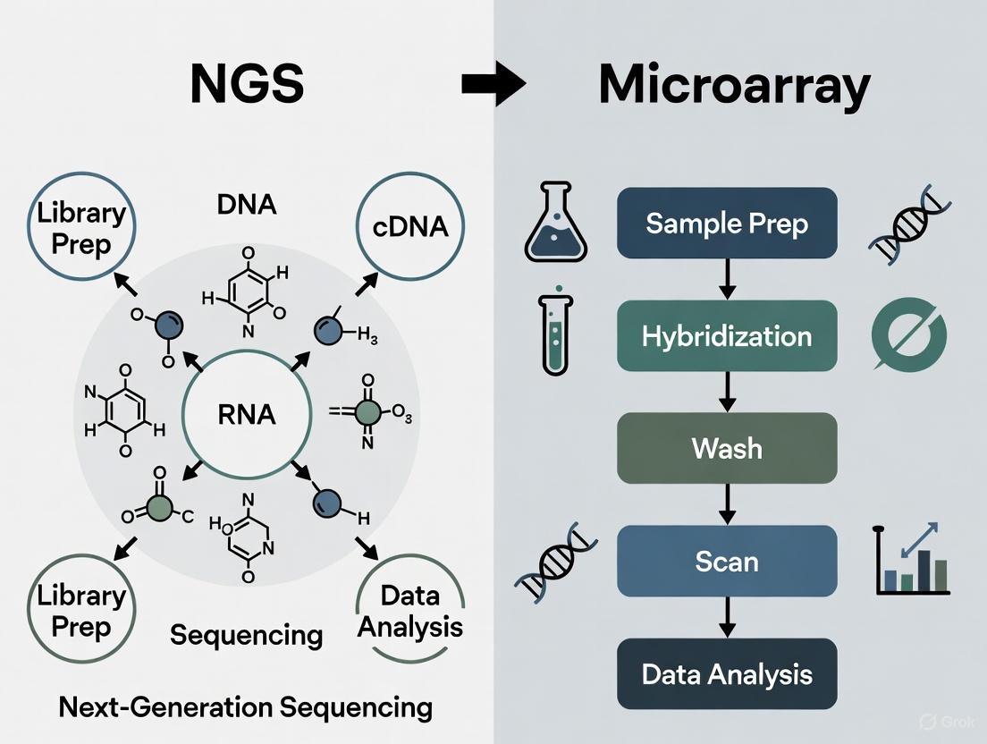
Microarray Experimental Workflow
The microarray workflow begins with total RNA extraction from biological samples, followed by a series of enzymatic reactions to generate biotin-labeled complementary RNA (cRNA). As detailed in cannabinol (CBN) exposure studies, this process typically involves generating single-stranded cDNA from 100 ng total RNA using reverse transcriptase and a T7-linked oligo(dT) primer, which is then converted to double-stranded cDNA [1]. Subsequently, cRNA is synthesized through in vitro transcription (IVT) with biotinylated UTP and CTP, using T7 RNA polymerase [1]. The biotin-labeled cRNA is fragmented and hybridized onto microarray chips, which are then stained, washed, and scanned to produce image files that are processed into cell intensity (CEL) files [1]. The robust multi-chip average (RMA) algorithm is commonly used for background adjustment, quantile normalization, and summarization of normalized expression data for each probe set on a log2 scale [1].
RNA-Seq Experimental Workflow
The RNA-Seq workflow shares the initial RNA extraction step but diverges significantly in subsequent processes. For sequencing library preparation, the Illumina Stranded mRNA Prep kit is commonly employed, beginning with purification of messenger RNAs (mRNAs) with polyA tails from 100 ng of total RNA using oligo(dT) magnetic beads [1]. The purified mRNA is then fragmented and converted to cDNA, followed by adapter ligation and potential amplification to create the final sequencing library [3]. These libraries are loaded onto flow cells where cluster generation occurs, amplifying single molecules to create thousands of identical copies for sequencing [3]. The actual sequencing employs sequencing-by-synthesis (SBS) chemistry, which tracks the addition of fluorescently-labeled nucleotides as the DNA chain is copied in a massively parallel fashion [3]. The resulting sequences are then aligned to a reference genome or transcriptome for quantification and analysis.
The Scientist's Toolkit: Essential Research Reagents and Platforms
Table 3: Essential Research Reagents and Platforms for Transcriptomics
| Item | Function | Example Products/Platforms |
|---|---|---|
| Gene Expression Microarrays | Pre-designed arrays for targeted transcript detection | GeneChip PrimeView Human Gene Expression Arrays [1] |
| NGS Platforms | Massively parallel sequencing instruments | Illumina NovaSeq, MiSeq; PacBio SMRT; Oxford Nanopore [7] [3] |
| Library Prep Kits | Prepare RNA samples for sequencing | Illumina Stranded mRNA Prep; Ion Torrent Transcriptome Sequencing kits [1] [6] |
| RNA Extraction & Purification Kits | Isolate high-quality RNA from samples | EZ1 RNA Cell Mini Kit; QIAshredder [1] |
| Hybridization & Staining Reagents | Process microarrays for detection | GeneChip Hybridization, Wash, and Stain Kits [1] |
| Data Analysis Software | Process, normalize, and analyze transcriptomic data | Affymetrix TAC; JMP Genomics; IPA; BaseSpace [1] [5] [3] |
Application in Chemical Perturbation Profiling
Case Study: Cannabinoid Exposure Research
The application of transcriptomic technologies in chemical perturbation profiling is well illustrated by recent research on cannabinoids. A 2025 study directly compared microarray and RNA-Seq platforms using two cannabinoids—cannabichromene (CBC) and cannabinol (CBN)—as case studies [1]. The experimental design exposed iPSC-derived hepatocytes to varying concentrations of each cannabinoid for 24 hours, with subsequent transcriptomic analysis performed using both platforms on the same biological samples [1]. This rigorous approach enabled direct comparison of platform performance in identifying functions and pathways impacted by compound exposure.
Both technologies successfully revealed similar overall gene expression patterns with regard to concentration for both CBC and CBN [1]. Despite RNA-Seq identifying a larger number of differentially expressed genes with wider dynamic ranges, including various non-coding RNA transcripts unavailable to microarray analysis, the platforms demonstrated equivalent performance in identifying impacted functions and pathways through gene set enrichment analysis (GSEA) [1]. Most notably, transcriptomic point of departure (tPoD) values derived through benchmark concentration (BMC) modeling showed no statistically significant differences between platforms for both compounds [1]. This finding has substantial implications for regulatory risk assessment, suggesting that for quantitative toxicogenomic applications, both technologies can provide equivalent points of departure for data-poor chemicals.
Analysis Pathways for Chemical Perturbation Studies
Implementation Considerations for Research Programs
Decision Framework for Technology Selection
Choosing between microarray and RNA-Seq technologies requires careful consideration of multiple factors specific to each research program. For established model organisms with comprehensive genomic annotations, where the research question focuses on known transcripts and the study design involves high sample throughput, microarrays offer significant advantages in cost-effectiveness and analytical simplicity [1] [5]. The technology benefits from decades of methodological refinement, with well-established normalization techniques and user-friendly analysis software that reduces the bioinformatics burden [5].
In contrast, RNA-Seq becomes the preferred option for non-model organisms, discovery-oriented research, and studies requiring detection of novel transcript features [5] [4]. The technology's ability to profile transcriptomes without a priori knowledge makes it indispensable for exploratory investigations and applications requiring the highest sensitivity [4]. However, researchers must be prepared for the bioinformatics challenges associated with RNA-Seq, including substantial data storage requirements, computational processing needs, and the need for specialized analytical expertise [5] [2].
Hybrid Approaches and Future Directions
Increasingly, researchers are adopting hybrid approaches that leverage the strengths of both technologies. One effective strategy involves using RNA-Seq for initial discovery phases to identify novel transcripts and biomarkers, followed by development of targeted microarrays for high-throughput screening applications [5]. This approach was successfully demonstrated in ecotoxicity testing on Chironomus riparius, where researchers used initial RNA-Seq data to create a microarray for routine monitoring [5]. This synergistic combination allows for both comprehensive discovery and cost-effective large-scale application.
The field continues to evolve with emerging methodologies that push transcriptomic analysis to higher resolution. Single-cell RNA sequencing is enabling researchers to move beyond bulk tissue analysis to examine transcriptomic responses at cellular resolution, revealing heterogeneity in chemical responses within seemingly uniform cell populations [8]. Similarly, spatial transcriptomics technologies are beginning to preserve geographical information within tissues, allowing researchers to map chemical effects within the architectural context of organs and tissue structures [8] [9]. These advances, combined with decreasing sequencing costs and improved computational methods, suggest that the transition to sequencing-based approaches will continue, while microarrays maintain their niche in targeted applications where their cost-effectiveness and analytical simplicity provide distinct advantages.
The evolution from hybridization-based microarrays to sequencing-driven RNA-Seq represents a paradigm shift in transcriptomics that has fundamentally expanded research capabilities. While RNA-Seq offers clear advantages in detection range, sensitivity, and discovery potential for novel transcripts, microarray technology maintains relevance for targeted applications where cost-effectiveness and analytical simplicity are paramount [1]. For chemical perturbation profiling specifically, both technologies can generate equivalent results for key endpoints including pathway analysis and benchmark concentration modeling [1]. The choice between platforms should be guided by specific research objectives, organism familiarity, discovery requirements, and resource constraints. As transcriptomics continues evolving toward single-cell and spatial resolutions, the integration of these complementary technologies will further empower researchers to unravel the complex molecular responses to chemical perturbations, advancing both basic science and regulatory decision-making.
In the field of chemical perturbation profiling research, scientists increasingly face a critical choice between established and emerging technologies for transcriptome analysis. Two platforms dominate this landscape: microarrays, the established workhorse relying on fluorescence-based hybridization to predefined transcripts, and RNA-Seq (Next-Generation Sequencing), the disruptive technology that enables direct, hypothesis-free sequencing of the entire transcriptome. Understanding the fundamental workings of microarrays—their strengths, limitations, and appropriate applications—is essential for designing effective toxicogenomic studies and accurately interpreting the resulting data. This guide provides an objective comparison of these platforms, supported by experimental data, to inform decision-making for researchers, scientists, and drug development professionals.
Core Technology: The Microarray Method
Fundamental Principles and Workflow
Gene expression microarrays function on the principle of complementary hybridization between immobilized probe sequences and fluorescently-labeled target transcripts. The technology provides a high-throughput method for quantifying the expression levels of thousands of predefined transcripts simultaneously [10].
A typical modern microarray consists of short oligonucleotide probes complementary to transcripts of interest, immobilized on a solid substrate [10]. Probe design is typically based on known genome sequences or predicted open reading frames, with multiple probes often designed per gene model to improve accuracy and reliability [10].
Table: Key Steps in a Microarray Experiment
| Step | Process Description | Key Considerations |
|---|---|---|
| 1. Probe Design | Oligonucleotides designed based on genomic sequences | Dependent on prior genomic knowledge; limited to annotated regions |
| 2. Sample Preparation | RNA extraction, purification, and fluorescent labeling | RNA quality (RIN ≥9) critical; may involve amplification |
| 3. Hybridization | Labeled transcripts bind to complementary probes on array | Stringency controls minimize cross-hybridization; typically 16-24 hours |
| 4. Washing & Scanning | Removal of non-specific binding; laser excitation of dyes | Eliminates background noise; captures fluorescence intensity |
| 5. Data Acquisition | Fluorescence intensity measured for each probe | Intensity correlates with expression level; specialized scanners required |
The process begins with transcript extraction from cells or tissues, followed by labeling with fluorescent dyes (either one-color or two-color approaches) [10]. The labeled transcripts are then hybridized to the arrays, washed to remove non-specifically bound material, and scanned with a laser [10]. Probes that correspond to transcribed RNA hybridize to their complementary targets, with light intensity serving as the quantitative measure of gene expression [10].
Visualization: Microarray Workflow
The following diagram illustrates the complete microarray experimental workflow, from probe design to data interpretation:
Comparative Analysis: Microarrays vs. RNA-Seq
Technical and Performance Comparison
Multiple studies have systematically compared the performance of microarray and RNA-Seq platforms for transcriptomic analysis. The table below summarizes key comparative findings from experimental studies:
Table: Experimental Comparison of Microarray and RNA-Seq Performance
| Parameter | Microarray | RNA-Seq | Experimental Context & Evidence |
|---|---|---|---|
| Dynamic Range | Limited [1] | Wider [1] [11] | RNA-Seq provides higher precision and wider dynamic range [1] |
| DEG Detection | Fewer DEGs typically identified [11] | More DEGs, including non-coding RNAs [1] [11] | RNA-Seq identified more differentially expressed protein-coding genes [11] |
| Platform Concordance | ~78% overlap with RNA-Seq DEGs [11] | High correlation with microarray (Spearman's 0.7-0.83) [11] | Both platforms detected similar pathway perturbations despite DEG differences [11] |
| Alternative Splicing | Requires specialized junction arrays [10] | Direct detection of splice junctions [10] | RNA-Seq enables identification of transcript isoforms without prior knowledge [10] |
| Species Flexibility | Limited to species with known sequences [10] | Can be used on species without full genome [10] | Microarrays require species-specific design or cross-species hybridization [10] |
| Cost per Sample | ~$100 [10] | ~$1,000 [10] | Microarrays offer significant cost advantage for large studies [10] |
Practical Considerations for Research Applications
Beyond technical specifications, practical considerations significantly impact platform selection for chemical perturbation studies:
Table: Practical Implementation Factors
| Factor | Microarray | RNA-Seq |
|---|---|---|
| Data Maturity | Well-understood biases; stable analytical solutions [10] | Evolving standards; biases still being researched [10] |
| Sample Throughput | High-throughput; streamlined workflows [1] | Moderate throughput; more complex preparation [12] |
| Infrastructure Needs | Standard computing resources [1] | Extensive computational resources needed [11] |
| Data Interpretation | Established pipelines and databases [1] | Complex bioinformatics; longer analysis times [11] |
| Regulatory Acceptance | Well-established for toxicogenomics [11] | Growing acceptance; expanding databases [11] |
Recent comparative studies demonstrate that despite their technological differences, both platforms can produce biologically concordant results. A 2024 study found that "despite some degree of discordance between the two platforms found during data analysis, very similar final results, i.e., impacted functional pathways and transcriptomic point of departure (tPoD) values, were obtained by the two platforms" [1]. This suggests that for many traditional transcriptomic applications, microarrays remain a viable and cost-effective option.
Experimental Evidence: Case Studies in Toxicogenomics
Hepatotoxicity Study
A comprehensive comparison study examined liver samples from rats treated with five hepatotoxicants using both platforms [11]. The research demonstrated that:
- Both platforms identified a larger number of DEGs in livers of rats treated with ANIT, MDA, and CCl4 compared to APAP and DCLF, correlating with histopathological findings [11]
- Consistent with established mechanisms of toxicity, both platforms detected dysregulation of key liver-relevant pathways including Nrf2, cholesterol biosynthesis, eiF2, hepatic cholestasis, glutathione, and LPS/IL-1 mediated RXR inhibition [11]
- RNA-Seq data enriched these pathways with additional DEGs and suggested modulation of additional liver-relevant pathways [11]
- RNA-Seq enabled identification of non-coding DEGs offering potential for improved mechanistic clarity [11]
Cannabinoid Profiling Study
A 2025 investigation compared microarray and RNA-Seq platforms using two cannabinoids (CBC and CBN) as case studies [1]. The experimental protocol included:
- Cell Culture: Commercial iPSC-derived hepatocytes (iCell Hepatocytes 2.0) cultured following manufacturer's protocol [1]
- Exposure: Cells exposed to varying concentrations of cannabinoids in triplicate for 24 hours [1]
- RNA Preparation: Total RNA purified using EZ1 Advanced XL automated instrument with DNase digestion [1]
- Quality Control: RNA concentration/purity measured by NanoDrop; quality checked by Agilent 2100 Bioanalyzer for RIN [1]
- Microarray Processing: Samples processed using GeneChip 3' IVT PLUS Reagent Kit and hybridized to GeneChip PrimeView Human Gene Expression Arrays [1]
- Data Generation: Arrays scanned using GeneChip Scanner 3000; CEL files processed with Affymetrix Transcriptome Analysis Console using RMA algorithm [1]
The study concluded that "considering the relatively low cost, smaller data size, and better availability of software and public databases for data analysis and interpretation, microarray is still a viable method of choice for traditional transcriptomic applications such as mechanistic pathway identification and concentration response modeling" [1].
The Scientist's Toolkit: Essential Research Reagents
Table: Key Research Reagent Solutions for Microarray Experiments
| Reagent/Instrument | Function | Application Notes |
|---|---|---|
| iCell Hepatocytes 2.0 (FUJIFILM) | Biologically relevant in vitro model | iPSC-derived; maintain hepatocyte functionality [1] |
| EZ1 RNA Cell Mini Kit (Qiagen) | Total RNA purification | Automated purification with DNase digestion step [1] |
| Agilent 2100 Bioanalyzer | RNA quality assessment | Determines RNA Integrity Number (RIN); essential for QC [1] |
| GeneChip PrimeView Arrays (Affymetrix) | Gene expression profiling | Predefined transcript coverage; consistent performance [1] |
| GeneChip 3' IVT PLUS Kit (Affymetrix) | Target preparation | Includes reverse transcription, IVT, and labeling [1] |
| TruSeq Stranded mRNA Kit (Illumina) | RNA-Seq library prep | Comparison methodology; enriches coding mRNAs [11] |
Decision Framework: Platform Selection Guide
The choice between microarray and RNA-Seq technologies depends on multiple factors, which can be visualized through the following decision pathway:
Microarray technology, based on fluorescence-based hybridization to predefined transcripts, remains a valuable and reliable platform for transcriptomic analysis in chemical perturbation profiling research. While RNA-Seq offers advantages in detecting novel transcripts and providing a wider dynamic range, microarrays provide a cost-effective, well-established alternative with mature analytical frameworks [10] [1]. The experimental evidence demonstrates that for many applications—including mechanistic pathway identification and concentration-response modeling—microarrays produce results functionally equivalent to RNA-Seq in terms of biological interpretation [1] [11]. The choice between platforms should be guided by specific research objectives, budgetary constraints, and the need for novel transcript discovery versus focused hypothesis testing.
Next-generation sequencing (NGS) has revolutionized genomic research by providing powerful tools to investigate biological systems. For researchers studying chemical perturbation profiling—analyzing how cells respond to drugs or chemical compounds—understanding the core technological advantages of NGS compared to traditional microarray platforms is crucial for experimental design and data interpretation. This guide explores the fundamental principles of sequencing-by-synthesis and massive parallel sequencing, objectively comparing NGS performance against microarrays for toxicogenomic and chemical perturbation applications.
Principles of NGS Technology
Sequencing-by-Synthesis (SBS) Chemistry
Sequencing-by-Synthesis forms the foundation of modern NGS platforms. Unlike the Sanger chain-termination method, SBS technology involves tracking the addition of fluorescently-labeled nucleotides as the DNA chain is copied in a cyclical process [3]. The core SBS process consists of repeated steps: polymerase-based extension using reversible terminator nucleotides, fluorescence imaging to identify the incorporated base, and chemical cleavage to remove the terminating group and fluorescent dye, preparing the template for the next incorporation cycle [13].
This reversible termination chemistry enables highly accurate base determination across millions of parallel reactions. Recent innovations like XLEAP-SBS chemistry have further increased sequencing speed and fidelity compared to standard Illumina SBS chemistry [3].
Massive Parallel Sequencing
Massive parallel sequencing refers to the simultaneous sequencing of millions to billions of DNA fragments in a single run [14]. This is achieved through clonal amplification of individual DNA fragments either on a solid surface (bridge amplification) or in emulsion droplets (emulsion PCR), creating clusters of identical DNA templates that generate sufficient signal for detection during sequencing [13].
The extraordinary throughput of massive parallel sequencing enables researchers to rapidly sequence whole genomes, deeply sequence target regions, and perform complex transcriptomic analyses that would be impractical with traditional methods [3]. Modern Illumina systems can generate data output ranging from 300 kilobases up to multiple terabases in a single run, depending on the instrument type and configuration [3].
NGS Versus Microarrays: Technical Comparison
The table below summarizes the core technical differences between NGS and microarray technologies for chemical perturbation studies:
| Feature | Next-Generation Sequencing (NGS) | Microarray |
|---|---|---|
| Fundamental Principle | Sequencing-by-synthesis via reversible terminator chemistry [13] | Fluorescence-based hybridization to predefined probes [1] |
| Dynamic Range | Orders of magnitude greater; digital counting of reads enables detection across wide expression levels [5] [3] | Limited; suffers from signal saturation at high expression levels and background noise at low levels [1] [5] |
| Transcript Discovery | Capable of detecting novel transcripts, splice variants, and non-coding RNAs without prior knowledge [1] [5] | Limited to predefined probes; cannot detect sequences not represented on the array [1] |
| Required A Priori Knowledge | Not required; can profile organisms with unsequenced genomes [5] | Extensive knowledge needed for probe design [5] |
| Background Noise | Low background signal [1] | High background noise due to nonspecific binding [1] |
| Cost Considerations | Higher per-sample cost; decreasing over time [5] | Lower per-sample cost; established economical option [1] |
Experimental Evidence: Performance Comparison in Chemical Perturbation Studies
Recent research directly compares these platforms for chemical perturbation profiling. A 2025 study examined two cannabinoids (cannabichromene and cannabinol) using both RNA-seq and microarrays, providing quantitative performance data [1].
Experimental Protocol
The comparative study followed this methodology [1]:
- Cell Culture: Human induced pluripotent stem cell (iPSC)-derived hepatocytes (iCell Hepatocytes 2.0) cultured in 24-well plates
- Chemical Exposure: Cells exposed to varying concentrations of CBC and CBN for 24 hours in triplicate
- RNA Extraction: Total RNA purified using EZ1 Advanced XL automated instrument with DNase digestion
- Platform-Specific Processing:
- Microarray: GeneChip PrimeView Human Gene Expression Arrays with 3' IVT PLUS Reagent Kit
- RNA-seq: Illumina Stranded mRNA Prep kit for library preparation
- Data Analysis: Differential expression analysis followed by gene set enrichment analysis (GSEA) and benchmark concentration (BMC) modeling
Comparative Performance Data
The table below summarizes the key findings from the cannabinoid perturbation study:
| Performance Metric | RNA-seq Results | Microarray Results | Conclusion |
|---|---|---|---|
| Differentially Expressed Genes (DEGs) | Larger numbers of DEGs identified with wider dynamic range [1] | Fewer DEGs detected [1] | RNA-seq more sensitive in DEG detection |
| Functional Pathway Identification | Equivalent performance in identifying impacted functions and pathways through GSEA [1] | Equivalent performance in identifying impacted functions and pathways through GSEA [1] | Both platforms equivalent for functional analysis |
| Transcriptomic Point of Departure (tPoD) | tPoD values on the same level for both cannabinoids [1] | tPoD values on the same level for both cannabinoids [1] | Both platforms equivalent for concentration-response modeling |
| Novel Transcript Detection | Detected non-coding RNAs (miRNA, lncRNA) and novel transcripts [1] | Limited to predefined probeset [1] | RNA-seq superior for novel biomarker discovery |
Despite RNA-seq's technical advantages in detecting more DEGs with wider dynamic range, both platforms produced equivalent results for the endpoints most relevant to chemical risk assessment: identification of impacted functional pathways and transcriptomic point of departure values [1]. This suggests that for traditional toxicogenomic applications like mechanistic pathway identification and concentration-response modeling, microarrays remain a viable option, particularly considering their lower cost, smaller data size, and better availability of analysis software and databases [1].
The Scientist's Toolkit: Essential Research Reagents
The table below details key reagents and materials essential for implementing NGS and microarray protocols in chemical perturbation studies:
| Reagent/Material | Function | Application Notes |
|---|---|---|
| iPSC-derived hepatocytes (e.g., iCell Hepatocytes 2.0) | Physiologically relevant in vitro model for chemical exposure studies [1] | Preferred over cancer cell lines for non-cancer chemical perturbation research [15] |
| Stranded mRNA Prep Kit (Illumina) | Library preparation for RNA-seq; preserves strand orientation [1] | Essential for accurate transcript annotation and quantification |
| GeneChip PrimeView Array (Affymetrix) | Microarray-based gene expression profiling [1] | Established platform with well-annotated databases |
| EZ1 RNA Cell Mini Kit (Qiagen) | Automated RNA purification with genomic DNA removal [1] | Critical for obtaining high-quality RNA (RIN > 8) for both platforms |
| Poly-A Selection Beads | mRNA enrichment for RNA-seq library prep [1] | Reduces ribosomal RNA contamination |
| Reversible Terminator Nucleotides | Core SBS chemistry for base identification [13] | XLEAP-SBS chemistry offers improved fidelity [3] |
Workflow and Pathway Diagrams
NGS Sequencing-by-Synthesis Workflow
Chemical Perturbation Transcriptomics Pathway
The choice between NGS and microarray technologies for chemical perturbation profiling depends on specific research objectives and resource constraints. NGS technologies, with their sequencing-by-synthesis chemistry and massive parallel sequencing capabilities, offer clear technical advantages for discovery-based research where novel transcript detection, broader dynamic range, and absence of pre-existing genomic knowledge are primary considerations [5] [3]. However, recent evidence demonstrates that microarray platforms remain competitive for traditional toxicogenomic applications, providing equivalent performance in functional pathway analysis and concentration-response modeling at lower cost and with less computational overhead [1].
For comprehensive chemical perturbation studies that require novel biomarker discovery or detection of non-coding RNAs, NGS is unquestionably superior. However, for well-defined hypothesis testing within annotated genomes, microarrays provide a cost-effective and analytically tractable alternative. The emerging trend of combining both approaches—using NGS for initial discovery and microarrays for routine screening—represents a pragmatic strategy that leverages the respective strengths of both platforms [5].
In the field of chemical perturbation profiling, researchers face a critical decision when selecting a genomic tool: next-generation sequencing (NGS) or microarray technology. Each platform possesses distinct technical characteristics that directly impact data quality and biological interpretation. For research aimed at understanding the mechanisms of action of chemical compounds, the choices between these technologies influence everything from experimental design to the validation of findings. This guide provides an objective comparison of three fundamental performance parameters—dynamic range, background noise, and specificity—between NGS and microarrays, drawing on experimental data to inform selection for toxicogenomics and drug development studies.
The core distinction between these platforms lies in their underlying biochemistry. Microarrays are a closed-architecture system that relies on the hybridization of fluorescently-labeled nucleic acids to predefined probes immobilized on a solid surface [16]. The signal intensity measured at each probe provides a relative quantification of the target sequence. In contrast, next-generation sequencing (NGS) is an open-architecture system that uses massively parallel sequencing-by-synthesis to directly determine the nucleotide sequence of millions of DNA fragments simultaneously [3]. This fundamental difference—indirect hybridization versus direct sequencing—is the origin of their performance distinctions.
Direct Performance Comparison
The table below summarizes the key technological distinctions between NGS and microarrays based on experimental comparisons.
Table 1: Key Performance Metrics for Microarray and NGS Platforms
| Performance Metric | Microarray | Next-Generation Sequencing (NGS) |
|---|---|---|
| Dynamic Range | Limited by signal saturation at high end and background noise at low end [3]. | Broader, digital counting of reads enables quantification across a wider concentration range [3]. |
| Background Noise | Susceptible to high background noise from nonspecific binding [1] [17]. | Lower background; noise primarily from sequencing errors or PCR duplicates [7]. |
| Specificity | Challenged by cross-hybridization between related sequences; difficult to distinguish paralogs or splice variants [18] [17]. | High specificity; can uniquely map reads to their genomic origin, identifying splice sites and single-nucleotide differences [3]. |
| Data Type | Relative, analog-like fluorescence intensity. | Digital, countable read counts. |
| Optimal Application | Profiling known transcripts; studies where cost-effectiveness and sample throughput are priorities [1]. | Discovery of novel transcripts, splice variants, and non-coding RNAs; quantifying rare transcripts [1] [7]. |
Experimental data reinforces these distinctions. A 2025 toxicogenomics study comparing the same cannabinoid samples on both platforms noted that RNA-seq identified larger numbers of differentially expressed genes (DEGs) with a wider dynamic range, consistent with its digital, counting-based nature [1]. In a separate investigation, the specificity of microarrays was compromised by cross-hybridization, a phenomenon where a probe binds to non-target sequences with high similarity, such as closely related members of a miRNA family [18].
Experimental Protocols for Platform Comparison
To ensure valid and reproducible comparisons between NGS and microarray technologies, a rigorous experimental protocol is essential.
Sample Preparation and Core Methodology
The foundational step for a fair comparison is using the same high-quality RNA sample for both platforms. RNA integrity should be verified using methods like microfluidic electrophoresis to obtain an RNA Integrity Number (RIN) [17] [19].
- Microarray Workflow: The protocol generally involves reverse transcribing RNA into cDNA, followed by in vitro transcription to produce biotin-labeled cRNA. This labeled cRNA is fragmented and hybridized to the microarray chip. After washing, the chip is scanned to produce fluorescence intensity data (DAT files), which are converted into cell intensity (CEL) files for analysis [1] [17].
- NGS Workflow (RNA-Seq): The library preparation starts by isolating and fragmenting RNA. Fragments are reverse-transcribed into cDNA, and platform-specific adapters are ligated to the ends. These libraries are then quantified and loaded onto a sequencer. In Illumina systems, fragments undergo clonal amplification on a flow cell, followed by sequencing-by-synthesis with reversible dye-terminators [3] [19].
The following diagram illustrates the core biochemical workflows for each technology.
Data Analysis and Validation
- Microarray Data Processing: Raw intensity files require significant pre-processing, including background correction, normalization (e.g., Robust Multi-array Average - RMA), and summarization to generate expression values for each probe set [1] [17].
- NGS Data Processing: The primary data analysis involves base calling, demultiplexing, and quality control. Reads are then aligned to a reference genome, and gene expression is quantified by counting the number of reads mapped to each gene [19].
- Validation: A common practice is to validate key findings using an orthogonal method, such as real-time quantitative PCR (qPCR) for a subset of genes [18].
The Scientist's Toolkit: Essential Research Reagents
Successful execution of a chemical perturbation study requires specific reagents and tools for both platforms.
Table 2: Essential Reagents and Materials for Perturbation Profiling
| Item | Function | Considerations |
|---|---|---|
| High-Quality Total RNA | Starting material for both library prep (NGS) and labeling (microarray). | Assess yield, purity (A260/280), and integrity (RIN > 8) [17] [19]. |
| NGS Library Prep Kit | Prepares RNA/DNA fragments for sequencing by adding platform-specific adapters. | Select based on application (e.g., mRNA-seq, total RNA-seq), input amount, and workflow simplicity [19]. |
| Microarray Platform | Pre-manufactured slide or chip with immobilized probes for hybridization. | Choose a platform with comprehensive and up-to-date gene coverage for your organism of interest [18] [17]. |
| qPCR Reagents | For orthogonal validation of differentially expressed genes identified by NGS or microarray. | Enables high-sensitivity and high-specificity confirmation of expression changes [18]. |
| Cell Painting Assay Reagents | For complementary morphological profiling; includes fluorescent dyes for staining cellular components. | Used to connect transcriptional changes with phenotypic outcomes in chemical perturbation studies [20]. |
Implications for Chemical Perturbation Research
The choice between NGS and microarray has direct consequences for interpreting chemical perturbation experiments.
- Mechanism of Action (MoA) Elucidation: NGS is superior for de novo discovery, as it can reveal novel transcripts, splice variants, and non-coding RNAs affected by a compound, providing a more complete picture of its MoA [7] [3]. Microarrays are confined to pre-defined transcripts.
- Toxicogenomics and Pathway Analysis: A 2025 study found that while NGS identified more DEGs, both platforms ultimately revealed similar enriched pathways and produced comparable transcriptomic points of departure (tPoD) in concentration-response modeling [1]. This suggests that for well-annotated pathways, microarrays remain a cost-effective option.
- Data Reproducability and Noise: Microarray data can be confounded by batch effects and cross-hybridization, requiring careful normalization and batch-effect correction [17] [21]. NGS data, while less prone to cross-hybridization, must be processed to account for sequencing artifacts and amplification biases [18] [7].
The relationship between data generation and biological insight in perturbation studies is summarized below.
The decision between NGS and microarray technology for chemical perturbation profiling is not one-size-fits-all. NGS offers clear technical advantages in dynamic range, specificity, and discovery power, making it the preferred tool for uncovering novel mechanisms and profiling complex transcriptomes. Microarrays, however, remain a viable and cost-effective option for focused studies where high sample throughput and well-established analytical pipelines are priorities, and where the target transcripts are well-annotated. The most appropriate technology depends on the specific research goals, budget, and bioinformatic capabilities of the project.
Next-generation sequencing (NGS) has revolutionized genomics research, bringing unparalleled capabilities to analyze DNA and RNA molecules in a high-throughput and cost-effective manner [7]. This transformative technology has become particularly crucial for chemical perturbation profiling, a field that systematically studies how small molecules affect biological systems. Unlike traditional microarray technologies, which rely on hybridization to predefined probes, NGS-based methods offer higher precision, wider dynamic range, and the ability to detect novel transcripts and modifications without prior sequence knowledge [22]. The transition from microarray to NGS has enabled researchers to move beyond simple gene expression profiling to comprehensive mechanism-of-action studies for drug discovery, fundamentally changing how we approach chemical genomics and toxicogenomics.
NGS Technology Generations: Core Platforms and Specifications
The evolution of sequencing technologies has progressed through distinct generations, each overcoming limitations of its predecessors while introducing new capabilities essential for detailed perturbation studies.
Second-Generation Sequencing: The Short-Read Workhorse
Second-generation or short-read sequencing platforms remain the workhorses of most NGS laboratories, dominating applications requiring high accuracy and throughput at low cost [23]. These technologies share a common principle of massively parallel sequencing of millions to billions of DNA fragments, typically ranging from 50-300 base pairs in length [7]. The Illumina platform, which accounts for the majority of the world's sequencing data, utilizes a sequencing-by-synthesis approach with reversible dye-terminators and bridge amplification on flow cells [7] [24]. Alternative short-read technologies like Ion Torrent employ semiconductor sequencing, detecting hydrogen ions released during DNA polymerization rather than using optical methods [7].
Table 1: Comparison of Major Short-Read Sequencing Platforms
| Platform | Technology | Amplification Method | Read Length | Key Advantages | Primary Limitations |
|---|---|---|---|---|---|
| Illumina NovaSeq X | Sequencing-by-Synthesis | Bridge PCR | 36-300 bp | Extremely high throughput (16 Tb/run); Low error rate (<1%) [25] [26] | Limited read length; GC bias [27] |
| Ion Torrent Genexus | Semiconductor | Emulsion PCR | 200-600 bp | Rapid results (1 day); Simple workflow [26] | Homopolymer errors [7] |
| MGI DNBSEQ-T7 | DNA Nanoball | Nanoball PCR | 50-150 bp | Cost-effective; Competitive accuracy [7] [27] | Multiple PCR cycles required [7] |
| Element AVITI | Sequencing-by-Binding | Proprietary | Up to 300 bp | Q40 accuracy (1 error/10,000 bases) [24] | Emerging platform with smaller user base |
Third-Generation Sequencing: Long-Read Technologies
Third-generation sequencing technologies overcome the read length limitations of short-read platforms by sequencing single DNA molecules without amplification [7]. Pacific Biosciences (PacBio) employs Single Molecule Real-Time (SMRT) sequencing, where DNA polymerase incorporates fluorescently labeled nucleotides in real-time within nanoscale wells called zero-mode waveguides (ZMWs) [7] [25]. The introduction of HiFi (High-Fidelity) reads circularizes DNA fragments, allowing multiple passes to generate reads of 10-25 kilobases with accuracy exceeding 99.9% (Q30) [25] [23].
Oxford Nanopore Technologies (ONT) utilizes a fundamentally different approach, measuring changes in electrical current as DNA strands pass through protein nanopores [7]. Recent developments like the Q20+ and Q30 Duplex kits have significantly improved accuracy, with duplex reads exceeding Q30 (>99.9% accuracy) while maintaining the ability to generate ultra-long reads exceeding 100 kilobases [25] [24]. This technology uniquely enables direct detection of epigenetic modifications and requires minimal instrumentation, from pocket-sized MinION devices to high-throughput PromethION platforms [7] [23].
Table 2: Comparison of Major Long-Read Sequencing Platforms
| Platform | Technology | Read Length | Accuracy | Key Advantages | Primary Limitations |
|---|---|---|---|---|---|
| PacBio Revio (HiFi) | SMRT Sequencing | 10-25 kb | >99.9% (Q30) | High accuracy; Uniform coverage [25] [23] | Higher cost per sample; Moderate throughput |
| Oxford Nanopore (Q30 Duplex) | Nanopore Sensing | 10-100+ kb | >99.9% (Q30) | Ultra-long reads; Direct epigenetic detection [25] | Higher DNA input requirements |
| PacBio Onso | Sequencing-by-Binding | 100-200 bp | Q40 | Exceptional accuracy (1 error/10,000 bases) [24] | Short-read platform |
Benchmarking NGS Performance for Chemical Genomics Applications
Technical Comparisons Across Platforms
Practical comparisons of NGS platforms reveal distinct performance characteristics critical for experimental design. A comprehensive evaluation of yeast genome assembly demonstrated that ONT reads generated more continuous assemblies than PacBio Sequel, though with persistent homopolymer-related errors [27]. The study further found Illumina NovaSeq 6000 provided more accurate assemblies in short-read-first pipelines, while MGI DNBSEQ-T7 offered a cost-effective alternative for polishing processes [27].
For transcriptomic applications, including chemical perturbation studies, RNA-seq demonstrates clear advantages over microarrays in detecting novel transcripts, splice variants, and non-coding RNAs with a wider dynamic range [22]. However, microarray technology remains competitive for traditional applications like mechanistic pathway identification and concentration-response modeling, offering lower costs, smaller data sizes, and better-established analytical pipelines [22].
NGS in Chemical Perturbation Profiling: The PROSPECT Case Study
The application of NGS to chemical genomics is exemplified by the PROSPECT (PRimary screening Of Strains to Prioritize Expanded Chemistry and Targets) platform for antibiotic discovery [28]. This methodology addresses a fundamental challenge in drug discovery—simultaneously identifying bioactive compounds and their mechanisms of action.
Experimental Protocol: PROSPECT Chemical-Genetic Profiling
Strain Pool Preparation: A pooled collection of hypomorphic Mycobacterium tuberculosis mutants, each depleted of a different essential protein and tagged with unique DNA barcodes, is prepared [28].
Chemical Perturbation: The mutant pool is exposed to chemical compounds across a range of concentrations, with untreated controls maintained for comparison [28].
Selective Growth: Following incubation, genomic DNA is extracted from both treated and control pools. The relative abundance of each mutant strain is quantified by amplifying and sequencing the barcode regions using NGS [28].
Data Analysis: Chemical-genetic interaction (CGI) profiles are generated as vectors representing each hypomorph's sensitivity. The Perturbagen Class (PCL) analysis then compares unknown compound profiles to a curated reference set of compounds with known mechanisms of action [28].
Figure 1: NGS-Based Chemical-Genetic Interaction Profiling Workflow. This diagram illustrates the PROSPECT platform workflow for elucidating small molecule mechanism of action through chemical-genetic interaction profiling [28].
This approach demonstrates how NGS transcends mere sequence detection to become a quantitative tool for measuring biological responses. In proof-of-concept validation, PCL analysis correctly predicted mechanism of action with 70% sensitivity and 75% precision in leave-one-out cross-validation, successfully identifying compounds targeting tuberculosis respiration [28].
The Scientist's Toolkit: Essential Reagents and Methodologies
Successful implementation of NGS-based chemical genomics requires specialized reagents and methodologies tailored to perturbation studies.
Table 3: Essential Research Reagent Solutions for NGS Chemical Genomics
| Reagent/Method | Function | Application in Perturbation Studies |
|---|---|---|
| Hypomorphic Mutant Libraries | Collection of essential gene knockdown strains | Enables genome-wide sensitivity profiling; Key to PROSPECT platform [28] |
| DNA Barcode Systems | Unique sequence tags for each strain or perturbation | Allows pooled screening by tracking strain abundance via NGS [28] [29] |
| Stranded mRNA Prep Kits | Library preparation preserving strand information | Maintains transcriptional directionality in perturbation transcriptomics [22] |
| Transposase-Based Library Construction | Efficient fragmentation and tagging of DNA | Streamlines library prep for both short- and long-read sequencing [23] |
| Multiplexed Library Prep Technologies (e.g., purePlex, ExpressPlex) | Simultaneous processing of multiple samples | Enables large-scale chemical screening with normalized libraries [23] |
| Batch Effect Correction Methods (e.g., ComBat, TVN) | Statistical adjustment for technical variation | Critical for integrating data across multiple screens or laboratories [29] [30] |
Integrated Data Analysis: From Sequences to Biological Insights
The computational transformation of NGS data into biological insights requires sophisticated pipelines, particularly for perturbation studies where distinguishing true signals from background variation is crucial.
Perturbative Map Building with EFAAR Pipeline
Large-scale chemical and genetic perturbation data can be integrated into unified "maps of biology" using the EFAAR pipeline (Embedding, Filtering, Aligning, Aggregating, Relating) [29]. This framework processes high-dimensional data from various perturbation types (CRISPR knockout, chemical treatment) into comparable embedding spaces [29].
Embedding reduces high-dimensional assay data (e.g., 20,000 gene expression values or million-pixel images) to tractable numerical representations using methods like principal component analysis or neural networks [29]. Filtering removes low-quality perturbation units based on predefined criteria. Aligning applies batch effect correction methods like Typical Variation Normalization (TVN) or ComBat to remove technical artifacts [29]. Aggregating combines replicate measurements using statistical methods, while Relating computes similarity measures between perturbations to identify biological relationships [29].
Figure 2: EFAAR Computational Pipeline for Perturbative Map Building. This workflow transforms raw perturbation data into unified maps that capture biological relationships [29].
Advanced Algorithms for Chemical-Genetic Data
The Bucket Evaluations (BE) algorithm addresses specific challenges in chemical-genomic data analysis by using leveled rank comparisons to minimize batch effects without requiring prior knowledge of confounding variables [30]. This method divides each profile's gene scores into "buckets" - smaller buckets for the most significant genes (highest fitness defects) and larger buckets for less significant genes [30]. A weighted scoring system then identifies profile similarities, awarding higher scores to genes in corresponding high-significance buckets across experiments [30].
The NGS landscape continues to evolve with emerging technologies that promise to further transform chemical perturbation profiling. Multi-omics integration represents a key frontier, with platforms like PacBio's SPRQ chemistry simultaneously capturing DNA sequence and chromatin accessibility information from the same molecule [25]. Ultra-high accuracy sequencing is another trend, with platforms like Element AVITI and PacBio Onso achieving Q40 accuracy (1 error in 10,000 bases), enabling more confident detection of rare variants in heterogeneous samples [24].
The ongoing competition between short-read and long-read technologies has driven remarkable cost reductions, with the price of sequencing a human genome falling below $100, outpacing Moore's Law [24]. This increased affordability, combined with continuous improvements in accuracy and throughput, ensures that NGS will remain the foundational technology for chemical perturbation profiling and drug discovery.
While microarrays retain niche applications in standardized toxicogenomic testing due to their lower cost and simpler data analysis [22], NGS provides unparalleled versatility for discovering novel biological mechanisms. The choice between short-read and long-read technologies increasingly depends on specific application requirements rather than technical limitations, with many researchers adopting hybrid approaches that leverage the complementary strengths of both platforms [23] [27].
For chemical genomics research, this expanding NGS landscape offers unprecedented opportunities to elucidate mechanisms of action, identify novel therapeutic targets, and accelerate drug discovery through more informative early-stage screening. As sequencing technologies continue to converge and improve, they will undoubtedly uncover deeper insights into biological systems and their chemical perturbations.
Designing Your Profiling Study: Methodological Approaches and Practical Applications
In chemical perturbation profiling research, selecting the appropriate transcriptomic platform is crucial for generating reliable, biologically relevant data. The choice between microarray technology and RNA sequencing (RNA-seq) represents a fundamental decision point that affects every subsequent stage of experimental workflow, data interpretation, and biological insight. While RNA-seq has increasingly become the dominant platform in modern transcriptomics, recent evidence suggests that microarrays remain surprisingly competitive for specific applications, particularly in studies focusing on pathway identification and concentration-response modeling [1].
This guide provides an objective comparison of sample preparation workflows for both platforms, focusing specifically on their application in chemical perturbation studies. We examine detailed experimental protocols, present quantitative performance data, and analyze the technical considerations researchers must evaluate when designing transcriptomic experiments for toxicogenomics and drug development applications.
The fundamental distinction between these platforms lies in their basic detection principles: microarrays utilize hybridization-based detection of predefined transcripts, while RNA-seq employs sequencing-by-synthesis to generate digital read counts [1] [4]. This core difference dictates substantial variations in their sample preparation requirements, data output, and analytical capabilities.
The following diagram illustrates the parallel workflows for both technologies, highlighting key decision points and procedural differences:
Detailed Experimental Protocols
Microarray Sample Preparation Protocol
The microarray workflow employs a hybridization-based approach with fluorescent detection. The following protocol is adapted from toxicogenomic studies of cannabinoids (CBC and CBN) using iPSC-derived hepatocytes [1]:
RNA Extraction and Quality Control: Isolate total RNA using silica-based membrane purification (e.g., EZ1 RNA Cell Mini Kit) with integrated DNase digestion to remove genomic DNA contamination. Assess RNA purity using UV spectrophotometry (260/280 ratio) and determine RNA integrity number (RIN) ≥7.0 using microfluidics-based analysis (e.g., Agilent 2100 Bioanalyzer) [1].
cDNA Synthesis and Amplification: Convert 100ng total RNA to double-stranded cDNA using reverse transcriptase with T7-linked oligo(dT) primers, followed by second-strand synthesis with DNA polymerase and RNase H. Perform in vitro transcription (IVT) using T7 RNA polymerase with biotin-labeled UTP and CTP to generate complementary RNA (cRNA) [1].
Fragmentation and Hybridization: Fragment 12μg of biotin-labeled cRNA using magnesium-induced cleavage (94°C). Hybridize to array (e.g., GeneChip PrimeView Human Gene Expression Array) at 45°C for 16 hours in a specialized hybridization oven [1].
Washing, Staining, and Scanning: Perform automated washing and staining on a fluidics station using streptavidin-phycoerythrin conjugate. Scan arrays using a high-resolution scanner (e.g., GeneChip Scanner 3000 7G) to generate DAT image files, which are processed into CEL files using vendor software [1].
RNA-Seq Sample Preparation Protocol
RNA-seq employs a sequencing-based approach that captures digital expression data. The following protocol is adapted from parallel analysis of the same cannabinoid samples [1]:
RNA Extraction and Quality Control: Use identical RNA extraction and quality assessment procedures as the microarray protocol to ensure comparable starting material. The consistency in initial sample processing allows for direct platform comparisons [1].
Library Preparation: Process 100ng total RNA using Illumina Stranded mRNA Prep, Ligation kit. Purify polyadenylated mRNA using oligo(dT) magnetic beads. Fragment RNA and synthesize cDNA with random hexamer priming. Perform end repair, A-tailing, and adapter ligation for library construction [1] [31].
Library Quality Control and Normalization: Assess library quality using microfluidics-based systems (e.g., Bioanalyzer) and quantify using fluorometric methods (e.g., Qubit). Normalize libraries to equimolar concentrations for pooling and multiplexed sequencing [31].
Sequencing: Load pooled libraries onto an NGS platform (e.g., Illumina HiSeq 2000) for cluster generation and sequencing-by-synthesis. Generate 50-100 million paired-end reads per sample (2×100bp configuration) to ensure sufficient coverage for transcript quantification [1].
Performance Comparison Data
Technical Capabilities and Limitations
Table 1: Technical comparison between microarray and RNA-seq platforms
| Parameter | Microarray | RNA-Seq |
|---|---|---|
| Detection Principle | Hybridization-based | Sequencing-based |
| Dynamic Range | ~10³ [4] | >10⁵ [4] |
| Background Noise | High background due to nonspecific binding [1] | Low background |
| Sample Throughput | High [16] | Moderate [16] |
| Required RNA Input | 100ng [1] | 100ng [1] |
| Novel Transcript Discovery | Limited to predefined probes [4] | Unlimited detection capability [4] |
| Variant Detection | Not available | SNP, splice variants, fusion genes [4] |
| Multiplexing Capability | Limited | High (with barcoding) |
| Startup Cost | Low | High |
| Cost per Sample | Low [5] | High [5] |
Experimental Outcomes in Chemical Perturbation Studies
Recent comparative studies using identical chemical perturbation samples reveal nuanced performance differences between the platforms:
Table 2: Experimental outcomes from comparative studies of cannabinoid perturbations
| Performance Metric | Microarray Results | RNA-Seq Results | Comparative Analysis |
|---|---|---|---|
| Differentially Expressed Genes | 427 DEGs identified in HIV study [32] | 2,395 DEGs identified in HIV study [32] | RNA-seq detects 5.6× more DEGs |
| Pathway Identification | 47 perturbed pathways [32] | 205 perturbed pathways [32] | 30 pathways shared between platforms |
| Correlation with Protein Expression | Variable by gene; superior for BAX, PIK3CA [33] | Variable by gene; superior for others [33] | Platform performance gene-dependent |
| Transcriptomic Point of Departure (tPoD) | Equivalent to RNA-seq [1] | Equivalent to microarray [1] | No significant difference in tPoD values |
| Gene Expression Correlation | Median Pearson R=0.76 with RNA-seq [32] | Median Pearson R=0.76 with microarray [32] | High correlation between platforms |
| Dynamic Fold Change Distribution | Similar distribution to RNA-seq [32] | Similar distribution to microarray [32] | No significant difference (KS test) |
Pathway Analysis and Bioinformatics
Functional analysis of chemical perturbation data reveals important similarities and differences between the platforms. The following diagram illustrates the bioinformatic workflow for pathway enrichment analysis from raw data through functional interpretation:
Despite detecting different numbers of differentially expressed genes, both platforms identify highly concordant biological pathways in chemical perturbation studies. Research comparing cannabinoid exposures found that "the two platforms displayed equivalent performance in identifying functions and pathways impacted by compound exposure through gene set enrichment analysis (GSEA)" [1]. This pathway-level concordance persists even when gene-level detection differs substantially.
The Scientist's Toolkit: Essential Research Reagents
Table 3: Key reagents and solutions for transcriptomic sample preparation
| Reagent/Kits | Function | Platform Application |
|---|---|---|
| PAXgene Blood RNA Tubes | RNA stabilization during blood collection | Both platforms [32] |
| EZ1 RNA Cell Mini Kit | Automated RNA purification with DNase treatment | Both platforms [1] |
| GLOBINclear Kit | Globin mRNA depletion (blood samples) | Both platforms [32] |
| GeneChip 3' IVT Express Kit | cDNA synthesis, IVT, and biotin labeling | Microarray only [1] [32] |
| Illumina Stranded mRNA Prep | RNA library preparation with poly(A) selection | RNA-seq only [1] |
| Agilent RNA 6000 Nano Kit | RNA quality assessment (RIN calculation) | Both platforms [1] |
| NEBNext Ultra II RNA Library Prep | High-efficiency library construction | RNA-seq only [32] |
The choice between microarray and RNA-seq technologies for chemical perturbation profiling involves trade-offs between discovery power and practical considerations. While RNA-seq offers superior detection of novel transcripts, wider dynamic range, and higher sensitivity for low-abundance genes, microarrays provide a cost-effective alternative with established analytical frameworks that deliver equivalent performance for pathway identification and concentration-response modeling [1].
Researchers should select platforms based on their specific study objectives: RNA-seq is preferable for comprehensive transcriptome characterization and novel biomarker discovery, while microarrays remain viable for focused hypothesis testing in well-annotated genomes, particularly when processing large sample sets with limited budgets. For chemical perturbation studies specifically, both platforms generate comparable transcriptomic points of departure for risk assessment, suggesting that legacy microarray data remains relevant for toxicogenomic applications [1] [33].
Transcriptomic Benchmark Concentration (BMC) modeling represents a pivotal advancement in toxicogenomics, providing quantitative information that is increasingly used in regulatory risk assessment of data-poor chemicals [22]. This methodology enables researchers to derive transcriptomic points of departure (tPoDs) that can inform chemical safety decisions. The emergence of New Approach Methodologies (NAMs) has accelerated the adoption of transcriptomic BMC modeling to address the 3Rs (Replacement, Reduction, and Refinement) in toxicology testing while generating human-relevant data for risk assessment [22]. As the field progresses, a critical question has emerged: which transcriptomic platform—microarray or RNA sequencing (RNA-seq)—offers superior performance for concentration-response studies? This guide provides an objective comparison of these platforms within the context of chemical perturbation profiling, drawing upon recent experimental evidence to inform researchers and drug development professionals.
Technological Platforms: Microarray vs. RNA-seq
Fundamental Principles and Evolution
Microarray technology, dominant for over a decade, employs a hybridization-based approach to profile transcriptome-wide gene expression by measuring fluorescence intensity of predefined transcripts [22]. This established platform offers relatively simple sample preparation, low per-sample cost, and well-established methodologies for data processing and analysis. However, microarrays suffer from limitations including restricted dynamic range, high background noise, and nonspecific binding [22].
RNA sequencing (RNA-seq) emerged in the mid-2000s as a transformative alternative, based on counting reads that can be aligned to a reference sequence [22]. This next-generation sequencing approach theoretically offers unlimited dynamic range of signal detection and can identify transcripts not typically detectable by microarrays, including splice variants, microRNAs, long non-coding RNAs, and pseudogenes [22]. With advancing technology and reduced costs, RNA-seq has gradually become the mainstream platform for transcriptomic studies [22].
Key Technical Differences
Table 1: Fundamental Comparison of Microarray and RNA-seq Technologies
| Feature | Microarray | RNA-seq |
|---|---|---|
| Underlying Principle | Hybridization-based | Sequencing-based |
| Dynamic Range | Limited [22] | Wide (theoretically unlimited) [22] |
| Background Noise | High [22] | Lower |
| Transcript Discovery | Limited to predefined transcripts | Capable of detecting novel transcripts, splice variants, non-coding RNAs [22] |
| Sample Preparation | Relatively simple [22] | More complex |
| Cost per Sample | Low [22] | Higher |
| Data Analysis Resources | Well-established software and databases [22] | Rapidly evolving but require more sophisticated bioinformatics |
| A Priori Genome Knowledge | Required [5] | Not required [5] |
Experimental Comparison in Toxicogenomics
Case Studies with Cannabinoids
Recent research provides direct comparisons between microarray and RNA-seq platforms for concentration-response transcriptomic studies. A 2025 investigation examined two cannabinoids—cannabichromene (CBC) and cannabinol (CBN)—as case studies [22] [34]. The study utilized the same biological samples to generate both microarray and RNA-seq data, allowing for direct platform comparison without biological variability confounding the results.
The experimental protocol involved several key stages [22]:
- Cell Culture: Human induced pluripotent stem cell (iPSC)-derived hepatocytes (iCell Hepatocytes 2.0) were cultured following manufacturer protocols and exposed to cannabinoids on day 6 of culture.
- Chemical Exposure: Cells were exposed to varying concentrations of CBC and CBN in triplicate, with DMSO vehicle controls (0.5% v/v), for 24 hours at 37°C and 5% CO₂.
- RNA Extraction: Total RNA was purified using automated RNA purification instruments with DNase digestion, followed by quality assessment using NanoDrop and Bioanalyzer.
- Microarray Processing: Samples were processed using the GeneChip 3' IVT PLUS Reagent Kit and hybridized to GeneChip PrimeView Human Gene Expression Arrays, with scanning performed using the GeneChip Scanner 3000 7G.
- RNA-seq Library Preparation: Sequencing libraries were prepared using the Illumina Stranded mRNA Prep, Ligation kit with polyA selection, followed by fragmentation and cDNA synthesis.
Figure 1: Experimental workflow for comparative transcriptomic studies
Performance Metrics and Benchmark Concentration Modeling
The critical assessment of both platforms focused on their performance in identifying differentially expressed genes (DEGs), enriching biological pathways, and deriving transcriptomic points of departure (tPoDs) through BMC modeling [22].
Table 2: Performance Comparison in Concentration-Response Transcriptomics
| Performance Metric | Microarray Results | RNA-seq Results |
|---|---|---|
| Overall Gene Expression Patterns | Similar patterns with regard to concentration for both CBC and CBN [22] | Similar patterns with regard to concentration for both CBC and CBN [22] |
| Differentially Expressed Genes (DEGs) | Standard numbers identified | Larger numbers with wider dynamic ranges identified [22] |
| Non-coding RNA Detection | Limited | Many varieties detected [22] |
| Functional Pathway Identification (GSEA) | Equivalent performance [22] | Equivalent performance [22] |
| Transcriptomic Point of Departure (tPoD) | Same level for both CBC and CBN [22] | Same level for both CBC and CBN [22] |
| BMC Modeling Performance | Equivalent | Equivalent |
Despite RNA-seq's technical advantages in detecting more DEGs with wider dynamic ranges and identifying non-coding RNAs, both platforms demonstrated equivalent performance in identifying functions and pathways impacted by compound exposure through gene set enrichment analysis (GSEA) [22]. Most significantly, transcriptomic point of departure values derived through BMC modeling were at the same levels for both CBC and CBN across platforms [22].
These findings align with earlier comparative studies, such as research on aristolochic acid effects in rat kidneys, which found that while RNA-seq was more sensitive in detecting genes with low expression levels, the biological interpretation was largely consistent between platforms [35].
The Scientist's Toolkit: Essential Research Reagents and Materials
Table 3: Key Research Reagent Solutions for Transcriptomic BMC Studies
| Reagent/Material | Function/Purpose | Example Products |
|---|---|---|
| iPSC-derived Hepatocytes | Metabolically competent in vitro model for chemical exposure | iCell Hepatocytes 2.0 [22] |
| RNA Stabilization Buffer | Preserves RNA integrity immediately after cell lysis | RLT buffer (Qiagen) [22] |
| RNA Purification Kit | High-quality total RNA extraction with genomic DNA removal | EZ1 RNA Cell Mini Kit [22] |
| RNA Quality Assessment | Evaluates RNA integrity for downstream applications | Bioanalyzer RNA 6000 Nano Kit [22] |
| Microarray Platform | Whole transcriptome expression profiling | GeneChip PrimeView Human Gene Expression Array [22] |
| RNA-seq Library Prep Kit | Preparation of sequencing libraries from total RNA | Illumina Stranded mRNA Prep, Ligation Kit [22] |
| BMC Modeling Software | Computational analysis of concentration-response data | BMD software [36] |
Experimental Design Considerations for BMC Studies
Concentration-Time Response Relationships
Recent research highlights the importance of considering both concentration and exposure time when designing transcriptomic studies for BMC derivation. A 2024 study demonstrated that BMC can vary with exposure time, and the degree of this variation is chemical-dependent [36]. For two of five chemicals tested, the point of departure varied by 0.5-1 log-order within a 48-hour timeframe [36].
The experimental approach utilized metabolically competent HepaRG cells exposed to five known toxicants over a range of concentrations and time points, followed by gene expression analysis using a targeted RNA expression assay (TempO-Seq) [36]. A non-parametric factor-modeling approach was employed to model the collective response of all significant genes, exploiting the interdependence of differentially expressed gene responses to determine an isobenchmark response (isoBMR) curve for each chemical [36].
Figure 2: Concentration-time modeling for BMC derivation
Platform Selection Decision Framework
Choosing between microarray and RNA-seq requires careful consideration of multiple factors:
Existing Expertise and Infrastructure: If a laboratory is already established for microarray analysis, transitioning to RNA-seq requires significant investment in expertise and computational resources [5].
Data Analysis Capabilities: Microarray data analysis benefits from decades of method development and user-friendly software, while RNA-seq analysis demands more sophisticated bioinformatics skills [5].
Genome Knowledge: For well-characterized organisms like humans or mice, both platforms are suitable. For non-model organisms, RNA-seq is necessary due to its independence from a priori genome knowledge [5].
Transcript Expression Levels: RNA-seq provides superior performance for detecting very low or high abundance transcripts due to its wider dynamic range [5].
Budget Constraints: Despite decreasing costs, RNA-seq remains more expensive than microarrays, particularly for large-scale studies involving hundreds of samples [5].
The comparative analysis between microarray and RNA-seq for transcriptomic BMC modeling reveals a nuanced landscape. While RNA-seq offers technical advantages including wider dynamic range, detection of novel transcripts, and superior sensitivity for low-abundance genes, these advantages do not necessarily translate to improved performance in deriving benchmark concentrations for chemical risk assessment [22]. Both platforms produce similar transcriptomic points of departure and biological interpretations through pathway analysis.
For traditional transcriptomic applications such as mechanistic pathway identification and concentration-response modeling, microarrays remain a viable and cost-effective choice, particularly considering their lower cost, smaller data size, and better availability of software and public databases for data analysis and interpretation [22]. However, for discovery-oriented research requiring detection of novel transcripts or comprehensive transcriptome characterization, RNA-seq provides distinct advantages. The decision between platforms should be guided by specific research goals, available resources, and the biological questions being addressed.
The accurate identification of differentially expressed genes (DEGs) represents a fundamental step in understanding biological responses to chemical perturbations, disease states, and developmental processes. The choice of transcriptomic technology significantly influences the sensitivity, scope, and reliability of DEG detection, with important implications for research conclusions and subsequent applications in drug development. Next-generation sequencing (NGS) and microarrays currently represent the two primary technologies for genome-wide expression profiling, each with distinct technical principles, capabilities, and limitations. While microarrays rely on hybridization-based detection of predefined transcripts, NGS (RNA sequencing) utilizes high-throughput sequencing to directly sequence cDNA fragments, theoretically offering broader dynamic range and the ability to detect novel transcripts [37] [1]. This review provides a comprehensive comparison of these platforms, focusing specifically on their performance in detecting DEGs—examining sensitivity, dynamic range, and transcriptome coverage—to guide researchers in selecting appropriate methodologies for chemical perturbation profiling and toxicogenomic applications.
Technological Foundations and Workflows
The fundamental differences between microarray and NGS technologies begin with their underlying detection principles, which directly influence their experimental workflows and analytical outputs.
Microarray Technology relies on hybridization between fluorescently-labeled cDNA and oligonucleotide probes fixed on a solid surface. In typical workflows, such as the Affymetrix 3'IVT platform, RNA is isolated, converted to cDNA, and then to biotin-labeled complementary RNA (cRNA) through in vitro transcription. After fragmentation, the cRNA is hybridized to the array, stained, washed, and scanned to produce fluorescence intensity data [17] [1]. The Agilent platform uses longer probes (60 nt) but fewer per gene, while Affymetrix systems employ shorter probes (25 nt) with multiple probes per transcript. Expression estimates derive from fluorescence intensity measurements, which reflect the amount of target transcript present through hybridization efficiency.
RNA-Seq Technology involves direct sequencing of cDNA fragments. In standard protocols, such as Illumina's Stranded mRNA Prep, polyA+ RNA is selected from total RNA, followed by cDNA synthesis, adapter ligation, and PCR amplification to create sequencing libraries. These libraries are then subjected to massive parallel sequencing, producing millions of short reads that are subsequently aligned to a reference genome or transcriptome [1]. Gene expression is quantified by counting the number of reads mapping to each genomic feature, typically normalized as reads per kilobase of exon model per million mapped reads (RPKM) or similar metrics. This digital counting method provides the theoretical foundation for RNA-Seq's wider dynamic range and single-base resolution.
The table below summarizes the core methodological differences between these platforms:
Table 1: Fundamental Technological Differences Between Microarrays and RNA-Seq
| Feature | Microarrays | RNA-Seq |
|---|---|---|
| Detection Principle | Hybridization-based | Sequencing-based |
| Throughput | Limited by probe design | Virtually unlimited |
| Dynamic Range | Limited, ~1000-fold [1] | >8,000-fold [38] |
| Resolution | Probe-level | Single-base |
| Background | Physical/optical noise [17] | Minimal with proper filtering |
| Dependence on Genome Annotation | Complete | Partial (can discover novel features) |
Figure 1: Comparative Workflows for Microarray and RNA-Seq Technologies. The microarray pathway (red) depends on hybridization and fluorescence detection, while the RNA-Seq pathway (green) utilizes direct sequencing and digital counting.
Comparative Performance in DEG Detection
Sensitivity and Dynamic Range
Sensitivity in DEG detection refers to a platform's ability to identify true expression differences, particularly for low-abundance transcripts or subtle fold-changes. Multiple comparative studies demonstrate that RNA-Seq consistently identifies a larger number of DEGs compared to microarrays, with superior performance for low-expression genes.
In a toxicogenomic study comparing rat liver responses to hepatotoxicants, RNA-Seq identified significantly more DEGs than microarrays across all compounds tested. For instance, with α-naphthylisothiocyanate (ANIT) exposure, RNA-Seq detected 2,183 DEGs compared to 1,426 with microarrays—a 53% increase in sensitivity. Similar advantages were observed for carbon tetrachloride (CCl₄; 2,010 vs. 1,317 DEGs) and methylenedianiline (MDA; 2,113 vs. 1,650 DEGs) [37]. This enhanced detection power stems from RNA-Seq's wider dynamic range, which exceeds 8,000-fold compared to approximately 1,000-fold for microarrays [38] [1].
The correlation of expression measurements between platforms varies by expression level. One study reported high correlation for moderately expressed genes (Spearman's ρ = 0.70-0.83) but poor correlation for low-abundance transcripts, where RNA-Seq demonstrated superior detection capability [37]. This advantage extends to transcripts with lower expression values, where RNA-Seq's digital counting method provides more precise quantification compared to the analog fluorescence signals from microarrays that approach background noise levels.
Table 2: Comparison of DEG Detection Performance Between Platforms
| Performance Metric | Microarrays | RNA-Seq | Experimental Evidence |
|---|---|---|---|
| Number of DEGs Detected | Moderate | High (25-50% more) | [37] |
| Low-Abundance Transcript Detection | Limited | Superior | [37] [38] |
| Dynamic Range | ~1000-fold | >8000-fold | [38] [1] |
| Correlation Between Platforms | N/A | Moderate (ρ=0.70-0.83) | [37] |
| Technical Reproducibility | High (R=0.97) | High (R=0.98) | [38] |
| Fold-Change Concordance | Moderate | Higher quantitative precision | [37] [1] |
Transcriptomic Scope and Novelty
Beyond sensitivity differences, RNA-Seq provides substantial advantages in transcriptomic scope, including the ability to detect novel transcripts, alternative splicing events, and non-coding RNA species not covered by conventional microarrays.
RNA-Seq enables comprehensive profiling of diverse RNA classes, including long non-coding RNAs (lncRNAs), microRNAs (miRNAs), and pseudogenes, which play crucial regulatory roles in chemical response pathways. In the hepatotoxicant study, RNA-Seq detected numerous differentially expressed non-coding transcripts that were completely undetectable by microarray analysis [37]. This expanded detection capability provides researchers with more complete mechanistic insights into toxicological responses and mode-of-action.
Additionally, RNA-Seq can identify sequence variations alongside expression changes, detecting single-nucleotide variants (SNVs) and insertions/deletions (indels) within expressed regions. However, it is important to note that conventional short-read NGS has limitations in detecting certain technically challenging variants, including large indels, small copy-number variants, and variants in low-complexity or segmentally duplicated regions. One comprehensive analysis found that 13.8% of pathogenic variants in clinical testing were technically challenging for NGS, with detection rates varying significantly across different laboratory workflows [39].
For basic transcript quantification, both platforms show substantial concordance in biological interpretation. A recent study of cannabinoid effects on hepatocytes found that while RNA-Seq identified more DEGs, the functional pathways enriched and transcriptomic benchmark concentrations (BMCs) were remarkably similar between platforms [1]. This suggests that for applications focused on known biological pathways rather than novel transcript discovery, microarrays can still provide valid results.
Experimental Design and Protocol Considerations
Sample Preparation and Quality Assessment
Proper experimental design begins with appropriate sample handling, as RNA quality significantly impacts data reliability for both platforms. For both microarray and RNA-Seq experiments, RNA integrity number (RIN) should be assessed using an Agilent Bioanalyzer, with values ≥8.0 generally recommended [37] [1]. Special consideration should be given to sample storage conditions, as clinically derived RNA often shows varying degrees of degradation that can affect platform performance differently.
Microarray protocols typically require 30-100 ng of total RNA for labeling and amplification [38] [1], while RNA-Seq library preparation generally utilizes 10-100 ng of input RNA [38] [1]. For degraded samples, RNA-Seq protocols incorporating ribosomal RNA depletion rather than polyA selection may provide better coverage, though this approach introduces different biases. Microarray performance degrades more predictably with RNA quality, as hybridization efficiency decreases systematically with fragmentation.
Platform-Specific Protocols
Microarray Protocol (Affymetrix Platform):
- Isolate total RNA and assess quality (RIN ≥8.0)
- Convert 100 ng RNA to single-stranded cDNA using T7-linked oligo(dT) primer
- Synthesize double-stranded cDNA
- Perform in vitro transcription with biotinylated nucleotides to produce cRNA
- Fragment 12 μg of cRNA to ~100 nt fragments
- Hybridize to microarray chip at 45°C for 16 hours
- Wash, stain with fluorescent dye, and scan array
- Process raw image files to generate intensity values (CEL files)
- Normalize data using Robust Multichip Average (RMA) algorithm [1]
RNA-Seq Protocol (Illumina Platform):
- Isolate total RNA and assess quality (RIN ≥8.0)
- Enrich polyA-containing mRNA using oligo(dT) magnetic beads
- Fragment RNA to 200-300 bp pieces
- Synthesize cDNA using random primers
- Ligate sequencing adapters and amplify library via PCR
- Sequence on Illumina platform (typically 75 bp single-end or 150 bp paired-end)
- Align reads to reference genome using tools like OSA4, NovoAlign, or STAR
- Quantify gene expression as read counts per gene
- Normalize data using RPKM, FPKM, or TPM metrics [37] [1]
Figure 2: Core Experimental Workflow for Transcriptomic Studies. Both microarray and RNA-Seq experiments share critical sample quality assessment steps, with divergence in raw data generation followed by convergent analytical approaches for DEG identification.
Key Reagent Solutions
Table 3: Essential Research Reagents for DEG Studies
| Reagent/Category | Function | Platform Application |
|---|---|---|
| TRIzol Reagent | RNA isolation and stabilization | Both platforms |
| DNase I Kit | Genomic DNA removal | Both platforms |
| Agilent Bioanalyzer | RNA quality assessment (RIN) | Both platforms |
| Biotin-labeled UTP/CTP | cRNA labeling for detection | Microarray-specific |
| TruSeq Stranded mRNA Kit | Library preparation | RNA-Seq-specific |
| PolyA Selection Beads | mRNA enrichment | RNA-Seq (typically) |
| Hybridization Buffer | Array hybridization optimization | Microarray-specific |
| Sequencing Adapters | Sample multiplexing and sequencing | RNA-Seq-specific |
Analytical Approaches for DEG Identification
Platform-Specific Statistical Methods
The distinct data structures generated by microarrays and RNA-Seq require different statistical approaches for robust DEG identification. Microarray data, represented as continuous intensity values, typically employs methods like Significance Analysis of Microarrays (SAM), linear models with empirical Bayes moderation (limma), or Rank Products. These methods effectively handle the moderate-dimensional data structure and technical variation characteristic of hybridization-based platforms [40].
RNA-Seq data, consisting of discrete count data, requires specialized statistical models that account for count distribution properties. Common approaches include negative binomial models (as implemented in edgeR and DESeq2), Poisson models with likelihood ratio tests (DEGseq), and Audic-Claverie statistics [40]. The negative binomial model has become the de facto standard as it effectively handles overdispersion common in sequencing count data.
A comparative evaluation of these methods found that for RNA-Seq data, the Poisson model with likelihood ratio test (DEGseq) identified the highest number of DEGs (approximately 11,523 out of 16,766 genes) at a 10% false discovery rate in a kidney-liver comparison study. For microarray data, the empirical Bayes method (limma) performed best, identifying 11,169 DEGs from the same gene set [40].
Cross-Platform Integration and Meta-Analysis
Combining datasets across platforms presents significant challenges but can enhance statistical power when properly executed. Successful integration requires careful normalization to address the different dynamic ranges and value distributions between continuous intensity data (microarrays) and discrete count data (RNA-Seq). Suggested approaches include:
- Data Transformation: Converting microarray data to linear scale (power of 2) or RNA-Seq data to log-scale to improve compatibility
- Quantile Normalization: Forcing both datasets to share identical distributions
- Batch Effect Correction: Using ComBat or similar methods to adjust for platform-specific technical variation
- Filtering: Handling genes with zero expression in RNA-Seq data by either removal or imputation [40]
While these methods enable basic integration, studies show that platform-specific effects remain substantial, with within-platform correlations (0.97-0.98) significantly higher than between-platform correlations (0.70-0.83) [38]. Thus, combined analysis should be approached cautiously, with rigorous quality control and appropriate statistical adjustments.
Applications in Chemical Perturbation Profiling
Transcriptomic technologies play an increasingly important role in toxicogenomics and chemical safety assessment, where they contribute to mode-of-action analysis and quantitative risk assessment. In concentration-response studies of cannabinoids (CBC and CBN), both microarray and RNA-Seq platforms produced similar transcriptomic points of departure (tPoDs) despite differences in absolute DEG numbers [1]. This demonstrates that for applications focused on benchmark concentration modeling and potency ranking, both technologies can provide valid and complementary results.
The expanded detection capability of RNA-Seq offers particular advantages for comprehensive chemical characterization. In a study of five hepatotoxicants with distinct mechanisms, RNA-Seq not only confirmed all pathways identified by microarrays (Nrf2 signaling, cholesterol biosynthesis, hepatic cholestasis) but also revealed additional impacted pathways through its enhanced detection of low-abundance transcripts [37]. This improved pathway resolution can provide deeper mechanistic insights into chemical toxicity.
For regulatory applications, microarrays maintain certain practical advantages, including established standardized protocols, smaller data storage requirements, and extensive reference databases [1]. However, the trend clearly favors RNA-Seq as costs decrease and analytical methods mature, particularly for applications requiring novel biomarker discovery or comprehensive transcriptome characterization.
The choice between microarray and RNA-Seq technologies for DEG identification involves careful consideration of research objectives, resource constraints, and desired outcomes. RNA-Seq demonstrates clear advantages in sensitivity, dynamic range, and transcriptomic scope, enabling detection of more DEGs—particularly low-abundance transcripts—and providing access to novel transcripts and non-coding RNA species. These capabilities make RNA-Seq particularly valuable for discovery-phase research and comprehensive chemical characterization.
Microarrays remain a viable option for targeted studies, especially in contexts with established analytical frameworks, limited bioinformatics resources, or budget constraints. Their performance in functional pathway analysis and concentration-response modeling often parallels RNA-Seq outcomes, despite identifying fewer individual DEGs [1].
For chemical perturbation profiling specifically, researchers should prioritize RNA-Seq when novel transcript discovery, alternative splicing analysis, or comprehensive non-coding RNA profiling are research priorities. Microarrays may suffice for studies focused on well-annotated pathways or when leveraging existing analytical frameworks and historical data. As sequencing costs continue to decline and analytical methods mature, RNA-Seq is positioned to become the dominant platform for DEG identification, though microarrays will likely maintain applications in specialized contexts for the foreseeable future.
In the field of chemical perturbation profiling research, a critical question persists: does the choice of transcriptomic platform significantly influence the biological insights derived from Gene Set Enrichment Analysis (GSEA)? As researchers increasingly employ transcriptomic technologies to understand mechanisms of chemical toxicity and drug action, the debate between traditional microarray and emerging RNA sequencing (RNA-seq) platforms has become central to experimental design decisions. Next-generation sequencing (NGS) technologies have revolutionized genomics by enabling massively parallel analysis, processing millions of DNA fragments simultaneously at a cost that has dropped from billions to under $1,000 per genome [41]. This technological shift has created a apparent transition in the field, with RNA-seq now comprising 85% of all submissions to the Gene Expression Omnibus repository as of 2023 [32].
Despite this trend, microarray technology maintains several distinct advantages, including relatively simple sample preparation, low per-sample cost, and well-established methodologies for data processing and analysis [1]. The fundamental difference between the platforms lies in their approach to transcript detection: microarrays use a hybridization-based method to profile predefined transcripts through fluorescence intensity, while RNA-seq provides a digital readout via counting of sequenced reads aligned to reference sequences [1] [32]. This technical distinction creates both opportunities and challenges for pathway enrichment analysis, particularly in the context of chemical perturbation studies where detecting subtle biological changes is critical for accurate risk assessment and mechanistic understanding.
This guide provides an objective comparison of GSEA outcomes between microarray and RNA-seq platforms, synthesizing evidence from recent studies to empower researchers in selecting the optimal platform for their chemical perturbation profiling research.
Technical Foundations: Platform Architectures and Capabilities
Fundamental Technological Principles
The architectural differences between microarrays and RNA-seq create fundamental distinctions in their approach to transcriptome profiling. Microarray technology operates as a closed system, relying on hybridization-based detection of fluorescently labeled cDNA to complementary probes immobilized on a solid surface [16] [32]. This approach requires a priori knowledge of transcript sequences for probe design, inherently limiting detection to predefined, known transcripts. The output is an analog fluorescence intensity value that serves as a proxy for expression level, with limitations including background noise, nonspecific binding, and a constrained dynamic range [1].
In contrast, RNA-seq functions as an open system that employs massively parallel sequencing of cDNA fragments without requiring prior knowledge of transcript sequences [16]. This next-generation sequencing approach generates digital read counts through alignment of sequences to a reference genome, providing several theoretical advantages: virtually unlimited dynamic range, single-base resolution, and the capability to detect novel transcripts including splice variants, long non-coding RNAs, microRNAs, and pseudogenes [1] [11]. The sequencing-by-synthesis chemistry used in platforms like Illumina enables millions of DNA fragments to be sequenced in parallel on a flow cell, with typical read lengths of 75-300 base pairs and exceptionally low error rates (0.1-0.6%) [42].
Analytical Performance Characteristics
The technological distinctions translate into measurable differences in analytical performance, particularly regarding sensitivity, dynamic range, and discovery power. RNA-seq demonstrates superior sensitivity in detecting low-abundance transcripts and low-fold-change differences in expression, with the ability to identify 1.5 to 4 times more differentially expressed genes (DEGs) compared to microarrays in toxicogenomic studies [11]. This enhanced detection capability stems from RNA-seq's wider dynamic range, which spans approximately 5 orders of magnitude compared to microarrays' more limited ~3 orders of magnitude [1].
However, this increased sensitivity comes with computational burdens. RNA-seq datasets are substantially larger than microarray data, requiring more extensive bioinformatics infrastructure and expertise for processing and analysis [11]. Microarray data analysis benefits from standardized, established pipelines like Robust Multi-Array Averaging (RMA) for background correction, normalization, and summarization, whereas RNA-seq analysis involves more complex workflows with multiple algorithm options for alignment, transcript assembly, and quantification [1] [11]. These analytical differences can significantly influence downstream GSEA results and require careful consideration in experimental design.
Table 1: Fundamental Platform Characteristics Comparison
| Feature | Microarray | RNA-seq |
|---|---|---|
| System Architecture | Closed system | Open system |
| Detection Principle | Hybridization-based | Sequencing-based |
| Throughput | Moderate | High (massively parallel) |
| Dynamic Range | Limited (~3 orders of magnitude) | Wide (~5 orders of magnitude) |
| Background Noise | Higher | Lower |
| Transcript Discovery | Limited to predefined probes | Capable of novel transcript detection |
| Data Output | Analog fluorescence intensity | Digital read counts |
Experimental Designs for Platform Comparison
Chemical Perturbation Case Studies
Recent comparative studies have employed rigorous experimental designs to evaluate platform performance in the context of chemical perturbation profiling. A 2025 study examining cannabinoids (cannabichromene and cannabinol) used the same biological samples for both microarray and RNA-seq analysis, with iPS-derived hepatocytes exposed to varying concentrations of each compound for 24 hours [1]. This controlled design enabled direct comparison of platform performance while eliminating biological variability as a confounding factor. The researchers performed transcriptomic benchmark concentration (BMC) modeling to derive quantitative points of departure, providing a robust framework for comparing the platforms' abilities to generate data suitable for chemical risk assessment [1].
Similarly, a toxicogenomic evaluation of five hepatotoxicants (α-naphthylisothiocyanate/ANIT, carbon tetrachloride/CCl4, methylenedianiline/MDA, acetaminophen/APAP, and diclofenac/DCLF) treated male Sprague Dawley rats for 5 days, using the same RNA samples for both microarray and RNA-seq analyses [11]. This approach included compounds with distinct mechanisms of toxicity at doses known to produce measurable hepatotoxic effects, allowing assessment of both platforms' abilities to detect mechanistically relevant pathway perturbations across diverse toxicological contexts. The concordance between histopathological findings and transcriptomic changes provided a crucial benchmark for evaluating biological relevance [11].
Analytical Methodologies
The computational approaches for GSEA can significantly influence cross-platform comparisons. A 2024 assessment of GSEA using RNA-seq-based benchmarks highlighted the importance of permutation strategy selection, finding that the classic gene-set permutation approach offered comparable or better sensitivity-specificity tradeoffs compared to more complex phenotype permutation methods [43]. This study leveraged harmonized RNA-seq datasets from The Cancer Genome Atlas (TCGA) combined with curated pathway collections from the Molecular Signatures Database to establish cancer-type-specific benchmark pathway lists [43].
Another critical methodological consideration is the statistical approach for identifying differentially expressed genes. A 2025 comparison study between microarray and RNA-seq demonstrated that applying consistent non-parametric statistical methods (Mann-Whitney U tests) to both platforms minimized discrepancies and enhanced concordance in downstream pathway analyses [32]. The researchers processed paired samples from whole blood of 35 participants, using the same statistical framework for both technologies to enable fair comparison of detected pathways and functions [32].
Diagram 1: Experimental workflow for platform comparison studies. Studies used shared RNA samples and consistent statistical approaches to enable fair comparison of GSEA outcomes [1] [32] [11].
Quantitative Comparison of GSEA Outcomes
Differentially Expressed Gene Detection
The most consistent finding across comparison studies is RNA-seq's ability to identify a larger number of differentially expressed genes compared to microarrays. In the cannabinoid study, RNA-seq detected wider dynamic ranges and larger numbers of DEGs, including non-coding RNA transcripts not detectable by microarrays [1]. Similarly, the toxicogenomic evaluation of hepatotoxicants found that RNA-seq identified more differentially expressed protein-coding genes across all five compounds, with approximately 78% of DEGs identified by microarrays overlapping with RNA-seq data (Spearman's correlation 0.7-0.83) [11].
A comprehensive comparison using peripheral blood cells from 35 participants revealed a stark contrast in DEG detection capacity: RNA-seq identified 2,395 differentially expressed genes, while microarray identified only 427 DEGs, with 223 DEGs shared between the platforms [32]. This represents a 5.6-fold increase in DEG detection by RNA-seq, though the overlapping genes showed high correlation (median Pearson correlation coefficient of 0.76) [32]. The enhanced detection power of RNA-seq is particularly evident for low-abundance transcripts and genes with subtle expression changes, which has important implications for pathway enrichment analysis.
Table 2: Quantitative Comparison of DEG Detection and Pathway Analysis Outcomes
| Performance Metric | Microarray | RNA-seq | Concordance |
|---|---|---|---|
| Typical DEG Detection | Lower (427 DEGs) | Higher (2,395 DEGs) | ~50% of microarray DEGs overlap [32] |
| Dynamic Range | Limited | Wider [1] | N/A |
| Non-coding RNA Detection | Limited or none | Comprehensive [1] [11] | N/A |
| Pathways Identified | Fewer (47 pathways) | More (205 pathways) | ~64% of microarray pathways overlap [32] |
| Toxicological Pathway Enrichment | Core pathways detected | Additional pathways enriched [11] | High for established mechanisms [1] |
| Transcriptomic Point of Departure | Equivalent levels [1] | Equivalent levels [1] | High concordance |
Pathway Enrichment Concordance
Despite substantial differences in DEG detection, multiple studies report remarkable concordance in pathway-level analyses. In the cannabinoid study, both platforms displayed equivalent performance in identifying functions and pathways impacted by compound exposure through GSEA, and most importantly, transcriptomic point of departure values derived through BMC modeling were at the same levels for both cannabinoids [1]. This finding suggests that for traditional toxicogenomic applications such as mechanistic pathway identification and concentration-response modeling, both platforms can generate functionally equivalent results.
The toxicogenomic evaluation of hepatotoxicants found that both platforms successfully identified dysregulation of liver-relevant pathways consistent with known mechanisms of toxicity, including Nrf2 signaling, cholesterol biosynthesis, eiF2 signaling, hepatic cholestasis, glutathione metabolism, and LPS/IL-1 mediated RXR inhibition [11]. However, RNA-seq data showed additional DEGs that not only significantly enriched these pathways but also suggested modulation of additional liver-relevant pathways not detected by microarray [11]. The enhanced pathway detection capability of RNA-seq was particularly valuable for discovering novel mechanisms or less-characterized biological responses.
A comparative analysis of HIV/youth samples found that while RNA-seq identified 205 perturbed pathways compared to 47 by microarray, the platforms shared 30 pathways, representing 64% of microarray's detected pathways [32]. This substantial overlap in significantly enriched pathways despite large differences in raw DEG numbers highlights the functional redundancy in pathway analysis, where multiple genes can contribute to the same biological functions.
The Researcher's Toolkit: Essential Materials and Reagents
Table 3: Key Research Reagent Solutions for Platform Comparison Studies
| Reagent/Kit | Function | Application |
|---|---|---|
| GeneChip 3' IVT Express Kit | Target labeling for microarray | Amplifies and biotinylates cRNA for microarray hybridization [1] [32] |
| TruSeq Stranded mRNA Prep Kit | RNA-seq library preparation | Prepares sequencing libraries with strand specificity [11] |
| Illumina Stranded mRNA Prep | RNA-seq library preparation | Prepares libraries from polyA-selected RNA [1] |
| PAXgene Blood RNA Kit | RNA stabilization and isolation | Preserves RNA integrity in whole blood samples [32] |
| GLOBINclear Kit | Globin mRNA depletion | Removes globin transcripts from blood RNA to improve detection sensitivity [32] |
| Qiagen RNeasy Kit | Total RNA purification | Isolves high-quality RNA with genomic DNA removal [1] [11] |
| Sophia DDM Software | Variant analysis and visualization | Uses machine learning for rapid variant analysis in NGS data [44] |
| Ingenuity Pathway Analysis (IPA) | Pathway analysis platform | Enables functional interpretation of transcriptomic data [32] |
Decision Framework: Platform Selection for Chemical Perturbation Studies
Application-Specific Recommendations
The choice between microarray and RNA-seq for chemical perturbation profiling depends heavily on the specific research objectives and resource constraints. For traditional toxicogenomic applications focused on established mechanisms and pathways, microarray technology remains a viable choice, offering significant cost advantages, smaller data size, and better availability of software and public databases for data analysis and interpretation [1]. This is particularly relevant for high-throughput screening environments where numerous compounds need evaluation under standardized conditions.
For discovery-oriented research aimed at identifying novel mechanisms, biomarkers, or unexpected biological responses, RNA-seq provides clear advantages due to its ability to detect non-coding RNAs, splice variants, and previously uncharacterized transcripts [11]. The technology is particularly valuable for exploring the "rare biosphere" of low-abundance transcripts that may play important roles in chemical-specific responses [16]. Additionally, for applications requiring absolute quantification of transcript levels or detection of sequence variations, RNA-seq is unquestionably superior.
Diagram 2: Decision framework for platform selection in chemical perturbation studies. The choice depends on research objectives, resources, and specific application requirements [1] [16] [11].
Emerging Trends and Future Directions
The landscape of transcriptomic technologies continues to evolve, with several emerging trends likely to influence platform selection in chemical perturbation research. Long-read sequencing technologies (third-generation sequencing) are addressing RNA-seq's limitations in resolving complex genomic regions and detecting full-length transcripts, though these platforms currently have higher error rates and costs [41] [42]. The integration of artificial intelligence and machine learning approaches with transcriptomics offers powerful tools for data integration, pattern recognition, and predictive modeling, potentially leveraging both legacy microarray and newer RNA-seq datasets [32].
For chemical perturbation profiling specifically, the development of more comprehensive reference databases for both coding and non-coding transcripts will be essential to fully leverage the additional data generated by RNA-seq [11]. Additionally, methodological advances in concentration-response modeling of transcriptomic data are creating new opportunities to exploit such data for regulatory toxicity testing paradigms [1]. As these trends mature, the complementary strengths of both platforms may be increasingly leveraged through integrated analysis approaches that maximize biological insights while optimizing resource utilization.
The comparative analysis of GSEA outcomes between microarray and RNA-seq platforms reveals a nuanced landscape where technological capabilities must be balanced against practical considerations. While RNA-seq demonstrates clear advantages in detection sensitivity, dynamic range, and ability to identify novel transcripts, these technical benefits do not always translate into substantially improved biological insights for traditional toxicogenomic applications. Both platforms show high concordance in identifying significantly enriched pathways and generating equivalent transcriptomic points of departure for chemical risk assessment [1].
The decision between platforms for chemical perturbation profiling should be guided by specific research objectives, with microarray offering a cost-effective solution for established pathways and high-throughput screening, and RNA-seq providing superior capabilities for discovery-oriented research and comprehensive mechanistic investigation. As the field continues to evolve, the integration of both technologies through appropriate statistical approaches and analytical frameworks will likely provide the most powerful approach for advancing chemical safety assessment and mechanistic toxicology.
Profiling Cannabinoids (CBC and CBN) with Both Platforms
This guide provides an objective comparison of microarray and RNA-seq platforms for transcriptomic profiling of chemical perturbations, using cannabichromene (CBC) and cannabinol (CBN) as case studies. While RNA-seq detects a wider range of transcripts and more differentially expressed genes (DEGs), both platforms yield functionally equivalent results in pathway analysis and generate comparable transcriptomic points of departure (tPoD), supporting microarray's continued viability for traditional toxicogenomic applications.
Quantitative Performance Comparison
The table below summarizes key performance metrics for microarray and RNA-seq platforms derived from a 2025 comparative study of cannabinoids CBC and CBN.
Table 1: Performance Metrics for Microarray and RNA-Seq in Cannabinoid Profiling [1]
| Performance Metric | Microarray | RNA-Seq |
|---|---|---|
| Overall Gene Expression Patterns | Similar patterns for both CBC and CBN | Similar patterns for both CBC and CBN |
| Dynamic Range | Limited | Wider |
| Number of DEGs Detected | Fewer | Larger |
| Non-Coding RNA Detection | Limited capability | Detects novel transcripts, lncRNA, miRNA |
| Functional Pathway Identification (GSEA) | Equivalent performance | Equivalent performance |
| Transcriptomic Point of Departure (tPoD) | Same level for CBC and CBN | Same level for CBC and CBN |
| Cost per Sample | Relatively low | Higher |
| Data Size | Smaller | Larger |
| Software & Database Availability | Well-established | Improving |
Experimental Protocols for Cannabinoid Profiling
Cell Culture and Cannabinoid Exposure
The foundational experiment for this comparison used human induced pluripotent stem cell (iPSC)-derived hepatocytes (iCell Hepatocytes 2.0) [1].
- Cell Preparation: Cells were thawed and seeded onto collagen-I coated 24-well plates at a density of 3 × 10^5 cells/cm² in a specialized plating medium, which was replenished daily for four days before switching to a maintenance medium [1].
- Cannabinoid Dosing: On day six of culture, cells were exposed to a concentration range of purified CBC or CBN for 24 hours. Stock solutions (40 mM in DMSO) were diluted to create dosing solutions with a constant DMSO concentration of 0.5% (v/v). Vehicle control groups received maintenance medium with 0.5% DMSO only [1].
RNA Sample Preparation
Following exposure, total RNA was purified for both platforms under identical conditions to ensure a fair comparison [1].
- Lysis and Purification: Cells were lysed in RLT buffer with β-mercaptoethanol. Total RNA was purified using an automated system (EZ1 Advanced XL) with an on-column DNase digestion step to remove genomic DNA [1].
- Quality Control: RNA concentration and purity (260/280 ratio) were measured via UV-vis spectrophotometry. RNA integrity was further assessed using a bioanalyzer to obtain RNA Integrity Numbers (RIN) [1].
Platform-Specific Data Generation
- Labeling and Amplification: 100 ng of total RNA from each sample was processed using the GeneChip 3' IVT PLUS Reagent Kit. This involves reverse transcription to create single-stranded cDNA, followed by synthesis of double-stranded cDNA. Biotin-labeled complementary RNA (cRNA) was then generated via in vitro transcription (IVT).
- Hybridization and Scanning: 12 µg of fragmented cRNA was hybridized onto GeneChip PrimeView Human Gene Expression Arrays for 16 hours. The chips were then washed, stained, and scanned to produce image files.
- Data Preprocessing: Scanned images were converted to cell intensity (CEL) files. The Robust Multi-chip Average (RMA) algorithm in the Affymetrix Transcriptome Analysis Console (v4.0) was used for background adjustment, quantile normalization, and summarization to generate normalized, log2-scale expression values.
- Library Preparation: Sequencing libraries were constructed from 100 ng of total RNA per sample using the Illumina Stranded mRNA Prep, Ligation Kit. This process involves purification of polyA-tailed mRNA, fragmentation, and the addition of adapters for sequencing.
- Sequencing: The prepared libraries were sequenced on an Illumina platform to generate short-read data. The specific sequencing instrument and depth were not detailed in the study.
- Data Analysis: The resulting sequencing reads were aligned to a reference genome, and transcript abundance was quantified using widely accepted bioinformatics pipelines in the research community.
Visualizing the Experimental Workflow
The following diagram illustrates the key steps of the experimental workflow that is common to both platforms, up to the point of platform-specific analysis.
Key Signaling Pathways and Functional Outcomes
Gene Set Enrichment Analysis (GSEA) of the data from both platforms identified equivalent functional pathways impacted by CBC and CBN exposure [1]. The following diagram illustrates the core analytical pathway from raw data to biological interpretation, a process that yielded concordant results despite platform differences.
The Scientist's Toolkit: Essential Research Reagents and Materials
The table below lists key reagents and materials required to perform a similar cannabinoid profiling study.
Table 2: Essential Research Reagents and Solutions [1]
| Item | Function / Application | Specific Example / Kit |
|---|---|---|
| iPSC-Derived Hepatocytes | Biologically relevant in vitro model for toxicogenomics | iCell Hepatocytes 2.0 (FUJIFILM Cellular Dynamics) [1] |
| Cannabinoids | Chemical perturbagens for exposure studies | Purified Cannabichromene (CBC), Cannabinol (CBN) [1] |
| Total RNA Purification Kit | Isolation of high-quality, genomic DNA-free RNA for downstream applications | EZ1 RNA Cell Mini Kit (Qiagen) with on-column DNase digestion [1] |
| Microarray Platform | For hybridization-based transcriptome profiling | GeneChip PrimeView Human Gene Expression Array (Affymetrix) [1] |
| Microarray Labeling Kit | For sample preparation, amplification, and biotin-labeling for microarray | GeneChip 3' IVT PLUS Reagent Kit (Affymetrix) [1] |
| RNA-Seq Library Prep Kit | For preparation of sequencing libraries from mRNA | Illumina Stranded mRNA Prep, Ligation Kit [1] |
Rather than being mutually exclusive, the two technologies can be used synergistically [5]. For instance, RNA-seq can be used for initial discovery in a non-model organism to identify key transcripts, which then informs the design of a custom microarray for cost-effective, high-throughput routine monitoring [5]. Conversely, RNA-seq can be used to validate specific findings from a microarray study on a larger set of genes without the need for extensive RT-PCR validation [5].
For traditional transcriptomic applications like mechanistic pathway identification and concentration-response modeling for cannabinoids, microarrays remain a scientifically sound and cost-effective choice [1]. However, for discovery-driven research where novel transcript detection, splice variants, or non-coding RNAs are of primary interest, or when working with organisms without a defined genome, RNA-seq is the superior platform [1] [5]. The decision should be guided by the specific research questions, available budget, and bioinformatics capabilities.
Navigating Practical Challenges: Cost, Analysis, and Data Management
In the field of chemical perturbation profiling, a critical decision faces every researcher: which transcriptomic technology is right for my project? Next-generation sequencing (NGS) has emerged as a powerful technology, yet microarray analysis remains a viable option in many scenarios. This guide provides an objective, data-driven comparison between these platforms specifically for chemical perturbation studies, helping you navigate this complex decision through a structured 5-question framework.
Understanding the Technologies: Core Principles and Evolution
Microarray Technology
Microarrays utilize a hybridization-based approach to profile genome-wide gene expression. The technology relies on measuring fluorescence intensity of predefined transcripts immobilized on a chip. Sample preparation involves converting RNA to biotin-labeled complementary RNA (cRNA), which is then fragmented and hybridized to the array. After staining and washing, scanners detect fluorescence intensity, and specialized software converts these signals into normalized expression values for each probe set. The technology is characterized by its predefined nature, measuring only transcripts for which probes have been designed and manufactured on the array [1].
Next-Generation Sequencing (NGS) Technology
NGS represents a fundamental shift from hybridization-based to sequencing-based detection. In RNA sequencing (RNA-seq), mRNAs are typically purified and converted to a sequencing library. The technology operates on a massively parallel sequencing architecture, enabling simultaneous analysis of millions of DNA fragments in a single run. This provides single-base resolution and allows for the identification and quantification of transcripts based on read counts that can be aligned to a reference genome or transcriptome. Unlike microarrays, NGS requires sophisticated bioinformatics pipelines for data analysis, including alignment, quantification, and differential expression analysis [1] [42].
Figure 1: Comparative workflows of microarray and RNA-seq technologies for transcriptomic analysis.
Head-to-Head Comparison: Key Performance Metrics
Quantitative Technology Comparison
Table 1: Direct comparison of performance characteristics between microarray and RNA-seq technologies
| Performance Metric | Microarray | RNA-Seq |
|---|---|---|
| Dynamic Range | Limited by fluorescence saturation [1] | Wide, limited only by sequencing depth [1] |
| Sensitivity | Lower, especially for low-abundance transcripts [45] | Higher, can detect low-abundance transcripts [45] |
| Precision | Moderate, with background noise issues [1] | High, with single-base resolution [45] |
| Novel Transcript Discovery | Cannot discover novel transcripts [1] | Can identify novel transcripts, splice variants, and fusion genes [1] [46] |
| Throughput | Lower, limited by predefined probes [45] | Very high, can sequence entire transcriptomes [45] |
| Cost Per Sample | Lower [1] | Higher [45] |
| Data Analysis Complexity | Moderate, well-established methods [1] | High, requires advanced bioinformatics [1] [45] |
| Sample Preparation | Relatively simple [1] | More complex library preparation [1] |
Experimental Evidence in Chemical Perturbation Studies
A direct comparative study published in 2025 examined both platforms using two cannabinoids (CBC and CBN) as case studies for chemical perturbation profiling. The research found that while RNA-seq detected larger numbers of differentially expressed genes (DEGs) with wider dynamic ranges and identified various non-coding RNA transcripts, both platforms ultimately showed equivalent performance in identifying impacted functions and pathways through gene set enrichment analysis (GSEA). Most significantly, transcriptomic point of departure (tPoD) values derived through benchmark concentration (BMC) modeling were at similar levels for both cannabinoids regardless of the platform used [1].
Table 2: Experimental results from cannabinoid perturbation study comparing platform performance
| Experimental Outcome | Microarray Results | RNA-Seq Results | Comparative Conclusion |
|---|---|---|---|
| Overall Gene Expression Patterns | Similar patterns with regard to compound concentration [1] | Similar patterns with regard to compound concentration [1] | Equivalent performance in capturing concentration-response relationships [1] |
| Differentially Expressed Genes (DEGs) | Standard numbers detected | Larger numbers with wider dynamic ranges detected | RNA-seq more sensitive, but functional interpretation similar [1] |
| Non-Coding RNA Detection | Limited detection | Comprehensive detection of miRNAs, lncRNAs, etc. | RNA-seq superior for non-coding transcriptome [1] |
| Pathway Identification (GSEA) | Effectively identified impacted pathways and functions [1] | Effectively identified impacted pathways and functions [1] | Equivalent performance despite differences in DEG numbers [1] |
| Transcriptomic Point of Departure (tPoD) | Similar values for CBC and CBN [1] | Similar values for CBC and CBN [1] | Equivalent performance for quantitative risk assessment [1] |
The 5-Question Decision Framework
Question 1: What Are Your Primary Research Objectives?
If your study focuses primarily on known transcripts and pathways for chemical perturbation, microarrays may be sufficient. For discovery-oriented research aiming to identify novel transcripts, splice variants, or non-coding RNAs, RNA-seq is clearly superior. RNA-seq can detect diverse RNA classes including miRNAs, circRNAs, and lncRNAs that are often missed by microarrays but play important regulatory roles in toxicological responses [47].
Question 2: What Is Your Project Budget and Timeline?
Microarrays maintain advantages in cost-effectiveness for traditional transcriptomic applications. With lower per-sample costs, smaller data sizes, and better availability of established software and public databases for analysis, microarrays provide a practical solution for projects with budget constraints or tight timelines. The 2025 cannabinoid study authors noted that considering "relatively low cost, smaller data size, and better availability of software and public databases for data analysis and interpretation, microarray is still a viable method of choice for traditional transcriptomic applications" [1].
RNA-seq demands substantial bioinformatics expertise and computational resources. The massive datasets generated require advanced processing, storage capabilities, and specialized personnel for analysis. Microarrays benefit from more straightforward analysis workflows and established statistical methods that are accessible to researchers without extensive bioinformatics support [1] [45].
Question 4: How Many Samples Will You Analyze?
For large-scale chemical screening projects involving hundreds of compounds or multiple concentrations, microarrays may be more practical due to lower costs and data management requirements. RNA-seq becomes more cost-effective when seeking comprehensive molecular information from fewer samples, as the rich data output provides more value per sample despite higher individual costs [48] [49].
Question 5: What Detection Sensitivity Do You Require?
If your research demands detection of low-abundance transcripts or rare variants, RNA-seq offers superior sensitivity. RNA-seq can identify low-frequency mutations and quantify gene expression at single-base resolution, making it preferable for detecting subtle transcriptional changes in response to chemical perturbations. Microarrays may lack the sensitivity for detecting modest transcriptional changes that could be biologically important [45].
Figure 2: Decision framework for selecting between microarray and RNA-seq technologies.
Experimental Protocols for Chemical Perturbation Studies
Standardized Chemical Perturbation Protocol
The cannabinoid comparison study employed a standardized approach that can be adapted for general chemical perturbation profiling:
Cell Culture and Exposure:
- Use relevant cell models (e.g., iPSC-derived hepatocytes for toxicology studies)
- Culture cells following manufacturer protocols with appropriate differentiation
- On day of exposure, prepare compound dilutions in DMSO followed by further dilution in maintenance medium
- Maintain constant DMSO concentration (e.g., 0.5%) across all treatments including vehicle controls
- Expose cells to varying concentrations of test compounds in triplicate
- Conduct exposure at standard culture conditions (e.g., 37°C, 5% CO₂ for 24 hours) [1]
RNA Isolation and Quality Control:
- Lyse cells in appropriate buffer (e.g., RLT buffer with β-mercaptoethanol)
- Purify total RNA using automated systems (e.g., EZ1 Advanced XL)
- Include on-column DNase digestion step to remove genomic DNA contamination
- Measure RNA concentration and purity (260/280 ratio) using spectrophotometry
- Assess RNA integrity using bioanalyzer systems to obtain RNA integrity numbers (RIN) [1]
Platform-Specific Processing
Microarray Processing:
- Process samples using platform-specific kits (e.g., GeneChip 3' IVT PLUS Reagent Kit)
- Generate single-stranded cDNA then convert to double-stranded cDNA
- Synthesize biotin-labeled cRNA through in vitro transcription
- Fragment cRNA and hybridize to microarray chips
- Stain, wash, and scan arrays according to manufacturer protocols
- Import data using platform software (e.g., Affymetrix GeneChip Command Console)
- Perform quality checks and normalize data using established algorithms (e.g., RMA) [1]
RNA-Seq Processing:
- Prepare sequencing libraries using standardized kits (e.g., Illumina Stranded mRNA Prep)
- Purify polyA mRNAs using oligo(dT) magnetic beads
- Process according to manufacturer protocols for library preparation
- Sequence using appropriate NGS platforms
- Process data through bioinformatics pipelines for alignment, quantification, and differential expression analysis [1]
Essential Research Reagent Solutions
Table 3: Key reagents and materials for transcriptomic perturbation studies
| Reagent/Material | Function | Platform Application |
|---|---|---|
| iPSC-derived hepatocytes | Biologically relevant in vitro model for chemical perturbation studies [1] | Both platforms |
| Compound dilution series | Enables concentration-response modeling and BMC analysis [1] | Both platforms |
| RNA stabilization buffers | Preserve RNA integrity immediately after cell lysis [1] | Both platforms |
| Automated RNA purification systems | Ensure consistent, high-quality RNA extraction [1] | Both platforms |
| DNase digestion kits | Remove genomic DNA contamination that could interfere with results [1] | Both platforms |
| RNA quality assessment tools | Verify RNA integrity before proceeding to expensive downstream applications [1] | Both platforms |
| Platform-specific labeling kits | Convert RNA to labeled form appropriate for each detection method [1] | Platform-specific |
| Hybridization reagents and arrays | Enable target-probe binding and detection for microarray [1] | Microarray |
| Sequencing library prep kits | Prepare RNA samples for massively parallel sequencing [1] | RNA-seq |
| Bioinformatics software packages | Analyze complex data and identify significantly altered pathways [1] [47] | Both (more critical for RNA-seq) |
The choice between NGS and microarray for chemical perturbation profiling depends primarily on your specific research questions and resources. For traditional applications focusing on known pathways and mechanisms, particularly in regulated environments or with budget constraints, microarrays remain a scientifically valid and practical choice. For discovery-oriented research requiring comprehensive transcriptome characterization, detection of novel features, or highest sensitivity, RNA-seq provides superior capabilities worth the additional investment. By applying the five-question framework outlined in this guide, researchers can make informed, justified decisions that optimize their experimental design and resource allocation for successful perturbation studies.
For researchers engaged in chemical perturbation profiling, selecting the optimal genomic profiling technology requires a careful balance between experimental goals, budgetary constraints, and data needs. Next-generation sequencing (NGS) and microarrays represent two foundational technologies for high-throughput molecular analysis, each with distinct strengths in cost, throughput, and data characteristics [50] [4]. While NGS provides an unbiased, comprehensive view of the transcriptome with a wider dynamic range, microarrays offer a proven, economical, and high-throughput alternative for well-defined genomic regions [4] [2]. This guide provides an objective comparison of NGS and microarrays, focusing on budget and throughput considerations to inform strategic decision-making for perturbation research.
Core Principles and Data Output
Microarrays operate on a closed architecture system, requiring a priori knowledge for probe design. They measure hybridization intensity to pre-defined probes, providing a quantitative but relative measure of gene expression or genotyping [2]. Their design bias means they cannot detect novel transcripts or genetic variants outside their designed scope [50] [4].
NGS (Next-Generation Sequencing) is an open architecture system that digitally sequences millions of DNA fragments in parallel [2]. It generates discrete, digital sequencing read counts, allowing for absolute quantification and the discovery of novel transcripts, splice variants, gene fusions, and single nucleotide variants without prior sequence knowledge [4].
Performance Characteristics for Perturbation Studies
For chemical perturbation profiling, understanding dynamic range and specificity is crucial. NGS provides a significantly wider dynamic range (>10⁵) compared to microarrays (10³), enabling more accurate quantification of both highly abundant and rare transcripts [4]. Studies indicate NGS has higher specificity and sensitivity, particularly for detecting differentially expressed genes at low expression levels [4].
A critical consideration in perturbation response prediction is systematic variation—consistent transcriptional differences between perturbed and control cells arising from selection biases or confounders [51]. Research shows that simple baselines capturing average perturbation effects can perform comparably to sophisticated state-of-the-art methods, suggesting that standard evaluation metrics may be susceptible to these systematic biases [51]. This highlights the importance of careful experimental design and data interpretation in perturbation studies, regardless of the chosen technology.
Direct Cost and Throughput Comparison
Cost per Sample Analysis
The table below summarizes direct cost comparisons from core facility pricing, effective 2025 and 2023, providing a realistic view of current expenses.
Table 1: Direct Cost per Sample Comparison
| Technology | Application | Specific Type | Cost per Sample | Notes |
|---|---|---|---|---|
| Microarrays | Gene Expression | Human Gene 2.0 ST Array | $365 - $395 | Includes arrays, reagents, processing, basic analysis [52] |
| Microarrays | Genotyping | Human CoreExome-24 v1.4 | $117 | Includes chemistry, hybridization, scanning [53] |
| Microarrays | Methylation | Human Methylation EPIC v2 | $412 | Includes chemistry, hybridization, scanning [53] |
| NGS | mRNA Sequencing | Illumina Stranded mRNA Prep | $225 - $255 | Library preparation cost only [52] |
| NGS | Sequencing | Illumina NextSeq 2000 (P2 400M reads) | ~$4.50 - $5.00 | Cost per sample (assuming 80-90 samples pooled on a $4150 flow cell) [52] |
Project-Scale Budget Considerations
Beyond per-sample costs, total project budget is influenced by sample number and data analysis needs.
Table 2: Project-Scale Budget Considerations
| Factor | Microarrays | Next-Generation Sequencing (NGS) |
|---|---|---|
| Typical Project Scope | Ideal for large-scale studies (hundreds to thousands of samples) [50] | Well-suited for studies with smaller sample numbers but requiring greater depth per sample [2] |
| Total Cost Drivers | Primarily the fixed cost per array; analysis costs are generally lower and more predictable [50] | Sum of library prep, sequencing reagents (scaled by data volume), and often substantial bioinformatics costs [52] [54] |
| Data Analysis Cost | Generally lower; methods are standardized and tried-and-true [50] [45] | Higher and more complex; requires specialized bioinformatics expertise and resources [54] [45] |
| Economies of Scale | Limited; each additional sample incurs a similar array cost [52] | Significant for sequencing; multiplexing allows many samples to be pooled and sequenced simultaneously, reducing per-sample cost [54] |
Experimental Design and Workflow
Typical Workflow for Perturbation Profiling
The following diagram illustrates the core steps in a chemical perturbation profiling experiment, highlighting key divergences between microarray and NGS pathways.
The Scientist's Toolkit: Essential Research Reagents and Materials
Table 3: Key Research Reagent Solutions for Genomic Profiling
| Item | Function | Technology Association |
|---|---|---|
| Specific Array Kits (e.g., Affymetrix GeneChip, Illumina BeadChip) | Glass slide or silicon chip with pre-synthesized probes for targeted genes/variants. The core consumable defining experiment scope. | Microarrays [52] [53] |
| Fluorescent Dyes (e.g., Cy3, Cy5) | Label cDNA or cRNA for detection during laser scanning of the array. | Microarrays [55] |
| Library Preparation Kits (e.g., Illumina Stranded mRNA, NEBNext) | Reagent sets to convert RNA into a sequence-ready library, including fragmentation, adapter ligation, and amplification. | NGS [52] |
| Sequence-Specific Oligos (e.g., PCR Primers, Barcoded Index Adapters) | Enable target amplification and sample multiplexing by providing unique molecular identifiers for each sample. | NGS [54] |
| Cluster Generation & Sequencing Kits (Flow Cell, Polymerase, Nucleotides) | Consumables for the sequencer itself to generate clusters of clonal amplicons and perform the cyclic sequencing chemistry. | NGS [54] |
| Nucleic Acid Quality Control Tools (e.g., Agilent Bioanalyzer/TapeStation) | Essential for verifying RNA Integrity Number (RIN) and library fragment size, a critical success factor for both technologies. | Both [52] |
Strategic Guidance for Technology Selection
Decision Framework for Perturbation Profiling
Choosing between NGS and microarrays involves multiple factors. The following diagram maps the primary decision logic based on research goals and practical constraints.
Application-Specific Recommendations
Gene Expression Profiling (Transcriptomics): NGS (RNA-Seq) is superior for discovery-driven work, providing an unbiased view of the entire transcriptome, including novel transcripts, splice junctions, and non-coding RNAs [50] [4]. Microarrays remain a valid, cost-effective choice for large-scale profiling studies (e.g., hundreds to thousands of samples) targeting well-annotated genes, where their design bias is not a limitation [50].
Genotyping/GWAS: Microarrays are still widely adopted for genome-wide association studies due to lower cost per sample and high throughput for processing thousands of samples [50]. However, NGS is gaining traction for capturing both common and rare variants; exome sequencing is a cost-effective compromise focusing on coding regions [50].
Methylation Profiling: The choice is nuanced. NGS provides a more complete picture of the methylome but whole-genome bisulfite sequencing remains expensive. Microarrays are a popular, cost-effective choice for high-throughput profiling of known methylation sites, while targeted NGS methods offer a middle ground [50].
Chemical Perturbation Screening: Be mindful of systematic variation inherent in many perturbation datasets [51]. While NGS offers a more detailed view, ensure your experimental design and analysis plan can distinguish perturbation-specific effects from systematic biases. For large-scale chemical screens targeting known pathways, microarrays can be a robust and economical platform.
The decision between NGS and microarrays for chemical perturbation profiling is not a matter of one technology being universally better, but rather which is more appropriate for a given research context. Researchers must weigh the trade-offs: NGS offers unparalleled discovery power and dynamic range, while microarrays provide proven reliability, simpler data analysis, and lower costs for high-throughput, targeted studies. By carefully considering the factors of budget, throughput, and experimental goals outlined in this guide, scientists can make an informed choice that optimally balances cost per sample with data volume and quality, thereby maximizing the impact of their research.
In chemical perturbation profiling research, the choice between Next-Generation Sequencing (NGS) and microarray technologies represents a critical decision point with significant bioinformatic implications. While RNA sequencing (RNA-Seq) has emerged as a powerful tool for comprehensive transcriptome analysis, microarrays maintain relevance in specific research contexts due to their lower cost, simpler data analysis, and well-established methodologies [1]. The evolving landscape of bioinformatics tools has substantially lowered the barrier to analyzing data from both platforms, yet significant hurdles remain in selecting appropriate analysis pipelines, interpreting complex results, and leveraging the full potential of each technology's output.
This guide provides an objective comparison of the performance characteristics of NGS and microarray platforms within the context of chemical perturbation studies. We present experimental data from recent investigations, detailed methodological protocols, and analysis workflows to equip researchers with the practical knowledge needed to navigate the bioinformatic challenges associated with each technology. By understanding the specific capabilities, limitations, and analytical requirements of NGS and microarrays, researchers can make informed decisions that align with their experimental goals, expertise, and resource constraints.
Technology Comparison: Capabilities and Performance Metrics
The fundamental differences between NGS and microarray technologies lead to distinct performance characteristics that influence their suitability for various research scenarios. Table 1 summarizes the core technical advantages and limitations of each platform, providing a framework for technology selection.
Table 1: Fundamental Technical Comparison of RNA-Seq and Microarray Platforms
| Feature | RNA Sequencing (NGS) | Microarray |
|---|---|---|
| Underlying Principle | Digital counting of sequencing reads [4] | Hybridization-based fluorescence measurement [56] |
| Dynamic Range | >10⁵ [4] | ~10³ [4] |
| Specificity & Sensitivity | Higher, especially for low-abundance transcripts [4] | Limited by background noise and signal saturation [4] [1] |
| Discovery Capability | Can detect novel transcripts, splice variants, gene fusions, and non-coding RNAs without prior knowledge [4] [3] | Limited to predefined probes based on existing genomic annotations [1] |
| Background Signal | Low, with precise mapping to reference genome [1] | Higher, due to nonspecific binding and cross-hybridization [1] |
| Sample Input Requirements | Often requires more complex sample preparation [50] | Generally robust with established protocols [50] |
| Cost Considerations | Higher per-sample sequencing costs; decreasing over time [57] | Lower per-sample cost; economically advantageous for large studies [1] [50] |
Beyond these fundamental characteristics, recent comparative studies have yielded quantitative performance data directly relevant to chemical perturbation profiling. Table 2 presents key findings from contemporary investigations that benchmarked both technologies in practical research scenarios.
Table 2: Experimental Performance Comparison in Perturbation Studies
| Performance Metric | RNA-Seq Findings | Microarray Findings | Experimental Context |
|---|---|---|---|
| Differentially Expressed Gene (DEG) Detection | Identifies larger numbers of DEGs with wider dynamic ranges [1] | Fewer DEGs detected, focused on higher abundance transcripts [1] | Cannabinoid exposure study in hepatocytes [1] |
| Correlation with Protein Expression (RPPA) | Stronger correlation for specific genes (e.g., CCNE1, CCNB1 in lung cancer) [58] | Stronger correlation for other genes (e.g., PIK3CA in renal and breast cancer) [58] | Multi-cancer analysis using TCGA data [58] |
| Pathway Identification Concordance | Equivalent performance in identifying impacted functions and pathways via GSEA [1] | Equivalent performance despite detecting fewer DEGs [1] | Cannabinoid exposure study [1] |
| Transcriptomic Point of Departure (tPoD) | Consistent tPoD values with microarray results [1] | Consistent tPoD values with RNA-Seq results [1] | Concentration-response modeling for risk assessment [1] |
| Clinical Endpoint Prediction | Better survival prediction in ovarian and endometrial cancers [58] | Better survival prediction in colorectal, renal, and lung cancers [58] | Random forest survival modeling across cancer types [58] |
Experimental Protocols for Technology Comparison
To ensure valid and reproducible comparisons between NGS and microarray platforms, researchers must implement standardized experimental protocols. The following methodology, adapted from a 2025 cannabinoid perturbation study, provides a robust framework for parallel profiling [1].
Sample Preparation and Treatment
- Cell Culture: Human induced pluripotent stem cell (iPSC)-derived hepatocytes (iCell Hepatocytes 2.0) are cultured in 24-well plates coated with rat tail collagen type I at a density of 3 × 10⁵ cells/cm².
- Chemical Exposure: Cells are treated with varying concentrations of chemical perturbagens (e.g., cannabinoids CBC and CBN) in triplicate, maintaining a constant DMSO concentration (0.5% v/v) across all treatments, including vehicle controls.
- Incubation Conditions: Exposure is conducted at 37°C with 5% CO₂ for 24 hours, after which cells are lysed in RLT buffer supplemented with 1% β-mercaptoethanol.
- RNA Extraction: Total RNA is purified using automated RNA purification systems (e.g., EZ1 Advanced XL) with included DNase digestion to remove genomic DNA contamination.
- Quality Control: RNA integrity is verified using spectrophotometry (260/280 ratio) and bioanalyzer systems (e.g., Agilent 2100 Bioanalyzer) to obtain RNA Integrity Numbers (RIN > 8.0 required).
Parallel Data Generation
Microarray Processing:
- Utilize commercial microarray platforms (e.g., Affymetrix GeneChip PrimeView Human Gene Expression Arrays).
- Process 100 ng total RNA using 3' IVT PLUS Reagent Kit for cDNA synthesis, in vitro transcription, and biotin labeling.
- Hybridize fragmented cRNA to arrays for 16 hours at 45°C.
- Perform staining, washing, and scanning according to manufacturer protocols.
- Generate CEL files using scanner software (e.g., GeneChip Command Console).
RNA-Seq Library Preparation:
- Process 100 ng total RNA using stranded mRNA library prep kits (e.g., Illumina Stranded mRNA Prep).
- Purify polyA-tailed mRNA using oligo(dT) magnetic beads.
- Fragment mRNA and perform cDNA synthesis with adaptor ligation.
- Amplify libraries via PCR and validate quality using bioanalyzer.
- Sequence on appropriate NGS platforms (e.g., Illumina NovaSeq or MiSeq systems) with sufficient depth (recommended: 20-30 million reads per sample).
Bioinformatics Analysis Workflows
The analysis of transcriptomic data requires distinct bioinformatic approaches for NGS and microarray technologies. The following workflow diagram illustrates the key stages and decision points in each pipeline, highlighting both divergent and shared elements.
Diagram Title: Transcriptomic Data Analysis Workflows
Microarray-Specific Analysis Considerations
Microarray data analysis requires specialized approaches to address technology-specific challenges:
- Background Correction and Normalization: Implement Robust Multi-array Average (RMA) algorithm to correct for background noise and perform quantile normalization across arrays [1]. This addresses issues of nonspecific binding and technical variation between arrays.
- Probe-Level Filtering: Identify and filter problematic probes containing runs of four or more consecutive guanines (G-quadruplexes) that cause hybridization artifacts [56]. Remove probes with sequences complementary to amplification primers (e.g., T7 spacer sequences) to avoid protocol-specific biases.
- Quality Assessment: Perform stringent quality control using metrics such as Relative Log Expression (RLE) and Normalized Unscaled Standard Error (NUSE) to identify outlier arrays. Check for spatial artifacts and edge effects that may require correction [56].
RNA-Seq-Specific Analysis Considerations
RNA-Seq analysis presents distinct bioinformatic challenges that require specialized tools:
- Read Processing and Alignment: Utilize quality control tools (FastQC) to assess read quality and perform adapter trimming. Align reads to reference genomes using splice-aware aligners (STAR, HISAT2) to accurately map reads across exon junctions [59].
- Gene Quantification and Normalization: Generate read counts per gene using featureCounts or similar tools. Apply normalization methods (TPM, FPKM) that account for sequencing depth and gene length. For differential expression analysis, use count-based methods (DESeq2, edgeR) that model the over-dispersed nature of count data [59].
- Batch Effect Correction: Identify and correct for technical batch effects using methods such as Combat or Remove Unwanted Variation (RUV). This is particularly important when processing samples across multiple sequencing runs.
Downstream Analysis Integration
Both technologies share common downstream analysis pathways once quantitative gene expression data is obtained:
- Differential Expression Analysis: Apply appropriate statistical models (limma for microarray, DESeq2 for RNA-Seq) to identify significantly dysregulated genes following chemical perturbation. Adjust for multiple testing using Benjamini-Hochberg or similar methods to control false discovery rates.
- Functional Enrichment Analysis: Perform Gene Set Enrichment Analysis (GSEA) or overrepresentation analysis using databases such as GO, KEGG, or Reactome to identify biologically relevant pathways affected by chemical treatment [1].
- Concentration-Response Modeling: Implement benchmark concentration (BMC) modeling to derive transcriptomic points of departure (tPoD) for quantitative risk assessment, an approach that shows strong concordance between microarray and RNA-Seq platforms [1].
Essential Research Reagent Solutions
Successful implementation of transcriptomic perturbation studies requires access to specialized reagents and computational tools. Table 3 catalogues key resources that facilitate robust experimental execution and data analysis.
Table 3: Essential Research Reagents and Computational Tools
| Category | Specific Product/Tool | Function and Application |
|---|---|---|
| RNA Isolation Kits | Qiagen EZ1 RNA Cell Mini Kit [1] | Automated purification of high-quality RNA with genomic DNA removal |
| Microarray Platforms | Affymetrix GeneChip PrimeView Arrays [1] | Comprehensive gene expression profiling with well-annotated content |
| RNA-Seq Library Prep | Illumina Stranded mRNA Prep [1] | Construction of strand-specific RNA sequencing libraries |
| CRISPR Screening | CRISPRko/i/a Libraries [59] [60] | Pooled guides for genetic perturbation studies prior to transcriptomic analysis |
| Alignment Tools | STAR, HISAT2 [59] | Spliced alignment of RNA-Seq reads to reference genomes |
| Differential Expression | DESeq2, limma, edgeR [59] | Statistical detection of differentially expressed genes |
| Quality Control | FastQC, Affymetrix TAC [1] [59] | Assessment of data quality for sequencing and array platforms |
| Pathway Analysis | GSEA, Enrichment Analysis [1] | Identification of biologically relevant pathways from gene lists |
| CRISPR Screen Analysis | MAGeCK, BAGEL [59] | Computational analysis of CRISPR screening data to identify hits |
The choice between NGS and microarray technologies for chemical perturbation profiling depends heavily on research objectives, resource constraints, and bioinformatic capabilities. RNA-Seq offers superior discovery power for novel transcript identification and comprehensive transcriptome characterization, while microarrays provide a cost-effective, standardized alternative for focused hypothesis testing [4] [1].
Recent evidence demonstrates that both platforms can generate functionally concordant results in pathway analysis and concentration-response modeling, despite differences in individual gene detection [1]. This suggests that for many applied toxicogenomics and risk assessment applications, microarray technology remains a scientifically valid and economically efficient choice. However, for discovery-phase research requiring detection of novel transcripts, splice variants, or non-coding RNAs, RNA-Seq provides capabilities beyond microarray limitations.
Bioinformatic challenges persist for both platforms, though the maturation of analysis pipelines and computational tools has substantially improved reproducibility and analytical standardization. By carefully considering the performance characteristics, analytical requirements, and practical constraints outlined in this guide, researchers can strategically select and implement the most appropriate transcriptomic technology for their specific chemical perturbation profiling applications.
Addressing Systematic Variation and Confounding Factors in Perturbation Studies
In chemical perturbation profiling research, accurately identifying true biological signals requires careful separation from non-biological noise and bias. Systematic variation—consistent technical or biological differences not related to the experimental treatment—and confounding factors—extraneous variables that correlate with both dependent and independent variables—represent fundamental challenges that can compromise data integrity and lead to false conclusions [51] [61]. As researchers increasingly employ high-throughput technologies like next-generation sequencing (NGS) and microarrays for perturbation studies, understanding how these platforms interact with sources of variation becomes essential for experimental design and data interpretation.
Confounding variables satisfy three specific criteria: they must associate with the disease or outcome, be unequally distributed between exposure groups, and not be an effect of the exposure itself [62]. In perturbation studies, common confounders include cell cycle stage, baseline chromatin accessibility, microenvironmental differences, and pre-existing genetic variations that may influence how cells respond to treatments [63]. Systematic variation may also arise from technical artifacts, platform-specific biases, or biological processes like stress responses that occur broadly across multiple perturbations [51]. This article examines how NGS and microarray technologies compare in their susceptibility to these factors and their capacity to reveal accurate biological insights in chemical perturbation profiling.
Technology Comparison: Fundamental Differences Between NGS and Microarrays
Core Technological Principles
Microarray technology operates on a "closed architecture" system that requires a priori knowledge of genomic sequences. This platform utilizes predefined probes immobilized on a solid surface to hybridize with labeled target sequences, with signal intensity indicating abundance [2]. The technology is fundamentally limited to detecting sequences complementary to the pre-designed probes, introducing what is known as "design bias" [50].
Next-generation sequencing represents an "open architecture" system that sequences DNA fragments in a massively parallel manner without requiring prior sequence knowledge [2]. NGS detects actual nucleotide sequences through various detection principles (sequencing-by-synthesis, ion semiconductor, etc.), providing direct rather than inferred measurements of nucleic acid abundance and identity [45] [44].
Key Performance Characteristics
Table 1: Core Technology Characteristics Comparing Susceptibility to Confounding
| Characteristic | Microarrays | Next-Generation Sequencing |
|---|---|---|
| System Architecture | Closed system [2] | Open system [2] |
| Prior Sequence Knowledge | Required [2] | Not required [2] |
| Throughput Capacity | High sample throughput [2] [50] | High sequence depth [2] [45] |
| Technical Reproducibility | High [50] | Moderate to high [44] |
| Sensitivity to Low-Abundance Targets | Limited [45] | High [45] |
| Dynamic Range | Limited [45] | Extensive [45] |
Systematic Variation and Confounding Factors: Platform-Specific Considerations
Both NGS and microarray platforms exhibit characteristic susceptibility to specific confounding factors:
Platform-specific technical artifacts: Microarrays demonstrate probe-specific hybridization efficiency variations, background fluorescence, and saturation effects at high signal intensities [2]. NGS platforms exhibit sequencing depth variations, GC-content biases, amplification artifacts, and base-calling errors, particularly in homopolymer regions for certain technologies [2] [44].
Biological confounders: Single-cell perturbation studies have revealed that factors including cell cycle stage, microenvironment, and pre-treatment chromatin accessibility can confound results [63]. Research shows that perturbed and control cells often display systematic differences in biological processes like stress response pathways and cell cycle distribution independent of the specific perturbation [51]. In one genome-scale perturbation screen, 46% of perturbed cells versus 25% of control cells were in G1 phase, demonstrating how underlying biology can introduce systematic variation [51].
Experimental design confounders: In chemical perturbation studies, factors like batch effects, sample processing time, and operator variability can introduce systematic variation that affects both platforms, though the manifestation in final data differs [61].
Impact on Data Interpretation
The presence of systematic variation can lead to overestimation of method performance in perturbation response prediction. Simple baselines that capture average treatment effects can perform comparably to sophisticated state-of-the-art methods, suggesting that many approaches primarily capture systematic differences between control and perturbed cells rather than perturbation-specific effects [51].
Table 2: Approaches to Address Confounding Across Experimental Stages
| Experimental Stage | Control Method | Microarray Compatibility | NGS Compatibility |
|---|---|---|---|
| Study Design | Randomization [61] [62] | High | High |
| Study Design | Restriction [61] [62] | High | High |
| Study Design | Matching [61] [62] | High | High |
| Data Analysis | Stratification [61] | Moderate | High |
| Data Analysis | Multivariate Regression [61] | High | High |
| Data Analysis | Causal Inference Frameworks [63] | Limited | High |
Experimental Design and Analytical Approaches for Confounding Control
Statistical Methods for Addressing Confounding
When experimental control of confounders is impractical, statistical approaches provide alternative adjustment strategies:
Stratification involves fixing the level of confounders to produce groups within which the confounder does not vary, then evaluating exposure-outcome associations within each stratum [61]. This approach works best with limited confounders and strata, making it particularly suitable for microarray studies with focused hypotheses.
Multivariate models including linear regression, logistic regression, and analysis of covariance (ANCOVA) can simultaneously adjust for multiple confounders [61]. These methods are equally applicable to both NGS and microarray data, though the higher dimensionality of NGS data may require specialized implementations.
Causal inference frameworks like CINEMA-OT (causal independent effect module attribution + optimal transport) apply independent component analysis and optimal transport to separate confounding sources of variation from perturbation effects, generating counterfactual cell pairs that permit causal treatment-effect estimation [63]. These advanced methods are particularly valuable for NGS-based single-cell perturbation studies where multiple latent confounders may be present.
Technology-Specific Experimental Protocols
Microarray-Based Perturbation Profiling Protocol:
- Sample Preparation: Isolate RNA from perturbed and control cells using column-based methods with DNase treatment [64]
- Quality Control: Verify RNA integrity using bioanalyzer (RIN > 8.0) and quantify by spectrophotometry
- Labeling: Convert RNA to cDNA and incorporate fluorescent dyes (Cy3/Cy5) during in vitro transcription [64]
- Hybridization: Apply labeled targets to microarray slides for 16-24 hours at appropriate hybridization temperature
- Washing: Remove non-specific binding through stringent washes with decreasing salt concentrations
- Scanning: Detect fluorescence signals using confocal laser scanners
- Normalization: Apply spatial and intensity-dependent normalization algorithms to correct for technical variability
NGS-Based Perturbation Profiling Protocol:
- Sample Preparation: Extract high-quality DNA/RNA using magnetic bead-based methods [44]
- Library Preparation: Fragment nucleic acids, repair ends, add adapters, and amplify via PCR (10-14 cycles) [44]
- Target Enrichment: For targeted approaches, use hybridization capture with biotinylated oligonucleotides or amplicon-based enrichment [44]
- Quality Control: Validate library size distribution and quantity using bioanalyzer and qPCR
- Sequencing: Load onto appropriate NGS platform (Illumina, MGI, etc.) to achieve sufficient coverage (typically >100× for targeted panels) [44]
- Base Calling: Convert raw signals to nucleotide sequences with platform-specific algorithms
- Quality Filtering: Remove low-quality reads and technical artifacts before alignment
Comparative Performance Data: NGS vs. Microarrays in Perturbation Studies
Quantitative Performance Metrics
Table 3: Experimental Performance Metrics for Perturbation Studies
| Performance Measure | Microarray Results | NGS Results | Experimental Context |
|---|---|---|---|
| Sensitivity | 97.14% [64] | 98.23% [44] | Variant detection in reference samples |
| Specificity | 99.99% [64] | 99.99% [44] | Variant detection in reference samples |
| Reproducibility | >99% [64] | 99.99% [44] | Inter-run precision |
| Dynamic Range | Limited (2-3 logs) [45] | Extensive (>5 logs) [45] | Detection of expression levels |
| Coverage Uniformity | Probe-dependent | >99% [44] | Across target regions |
| Novel Discovery Capacity | None [2] [50] | High [2] [50] | Identification of unknown transcripts/variants |
Application-Specific Performance
The relative performance of NGS and microarrays varies significantly by application:
Gene expression profiling: NGS provides more comprehensive transcriptome coverage without design bias, enabling discovery of novel transcripts, splice variants, and noncoding RNAs [50]. Microarrays remain economically advantageous for large-scale studies targeting known transcripts, with simpler data analysis requirements [50].
Epigenetic studies: For DNA methylation analysis, NGS provides base-resolution methylome data but at higher cost, while microarrays offer cost-effective profiling of predefined CpG sites [50]. Many researchers employ a hybrid approach, using NGS for discovery and microarrays for validation or large-scale screening [50].
Variant detection: NGS demonstrates superior capability in identifying both common and rare variants, while microarrays are limited to predefined polymorphisms [50]. For large-scale genotyping studies requiring thousands of samples, microarrays remain more cost-effective [50].
Visualizing Experimental Strategies and Analytical Frameworks
Causal Inference Framework for Perturbation Studies
Causal Inference in Perturbation Analysis
Technology Selection Decision Framework
Technology Selection Decision Framework
Essential Research Reagent Solutions for Perturbation Studies
Table 4: Key Research Reagents and Their Applications in Perturbation Studies
| Reagent Category | Specific Examples | Function in Perturbation Studies | Technology Compatibility |
|---|---|---|---|
| Nucleic Acid Extraction Kits | Magnetic bead-based RNA/DNA kits [44] | High-quality nucleic acid isolation preserving integrity | NGS & Microarrays |
| Library Preparation Kits | Hybridization-capture kits [44] | Target enrichment and sequencing library construction | NGS |
| Target Enrichment Panels | Custom pan-cancer gene panels [44] | Focused analysis of disease-relevant genomic regions | NGS |
| Amplification Reagents | Multiplex PCR master mixes | Target amplification with minimal bias | NGS & Microarrays |
| Labeling Reagents | Fluorescent dyes (Cy3/Cy5) [64] | Sample tagging for detection and quantification | Microarrays |
| Quality Control Assays | Bioanalyzer kits, qPCR assays [44] | Assessment of nucleic acid quality and quantity | NGS & Microarrays |
| Normalization Controls | Spike-in RNAs, reference standards [64] | Technical variation correction across samples | NGS & Microarrays |
The choice between NGS and microarrays for chemical perturbation profiling involves careful consideration of multiple factors, including research objectives, confounding control requirements, and resource constraints. NGS technologies offer superior capabilities for novel discovery, detection of rare variants, and comprehensive genome-wide profiling without design bias, making them ideal for exploratory studies where systematic variation can be addressed through advanced computational methods [2] [45] [50]. Microarrays provide cost-effective, reproducible solutions for focused perturbation studies targeting known genomic regions, particularly when processing large sample numbers where technical reproducibility is paramount [2] [50].
As perturbation studies increasingly focus on subtle cellular responses and heterogeneous effects, the ability to control for systematic variation and confounding factors becomes a critical determinant of technological selection. While NGS provides more comprehensive data, it also introduces greater analytical complexity in distinguishing true biological signals from confounding noise [51] [63]. Microarrays offer analytical simplicity but may miss important biological phenomena outside their design specifications. The optimal approach often involves strategic combination of both technologies—using NGS for initial discovery and microarray for large-scale validation—while implementing robust statistical frameworks to account for sources of bias and confounding throughout the analytical pipeline.
In the field of chemical perturbation profiling research, scientists increasingly face a critical decision: which genomic technology to employ for their expression studies. Next-generation sequencing (NGS) and microarrays represent two foundational technologies with complementary strengths and limitations [50]. While NGS offers unparalleled discovery power for novel transcripts and comprehensive transcriptome characterization, microarrays provide a cost-effective, standardized platform for high-throughput validation and profiling [50] [2]. This guide objectively compares the performance of these technologies and presents experimental data supporting a hybrid approach that leverages NGS for initial discovery phases followed by microarrays for validation and large-scale screening applications. This strategic integration enables researchers to maximize scientific insights while managing budgetary constraints, particularly valuable in drug development workflows where both innovation and reproducibility are paramount.
Technology Comparison: NGS vs. Microarrays
Performance Characteristics Across Applications
Table 1: Comparative analysis of NGS and microarray performance for key applications in genomic research.
| Application | Technology | Strengths | Limitations | Optimal Use Case |
|---|---|---|---|---|
| Gene Expression | NGS | No design bias; detects novel transcripts, splice junctions, and non-coding RNAs; broader dynamic range [50] [3] | Higher cost per sample; more complex data analysis [50] | Discovery-phase research; comprehensive transcriptome characterization |
| Microarrays | Established protocols; cost-effective for large sample numbers; simpler data analysis [50] | Design bias (limited to probes on array); signal saturation at high expression levels [50] [3] | High-throughput profiling; targeted expression studies | |
| Genotyping/Variant Discovery | NGS | Identifies both common and rare variants; provides complete sequence context [50] | Cost-prohibitive for whole-genome sequencing of large cohorts [50] | Comprehensive variant discovery; rare variant detection |
| Microarrays | Highly cost-effective for large sample numbers; ideal for genome-wide association studies [50] | Limited to known variants on the array; focuses on common polymorphisms [50] | Large-scale genotyping studies; population screening | |
| Epigenetics (Methylation) | NGS | Provides complete methylome picture; base-resolution data [50] | Expensive for whole-genome approaches [50] | Discovery-based methylation studies |
| Microarrays | Cost-effective; high throughput; standardized analysis [50] | Limited to pre-designed CpG sites [50] | Profiling known methylation sites; clinical applications | |
| Forensic Analysis | NGS | Better for degraded DNA; improved mixture deconvolution; detects more marker types [65] | Higher cost; technical complexity; limited standardized protocols [65] | Complex kinship cases; degraded samples; investigative genetic genealogy |
| Microarrays | Cost-effective for extended kinship testing; established frameworks [65] | Less effective with low-quality samples [65] | Routine forensic screening; large-scale kinship studies |
Quantitative Performance Metrics
Table 2: Experimental performance metrics for NGS and microarray platforms.
| Performance Parameter | NGS | Microarrays | Experimental Context |
|---|---|---|---|
| Dynamic Range | Digital read counts offer broader dynamic range [3] | Signal saturation at high end, noise at low end [3] | Gene expression quantification [3] |
| Sensitivity (Low Abundance) | Detects low-level transcripts | High sensitivity, especially at low concentrations [55] | Synthetic RNA spike-in studies [55] |
| Absolute Quantification Correlation | Moderate correlation with known RNA content (r=0.50) [55] | Better correlation with known RNA content (r=0.69) [55] | Controlled synthetic RNA samples [55] |
| Differential Expression Concordance | High correlation for relative quantification (r=0.93 with expected ratios) [55] | High correlation for relative quantification (r=0.96 with expected ratios) [55] | Comparison of expression ratios between platforms [55] |
| Reproducibility | Highly reproducible (r≈1) [55] | Highly reproducible (r≈1) [55] | Technical replication studies [55] |
| Cost per Sample | Higher cost, especially for whole-genome approaches [50] [65] | More economical, especially for large studies [50] [65] | Platform comparison for typical study designs [50] |
Experimental Protocols for Technology Evaluation
Cross-Platform Validation Methodology
Objective: To validate gene expression findings across NGS and microarray platforms using a standardized approach.
Sample Preparation:
- Extract high-quality RNA from chemically perturbed and control samples
- Process samples in parallel for NGS and microarray analysis
- For NGS: Prepare sequencing libraries using TruSeq RNA Library Preparation Kit
- For microarrays: Process samples using Affymetrix GeneChip platform
Experimental Replication:
- Include minimum of three biological replicates per condition
- Incorporate technical replicates to assess platform reproducibility
- Use reference RNA samples for cross-platform normalization
Data Analysis Pipeline:
- For NGS data: Align reads to reference genome, generate count data using tools like HISAT2 and featureCounts
- For microarray data: Process raw intensity files using RMA normalization
- Implement cross-platform normalization using quantile normalization methods [40]
- Identify differentially expressed genes using platform-appropriate statistical methods (e.g., DESeq2 for NGS, limma for microarrays)
Validation Protocol:
- Select genes for validation using random-stratified sampling to avoid bias toward large effects [66]
- Perform qPCR validation using TaqMan Gene Expression Assays [67]
- Calculate concordance correlation coefficient (CCC) to assess agreement between platforms [66]
Targeted Sequencing Validation Workflow
Objective: To confirm microarray findings using targeted NGS approaches.
Target Enrichment Strategy:
- Design custom hybridization probes for genes of interest identified in microarray screening [68]
- Use solution-based hybridization capture with biotinylated oligonucleotide probes [68]
- Implement PCR-based amplicon sequencing for specific variant validation
Sequencing Parameters:
- Sequence to high depth of coverage (minimum 100x) for confident variant calling
- Include both positive and negative controls in each sequencing run
- Use unique molecular identifiers to correct for amplification biases
Data Integration:
- Develop standardized pipeline for combining microarray and NGS data [40]
- Implement cross-platform normalization accounting for different dynamic ranges [40]
- Apply stringent statistical thresholds for cross-platform validation (FDR < 0.05)
Decision Framework for Technology Selection
The following workflow diagram illustrates the decision process for selecting between NGS and microarray technologies in chemical perturbation studies, and how they can be integrated in a hybrid approach:
Implementation Guide: Research Reagent Solutions
Table 3: Essential research reagents and platforms for implementing hybrid NGS-microarray strategies.
| Product Category | Specific Solutions | Application in Hybrid Workflow | Key Performance Characteristics |
|---|---|---|---|
| NGS Library Prep | TruSeq RNA Library Prep Kit | Discovery-phase transcriptome sequencing | Compatibility with degraded samples; strand-specificity |
| Target Enrichment | Hybridization capture probes [68] | Targeted validation of microarray findings | High on-target rates; uniform coverage |
| Microarray Platforms | Affymetrix GeneChip arrays [56] | High-throughput validation screening | Established QC metrics; reproducible results |
| Validation Assays | TaqMan Gene Expression Assays [67] | Cross-platform technical validation | Gold-standard qPCR methodology; predefined assays |
| Automation Systems | Automated liquid handlers | High-throughput sample processing for microarrays | Reduced technical variability; increased throughput |
| Data Analysis Tools | Integrated bioinformatics pipelines | Cross-platform data normalization and analysis [40] | Compatibility with multiple data types; robust normalization methods |
The strategic integration of NGS and microarray technologies provides an optimal framework for chemical perturbation profiling research. NGS offers superior capabilities for comprehensive discovery of novel transcriptional events, alternative splicing, and epigenetic modifications, while microarrays provide a cost-effective platform for high-throughput validation across large sample sets. This hybrid approach leverages the distinct advantages of each technology, maximizing both discovery potential and practical scalability. For drug development professionals, this strategy balances the need for innovative target identification with the requirement for rigorous, reproducible validation—ultimately accelerating the translation of chemical perturbation findings into therapeutic applications.
Head-to-Head Validation: Performance Metrics and Concordance Analysis
When embarking on transcriptomic studies to profile chemical perturbations, a critical first decision is the choice of platform. This guide provides an objective, data-driven comparison of Next-Generation Sequencing (RNA-seq) and microarrays, focusing on how well their results agree and the practical implications for your research.
Head-to-Head: Platform Performance at a Glance
The table below summarizes key quantitative comparisons between RNA-seq and microarray platforms from controlled studies.
| Performance Metric | Microarray | RNA-seq | Context & Concordance |
|---|---|---|---|
| Overall Gene Expression Pattern | Similar overall patterns | Similar overall patterns | High visual concordance in concentration-response studies of cannabinoids [1]. |
| Differentially Expressed Genes (DEGs) | Fewer, smaller dynamic range | More numerous, wider dynamic range | RNA-seq detects a larger and more diverse set of DEGs and non-coding RNAs [1]. |
| Functional/Pathway Enrichment (GSEA) | Equivalent performance | Equivalent performance | Despite different DEG lists, biological interpretation is highly concordant [1]. |
| Transcriptomic Point of Departure (tPoD) | Same level | Same level | Quantitative BMC models yield tPoD values on the same order of magnitude [1]. |
| Per-Sample Genotype Concordance | N/A | 97.2% | Compared to orthogonal clinical genotyping in a large pharmacogenetic study [69]. |
| Per-Variant Genotype Concordance | N/A | 99.7% | High base-level accuracy in a large pharmacogenetic study [69]. |
Experimental Protocols: A Glimpse into the Data
The comparative data presented above are derived from rigorous experimental designs. Here are the methodologies from key cited studies.
Protocol 1: In Vitro Chemical Perturbation & Transcriptomics
This protocol from a 2025 study directly compared both platforms using the same biological samples [1].
- Cell Culture: Human induced pluripotent stem cell (iPSC)-derived hepatocytes (iCell Hepatocytes 2.0) were cultured in 24-well plates according to the manufacturer's protocol [1].
- Chemical Exposure: Cells were exposed to varying concentrations of cannabichromene (CBC) and cannabinol (CBN) for 24 hours. Dosing solutions were prepared in maintenance medium with a constant DMSO concentration (0.5% v/v) across all treatments and vehicle controls [1].
- RNA Sample Preparation: Post-exposure, cells were lysed and total RNA was purified using an automated system with a DNase digestion step. RNA concentration, purity (260/280), and integrity (RIN) were quality-controlled [1].
- Microarray Data Generation: Total RNA (100 ng) was processed using the GeneChip 3' IVT PLUS Reagent Kit and hybridized to GeneChip PrimeView Human Gene Expression Arrays. Scanned images were processed using Affymetrix Command Console and normalized using the Robust Multi-chip Average (RMA) algorithm in the Transcriptome Analysis Console software [1].
- RNA-seq Data Generation: Sequencing libraries were prepared from 100 ng of total RNA using the Illumina Stranded mRNA Prep, Ligation Kit, which includes a step for purification of polyA-tailed mRNA [1].
Protocol 2: Clinical Genotyping Concordance
This study compared research-based NGS to clinical genotyping in the eMERGE-PGx program [69].
- Sample & Study Population: 4,077 subjects from nine clinical sites were included, all of whom had at least one pharmacogenetic variant called by both NGS and an orthogonal clinical platform [69].
- Research NGS: The PGRNseq panel (84 pharmacogenes) was sequenced on Illumina HiSeq platforms with a mean depth of 496x [69].
- Orthogonal Clinical Genotyping: Clinical genotyping was performed in CLIA-approved labs using platforms including the Illumina ADME array, custom Agena Bioscience panels, Sanger sequencing, and TaqMan assays [69].
- Concordance Analysis: Concordance was calculated as percentage agreement. For a subset of 1,792 samples, discrepancies were investigated and attributed to preanalytical, analytical, or postanalytical phases [69].
The Scientist's Toolkit: Essential Research Reagents
The following table lists key materials and tools used in the featured experiments, which are essential for designing similar studies.
| Item | Function in the Experiment |
|---|---|
| iPSC-derived Hepatocytes | Human-relevant in vitro model system for studying hepatotoxicity and metabolic perturbations [1]. |
| GeneChip PrimeView Array | A predefined "closed" platform for measuring the expression of well-annotated human genes [1]. |
| Illumina Stranded mRNA Prep | Library preparation kit for RNA-seq; converts purified mRNA into a library of fragments ready for sequencing [1]. |
| PGRNseq Panel | A targeted NGS panel of 84 pharmacogenes used for high-depth sequencing of clinically relevant variants [69]. |
| Agena Bioscience MassARRAY | A platform used for orthogonal clinical genotyping via multiplexed PCR and mass spectrometry [69]. |
Logical Workflow: From Sample to Interpretation
The diagram below illustrates the logical workflow and key decision points for a comparative transcriptomics study.
Key Insights for Your Research
- For Novel Discovery, RNA-seq is superior for detecting novel transcripts, splice variants, and non-coding RNAs without a priori sequence knowledge [1] [5].
- For Targeted & Cost-Effective Studies, microarrays remain a viable choice for well-annotated organisms when the goal is pathway analysis or concentration-response modeling, offering lower cost and simpler data analysis [1] [5].
- Leverage Synergy, using RNA-seq for initial discovery to inform the design of a custom microarray can be a powerful and cost-effective strategy for large-scale follow-up studies [5].
Within chemical perturbation research, a fundamental objective is to accurately measure the resulting changes in molecular profiles. Next-generation sequencing (NGS) and microarrays represent two pivotal technologies for this transcriptomic profiling. A critical question for researchers is how these platforms compare in performance when tasked with analyzing the same biological samples. This case study examines a direct, experimental comparison using identical RNA samples to evaluate the correlation, accuracy, and practical performance of NGS and microarray technologies. Such head-to-head comparisons are essential for informing platform selection in drug discovery and development workflows.
Experimental Protocol for Direct Technology Comparison
Sample Preparation and Design
The foundational step for a rigorous comparison involves the use of well-defined, common RNA samples across all platforms.
- Biological Samples: One cited large-scale study used a pool of commercial RNAs from normal breast tissue (N), the luminal breast cancer cell line MCF7 (M), and a breast progenitor cancer cell line PMC42 (P). These were chosen to represent a realistic application in cancer research [18].
- Artificial Reference Samples: Another approach utilized synthetic RNA samples, which provide a known ground truth. In this design, two artificial samples (A and B) were constructed by mixing 744 synthetic RNA oligos in different amounts, creating 14 different concentrations and 11 different expected log2 ratios. This design maximally challenges the technologies' specificity by distributing highly similar sequences into separate pools [55].
Platform Profiling and Validation
- Microarray Platforms: The referenced study profiled samples across six commercially available miRNA microarray platforms: Agilent Human miRNA Microarray 1.0; Exiqon miRCURY LNA Array; Illumina Sentrix Array Matrix; Ambion mirVana Bioarray; Combimatrix microRNA Microarray; and Invitrogen NCode Multi-Species miRNA Microarray. All labeling and hybridization steps were performed in quadruplicate following manufacturers' protocols [18].
- NGS Platform: The same RNA samples were also subjected to analysis by next-generation sequencing (pyrosequencing) [18]. In the synthetic sample study, the Illumina Genome Analyzer II (GA-II) was used [55].
- Validation Method: To anchor the findings, results for a subset of miRNAs (89 in one study) were validated using real-time reverse transcription PCR (qRT-PCR) [18].
Key Comparative Data and Performance Metrics
The direct comparison of platforms using identical samples yields quantitative data on their performance across several key parameters.
Correlation of Relative Quantification
A primary finding across studies is the high correlation between NGS and microarrays for measuring relative changes in expression (e.g., fold-changes between sample A and B).
Table 1: Correlation of Expression Ratios Between Platforms
| Metric | NGS vs. Microarrays | NGS vs. Expected Ratio | Microarrays vs. Expected Ratio |
|---|---|---|---|
| Correlation Coefficient (r) | 0.93 [55] | 0.96 [55] | 0.96 [55] |
| Slope of Fit | ~0.97 (NGS) vs. ~0.8 (Microarrays) [55] | Not Reported | Not Reported |
Performance in Absolute Quantification
Despite strong correlation for relative ratios, studies show a difference in performance when estimating absolute RNA concentration.
Table 2: Performance in Absolute Quantification and Sensitivity
| Performance Aspect | Microarray Findings | NGS Findings | Statistical Significance |
|---|---|---|---|
| Correlation with Known RNA Content | r = 0.69 [55] | r = 0.50 [55] | P-value < 2.74e-08 [55] |
| Minimum Concentration for Significant Difference | 72 amol/μL [55] | 125 amol/μL [55] | Not Reported |
| Reproducibility (Correlation between replicates) | r ≈ 1 [55] | r ≈ 1 [55] | Comparable |
| Sensitivity (Detection at low concentrations) | More sensitive, especially at lowest concentration [55] | Lower sensitivity when counting one read as detection [55] | Not Reported |
Platform-Specific Challenges and Artifacts
- Microarray Limitations: A key challenge for microarrays is differentiating between closely related miRNA family members due to potential cross-hybridization. This is often addressed using locked nucleic acid (LNA) probes and optimized hybridization stringency [18].
- NGS Limitations: NGS data can contain significant artifactual sequence variation. One study observed over 130,000 different read sequences from a sample containing only 744 synthetic RNAs, with a majority being variants of the true sequences. This "cross-sequencing" issue complicates the distinction of closely related RNAs [55]. Furthermore, the overall miRNA content can influence data quality, with lower total miRNA content (e.g., in cell lines) leading to a lower signal-to-noise ratio on microarrays, a factor that also impacts NGS library preparation [18].
Visualizing the Experimental Workflow and Correlation
The following diagram illustrates the typical workflow for a direct comparison study and the high-correlation outcome for relative quantification.
The Scientist's Toolkit: Key Research Reagent Solutions
The following table details essential materials and technologies used in the featured comparative experiments.
Table 3: Key Reagents and Platforms for Perturbation Profiling
| Item Name | Function in Experiment | Pertinent Features |
|---|---|---|
| Exiqon miRCURY LNA Array | miRNA expression profiling | Locked Nucleic Acid (LNA) probes for higher affinity and specificity, improved discrimination of miRNA families [18]. |
| Illumina Genome Analyzer (GA-II) | Digital gene expression via sequencing | Next-generation sequencing platform for "digital" counting of transcript frequency [55]. |
| Agilent Human miRNA Microarray | miRNA expression profiling | Utilizes stem-loop probes for enhanced specificity during the hybridization process [18]. |
| Synthetic RNA Oligo Pool | Reference material for accuracy assessment | Comprises known quantities of RNA sequences, providing a ground truth for evaluating quantification accuracy and cross-hybridization/reactivity [55]. |
| Real-time RT-PCR (qPCR) | Independent validation of results | Used as a benchmark to validate differential expression findings from NGS and microarray platforms [18]. |
Direct comparisons using the same samples demonstrate that while NGS and microarrays exhibit very high correlation for relative quantification—the measurement most critical for many perturbation studies—they possess distinct performance characteristics. Microarrays showed a stronger correlation with known absolute RNA content in controlled studies, while NGS provides a digital count and is not limited by predefined probes. The choice between technologies should therefore be guided by the specific research goals: microarrays offer a robust, cost-effective solution for focused, high-throughput studies of known targets, whereas NGS is indispensable for discovery-driven research aiming to identify novel transcripts or genetic variations.
The Transcriptomic Point of Departure (tPOD) is a quantitative measure of chemical potency derived from analyzing genome-wide gene expression changes in response to chemical exposure [70]. This methodology represents a significant advancement in toxicogenomics, enabling researchers to identify the lowest exposure level at which a chemical induces significant biological perturbations at the molecular level. The tPOD concept is grounded in the understanding that molecular changes precede and predict adverse effects observed in traditional toxicity studies [71]. By applying benchmark dose (BMD) modeling to transcriptomic data, scientists can establish a point of departure that demonstrates strong concordance with apical points of departure derived from chronic toxicity studies, including both non-cancer and cancer endpoints [72] [71]. This approach offers greater sensitivity and accuracy compared to traditional toxicity testing methods, which rely on observing adverse effects on whole organisms and often fail to detect subtle early molecular changes [70].
The fundamental workflow for deriving a tPOD involves exposing biological systems to varying chemical concentrations, measuring transcriptomic responses, and applying statistical models to determine the dose at which significant gene expression changes occur [70] [71]. This data-driven approach provides a more mechanistic understanding of chemical toxicity while supporting the 3Rs (Replacement, Reduction, and Refinement) in toxicology testing through New Approach Methodologies (NAMs) [1]. The resulting tPOD values serve as robust proxies for compound toxicity, enabling comparative potency assessment and informing chemical risk assessment decisions [70].
Technology Platform Comparison: Microarray vs. RNA-Seq
The determination of transcriptomic points of departure relies on technologies capable of genome-wide gene expression profiling. The two principal platforms for these applications are microarray and RNA sequencing (RNA-seq), each with distinct technical characteristics, advantages, and limitations that influence their suitability for concentration-response modeling.
Table 1: Technical Comparison of Microarray and RNA-Seq Platforms for tPOD Applications
| Feature | Microarray | RNA-Seq |
|---|---|---|
| Underlying Principle | Hybridization-based detection using predefined probes [1] | Sequencing-based counting of aligned reads [1] |
| Dynamic Range | Limited, subject to background noise and signal saturation [1] [5] | Wide dynamic range (orders of magnitude greater) [38] [5] |
| Prior Knowledge Requirement | Requires complete genome annotation and predefined probes [5] | Can detect novel transcripts without prior knowledge [38] [5] |
| Transcript Coverage | Limited to annotated genes on the array [1] | Can detect novel transcripts, splice variants, and non-coding RNAs [1] [38] |
| Cost Considerations | Lower per sample cost, especially for large studies [1] [5] | Higher per sample cost, though decreasing [5] |
| Data Analysis Complexity | Well-established, user-friendly tools available [5] | Requires sophisticated bioinformatics expertise [38] [5] |
| Performance in tPOD Studies | Produces tPOD values equivalent to RNA-seq despite technical limitations [1] | Produces tPOD values equivalent to microarray despite technical advantages [1] |
Functional Performance Equivalence in tPOD Determination
Despite their technical differences, multiple studies have demonstrated that microarray and RNA-seq platforms yield functionally equivalent tPOD values. A 2024 systematic comparison specifically evaluated both platforms using two cannabinoids - cannabichromene (CBC) and cannabinol (CBN) - as case studies [1]. The research revealed that both technologies identified similar overall gene expression patterns in response to chemical exposure and produced equivalent transcriptomic point of departure values for both compounds [1].
This functional equivalence extends to pathway-level analyses critical for tPOD determination. Although RNA-seq detected larger numbers of differentially expressed genes with wider dynamic ranges, including various non-coding RNA species, both platforms identified similar enriched pathways and biological functions through gene set enrichment analysis (GSEA) [1]. This convergence at the pathway level is significant because tPOD values are typically derived from the gene set with the lowest median benchmark dose rather than from individual genes [72] [71]. The consistency in pathway identification explains why both technologies ultimately produce comparable tPOD estimates despite their methodological differences.
Experimental Protocols for tPOD Determination
Standardized Workflow for Transcriptomic Point of Departure
The derivation of transcriptomic points of departure follows a well-established computational workflow that can be applied to data from either microarray or RNA-seq platforms. The standardized tPOD pipeline consists of five main steps that transform raw gene expression data into a robust point of departure for chemical risk assessment [71]:
Input Normalized Gene Expression Data: Processed and normalized gene expression data from either platform serves as the input for analysis.
Dose-Responsive Gene Filtering: Statistical filtering identifies genes demonstrating dose-dependent behavior with sufficient magnitude of change (e.g., using Williams' Trend Test with fold-change thresholds) [72].
Benchmark Dose Modeling: Individual dose-response models are fit to each gene's expression data across concentrations to calculate gene-level benchmark doses [72] [71].
Gene Set Enrichment Analysis: Genes with calculable BMD values are mapped to biologically relevant gene sets (pathways, ontologies, networks).
tPOD Determination: The transcriptomic point of departure is derived from the gene set with the lowest median BMD value [72] [71].
Table 2: Comparison of tPOD Determination Methods
| Method Type | Specific Approach | Key Features | Reference |
|---|---|---|---|
| Gene Set-Based | Lowest median BMD of enriched gene sets | Most common approach; utilizes biological knowledge | [72] [71] |
| Distribution-Based | Percentile methods (5th, 10th) | Simplified approach; avoids gene set dependencies | [71] |
| Distribution-Based | 25th lowest ranked BMD | Consistent performance across studies | [71] |
| Distribution-Based | First mode of BMD distribution | Captures dominant responsive gene population | [71] |
| Distribution-Based | Accumulation plot curvature | Identifies point of maximal acceleration in response | [71] |
Experimental Design Considerations
The EPA Transcriptomic Assessment Products (ETAP) program employs a study design largely based on the National Toxicology Program's approach to genomic dose-response modeling [72]. This involves a 5-day repeated dose in vivo study in rats with an extended dose-response range at multiple dose levels [72]. Transcriptomic measurements are performed on multiple tissues - including liver, kidney, brain, heart, and endocrine organs - to increase the breadth of biological responses evaluated [72].
For both platforms, proper experimental design is crucial for reliable tPOD determination. This includes adequate sample replication, appropriate dose selection covering both no-effect and effect levels, and careful consideration of exposure duration. The TempO-Seq rat S1500+ platform has emerged as a pragmatic choice for regulatory applications, providing a balance between curated gene coverage and cost-effectiveness across multiple tissues, doses, and chemicals [72].
Visualization of tPOD Workflow and Platform Decision Pathway
Core tPOD Determination Workflow
The following diagram illustrates the standardized computational workflow for deriving transcriptomic points of departure from gene expression data, highlighting steps common to both microarray and RNA-seq platforms:
Core tPOD Determination Workflow
Technology Selection Decision Pathway
Researchers must consider multiple factors when selecting between microarray and RNA-seq platforms for tPOD studies. The following decision pathway outlines key considerations:
Technology Selection Decision Pathway
Research Reagent Solutions for tPOD Studies
The experimental determination of transcriptomic points of departure relies on specialized reagents and platforms designed for robust gene expression profiling. The following table details key research solutions used in tPOD studies:
Table 3: Essential Research Reagents and Platforms for tPOD Studies
| Reagent/Platform | Function | Application Context |
|---|---|---|
| TempO-Seq rat S1500+ | Targeted transcriptomics platform measuring curated gene set | EPA ETAP studies; balances coverage with cost-effectiveness [72] |
| Affymetrix GeneChip Arrays | Whole-genome expression profiling using hybridization | Traditional microarray studies; used in TG-GATEs database [71] |
| Illumina Stranded mRNA Prep | Library preparation for RNA-seq | Sequencing-based transcriptome profiling [1] |
| BioSpyder TempO-Seq | Ligation-based targeted sequencing | Alternative to whole transcriptome sequencing; cost-effective [70] |
| BMDExpress Software | Benchmark dose modeling of transcriptomic data | Primary tool for tPOD determination; implements NTP workflow [71] |
| iCell Hepatocytes 2.0 | iPSC-derived human hepatocytes | In vitro toxicogenomics studies [1] |
The determination of transcriptomic points of departure represents a significant advancement in chemical safety assessment, providing a sensitive, mechanistic approach to toxicity evaluation. Both microarray and RNA-seq technologies demonstrate functional equivalence in deriving tPOD values despite their substantial technical differences. This equivalence enables researchers to select platforms based on practical considerations including experimental goals, budget constraints, and analytical capabilities rather than concerns about data quality for concentration-response modeling.
The standardized computational workflow for tPOD determination continues to be refined through initiatives such as the EPA's ETAP program, with ongoing optimization of pre-modeling filters, BMD modeling parameters, and gene set summarization approaches [72]. The demonstration that distribution-based methods provide tPOD values concordant with traditional gene set-based approaches further simplifies analysis, particularly for species with limited genomic annotation [71].
For chemical perturbation profiling, both platforms offer viable paths to reliable tPOD determination, allowing the scientific community to leverage existing microarray data while transitioning to sequencing-based approaches as resources and expertise develop. This methodological flexibility ensures that transcriptomic points of departure will continue to grow as essential tools in modern risk assessment and regulatory decision-making.
The choice between Next-Generation Sequencing (NGS) and microarray technologies is a fundamental consideration for researchers designing chemical perturbation profiling studies. While microarrays have served as a reliable tool for nearly two decades, offering ease of use and cost-effectiveness for large studies, NGS has emerged as a powerful technology that provides an unbiased view of the transcriptome with a wider dynamic range and the ability to discover novel features [50] [4]. This guide provides an objective, data-driven comparison of these two platforms to inform scientists and drug development professionals in their experimental planning.
Technical Performance Comparison
The table below summarizes the core strengths and limitations of NGS and microarrays across key technical parameters.
| Parameter | Next-Generation Sequencing (NGS) | Microarrays |
|---|---|---|
| Fundamental Principle | Sequencing and digital counting of DNA fragments [4] | Hybridization-based fluorescence measurement [1] |
| Throughput & Dynamic Range | High throughput; dynamic range > 10⁵ [4] | Lower throughput; dynamic range ~ 10³ due to background noise and signal saturation [4] |
| Genome Interrogation | "Open" system; does not require prior sequence knowledge; can identify novel transcripts, splice variants, and gene fusions [50] [4] | "Closed" system; dependent on pre-designed probes; limited to known genomic sequences [50] [2] |
| Sensitivity & Specificity | Higher sensitivity and specificity; better at detecting low-abundance and differentially expressed genes [4] | Lower sensitivity; can struggle to detect rare transcripts or genes with very low expression [50] [4] |
| Absolute Quantification | Sequencing data may correlate less with known RNA content compared to microarrays in controlled synthetic RNA experiments [55] | Microarray expression measures can correlate better with sample RNA content than sequencing data in some controlled studies [55] |
| Relative Quantification (e.g., Fold-change) | Correlates extremely well with expected ratios (r=0.96 in synthetic RNA studies); ratios are close to expected values [55] | Also highly correlated with expected ratios (r=0.96); may slightly underestimate fold-changes, a known phenomenon for microarrays [55] |
| Reproducibility | Highly reproducible (r ≈ 1) [55] | Highly reproducible (r ≈ 1) [55] |
| Typical Applications in Perturbation Studies | Discovery-driven research; identifying novel transcripts, splice variants, and non-coding RNA; ChIP-Seq (provides better resolution) [50] | Rapid profiling of known targets; genotyping (e.g., GWAS); cytogenetics; diagnostics (stable, proven platform) [50] |
Experimental Workflows for Chemical Perturbation Studies
Detailed and standardized protocols are critical for generating reliable and reproducible data. The workflows below outline the key steps for profiling transcriptional responses to chemical perturbations using each technology.
Microarray Workflow for Gene Expression Analysis
Detailed Protocol:
- Cell Culture & Chemical Exposure: Culture cells (e.g., iPSC-derived hepatocytes) and expose them to a range of chemical concentrations (e.g., cannabinoids like CBC and CBN) and a vehicle control, typically for 24 hours [1].
- Total RNA Extraction: Lyse cells and purify total RNA using a kit-based method (e.g., EZ1 RNA Cell Mini Kit). Include a DNase digestion step to remove genomic DNA contamination. Assess RNA concentration, purity (via Nanodrop), and integrity (using RIN from a Bioanalyzer) [1].
- cDNA Synthesis and In Vitro Transcription (IVT): Convert 100 ng of total RNA to double-stranded cDNA using a reverse transcriptase and a T7-linked oligo(dT) primer. Then, perform IVT using T7 RNA polymerase and biotin-labeled nucleotides to produce biotinylated complementary RNA (cRNA) [1].
- Fragmentation and Hybridization: Fragment the cRNA to uniform size and hybridize it to the microarray (e.g., Affymetrix GeneChip) for 16 hours at 45°C [1].
- Washing, Staining, and Scanning: Wash the array to remove non-specific binding, stain with a fluorescent dye (e.g., streptavidin-phycoerythrin), and scan the chip to generate a digital image file (DAT) [1].
- Data Pre-processing: Process the raw image file into a cell intensity file (CEL). Then, using software like Affymetrix Transcriptome Analysis Console, perform background adjustment, quantile normalization, and summarization of probe-level data to generate normalized log2 expression values for each probeset [1].
RNA-Seq Workflow for Gene Expression Analysis
Detailed Protocol:
- Cell Culture & Chemical Exposure: This initial step is identical to the microarray protocol, ensuring comparable biological starting material [1].
- Total RNA Extraction: Identical to the microarray protocol [1].
- Library Preparation: Using a kit such as Illumina Stranded mRNA Prep, purify poly-A mRNA from total RNA (e.g., 100 ng) using oligo(dT) magnetic beads. The RNA is then fragmented, reverse-transcribed into cDNA, and indexing adapters are ligated to create the sequencing library [1] [4].
- Sequencing: The library is loaded onto a sequencer (e.g., Illumina GAII, Illumina NovaSeq) for massively parallel sequencing, generating millions of short sequence reads [55] [4].
- Bioinformatic Analysis: Process raw sequencing reads through a quality control step (e.g., FastQC). Then, align the reads to a reference genome (e.g., using the
bwaaligner) allowing for a small number of mismatches. For genes with multiple possible alignments, the read contribution can be fractionally assigned (e.g., 1/N for N alignment locations) [40]. - Quantification: Calculate gene expression values based on aligned reads. A common metric is Reads Per Kilobase of exon per Million mapped reads (RPKM). This normalizes for both gene length and total sequencing depth, allowing for cross-sample comparison [40].
Application in Chemical Perturbation Profiling
In the specific context of profiling transcriptional responses to chemical perturbations, studies have shown that the choice of technology can depend on the ultimate research goal.
- For Discovery and Novelty: NGS is unparalleled when the goal is to uncover novel transcripts, non-coding RNAs, splice junctions, or gene fusions induced by a chemical treatment, as it requires no prior knowledge of the transcriptome [50] [4]. For example, ChIP experiments largely transitioned to sequencing (ChIP-Seq) because it provides much better peak resolution [50].
- For Rapid Profiling and Diagnostics: Microarrays remain a popular and robust choice for focused studies where the relevant pathways are well-understood, and for processing very large numbers of samples (e.g., hundreds to thousands) due to lower cost and simpler data analysis [50] [1]. Their stability and proven track record also make them suitable for clinical diagnostic settings where consistent results and regulatory approval are paramount [50].
- Functional Concordance: Despite technological differences, both platforms can lead to similar biological interpretations. A 2025 study on cannabinoids found that while RNA-seq identified more differentially expressed genes, both platforms revealed equivalent performance in identifying impacted functions and pathways through Gene Set Enrichment Analysis (GSEA). Furthermore, transcriptomic points of departure (tPoD) derived from benchmark concentration (BMC) modeling were on the same level for both technologies [1].
The Scientist's Toolkit: Essential Research Reagents
The table below lists key reagents and materials required for conducting gene expression profiling experiments.
| Item | Function in Experiment |
|---|---|
| iPSC-derived Hepatocytes (e.g., iCell Hepatocytes 2.0) | A biologically relevant in vitro model system for studying human hepatic responses to chemical perturbations [1]. |
| Chemical Compounds (e.g., Cannabichromene/CBC, Cannabinol/CBN) | The chemical perturbagens whose transcriptomic impact is being investigated [1]. |
| Total RNA Purification Kit (e.g., EZ1 RNA Cell Mini Kit) | For the isolation of high-quality, genomic DNA-free total RNA from cell lysates, a critical starting point for both platforms [1]. |
| Microarray Platform (e.g., GeneChip PrimeView Human Array) | The solid-phase platform containing pre-synthesized probes for known transcripts used for hybridization-based expression profiling [1]. |
| 3' IVT PLUS Reagent Kit | A microarray-specific kit for converting total RNA into biotin-labeled, fragmented cRNA suitable for hybridization [1]. |
| RNA Sequencing Kit (e.g., Illumina Stranded mRNA Prep) | For preparing sequencing libraries from total RNA, including mRNA enrichment, fragmentation, cDNA synthesis, and adapter ligation [1]. |
| Bioanalyzer Instrument (e.g., Agilent 2100 Bioanalyzer) | For assessing RNA Integrity Number (RIN), a crucial quality control metric to ensure only high-quality RNA is used in downstream applications [1]. |
| Alignment Software (e.g., bwa) | A core bioinformatics tool for mapping millions of short sequencing reads to a reference genome to determine their genomic origin [40]. |
Modern toxicology and chemical risk assessment are undergoing a fundamental transformation, increasingly relying on New Approach Methodologies (NAMs) to address the 3Rs (Replacement, Reduction, and Refinement) and generate human-relevant data for the growing number of chemicals requiring safety evaluation [1]. Among these NAMs, transcriptomics provides a powerful high-throughput tool for exploring genome-wide biological perturbations resulting from chemical exposures. The combination of transcriptomics with benchmark concentration (BMC) modeling provides quantitative toxicogenomic information that is increasingly being used in regulatory risk assessment for data-poor chemicals [1]. For over a decade, whole-genome microarrays were the primary platform for transcriptomic applications. However, next-generation RNA sequencing (RNA-seq) has gradually emerged as a mainstream alternative, promising higher precision, wider dynamic range, and capability for novel transcript detection [1]. This comparison guide objectively evaluates the performance, reliability, and regulatory suitability of both platforms for chemical perturbation profiling and risk assessment applications.
Technology Comparison: Performance Characteristics and Experimental Data
Technical Foundations and Performance Characteristics
The fundamental technological differences between microarrays and RNA-seq lead to distinct performance characteristics that influence their application in regulatory settings.
Quantitative Performance Comparison in Toxicogenomic Studies
Recent comparative studies provide experimental data on how these platforms perform in practical toxicogenomic applications. The following table summarizes key findings from a 2025 study comparing microarray and RNA-seq for concentration-response modeling of cannabinoids [1].
Table 1: Experimental Performance Comparison for Toxicogenomic Application
| Performance Metric | Microarray Findings | RNA-seq Findings | Regulatory Implications |
|---|---|---|---|
| Differentially Expressed Genes (DEGs) | Identified 427 DEGs in human blood study [32] | Identified 2,395 DEGs in same study [32] | RNA-seq provides more comprehensive hazard identification |
| Dynamic Range | Limited dynamic range, signal saturation at high expression [5] | Orders of magnitude greater dynamic range [5] | RNA-seq better for quantifying strong transcriptional responses |
| Pathway Identification | 47 perturbed pathways identified [32] | 205 perturbed pathways identified [32] | RNA-seq detects more comprehensive pathway perturbations |
| Concentration-Response Modeling | Equivalent tPoD values to RNA-seq [1] | Equivalent tPoD values to microarray [1] | Both platforms provide equivalent point of departure data |
| Platform Concordance | 223 shared DEGs with RNA-seq (52% of array DEGs) [32] | 223 shared DEGs with microarray (9% of RNA-seq DEGs) [32] | Microarray DEGs represent a robust, high-confidence subset |
| Functional Enrichment | Equivalent performance in GSEA despite fewer DEGs [1] | Identified additional non-coding RNA functions [1] | Core biological pathways consistently identified by both |
A separate 2025 study analyzing human blood samples found a high correlation (median Pearson correlation coefficient of 0.76) in gene expression profiles between the platforms when consistent statistical methods were applied [32]. This suggests that despite differences in absolute DEG numbers, both technologies capture similar biological signals when analyzed appropriately.
Experimental Protocols and Methodologies
Standardized Experimental Workflows
Robust transcriptomic analysis for regulatory applications requires standardized experimental protocols. The following workflow diagrams illustrate typical procedures for both platforms.
Microarray Experimental Protocol
RNA-seq Experimental Protocol
The Scientist's Toolkit: Essential Research Reagents and Materials
Table 2: Essential Research Reagents and Platforms
| Category | Specific Products/Platforms | Function in Experiment |
|---|---|---|
| Microarray Platforms | Affymetrix GeneChip PrimeView Human Gene Expression Array [1] | Predefined probe sets for transcript quantification |
| RNA-seq Platforms | Illumina HiSeq 3000 [32] | High-throughput sequencing of cDNA libraries |
| RNA Isolation | PAXgene Blood RNA Kit [32] | Preservation and extraction of high-quality RNA |
| Sample Quality Control | Agilent 2100 Bioanalyzer [1] | RNA Integrity Number (RIN) assessment |
| Microarray Processing | GeneChip 3' IVT PLUS Reagent Kit [1] | cDNA synthesis, amplification, and labeling |
| RNA-seq Library Prep | NEBNext Ultra II RNA Library Prep Kit [32] | Library construction for sequencing |
| Globin Reduction | GLOBINclear Kit [32] | Depletion of globin mRNA from blood samples |
| Data Analysis Software | Affymetrix TAC, DESeq2, IPA [1] [32] | Statistical analysis and pathway enrichment |
Regulatory Landscape and Validation Considerations
Quality Assurance and Regulatory Frameworks
The regulatory acceptance of genomic data requires rigorous quality assurance. The MicroArray/Sequencing Quality Control (MAQC/SEQC) consortium, led by the FDA, has developed standards and quality measures to ensure reliable application of these technologies in regulatory decision-making [73]. The project has yielded approximately 60 publications establishing best practices for analytical validation.
For clinical applications, the Next-Generation Sequencing Quality Initiative (NGS QI) addresses challenges in implementing NGS in clinical and public health laboratories, developing tools for quality management systems, method validation, and personnel competency assessment [74]. These initiatives highlight the importance of proper validation plans, key performance indicators, and locked-down workflows once validated [74].
Analytical Validation Requirements for Clinical Applications
The Association of Molecular Pathology and College of American Pathologists have established consensus recommendations for analytical validation of NGS-based tests, including requirements for:
- Positive percentage agreement and positive predictive value for each variant type [75]
- Minimum depth of coverage and sample numbers for establishing performance [75]
- Reference materials and cell lines for evaluation of assay performance [75]
- Error-based approaches identifying potential sources of errors throughout the analytical process [75]
These validation requirements are particularly important for targeted gene panels used in molecular oncology, which must reliably detect single-nucleotide variants, small insertions/deletions, copy number alterations, and structural variants [75].
The choice between microarray and RNA-seq technologies for chemical perturbation profiling and risk assessment involves balancing multiple factors including research objectives, regulatory requirements, and practical considerations.
Table 3: Technology Selection Guide for Risk Assessment Applications
| Application Scenario | Recommended Technology | Rationale |
|---|---|---|
| High-Throughput Chemical Screening | Microarray | Lower cost per sample, established analysis pipelines [1] |
| Mechanistic Pathway Identification | Either (equivalent performance) | Both platforms identify similar enriched pathways [1] |
| Novel Transcript Discovery | RNA-seq | Unbiased detection of splice variants, non-coding RNAs [1] |
| Concentration-Response Modeling | Either (equivalent performance) | Similar tPoD values generated by both platforms [1] |
| Regulatory Submission for Data-Poor Chemicals | Microarray | Extensive historical data, well-established for risk assessment [1] |
| Comprehensive Hazard Characterization | RNA-seq | Wider dynamic range, detection of more DEGs and pathways [32] |
For traditional toxicogenomic applications such as mechanistic pathway identification and concentration-response modeling, microarray remains a viable method considering its relatively low cost, smaller data size, and better availability of software and public databases for data analysis and interpretation [1]. However, for comprehensive hazard characterization requiring detection of novel transcripts or exceptional dynamic range, RNA-seq offers distinct advantages. The technologies should be viewed as complementary rather than competing, with the potential for combined use where initial RNA-seq discovery informs targeted microarray development for routine testing [5]. Ultimately, both platforms can provide reliable data for chemical risk assessment when implemented with appropriate quality control and validation procedures.
Conclusion
The choice between NGS and microarrays for chemical perturbation profiling is not a simple matter of one technology being superior. Recent studies demonstrate that both platforms can produce highly concordant results in functional pathway analysis and yield equivalent transcriptomic points of departure for risk assessment. NGS provides unparalleled discovery power for novel transcripts and offers a wider dynamic range, making it ideal for exploratory research. Meanwhile, microarrays remain a viable, cost-effective option for well-defined genomic studies, especially with large sample sizes or limited bioinformatics resources. The future lies in strategic selection based on project goals and, increasingly, in hybrid approaches that leverage the strengths of both technologies to build more robust and comprehensive toxicogenomic models for biomedical research and drug development.
References
- 1. An updated comparison of microarray and RNA-seq for ... [https://pmc.ncbi.nlm.nih.gov/articles/PMC12016467/]
- 2. Comparing microarrays and next-generation sequencing ... [https://www.sciencedirect.com/science/article/pii/S0167779910000491]
- 3. Next-Generation Sequencing (NGS) | Explore the technology [https://www.illumina.com/science/technology/next-generation-sequencing.html]
- 4. RNA-Seq vs Microarrays | Compare technologies [https://www.illumina.com/science/technology/next-generation-sequencing/beginners/advantages/rna-seq-vs-arrays.html]
- 5. Next Gen Sequencing or Microarray: How to Make the ... [https://bitesizebio.com/20870/next-gen-sequencing-or-microarray-how-to-make-the-choice/]
- 6. Microarrays and RNA Sequencing Education [https://www.thermofisher.com/us/en/home/life-science/sequencing/sequencing-learning-center/next-generation-sequencing-information/microarrays-rna-sequencing.html]
- 7. Next-Generation Sequencing Technology: Current Trends ... [https://pmc.ncbi.nlm.nih.gov/articles/PMC10376292/]
- 8. Evolution of comparative transcriptomics: biological scales ... [https://www.sciencedirect.com/science/article/pii/S0959437X25000796]
- 9. Sequencing | Nature Methods [https://www.nature.com/subjects/sequencing/nmeth]
- 10. Microarrays, deep sequencing and the true measure of the ... [https://pmc.ncbi.nlm.nih.gov/articles/PMC3104486/]
- 11. Comparison of RNA-Seq and Microarray Gene Expression ... [https://www.frontiersin.org/journals/genetics/articles/10.3389/fgene.2018.00636/full]
- 12. A comparison of the TempO-Seq and Affymetrix microarray ... [https://bmcgenomics.biomedcentral.com/articles/10.1186/s12864-024-10586-7]
- 13. Review of massively parallel DNA sequencing technologies [https://pmc.ncbi.nlm.nih.gov/articles/PMC3238019/]
- 14. Massive parallel sequencing [https://en.wikipedia.org/wiki/Massive_parallel_sequencing]
- 15. ChemPert: mapping between chemical perturbation and ... [https://pmc.ncbi.nlm.nih.gov/articles/PMC9825489/]
- 16. Comparing microarrays and next-generation sequencing ... [https://www.sciencedirect.com/science/article/abs/pii/S0167779910000491]
- 17. Microarray experiments and factors which affect their reliability [https://pmc.ncbi.nlm.nih.gov/articles/PMC4559324/]
- 18. Systematic comparison of microarray profiling, real-time ... [https://pmc.ncbi.nlm.nih.gov/articles/PMC2856892/]
- 19. Next-Generation Sequencing Illumina... | Thermo Fisher Scientific - US [https://www.thermofisher.com/us/en/home/life-science/cloning/cloning-learning-center/invitrogen-school-of-molecular-biology/next-generation-sequencing/illumina-workflow.html]
- 20. Three million images and morphological profiles of... | Nature Methods [https://www.nature.com/articles/s41592-024-02241-6?error=cookies_not_supported&code=49287ed0-314d-4420-8f48-6047805fa30c]
- 21. Network-based analysis of transcriptional profiles from chemical ... [https://pmc.ncbi.nlm.nih.gov/articles/PMC5374700/]
- 22. An updated comparison of microarray and RNA-seq for ... [https://bmcgenomics.biomedcentral.com/articles/10.1186/s12864-025-11548-3]
- 23. Short-Read Sequencing vs. Long ... [https://seqwell.com/short-read-sequencing-vs-long-read-sequencing-which-technology-is-right-for-your-research/]
- 24. The Latest Developments in Sequencing Technologies [https://frontlinegenomics.com/the-latest-developments-in-sequencing-technologies/]
- 25. A Primer on NGS Technologies and Their Specs (2025) [https://blog.latch.bio/p/a-primer-on-ngs-technologies-and]
- 26. DNA Sequencing: How to Choose the Right Technology [https://frontlinegenomics.com/dna-sequencing-how-to-choose-the-right-technology/]
- 27. A practical comparison of the next-generation sequencing ... [https://www.life-science-alliance.org/content/6/4/e202201744]
- 28. Reference-based chemical-genetic interaction profiling to ... [https://www.nature.com/articles/s41467-025-64662-x]
- 29. Building, benchmarking, and exploring perturbative maps ... [https://pmc.ncbi.nlm.nih.gov/articles/PMC11469686/]
- 30. An algorithm for chemical genomic profiling that minimizes ... [https://pmc.ncbi.nlm.nih.gov/articles/PMC3780717/]
- 31. Comparative analysis of library preparation approaches for ... [https://www.nature.com/articles/s41598-025-12992-7]
- 32. The Role of Microarray in Modern Sequencing [https://www.mdpi.com/2673-6284/14/3/55]
- 33. Comparison of RNA-Seq and microarray in the prediction ... [https://www.frontiersin.org/journals/genetics/articles/10.3389/fgene.2024.1342021/full]
- 34. An updated comparison of microarray and RNA-seq for ... [https://pubmed.ncbi.nlm.nih.gov/40264021/]
- 35. Comparing next-generation sequencing and microarray ... [https://pubmed.ncbi.nlm.nih.gov/21834575/]
- 36. Investigating the dependency of in vitro benchmark ... [https://www.sciencedirect.com/science/article/pii/S0887233323002102]
- 37. Comparison of RNA-Seq and Microarray Gene Expression ... [https://pmc.ncbi.nlm.nih.gov/articles/PMC6349826/]
- 38. Advantages of RNA-seq Compared to RNA Microarrays for ... [https://pmc.ncbi.nlm.nih.gov/articles/PMC5787041/]
- 39. One in seven pathogenic variants can be challenging to ... [https://www.nature.com/articles/s41436-021-01187-w]
- 40. Exploring the feasibility of next-generation sequencing and ... [https://pmc.ncbi.nlm.nih.gov/articles/PMC5003043/]
- 41. Next-generation sequencing: 2025 Revolution [https://lifebit.ai/blog/next-generation-sequencing/]
- 42. Next-Generation Sequencing: A Review of Its Transformative ... [https://pmc.ncbi.nlm.nih.gov/articles/PMC12523276/]
- 43. Assessment of Gene Set Enrichment Analysis using ... [https://pubmed.ncbi.nlm.nih.gov/38753612/]
- 44. A targeted next-generation sequencing panel for ... [https://www.nature.com/articles/s41598-025-08039-6]
- 45. Next-Generation Sequencing versus qPCR and Microarrays [https://www.rna-seqblog.com/next-generation-sequencing-versus-qpcr-and-microarrays/]
- 46. RNA hybrid-capture next-generation sequencing has high ... [https://pubmed.ncbi.nlm.nih.gov/40385986/]
- 47. Integrating AI and RNA biomarkers in cancer: advances in ... [https://biosignaling.biomedcentral.com/articles/10.1186/s12964-025-02434-2]
- 48. Advances in high-throughput drug screening based on ... [https://www.sciencedirect.com/science/article/pii/S2090123225006897]
- 49. The Multidimensional Perturbation Value: A Single Metric ... [https://www.sciencedirect.com/science/article/pii/S2472555222075001]
- 50. Next-Generation Sequencing vs. Microarrays [https://www.genengnews.com/news/next-generation-sequencing-vs-microarrays/]
- 51. Systema: a framework for evaluating genetic perturbation ... [https://www.nature.com/articles/s41587-025-02777-8]
- 52. Pricing » Microarray and Sequencing Resource [https://www.bumc.bu.edu/microarray/pricing/]
- 53. Microarrays and Fees | Iowa Institute of Human Genetics [https://humangenetics.medicine.uiowa.edu/genomics-division/microarray/microarrays-and-fees]
- 54. Cost of NGS | Comparisons and budget guidance [https://www.illumina.com/science/technology/next-generation-sequencing/beginners/ngs-cost.html]
- 55. Comparing microarrays with next-generation sequencing [https://pmc.ncbi.nlm.nih.gov/articles/PMC2764476/]
- 56. Physico-chemical foundations underpinning microarray and ... [https://pmc.ncbi.nlm.nih.gov/articles/PMC3597649/]
- 57. Saudi Arabia Next Generation Sequencing Market Analysis and... [https://www.businesswire.com/news/home/20250319247884/en/Saudi-Arabia-Next-Generation-Sequencing-Market-Analysis-and-Forecasts-2025-2035-Adoption-of-NGS-Vs-PCR-VS-Microarrays-Vs-Others-Customer-Groups-Market-Dynamics-Prominent-Players---ResearchAndMarkets.com]
- 58. Comparison of RNA-Seq and microarray in the prediction of ... [https://pmc.ncbi.nlm.nih.gov/articles/PMC10920353/]
- 59. Bioinformatics approaches to analyzing CRISPR screen data [https://pmc.ncbi.nlm.nih.gov/articles/PMC10019185/]
- 60. High-content CRISPR screening [https://www.nature.com/articles/s43586-021-00093-4]
- 61. How to control confounding effects by statistical analysis [https://pmc.ncbi.nlm.nih.gov/articles/PMC4017459/]
- 62. Confounding: What it is and how to deal with it [https://www.sciencedirect.com/science/article/pii/S0085253815529748]
- 63. Causal identification of single-cell experimental perturbation ... [https://pmc.ncbi.nlm.nih.gov/articles/PMC10630139/]
- 64. Systematic comparison of microarray profiling, real-time ... [https://pubmed.ncbi.nlm.nih.gov/20360395/]
- 65. Implementation of NGS and SNP microarrays in routine ... [https://pmc.ncbi.nlm.nih.gov/articles/PMC12117683/]
- 66. A methodology for global validation of microarray experiments [https://pmc.ncbi.nlm.nih.gov/articles/PMC1539027/]
- 67. Verification of NGS and Microarray Results using TaqMan ... [https://www.thermofisher.com/us/en/home/life-science/pcr/real-time-pcr/real-time-pcr-applications/gene-expression-using-real-time-pcr/verification-ngs-microarray-results.html]
- 68. Hybridization-Based Target Enrichment for NGS [https://sequencing.roche.com/global/en/article-listing/hybridization-based-target-enrichment.html]
- 69. Concordance between Research Sequencing and Clinical ... [https://pmc.ncbi.nlm.nih.gov/articles/PMC5500823/]
- 70. Transcriptomic Points of Departure to Determine... [https://scitovation.com/using-transcriptomic-points-of-departure-to-determine-compound-toxicity/]
- 71. Transcriptomic point of departure determination [https://pmc.ncbi.nlm.nih.gov/articles/PMC11112360/]
- 72. DEVELOPMENT OF TRANSCRIPTOMIC POINTS OF ... - NCBI [https://www.ncbi.nlm.nih.gov/books/NBK606200/]
- 73. MicroArray/Sequencing Quality Control (MAQC/SEQC) [https://www.fda.gov/science-research/bioinformatics-tools/microarraysequencing-quality-control-maqcseqc]
- 74. The Next-Generation Sequencing Quality Initiative and ... [https://wwwnc.cdc.gov/eid/article/31/13/24-1175_article]
- 75. Guidelines for Validation of Next-Generation Sequencing– ... [https://pmc.ncbi.nlm.nih.gov/articles/PMC6941185/]