Multiplexing in Chemogenomic NGS Screens: Strategies for High-Throughput Discovery and Optimization
This article provides a comprehensive guide for researchers and drug development professionals on implementing multiplexing strategies in chemogenomic Next-Generation Sequencing (NGS) screens.
Multiplexing in Chemogenomic NGS Screens: Strategies for High-Throughput Discovery and Optimization
Abstract
This article provides a comprehensive guide for researchers and drug development professionals on implementing multiplexing strategies in chemogenomic Next-Generation Sequencing (NGS) screens. It covers foundational principles of sample multiplexing and its critical role in enhancing throughput and reducing costs in large-scale functional genomics studies. The content explores practical methodological approaches, including barcoding strategies and library preparation protocols, alongside advanced techniques like single-cell multiplexing and CRISPR-based screens. A significant focus is placed on troubleshooting common experimental challenges and optimizing workflows for accuracy. Furthermore, the article delivers a comparative analysis of multiplexing performance against other sequencing methods, supported by validation frameworks to ensure data reliability. This resource aims to equip scientists with the knowledge to effectively design, execute, and interpret multiplexed chemogenomic screens, thereby accelerating drug discovery and functional genomics research.
Unlocking Scale and Efficiency: The Core Principles of Sample Multiplexing in NGS
Sample multiplexing, also referred to as multiplex sequencing, is a foundational technique in next-generation sequencing (NGS) that enables the simultaneous processing of numerous DNA libraries during a single sequencing run [1]. This methodology is particularly vital in high-throughput applications such as chemogenomic CRISPR screens, where researchers need to evaluate thousands of genetic perturbations against various chemical compounds. By allowing large numbers of libraries to be pooled and sequenced together, multiplexing exponentially increases the number of samples analyzed without a corresponding exponential increase in cost or time [1]. The core mechanism that makes this possible is the use of barcodes or index adapters—short, unique nucleotide sequences added to each DNA fragment during library preparation [1] [2]. After sequencing, these barcodes act as molecular passports, allowing bioinformatic tools to identify the sample origin of each read and sort the complex dataset into its constituent samples before final analysis.
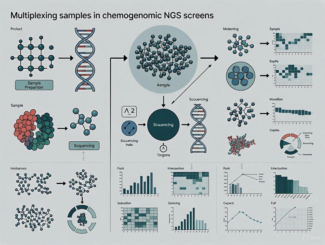
The integration of sample multiplexing is transformative for research scalability. For functional genomic screens, including those utilizing pooled shRNA or CRISPR libraries, sequencing the resulting mixed-oligo pools is a key challenge [3]. Multiplexing not only makes large-scale projects feasible but also optimizes resource utilization. The ability to pool samples means that sequencers can operate at maximum capacity, significantly reducing per-sample costs and reagent usage while dramatically increasing experimental throughput [1]. This efficiency is crucial in drug development, where screening campaigns may involve thousands of gene-compound interactions. The following diagram illustrates the logical workflow of a multiplexed NGS experiment, from sample preparation to data demultiplexing.
Core Concepts: Barcodes, Indexes, and Adapters
In multiplexed NGS, the terms barcode and index are often used interchangeably to refer to the short, known DNA sequences (typically 6-12 nucleotides) that are attached to each fragment in a library, uniquely marking its sample of origin [4]. These sequences are embedded within the adapters—longer, universal oligonucleotides that are covalently attached to the ends of the DNA fragments during library preparation [2]. The adapters serve multiple critical functions: they contain the primer-binding sites for the sequencing reaction and, crucially, the flow cell attachment sequences that allow the library fragments to bind to the sequencing platform [2]. The barcodes are strategically positioned within these adapter structures.
There are two primary indexing strategies, which differ in the location of the barcode sequence within the adapter, as shown in the diagram below.
Inline Indexing (Sample-Barcoding): With this strategy, the index sequence is located between the sequencing adapter and the actual genomic insert [4]. A key consequence of this design is that the barcode must be read out as part of the primary sequencing read (Read 1 or Read 2), which effectively reduces the available read length for the genomic insert itself [4]. The major advantage of inline indexing is that it permits early pooling of samples. Since the barcode is added in the initial reverse transcription or amplification step, hundreds of samples can be combined and processed simultaneously through subsequent workflow steps, leading to significant savings in consumables and hands-on time [4]. This makes inline indexing ideal for ultra-high-throughput applications, such as massive single-cell RNA sequencing or high-throughput drug screening.
Multiplex Indexing: In this more common strategy, the index sequences are located within the dedicated adapter regions, not the insert [4]. This requires designated Index Reads during the sequencing process, which are separate from the reads that sequence the genomic insert. Because the index is read independently, it has no impact on the insert read length [4]. Multiplex indexing can be further divided into single and dual indexing. Single indexing uses only one index (e.g., the i7 index), while dual indexing uses two separate indexes (the i7 and the i5 index) [1] [4]. Dual indexing is now considered best practice for most applications, as it provides a powerful mechanism for error correction and drastically reduces the rate of index hopping—a phenomenon where index sequences are incorrectly reassigned between molecules [1] [4].
Indexing Strategies for Optimal Experimental Design
Choosing the correct indexing strategy is a critical step in experimental design that directly impacts data quality, multiplexing capacity, and cost. The following table compares the primary indexing methods used in NGS.
Table 1: Comparison of NGS Indexing Strategies
| Strategy | Index Location | Read Method | Key Advantages | Key Limitations | Ideal Use Cases |
|---|---|---|---|---|---|
| Inline Indexing [4] | Within genomic insert | Part of primary sequencing read (Read 1/Read 2) | Enables early pooling; maximizes throughput; reduces hands-on time and cost for 1000s of samples | Reduces available insert read length; less error correction capability | Ultra-high-throughput screens, single-cell RNA-seq, QuantSeq-Pool |
| Single Indexing [4] | Within adapter (i7 only) | Dedicated Index Read | Shorter sequencing time; simpler design | Higher risk of index misassignment due to errors; no built-in error correction | Low-plexity studies, older sequencing platforms |
| Dual Indexing (Combinatorial) [1] [4] | Within adapter (i7 and i5) | Two Dedicated Index Reads | High multiplexing capacity; reduced index hopping vs. single indexing | Individual barcodes are re-used, limiting error correction | Most standard applications, general RNA-seq, exome sequencing |
| Unique Dual Indexing (UDI) [1] [4] | Within adapter (unique i7 and i5) | Two Dedicated Index Reads | Highest accuracy; enables index error correction; minimizes index hopping and misassignment | Requires more complex primer design and inventory | Chemogenomic screens, rare variant detection, sensitive applications |
For sensitive applications like chemogenomic CRISPR screens, Unique Dual Indexes (UDIs) are strongly recommended [4]. In a UDI system, each individual i5 and i7 index is used only once in the entire experiment. This creates a unique pair for each sample, which serves as two independent identifiers. The primary advantage is enhanced error correction: if a sequencing error occurs in one index of the pair, the second, error-free index can be used as a reference to pinpoint the correct sample identity and salvage the read [4]. This process, known as index error correction, can rescue approximately 10% of reads that would otherwise be discarded, maximizing data yield and ensuring the integrity of sample identity—a non-negotiable requirement in a quantitative screen where accurately tracking sgRNA abundance is paramount [4].
Practical Protocol for a Multiplexed Chemogenomic CRISPR Screen
The following section provides a detailed, step-by-step protocol for preparing sequencing libraries from a pooled chemogenomic CRISPR screen, incorporating best practices for multiplexing. This protocol is adapted from established methodologies for sequencing sgRNA libraries from genomic DNA [5] [3].
Step-by-Step Workflow
Genomic DNA (gDNA) Extraction:
- Input Material: Harvest cells from the completed CRISPR screen. The number of cells to collect is critical and must be calculated based on the desired library representation (see Table 2) [5].
- Procedure: Extract gDNA using a commercial kit (e.g., PureLink Genomic DNA Mini Kit). CRITICAL: Do not process more than 5 million cells per spin column to avoid clogging. For larger cell numbers, use multiple columns and pool the eluted gDNA [5].
- Quality Control: Quantify gDNA using a fluorometric method (e.g., Qubit dsDNA BR Assay). Assess purity via spectrophotometer (e.g., Nanodrop); 260/280 ratios should be 1.8-2.0 [5] [6]. Aim for a high concentration (>190 ng/µL) to minimize volume in subsequent PCR.
PCR Amplification and Indexing:
- Primer Design: Design primers to amplify the sgRNA integrated in the host genome. The forward primer should bind upstream of the guide spacer sequence and introduce the P5 Illumina adapter, stagger sequences (to increase nucleotide diversity), and the i7 index [5]. The reverse primer should bind downstream and introduce the P7 adapter and the i5 index [5]. For the highest data fidelity, use Unique Dual Index (UDI) primers.
- PCR Setup: Set up reactions in a decontaminated PCR workstation to avoid cross-contamination. UV-irradiate all tubes and tips before use [5].
- Reaction Conditions: Use a high-fidelity polymerase (e.g., Herculase). The number of parallel PCR reactions is determined by the total gDNA input required (see Table 2). To minimize heteroduplex formation (a major source of sequencing errors), use the minimum number of PCR cycles necessary for sufficient amplification and use magnetic beads for post-PCR clean-up instead of columns [3].
Library Purification and Quality Control:
- Purification: Pool all PCR reactions and purify using a magnetic bead-based clean-up system (e.g., GeneJET PCR Purification Kit). Beads effectively remove primers, enzymes, and small fragments while selecting for the desired library size [5] [3].
- Quality Control: Assess the final library concentration using a high-sensitivity fluorometric assay (e.g., Qubit dsDNA HS Assay). Validate the library size distribution using a bioanalyzer or agarose gel electrophoresis.
Pooling and Sequencing:
- Normalization and Pooling: Quantify all indexed libraries by qPCR or a high-sensitivity fluorometer. Normalize each library to an equimolar concentration and pool them together to create the final sequencing pool.
- Sequencing: Dilute the pooled library to the optimal concentration for clustering on your specific Illumina sequencing platform. A paired-end run is standard, with read lengths sufficient to cover the entire sgRNA sequence.
Table 2: Calculation of Input Requirements for CRISPR Library Representation (based on the Saturn V library example) [5]
| Saturn V Pool | Number of Guides | Library Representation at 300X | Minimum No. Cells for gDNA Extraction | Total Input gDNA Required (μg) | Parallel PCR Reactions (4 μg gDNA/reaction) |
|---|---|---|---|---|---|
| Pool 1 | 3,427 | 530X | 2,300,000 | 12 | 3 |
| Pool 2 | 3,208 | 567X | 2,300,000 | 12 | 3 |
| Pool 3 | 3,184 | 571X | 2,300,000 | 12 | 3 |
| Pool 4 | 1,999 | 606X | 1,500,000 | 8 | 2 |
| Pool 5 | 2,168 | 559X | 1,500,000 | 8 | 2 |
The Scientist's Toolkit: Essential Reagents and Materials
Table 3: Key Research Reagent Solutions for Multiplexed CRISPR Screen NGS
| Item | Function/Application | Example Products (Supplier) |
|---|---|---|
| gDNA Extraction Kit | Isolate high-quality, high-molecular-weight genomic DNA from screened cells. | PureLink Genomic DNA Mini Kit (Invitrogen) [5], QIAamp DNA Blood Maxi Kit (QIAGEN) [3] |
| High-Fidelity DNA Polymerase | Accurate amplification of the sgRNA region from gDNA with low error rate. | Herculase (Agilent Technologies) [5], Platinum Pfx (Invitrogen) [3] |
| Unique Dual Index (UDI) Primers | Provides unique i5/i7 index pairs for each sample to enable sample multiplexing with minimal index hopping. | xGen NGS Adapters & Indexing Primers (IDT) [2], NEXTFLEX UDI Barcodes (Revvity) [7] |
| PCR Purification Kit | Post-amplification clean-up to remove enzymes, salts, and short fragments. Magnetic beads help reduce heteroduplexes. | GeneJET PCR Purification Kit (Thermo Scientific) [5] [3] |
| DNA Quantification Kits | Fluorometric assays for precise quantification of gDNA (Broad Range) and final libraries (High Sensitivity). | Qubit dsDNA BR/HS Assay Kits (Invitrogen) [5] |
Troubleshooting and Technical Considerations
Even with a robust protocol, challenges can arise. Below are common issues and their solutions:
Challenge: Index Hopping. This occurs when index sequences are incorrectly assigned to reads, leading to sample misidentification. It is more prevalent on pattern flow cells (e.g., Illumina NovaSeq) [1] [4].
- Solution: Implement Unique Dual Indexes (UDIs). UDIs provide two unique identifiers per sample, allowing bioinformatic filters to detect and discard reads with non-matching index pairs, thus preventing misassignment [4].
Challenge: Heteroduplex Formation. During the final PCR amplification of a mixed library, incomplete extension can create heteroduplex molecules that lead to polyclonal clusters and failed sequencing reads [3].
- Solution: Minimize PCR cycles and use magnetic bead-based clean-up instead of spin columns, as beads are more effective at removing these heteroduplex structures [3].
Challenge: Mixing Indexes of Different Lengths. Combining libraries from different kits or vendors may result in a pool with varying index lengths (e.g., 8-nt and 10-nt indexes) [7].
- Solution: This is feasible. In the sample sheet, set the index length to the longest one in the pool (e.g., 10-nt). For shorter indexes, "pad" the sequence by adding bases (e.g., 'AT') to the end to match the required length. To maintain base diversity during the index read, ensure that over 50% of the pool consists of libraries with the longest index [7].
Sample multiplexing via barcodes and index adapters is an indispensable technique that underpins the scale and efficiency of modern NGS, most notably in complex, high-value applications like chemogenomic CRISPR screening. A deep understanding of the different indexing strategies—from inline to the highly recommended Unique Dual Indexing—empowers researchers to design robust, cost-effective, and high-quality studies. By adhering to the detailed protocols outlined herein, including careful calculation of library representation, meticulous PCR setup, and the use of UDIs, scientists can confidently execute multiplexed screens. This approach ensures the generation of reliable, high-integrity data that is crucial for identifying novel genetic interactions and accelerating the journey toward new therapeutic discoveries.
In the field of chemogenomics, next-generation sequencing (NGS) has become an indispensable tool for unraveling the complex interactions between chemical compounds and biological systems. Chemogenomic screens, which utilize pooled shRNA or CRISPR libraries, enable the systematic interrogation of gene function and drug-target relationships on a genome-wide scale [3]. A central challenge in these studies is managing the immense scale of data generation in a cost- and time-efficient manner. Sample multiplexing, also known as multiplex sequencing, addresses this challenge by allowing large numbers of libraries to be sequenced simultaneously during a single NGS run [1]. This approach transforms the economics of large-scale genetic screens by exponentially increasing the number of samples analyzed without proportionally increasing costs or experimental time [1]. The core principle involves labeling individual DNA fragments from different samples with unique DNA barcodes (indexes) during library preparation, which enables computational separation of the data after sequencing [1]. For chemogenomic research, where screening entire libraries of compounds against comprehensive genetic backgrounds is essential, multiplexing provides the throughput necessary to achieve statistical power and biological relevance.
Economic and Operational Advantages
The implementation of multiplexing strategies confers significant economic and operational benefits, making large-scale chemogenomic projects feasible for individual laboratories.
Table 1: Economic Advantages of Multiplexed NGS in Chemogenomic Screens
| Factor | Standard NGS | Multiplexed NGS | Impact on Chemogenomic Screens |
|---|---|---|---|
| Cost per Sample | High | Dramatically reduced [1] | Enables screening of more compounds/conditions within same budget |
| Sequencing Time | Linear increase with sample number | Minimal increase with sample number [1] | Accelerates target discovery and validation cycles |
| Reagent Consumption | Proportional to sample number | Significantly reduced [1] | Lowers per-datapoint cost in high-throughput compound profiling |
| Labor & Hands-on Time | High for multiple library preps | Consolidated into fewer, larger runs [1] | Increases research efficiency in functional genomics labs |
| Data Generation Rate | Limited by sequential processing | High-throughput; 100s of samples in parallel [1] | Facilitates robust, statistically powerful screens |
The economic imperative for multiplexing is clear. By pooling samples, researchers optimize instrument use, reduce reagent consumption, and decrease the hands-on time required per sample [1]. This is particularly critical in chemogenomic screens, where researchers often need to test multiple compound concentrations, time points, and genetic backgrounds against entire shRNA or CRISPR libraries [3]. The alternative—running samples individually—is prohibitively expensive and slow. The global NGS market's rapid growth, driven by factors like increased adoption in clinical diagnostics and drug discovery, underscores the technology's central role in modern bioscience [8]. Multiplexing ensures that chemogenomic studies can remain at the cutting edge without being constrained by resource limitations.
Key Multiplexing Methodologies and Protocols
Core Principles: Barcoding and Indexing Strategies
At the heart of sample multiplexing is the use of unique DNA barcodes, or indexes. These short, known DNA sequences are ligated to the fragments of each sample library during preparation [1]. When samples are pooled and sequenced, the sequencer reads both the genomic DNA and the barcode. Sophisticated bioinformatics software then uses these barcode sequences to demultiplex the data, sorting the sequenced reads back into their respective sample-specific files for downstream analysis [9] [10]. The choice of indexing strategy is critical for minimizing errors and maximizing multiplexing capacity.
- Unique Dual Indexes (UDI): This is the recommended strategy for complex chemogenomic screens. UDI employs two unique barcodes on each fragment—one on each end. This provides an error-correction mechanism, as an index hop (where a barcode is incorrectly assigned) is highly unlikely to occur for both indexes simultaneously. This dramatically reduces misassignment and cross-talk between samples, ensuring the integrity of sample identity in pooled screens [1].
- Unique Molecular Identifiers (UMIs): For applications requiring ultra-high accuracy in quantifying allele frequencies or transcript counts, UMIs are incorporated. UMIs are random molecular barcodes added to each molecule before amplification. This allows bioinformatics tools to distinguish between biologically unique molecules and PCR duplicates, thereby reducing false-positive variant calls and increasing the sensitivity of detection [1]. This is vital in chemogenomics for accurately determining guide RNA abundances in CRISPR screens or quantifying dropout of shRNAs in response to compound treatment.
Detailed Protocol: Maximizing Throughput in Pooled shRNA/CRISPR Screens
Pooled chemogenomic screens are highly susceptible to sequencing failures due to the formation of secondary structures (hairpins) and heteroduplexes in mixed-oligo PCR reactions [3]. The following optimized protocol mitigates these issues to maximize usable data from a single run.
A. Library Amplification from Genomic DNA
- Isolate Genomic DNA: From cells transduced with the pooled shRNA or CRISPR library, using a kit such as the QIAamp DNA Blood Maxi Kit [3].
- Amplify the Library: Perform PCR amplification using a high-fidelity DNA polymerase (e.g., Platinum Pfx). Critical parameters include:
- Template: Use ~20 µg of genomic DNA to ensure sufficient representation of each shRNA/sgRNA in the library [3].
- Primers: Design primers compatible with your sequencer and which flank the shRNA/sgRNA sequence.
- PCR Cycles: Minimize the number of cycles (e.g., 30 cycles) to reduce the formation of heteroduplexes, which are a major cause of low-quality, polyclonal reads [3].
- Purify the Product: Pool PCR reactions and purify using a magnetic bead-based purification system (e.g., GeneJET PCR Purification Kit). Beads are preferred over gel extraction at this stage for speed and to minimize heteroduplex formation [3].
B. Overcoming Hairpin Structures (Half-shRNA Method)
This step is crucial for shRNA libraries, which contain palindromic sequences that form hairpins, leading to incomplete and failed sequencing reads [3].
- Restriction Digest: Digest the purified PCR product with a restriction enzyme (e.g., XhoI) that cuts specifically within the loop region of the shRNA hairpin. Perform the digestion immediately after PCR purification to avoid cruciform formation [3].
- Ligate Adapter: Purify the digested product to remove the small, cut loop fragment. Ligate a custom adapter oligonucleotide to the end of the now-linearized shRNA fragment. This adapter provides the sequence necessary for binding to the sequencing flow cell [3].
- Final Amplification: Perform a second, limited-cycle PCR with primers that bind the adapter and the original library-specific sequence to generate the final sequencing library.
C. Library Quantification and Pooling
- Quantify each barcoded library accurately using a fluorometric method (e.g., Qubit).
- Pool Equimolar amounts of each uniquely barcoded library into a single tube. Use a pooling calculator to normalize contributions and ensure even sequencing coverage across all samples [1].
D. Sequencing
Sequence the pooled library on an appropriate Illumina sequencer (e.g., MiSeq, NextSeq, or NovaSeq), following the manufacturer's instructions for loading and data generation [1].
Diagram: Multiplexing Workflow for Pooled Screens. This workflow illustrates the key steps, from library preparation to computational demultiplexing, highlighting stages critical for overcoming technical challenges like hairpins.
Data Analysis Workflow for Multiplexed Screens
The data analysis pipeline for a multiplexed chemogenomic screen is a multi-stage process that transforms raw sequencer output into biologically interpretable results.
Primary Analysis occurs on the sequencer and involves the conversion of raw signal data (e.g., fluorescence, pH change) into nucleotide base calls. The key output of this stage is the FASTQ file, which contains the sequence of each read and its corresponding per-base quality score (Phred score) [9] [10]. A critical step in primary analysis is demultiplexing, where the sequencer's software uses the index reads to sort all sequences into separate FASTQ files, one for each sample in the pool [9].
Secondary Analysis begins with quality control and alignment.
- Read Cleanup: Tools like Trimmomatic or FastQC are used to trim adapter sequences and remove low-quality reads or portions of reads (typically with a Phred score cutoff of <30) [10]. This step generates a "cleaned" FASTQ file.
- Alignment (Mapping): The cleaned reads are aligned to a reference genome (e.g., hg38 for human) using specialized aligners like BWA or Bowtie. The output is a BAM (Binary Alignment Map) file, a compressed, efficient format storing how each read maps to the genome [9] [10] [11].
- Variant Calling: For chemogenomic screens, the crucial step is not variant calling but molecular barcode counting. Custom scripts or tools are used to count the number of reads corresponding to each unique shRNA or sgRNA sequence from the BAM file, generating a count table [10] [3].
Tertiary Analysis involves the biological interpretation of the data. The count table for each sample (condition, compound treatment) is analyzed to identify shRNAs/sgRNAs that are significantly enriched or depleted compared to a control (e.g., DMSO-treated cells). This statistical analysis, often using specialized software, reveals genes essential for survival under specific chemical treatments, thereby identifying potential drug targets or resistance mechanisms [10].
Diagram: NGS Data Analysis Pipeline. The three-stage workflow from raw data to biological interpretation, showing key file types and processes.
The Scientist's Toolkit: Essential Reagents and Materials
Table 2: Key Research Reagent Solutions for Multiplexed Chemogenomic Screens
| Item | Function | Application Note |
|---|---|---|
| NGS Library Prep Kit | Provides enzymes and buffers for end-repair, A-tailing, and adapter ligation. | Select kits designed for complex genomic DNA inputs and that support dual indexing [3]. |
| Unique Dual Indexed (UDI) Adapters | Contains the unique barcode sequences for multiplexing. | UDIs are essential for minimizing index hopping in pooled screens, ensuring sample identity integrity [1]. |
| High-Fidelity DNA Polymerase | Amplifies the library from genomic DNA with low error rates. | Critical for accurate representation of the shRNA/sgRNA pool; minimizes PCR-introduced errors [3]. |
| Magnetic Bead-based Purification Kits | For size selection and cleanup of DNA after enzymatic reactions. | Preferred over column-based or gel extraction for higher yield and to reduce heteroduplex formation [3]. |
| Restriction Enzyme (e.g., XhoI) | Digests hairpin structures in shRNA libraries. | Key for the "half-shRNA" method to prevent sequencing failures due to secondary structures [3]. |
| Fluorometric Quantification Assay | Accurately measures DNA concentration. | Essential for normalizing library concentrations before pooling to ensure even sequencing coverage [3]. |
| Pooled shRNA/CRISPR Library | The core reagent containing the collection of genetic perturbagens. | Libraries targeting specific gene families (e.g., kinome) are ideal for focused chemogenomic screens [3]. |
The strategic implementation of multiplexing is a cornerstone of modern, high-throughput chemogenomics. By enabling the processing of hundreds of samples in a single NGS run, it provides an undeniable economic and throughput advantage, making large-scale, statistically robust screens routine. Adhering to optimized protocols that address technical challenges like heteroduplex formation and hairpin structures, combined with the use of robust bioinformatics pipelines, ensures the generation of high-quality, reliable data. As NGS technology continues to evolve, becoming faster and more cost-effective, its synergy with advanced multiplexing strategies will further empower researchers to deconvolute the complex interplay between genes and small molecules, accelerating the pace of drug discovery and therapeutic development.
Multiplexing as a Pillar of Modern Chemogenomics and Functional Genomics Screens
Multiplexing has emerged as a foundational methodology that has fundamentally transformed the scale and efficiency of chemogenomic and functional genomic research. This approach, which enables the simultaneous processing and analysis of numerous samples or perturbations within a single experiment, provides the technical framework for high-throughput screening campaigns essential for modern drug discovery and functional genomics. The core principle of multiplexing involves strategically "barcoding" individual samples or perturbations with unique identifiers, allowing them to be pooled and processed collectively while maintaining the ability to deconvolute results back to their origin through computational demultiplexing [1] [12]. This paradigm has become indispensable for addressing the complexity of biological systems, where understanding the relationships between genetic variants, chemical perturbations, and phenotypic outcomes requires testing thousands to millions of experimental conditions.
The adoption of multiplexing strategies across genomics, transcriptomics, proteomics, and chemogenomics has accelerated the transition from reductionist, single-target approaches to systems-level investigations. In chemogenomics, where small molecule libraries are screened against biological systems to identify bioactive compounds and their mechanisms of action, multiplexing enables the efficient profiling of extensive compound libraries [13]. Similarly, in functional genomics, which seeks to understand gene function and regulation, multiplexed assays make it feasible to systematically interrogate the consequences of thousands of genetic perturbations in parallel [14] [15]. The integration of these fields through multiplexed approaches provides unprecedented opportunities to link chemical and genetic perturbations to molecular and cellular phenotypes, offering comprehensive insights into disease mechanisms and therapeutic strategies.
Fundamental Principles and Advantages of Multiplexing
Core Concepts and Methodological Framework
At its essence, multiplexing relies on the incorporation of unique molecular tags, or barcodes, that serve as sample identifiers throughout experimental workflows. These barcodes can be introduced at various stages: during library preparation for next-generation sequencing (NGS) [1], through metabolic or chemical labeling in proteomic studies [16], via lentiviral vectors for genetic perturbations [12], or through antibody-based tagging methods in single-cell studies [12]. The strategic application of these identifiers enables researchers to combine multiple experimental conditions, significantly reducing reagent costs, instrument time, and technical variability while dramatically increasing experimental throughput.
Two primary indexing strategies dominate multiplexed NGS approaches: single indexing and dual indexing. Single indexing employs one barcode sequence per sample, while dual indexing uses two separate barcode sequences, providing a much larger combinatorial space for sample identification [1]. Dual indexing is particularly valuable in large-scale screens as it exponentially increases the number of samples that can be uniquely tagged and pooled. For example, a dual indexing system with 24 unique i5 indexes and 24 unique i7 indexes can theoretically multiplex 576 samples in a single sequencing run. This strategy also helps mitigate index hopping—a phenomenon where barcode sequences are incorrectly assigned during sequencing—which can compromise data integrity in highly multiplexed experiments [1].
Key Advantages in Screening Applications
The implementation of multiplexing strategies confers several critical advantages that make large-scale chemogenomic and functional genomic screens technically and economically feasible:
Cost Efficiency: Pooling samples exponentially increases the number of samples analyzed in a single sequencing run or mass spectrometry injection without proportionally increasing costs. This efficiency makes large-scale screens accessible even with limited resources [1] [16].
Reduced Technical Variability: Processing all samples simultaneously under identical conditions minimizes batch effects and technical noise, enhancing the statistical power to detect true biological signals [12].
Increased Throughput: Multiplexing enables the processing of hundreds to thousands of samples in timeframes previously required for just a handful of samples, dramatically accelerating screening timelines [14] [15].
Internal Controls: Multiplexed designs naturally incorporate internal controls and reference standards within the same experiment, improving normalization and quantitative accuracy [16].
Resource Conservation: By reducing the consumption of expensive reagents, antibodies, and sequencing capacity, multiplexing extends research budgets while maximizing data output [1] [17].
Technological Approaches for Multiplexed Screens
Massively Parallel Reporter Assays (MPRAs)
Massively Parallel Reporter Assays represent a powerful multiplexing approach for functionally characterizing noncoding genetic variants. MPRAs utilize synthetic oligonucleotide libraries containing thousands to millions of putative regulatory elements, each coupled to a unique barcode sequence. These libraries are introduced into cells, where the transcriptional activity of each element drives the expression of its associated barcode. By quantifying barcode abundance through high-throughput sequencing, researchers can simultaneously assess the regulatory potential of thousands of sequences in a single experiment [14].
The key advantage of MPRA lies in its direct measurement of regulatory function and its ability to test sequences outside their native genomic context, eliminating confounding effects from local chromatin environment or three-dimensional genome architecture. However, this strength also represents MPRAs' primary limitation: the artificial context may not fully recapitulate endogenous regulatory dynamics. Additionally, MPRAs cannot inherently identify the target genes of regulatory elements, requiring complementary approaches to establish physiological relevance [14].
CRISPR-Based Pooled Screens
CRISPR-based technologies have revolutionized functional genomics by enabling precise genetic perturbations at unprecedented scale. Pooled CRISPR screens introduce complex libraries of guide RNAs (gRNAs) targeting thousands of genomic loci into populations of cells, with each gRNA acting as both a perturbation agent and a unique barcode for that perturbation [14] [15]. The power of this approach lies in its flexibility—different CRISPR systems can be employed to achieve diverse perturbation modalities:
- CRISPR Knockout: Utilizes Cas9 nuclease to create double-strand breaks, resulting in gene disruption through imperfect repair [14].
- CRISPR Interference (CRISPRi): Employs catalytically dead Cas9 (dCas9) fused to repressor domains to reversibly suppress gene expression without altering DNA sequence [14].
- CRISPR Activation (CRISPRa): Uses dCas9 fused to transcriptional activator domains to enhance gene expression [14].
- Base Editing: Leverages Cas9 nickase fused to deaminase enzymes to directly convert one base to another without double-strand breaks [15].
- Prime Editing: Utilizes Cas9 nickase fused to reverse transcriptase to mediate all possible base-to-base conversions, small insertions, and small deletions [15].
These diverse CRISPR tools enable researchers to tailor their screening approach to specific biological questions, from essential gene identification to nuanced studies of transcriptional regulation or specific mutational effects.
Single-Cell Multiomic Technologies
Recent advances in single-cell technologies have enabled multiplexed analysis at unprecedented resolution. Single-cell DNA-RNA sequencing (SDR-seq) simultaneously profiles up to 480 genomic DNA loci and gene expression in thousands of single cells, enabling accurate determination of coding and noncoding variant zygosity alongside associated transcriptional changes [18]. This joint profiling confidently links precise genotypes to gene expression in their endogenous context, overcoming limitations of methods that use guide RNAs as proxies for variant perturbation [18].
Several sample-multiplexing strategies have been developed for single-cell sequencing to overcome challenges of inefficient sample processing, high costs, and technical batch effects:
- Natural Genetic Variation: Demultiplexing based on naturally occurring genetic variants using tools like demuxlet, scSplit, Vireo, or Souporcell [12].
- Cell Hashing: Uses oligo-tagged antibodies against ubiquitous cell-surface proteins to label cells from different samples prior to pooling [12].
- MULTI-seq: Employs lipid-modified oligonucleotides that incorporate into cell membranes to barcode live cells [12].
- Nucleus Hashing: Adapts cell hashing principles for nuclei isolation using DNA-barcoded antibodies targeting the nuclear pore complex [12].
These approaches enable "super-loading" of single cells, significantly increasing throughput while reducing multiplet rates and identifying technical artifacts [12]. The ability to pool multiple samples prior to single-cell processing also minimizes batch effects and reduces per-sample costs, making large-scale single-cell studies more feasible.
Table 1: Comparison of Major Multiplexing Technologies
| Technology | Multiplexing Capacity | Primary Applications | Key Advantages | Limitations |
|---|---|---|---|---|
| MPRA | 10³-10⁶ variants/experiment | Functional characterization of noncoding variants | Direct measurement of regulatory function; High throughput | Artificial genomic context; Cannot infer endogenous target genes |
| CRISPR Screens | 10³-10⁵ gRNAs/experiment | Functional genomics; Gene discovery; Mechanism of action studies | Endogenous genomic context; Diverse perturbation modalities; Target gene identification | Relatively lower throughput; Potential for confounding off-target effects |
| Single-Cell Multiomics | 10³-10⁵ cells/experiment; 2-8 samples/pool | Cellular heterogeneity; Gene regulation studies; Tumor evolution | Single-cell resolution; Combined genotype-phenotype information | Technical complexity; Higher cost per cell; Limited molecular targets per cell |
| Isobaric Labeling (Proteomics) | 2-54 samples/experiment [16] | Quantitative proteomics; Drug mechanism studies | Reduced instrument time; Internal controls; High quantitative accuracy | Potential for reporter ion interference; Limited multiplexing compared to genetic approaches |
Experimental Protocols for Multiplexed Functional Genomics
Saturation Genome Editing for Variant Functionalization
Multiplexed Assays for Variant Effects (MAVEs) enable comprehensive functional assessment of all possible genetic variations within specific genomic regions. The following protocol outlines the steps for saturation genome editing to study variant effects:
Step 1: sgRNA Sequence Design
- Import the fully annotated gene of interest (e.g., from Ensembl) into a sequence analysis platform like Benchling.
- Select the target exon and use the guide RNA algorithm to generate 20 bp sgRNA sequences with high on-target and low off-target scores.
- Incorporate a synonymous change that disrupts the Protospacer Adjacent Motif (PAM) site to serve as a fixed marker that blocks Cas9 recutting after editing.
- Verify that PAM modification does not impact splicing using SpliceAI prediction (score > 0.2 indicates potential splicing impact).
- Order sgRNA sequences and forward/reverse primers for cloning (forward primer: 5′-CACCG + 20 bp sgRNA sequence; reverse primer: 5′-AAAC + reverse complement + C) [15].
Step 2: Oligo Donor Library Design
- Design 180 bp antisense oligos covering the Cas9 cut site with homology arms positioned such that the cut site is in the middle.
- Incorporate the fixed PAM modification into all oligos as a homologous directed repair (HDR) marker.
- Systematically change each nucleotide position across the saturation region to all three non-wild-type bases using standard mixed base nomenclature (e.g., H = A/C/T, B = G/C/T, V = A/C/G, D = A/G/T).
- Order the oligo library as 180 bp ultramers for direct use in nucleofection without cloning [15].
Step 3: Cell Culture and Nucleofection
- Culture mouse embryonic stem cells (mESCs) containing a single copy of the human gene of interest integrated into the mouse genome on SNLP feeder dishes in mESC maintenance media.
- Prepare cells for nucleofection by trypsinization and resuspension in nucleofection solution.
- Combine sgRNA (or Cas9-sgRNA ribonucleoprotein complex) with the oligo donor library and nucleofect into mESCs.
- Plate transfected cells and allow recovery for 48-72 hours before drug selection or phenotypic analysis [15].
Step 4: Genomic DNA Amplification and Sequencing
- Harvest genomic DNA from pooled edited cells after phenotypic selection or at designated timepoints.
- Amplify edited genomic regions using primers flanking the target site with Illumina adapter overhangs.
- Purify PCR products and prepare sequencing libraries using standard Illumina library preparation methods.
- Sequence on an appropriate Illumina platform to achieve sufficient coverage for variant quantification (typically >1000x coverage) [15].
Step 5: Computational Analysis
- Process sequencing data to quantify variant abundance and calculate indel rates.
- Normalize variant counts to control for amplification bias and sequencing depth.
- Determine functional impact of variants based on their enrichment or depletion under selective conditions compared to non-selective conditions.
- Classify variants as functional (enriched/depleted) or neutral (no change in abundance) [15].
Protocol for Single-Cell DNA-RNA Sequencing (SDR-seq)
SDR-seq enables simultaneous profiling of genomic DNA loci and gene expression in thousands of single cells, providing a powerful approach to link genotypes to transcriptional phenotypes:
Step 1: Cell Preparation and Fixation
- Dissociate cells into a single-cell suspension using appropriate enzymatic or mechanical methods.
- Count cells and assess viability using trypan blue exclusion or similar methods.
- Fix cells using either paraformaldehyde (PFA) or glyoxal. Glyoxal is preferred for better nucleic acid preservation as it does not cross-link nucleic acids [18].
- Permeabilize fixed cells to enable access to intracellular components for subsequent molecular reactions.
Step 2: In Situ Reverse Transcription
- Perform in situ reverse transcription using custom poly(dT) primers that add a unique molecular identifier (UMI), sample barcode, and capture sequence to cDNA molecules.
- This step preserves the cellular origin of RNA molecules while adding the necessary information for downstream demultiplexing and sequencing.
Step 3: Droplet-Based Partitioning and Amplification
- Load cells containing cDNA and gDNA onto the Tapestri platform (Mission Bio) for droplet-based partitioning.
- Generate first droplets containing individual cells, then lyse cells and treat with proteinase K to release nucleic acids.
- Mix with reverse primers for each intended gDNA or RNA target and generate second droplets containing forward primers with capture sequence overhangs, PCR reagents, and barcoding beads with cell barcode oligonucleotides.
- Perform multiplexed PCR within droplets to amplify both gDNA and RNA targets, with cell barcoding achieved through complementary capture sequence overhangs [18].
Step 4: Library Preparation and Sequencing
- Break emulsions and purify amplified products.
- Prepare separate sequencing libraries for gDNA and RNA using distinct overhangs on reverse primers (R2N for gDNA, R2 for RNA).
- This separation enables optimized sequencing for each data type: full-length coverage for gDNA variants and standard RNA-seq libraries for transcriptome analysis.
- Sequence libraries on an Illumina platform with appropriate read lengths to cover the targeted regions [18].
Step 5: Data Integration and Analysis
- Process sequencing data to assign reads to individual cells based on cell barcodes.
- Demultiplex samples based on genetic variants or sample barcodes introduced during in situ RT.
- Call variants from gDNA reads and quantify gene expression from RNA reads.
- Integrate data to associate specific genotypes with transcriptional changes at single-cell resolution [18].
Research Reagent Solutions for Multiplexed Screens
Successful implementation of multiplexed screening approaches requires carefully selected reagents and materials optimized for high-throughput applications. The following table details essential research reagent solutions for establishing multiplexed functional genomics and chemogenomics workflows:
Table 2: Essential Research Reagents for Multiplexed Genomic Screens
| Reagent Category | Specific Examples | Function in Multiplexed Screens | Key Considerations |
|---|---|---|---|
| Barcoding Reagents | Unique Dual Indexes (Illumina) [1]; Cell Hashing Antibodies [12]; MULTI-seq Lipids [12] | Sample multiplexing; Sample origin identification | Barcode diversity; Minimal sequence similarity; Compatibility with downstream applications |
| Library Preparation Kits | Illumina DNA Prep; Nextera XT; NEBNext Ultra II [17] | NGS library construction; Adapter ligation; Library amplification | Efficiency for low-input samples; Compatibility with automation; Fragment size distribution |
| CRISPR Components | Cas9 enzymes; sgRNA libraries; Base editors; Prime editors [14] [15] | Genetic perturbation; Screening libraries; Precision genome editing | Editing efficiency; Specificity; Delivery method; Off-target effects |
| Single-Cell Platforms | 10x Genomics Chromium; BD Rhapsody; Mission Bio Tapestri [18] [12] | Single-cell partitioning; Barcoding; Library preparation | Cell throughput; Multiplexing capacity; Multiomics capabilities; Cost per cell |
| Quantitative Proteomics Reagents | TMT & iTRAQ isobaric tags [16]; DiLeu tags [16]; SILAC amino acids [16] | Multiplexed protein quantification; Sample multiplexing in MS | Number of plex; Labeling efficiency; Cost; Reporter ion interference |
| Cell Painting Reagents | Cell Painting kit (Broad Institute); Fluorescent dyes [13] | Morphological profiling; Phenotypic screening | Image quality; Stain specificity; Compatibility with automation; Feature extraction |
Data Analysis and Computational Considerations
The computational demultiplexing and analysis of data generated from multiplexed screens present unique challenges and considerations. Effective analysis pipelines must address several key aspects:
Demultiplexing Strategies: The approach to sample demultiplexing depends on the barcoding method employed. For genetically multiplexed samples, tools like demuxlet, scSplit, Vireo, and Souporcell use natural genetic variation to assign cells to their sample of origin [12]. These tools employ different statistical approaches—including maximum likelihood models, hidden state models, and Bayesian methods—to confidently assign cells to samples based on reference or reference-free genotyping. For antibody-based hashing methods, demultiplexing involves detecting the antibody-derived tags (ADTs) associated with each cell and comparing their expression patterns to assign sample identity [12].
Multiomic Data Integration: Advanced multiplexing approaches like SDR-seq generate coupled DNA and RNA measurements from the same single cells, requiring specialized integration methods [18]. These analyses must account for technical factors such as allelic dropout (where one allele fails to amplify), cross-contamination between cells, and the sparsity inherent in single-cell data. Successful integration enables researchers to directly link genotypes (e.g., specific mutations) to transcriptional phenotypes (e.g., differential expression) within the same cells, providing powerful insights into variant function [18].
Hit Identification and Validation: In pooled screening approaches, identifying true hits requires careful statistical analysis to distinguish biologically significant signals from technical noise. Methods like MAGeCK, BAGEL, and drugZ implement specialized statistical models that account for guide-level efficiency, screen dynamics, and multiple testing correction. For chemogenomic screens integrating chemical and genetic perturbations, network-based approaches can help identify functional modules and pathways affected by compound treatment [13].
Application in Chemogenomics and Drug Discovery
Multiplexed approaches have become indispensable tools in modern drug discovery, particularly in the emerging field of network pharmacology which considers the complex interactions between drugs and multiple biological targets [13]. Chemogenomic libraries comprising 5,000 or more small molecules representing diverse target classes enable systematic profiling of compound activities against biological systems [13]. When combined with multiplexed readouts, these libraries provide unprecedented insights into compound mechanism of action, polypharmacology, and cellular responses.
Morphological Profiling: The Cell Painting assay represents a powerful multiplexed phenotypic screening approach that uses multiplexed fluorescence imaging to capture thousands of morphological features in treated cells [13]. When applied to chemogenomic libraries, this approach generates high-dimensional phenotypic profiles that can be used to cluster compounds with similar mechanisms of action, identify novel bioactive compounds, and deconvolute the cellular targets of uncharacterized compounds. The integration of morphological profiles with chemical and target information in network pharmacology databases enables predictive modeling of compound activities [13].
Target Deconvolution: A major challenge in phenotypic drug discovery is identifying the molecular targets responsible for observed phenotypic effects. Multiplexed chemogenomic approaches address this challenge by screening compound libraries against diverse genetic backgrounds or in combination with genetic perturbations. For example, profiling compound sensitivity across cell lines with different genetic backgrounds or in combination with CRISPR-based genetic perturbations can help identify synthetic lethal interactions and resistance mechanisms, providing clues about compound mechanism of action [13].
Network Pharmacology: The integration of multiplexed screening data with biological networks enables a systems-level understanding of drug action. By mapping compound-target interactions onto protein-protein interaction networks, signaling pathways, and gene regulatory networks, researchers can identify network neighborhoods and functional modules affected by compound treatment [13]. This network pharmacology perspective moves beyond the traditional "one drug, one target" paradigm to consider the systems-level effects of pharmacological intervention, potentially leading to more effective therapeutic strategies with reduced side effects.
Visualizing Multiplexed Screening Workflows
The following diagrams illustrate key experimental workflows and conceptual frameworks for multiplexed screening approaches:
Diagram 1: Conceptual workflow for sample multiplexing approaches showing the integration of multiple samples through barcoding and pooling, followed by unified processing and computational demultiplexing. Key advantages include cost efficiency, reduced technical variability, and increased throughput.
Diagram 2: Workflow for multiplexed CRISPR screening showing key steps from library design and delivery through phenotypic selection and sequencing-based readout. Different CRISPR modalities enable diverse perturbation types including gene knockout, transcriptional modulation, and precise base editing.
Multiplexing technologies have fundamentally transformed the scale and scope of chemogenomic and functional genomic research, enabling systematic interrogation of biological systems at unprecedented resolution. The integration of diverse multiplexing approaches—from pooled genetic screens to single-cell multiomics and high-content phenotypic profiling—provides complementary insights into gene function, regulatory mechanisms, and compound mode of action. As these technologies continue to evolve, several exciting directions promise to further enhance their capabilities and applications.
The ongoing development of higher-plex methods will enable even more comprehensive profiling in single experiments. In proteomics, recent advances have expanded isobaric tagging from 2-plex to 54-plex approaches [16], while single-cell technologies now routinely profile tens of thousands of cells in individual runs [12]. Future improvements will likely focus on increasing multiplexing capacity while reducing technical artifacts such as index hopping in sequencing [1] and reporter ion interference in mass spectrometry [16].
The integration of multiplexed functional data with large-scale biobanks and clinical datasets represents another promising direction. As multiplexed assays are applied to characterize the functional impact of variants identified in population-scale sequencing studies, they will provide mechanistic insights into disease pathogenesis and potential therapeutic strategies [14]. Similarly, the application of multiplexed chemogenomic approaches to patient-derived samples, including organoids and primary cells, will enhance the translational relevance of screening findings.
Finally, advances in artificial intelligence and machine learning will revolutionize the analysis and interpretation of multiplexed screening data. These approaches can identify complex patterns in high-dimensional data, predict variant functional effects, and prioritize candidate compounds or targets for further investigation. As multiplexed screening technologies continue to generate increasingly large and complex datasets, sophisticated computational methods will be essential for extracting biologically and clinically meaningful insights.
In conclusion, multiplexing has established itself as an indispensable pillar of modern chemogenomics and functional genomics, providing the technical foundation for large-scale, systematic investigations of biological systems. Through continued methodological refinement and innovative application, these approaches will continue to drive advances in basic research and therapeutic development for years to come.
Unique Dual Indexes (UDIs) and Unique Molecular Identifiers (UMIs) for Error Correction
In the context of chemogenomic NGS screens, where the parallel testing of numerous chemical compounds on multiplexed biological samples is standard, ensuring data integrity is paramount. Accurate demultiplexing and variant calling are critical for correlating chemical perturbations with genomic outcomes. Unique Dual Indexes (UDIs) and Unique Molecular Identifiers (UMIs) are two powerful barcoding strategies that, when integrated into next-generation sequencing (NGS) workflows, provide robust error correction and mitigate common artifacts. UDIs are essential for accurate sample multiplexing, effectively preventing sample misassignment—a phenomenon known as index hopping [19] [20] [21]. In contrast, UMIs are molecular barcodes that tag individual nucleic acid fragments before amplification, enabling bioinformaticians to distinguish true biological variants from errors introduced during PCR amplification and sequencing, thereby increasing the sensitivity of detecting low-frequency variants [22] [23]. For chemogenomic screens, which often involve limited samples like single cells or low-input DNA/RNA, the combination of these technologies provides a framework for highly accurate, quantitative, and multiplexed analysis.
Table 1: Core Functions of UDIs and UMIs
| Feature | Unique Dual Indexes (UDIs) | Unique Molecular Identifiers (UMIs) |
|---|---|---|
| Primary Function | Sample multiplexing and demultiplexing | Identification and correction of PCR/sequencing errors |
| Level of Application | Per sample library | Per individual molecule |
| Key Benefit | Prevents sample misassignment due to index hopping | Enables accurate deduplication and rare variant detection |
| Impact on Cost | Reduces per-sample cost by enabling higher multiplexing | Prevents wasteful analysis of false positives, improving data quality |
Understanding Unique Dual Indexes (UDIs)
Principles and Design
Unique Dual Indexes consist of two unique nucleotide sequences—an i7 and an i5 index—ligated to opposite ends of each DNA fragment in a sequencing library [19] [21]. In a pool of 96 samples, for instance, each sample receives a truly unique pair of indexes; these index combinations are not reused or shared across any other sample in the pool [19] [20]. This design is a significant improvement over combinatorial dual indexing, where a limited set of indexes (e.g., 8 i7 and 8 i5) are combined to create a theoretical 64 unique pairs, but where sequences are repeated across a plate, increasing the risk of misassignment [19]. The uniqueness of the UDI pair is the key to its error-correction capability. During demultiplexing, the sequencing software expects only a specific set of i7-i5 combinations. Reads that exhibit an unexpected index pair—a result of index hopping where an index dimer erroneously attaches to a different library molecule—can be automatically identified and filtered out, thus preserving the integrity of sample identity [19] [21]. This is particularly crucial when using modern instruments with patterned flow cells, like the Illumina NovaSeq 6000, where index hopping rates can be significant [19] [21].
Application Note for Chemogenomic Screens
In a typical chemogenomic screen, researchers may treat hundreds of cell lines or pools with different chemical compounds and need to sequence them all in parallel. UDIs enable the precise pooling of these libraries, ensuring that the genomic data for a cell line treated with compound "A" is never confused with that treated with compound "B." This accurate sample tracking is the foundation for a reliable screen.
Protocol: Implementing UDI-Based Multiplexing
- Library Preparation and UDI Ligation: During the NGS library prep, use a kit or system that incorporates UDIs. Examples include the IDT for Illumina UD Indexes or the Twist Bioscience HT Universal Adapter System [19] [20]. The UDI adapters are ligated to the fragmented genomic DNA or cDNA.
- Library Pooling: Quantify the final concentration of each uniquely indexed library. Combine equimolar amounts of each library into a single pool. With a single UDI plate, you can confidently pool up to 96 samples [19].
- Sequencing: Sequence the pooled library on your chosen Illumina platform. Ensure the sequencing run includes the additional cycles required to read both the i7 and i5 indexes.
- Demultiplexing and Data Analysis: Use Illumina's standard demultiplexing software (e.g., Illumina DRAGEN BaseSpace App or bcl2fastq). The software will assign reads to their correct sample based on the expected UDI pairs and will filter out reads with index combinations not present in the sample sheet, effectively mitigating the effects of index hopping [19] [21].
Diagram 1: UDI Workflow for Error-Free Multiplexing. This diagram illustrates the process from library preparation to demultiplexing, highlighting the step where unexpected index pairs are filtered out.
Understanding Unique Molecular Identifiers (UMIs)
Principles and Design
Unique Molecular Identifiers are short, random nucleotide sequences (e.g., 8-12 bases) that are used to tag each individual DNA or RNA molecule in a sample library before any PCR amplification steps [22] [23]. The central premise is that every original molecule receives a random, unique "barcode." When this molecule is subsequently amplified by PCR, all resulting copies (PCR duplicates) will carry the identical UMI sequence. During bioinformatic analysis, reads that align to the same genomic location and share the same UMI are collapsed into a single "read family" and counted as a single original molecule [22] [23]. This process, known as deduplication, provides two major benefits: First, it removes PCR amplification bias, allowing for accurate quantification of transcript abundance in RNA-Seq or original fragment coverage in DNA-Seq [23]. Second, by generating a consensus sequence from the read family, random errors introduced during PCR or sequencing can be corrected, dramatically improving the sensitivity and specificity for detecting low-frequency variants [22] [24]. This is especially critical in chemogenomics for identifying rare somatic mutations induced by chemical treatments.
Application Note for Chemogenomic Screens
In screens aiming to quantify subtle changes in gene expression or to detect rare mutant alleles following chemical exposure, standard NGS workflows can be confounded by PCR duplicates and sequencing errors. UMIs allow researchers to trace the true molecular origin of each read, ensuring that quantitative measures of gene expression or variant allele frequency are accurate and reliable.
Protocol: Incorporating UMIs for Variant Detection
- Early UMI Incorporation: Introduce UMIs at the earliest possible step in library preparation to tag original molecules. For RNA-Seq, this can be during reverse transcription by using oligo(dT) primers containing a UMI sequence [23]. For DNA-Seq, use UMI-containing adapters, such as the NEBNext Unique Dual Index UMI Adaptors, during ligation [25] [24].
- Library Amplification and Sequencing: Proceed with PCR amplification and sequencing as normal. The UMI sequences will be co-amplified and sequenced along with the genomic DNA.
- Bioinformatic Processing with UMI-Aware Tools: Process the raw sequencing data using specialized tools like UMI-tools or AmpUMI [26]. The typical workflow involves:
- Extraction: Identifying and extracting the UMI sequence from each read.
- Consensus Building: Grouping reads into families based on their alignment coordinates and UMI sequence.
- Error Correction: Generating a high-quality consensus sequence for each read family, which corrects for random errors in individual reads.
- Deduplication: Collapsing each read family into a single, high-quality representative read for accurate quantification [26] [23] [24].
Diagram 2: UMI Workflow for Error Correction and Deduplication. The process shows how original molecules are tagged, amplified, and then bioinformatically processed to generate a consensus, correcting for PCR and sequencing errors.
The Combined Power of UDIs and UMIs
For the highest data integrity in demanding applications like chemogenomic NGS screens, UDIs and UMIs can and should be used together [21] [24]. They address orthogonal sources of error: UDIs correct for sample-level misassignment, while UMIs correct for molecule-level errors and biases. Using both technologies creates a powerful, multi-layered error-correction system. A study demonstrated that combining unique dual sample indexing with UMI molecular barcoding significantly improves data analysis accuracy, especially on patterned flow cells [24]. Furthermore, traditional methods for identifying PCR duplicates based on read mapping coordinates can be highly inaccurate, with one analysis showing that up to 90% of reads flagged as duplicates this way were, in fact, unique molecules [24]. UMI-based deduplication prevents this loss of valuable data, ensuring maximum use of sequencing depth.
Table 2: Comparison of Error Correction Strategies
| Error Source | Impact on Data | Corrective Technology | Mechanism of Correction |
|---|---|---|---|
| Index Hopping | Sample misassignment; cross-contamination of samples | UDIs | Bioinformatic filtering of reads with invalid i7-i5 index pairs |
| PCR Duplication | Amplification bias; inaccurate quantification of gene expression/variant frequency | UMIs | Bioinformatic grouping and deduplication of reads sharing a UMI and alignment |
| PCR/Sequencing Errors | False positive variant calls, especially for low-frequency variants | UMIs | Generating a consensus sequence from a family of reads sharing a UMI |
The Scientist's Toolkit: Research Reagent Solutions
Selecting the appropriate reagents is critical for successfully implementing UDI and UMI protocols. The following table details key commercially available solutions.
Table 3: Essential Research Reagents for UDI and UMI Workflows
| Product Name | Supplier | Function | Key Application |
|---|---|---|---|
| IDT for Illumina UD Indexes | Illumina/IDT | Provides a plate of unique dual indexes for highly accurate sample multiplexing. | Whole-genome sequencing, complex multiplexing [19] |
| Twist Bioscience HT Universal Adapter System | Twist Bioscience | Offers 3,072 empirically tested unique indexes for large-scale multiplexing with minimal barcode collisions. | Population-scale genomics, rare disease gene panels [20] |
| NEBNext Unique Dual Index UMI Adaptors | New England Biolabs | Provides pre-annealed adapters containing both UMIs and UDIs in a single system. | Sensitive detection of low-frequency variants in DNA-Seq (including PCR-free) [25] [24] |
| Zymo-Seq SwitchFree 3' mRNA Library Kits | Zymo Research | All-in-one kit for RNA-Seq with built-in UMIs and UDIs, requiring no additional purchases. | Accurate gene expression quantification, especially for low-input RNA [21] |
| UMI-tools | Open Source | A comprehensive bioinformatics package for processing UMI data, including extraction, deduplication, and error correction. | Downstream analysis of UMI-tagged sequencing data [26] |
The integration of Unique Dual Indexes and Unique Molecular Identifiers represents a significant advancement in the reliability of next-generation sequencing. For researchers conducting chemogenomic screens, where the cost of error is high and the signals of interest can be subtle, these technologies are no longer optional luxuries but essential components of a robust NGS workflow. UDIs ensure that the complex data from multiplexed samples are assigned correctly, while UMIs peel back the layers of technical noise to reveal the true biological signal. By adopting the detailed protocols and reagent solutions outlined in this application note, scientists can achieve unprecedented levels of accuracy in their data, leading to more confident and impactful discoveries in drug development and chemical genomics.
Integrating Multiplexing with Multi-Omics Approaches for Comprehensive Biological Insight
The convergence of multiplexing technologies and multi-omics approaches represents a paradigm shift in biological research, enabling unprecedented depth and breadth in molecular profiling. Multiplexing, the simultaneous analysis of multiple molecules or samples, synergizes with multi-omics—the integrative study of various molecular layers—to provide a holistic view of biological systems [27]. This integration is particularly transformative for chemogenomic NGS screens, where understanding compound-genome interactions requires capturing complex, multi-layered molecular responses. The ability to pool hundreds of samples through multiplex sequencing exponentially increases experimental throughput while reducing per-sample costs, making large-scale chemogenomic studies feasible [1]. However, this powerful combination introduces computational and analytical challenges related to data heterogeneity, integration complexity, and interpretive frameworks that must be addressed through sophisticated computational strategies [28] [29].
Foundational Concepts and Integration Strategies
Multiplexing and Multi-Omics: A Synergistic Relationship
Multiplexing technologies and multi-omics approaches are intrinsically complementary. Multiplexing addresses the "who" and "what" by enabling simultaneous measurement of multiple analytes, while multi-omics contextualizes these measurements across biological layers to reveal functional interactions [27]. In chemogenomic screens, this synergy allows researchers to not only identify hits but also understand the mechanistic basis of compound action across genomic, transcriptomic, and proteomic dimensions.
Spatial multiplexing adds crucial contextual information by preserving the anatomical location of molecular measurements, revealing how cellular microenvironment influences compound response [27]. This is particularly valuable in complex tissues like tumors, where drug penetration and activity vary across regions. Temporal multiplexing through longitudinal sampling captures dynamic molecular responses to compounds over time, illuminating pathway activation kinetics and adaptive resistance mechanisms.
Multi-Omics Integration Strategies for Chemogenomics
Integrating diverse molecular data types requires strategic approaches that balance completeness with computational feasibility. Three principal integration strategies have emerged, each with distinct advantages for chemogenomic applications:
Table: Multi-Omics Integration Strategies for Chemogenomic Screens
| Integration Strategy | Timing of Integration | Advantages | Limitations | Best Applications in Chemogenomics |
|---|---|---|---|---|
| Early Integration (Concatenation-based) | Before analysis | Captures all cross-omics interactions; preserves raw information | High dimensionality; computationally intensive; prone to overfitting | Discovery of novel, complex biomarker patterns across omics layers [29] [30] |
| Intermediate Integration (Transformation-based) | During analysis | Reduces complexity; incorporates biological context through networks | May lose some raw information; requires domain knowledge | Pathway-centric analysis; network pharmacology studies [28] [29] |
| Late Integration (Model-based) | After individual analysis | Handles missing data well; computationally efficient; robust | May miss subtle cross-omics interactions | Predictive modeling of drug response; patient stratification [29] [31] |
Early integration (also called concatenation-based or low-level integration) merges raw datasets from multiple omics layers into a single composite matrix before analysis [30]. While this approach preserves all potential interactions, it creates extreme dimensionality that requires careful handling through regularization or dimensionality reduction techniques.
Intermediate integration (transformation-based or mid-level) first transforms each omics dataset into intermediate representations—such as biological networks or latent factors—before integration [29]. Network-based approaches are particularly powerful for chemogenomics, as they can map compound-induced perturbations across molecular interaction networks to identify key regulatory nodes and emergent properties [28].
Late integration (model-based or high-level) builds separate models for each omics data type and combines their outputs [29] [31]. This approach is exemplified by ensemble methods that aggregate predictions from omics-specific models, making it robust to missing data types—a common challenge in large-scale screens.
Experimental Design and Workflow
Sample Preparation and Multiplexing Considerations
Robust sample preparation is foundational to successful multi-omics studies. The general workflow for NGS sample preparation involves four critical steps: (1) nucleic acid extraction, (2) library preparation, (3) amplification, and (4) purification and quality control [17]. Each step requires careful optimization to maintain compatibility across omics layers.
For multiplexed chemogenomic screens, unique dual indexes (UDIs) are essential for sample pooling and demultiplexing [1]. UDIs contain two separate barcode sequences that uniquely identify each sample, dramatically reducing index hopping and cross-contamination between samples. Unique Molecular Identifiers (UMIs) provide an additional layer of accuracy by tagging individual molecules before amplification, enabling error correction and accurate quantification by accounting for PCR duplicates [1].
Table: Research Reagent Solutions for Multiplexed Multi-Omics Studies
| Reagent/Material | Function | Key Considerations | Application in Chemogenomics |
|---|---|---|---|
| Unique Dual Indexes | Sample identification during multiplex sequencing | Minimize index hopping; enable high-level multiplexing | Track multiple cell lines/conditions in pooled screens [1] |
| Unique Molecular Identifiers | Molecular tagging for error correction | Account for PCR amplification bias; improve variant detection | Accurate quantification of transcriptional responses to compounds [1] |
| Cross-linking Reversal Reagents | Epitope retrieval for FFPE samples | Overcome formalin-induced crosslinks; optimize antibody binding | Enable archival sample analysis for longitudinal studies [27] |
| Multiplexed Imaging Panels | Simultaneous detection of multiple proteins | Validate compound effects across signaling pathways | Spatial resolution of drug target engagement in complex tissues [27] |
| Automated Liquid Handlers | High-throughput library preparation | Reduce manual errors; improve reproducibility | Enable large-scale compound library screening [17] |
Sample Type Considerations for Multi-Omics
Sample selection and processing directly impact data quality and integration potential. The two primary sample types—FFPE (Formalin-Fixed Paraffin-Embedded) and frozen samples—offer complementary advantages and limitations for multi-omics studies [27]:
FFPE samples represent the most widely available archival material, offering structural preservation and stability at room temperature. However, formalin fixation creates protein-DNA and protein-protein crosslinks that can compromise nucleic acid quality and antigen accessibility. Lipid removal during processing eliminates lipidomic analysis potential. Recent advances in antigen retrieval methods have significantly improved FFPE compatibility with proteogenomic approaches [27].
Frozen samples preserve molecular integrity without crosslinking, making them ideal for lipidomics, metabolomics, and native protein complex analysis. While requiring continuous cold storage, frozen tissues provide superior quality for most omics applications, particularly when analyzing labile metabolites or post-translational modifications [27].
Workflow for multiplexed multi-omics sample processing. The diagram illustrates parallel processing paths for different sample types (FFPE, frozen) and molecular analyses, converging through multiplexing before integrated data analysis.
Computational Integration and Analysis
AI and Machine Learning Approaches
The complexity of multi-omics data demands advanced computational approaches to extract meaningful biological insights. Deep learning models have emerged as powerful tools for handling high-dimensional, non-linear relationships inherent in integrated omics datasets [29] [31].
Autoencoders and Variational Autoencoders learn compressed representations of high-dimensional omics data in a lower-dimensional latent space, facilitating integration and revealing underlying biological patterns [29]. These unsupervised approaches are particularly valuable for hypothesis generation and data exploration in chemogenomic screens.
Graph Convolutional Networks operate directly on biological networks, aggregating information from connected nodes to make predictions [29]. In chemogenomics, GCNs can model how compound-induced perturbations propagate through molecular interaction networks to identify key regulatory nodes and emergent properties.
Multi-task learning frameworks like Flexynesis enable simultaneous prediction of multiple outcome variables—such as drug response, toxicity, and mechanism of action—from integrated omics data [31]. This approach mirrors the multi-faceted decision-making required in drug development, where therapeutic candidates must be evaluated across multiple efficacy and safety dimensions.
Addressing Analytical Challenges
Multi-omics integration introduces several analytical challenges that must be addressed to ensure robust conclusions:
Batch effects represent systematic technical variations that can obscure biological signals [29]. Experimental design strategies such as randomization and blocking, combined with statistical correction methods like ComBat, are essential for mitigating these effects. The inclusion of reference standards and control samples further improves cross-batch comparability.
Missing data is inevitable in large-scale multi-omics studies, particularly when integrating across platforms and timepoints [29]. Imputation methods ranging from simple k-nearest neighbors to sophisticated matrix factorization approaches can estimate missing values based on patterns in the observed data. The selection of appropriate imputation strategies depends on the missingness mechanism and proportion.
Data harmonization ensures that measurements from different platforms and laboratories are comparable [29]. This process includes normalization to adjust for technical variations, standardization of data formats, and annotation using common ontologies. Frameworks like MOFA (Multi-Omics Factor Analysis) provide robust implementations of these principles for integrative analysis [32].
Applications in Precision Oncology and Drug Development
Biomarker Discovery and Patient Stratification
Integrated multi-omics has demonstrated particular promise in oncology, where molecular heterogeneity complicates treatment decisions. By combining genomic, transcriptomic, and proteomic data, researchers can identify composite biomarkers that more accurately predict therapeutic response than single-omics approaches [29].
For example, microsatellite instability status—a key predictor of response to immune checkpoint inhibitors—can be accurately classified from gene expression and methylation profiles alone, enabling identification of eligible patients even when mutational data is unavailable [31]. Similarly, integrative analysis of lower grade glioma and glioblastoma multiforme has improved survival prediction and patient risk stratification compared to clinical variables alone [31].
Drug Response Prediction and Mechanism Elucidation
Multi-omics approaches significantly enhance our ability to predict compound sensitivity and resistance mechanisms. In a notable application, integration of gene expression and copy number variation data from cancer cell lines enabled accurate prediction of response to targeted therapies like Lapatinib and Selumetinib across independent datasets [31].
Beyond prediction, multi-omics profiling can elucidate mechanisms of action for uncharacterized compounds by comparing their molecular signatures to those of well-annotated reference compounds. This approach, termed chemical genomics, leverages pattern-matching across transcriptomic, proteomic, and metabolomic spaces to infer functional similarities and novel targets.
Multi-omics data analysis workflow for compound treatment studies. The diagram shows parallel integration strategies feeding into AI/ML analysis to extract biological insights from multi-omics profiles following compound treatment.
Protocol: Implementing Multiplexed Multi-Omics in Chemogenomic Screens
Step-by-Step Experimental Protocol
This protocol outlines a standardized workflow for implementing multiplexed multi-omics in chemogenomic NGS screens, with specific steps for quality control and data generation.
Step 1: Experimental Design and Sample Preparation
- Define treatment conditions and controls, ensuring adequate replication for statistical power
- For cell-based screens, seed cells at optimized densities and treat with compound libraries for predetermined durations
- Harvest samples, dividing material for different omics analyses: RNA for transcriptomics, protein for proteomics, etc.
- Process samples according to type: flash-freeze for frozen protocols or formalin-fix and paraffin-embed for FFPE [27]
Step 2: Nucleic Acid Extraction and Quality Control
- Extract DNA/RNA using validated kits optimized for your sample type
- Assess nucleic acid quality using appropriate methods: Bioanalyzer for RNA Integrity Number (RIN), spectrophotometry for purity (A260/280 ratio)
- Quantify using fluorometric methods for accuracy
- Proceed only with samples passing quality thresholds (e.g., RIN > 8 for transcriptomics) [17]
Step 3: Library Preparation and Multiplexing
- Prepare sequencing libraries using platform-specific kits
- Fragment DNA/RNA to optimal size distributions (e.g., 200-500bp for Illumina)
- Ligate platform-specific adapters containing unique dual indexes for sample multiplexing [1]
- For targeted approaches, incorporate hybridization capture or amplicon generation steps
- Perform limited-cycle PCR to amplify libraries while minimizing duplicates [17]
Step 4: Library Quality Control and Pooling
- Quantify final libraries using qPCR or fluorometry
- Assess size distribution via Bioanalyzer or TapeStation
- Normalize libraries to equal concentration based on accurate quantification
- Pool normalized libraries in equimolar ratios for multiplexed sequencing [1]
Step 5: Sequencing and Primary Analysis
- Sequence pooled libraries on appropriate NGS platform with sufficient depth
- Demultiplex based on dual indexes, allowing minimal mismatch
- Perform primary quality control: base quality, duplication rates, alignment metrics
- Generate count tables or alignment files for downstream integration [17] [1]
Data Integration and Analysis Protocol
Step 1: Data Preprocessing and Normalization
- Process raw data using platform-specific pipelines: STAR for RNA-seq, MaxQuant for proteomics, etc.
- Normalize within omics layers using appropriate methods: TPM for RNA-seq, variance-stabilizing transformation for proteomics
- Annotate features using standard databases (Ensembl, UniProt) [29]
Step 2: Data Integration and Multivariate Analysis
- Select integration strategy based on research question: early, intermediate, or late integration
- Implement chosen method: MOFA for factor analysis, mixOmics for multivariate analysis, or custom deep learning architectures
- Assess integration quality: variance explained, sample clustering, batch effect correction [32]
Step 3: Biological Interpretation and Validation
- Perform functional enrichment analysis on identified factors or features
- Map findings to biological pathways (KEGG, Reactome)
- Construct molecular networks to contextualize results
- Design validation experiments: orthogonal assays, targeted quantification, perturbation studies [28] [29]
The integration of multiplexing technologies with multi-omics approaches represents a powerful framework for advancing chemogenomic research. By enabling comprehensive molecular profiling at scale, this synergy accelerates biomarker discovery, therapeutic target identification, and mechanism of action elucidation. While computational and analytical challenges remain, continued development of integration methodologies and AI-powered analysis tools is rapidly enhancing our ability to extract meaningful insights from these complex datasets. As the field progresses, standardized protocols like those outlined here will be essential for ensuring reproducibility and translational impact across diverse applications in precision medicine and drug development.
From Theory to Bench: A Practical Guide to Multiplexing Workflows and Applications
In chemogenomic screens, researchers systematically study the interactions between chemical compounds and genetic perturbations to discover new drug targets and mechanisms of action. Next-generation sequencing (NGS) has revolutionized this field by enabling high-throughput analysis of complex pooled samples. Sample multiplexing, the simultaneous processing of numerous samples through the addition of unique molecular barcodes, is fundamental to this approach as it dramatically reduces costs and increases throughput without compromising data quality [1]. This protocol details the library preparation and barcode ligation processes specifically optimized for chemogenomic screens, framed within the critical context of effective sample multiplexing.
Background: Core NGS Library Preparation Concepts
Understanding Sequencing Libraries
A sequencing library is a collection of DNA fragments that have been prepared for sequencing on a specific platform. The primary goal of library preparation is to convert a diverse population of nucleic acid fragments into a standardized format that can be recognized by the sequencing instrument [17] [2]. In chemogenomic screens, this typically involves fragmenting genomic DNA, repairing the ends, and attaching platform-specific adapters and sample-specific barcodes.
The Critical Importance of Barcoding and Multiplexing
Multiplex sequencing allows large numbers of libraries to be pooled and sequenced simultaneously during a single run on NGS instruments [1]. This is achieved through the use of barcodes (or indexes), which are short, unique DNA sequences ligated to each sample's DNA fragments. After sequencing, computational methods use these barcodes to demultiplex the data—sorting the combined read output back into individual samples [1]. For chemogenomic screens that may involve hundreds of compound treatments across multiple genetic backgrounds, this multiplexing capability is not just convenient but essential for practical and economic reasons.
Table 1: Common Sequencing Types in Chemogenomic Research
| Sequencing Type | Primary Application in Chemogenomics | Key Library Preparation Notes |
|---|---|---|
| Whole Genome Sequencing (WGS) | Identifying mutations or structural variants that confer compound resistance/sensitivity | Requires fragmentation of entire genome; no target enrichment [17] |
| Targeted Sequencing | Deep sequencing of specific gene panels or amplified regions | Uses hybridization capture or amplicon sequencing to enrich targets [17] |
| RNA Sequencing | Profiling gene expression changes in response to compound treatment | RNA must first be reverse transcribed to cDNA before library prep [17] |
Materials and Equipment
Essential Reagents and Kits
Table 2: Essential Research Reagent Solutions for Library Preparation and Barcoding
| Reagent / Kit | Function / Application | Specific Example |
|---|---|---|
| Native Barcoding Kit 96 | Provides unique barcodes for multiplexing up to 96 samples in a single run | SQK-NBD114.96 (Oxford Nanopore) [33] |
| NEB Blunt/TA Ligase Master Mix | Ligates barcodes and adapters to prepared DNA fragments | M0367 (New England Biolabs) [33] |
| NEBNext Ultra II End Repair/dA-Tailing Module | Repairs fragmented DNA ends and prepares them for adapter ligation | E7546 (New England Biolabs) [33] |
| DNA Clean-up Beads | Purifies DNA fragments between enzymatic steps and removes unwanted reagents | AMPure XP Beads [33] |
| Qubit dsDNA HS Assay Kit | Precisely quantifies DNA concentration before and after library preparation | Q32851 (Thermo Fisher Scientific) [33] |
| Flow Cell | The surface where sequencing occurs; must match library prep chemistry | R10.4.1 Flow Cells (for SQK-NBD114.96) [33] |
Required Laboratory Equipment
- Thermal cycler
- Magnetic separation rack
- Microcentrifuge and microplate centrifuge
- Hula mixer (gentle rotator mixer)
- Vortex mixer
- Pipettes (multichannel and single-channel, covering P2-P1000 range)
- Qubit fluorometer or equivalent DNA quantification system
- Eppendorf twin.tec PCR plates (96-well, LoBind, semi-skirted) with heat seals [33]
- LoBind DNA tubes (1.5 mL and 2 mL) [33]
Step-by-Step Protocol Workflow
The following diagram illustrates the complete workflow for library preparation and barcode ligation:
Input DNA Quality Control and Quantification
Critical Step: The success of your chemogenomic screen heavily depends on starting with high-quality DNA.
- Quantity Assessment: Use the Qubit dsDNA HS Assay to accurately measure DNA concentration. The protocol requires 400 ng of gDNA per barcode [33].
- Quality Assessment: Check DNA integrity using agarose gel electrophoresis or a Fragment Analyzer. High-molecular-weight DNA is ideal, though fragmentation may be intentionally introduced later.
- Purity Check: Use a Nanodrop spectrophotometer to check for contaminants (e.g., salts, phenols, proteins). Acceptable 260/280 ratios are ~1.8-2.0 [33].
DNA Repair and End-Preparation (Time: 20 minutes)
This step ensures all DNA fragments have blunt ends, which is necessary for efficient ligation of barcodes and adapters.
- Prepare the following reaction mix in a LoBind tube or plate:
- 400 ng gDNA (per sample)
- NEBNext FFPE Repair Mix
- NEBNext Ultra II End Repair/dA-tailing Module components [33]
- Mix thoroughly by pipetting and incubate at 20°C for 5 minutes, then 65°C for 5 minutes in a thermal cycler [33].
- Stop Option: The reaction can be held at 4°C overnight if necessary [33].
Native Barcode Ligation (Time: 60 minutes)
This is the core multiplexing step where unique barcodes are attached to each sample, allowing for pooling.
- Add the Native Barcode (from the SQK-NBD114.96 kit) and NEB Blunt/TA Ligase Master Mix to the end-prepped DNA [33].
- Incubate the reaction at room temperature for 60 minutes [33].
- Purification: Add Short Fragment Buffer (SFB) and clean up the reaction using DNA clean-up beads to remove excess barcodes and enzymes. Elute in Elution Buffer (EB) [33].
- Stop Option: This step can also be paused by storing at 4°C overnight [33].
Adapter Ligation and Final Clean-up (Time: 50 minutes)
Adapters are ligated to the barcoded DNA fragments, enabling binding to the flow cell for sequencing.
- To the barcoded DNA, add the Native Adapter (NA), Sequencing Buffer (SB), and Ligation Mix [33].
- Incubate at room temperature for 50 minutes [33].
- Purification: Add Long Fragment Buffer (LFB) and perform a final clean-up using DNA beads to remove unligated adapters. Elute the final library in EB.
- The prepared library can be stored at 4°C for short-term storage or -80°C for long-term preservation [33].
Library Quantification and Pooling
- Quantify the final library using the Qubit dsDNA HS Assay.
- Pooling: Combine equimolar amounts of each uniquely barcoded library into a single tube. This pooled library is now ready for sequencing.
- Critical Note: The protocol specifically advises against mixing barcoded libraries with non-barcoded libraries prior to sequencing, as this can complicate the demultiplexing process [33].
Quality Control and Troubleshooting
Essential QC Checkpoints
- Post-Repair/End-Prep: Confirm recovery of expected DNA quantity.
- Post-Barcode Ligation: Check for successful ligation and purity.
- Final Library: Quantify and assess fragment size distribution (e.g., via TapeStation).
Common Challenges and Solutions
Table 3: Troubleshooting Common Library Preparation Issues
| Challenge | Potential Impact on Data | Recommended Solution |
|---|---|---|
| Low Input DNA | Poor library complexity, low coverage | Incorporate a PCR amplification step (if not using PCR-free protocol); optimize fragmentation to increase molecule count [17] [33] |
| PCR Amplification Bias | Uneven coverage, false variants | Use PCR enzymes designed to minimize bias; employ unique molecular identifiers (UMIs) for error correction [17] [1] |
| Inefficient Library Construction | Low final yield, high rate of chimeric reads | Ensure efficient A-tailing of PCR products; use chimera detection programs in analysis [17] |
| Sample Cross-Contamination | Inaccurate sample assignment, false positives | Dedicate pre-PCR areas; use unique dual indexes to identify and filter index hopping events [17] [1] |
Robust library preparation and precise barcode ligation form the technical foundation of successful, high-throughput chemogenomic screens. This protocol, leveraging modern kits and stringent QC measures, ensures that the multiplexed samples entering the sequencer will yield high-quality, demultiplexable data. The resulting data integrity directly empowers the downstream statistical analyses and biological interpretations that drive discovery in drug development and chemical biology.
Calculating Library Representation and Sequencing Depth for Robust Data Quality
In multiplexed chemogenomic next-generation sequencing (NGS) screens, the quality of biological conclusions directly depends on appropriate experimental design, specifically the calculation of library representation and sequencing depth. These parameters determine the statistical power to distinguish true biological signals from technical noise, especially when screening multiple samples pooled together. Chemogenomic libraries, such as genome-wide CRISPR knockout collections, introduce immense complexity that must be adequately captured through sequencing. Sufficient depth ensures that even subtle phenotypic changes—such as modest drug sensitivities or resistance mechanisms—can be detected with confidence across the entire multiplexed sample set. This application note provides a structured framework and detailed protocols for calculating these critical parameters to ensure robust, reproducible data quality in complex screening experiments.
Core Principles: Library Representation and Sequencing Depth
Defining Key Parameters
Library complexity refers to the total number of unique molecular entities within a screening library, such as the distinct single guide RNAs (sgRNAs) in a CRISPR knockout library. In a well-designed screen, the cellular representation—the number of cells transduced with each unique library element—must be sufficient to ensure that the loss or enrichment of any single element can be detected statistically. For most genome-wide screens, maintaining a representation of 200-500 cells per sgRNA is considered adequate to account for stochastic losses during experimental procedures.
Sequencing depth (also called depth of coverage) is technically defined as the number of times a given nucleotide is read during sequencing. In the context of chemogenomic screens, it more practically represents the number of sequencing reads that successfully map to each library element (e.g., each sgRNA) after sample demultiplexing. The required depth is primarily determined by the complexity of the peptide or sgRNA pool and the specific biological question [34]. As depth increases, so does the accuracy of quantifying library element abundance and the ability to detect smaller effect sizes.
The Critical Impact of Sequencing Depth
Recent systematic comparisons of sequencing platforms with different throughput capacities demonstrate that higher sequencing depth fundamentally transforms library characterization. The table below summarizes key differences observed when sequencing the same phage display library using lower-throughput (LTP) versus higher-throughput (HTP) approaches:
Table 1: Impact of Sequencing Depth on Library Characterization Metrics
| Characterization Metric | Lower-Throughput (LTP) Sequencing | Higher-Throughput (HTP) Sequencing | Impact of Increased Depth |
|---|---|---|---|
| Unique Sequences Detected | 5.21×10⁵ (1 µL sample) | 3.70×10⁶ (1 µL sample) | 7.1-fold increase in detected diversity [34] |
| Singleton Population | 72.4% (1 µL sample) | 52.7% (1 µL sample) | More accurate quality assessment [34] |
| Distinguishing Capacity | Limited | Enhanced | Better resolution of peptide frequencies [34] |
| Composition Assessment | Potentially misleading | Comprehensive | Reveals true heterogeneity [34] |
These findings demonstrate that higher sequencing depth provides a dramatically more complete picture of library diversity and composition, enabling more reliable conclusions in chemogenomic screens [34].
Experimental Design and Calculation Framework
Calculating Library Representation
For a pooled CRISPR knockout screen, follow these steps to determine the minimum number of cells required:
- Identify Library Complexity: Determine the total number of unique sgRNAs in your library (e.g., ~80,000 sgRNAs for a human genome-wide Brunello library).
- Determine Representation Factor: Select an appropriate representation factor based on screen type (typically 200-500 cells/sgRNA for negative selection screens).
- Calculate Minimum Cells: Multiply library complexity by the representation factor.
- Example Calculation: 80,000 sgRNAs × 500 cells/sgRNA = 40,000,000 cells.
- Account for Transduction Efficiency: Adjust for your actual transduction efficiency. For 40% transduction: 40,000,000 cells ÷ 0.4 = 100,000,000 cells needed at transduction.
This ensures each sgRNA is represented in sufficient copies to withstand stochastic losses during screening and detect true biological signals.
Determining Sequencing Depth Requirements
The required sequencing depth varies significantly based on screen type and desired sensitivity:
Table 2: Sequencing Depth Recommendations for Different Screen Types
| Screen Type | Recommended Minimum Read Depth | Biological Context | Special Considerations |
|---|---|---|---|
| Positive Selection | ~1×10⁷ reads [35] | Drug resistance, survival advantage | Fewer cells survive selection; dominated by enriched guides |
| Negative Selection | Up to ~1×10⁸ reads [35] | Essential genes, fitness defects | Most cells survive; detecting depletion requires greater depth |
| Quality Assessment | Platform-dependent [34] | Naïve library quality control | HTP sequencing recommended for comprehensive diversity assessment |
These depth requirements ensure sufficient reads per sgRNA after demultiplexing to accurately quantify enrichment or depletion. Deeper sequencing is particularly crucial for negative screens where detecting subtle depletion signals against a background of mostly unchanged sgRNAs requires greater statistical power [35].
Detailed Experimental Protocol for a Multiplexed CRISPR Screen
Pre-Sequencing Steps: Library Transduction and Screening
The following workflow outlines the key steps in a multiplexed chemogenomic screen, from initial setup to sequencing preparation:
Step 1: Cell Line Preparation
- Transduce your target cells with Cas9-expressing lentivirus and apply appropriate selection (e.g., puromycin for stable integrants) to generate a homogeneous, Cas9-expressing cell population [35].
- Critical Note: Isolate cells expressing Cas9 at optimal levels, as this dramatically impacts editing efficiency and screen quality.
Step 2: sgRNA Library Transduction
- Produce sgRNA library lentivirus stock. For the Guide-it CRISPR Genome-Wide sgRNA Library System, add water to the transfection mix and transfer to Lenti-X 293T cells; collect virus at 48 and 72 hours post-transfection [35].
- Titrate the virus using your Cas9+ cell line to determine the Multiplicity of Infection (MOI) needed to achieve 30-40% transduction efficiency [35]. This low MOI is crucial to ensure most cells receive only a single sgRNA, simplifying phenotype-genotype linkage.
- Scale up transduction using the calculated virus amount, aiming to maintain the 30-40% efficiency. For a typical genome-wide screen, this requires approximately 76 million cells transduced at 40% efficiency [35].
Step 3: Phenotypic Screening
- Apply your selective pressure (e.g., drug treatment, growth factor withdrawal). The duration varies but typically spans 10-14 days to allow full manifestation of knockout phenotypes [35].
- Include appropriate reference controls (e.g., untreated cells, DMSO vehicle controls).
Step 4: Genomic DNA Harvest
- Extract genomic DNA from a sufficient number of cells to maintain sgRNA representation. Isolate DNA from ~100-200 million cells post-selection [35].
- Critical Note: Use maxiprep-scale DNA isolation methods. Miniprep protocols cannot handle this scale, and overloading columns reduces sample diversity. Recover ~400-1000 cells per original sgRNA to maintain representation for sequencing.
NGS Library Preparation and Multiplexing
Step 5: NGS Library Construction
- Prepare NGS libraries from harvested genomic DNA using primers containing all necessary features for Illumina sequencing: P5 and P7 flow cell attachment sequences, unique dual indexes for sample multiplexing, and staggered sequences to maintain library complexity [35].
- Use unique dual indexes to increase the number of samples sequenced per run and reduce index hopping compared to other indexing strategies [1].
Step 6: Library Pooling and Multiplexing
- Pool completed NGS libraries from different experimental conditions (e.g., treated vs. control) in equimolar ratios.
- When performing multiplexed target enrichment, use 500 ng of each barcoded library as input during hybridization capture to minimize PCR duplication rates. Maintaining this input amount keeps duplication rates low and stable (~2.5%) even when multiplexing 16 libraries, whereas reducing input disproportionately increases duplicates [36].
Step 7: Sequencing
- Sequence the pooled library on an appropriate Illumina platform (e.g., NextSeq 500/550 for higher-throughput requirements). The specific platform and reagent kit should be selected based on the total depth required across all multiplexed samples [34].
- Follow the calculated depth requirements from Section 3.2, ensuring sufficient reads per sample after computational demultiplexing.
The Scientist's Toolkit: Essential Research Reagent Solutions
Table 3: Key Reagents for Robust Chemogenomic Screening
| Reagent / Solution | Function in Screening Workflow | Technical Considerations |
|---|---|---|
| Genome-Wide sgRNA Library | Provides pooled knockouts targeting entire genome; links genotype to phenotype | Designed with multiple guides/gene to control for off-target effects [35] |
| Lentiviral Packaging System | Delivers sgRNAs for stable genomic integration | Essential for single-copy delivery; enables controlled MOI [35] |
| Cas9-Expressing Cell Line | Provides DNA cleavage machinery for gene knockout | Stable, homogeneous expression critical for uniform editing [35] |
| Selection Antibiotics | Enriches successfully transduced cells (e.g., puromycin) | Concentration must be determined empirically for each cell line |
| NGS Library Prep Kit with Unique Dual Indexes | Prepares sequencing libraries; enables sample multiplexing | Reduces index hopping versus single indexes [1] |
| Hybridization Capture Panel | Enriches target regions in multiplexed sequencing | Using 500 ng per library input maintains uniformity, minimizes duplicates [36] |
Accurately calculating library representation and sequencing depth is not merely a preliminary step but a fundamental determinant of success in multiplexed chemogenomic screens. By applying the systematic calculations and detailed protocols outlined here—particularly ensuring adequate cellular representation during screening and sufficient sequencing depth during analysis—researchers can dramatically enhance the robustness and reproducibility of their findings. These practices enable the detection of subtle yet biologically significant phenotypes across multiplexed samples, ultimately accelerating drug discovery and functional genomics research.
Sample multiplexing represents a transformative methodological paradigm in single-cell RNA sequencing (scRNA-seq), enabling researchers to pool multiple samples prior to library preparation and computationally demultiplex them after sequencing [12]. This approach addresses several critical challenges in single-cell research, including the reduction of technical batch effects, significant cost savings, more robust identification of cell multiplets (droplets containing cells from more than one sample), and increased experimental throughput [37] [38]. For chemogenomic Next-Generation Sequencing (NGS) screens, where evaluating cellular responses to numerous chemical or genetic perturbations across diverse cellular contexts is essential, multiplexing provides a powerful framework for scalable experimental design [39].
Two prominent techniques have emerged for sample multiplexing: Cell Hashing and Nucleus Hashing. Cell Hashing utilizes oligo-tagged antibodies against ubiquitously expressed surface proteins to label cells from distinct samples [37], while Nucleus Hashing adapts this concept for nuclear transcriptomics using DNA-barcoded antibodies targeting the nuclear pore complex [40]. Both methods allow sample-specific barcodes (hashtags) to be sequenced alongside the cellular transcriptome, creating a lookup table to assign each cell to its original sample post-sequencing. This technical advance is particularly valuable for large-scale chemogenomic screens, where it facilitates the direct comparison of transcriptional responses to hundreds of perturbations across diverse cellular contexts while minimizing technical variability and costs [39].
Technical Principles and Comparative Analysis
Fundamental Methodological Concepts
The core principle of hashing technologies involves labeling cells or nuclei with sample-specific barcodes prior to pooling. In Cell Hashing, cells from each sample are stained with uniquely barcoded antibodies that recognize ubiquitously expressed surface antigens, such as CD298 or β2-microglobulin [37] [38]. The oligonucleotide conjugates on these antibodies contain a sample-specific barcode sequence (hashtag oligonucleotide or HTO), a PCR handle, and a poly-A tail, enabling them to be captured alongside endogenous mRNA during library preparation [37].
Nucleus Hashing operates on a similar principle but is optimized for nuclei isolated from fresh-frozen or archived tissues. This method uses DNA-barcoded antibodies targeting the nuclear pore complex, with the conjugated oligos containing a polyA tail that allows them to be reverse-transcribed and sequenced similarly to nuclear transcripts [40]. This approach has proven particularly valuable for tissues difficult to dissociate into viable single cells, such as neuronal tissue, or for working with archived clinical specimens [40].
Both methods generate two parallel sequencing libraries: the traditional scRNA-seq library for gene expression analysis and an HTO library containing the sample barcodes. Computational tools then use the HTO count matrix to assign each cell barcode to its sample of origin and identify cross-sample multiplets.
Comparative Performance of Multiplexing Strategies
Table 1: Comparison of Sample Multiplexing Methods for Single-Cell RNA-Seq
| Method | Target | Labeling Mechanism | Optimal Application Context | Key Advantages |
|---|---|---|---|---|
| Cell Hashing | Live cells | Oligo-tagged antibodies against surface proteins (e.g., CD45, CD298) | Immune cells, cell lines, fresh tissues [37] [38] | High multiplexing accuracy; compatibility with CITE-seq [38] |
| Nucleus Hashing | Nuclei | DNA-barcoded antibodies against nuclear pore complex | Frozen tissues, clinical archives, neural tissues [40] | Preserves transcriptome quality; enables frozen tissue workflows [40] |
| MULTI-seq | Live cells/nuclei | Lipid-modified oligonucleotides (LMOs/CMOs) | Diverse cell types; nucleus workflows [12] [38] | Antigen-independent; broad species compatibility [38] |
| Genetic Multiplexing | Live cells/nuclei | Natural genetic variations (SNPs) | Genetically diverse samples (e.g., human cohorts) [12] [41] | No additional wet-lab steps; leverages inherent genetic variation [12] |
Table 2: Performance Characteristics of Hashing Methods
| Method | Multiplexing Efficiency | Cell/Nucleus Recovery | Transcriptome Compatibility | Required Sequencing |
|---|---|---|---|---|
| Cell Hashing (TotalSeq-A) | High (OCA: 0.96) [38] | High for compatible cell types | 3' scRNA-seq (any platform) [38] | HTO library: 5-10% of total reads [37] |
| Cell Hashing (TotalSeq-B/C) | High (OCA: 0.96) [38] | High for compatible cell types | 10x Genomics 3' or 5' workflows [38] | HTO library: 5-10% of total reads [37] |
| Nucleus Hashing | High (94.8% agreement with genetic validation) [40] | ~33% yield loss during staining [40] | snRNA-seq workflows [40] | Similar to Cell Hashing |
| Lipid-based (MULTI-seq) | Moderate (OCA: 0.84) [38] | Variable across cell types [38] | Broad platform compatibility [12] | Similar to Cell Hashing |
Diagram 1: Generalized workflow for sample multiplexing using hashing technologies. Individual samples are stained with unique Hashtag Oligonucleotides (HTOs) before pooling and processing through single-cell RNA sequencing. Computational demultiplexing uses HTO counts to assign cells to their sample of origin.
Detailed Experimental Protocols
Cell Hashing Protocol
Reagents and Equipment:
- TotalSeq antibodies (BioLegend) or custom-conjugated hashtag antibodies
- Single-cell suspension with viability >70% [42]
- Cell staining buffer (PBS with 0.04% BSA recommended) [42]
- 10x Genomics Chromium controller and appropriate reagent kits
Procedure:
- Prepare Single-Cell Suspension: Generate a high-quality single-cell suspension using standard dissociation protocols. Ensure viability exceeds 70% and cell concentration is optimized for your platform (typically 1,000-1,600 cells/μL for 10x Genomics) [42].
- Hashtag Antibody Staining:
- Aliquot cells for each sample (approximately 100,000-150,000 cells per sample)
- Resuspend each sample in 100μL staining buffer containing the appropriate hashtag antibody (1:200 dilution recommended for TotalSeq antibodies)
- Incubate for 30 minutes on ice with occasional gentle mixing
- Wash and Pool Samples:
- Add 2mL of staining buffer to each sample and centrifuge at 300-400g for 5 minutes
- Carefully aspirate supernatant and repeat wash step
- Resuspend each sample in appropriate volume of staining buffer
- Count cells and pool samples in desired proportions
- Proceed with scRNA-seq:
- Process pooled samples through standard 10x Genomics workflow (3' or 5' gene expression)
- Include HTO library preparation according to manufacturer's instructions
- Sequencing:
Critical Considerations:
- Titrate hashtag antibodies for each cell type to optimize signal-to-noise ratio
- Include negative controls (unstained cells) to assess background signal
- Balance cell numbers across samples to facilitate multiplet detection
Nucleus Hashing Protocol
Reagents and Equipment:
- Nucleus hashing antibodies (custom-conjugated to nuclear pore complex targets)
- Nuclei isolation buffer (sucrose-based recommended)
- Fixed nuclei or fresh-frozen tissue
- Nuclear staining and washing buffers (optimized for nuclei) [40]
Procedure:
- Nuclei Isolation:
- Isolate nuclei from fresh-frozen tissue using appropriate dissociation and homogenization methods
- Filter nuclei through appropriate strainers (30-40μm) to remove debris
- Count nuclei and assess quality
- Nucleus Staining:
- Aliquot nuclei for each sample
- Resuspend in 100μL optimized nuclear staining buffer containing hashing antibodies
- Incubate for 30 minutes on ice with occasional gentle mixing
- Wash and Pool Samples:
- Add 2mL of optimized nuclear washing buffer
- Centrifuge at 500g for 5 minutes at 4°C
- Carefully aspirate supernatant and repeat wash
- Resuspend each sample and pool in desired proportions
- Proceed with snRNA-seq:
- Process pooled nuclei through standard 10x Genomics single nucleus RNA-seq workflow
- Prepare HTO library alongside gene expression library
- Sequencing:
- Use similar sequencing parameters as cell hashing, allocating 5-10% of reads to HTO library
Critical Considerations:
- Expect approximately 33% nucleus loss during staining and washing steps [40]
- Optimized staining and washing buffers significantly improve library quality compared to PBS-based buffers [40]
- Nucleus hashing demonstrates minimal effects on transcriptome quality and cell type distributions [40]
The Scientist's Toolkit: Essential Research Reagents
Table 3: Key Research Reagent Solutions for Hashing Experiments
| Reagent Category | Specific Examples | Function | Compatibility & Notes |
|---|---|---|---|
| Commercial Hashing Antibodies | TotalSeq-A (BioLegend) | Sample barcoding for poly-dT based capture | Compatible with any scRNA-seq platform using poly-dT capture [38] |
| TotalSeq-B/C (BioLegend) | Sample barcoding for 10x Genomics | Designed for 10x Genomics 3' (v3) and 5' workflows respectively [38] | |
| CellPlex (10x Genomics) | Commercial cell multiplexing kit | Optimized for 10x Genomics platform [38] | |
| Lipid-based Barcodes | MULTI-seq Lipid-Modified Oligos | Antigen-independent cell labeling | Broad species and cell type compatibility [38] |
| Custom Conjugation Kits | iEDDA Click Chemistry Kits | Custom antibody-oligo conjugation | Enables flexible panel design [37] |
| Computational Tools | DemuxEM [40], MULTIseqDemux [38], HTOreader [41] | HTO data processing and sample assignment | DemuxEM specifically optimized for nucleus hashing [40] |
| Buffer Systems | Optimized Nuclear Staining Buffer [40] | Preserves nuclear integrity during hashing | Critical for nucleus hashing performance |
Applications in Chemogenomic Screens and Data Analysis
Implementing Hashing in Chemogenomic Studies
The integration of hashing technologies with chemogenomic screening approaches enables unprecedented scalability in perturbation studies. The MIX-Seq methodology demonstrates this powerful combination by pooling hundreds of cancer cell lines, treating them with compounds, and using genetic demultiplexing to resolve cell line-specific transcriptional responses [39]. When combined with hashing, this approach can be further extended to include multiple time points, doses, or perturbation conditions within a single experiment.
For mechanism of action (MoA) studies, hashing facilitates the profiling of transcriptional responses across diverse cellular contexts, revealing both shared and context-specific drug effects [39]. This is particularly valuable for identifying biomarkers of drug sensitivity and understanding how genomic background influences therapeutic response. For example, MIX-Seq successfully captured the selective activation of p53 pathway specifically in TP53 wild-type cell lines treated with Nutlin, while TP53 mutant cell lines showed minimal response [39].
Diagram 2: Application of hashing in chemogenomic screens. Cell line pools and treatment conditions are multiplexed using hashing, enabling efficient profiling of context-specific transcriptional responses and mechanism of action analysis.
Computational Analysis Pipeline
Robust computational analysis is essential for leveraging the full potential of hashed datasets. The following workflow represents best practices:
Preprocessing and Quality Control:
- Process gene expression and HTO libraries through standard scRNA-seq pipelines (Cell Ranger, etc.)
- Perform initial quality control using gene expression metrics (UMI counts, gene detection, mitochondrial percentage)
Sample Demultiplexing:
- Apply dedicated hashing demultiplexing algorithms (MULTIseqDemux, HTODemux, or DemuxEM)
- For nucleus hashing, DemuxEM uses an expectation-maximization algorithm to distinguish signal from background HTO counts [40]
- The recently developed HTOreader improves cutoff calling accuracy using finite mixture modeling [41]
Multiplet Identification:
- Leverage hashtag information to identify cross-sample multiplets
- In Cell Hashing, this enables "super-loading" of commercial systems with significant cost reduction while maintaining manageable multiplet rates [37]
Downstream Analysis:
- Process demultiplexed samples through standard scRNA-seq analysis workflows
- For differential expression across conditions, employ pseudobulk approaches to account for biological replication and avoid false positives [42]
Hybrid Demultiplexing Strategies: Recent advances demonstrate the power of combining hashing with genetic demultiplexing. This hybrid approach increases cell recovery and accuracy, particularly when hashtag staining quality is suboptimal [41]. By leveraging both artificial barcodes and natural genetic variation, this strategy provides redundant assignment mechanisms and enables each method to validate the other.
Cell Hashing and Nucleus Hashing have established themselves as foundational technologies for scalable single-cell genomics, particularly in the context of chemogenomic screening. By enabling efficient sample multiplexing, these methods reduce costs, minimize batch effects, and improve multiplet detection—critical considerations for large-scale perturbation studies.
The continuing evolution of hashing technologies includes improvements in barcode chemistry, expanded compatibility with diverse sample types and single-cell modalities, and more sophisticated computational methods for data analysis. The integration of hashing with other emerging technologies, such as spatial transcriptomics and single-cell multiomics, promises to further enhance our ability to dissect complex biological responses to chemical and genetic perturbations.
For researchers embarking on chemogenomic screens, the strategic implementation of hashing technologies—whether antibody-based, lipid-based, or genetically encoded—provides a pathway to more robust, reproducible, and scalable experimental designs. As these methods continue to mature, they will undoubtedly play an increasingly central role in accelerating therapeutic discovery and understanding cellular responses to perturbation at unprecedented resolution.
Leveraging Multiped CRISPR Screens for High-Throughput Gene-Drug Interaction Studies
The identification of gene-drug interactions is a cornerstone of modern functional genomics and targeted drug development. Multiplexed CRISPR screens represent a powerful evolution in this field, enabling the systematic perturbation of thousands of genetic targets alongside compound treatment to identify synthetic lethal interactions, resistance mechanisms, and therapeutic opportunities. Unlike earlier screening approaches, modern CRISPR systems allow for combinatorial targeting and sophisticated readouts that capture the complexity of biological systems. These screens are particularly transformative in chemogenomics, where understanding the genetic determinants of drug response can stratify patient populations, identify rational combination therapies, and overcome treatment resistance.
The integration of multiplexing capabilities—simultaneously targeting multiple genomic loci—with complex phenotypic readouts in physiologically relevant models has significantly accelerated the pace of therapeutic discovery. This application note details the experimental and computational frameworks for implementing multiplexed CRISPR screens specifically for gene-drug interaction studies, providing researchers with validated protocols and analytical approaches to advance their chemogenomic research programs.
Key CRISPR Systems for Multiplexed Screening
The selection of an appropriate CRISPR system is fundamental to screen design, with each offering distinct advantages for specific research questions in gene-drug interaction studies.
Table 1: Comparison of CRISPR Systems for Multiplexed Screening
| System | Mechanism | Best Applications | Multiplexing Advantages | Key Considerations |
|---|---|---|---|---|
| CRISPRko | Cas9-induced double-strand breaks cause frameshift mutations and gene knockout | Identification of essential genes; synthetic lethal interactions with drugs | Well-established; high efficiency; comprehensive knockout | Potential for confounding toxicity from DNA damage [43] |
| CRISPRi | dCas9-KRAB fusion protein represses transcription | Studying essential genes; dose-dependent responses; non-coding elements | Reduced toxicity; tunable repression; enables finer dissection of gene function | Requires careful sgRNA design for promoter targeting [44] |
| CRISPRa | dCas9-VPR fusion protein activates transcription | Gain-of-function studies; gene expression modulation; non-coding elements | Identifies genes whose overexpression confers drug resistance or sensitivity | Can be limited by chromatin context [44] |
| Cas12a Systems | dCas12a fused to effector domains; processes its own crRNA arrays | Highly multiplexed screens; combinatorial targeting | Superior multiplexing capacity; streamlined array design; efficient processing of long crRNA arrays [45] |
Recent advances in Cas12a systems have particularly enhanced multiplexing capabilities. Engineered variants such as dHyperLbCas12a and dEnAsCas12a demonstrate strong epigenome editing activity, with dHyperLbCas12a showing the strongest effects for both activation and repression in comparative studies [45]. A critical innovation for highly multiplexed screens is the implementation of RNA polymerase II promoters for expressing long pre-crRNA arrays, which overcome the limitations of RNA Pol III systems that typically experience reduced expression beyond approximately 4 crRNAs. This approach enables robust arrays of 10 or more crRNAs, dramatically expanding combinatorial screening possibilities [45].
Experimental Models and Phenotypic Readouts
Advanced Model Systems
The transition from conventional 2D cell lines to more physiologically relevant models has significantly enhanced the predictive value of gene-drug interaction studies:
- Primary Human 3D Organoids: Recent research demonstrates the successful implementation of large-scale CRISPR screens in primary human gastric organoids, preserving tissue architecture, genomic alterations, and pathology of primary tissues. These models more accurately recapitulate therapeutic vulnerabilities observed in clinical settings [43].
- Engineered Tumor Organoids: TP53/APC double knockout gastric organoid lines provide a relatively homogeneous genetic background that minimizes variability and enables precise identification of gene-function relationships in CRISPR-based screens [43].
High-Content Phenotypic Assessment
Moving beyond simple viability readouts enriches the understanding of gene-drug interactions:
- Single-Cell RNA Sequencing: Coupling CRISPR perturbations with scRNA-seq enables comprehensive analysis of genetic regulatory networks at single-cell resolution, revealing how genetic alterations interact with compounds at the level of individual cells and cellular heterogeneity in response [43].
- Fluorescence-Activated Cell Sorting (FACS): Enables screening based on cell surface markers, intracellular reporters, or specific cell types, expanding the phenotypic space that can be investigated [44].
Experimental Protocol: Multiplexed CRISPRi Screen for Gene-Drug Interactions
Screen Design and Library Selection
Duration: 2-3 weeks
- Step 1: System Selection - Choose dCas9-KRAB for CRISPRi or dHyperLbCas12a-VPR for activation based on research question. For highly multiplexed screens (>4 targets simultaneously), Cas12a systems are preferred [45].
- Step 2: Library Design - For focused screens, select 10-12 sgRNAs per gene including non-targeting controls. For genome-wide screens, use validated libraries (e.g., Brunello, Calabrese). Maintain >1000x cellular coverage per sgRNA throughout the screen to ensure library representation [43].
- Step 3: Controls - Include non-targeting sgRNAs (≥750) for normalization and positive control sgRNAs targeting essential genes to monitor screen performance [43].
Lentiviral Production and Transduction
Duration: 1 week
Materials:
- HEK293T cells for virus production
- Lentiviral packaging plasmids (psPAX2, pMD2.G)
- Polyethylenimine (PEI) transfection reagent
- Ultracentrifugation tubes
- Target cells (organoids or cell lines)
Procedure:
- Transfect HEK293T cells with library plasmid and packaging vectors using PEI
- Harvest virus-containing supernatant at 48 and 72 hours post-transfection
- Concentrate virus by ultracentrifugation
- Transduce target cells at MOI of 0.3-0.4 to ensure most cells receive single integration
- Add polybrene (8μg/mL) to enhance transduction efficiency
- Select with puromycin (dose determined by kill curve) 48 hours post-transduction for 5-7 days
Drug Treatment and Sample Collection
Duration: 2-4 weeks
- Step 1: Split transduced cells into vehicle and drug treatment groups once selection is complete
- Step 2: Determine appropriate drug concentration using IC50 values from dose-response curves
- Step 3: Maintain cells in drug or vehicle for 14-28 days, passaging regularly while maintaining >1000x coverage for each sgRNA
- Step 4: Harvest minimum of 50 million cells per condition at endpoint for genomic DNA extraction
- Step 5: Isolve genomic DNA using commercial kits (e.g., Qiagen Blood & Cell Culture DNA Maxi Kit)
Sequencing Library Preparation and Analysis
Duration: 1-2 weeks
- Procedure:
- Amplify integrated sgRNA sequences from 50μg genomic DNA per sample in 50μL PCR reactions
- Use 8-10 PCR cycles with barcoded primers to enable sample multiplexing
- Purify PCR products with SPRI beads
- Quantify libraries by fluorometry and validate quality by Bioanalyzer
- Sequence on Illumina platform (minimum 75bp single-end)
Table 2: Essential Research Reagents and Solutions
| Reagent/Solution | Function | Example Products/Components |
|---|---|---|
| dCas9-KRAB/dCas9-VPR | Transcriptional repression/activation | Lentiviral constructs with puromycin resistance |
| dHyperLbCas12a/dEnAsCas12a | High-efficiency Cas12a variants for multiplexing | Engineered variants with nuclear localization signals |
| sgRNA/crRNA Library | Guides CRISPR machinery to genomic targets | Custom-designed or validated libraries (Brunello) |
| Lentiviral Packaging System | Production of viral particles for delivery | psPAX2, pMD2.G packaging plasmids |
| Polybrene | Enhances viral transduction efficiency | Hexadimethrine bromide, typically 8μg/mL |
| Puromycin | Selection of successfully transduced cells | Concentration determined by kill curve (typically 1-5μg/mL) |
| Next-Generation Sequencing Kit | sgRNA abundance quantification | Illumina NextSeq 500/550 High Output Kit |
Bioinformatics Analysis of Screen Data
Quality Control and Read Processing
- Sequence Quality Assessment: Use Fastp to remove adapter sequences, ambiguous nucleotides, and low-quality reads [46]
- Read Alignment: Map reads to the reference sgRNA library using BWA or Bowtie
- Read Counting: Generate count table for each sgRNA in all samples
- Library Representation: Verify >90% of sgRNAs are detected with minimum 100 reads in initial timepoint [43]
Hit Identification and Statistical Analysis
Multiple algorithms have been developed specifically for CRISPR screen analysis, each with different statistical approaches:
Table 3: Bioinformatics Tools for CRISPR Screen Analysis
| Tool | Statistical Approach | Key Features | Best For |
|---|---|---|---|
| MAGeCK | Negative binomial distribution + Robust Rank Aggregation (RRA) | First specialized CRISPR tool; identifies positive and negative selections | General CRISPRko screens [44] |
| MAGeCK-VISPR | Maximum likelihood estimation | Integrated workflow with quality control visualization | Chemogenetic screens with multiple conditions [44] |
| BAGEL | Bayesian classifier with reference gene sets | Uses known essential genes as reference; reports Bayes factor | Essential gene identification [44] |
| DrugZ | Normal distribution + sum z-score | Specifically designed for drug-gene interaction screens | Identifying drug resistance/sensitivity genes [44] |
| scMAGeCK | RRA or linear regression | Designed for single-cell CRISPR screens | Connecting perturbations to transcriptomic phenotypes [44] |
| GLiMMIRS | Generalized linear modeling framework | Analyzes single-cell CRISPR perturbation data; tests enhancer interactions | Enhancer interaction studies [47] |
Context-Specific Reproducibility Assessment
Traditional correlation metrics (e.g., Pearson correlation) can be misleading for assessing reproducibility in context-specific screens where true hits are sparse. The Within-vs-Between context replicate Correlation (WBC) score provides a more accurate measure by comparing similarity of replicates within the same condition versus between different conditions [48]. This is particularly important in gene-drug interaction screens where treatment-specific effects may be limited to a small subset of genes.
Case Study: Cisplatin Response Screen in Gastric Organoids
A recent study demonstrated the power of multiplexed CRISPR screening in primary human 3D gastric organoids to identify genes modulating response to cisplatin, a common chemotherapeutic [43]. The screen employed multiple CRISPR modalities (CRISPRko, CRISPRi, CRISPRa) in TP53/APC double knockout gastric organoids, revealing:
- DNA Repair Pathway-Specific Transcriptomic Convergence: Single-cell CRISPR screens revealed distinct gene expression profiles in cisplatin-treated organoids, demonstrating how genetic perturbations lead to shared transcriptional programs in response to DNA damage [43].
- Functional Connection to Protein Fucosylation: An unexpected link between protein fucosylation and cisplatin sensitivity was uncovered, highlighting the ability of unbiased screens to reveal novel biological mechanisms [43].
- TAF6L in Recovery from DNA Damage: TAF6L was identified as a key regulator of cell proliferation during the recovery phase following cisplatin-induced DNA damage, suggesting potential therapeutic targets for combination therapies [43].
This study established a robust platform spanning CRISPRko, CRISPRi, and CRISPRa screens in physiologically relevant organoid models, demonstrating the feasibility of systematic gene-drug interaction mapping in human tissue-derived systems.
Visualization of Experimental Workflows
CRISPR Screening Workflow: This diagram outlines the major stages in a multiplexed CRISPR screen for gene-drug interactions, from initial design through experimental execution and computational analysis.
Gene-Drug Interaction Outcomes: This diagram illustrates the possible outcomes when genetic perturbations are combined with drug treatment, highlighting sensitization, resistance, and synthetic lethal interactions.
Multiplexed CRISPR screens represent a transformative approach for systematically mapping gene-drug interactions at scale. The integration of advanced CRISPR systems like HyperCas12a with physiologically relevant models such as 3D organoids and sophisticated single-cell readouts provides unprecedented resolution for identifying genetic modifiers of drug response. The protocols and analytical frameworks outlined in this application note provide researchers with a comprehensive roadmap for implementing these powerful approaches in their chemogenomics research, ultimately accelerating the discovery of novel therapeutic targets and precision medicine strategies.
Multiplexed CRISPR screening represents a powerful functional genomics approach that enables the systematic interrogation of gene function across multiple targets simultaneously. Unlike traditional single-gene editing methods, multiplex genome editing (MGE) allows researchers to modify several genomic loci within a single experiment, dramatically expanding the scope for studying gene networks, synthetic lethality, and complex metabolic pathways [49]. The Saturn V CRISPR library builds upon this foundation by incorporating recent advances in CRISPR effectors, guide RNA design, and barcoding strategies to achieve unprecedented scale and precision in chemogenomic next-generation sequencing (NGS) screens.
The core innovation of the Saturn V platform lies in its ability to seamlessly integrate multiplexed perturbation with single-cell readouts, enabling researchers to deconvolve complex cellular responses and genetic interactions that would be obscured in bulk analyses. This case study details the implementation of a Saturn V screen to investigate the mammalian unfolded protein response (UPR), showcasing how this platform can bridge the gap between perturbation scale and phenotypic complexity [50]. By combining CRISPR-mediated genetic perturbations with droplet-based single-cell RNA sequencing, the Saturn V system facilitates the high-throughput functional annotation of genes within complex biological pathways.
Saturn V Library Design and Architecture
Core Library Components
The Saturn V CRISPR library employs a sophisticated vector system designed to concurrently encode multiple guide RNAs and track perturbations through expressed barcodes. The library's architecture centers on the Perturb-seq vector, a third-generation lentiviral construct containing two essential expression cassettes [50]:
- Guide Barcode (GBC) Expression Cassette: An RNA polymerase II-driven component featuring a unique barcode sequence and strong polyadenylation signal (BGH pA) to ensure efficient capture in single-cell RNA-seq libraries.
- sgRNA Expression Cassette: An RNA polymerase III-driven element that enables the transcription of single guide RNAs for targeted genetic perturbations.
To enable high-order multiplexing while maintaining structural stability, the Saturn V system incorporates three different RNA Polymerase III-dependent promoters (AtU6-26, AtU3b, and At7SL-2) to drive sgRNA expression. This design minimizes intramolecular recombination that can occur during lentiviral transduction with highly repetitive sequences [51] [50]. Each sgRNA module is engineered with adaptive restriction sites that facilitate seamless assembly of multiple fragments through a streamlined three-step cloning strategy.
Multiplexing Capacity and Guide RNA Design
The Saturn V platform demonstrates robust performance in simultaneous targeting of up to six gene loci, a significant advancement over first-generation CRISPR systems limited to one or two targets [51]. This expanded capacity is particularly valuable for interrogating gene families or pathways, as evidenced by successful targeting of six members of the fourteen PYL families of ABA receptor genes in a single transformation experiment [51].
Table 1: Saturn V Library Specifications and Performance Metrics
| Parameter | Specification | Performance Metric |
|---|---|---|
| Multiplexing Capacity | Up to 6 sgRNAs per construct | 93% mutagenesis frequency for optimal targets [51] |
| Library Design | 4 sgRNAs per gene on average | Improved essential gene distinction (dAUC = 0.80) [52] |
| Barcoding Efficiency | Guide barcode (GBC) system | 92.2% confident cell-to-perturbation mapping [50] |
| Vector System | 3rd generation lentiviral | 95.4% repression efficiency with CRISPRi [50] |
Guide RNA selection for the Saturn V library employs Rule Set 2 design principles, which optimize on-target activity while minimizing off-target effects without training data from negative selection screens [52]. This approach has demonstrated superior performance compared to earlier library designs, with the Brunello CRISPRko library (which shares design principles with Saturn V) showing greater depletion of sgRNAs targeting essential genes (AUC = 0.80) compared to previous generations [52].
Experimental Protocol: Implementing a Saturn V Screen
Library Delivery and Cell Preparation
The following protocol outlines the critical steps for implementing a multiplexed screen with the Saturn V CRISPR library:
Step 1: Library Delivery and Transduction
- Seed A375 melanoma cells (or other appropriate cell line) at 30% confluency in T-75 flasks 24 hours prior to transduction. Engineer cells to express Cas9 or dCas9 based on screening modality (CRISPRko, CRISPRi, or CRISPRa).
- Prepare lentivirus containing the Saturn V library in the lentiGuide vector. Transduce cells at a multiplicity of infection (MOI) of ~0.3-0.5 to ensure most transduced cells receive only a single viral integrant.
- Include a minimum of 500x coverage for each sgRNA to maintain library representation throughout the screen [52].
- Remove uninfected cells by applying puromycin selection (1-2 μg/mL) 48 hours post-transduction for 5-7 days.
Step 2: Experimental Processing and Sample Multiplexing
- For perturbation screens, passage cells at consistent densities to maintain logarithmic growth. Maintain a minimum of 500x coverage for each sgRNA at each passage.
- For time-course experiments, harvest aliquots at predetermined time points (e.g., day 5, 10, 15 post-selection) for genomic DNA extraction and single-cell RNA sequencing.
- For complex experimental designs, incorporate sample barcoding at the cell level to enable pooling of multiple conditions while maintaining the ability to deconvolve results computationally.
Single-Cell RNA Sequencing and Guide Barcode Capture
Step 3: Single-Cell Library Preparation
- Prepare single-cell suspensions with >90% viability and target concentration of 700-1,200 cells/μL.
- Load cells onto the Chromium Controller (10x Genomics) to generate single-cell gel beads-in-emulsion (GEMs).
- Perform reverse transcription within GEMs to add cell barcodes (CBC) and unique molecular identifiers (UMI) to cDNA molecules.
- Following GEM cleanup and cDNA amplification, employ a specialized PCR protocol to enrich guide-mapping amplicons from the single-cell RNA-seq libraries to facilitate perturbation tracking [50].
Step 4: Sequencing and Data Generation
- Pool libraries and sequence on an Illumina NextSeq500 or similar platform using a 75-cycle sequencing kit.
- Target 10-20 million reads per sample for adequate coverage of both transcriptomes and guide barcodes [53].
- Include negative controls (non-targeting sgRNAs) and positive controls (essential gene-targeting sgRNAs) to quality control screening performance.
Data Analysis Workflow
The Saturn V platform generates complex datasets requiring specialized computational approaches for meaningful interpretation. The analysis pipeline encompasses three major phases:
Phase 1: Preprocessing and Demultiplexing
- Process raw sequencing data through Cell Ranger (10x Genomics) or similar tools to align reads to the reference genome and generate gene expression matrices.
- Extract GBC sequences from guide-mapping amplicons and map them to the Saturn V library design to establish perturbation identities for each cell.
- Filter cells based on quality control metrics: minimum gene counts (500-1,000), maximum mitochondrial percentage (10-20%), and GBC UMI counts (>10 per cell for confident assignment) [50].
Phase 2: Single-Cell Analysis and Dimensionality Reduction
- Normalize gene expression values using regularized negative binomial regression (SCTransform).
- Perform dimensionality reduction using principal component analysis (PCA) followed by uniform manifold approximation and projection (UMAP) to visualize cellular states.
- Cluster cells using graph-based clustering methods (Louvain algorithm) to identify distinct cell populations.
Phase 3: Perturbation Effect Analysis
- For each perturbation, compare gene expression profiles in targeted cells versus control cells (non-targeting sgRNAs) using differential expression testing (MAST, Wilcoxon rank-sum test).
- Project perturbation effects onto reduced dimension spaces to visualize how genetic perturbations influence cellular trajectories.
- Perform gene set enrichment analysis (GSEA) to identify biological pathways significantly altered by specific perturbations.
Application to Unfolded Protein Response Research
To demonstrate the capabilities of the Saturn V platform, we implemented a multiplexed screen targeting genes involved in the mammalian unfolded protein response (UPR). The UPR represents an ideal case study, as it comprises three partially overlapping branches (IRE1α, PERK, and ATF6) that integrate diverse stress signals into coordinated transcriptional outputs [50].
Experimental Design
We designed a Saturn V library targeting 100 genes previously identified in genome-wide CRISPRi screens as modifiers of ER homeostasis [50]. The library included:
- Single perturbations targeting each of the three UPR sensors (IRE1α, PERK, ATF6)
- Double and triple perturbations to probe genetic interactions and branch redundancy
- Non-targeting control sgRNAs for background normalization
- Essential and non-essential gene targeting sgRNAs for quality control
K562 cells expressing dCas9-KRAB (for CRISPRi) were transduced with the Saturn V library and processed for single-cell RNA sequencing after 14 days of selection.
Key Findings and Biological Insights
The Saturn V screen revealed several novel aspects of UPR regulation:
Bifurcated UPR Activation: Single-cell analysis uncovered substantial cell-to-cell heterogeneity in UPR branch activation, even within clonal populations subjected to identical genetic perturbations. Specifically, IRE1α and PERK activation demonstrated mutually exclusive patterns in a subset of cells, suggesting competitive regulation or stochastic signaling decisions [50].
Differential Branch Sensitivities: Systematic profiling across the 100 gene perturbations revealed distinct patterns of UPR branch activation. While perturbations affecting protein glycosylation preferentially activated the IRE1α branch, disturbances in ER calcium homeostasis predominantly engaged the PERK pathway.
Translocon-IRE1α Feedback Loop: The screen identified a dedicated feedback mechanism between the Sec61 translocon complex and IRE1α activation, demonstrating how Saturn V can elucidate specialized regulatory circuits within broader stress response networks [50].
Table 2: Quantitative Results from UPR Saturn V Screen
| Perturbation Class | Cells Analyzed | Differential Genes | IRE1α Activation | PERK Activation |
|---|---|---|---|---|
| IRE1α Knockdown | 4,521 | 347 | N/A | 28% |
| PERK Knockdown | 3,987 | 294 | 42% | N/A |
| ATF6 Knockdown | 4,215 | 187 | 15% | 19% |
| Translocon Defects | 5,632 | 512 | 89% | 34% |
| Glycosylation Defects | 4,873 | 426 | 76% | 41% |
Research Reagent Solutions
Successful implementation of Saturn V screens requires carefully selected reagents and tools. The following table details essential components and their functions:
Table 3: Essential Research Reagents for Saturn V CRISPR Screens
| Reagent/Tool | Function | Specifications | Source/Reference |
|---|---|---|---|
| Saturn V Library | Multiplexed perturbation | 4 sgRNAs/gene, 1000 non-targeting controls | This study |
| lentiGuide Vector | sgRNA delivery | Puromycin resistance, U6 promoter | [52] |
| dCas9-KRAB | CRISPR interference | Krüppel-associated box repressor domain | [50] |
| 10x Chromium | Single-cell partitioning | Single-cell 3' RNA-seq v3 | [50] |
| Cell Ranger | Single-cell data processing | Alignment, barcode counting, matrix generation | 10x Genomics |
| Perturb-seq Pipeline | Perturbation analysis | Differential expression, trajectory analysis | [50] |
Technical Considerations and Optimization
Critical Parameters for Success
Implementing robust Saturn V screens requires attention to several technical considerations:
Library Representation and Coverage: Maintain a minimum of 500x coverage for each sgRNA throughout the screen to prevent stochastic dropout and ensure statistical power. For a library targeting 100 genes with 4 sgRNAs per gene, this requires at least 200,000 successfully transduced cells [52].
Guide Barcode Detection: Optimize GBC capture through careful primer design and dedicated PCR enrichment. Target a median of 45 GBC UMIs per cell to achieve >90% confident perturbation assignments [50].
Controls and Quality Metrics: Include non-targeting control sgRNAs (≥1,000 sequences) to establish background distributions of gene expression. Monitor essential gene targeting sgRNAs throughout the screen to quantify expected depletion dynamics (AUC ≥0.8 for essential genes) [52].
Troubleshooting Common Issues
- Low GBC Recovery: Implement additional GBC-enrichment PCR cycles and verify reverse transcription efficiency.
- Poor Cell Viability After Transduction: Titrate viral concentration to reduce MOI and potential CRISPR toxicity; consider using milder selection conditions.
- Inadequate Library Complexity: Increase cell numbers during transduction and harvesting to maintain sufficient sgRNA representation.
- High Multiplet Rate: Adjust cell concentration loading to target ~5,000-10,000 cells per channel to minimize multiple cell captures per droplet.
The Saturn V CRISPR library represents a significant advancement in multiplexed functional genomics, enabling researchers to simultaneously probe multiple genetic targets while capturing complex phenotypic readouts at single-cell resolution. By integrating optimized sgRNA design, robust barcoding strategies, and scalable single-cell sequencing, this platform provides unprecedented capability to dissect complex biological pathways like the UPR.
The case study presented herein demonstrates how Saturn V screens can reveal nuanced biological insights, including cell-to-cell heterogeneity in pathway activation, differential branch sensitivities, and specialized regulatory circuits. These findings would be challenging or impossible to obtain through conventional single-gene perturbation approaches.
As multiplexed screening technologies continue to evolve, platforms like Saturn V will play an increasingly important role in functional genomics, drug target discovery, and systems biology. The protocols and considerations outlined in this application note provide a foundation for researchers to implement these powerful approaches in their own investigations of gene function and genetic interactions.
Solving Common Pitfalls: Strategies for Optimizing Multiplexed Screen Quality and Accuracy
Within the context of multiplexing samples in chemogenomic NGS screens, achieving robust and reproducible results is paramount for generating high-quality data on compound-genome interactions. However, several technical failure modes consistently challenge researchers, potentially compromising data integrity and leading to costly reagent waste and project delays. This application note details the identification and resolution of three predominant issues: low library yield, adapter dimer contamination, and amplification bias. By providing targeted protocols and quantitative data, we aim to equip scientists with the tools to enhance the reliability and performance of their next-generation sequencing workflows.
Understanding and Quantifying Common Failure Modes
A systematic analysis of failure modes is the first step toward mitigation. The table below summarizes the primary causes and observable signals for these common issues.
Table 1: Common NGS Failure Modes: Causes and Detection
| Failure Mode | Typical Failure Signals | Common Root Causes |
|---|---|---|
| Low Library Yield | Low final library concentration; low library complexity; smear in electropherogram [54]. | Degraded DNA/RNA; sample contaminants (phenol, salts); inaccurate quantification; suboptimal adapter ligation; overly aggressive purification [54]. |
| Adapter Dimers | Sharp peak at ~120-170 bp on BioAnalyzer; low library diversity; high levels of "A" base calling at read ends during sequencing [55] [56]. | Insufficient starting material; poor quality of starting material; inefficient bead clean-up; improper adapter-to-insert molar ratio [54] [56]. |
| Amplification Bias | High duplicate read rate; uneven coverage across amplicons; overamplification artifacts [57] [54]. | Too many PCR cycles; inefficient polymerase or presence of inhibitors; primer exhaustion or mispriming [54]. |
The presence of adapter dimers is particularly detrimental. These structures, formed when 5' and 3' adapters ligate without a DNA insert, contain full adapter sequences and cluster on the flow cell with high efficiency [55] [56]. This not only wastes sequencing capacity but can also cause runs to stop prematurely and obscure data from low-abundance targets, leading to false negatives [55]. For patterned flow cells, Illumina recommends limiting adapter dimers to 0.5% or lower of the total library, as any level will consume reads intended for the proper library fragments [56].
Experimental Protocols for Mitigation
Protocol: Low-Cycle Multiplex PCR for Reduced Bias
This protocol is adapted from Lu et al. (2024) for constructing highly uniform amplicon libraries with minimal bias, a critical concern in chemogenomic screens [57].
- Principle: Using a low number of PCR cycles (<10) reduces overamplification artifacts and bias, improving amplicon uniformity. Carrier DNAs and bead cleanups are then used to select for targeted products.
- Key Reagents:
- Primer pairs for targeted SNP sites or genomic regions.
- High-fidelity DNA polymerase.
- Carrier DNA (e.g., linear acrylamide, glycogen).
- AMPure XP or equivalent SPRI magnetic beads.
- Methodology:
- Multiplex PCR Setup: Perform multiplex PCR reactions using a precisely quantified DNA template (e.g., 120 DNA fragments from a mouse genome). Use a fluorometric method for accurate quantification.
- Low-Cycle Amplification: Run the PCR for a low number of cycles, such as 7 cycles [57].
- Product Selection: Add carrier DNA to the reaction to improve recovery of small quantities of product.
- Size Selection and Cleanup: Purify the amplified products using magnetic beads to remove primer-dimers and other artifacts. Optimize the bead-to-sample ratio (e.g., 0.8x to 1x) to retain target amplicons while excluding short fragments [57] [56].
- Validation: The described method achieved a mapping rate of 95.8% of targeted SNP sites with a coverage of at least 1x. The average sequencing depth was 1705.79 ± 1205.30x, with 87% of amplicons reaching a coverage depth exceeding 0.2-fold of the average, demonstrating superior uniformity compared to other methods like Hi-Plex (53.3%) [57].
Protocol: Adapter Dimer Prevention and Removal
A robust strategy to prevent and remove adapter dimers is essential for successful library preparation.
- Principle: Minimize dimer formation through optimal input material and adapter ratios, followed by stringent size-selective cleanup.
- Key Reagents:
- High-quality, intact input DNA/RNA.
- Fluorometric quantification kits (e.g., Qubit assays).
- AMPure XP or equivalent SPRI magnetic beads.
- Methodology:
- Input Material QC: Use a fluorometric-based method (e.g., Qubit) to ensure accurate input quantification. Verify RNA/DNA integrity and purity (260/280 ~1.8; 260/230 > 1.8) [54].
- Optimize Ligation Conditions: Titrate the adapter-to-insert molar ratio during ligation to find the optimal balance that maximizes yield while minimizing adapter-dimer formation [54].
- Double-Sided Bead Cleanup: Perform two consecutive rounds of bead-based purification.
- First cleanup: Use a standard bead ratio (e.g., 0.8x-1.0x) to remove the bulk of reaction components.
- Second cleanup: Repeat the bead cleanup to rigorously remove any residual adapter dimers [56].
- Validation: Post-cleanup, analyze the library on a BioAnalyzer, Fragment Analyzer, or similar system. A successful cleanup will show the elimination of the ~120-170 bp peak corresponding to adapter dimers [56].
The following workflow diagram summarizes the logical relationship between the primary failure modes, their root causes, and the recommended corrective and preventive actions.
The Scientist's Toolkit: Essential Reagents and Materials
The following table lists key reagents and their critical functions in preventing the failure modes discussed.
Table 2: Key Research Reagent Solutions for Robust NGS Library Prep
| Reagent/Material | Function | Role in Mitigating Failure Modes |
|---|---|---|
| Fluorometric Quantification Kits (e.g., Qubit) | Accurately measures concentration of dsDNA or RNA, ignoring contaminants. | Prevents low yield and adapter dimers caused by inaccurate input quantification [54] [56]. |
| High-Fidelity DNA Polymerase | Enzyme for accurate DNA amplification with low error rates. | Reduces PCR artifacts and bias, crucial for low-cycle number protocols [57] [54]. |
| SPRI Magnetic Beads (e.g., AMPure XP) | Size-selective purification and cleanup of nucleic acids. | Removes adapter dimers, salts, and other contaminants; critical for double-sided cleanup [57] [56]. |
| Carrier DNA (e.g., Linear Acrylamide) | Improves precipitation and recovery of low-concentration nucleic acids. | Enhances yield from low-input samples and improves recovery after bead clean-up [57]. |
| Validated Primer Pools | Pre-optimized sets of primers for specific multiplex PCR targets. | Minimizes mispriming and primer-dimer formation, reducing bias and improving uniformity [57]. |
Success in chemogenomic NGS screens hinges on the ability to produce high-quality sequencing libraries consistently. By understanding the root causes of low yield, adapter dimers, and bias, researchers can implement proactive strategies to overcome them. The protocols and tools detailed herein—emphasizing rigorous quality control, optimized low-cycle amplification, and stringent size selection—provide a robust framework for enhancing the sensitivity, specificity, and reproducibility of multiplexed NGS workflows. This enables the generation of more reliable data, ultimately accelerating discoveries in drug development and functional genomics.
Mitigating Index Hopping and Cross-Contamination with Unique Dual Indexes
In the context of chemogenomic Next-Generation Sequencing (NGS) screens, where multiple compound treatments are evaluated in parallel, sample multiplexing is indispensable for efficient experimental design. However, this practice introduces the risk of index misassignment, a phenomenon where sequencing reads are incorrectly assigned to samples, potentially compromising data integrity and leading to false discoveries [58] [59]. This application note details the implementation of Unique Dual Indexing (UDI) strategies to effectively mitigate this risk, ensuring the reliability of high-throughput screening data.
Index hopping (or index switching) occurs when an index sequence from one library molecule becomes erroneously associated with a different molecule during library preparation or cluster amplification on the flow cell [59] [60]. On Illumina platforms utilizing patterned flow cells and exclusion amplification (ExAmp) chemistry, such as the NovaSeq 6000, HiSeq 4000, and NextSeq 2000, typical index hopping rates range from 0.1% to 2% [60]. While this rate appears small, in a billion-read sequencing run, it can translate to millions of misassigned reads, which is unacceptable in sensitive applications like low-frequency variant detection in chemogenomic studies [60] [61].
Understanding Indexing Strategies and Their Limitations
Comparison of Indexing Approaches
Different indexing methods offer varying levels of protection against index misassignment, which is crucial for interpreting multiplexed chemogenomic screen results.
Table 1: Characteristics of Indexing Strategies for Multiplexed NGS
| Indexing Strategy | Principle | Multiplexing Capacity | Vulnerability to Index Hopping | Suitability for Sensitive Applications |
|---|---|---|---|---|
| Single Indexing | A single sample-specific index (i7) is used. | Limited by the number of unique i7 indices. | High - A single hopping event leads to misassignment. | Not recommended [19]. |
| Combinatorial Dual Indexing (CDI) | A limited set of i7 and i5 indices is recombined to create unique pairs. | For example, 8 i7 and 8 i5 indices can create 64 combinations. | Medium - A hopped read may still form a valid, but incorrect, index pair and be misassigned [19] [61]. | Inappropriate for sensitive applications due to unacceptable misassignment rates [61]. |
| Unique Dual Indexing (UDI) | Each sample receives a completely unique combination of i7 and i5 indices that is not reused in the pool. | A single plate can index 96 samples; multiple plates can index 384+ samples [19] [62]. | Very Low - A hopped read will contain an invalid, non-existent index pair and can be filtered out bioinformatically [58] [59] [60]. | Critical - Effectively eliminates index cross-talk, making it the gold standard [60] [61]. |
The Impact of Index Hopping on Data Integrity
Index misassignment can lead to cross-contamination between samples in a pool. In a chemogenomic screen, this could result in a variant or expression signal from a DMSO-treated control being incorrectly assigned to a compound-treated sample, generating a false positive hit. Studies have demonstrated that using standard combinatorial adapters can result in cross-talk rates up to 0.29%, which can equate to over one million misassigned reads in a single patterned flow cell lane [61] [63]. The use of UDIs dramatically reduces this to nearly undetectable levels—≤1 misassigned read per flow cell lane—thereby preserving the integrity of the data and the validity of downstream conclusions [61].
Quantitative Evidence: UDI Performance in Assay Sensitivity
Experimental data from multiple sources validates the significant improvement in assay sensitivity and specificity achieved by implementing UDI.
In a study using well-characterized cell lines (NA12878/NA24385) and tumor-derived FFPE samples to model low-frequency variants, the use of UDI adapters with Unique Molecular Identifiers (UMIs) drastically improved variant calling. In cell line samples, UMI consensus calling enhanced the Positive Predictive Value (PPV) from 69.6% to 98.6% and reduced false-positive calls from 136 to 4 [58]. Similar improvements were observed in FFPE samples, particularly for variants with allele frequencies below 1%, a critical range for detecting rare cellular events in chemogenomic screens [58].
Table 2: Quantitative Impact of UDI-UMI Adapters on Variant Calling Accuracy
| Sample Type | Analysis Method | Positive Predictive Value (PPV) | False Positive Calls | Key Finding |
|---|---|---|---|---|
| Cell Line (25 ng input) | Standard Analysis (no UMI) | 69.6% | 136 | High false positive rate unsuitable for sensitive detection. |
| UMI Consensus Calling | 98.6% | 4 | Drastic improvement in specificity with minimal impact on resolution. | |
| FFPE DNA (25-100 ng input) | Standard Analysis (no UMI) | Data not specified | Data not specified | Lower precision for <1% allele frequency variants. |
| UMI Consensus Calling | Higher PPV, especially for <1% AF variants | Data not specified | Increased variant calling precision for low-frequency variants. |
Another experiment directly measured index cross-talk by sequencing libraries prepared with combinatorial dual indexes (TS-96 adapters) on MiSeq and HiSeq platforms. The results showed misassignment rates of 0.10% and 0.16%, respectively, with tens to hundreds of thousands of reads incorrectly assigned [61] [63]. When the same type of analysis was performed with unique dual-matched indexed adapters, index cross-talk was reduced to negligible levels—effectively one misassigned read or fewer per lane [61].
Recommended Protocols for Implementing UDI
Experimental Workflow for Robust Multiplexed Sequencing
The following diagram and protocol outline the key steps for incorporating UDIs into a chemogenomic NGS screen workflow to minimize index hopping.
Diagram Title: UDI Integration in NGS Workflow
Detailed Protocol Steps:
- Library Preparation with UDI Adapters: During library construction, use UDI-containing adapters to tag each sample's DNA fragments. For example, the xGen UDI-UMI Adapters from IDT or the Unique Dual Index Kits from Takara Bio are designed for this purpose and are compatible with many common library prep kits [58] [62].
- Critical Cleanup to Remove Free Adapters: After adapter ligation and any subsequent PCR amplification, perform a thorough cleanup using solid-phase reversible immobilization (SPRI) beads or other methods to remove excess, unbound adapters. This is a crucial step, as free adapters in the library pool are a primary contributor to index hopping [59] [36].
- Library Pooling for Multiplexing: Quantify the final libraries accurately and pool them in equimolar ratios for simultaneous hybrid capture or direct sequencing.
- Hybrid Capture (for Targeted Sequencing): If performing target enrichment, pool libraries before capture. Use a sufficient mass of each barcoded library (e.g., 500 ng per library) to minimize PCR duplication rates and ensure uniform coverage [36].
- Post-Capture Cleanup: After hybrid capture and any post-capture PCR, perform another cleanup step to remove free adapters generated during the process, further reducing the potential for index hopping [61].
- Sequencing: Sequence the pooled library on the chosen Illumina platform. Ensure the sequencing kit and cycle settings are configured to read both the i7 and i5 indices.
- Bioinformatic Demultiplexing: Use demultiplexing software (e.g., Illumina's BCL Convert or DRAGEN) that recognizes the unique i5-i7 pairs. Any read with an index combination not explicitly defined in the sample sheet will be automatically filtered into an "undetermined" file, thereby eliminating hopped reads from downstream analysis [59] [60].
The Scientist's Toolkit: Essential Reagents for UDI Implementation
Table 3: Key Research Reagent Solutions for UDI-Based Sequencing
| Reagent / Kit | Function | Key Features | Example Provider |
|---|---|---|---|
| UDI Adapter Plates | Provide the unique dual-indexed oligonucleotides for library tagging. | 96- or 384-well formats; pre-validated for Illumina systems; some include UMIs for superior error correction. | IDT (xGen UDI-UMI) [58], Takara Bio [62] |
| Compatible Library Prep Kits | Prepare sequencing libraries from various input types (gDNA, RNA, cfDNA). | T/A ligation-based or tagmentation-based kits designed for use with specific UDI adapter sets. | Illumina, Takara Bio [19] [62] |
| Hybrid Capture Panels | Enrich for specific genomic regions of interest in a multiplexed pool. | Used in conjunction with UDI adapters; requires sufficient library input mass (500 ng/library) for optimal performance. | IDT (xGen Panels) [36] |
| Post-Ligation Cleanup Reagents | Remove unligated, free adapters to minimize index hopping substrate. | SPRI beads or other purification methods. A critical, often kit-provided, component. | Various |
For chemogenomic NGS screens, where data accuracy is paramount for identifying true compound-induced effects, mitigating index hopping is not optional but essential. The implementation of Unique Dual Indexes provides a robust and effective solution, reducing index cross-talk by up to 100-fold compared to combinatorial indexing methods [60]. By adhering to the detailed protocols—including thorough cleanup of free adapters and using sufficient library input during multiplexed capture—researchers can confidently generate high-integrity sequencing data. The integration of UDIs, and optionally UMIs, into the workflow ensures that the conclusions drawn from complex, multiplexed chemogenomic screens are built upon a reliable and uncontaminated data foundation.
Optimizing PCR Conditions to Prevent Over-Amplification and Duplication Artifacts
In the context of chemogenomic Next-Generation Sequencing (NGS) screens, where multiplexing samples is essential for high-throughput analysis, preventing PCR artifacts is not merely an optimization step but a fundamental requirement for data integrity. Over-amplification and duplication artifacts pose significant threats to the accuracy of variant calling and quantitative interpretation, particularly when dealing with complex pooled samples. These artifacts manifest as false-positive variants, skewed quantitative measurements, and reduced reproducibility, ultimately compromising the validity of chemogenomic study conclusions [64].
The core of the problem lies in the inherent limitations of conventional PCR when applied to NGS library preparation. During amplification, duplicates arise when identical copies of an original DNA molecule are resampled and amplified. In later cycles, polymerase errors can become fixed in the amplification products, creating sequence changes not present in the original sample. These "polymerase artifacts" are particularly problematic for detecting low-frequency variants, such as somatic mutations in cancer or rare clones in a chemogenomic library [64]. Furthermore, PCR amplification bias—the non-uniform amplification of different targets—distorts the representation of original molecule abundances, making it difficult to accurately quantify genetic elements in a pooled screen [64]. This application note details protocols and strategies to mitigate these issues through optimized conditions and molecular barcoding.
Key Principles and Definitions
- Amplification Artifacts: Undesirable products generated during the PCR process. This includes both non-specific amplification (e.g., primer-dimers) and errors incorporated by the DNA polymerase.
- Duplicate Reads: In NGS data, multiple sequence reads that are suspected to have originated from a single original molecule due to PCR-mediated copying, rather than from independent original molecules.
- Molecular Barcodes (Unique Molecular Identifiers - UMIs): Short, random nucleotide sequences ligated to or incorporated within individual DNA fragments before any amplification steps. This allows bioinformatic tracking of which reads originated from the same original molecule, enabling accurate deduplication and error correction [64] [1].
- Multiplex PCR: A PCR reaction that uses multiple primer pairs to simultaneously amplify many different target sequences in a single tube. This is central to enriching specific genomic regions in targeted NGS screens [64].
- Amplification Bias: The phenomenon where some DNA fragments are amplified more efficiently than others during PCR, leading to uneven coverage that does not reflect the true abundance of fragments in the original sample [64].
Materials and Reagents
Research Reagent Solutions
Table 1: Essential Reagents for Optimized PCR in NGS Applications
| Item | Function | Key Considerations |
|---|---|---|
| High-Fidelity DNA Polymerase | Catalyzes DNA synthesis with low error rate. | Lower error rate than Taq polymerase, reducing introduced mutations [65]. |
| Molecular Barcoded Primers | Uniquely tags original molecules during amplification. | Contains random nucleotide sequences (e.g., 6-12mer) [64]. |
| dNTPs | Building blocks for new DNA strands. | High-quality, balanced mix to prevent misincorporation [65]. |
| MgCl₂ Solution | Cofactor for DNA polymerase. | Concentration must be optimized; affects specificity and yield [65]. |
| Nuclease-Free Water | Solvent for reaction components. | Ensures no contaminating nucleases degrade reagents. |
| Purification Beads (e.g., SPRI) | Size-selection and cleanup of PCR products. | Removes primers, dimers, and unwanted byproducts [64] [17]. |
Optimized Protocols
Protocol 1: High Multiplex PCR with Molecular Barcodes
This protocol is adapted for incorporating molecular barcodes in high multiplex PCR reactions with hundreds of amplicons, significantly reducing duplication and artifact rates in subsequent NGS analysis [64].
Primer and Template Preparation:
- Design primers with molecular barcodes (a random 6-12mer) located between a 5' universal sequence and the 3' target-specific sequence for one primer per amplicon [64].
- Pool all barcoded (BC) primers together. Pool all non-barcoded (non-BC) primers separately.
- Use high-quality, input DNA (10-40 ng for cDNA, up to 1 µg for genomic DNA) [65].
Initial Barcoding Extension:
- Combine template DNA with the pool of BC primers.
- Thermocycler Conditions:
- Denaturation: 95°C for 2 min.
- Annealing & Extension: 60-65°C for 10-15 min (primer-specific).
- Purpose: Each original DNA molecule is copied and tagged with a unique molecular barcode.
Purification of Extended Products:
- Purify the reaction product using magnetic bead-based cleanup to remove unused BC primers completely.
- Critical Step: This prevents barcode resampling and primer dimer formation in subsequent steps [64].
Limited Amplification with Non-BC Primers:
- To the purified product, add the pool of non-BC primers and a universal primer matching the universal sequence on the BC primer.
- Thermocycler Conditions: 10-15 cycles of standard amplification (e.g., 95°C for 15s, 60°C for 30s, 72°C for 1 min/kb).
Second Purification:
- Clean up the amplicons to remove all unused primers.
Final Library Amplification:
- Perform a second, short PCR (e.g., 8-10 cycles) using universal primers that contain the full Illumina adapter sequences.
- Purpose: Amplifies the library to the desired quantity and adds platform-specific sequencing adapters.
Figure 1: Workflow for High Multiplex PCR with Molecular Barcodes. This protocol physically separates primer pools to minimize artifacts [64].
Protocol 2: General PCR Optimization for NGS
For any PCR-based NGS library preparation, these foundational optimization steps are critical to minimize over-amplification and improve specificity.
Optimize Primer Design and Concentration:
- Design primers with closely matched melting temperatures (Tm). Calculate Tm using the formula: Tm = 2(A+T) + 4(G+C) [65].
- Set the annealing temperature (Ta) 3°C below the lowest primer Tm [65].
- Use a total primer concentration below 1 µM (e.g., 0.1-0.5 µM) to reduce non-specific binding and primer-dimer formation [65].
Optimize Reaction Components:
- MgCl₂ Concentration: Start with the manufacturer's recommendation (often 1.5-2.0 mM) and titrate in 0.5 mM increments if needed for specificity [65].
- dNTPs: Use a concentration of 50-200 µM. Lower concentrations (e.g., 50 µM) can favor specificity, while higher concentrations may increase yield [65].
Minimize Cycles and Template Input:
- Use the minimum number of PCR cycles required to generate sufficient library mass. This is the single most effective way to reduce duplicates and artifacts.
- Use minimal template input: ≤1 ng for plasmid, 10-40 ng for cDNA, and up to 1 µg for gDNA to maintain specificity [65].
Employ Touchdown PCR:
- Start with an annealing temperature 1-2°C above the calculated Ta.
- Decrease the Ta by 1-2°C every 2-3 cycles for the first 10-12 cycles.
- Complete the remaining cycles at the final, calculated Ta.
- Benefit: Early, high-stringency cycles preferentially amplify the correct target, which then out-competes non-specific products in later cycles [65].
Optimize Extension Time:
- Use 15-20 seconds per cycle for amplicons ≤ 500 bp.
- Use 60 seconds per kb for larger amplicons. Avoid excessively long extension times, which can promote unwanted side reactions [65].
Data Analysis and Interpretation
Quantitative Impact of Optimization Strategies
Table 2: Comparison of PCR Methods and Their Impact on Key NGS Metrics
| Method / Parameter | Impact on Duplicates | Impact on False Positives | Quantitative Accuracy | Key Consideration |
|---|---|---|---|---|
| Standard PCR | High (>30% common) | High for low-allele fractions | Low (Skewed by bias) | Simple but unreliable for quantitation [64] |
| Molecular Barcodes | Enabled deduplication | Dramatically reduced [64] | High (Counts unique barcodes) [64] | Essential for detecting ≤1% mutations [64] |
| Cycle Number Reduction | Directly reduces rate | Moderately reduces | Improved | Most straightforward intervention |
| Touchdown PCR | Reduces indirectly | Moderately reduces | Improved | Improves initial specificity [65] |
| dPCR (for calibration) | N/A | N/A | Absolute quantification [66] | Useful as a reference method, not for NGS itself [66] |
Bioinformatic Considerations
Following wet-lab optimization, bioinformatic tools are required to finalize artifact removal.
- Duplicate Removal: Standard tools like
Picard MarkDuplicatesorSAMToolscan remove PCR duplicates based on their genomic coordinates. However, they cannot distinguish between PCR duplicates and true biological duplicates from independent original molecules that happen to have the same start and end points [17]. - Molecular Barcode-Aware Processing: When UMIs are used, dedicated tools (e.g.,
fgbio,UMI-tools) must be used. These tools group reads by their UMI sequence and genomic location, then perform error correction on the UMI and consensus building for the read, which also eliminates polymerase errors that occurred in early PCR cycles [64] [1].
Figure 2: Bioinformatic Workflow for PCR Duplicate Removal. The path diverges based on the use of molecular barcodes, with the barcode-aware path providing superior artifact resolution [64] [17].
Troubleshooting
Common issues and solutions during optimization:
- High Duplicate Rate Even After Optimization:
- Cause: Insufficient starting material, leading to excessive PCR cycles.
- Solution: Increase input DNA if possible, or use PCR enzymes designed for low input. Verify the library complexity after preparation [17].
- Persistent Primer Dimers:
- Cause: Overabundance of primers, inefficient purification, or mis-annealing.
- Solution: Lower primer concentration, optimize purification (e.g., increase bead-to-sample ratio), and increase annealing temperature. Physically separating primer pools, as in Protocol 1, is highly effective in high multiplex PCR [64].
- Low Library Yield:
- Cause: Too few PCR cycles, inefficient polymerase, or poor primer design.
- Solution: Increase cycle number slightly, ensure polymerase is active, and check primer specificity and secondary structures [65].
- Uneven Coverage (Amplification Bias):
- Cause: Inherent sequence-dependent amplification differences.
- Solution: This is difficult to eliminate entirely. Using molecular barcodes and quantifying results based on unique barcode counts, rather than raw read counts, corrects for this bias [64].
Next-generation sequencing (NGS) has revolutionized chemogenomic research by enabling high-throughput screening of cellular responses to chemical perturbations. A cornerstone of this approach is sample multiplexing, where numerous samples are processed simultaneously through molecular barcoding, dramatically reducing costs and batch effects [67] [68]. However, the resulting data complexity demands sophisticated bioinformatic clean-up strategies to ensure accuracy and reliability. In chemogenomic NGS screens, where precise genotype-phenotype linkages are paramount, computational demultiplexing and error correction become critical determinants of success [69]. This Application Note details standardized protocols for two fundamental bioinformatic processes: accurate sample demultiplexing using advanced mixture models and computational noise reduction in sequencing data to enhance differential expression detection. The methodologies outlined herein are specifically framed within the context of multiplexed chemogenetic screens, providing researchers with robust frameworks for data refinement prior to downstream analysis.
Demultiplexing Strategy: Regression Mixture Modeling
Background and Principle
In pooled CRISPR screens or single-cell RNA sequencing (scRNA-seq) experiments, cells from different samples or conditions are labeled with hashtag oligonucleotides (HTOs) before being combined for processing [67]. Demultiplexing is the computational process of assigning each sequenced droplet or cell to its original sample based on HTO read counts. Traditional threshold-based methods often struggle with background HTOs, low-quality cells, and multiplets (droplets containing more than one cell) [67]. The demuxmix method overcomes these limitations through a probabilistic framework based on negative binomial regression mixture models. This approach leverages the positive association between the number of detected genes in a cell and its HTO counts to explain variance in the data, resulting in more accurate sample assignments [67].
Experimental Protocol
Sample Preparation and HTO Labeling
- Cell Preparation: Harvest and wash cells from each sample to be multiplexed. Ensure high cell viability (>90%) to minimize background noise.
- HTO Labeling: Resuspend each cell sample in a separate staining reaction with a uniquely barcoded HTO-conjugated antibody. Use commercially available hashtag kits or custom-designed oligonucleotides.
- Staining Procedure: Incubate cells with HTO antibodies for 30 minutes on ice in the dark using a 1:100-1:200 antibody dilution in PBS + 0.04% BSA.
- Washing: Remove unbound HTOs by washing cells twice with excess PBS + 0.04% BSA.
- Pooling: Combine all HTO-labeled cell samples into a single tube in approximately equal numbers. The resulting pool is ready for single-cell library preparation and sequencing.
- Sequencing: Process the pooled sample through standard scRNA-seq workflows (e.g., 10x Genomics). Ensure sequencing includes HTO reads in addition to cDNA.
Computational Demultiplexing with demuxmix
Data Preprocessing:
- Load HTO count matrix and RNA read count matrix from Cell Ranger or similar pipeline output.
- Perform initial quality control on RNA data to remove empty droplets and low-quality cells using tools like
DropletUtils[67]. - Format HTO count matrix with cells as rows and HTOs as columns.
Model Fitting:
- For each HTO, fit a two-component negative binomial regression mixture model using the number of detected genes per cell as a covariate.
- The model parameters are estimated using the Expectation-Maximization (EM) algorithm, initialized with k-means clustering (k=2) on log-transformed HTO counts [67].
Droplet Classification:
Calculate posterior probabilities for each droplet belonging to positive (tagged) and negative (untagged) classes using Equation 3:
P(Ci,j = 1) = [ πj,2 × h(yi,j | θj,2, xi) ] / [ Σ(k=1)^2 πj,k × h(yi,j | θj,k, xi) ]
where Ci,j indicates whether droplet i contains a cell tagged with HTO j, πj,k represents mixture proportions, h is the negative binomial probability mass function, and θ_j,k contains regression parameters [67].
- Assign droplets to samples based on the highest posterior probability.
- Identify multiplets as droplets with high probabilities for more than one HTO.
Output and Quality Assessment:
- Generate a summary table of droplet assignments (singlets, multiplets, unassigned).
- Calculate confidence metrics for each assignment.
- Visualize HTO counts and assignments using dimensionality reduction plots.
Table 1: Key Input Parameters for demuxmix Implementation
| Parameter | Description | Recommended Setting |
|---|---|---|
| HTO Count Matrix | Raw count matrix from sequencing | Required input |
| RNA Count Matrix | Gene expression count matrix | Required for detected genes covariate |
| Minimum Genes | Threshold for cell filtering | 200-500 genes/cell |
| Maximum Genes | Threshold to remove outliers | 1.5×IQR above third quartile |
| EM Iterations | Maximum iterations for model convergence | 100 |
| Probability Threshold | Minimum confidence for assignment | 0.9 |
Error Correction Strategy: Technical Noise Removal
Background and Principle
RNA-seq data, particularly from chemogenomic screens, contains significant technical noise that obscures true biological signals, especially for low-abundant transcripts. Traditional approaches apply arbitrary count thresholds to remove noise, but these risk eliminating genuine low-expression signals [70]. The RNAdeNoise algorithm implements a data-driven modeling approach that decomposes observed mRNA counts into real signal and random technical noise components. This method models the noise as exponentially distributed and the true signal as negative binomially distributed, allowing for precise subtraction of the random component without introducing bias toward low-count genes [70].
Experimental Protocol
Data Modeling and Cleaning with RNAdeNoise
Input Data Preparation:
- Format RNA-seq count data as a matrix with genes in rows and samples in columns, compatible with standard formats (e.g., DESeq2, EdgeR).
- Normalize raw counts for library size differences using TMM (EdgeR) or median-of-ratios (DESeq2) methods.
Distribution Modeling:
For each sample, model the distribution of mRNA counts as a mixture of two independent processes:
Nf,i,r = Nf,i,r^(NegBinom) + N_f,r^(Exponential)
where N_f,i,r is the raw count for gene i in fraction f and replicate r, with negative binomial and exponential components representing real signal and technical noise, respectively [70].
- Fit an exponential decay model (y = Ae^(-αx)) to the first four points of the count distribution, which represent pure technical noise.
Noise Subtraction:
Calculate the subtraction value (x) where the exponential tail falls below a significance threshold (default = 0.01), satisfying:
∫1^x Ae^(-αt) dt ≤ (1-0.01) ∫1^∞ Ae^(-αt) dt ≤ ∫_1^(x+1) Ae^(-αt) dt [70]
- Subtract x from each mRNA count in the sample. Set any resulting negative values to zero.
Validation and Downstream Analysis:
- Verify cleaned data distribution approximates negative binomial.
- Proceed with differential expression analysis using standard tools (DESeq2, EdgeR).
- Compare results with and without cleaning, particularly for low-to-moderately expressed genes.
Table 2: Performance Comparison of RNAdeNoise Against Alternative Filtering Methods
| Filtering Method | DEGs Detected | Bias Toward Low-Count Genes | Handling of Technical Replicates | Implementation Complexity |
|---|---|---|---|---|
| RNAdeNoise | +++ (Highest) | No bias | Excellent | Medium |
| Fixed Threshold (>10) | + (Lowest) | Strong bias | Poor | Low |
| FPKM > 0.3 | ++ (Moderate) | Moderate bias | Moderate | Low |
| HTSFilter | ++ (Moderate) | Mild bias | Good | Medium |
| Samples-Based (½ > 5) | + (Low) | Strong bias | Moderate | Low |
The Scientist's Toolkit: Essential Research Reagents and Computational Tools
Table 3: Key Research Reagent Solutions for Multiplexed NGS Workflows
| Item | Function | Application Notes |
|---|---|---|
| Hashtag Oligonucleotides (HTOs) | Sample-specific barcoding for cell multiplexing | Available commercially; design should consider orthogonality to RNA sequences [67] |
| HTO-Conjugated Antibodies | Binding to ubiquitous surface proteins for cell labeling | Use against CD45, CD298, or similar pan-cell surface markers [67] |
| RNase H Enzyme | Ribodepletion for virome analysis and RNA-seq | Critical for targeted rRNA removal; thermostable version recommended [71] |
| NEBnext Ultra II Library Kit | Library preparation for Illumina sequencing | Compatible with automated microfluidic platforms [72] |
| Mag-Bind Total Pure NGS Beads | Solid-phase reversible immobilization for nucleic acid purification | 1.8X ratio recommended for clean-up; 0.65X for size selection [71] |
| Cell-Free DNA Reference Materials | Controls for library preparation and sequencing validation | Should include variants with different allelic frequencies (0.1%-5%) [72] |
Workflow Visualization
Diagram 1: Integrated bioinformatic clean-up workflow showing parallel demultiplexing and error correction processes.
Concluding Remarks
The computational strategies detailed in this Application Note provide robust solutions for two critical challenges in multiplexed chemogenomic NGS screens. The demuxmix method delivers superior sample demultiplexing accuracy by leveraging the relationship between gene detection and HTO counts through regression mixture models, while RNAdeNoise enables sensitive detection of differentially expressed genes by implementing data-driven technical noise removal. When implemented as part of a standardized bioinformatics pipeline, these methods significantly enhance data quality and reliability, ultimately strengthening genotype-phenotype associations in chemogenomic research. As multiplexing complexity continues to increase with advancing sequencing technologies, these computational clean-up approaches will become increasingly indispensable for extracting meaningful biological insights from high-throughput screening data.
Best Practices for gDNA Extraction, Quantification, and Purification to Maximize Library Complexity
In the context of multiplexed chemogenomic NGS screens, the quality of genomic DNA (gDNA) serves as the foundational determinant of experimental success. Sample preparation is no longer just a preliminary step but a critical process that, if performed poorly, will compromise sequencing results and jeopardize downstream analysis [17]. The overarching goal is to maximize library complexity—the diversity and abundance of unique DNA fragments in a sequencing library. High-complexity libraries directly enhance the detection of true biological variants while minimizing PCR-derived artifacts, a consideration of paramount importance in chemogenomic studies where discerning subtle phenotypic effects across multiplexed samples is essential [73] [36].
Library complexity is intrinsically linked to the quality, quantity, and integrity of the input gDNA. Suboptimal starting material leads to biased library construction, uneven sequencing coverage, and increased duplicate reads, which can obscure rare variants and complicate the interpretation of chemogenomic interactions [73] [36]. This application note details a standardized protocol for gDNA extraction, quantification, and purification, designed specifically to maximize library complexity for robust and reproducible multiplexed NGS screens.
gDNA Extraction: Methods and Optimization
The initial step of nucleic acid extraction sets the stage for all downstream processes. High-quality extraction is crucial for preventing contamination, improving accuracy, and minimizing the risk of biases [17].
Sample Lysis and Homogenization
Proper sample lysis and homogenization are critical for obtaining high-molecular-weight gDNA.
- Cell Lysis: Utilize lysis buffers tailored to your sample type (e.g., blood, cultured cells, tissue) [74]. For tough-to-lyse samples like bacteria or yeast, include Proteinase K in the homogenization step to ensure complete digestion of cellular components and efficient gDNA release [74].
- RNase Treatment: Following lysis, employ RNase A to effectively remove contaminated RNA from the lysate. This step is vital for ensuring accurate fluorometric quantification of gDNA, as RNA co-purification can lead to overestimation of DNA concentration [74].
gDNA Purification Techniques
Silica spin column-based purification is a widely adopted and reliable method.
- Binding and Washing: The conditioned lysate is applied to a silica spin column where gDNA selectively binds under high-salt conditions. Subsequent wash steps are essential to remove salts, proteins, and other contaminants that can inhibit downstream enzymatic reactions during library preparation [74].
- Elution: Elute the purified gDNA in a low-salt buffer or nuclease-free water. The eluted DNA should exhibit high yield and purity, with excellent integrity (high molecular weight), ready for use in downstream applications including NGS library prep [74].
Comparison of gDNA Extraction Methods
Table 1: Key Characteristics of gDNA Extraction Methods Relevant to NGS Library Prep
| Method | Typical Input Sample | Key Advantages | Considerations for Library Complexity |
|---|---|---|---|
| Silica Spin Column [74] | Blood, cells, tissues, bacteria, yeast | Universal application, high purity, good yield | Consistent high-quality input maximizes unique fragment diversity. |
| High Molecular Weight (HMW) Kits [74] | Cells, tissues | Optimized for extremely long, intact DNA fragments | Superior for long-read sequencing; minimizes shearing artifacts. |
| Magnetic Beads | Automated high-throughput systems | Amenable to automation, reduced hands-on time | Excellent for scalability in multiplexed screens; ensure bead quality to prevent sample loss. |
gDNA Quantification and Quality Control
Rigorous Quality Control (QC) of the starting gDNA is the first and most crucial checkpoint in preparing high-quality libraries. Inadequate QC can lead to biased or unreliable data, wasting valuable resources [75].
Essential QC Parameters and Methods
A multi-faceted approach to QC is recommended to fully characterize the gDNA.
- Quantity: Accurate quantification is essential. Fluorometric methods (e.g., Qubit, PicoGreen) are strongly preferred due to their specificity for DNA, as they do not measure RNA or nucleotide contaminants [76]. This precision ensures optimal input mass for library construction, preventing under- or over-sequencing.
- Purity: Assess purity by measuring absorbance ratios via spectrophotometry (e.g., NanoDrop). For DNA, the A260/A280 ratio should be 1.8-2.0 and the A260/A230 ratio should be >2.0. Ratios outside these ranges indicate contamination from proteins, organic compounds, or salts, which can interfere with enzymatic reactions in library prep [76] [77].
- Integrity: Evaluate the intactness of the gDNA. Gel electrophoresis (e.g., Agarose gel) or automated systems (e.g., Bioanalyzer, TapeStation) can confirm the presence of high-molecular-weight DNA. Intact gDNA is crucial because pre-fragmented DNA will be cut into even smaller pieces during library preparation, leading to an overrepresentation of short fragments and loss of complexity [75] [77].
The following workflow outlines the critical checkpoints for gDNA and library QC in the NGS process:
Quantitative Specifications for gDNA QC
Table 2: gDNA QC Specifications for NGS Library Preparation
| QC Parameter | Recommended Method(s) | Optimal Value/Specification | Impact on Library Complexity |
|---|---|---|---|
| Quantity | Fluorometry (Qubit, PicoGreen) [76] | Follow NGS kit input requirements (e.g., 100-1000 ng) | Prevents low-input bias; ensures sufficient unique starting molecules. |
| Purity (A260/A280) | Spectrophotometry (NanoDrop) [76] [77] | 1.8 - 2.0 | Contaminants (proteins) inhibit enzymes, reducing ligation efficiency. |
| Purity (A260/A230) | Spectrophotometry (NanoDrop) [76] [77] | > 2.0 | Contaminants (salts, organics) inhibit enzymes, reducing ligation efficiency. |
| Integrity | Gel Electrophoresis, Bioanalyzer [75] [77] | Sharp, high-molecular-weight band; RIN-like score for DNA. | Degraded DNA produces short fragments, skewing size selection and reducing complexity. |
From Purified gDNA to High-Complexity Libraries
The quality of the prepared gDNA directly influences the efficiency of the subsequent NGS library preparation. The ultimate goal of library preparation is to convert the extracted gDNA into a format compatible with the sequencing platform while preserving the original complexity of the genome [17] [73].
Key Library Preparation Steps Influenced by gDNA Quality
- Fragmentation: High-quality, intact gDNA is essential for controlled and uniform fragmentation, whether by mechanical shearing (e.g., acoustic shearing) or enzymatic methods (e.g., tagmentation). Degraded DNA leads to an unpredictable and skewed fragment size distribution [73] [78].
- Adapter Ligation: The efficiency of end-repair, A-tailing, and adapter ligation is highly dependent on having pure, contaminant-free gDNA. Any residual contaminants can inhibit the enzymes (e.g., T4 DNA Polymerase, Polynucleotide Kinase, T4 DNA Ligase), leading to a low yield of adapter-ligated fragments and a subsequent loss of complexity [73] [78].
- Amplification: While PCR amplification is often necessary, it should be minimized. Over-amplification of a low-complexity library, resulting from poor-quality or insufficient gDNA, exponentially increases PCR duplicate rates, where multiple reads originate from the same original molecule, thereby masking true biological variation [17] [36].
The Scientist's Toolkit: Essential Reagents for gDNA Workflows
Table 3: Key Research Reagent Solutions for gDNA and Library Preparation
| Reagent / Kit | Function | Key Consideration |
|---|---|---|
| Monarch Spin gDNA Purification Kit [74] | Silica column-based extraction of high-quality gDNA from diverse samples. | Universal for blood, cells, tissues; includes RNase and lysis buffers. |
| Proteinase K [74] | Enzyme for digesting proteins and disrupting cellular structures during lysis. | Essential for homogenizing tough samples (e.g., tissue, bacteria). |
| RNase A [74] | Enzyme that degrades RNA contaminants in the gDNA lysate. | Critical for obtaining accurate gDNA concentration and purity. |
| Fluorometric Assay Kits (Qubit) [76] | DNA-specific dyes for accurate quantification of gDNA concentration. | Superior to spectrophotometry for NGS input normalization. |
| NGS Library Prep Kit [73] [78] | Contains enzymes and buffers for fragmentation, end repair, A-tailing, and adapter ligation. | Select kits validated for your sample type (e.g., low-input, FFPE). |
| High-Fidelity DNA Polymerase [73] [78] | Enzyme for PCR amplification of the library with minimal errors. | Minimizes amplification bias; essential for maintaining sequence fidelity. |
| AMPure XP Beads [73] | Magnetic beads for post-ligation and post-amplification library clean-up and size selection. | Effectively removes adapter dimers and selects optimal fragment sizes. |
In multiplexed chemogenomic NGS screens, where data quality and reproducibility are paramount, adhering to rigorous best practices for gDNA extraction, quantification, and purification is non-negotiable. By prioritizing the isolation of high-integrity, pure gDNA and implementing stringent QC checkpoints, researchers can directly maximize NGS library complexity. This, in turn, ensures uniform coverage, minimizes PCR duplicates, and provides the robust, high-fidelity data required to confidently uncover novel chemogenomic interactions and drive therapeutic discovery.
Benchmarking Performance: Validating and Comparing Multiplexing Against Other NGS Modalities
The integration of multiplexed next-generation sequencing (NGS) into chemogenomic research represents a transformative approach for high-throughput functional genomics and drug discovery. Multiplex sequencing, the simultaneous processing of multiple samples in a single NGS run through molecular "barcoding," exponentially increases experimental throughput while reducing per-sample costs and reagent usage [1]. Establishing robust validation frameworks for these multiplexed screens is paramount for generating reliable, reproducible data that accurately captures the complex gene-compound interactions central to drug development.
Validation in this context requires a comprehensive error-based approach that identifies potential sources of inaccuracy throughout the analytical process [79]. This application note provides researchers, scientists, and drug development professionals with structured protocols and metrics for validating multiplex NGS assays, with particular emphasis on establishing accuracy, sensitivity, and specificity parameters appropriate for chemogenomic screening applications.
Performance Metrics for Multiplex NGS Assays
Comprehensive validation of multiplex NGS screens requires establishing benchmark values for key performance metrics across multiple variant types and experimental conditions.
Core Validation Metrics
Table 1: Key Performance Metrics for Multiplex NGS Validation
| Metric | Definition | Target Value | Application in Chemogenomics |
|---|---|---|---|
| Sensitivity | Proportion of true positives correctly identified | >95% for SNVs at 10% AF [80] | Critical for detecting subtle phenotype-inducing variants in pooled screens |
| Specificity | Proportion of true negatives correctly identified | >99% for coding SNVs [80] | Minimizes false hits in compound target identification |
| Accuracy | Overall agreement with reference standards | 93-100% across variant types [81] [82] | Ensures reliability of genotype-phenotype correlations |
| Positive Predictive Value (PPV) | Proportion of positive results that are true positives | 91.5-100% [82] | Directly impacts resource allocation for follow-up studies |
| Reproducibility | Consistency of results across replicates | >99% for indels and SNVs [82] | Essential for dose-response and time-course studies |
Advanced Analytical Parameters
Beyond core metrics, validation frameworks must address parameters particularly relevant to pooled screens:
Limit of Detection (LoD) establishes the minimum variant allele frequency or representation in a pool that can be reliably detected. For tumor samples, validation should demonstrate sensitivity for detecting variants at ≤20% allele fraction [80], which translates to detecting individual clones within complex pooled screens.
Tumor Mutational Burden (TMB) assessment requires high correlation with orthogonal methods (Pearson r ≥ 0.96) [82], analogous to validating mutational spectrum analysis in chemical mutagenesis screens.
Linearity across a range of sample inputs and pooling ratios ensures quantitative detection in dose-response chemogenomic applications.
Experimental Protocols for Validation
Sample Multiplexing and Library Preparation
Principle: Multiplexing employs unique "barcode" sequences (indexes) added to each sample during library preparation, enabling pooled sequencing and subsequent bioinformatic sorting [1]. The protocol below outlines a robust approach for validation libraries.
Materials:
- Fragmented genomic DNA or cDNA (10-100 ng/µL)
- Multiplexing-compatible library preparation kit (e.g., Illumina)
- Unique dual index adapters (recommended over single indexes)
- Size selection beads (e.g., SPRIselect)
- Qubit fluorometer and DNA HS assay kit
- Bioanalyzer or TapeStation
Procedure:
- Library Preparation: Perform end repair, A-tailing, and adapter ligation according to manufacturer protocols, incorporating unique dual indexes for each sample [1].
- Quality Assessment: Verify library quality using fluorometric quantification and fragment analysis.
- Pooling Equimolar: Quantify libraries by qPCR and pool in equimolar ratios.
- Sequencing: Sequence on appropriate NGS platform with sufficient coverage to accommodate multiplexing level.
Technical Notes:
- Use unique dual indexes to minimize index hopping and improve demultiplexing accuracy [1]
- Include both positive and negative controls in each pool
- For hybridization capture-based multiplexing, refer to established targeted sequencing methods [79]
Establishing Analytical Sensitivity and Specificity
Principle: Determine the detection limits and false positive rates using reference materials with known variant status.
Materials:
- Certified reference DNA (e.g., Coriell Institute, Horizon Discovery)
- In-house characterized cell lines or samples
- Orthogonal validation method (e.g., Sanger sequencing, digital PCR)
Procedure:
- Reference Material Dilution: Create dilution series of positive reference materials in negative background to establish allele frequency gradients (e.g., 1%, 5%, 10%, 20%).
- Multiplexed Sequencing: Process dilution series through full multiplex NGS workflow alongside unmplexed controls.
- Variant Calling: Perform variant detection using established bioinformatics pipelines.
- Comparison Analysis: Calculate sensitivity and specificity at each allele frequency level by comparing NGS results to expected variants.
Calculation:
Where TP = true positive, TN = true negative, FP = false positive, FN = false negative
Validation Acceptance Criteria:
- Sensitivity ≥95% for SNVs at 10% allele frequency [80]
- Specificity ≥99% for coding region SNVs [80]
- Minimum coverage of 1000× at critical positions [80]
Reproducibility and Precision Assessment
Principle: Evaluate inter-run, intra-run, and inter-operator variability to establish assay robustness.
Procedure:
- Prepare multiple aliquots of 3-5 reference samples representing different variant types (SNV, indel, CNA).
- Process replicates across different sequencing runs, different days, and by different operators.
- Include the same samples in different multiplexing pools where applicable.
- Calculate concordance between replicates for all variant calls.
Acceptance Criterion: ≥99% reproducibility for indels and SNVs [82]
Workflow Visualization
Validation Workflow for Multiplex NGS
The Scientist's Toolkit
Table 2: Essential Research Reagent Solutions for Multiplex NGS Validation
| Category | Specific Product/Type | Function in Validation |
|---|---|---|
| Reference Materials | Coriell DNA, Horizon Discovery references | Provide ground truth for sensitivity/specificity calculations |
| Library Prep Kits | Illumina DNA Prep, NEBNext Ultra II | Generate sequencing libraries with incorporated barcodes |
| Multiplexing Adapters | Illumina CD indexes, IDT for Illumina | Uniquely tag individual samples for pooling |
| Target Enrichment | Illumina AmpliSeq, Agilent SureSelect | Enrich specific genomic regions of interest |
| Quality Control | Qubit dsDNA HS assay, Bioanalyzer HS DNA | Quantify and qualify input DNA and final libraries |
| Negative Controls | Human genomic DNA (wild type), NTC | Monitor contamination and background signals |
| Bioinformatics Tools | FastQC, BWA, GATK, Centrifuge, Kraken2 | Process data, call variants, and classify organisms [83] [84] |
Analysis and Interpretation
Threshold Establishment and Result Adjudication
Establishing appropriate thresholds for variant calling requires balancing sensitivity and specificity. For multiplexed assays, this includes:
Read Depth Thresholds: Minimum coverage of 1000× provides high sensitivity for variants at 10% allele frequency [80].
Variant Allele Frequency Cutoffs: Setting appropriate VAF thresholds based on validation data minimizes false positives while maintaining sensitivity.
Background Contamination Management: In mNGS applications, commensal and environmental organisms were reported as potential contaminants in 10.6% of samples [81]. Establishing background thresholds is essential.
Bioinformatics Validation
Bioinformatics pipelines require separate validation to ensure accurate variant calling and species identification in multiplexed data:
- For mNGS applications, customized bioinformatics pipelines demonstrated superior performance (F1 score of 92.26%) compared to generic approaches [84]
- Database selection significantly impacts detection capability, with customized databases detecting 100% of known pathogens compared to 81.29% with generic databases [84]
- For targeted NGS, optimized bioinformatics pipelines achieved >99% accuracy for mutations and fusions [82]
Robust validation of multiplex NGS screens requires a comprehensive, error-based approach that addresses potential failure points from sample preparation through data analysis. By implementing the structured validation framework outlined here—incorporating appropriate reference materials, stringent performance metrics, and optimized bioinformatics—research laboratories can establish highly reliable multiplex NGS assays suitable for chemogenomic applications. The provided protocols and metrics create a foundation for generating high-quality, reproducible data that accelerates drug discovery and functional genomics research while maintaining rigorous analytical standards.
Next-generation sequencing (NGS) technologies have revolutionized pathogen detection in clinical and research settings, offering solutions to limitations of traditional culture-based methods and targeted molecular assays [85]. Two principal approaches have emerged: metagenomic NGS (mNGS), which sequences all nucleic acids in a sample without prior targeting, and multiplexed Targeted NGS (tNGS), which uses enrichment techniques to selectively sequence predefined pathogens. For researchers conducting chemogenomic screens and infectious disease surveillance, understanding the performance characteristics, limitations, and appropriate applications of each method is crucial for experimental design and resource allocation. This analysis provides a comparative evaluation of these platforms based on recent clinical studies, with a focus on their implementation in diagnostic and research workflows.
Performance Comparison: mNGS vs. tNGS
Multiple clinical studies have directly compared the diagnostic performance of mNGS and tNGS across various sample types and infectious syndromes. The table below summarizes key performance metrics from recent investigations.
Table 1: Comprehensive Performance Metrics of mNGS and tNGS from Recent Clinical Studies
| Study & Sample Type | Metric | mNGS | tNGS | Notes |
|---|---|---|---|---|
| Lower Respiratory Infections (n=205) [46] | Accuracy | - | 93.17% (Capture-based) | Benchmark: Comprehensive Clinical Diagnosis |
| Sensitivity (Gram-positive bacteria) | - | 40.23% (Amplification-based) | ||
| Sensitivity (Gram-negative bacteria) | - | 71.74% (Amplification-based) | ||
| Specificity (DNA virus) | 74.78% | 98.25% (Amplification-based) | ||
| Infectious Keratitis (n=60) [86] | Overall Detection Rate | 73.3% | 86.7% (Hybrid Capture-based) | hc-tNGS detected additional low-abundance pathogens |
| Normalized Reads (vs. mNGS) | 1X (Baseline) | Viruses: 57.2X; Bacteria: 2.7X; Fungi: 3.3X | ||
| Periprosthetic Joint Infection (Meta-Analysis) [87] | Pooled Sensitivity | 0.89 | 0.84 | No significant difference in AUC |
| Pooled Specificity | 0.92 | 0.97 | ||
| Diagnostic Odds Ratio (DOR) | 58.56 | 106.67 | ||
| Infant Severe Pneumonia (n=91) [88] | Pathogen Detection Rate | 81.3% | 84.6% | Not statistically significant (P=0.55) |
| Invasive Pulmonary Fungal Infection (n=115) [89] | Sensitivity | 95.08% | 95.08% | Both superior to conventional tests |
| Specificity | 90.74% | 85.19% |
Analysis of Performance Data
The comparative data reveals that neither method is universally superior; instead, they offer complementary strengths. The significantly higher normalized reads for viruses (57.2X) with hc-tNGS [86] highlights its exceptional sensitivity for low-abundance pathogens, a critical factor in immunocompromised patients. Meanwhile, mNGS demonstrates strength in broad detection, identifying the highest number of species (80 species) in a lower respiratory infection study compared to tNGS methods [46].
The high specificity (97%) and DOR (106.67) of tNGS [87] make it particularly valuable for confirming infections, especially when empirical therapy has already been initiated. However, the markedly low sensitivity of amplification-based tNGS for Gram-positive (40.23%) and Gram-negative (71.74%) bacteria [46] indicates that panel design and enrichment methodology critically influence performance.
Methodologies and Protocols
Metagenomic NGS (mNGS) Workflow
The mNGS protocol involves comprehensive nucleic acid extraction followed by untargeted sequencing [46] [53].
Sample Processing:
- Sample Type: Bronchoalveolar lavage fluid (BALF), tissue, cerebrospinal fluid.
- Input Volume: 0.5-1 mL BALF [46] [53].
- Host DNA Depletion: Treatment with Benzonase and Tween20 [46] or commercial kits like MolYsis Basic5 [90].
- Nucleic Acid Extraction: Using kits such as QIAamp UCP Pathogen DNA Kit (Qiagen) for DNA and QIAamp Viral RNA Kit for RNA [46].
Library Preparation and Sequencing:
- Fragmentation: Mechanical or enzymatic fragmentation of nucleic acids.
- Library Construction: Using kits such as Ovation Ultralow System V2 (NuGEN) [46] or VAHTS Universal Plus DNA Library Prep Kit for MGI [90].
- Sequencing Platform: Typically Illumina platforms (NextSeq 550, NextSeq500) [46] [53].
- Sequencing Depth: ~20 million single-end 75-bp reads per sample [46].
Bioinformatic Analysis:
- Quality Control: Fastp for adapter removal and quality filtering [46] [90].
- Host Read Removal: Alignment to human reference genome (hg38) using BWA [46] [90].
- Pathogen Identification: Alignment to curated microbial databases using tools like Kraken2, Bowtie2, or BLAST [90] [53].
Targeted NGS (tNGS) Workflow
tNGS uses targeted enrichment, with two primary methods: amplification-based and hybrid capture-based [46] [86].
Amplification-Based tNGS:
- Principle: Ultra-multiplex PCR with pathogen-specific primers.
- Panel Size: Typically 198-306 primers targeting bacteria, viruses, fungi, mycoplasma, and chlamydia [46] [88].
- Protocol: Two rounds of PCR amplification; first with target-specific primers, second with barcoded adapters [46] [88].
- Sequencing: Lower depth requirements (~0.1-1 million reads) on platforms like Illumina MiniSeq [46] [86].
Hybrid Capture-Based tNGS:
- Principle: Solution-based hybridization with biotinylated probes to enrich pathogen sequences.
- Panel Design: Probes targeting thousands of microbial species (e.g., 21,388 species in one study) [86].
- Protocol: Library preparation followed by hybridization with specific probes (e.g., 0.5 hours) using kits like MetaCAP Pathogen Capture Metagenomic Assay Kit [86].
- Advantages: Higher specificity and better performance with degraded samples [86] [91].
Table 2: Key Research Reagent Solutions for NGS-Based Pathogen Detection
| Reagent/Kit | Function | Application |
|---|---|---|
| QIAamp UCP Pathogen DNA Kit (Qiagen) | Nucleic Acid Extraction | mNGS [46] |
| MolYsis Basic5 (Molzym) | Host DNA Depletion | mNGS [90] |
| Ovation Ultralow System V2 (NuGEN) | Library Preparation | mNGS [46] |
| Respiratory Pathogen Detection Kit (KingCreate) | Multiplex PCR Enrichment | Amplification-based tNGS [46] [89] |
| MetaCAP Pathogen Capture Assay Kit (KingCreate) | Probe-Based Enrichment | Hybrid capture-based tNGS [86] |
| KAPA Target Enrichment (Roche) | Hybridization-Based Capture | tNGS [91] |
Workflow Visualization
Operational and Economic Considerations
Beyond pure diagnostic performance, operational factors significantly impact the choice between mNGS and tNGS in research and clinical practice.
Table 3: Operational and Economic Comparison of mNGS and tNGS
| Parameter | mNGS | tNGS | Implications |
|---|---|---|---|
| Turnaround Time | 20-24 hours [46] [88] | 12-18 hours [46] [88] | Faster results with tNGS enables more timely intervention |
| Cost per Sample | $500-$840 [46] [88] | $150 [88] | tNGS offers significant cost savings for high-throughput applications |
| Sequencing Data Volume | ~20-30 million reads [46] [86] | ~1-1.5 million reads [86] | Reduced data storage and analysis burden with tNGS |
| Bioinformatics Complexity | High [90] [92] | Moderate [92] [86] | tNGS requires less specialized computational expertise |
| Panel Flexibility | Unbiased, hypothesis-free | Limited to predefined targets | mNGS essential for novel pathogen discovery |
Additional Diagnostic Capabilities
mNGS offers unique secondary benefits beyond pathogen detection. The same sequencing data can be repurposed for host chromosomal copy number variation (CNV) analysis, providing valuable information for differentiating infections from malignancies [53]. Studies have demonstrated that CNV analysis from BALF mNGS data achieved 38.9% sensitivity and 100% specificity for diagnosing lung cancer, proving particularly useful in complex cases with overlapping symptoms of infection and malignancy [53].
The choice between multiplexed tNGS and mNGS represents a strategic decision balancing breadth of detection, sensitivity, cost, and turnaround time. For routine diagnostic testing and surveillance of known pathogens, particularly in resource-limited settings, tNGS offers superior cost-effectiveness, faster turnaround, and enhanced sensitivity for low-abundance targets [46] [86] [88]. Conversely, for exploratory research, outbreak investigation of unknown etiology, or detection of rare/novel pathogens, mNGS remains the unparalleled tool despite its higher cost and analytical complexity [46] [53].
Future developments in NGS technologies, including single-molecule sequencing and improved bioinformatic tools for host depletion, will continue to enhance both platforms. For now, a strategic approach that leverages the complementary strengths of both methods—using tNGS for focused screening and mNGS for comprehensive analysis—will provide the most effective pathogen detection strategy for clinical diagnostics and chemogenomic research.
Targeted next-generation sequencing (tNGS) has emerged as a powerful methodology for focusing sequencing efforts on specific genomic regions of interest, enabling deeper sequencing at a lower cost compared to whole-genome approaches [93] [94]. This focused strategy is particularly valuable in chemogenomic screens and diagnostic applications where specific genetic variants, pathogens, or resistance markers are of primary interest. The core principle of tNGS involves the enrichment of target sequences from the vast background of the entire genome prior to sequencing [93]. Two principal methodologies dominate the field of target enrichment: amplification-based (amplicon) approaches and capture-based (hybridization) methods [93] [94]. The selection between these approaches involves careful consideration of multiple factors including the number of targets, DNA input requirements, sensitivity, specificity, and workflow complexity [94] [95]. Within the context of multiplexed chemogenomic screens, this decision directly impacts the scale, cost, and quality of the generated data, making a thorough comparative understanding essential for researchers and drug development professionals.
Principles of Amplification-Based and Capture-Based Enrichment
Amplification-Based Target Enrichment
Amplification-based enrichment, also known as amplicon sequencing, utilizes the polymerase chain reaction (PCR) with primers flanking the genomic regions of interest to generate thousands of copies of these target sequences [93]. In this approach, multiple primers are designed to work simultaneously in a single multiplexed PCR reaction, amplifying all desired genomic regions [93]. The resulting amplicons subsequently have sequencing adapters ligated to create a library ready for sequencing [93]. This method has proven exceptionally effective with samples of limited quantity or quality, such as formalin-fixed paraffin-embedded (FFPE) tissues, due to its powerful amplification capabilities [93].
Several technological variations have enhanced the utility of amplification-based methods. Long-range PCR enables the amplification of longer DNA fragments (3–20 kb), reducing the number of primers needed and improving amplification uniformity [93]. Anchored multiplex PCR represents another significant advancement, requiring only one target-specific primer while the other end utilizes a universal primer [93]. This open-ended amplification is particularly valuable for detecting novel fusion genes without prior knowledge of the fusion partner [93]. Droplet PCR and microfluidics-based PCR compartmentalize the enrichment reaction into millions of individual microreactors, minimizing primer interference and enabling uniform target enrichment across all regions of interest [93].
Capture-Based Target Enrichment
Capture-based enrichment, or hybrid capture, employs sequence-specific oligonucleotide probes (baits) that are hybridized to the regions of interest within a fragmented DNA library [93] [96]. These baits are typically labeled with biotin, allowing for immobilization on streptavidin-coated beads after hybridization [96]. The non-target genomic background is then washed away, physically isolating the enriched targets for subsequent sequencing [96]. This method can utilize either DNA or RNA baits, with RNA probes generally offering higher hybridization specificity and stability, though DNA probes remain more commonly used due to their handling convenience [93].
The fundamental workflow for hybrid capture begins with fragmentation of genomic DNA via sonication or enzymatic cleavage [93]. The fragmented DNA is denatured and hybridized with biotin-labeled capture probes [93]. Following hybridization, the target-probe complexes are immobilized on streptavidin-coated beads, and non-hybridized DNA is removed through washing steps [93]. The enriched targets are then eluted and prepared for sequencing library construction [93]. This physical isolation method avoids the amplification biases and potential polymerase errors associated with PCR-based approaches, making it particularly suitable for detecting rare variants and applications requiring high uniformity of coverage [96].
Comparative Analysis of Key Performance Parameters
The selection between amplification-based and capture-based enrichment strategies requires careful evaluation of multiple performance parameters. The table below provides a systematic comparison of these critical characteristics based on current literature and commercial implementations.
Table 1: Comprehensive comparison of amplification-based and capture-based enrichment methods
| Feature | Amplification-Based | Capture-Based | References |
|---|---|---|---|
| Basic Principle | PCR amplification with target-specific primers | Hybridization with biotinylated probes & physical capture | [93] [94] |
| Workflow Complexity | Simple, fast, fewer steps | Complex, more steps, longer procedure | [94] [95] |
| DNA Input Requirement | 10–100 ng | >1 μg | [95] |
| Number of Targets | Limited (usually <10,000 amplicons) | Virtually unlimited | [94] [95] |
| Sensitivity | Down to 5% variant frequency | Down to 1% variant frequency | [95] |
| Variant Detection | Excellent for known SNVs, indels | Superior for CNVs, fusions, rare variants | [93] [96] |
| Uniformity of Coverage | Variable, prone to dropout | High uniformity | [94] [96] |
| Best-Suited Applications | Smaller panels, mutation hotspots, low DNA input | Large panels, exome sequencing, rare variants, oncology | [94] [95] |
Beyond the parameters summarized in Table 1, several additional factors warrant consideration. Amplification-based methods generally exhibit higher on-target rates due to the inherent specificity of primer design, though they may suffer from amplification biases that create coverage irregularities [94] [95]. In contrast, hybridization capture demonstrates superior uniformity and lower false-positive rates for single nucleotide variants, though it may require additional optimization to minimize off-target capture [94]. For multiplexing applications, amplification-based approaches face challenges with primer-primer interactions as panel size increases, while hybridization capture panels can be scaled more readily to encompass thousands of targets [96].
Recent comparative studies in clinical diagnostics further illuminate these performance differences. A 2025 analysis of lower respiratory infections demonstrated that capture-based tNGS identified 71 pathogen species compared to 65 species detected by amplification-based methods [46]. The same study reported significantly higher sensitivity for capture-based tNGS (99.43%) compared to amplification-based approaches, particularly for gram-positive (40.23%) and gram-negative bacteria (71.74%) [46]. However, amplification-based tNGS showed superior specificity for DNA virus identification (98.25% vs. 74.78%) [46], highlighting the context-dependent advantages of each method.
Table 2: Performance metrics from clinical comparative studies (2025)
| Parameter | Amplification-Based tNGS | Capture-Based tNGS | Context |
|---|---|---|---|
| Species Identified | 65 | 71 | Respiratory pathogens [46] |
| Overall Sensitivity | Lower | 99.43% | Against clinical diagnosis [46] |
| Gram-positive Bacteria Sensitivity | 40.23% | Higher | Detection performance [46] |
| DNA Virus Specificity | 98.25% | 74.78% | Identification accuracy [46] |
| Cost per Sample | Lower | Varies | Reagent and sequencing costs [94] [95] |
| Turnaround Time | ~12 hours | 20+ hours | Library prep to sequencing [46] [97] |
Workflow Visualization and Procedural Protocols
Workflow Diagrams
Detailed Experimental Protocols
Amplification-Based tNGS Protocol for Respiratory Pathogen Detection
This protocol is adapted from a large-scale clinical study analyzing 20,059 samples [98] and exemplifies a highly multiplexed amplification approach suitable for chemogenomic screening applications.
Sample Processing and Nucleic Acid Extraction
- Collect samples (throat swabs, sputum, bronchoalveolar lavage fluid) in sterile containers [98].
- Liquefy viscous samples using dithiothreitol (DTT) followed by vortex mixing [98].
- Extract total nucleic acid using ISO 13485-certified purification systems (e.g., MagPure Pathogen DNA/RNA Kit) according to manufacturer's instructions [98].
- Elute purified nucleic acids in dedicated reaction buffer (e.g., UP50 Premix Kit) [98].
Library Construction via Two-Step Amplification
- Primer Design Criteria: Design primers with length 18-26 bp, melting temperature ~60°C, GC content 40-60%, and absence of self-dimers or hairpin structures [98].
- First Amplification: Perform under the following conditions: 95°C for 3 min; 25 cycles of 95°C for 30 s and 68°C for 1 min [98].
- Second Amplification: Conduct with 30 cycles of 95°C for 30 s, 60°C for 30 s, and 72°C for 30 s, followed by final extension at 72°C for 1 min [98].
- Purification: Clean amplified products using magnetic bead-based cleanup (e.g., UP50 Premix Kit) [98].
- Quantification: Measure library concentration using fluorometric methods (e.g., EqualBit DNA HS Assay Kit on Qubit Fluorometer) [98].
Sequencing and Analysis
- Normalize libraries to minimum concentration of 0.5 ng/μL [98].
- Pool equimolar amounts of libraries, denature, and load onto sequencing platform (e.g., KM Miniseq Dx-CN Sequencer) for 2×150 bp paired-end sequencing [98].
- Process raw data through quality control (FastQC, MultiQC), adapter trimming (Fastp), and host sequence subtraction (BWA-mem against hg19) [98].
- Align non-human reads to pathogen databases (Bowtie2) and compute alignment statistics (Samtools, Bamdst) [98].
Capture-Based tNGS Protocol for Mycobacterium tuberculosis Detection
This protocol, validated against WHO recommendations for tuberculosis diagnosis [97], demonstrates the application of capture-based methods for challenging clinical samples with low pathogen burden.
Sample Preparation and DNA Extraction
- Process sputum and viscous BALF samples for liquefaction prior to nucleic acid extraction [97].
- Mince and homogenize fresh tissue samples by oscillation [97].
- Aliquot 1.3 mL of processed samples and subject to high-speed centrifugation [97].
- Retain 500 μL of supernatant, add 10 μL exogenous internal control, and process in tissue homogenizer for mechanical disruption [97].
- Centrifuge at 12,000 rpm for 5 minutes and collect 250 μL supernatant for nucleic acid extraction [97].
- Extract nucleic acids using specialized purification reagents (e.g., Guangzhou KingCreate Biotechnology Co. Ltd.) [97].
Library Construction and Target Capture
- Enrich target regions using specialized kits (e.g., MTBC and drug-resistance gene Extraction Kit) [97].
- Perform library purification steps to complete library construction [97].
- Utilize solution-based hybridization with biotinylated probes complementary to MTBC and drug-resistance genes [97].
- Capture probe-target complexes on streptavidin-coated beads [97].
- Perform stringent wash steps to remove non-specifically bound DNA [97].
- Elute captured targets for subsequent sequencing [97].
Quality Control and Sequencing
- Include non-template controls (nuclease-free water) with each batch [97].
- Process external controls (Bacillus subtilis and saline) alongside clinical samples throughout all stages [97].
- Sequence on appropriate NGS platforms with coverage sufficient for variant calling in drug-resistance genes [97].
- Validate detected mutations via Sanger sequencing in a subset of samples for confirmation [97].
The Scientist's Toolkit: Essential Reagents and Platforms
Table 3: Key research reagent solutions for targeted NGS workflows
| Category | Specific Product/Kit | Vendor/Manufacturer | Primary Function | Applications |
|---|---|---|---|---|
| Amplification-Based Kits | Respiratory Pathogen Detection Kit | KingCreate, Guangzhou, China | Ultra-multiplex PCR enrichment | Respiratory pathogen detection [46] [98] |
| Custom Amplicon Panels | Integrated DNA Technologies | Targeted amplification | Custom gene panels [93] | |
| Capture-Based Kits | MTBC and DR-gene Extraction Kit | KingCreate, Guangzhou, China | Hybridization capture | Tuberculosis & drug resistance [97] |
| Custom Hybridization Panels | Twist Bioscience | Solution-based capture | Custom target enrichment [96] | |
| Nucleic Acid Extraction | QIAamp UCP Pathogen DNA Kit | Qiagen, Valencia, CA, USA | Pathogen DNA isolation | mNGS and tNGS workflows [46] |
| MagPure Pathogen DNA/RNA Kit | Magen, Guangzhou, China | Total nucleic acid extraction | Amplification-based tNGS [98] | |
| Automation Platforms | Nanofluidic PCR Systems | Fluidigm, San Francisco, CA, USA | Microfluidic amplification | Low-volume multiplex PCR [93] |
| Automated Library Prep | Various | Library preparation | High-throughput workflows [1] | |
| Sequencing Platforms | MiniSeq System | Illumina, San Diego, CA, USA | Mid-output sequencing | Targeted panels [46] |
| NextSeq 550Dx | Illumina, San Diego, CA, USA | Clinical diagnostics sequencing | mNGS applications [46] |
Application Contexts and Implementation Guidelines
Diagnostic Applications and Performance Characteristics
The comparative performance of amplification-based and capture-based tNGS varies significantly across diagnostic contexts. In respiratory infection diagnostics, a comprehensive 2025 study demonstrated that capture-based tNGS achieved superior overall accuracy (93.17%) and sensitivity (99.43%) compared to amplification-based approaches when benchmarked against comprehensive clinical diagnosis [46]. This study, encompassing 205 patients with suspected lower respiratory tract infections, revealed significant weaknesses in amplification-based methods for detecting gram-positive (40.23% sensitivity) and gram-negative bacteria (71.74% sensitivity) [46]. However, amplification-based tNGS showed excellent specificity for DNA viruses (98.25%), outperforming capture-based methods (74.78%) in this specific domain [46].
For tuberculosis diagnosis, capture-based tNGS has demonstrated remarkable sensitivity, particularly in paucibacillary specimens that challenge conventional diagnostic methods [97]. When compared to the composite reference standard, tNGS showed sensitivity of 0.760, outperforming culture (0.458) and Xpert MTB/RIF (0.614) [97]. This performance advantage extends to drug resistance profiling, with tNGS capable of detecting resistance-associated mutations in 13.2% of cases, including 52.7% of culture-negative TB cases where conventional methods provide no drug susceptibility information [97]. The implementation of tNGS for TB diagnosis aligns with WHO recommendations and offers a cost-effective ($96 per test) solution with rapid turnaround time (12 hours) [97].
Selection Guidelines for Research Applications
The choice between amplification and capture-based enrichment should be guided by specific research objectives and practical constraints:
Select Amplification-Based Approaches When:
- Working with limited or degraded DNA samples (10-100 ng input) [95]
- Targeting small to medium panels (<10,000 amplicons) with well-characterized targets [94]
- Rapid turnaround time is critical (protocols can be completed in hours) [46]
- Budget constraints necessitate lower per-sample costs [94] [95]
- High specificity for DNA viruses is required [46]
Select Capture-Based Approaches When:
- Comprehensive coverage of large genomic regions or entire exomes is needed [94] [96]
- Detecting rare variants with low allele frequency (<5%) is essential [95] [96]
- Uniform coverage across targets is prioritized [94] [96]
- Analyzing complex genomic alterations (CNVs, fusions) is required [93]
- Maximum sensitivity for bacterial detection is needed [46]
For chemogenomic screening applications involving multiplexed sample processing, researchers should consider implementing unique dual indexes to increase sample throughput and reduce index hopping concerns [1]. Incorporation of unique molecular identifiers (UMIs) provides error correction and increases variant detection accuracy, particularly valuable for low-frequency variant calling in pooled screens [1]. The emerging approach of combining both methods—using amplification for low-input scenarios and hybridization capture for comprehensive variant detection—represents a promising direction for maximizing data quality across diverse sample types and research questions.
In the field of chemogenomic research, next-generation sequencing (NGS) has revolutionized our ability to probe gene-function relationships on an unprecedented scale. A critical application of this technology lies in multiplexed screening, which enables the simultaneous analysis of thousands of genetic perturbations in a single experiment. However, researchers must navigate a complex landscape of technical trade-offs when designing these studies. This application note examines the fundamental trade-offs between multiplexing scale, cost, turnaround time, and detection limit within chemogenomic NGS screens. We provide detailed protocols and data-driven insights to guide experimental design, ensuring researchers can optimize these parameters for their specific research contexts, from early target discovery to validation studies.
Quantitative Trade-Off Analysis of Multiplexed NGS Screens
The design of a multiplexed NGS screen requires balancing multiple, often competing, experimental parameters. The table below summarizes key quantitative relationships and their implications for chemogenomic studies.
Table 1: Core Trade-Offs in Multiplexed Chemogenomic NGS Screens
| Parameter | Technical Definition | Impact on Other Parameters | Optimal Use Case |
|---|---|---|---|
| Multiplexing Scale | Number of unique genetic elements (e.g., guides, barcodes) pooled in a single screen. [99] | ↑ Scale → ↑ Sequencing Depth Required → ↑ Cost.↑ Scale → Potential ↑ in Background Noise.↑ Scale → Can ↓ Per-Sample Cost. [100] | Primary, genome-wide screens for novel target discovery. |
| Cost | Total expenditure per data point, encompassing library prep, sequencing, and bioinformatics. | ↓ Cost often pursued via ↑ Multiplexing Scale.↓ Cost can be achieved by ↓ Sequencing Depth, risking ↓ Detection Limit. [101] | Large-scale screening with fixed budgets; requires careful balance with depth. |
| Turnaround Time | Duration from sample preparation to analyzable data. | ↓ Time (e.g., via PCR-based panels) often sacrifices Multiplexing Scale. [102]↓ Time (via rapid NGS) can ↑ Cost. [103] | Clinical diagnostics; rapid validation of candidate hits. |
| Detection Limit | Minimum frequency of a variant or phenotype that can be reliably detected. | ↑ Detection Limit (higher sensitivity) requires ↑ Sequencing Depth → ↑ Cost and ↑ Time. [102]Low-purity samples demand a higher limit. [102] | Detecting rare clones or subtle phenotypes; low-input samples. |
Different sequencing technologies inherently shape these trade-offs. For instance, while Illumina-based short-read sequencing offers high accuracy and throughput suitable for highly multiplexed screens, Pacific Biosciences (PacBio) and Oxford Nanopore long-read technologies can resolve complex regions but at a higher cost and with greater computational demands [103]. The choice of technology is thus a primary determinant in the experimental design matrix.
Table 2: Technology-Specific Trade-Offs in NGS Screening
| Technology | Typical Read Length | Relative Cost | Relative Multiplexing Scalability | Key Applications in Chemogenomics |
|---|---|---|---|---|
| Short-Read (e.g., Illumina) | 100-300 bp [103] | Moderate [103] | High | Genome-wide CRISPR screens, bulk RNA-Seq, high-variant-count panels. |
| Long-Read (e.g., PacBio) | 10,000-25,000 bp [103] | High [103] | Moderate | Resolving complex genomic regions, haplotyping, full-length transcript sequencing. |
| Multiplex PCR Panels | Targeted | Low | Lower (Targeted) | Rapid, focused validation of known driver mutations. [102] |
Experimental Protocols for Multiplexed Chemogenomic Screening
Protocol: A Multiplexed Barcode Sequencing (Bar-Seq) Screen for Modifiers of Proteotoxicity
This protocol is adapted from a high-throughput yeast screening platform designed to identify genetic modifiers of neurodegenerative disease-associated protein toxicity [99].
1. Principle A pooled library of DNA-barcoded yeast strains, each expressing a different neurodegenerative disease (NDD)-associated protein, is cultured in the presence of a chemical or genetic perturbation library. Growth differences, measured by tracking barcode abundance via NGS, reveal modifiers of proteotoxicity.
2. Reagents and Equipment
- Library of Barcoded Yeast Strains: Each strain harbors a unique DNA barcode and inducible expression construct for an NDD protein (e.g., TDP-43, FUS, α-synuclein) [99].
- Genetic Modifier Library: For example, a collection of ~1,000 human cDNAs or molecular chaperones [99].
- NGS Library Preparation Kit: (e.g., Illumina Nextera XT).
- Liquid Handling Robot: For high-throughput plating and replication.
- Next-Generation Sequencer: (e.g., Illumina NextSeq 500).
3. Procedure Step 1: Pool Assembly and Redundant Barcoding.
- Combine all uniquely DNA-barcoded yeast strains into a single pooled culture. Each "model" (e.g., TDP-43 expression) should be represented by 5-7 independently barcoded strains to enable robust statistical analysis and noise reduction [99].
- Include control strains expressing non-toxic proteins (e.g., mCherry) and other aggregation-prone controls.
Step 2: Genetic Perturbation.
- Transform the entire pooled yeast culture with the plasmid library of genetic modifiers (e.g., human chaperones). A control pool is transformed with an inert plasmid (e.g., mCherry).
- Plate the transformed pools onto selective solid media to induce the expression of both the NDD protein and the genetic modifier.
Step 3: Growth and Harvest.
- Allow colonies to grow for a standardized duration.
- Harvest all cells from the plate by scraping. This population represents the "output" of the screen.
Step 4: DNA Extraction and Barcode Amplification.
- Extract genomic DNA from the harvested cell pool.
- Amplify the unique DNA barcodes from each strain using primers containing Illumina adapters and sample indices in a PCR reaction.
Step 5: NGS and Data Analysis.
- Purify the amplified library and quantify.
- Sequence the library on an NGS platform to a depth of 10-20 million reads [53].
- Align sequencing reads to a barcode reference file.
- Calculate the fold-change in abundance for each barcode (and by aggregation, each model) in the test condition versus the mCherry control. A significant increase indicates a genetic suppressor of toxicity [99].
Figure 1: Workflow for a multiplexed barcode sequencing screen. Growth under selective pressure is quantified by tracking strain-specific barcode abundance via NGS.
Protocol: Comparative Performance Validation Using Multiplex Panels
This protocol outlines a method for comparing the performance of a high-plex NGS panel against a low-plex, rapid PCR panel, which is critical for validating findings or transitioning to clinical application [102].
1. Principle The same set of patient-derived NSCLC samples is analyzed in parallel using a comprehensive NGS panel (e.g., Oncomine Dx Target Test) and a targeted PCR panel (e.g., AmoyDx Pan Lung Cancer PCR Panel). The success rates, detection rates, and discordant results are systematically compared.
2. Reagents and Equipment
- Tumor Samples: Formalin-fixed, paraffin-embedded (FFPE) tissue sections.
- High-Plex NGS Panel: e.g., ODxTT-M (46 genes) [102].
- Low-Plex PCR Panel: e.g., AmoyDx PLC panel (9 genes) [102].
- Nucleic Acid Extraction Kits.
- Real-time PCR System.
- NGS Platform.
3. Procedure Step 1: Sample Selection and Preparation.
- Select NSCLC samples with a tumor content ≥30% as recommended for NGS. A minimum of 10 slides of 5μm-thick FFPE sections is typical [102].
- Divide the slides into two identical sets for parallel processing.
Step 2: Nucleic Acid Extraction.
- Extract DNA and RNA from both sample sets using the standard protocols for each respective platform.
Step 3: Parallel Testing.
- Process one set of extracts through the full NGS workflow (library prep, sequencing, bioinformatic analysis).
- Process the other set through the multiplex PCR panel according to the manufacturer's protocol.
Step 4: Data Analysis and Concordance Assessment.
- Calculate the success rate for each method (percentage of samples yielding a result).
- Compare the detection rate for overlapping genes (e.g., EGFR, ALK, ROS1).
- Investigate any discordant calls using an orthogonal method (e.g., digital PCR) to resolve the discrepancy, which may arise from differences in detection limits or variant coverage [102].
The Scientist's Toolkit: Essential Research Reagent Solutions
Successful execution of multiplexed NGS screens relies on a suite of specialized reagents and tools. The following table details key solutions for constructing and analyzing complex chemogenomic pools.
Table 3: Key Research Reagent Solutions for Multiplexed NGS Screens
| Reagent / Solution | Function | Key Characteristics |
|---|---|---|
| DNA-Barcoded Strain Collection | Enables pooling of hundreds of unique genotypes; basis for tracking fitness. | Requires 5-7 redundant barcodes per model for statistical power and noise reduction. [99] |
| Molecular Chaperone Library | Targeted genetic modifier library for probing proteostasis networks. | Contains 132 chaperones from yeast and humans for systematic interaction mapping. [99] |
| Multiplex PCR Panels (e.g., AmoyDx PLC) | Targeted, rapid mutation detection for validation. | Covers 9 lung cancer driver genes; high success rate with low DNA input. [102] |
| NGS Library Prep Kits (Automated) | Standardizes and scales library construction for high-throughput workflows. | Reduces manual handling time and variability; crucial for processing large sample batches. [53] |
| AI/ML Bioinformatics Tools | Analyzes high-dimensional data from multi-omic screens. | Identifies complex patterns and pathways from pharmacotranscriptomic profiles. [104] [101] |
Navigating the interconnected trade-offs of scale, cost, time, and sensitivity is fundamental to the successful design and execution of multiplexed chemogenomic screens. There is no universal optimal design; the choice depends heavily on the research question. Foundational, discovery-phase research benefits from maximizing multiplexing scale with technologies like Illumina, accepting higher costs and complexity. In contrast, translational validation and clinical application often prioritize speed and cost-effectiveness, making targeted PCR panels or focused NGS assays the superior choice [102]. As the field advances, the integration of automated workflows and AI-driven data analysis will continue to push the boundaries of these trade-offs, enabling more powerful, efficient, and insightful chemogenomic studies [100] [104].
Leveraging AI-Powered Tools like DeepVariant for Enhanced Variant Calling in Multiplexed Data
The integration of artificial intelligence (AI) into next-generation sequencing (NGS) analysis has revolutionized genomic research, offering unprecedented advancements in data analysis, accuracy, and scalability [105]. In chemogenomic CRISPR screens, where multiplexing enables high-throughput assessment of gene-drug interactions across thousands of genetic perturbations, accurate variant calling is paramount. Traditional variant calling methods often struggle with the complexities of multiplexed data, including low-frequency variants, sequencing artifacts, and the distinct error profiles of different sequencing platforms [106]. AI-powered tools, particularly deep learning models, now provide sophisticated solutions that significantly enhance variant detection by learning complex patterns from vast genomic datasets, thereby improving the reliability of chemogenomic screen results [105] [106] [107].
These AI-driven approaches are especially valuable in precision oncology, where detecting rare genetic variants containing crucial information for early cancer detection and treatment success is essential but complicated by inherent background noise in sequencing data [108]. The transformative potential of AI in genomic analysis stems from its ability to model nonlinear patterns, automate feature extraction, and improve interpretability across large-scale datasets that surpass the capabilities of traditional computational approaches [105]. For researchers conducting multiplexed chemogenomic screens, this translates to more accurate identification of genetic vulnerabilities and drug-gene interactions, ultimately accelerating therapeutic discovery.
AI-Powered Variant Calling Tools: Features and Performance
Multiple AI-powered variant calling tools have been developed, each with unique architectures and strengths suited to different aspects of multiplexed NGS data analysis. The table below summarizes the key features of major AI-powered variant callers relevant to chemogenomic screening applications:
Table 1: AI-Powered Variant Calling Tools for NGS Data Analysis
| Tool Name | AI Architecture | Supported Sequencing Platforms | Key Strengths | Primary Use Cases |
|---|---|---|---|---|
| DeepVariant | Deep Convolutional Neural Networks (CNNs) [106] | Illumina, PacBio HiFi, Oxford Nanopore [106] | High accuracy, automatic variant filtering, reduced false positives [106] [107] | Whole genome/exome sequencing, large-scale genomic studies [106] [107] |
| DeepTrio | Deep CNNs optimized for trio analysis [106] | Illumina, PacBio HiFi, Oxford Nanopore [106] | Familial context integration, improved de novo mutation detection [106] | Family-based studies, inherited disease research |
| Clair3 | Deep learning integrating pileup and full-alignment [106] [107] | Oxford Nanopore, PacBio [106] [107] | Speed optimization, excellent performance at lower coverages [106] | Long-read sequencing projects, rapid analysis |
| DNAscope | Machine learning-enhanced [106] | Illumina, PacBio HiFi, Oxford Nanopore [106] | Computational efficiency, high SNP/InDel accuracy [106] | High-throughput processing, resource-limited environments |
| Clair3-MP | Multi-platform deep learning [109] | ONT-Illumina, ONT-PacBio, PacBio-Illumina [109] | Leverages strengths of multiple platforms, excels in difficult genomic regions [109] | Complex genomic regions, integrative multi-platform studies |
| NeuSomatic | CNNs for somatic detection [107] | Illumina [107] | Enhanced sensitivity for low-frequency mutations [107] | Cancer genomics, tumor heterogeneity studies |
Performance Characteristics
The performance advantages of AI-powered variant callers are particularly evident in challenging genomic contexts encountered in chemogenomic screens. DeepVariant demonstrates remarkable accuracy by transforming sequencing reads into pileup image tensors and processing them through convolutional neural networks, effectively distinguishing true variants from sequencing artifacts [106]. In comprehensive benchmarking, DeepVariant has shown superior performance compared to traditional tools like GATK, FreeBayes, and SAMtools [106].
For multiplexed data analysis, Clair3-MP offers unique advantages by integrating data from multiple sequencing platforms. Experimental results demonstrate that combining Oxford Nanopore (30× coverage) with Illumina data (30× coverage) significantly improves variant calling performance in difficult genomic regions, including large low-complexity regions (SNP F1 score: 0.9973 vs. 0.9963 for ONT-only or 0.9844 for Illumina-only), segmental duplication regions (SNP F1 score: 0.9653 vs. 0.9565 or 0.9177), and collapse duplication regions (SNP F1 score: 0.8578 vs. 0.7797 or 0.4263) [109]. This enhanced performance in challenging regions is particularly valuable for chemogenomic screens aiming for comprehensive coverage of all potential genetic interactions.
Specialized tools like NeuSomatic address the specific challenge of detecting low-frequency somatic variants in heterogeneous cancer samples, a common scenario in oncology-focused chemogenomic screens [107]. By employing CNN architectures specifically trained on simulated and real tumor data, such tools demonstrate improved sensitivity in detecting mutations with low variant allele frequencies that might be missed by conventional variant callers [107].
Table 2: Performance Comparison in Challenging Genomic Regions (F1 Scores)
| Genomic Region | Variant Type | Clair3 (ONT-only) | Clair3 (Illumina-only) | Clair3-MP (ONT+Illumina) |
|---|---|---|---|---|
| Large low-complexity regions | SNP | 0.9963 | 0.9844 | 0.9973 |
| Large low-complexity regions | Indel | 0.9392 | 0.9661 | 0.9679 |
| Segmental duplication regions | SNP | 0.9565 | 0.9177 | 0.9653 |
| Segmental duplication regions | Indel | 0.9022 | 0.9300 | 0.9566 |
| Collapse duplication regions | SNP | 0.7797 | 0.4263 | 0.8578 |
| Collapse duplication regions | Indel | 0.8069 | 0.6686 | 0.8444 |
Wet-Lab Protocol for Multiplexed CRISPR Screen Sample Preparation
gDNA Extraction and Quality Control
The following protocol adapts established methodologies for CRISPR screen sample preparation optimized for subsequent AI-powered variant calling [5] [110]:
Cell Harvesting: Harvest and centrifuge the appropriate number of cells (calculated based on desired library representation) in 1.5 mL microcentrifuge tubes at 300 × g for 3 minutes at 20°C. Do not pellet more than 5 million cells per tube to ensure efficient gDNA extraction [5].
gDNA Extraction: Use the PureLink Genomic DNA Mini Kit or equivalent, following manufacturer's protocols. Critical: Do not process more than 5 million cells per spin column to prevent clogging and reduced yield. For larger cell quantities, extract gDNA using multiple columns and pool after extraction [5].
Quality Assessment: Determine gDNA concentration using Qubit dsDNA BR Assay Kit. Aim for a final concentration of at least 190 ng/μL to enable input of 4 μg of gDNA into a single 50 μL PCR reaction. Typical yields from 5 million cells eluted in 50 μL Molecular Grade Water exceed 200 ng/μL [5].
Storage: Store gDNA samples at -20°C if not proceeding immediately to PCR preparation. gDNA remains stable for over 10 years under these conditions [5].
One-Step PCR Library Preparation for Multiplexing
PCR Workstation Preparation: Decontaminate the PCR workstation with RNase AWAY or equivalent DNA decontaminant. UV-irradiate all tubes, racks, and pipette tips for at least 20 minutes to eliminate contaminating DNA [5].
PCR Reaction Setup: Prepare 50 μL reactions containing:
- 4 μg gDNA template
- NGS-adapted forward and reverse primers with barcodes
- Herculase or equivalent high-fidelity polymerase [5]
Thermocycling Conditions:
- Initial denaturation: 95°C for 2 minutes
- 25-30 cycles of: 95°C for 20 seconds, 60°C for 30 seconds, 72°C for 30 seconds
- Final extension: 72°C for 3 minutes [5]
PCR Product Purification: Purify amplified products using the GeneJET PCR Purification Kit according to manufacturer's instructions. Include Exonuclease I treatment to remove residual primers [5].
Library Pooling and QC: Pool barcoded libraries in equimolar ratios based on Qubit quantification. Verify library quality and fragment size using Bioanalyzer or TapeStation before sequencing [5].
The following workflow diagram illustrates the complete process from sample preparation to AI-enhanced analysis:
Bioinformatic Processing with AI-Powered Variant Callers
Data Preprocessing for AI Analysis
Proper data preprocessing is essential for optimal performance with AI-based variant callers:
Base Calling and Demultiplexing: Process raw sequencing data using platform-specific base callers (e.g., Illumina bcl2fastq) while demultiplexing samples based on their unique dual indexes [1]. For Oxford Nanopore data, AI-enhanced base callers like Bonito or Dorado can improve accuracy [107].
Read Alignment: Align reads to the appropriate reference genome (GRCh37/hg19 or GRCh38) using aligners such as BWA (Illumina) or Minimap2 (long-read data) [109]. The alignment step is critical as mapping errors can propagate through the variant calling process.
Post-Alignment Processing: Sort and index BAM files, then perform duplicate marking. While some AI variant callers are less sensitive to PCR duplicates, consistent processing improves cross-sample comparisons [106].
Data Formatting for AI Tools: Prepare input data according to specific requirements of each AI variant caller. For example, DeepVariant can process aligned BAM files directly, while other tools may require specific pre-processing steps [106].
Implementing AI Variant Calling in Chemogenomic Screens
Tool Selection: Choose an AI variant caller based on your sequencing platform, sample type, and research question. For multiplexed chemogenomic screens with Illumina data, DeepVariant offers robust performance, while Clair3 is optimized for long-read technologies [106] [107].
Variant Calling Execution: Run selected variant caller with parameters appropriate for your experimental design. For germline variants in chemogenomic screens, use default parameters initially, then adjust sensitivity based on validation results. For somatic variant detection in cancer models, use tools specifically designed for this purpose like NeuSomatic [107].
Multi-Platform Integration: When combining data from multiple sequencing technologies (e.g., Illumina and Oxford Nanopore), utilize Clair3-MP to leverage the complementary strengths of each platform, particularly for difficult genomic regions [109].
Variant Filtering and Annotation: While AI callers like DeepVariant output pre-filtered variants, additional filtering based on quality metrics, population frequency, and functional impact may be necessary. Annotate variants using established databases and prediction tools to prioritize biologically significant hits [111].
The following diagram illustrates the bioinformatic workflow with AI-powered analysis:
Essential Research Reagents and Computational Tools
Successful implementation of AI-powered variant calling in multiplexed chemogenomic screens requires both wet-lab reagents and computational resources:
Table 3: Essential Research Reagent Solutions for Multiplexed NGS
| Reagent/Tool | Function | Example Products/Platforms |
|---|---|---|
| gDNA Extraction Kit | High-quality genomic DNA isolation | PureLink Genomic DNA Mini Kit [5] |
| High-Fidelity Polymerase | Accurate amplification of library constructs | Herculase [5] |
| Unique Dual Indexes | Sample multiplexing and demultiplexing | Illumina dual index adapters [1] |
| DNA Quantitation Kits | Accurate nucleic acid concentration measurement | Qubit dsDNA BR/HS Assay Kits [5] |
| Library Purification Kits | PCR product clean-up | GeneJET PCR Purification Kit [5] |
| AI-Variant Callers | Genetic variant detection | DeepVariant, Clair3, DNAscope [106] |
| Alignment Tools | Sequencing read mapping | BWA, Minimap2 [109] |
| Bioinformatics Platforms | Data analysis and pipeline execution | Illumina BaseSpace, DNAnexus [105] |
The integration of AI-powered variant calling tools into multiplexed chemogenomic NGS screens represents a significant advancement in functional genomics research. These technologies enable researchers to more accurately identify genetic variants and their functional consequences in high-throughput experiments, providing deeper insights into gene-drug interactions and potential therapeutic targets. The continuous improvement of AI tools, including multi-platform integration and enhanced performance in difficult genomic regions, promises even greater advances in the coming years [109].
As AI methodologies continue to evolve, we anticipate increased automation, improved interpretation of variants of uncertain significance, and more sophisticated integration of multi-omics data [105] [111]. For the drug development community, these advancements translate to more reliable target identification and validation, ultimately accelerating the therapeutic discovery pipeline. By adopting these AI-enhanced approaches now, researchers can position themselves at the forefront of precision medicine and chemogenomic innovation.
Conclusion
Multiplexing samples in chemogenomic NGS screens has fundamentally transformed functional genomics and drug discovery by enabling the parallel, cost-effective analysis of thousands of experimental conditions. As demonstrated, a successful multiplexing strategy rests on a solid foundation of core principles, is executed through rigorous methodological workflows, is refined by proactive troubleshooting, and is validated through robust comparative benchmarking. The integration of advanced barcoding techniques, error-correction methods like UMIs, and sophisticated bioinformatic pipelines is crucial for generating high-fidelity data. Looking forward, the convergence of multiplexing with emerging technologies—including long-read sequencing, AI-driven data analysis, and sophisticated single-cell multi-omics platforms—promises to further deepen our understanding of gene function and compound mechanism of action. This progression will undoubtedly accelerate the development of targeted therapies and solidify the role of multiplexed chemogenomic screens as an indispensable tool in precision medicine.
References
- 1. Sample Multiplexing | Multiplex sequencing with indexes [https://www.illumina.com/techniques/sequencing/ngs-library-prep/multiplexing.html]
- 2. Master NGS library preparation for high-quality sequencing [https://www.idtdna.com/pages/technology/next-generation-sequencing/library-preparation]
- 3. Enhancing the throughput and multiplexing capabilities of ... [https://www.nature.com/articles/s41598-017-01170-z]
- 4. RNA Lexicon - Indexing Strategies and Solutions [https://www.lexogen.com/blog/rna-lexicon-indexing-strategies-and-solutions/]
- 5. An optimized NGS sample preparation protocol for in vitro ... [https://pmc.ncbi.nlm.nih.gov/articles/PMC9680194/]
- 6. NGS Library Construction | DNA Technologies Core - UC Davis [https://dnatech.ucdavis.edu/ngs-library-construction]
- 7. Barcode series: multiplexing with indexes of varying lengths. [https://www.revvity.com/blog/barcode-series-multiplexing-indexes-varying-lengths]
- 8. Next Generation Sequencing Market Size, Top Share And ... [https://straitsresearch.com/report/next-generation-sequencing-market]
- 9. NGS Data Analysis for Illumina Platform—Overview and ... [https://www.thermofisher.com/us/en/home/life-science/cloning/cloning-learning-center/invitrogen-school-of-molecular-biology/next-generation-sequencing/ngs-data-analysis-illumina.html]
- 10. Intro to NGS Data Analysis Workflow [https://diagnostech.co.za/intro-to-ngs-data-analysis-workflow/]
- 11. The Complete Guide to NGS Data Types and Formats [https://ngs101.com/the-complete-guide-to-ngs-data-types-and-formats-from-raw-reads-to-analysis-ready-files/]
- 12. Sample-multiplexing approaches for single-cell sequencing [https://pmc.ncbi.nlm.nih.gov/articles/PMC11073057/]
- 13. Development of a chemogenomics library for phenotypic ... [https://pmc.ncbi.nlm.nih.gov/articles/PMC8611952/]
- 14. Multiplexed functional genomic assays to decipher the ... [https://pmc.ncbi.nlm.nih.gov/articles/PMC9585676/]
- 15. Protocol for the saturation and multiplexing of genetic ... [https://pmc.ncbi.nlm.nih.gov/articles/PMC10658368/]
- 16. Sample Multiplexing Strategies in Quantitative Proteomics [https://pmc.ncbi.nlm.nih.gov/articles/PMC6951796/]
- 17. Sample Preparation for NGS – A Comprehensive Guide [https://frontlinegenomics.com/sample-preparation-for-ngs-a-comprehensive-guide/]
- 18. Functional phenotyping of genomic variants using joint ... [https://www.nature.com/articles/s41592-025-02805-0]
- 19. Dual Index Sequencing | Reduce Per-Sample Costs with UDIs [https://www.illumina.com/techniques/sequencing/ngs-library-prep/multiplexing/unique-dual-indexes.html]
- 20. The Little Sequences That Could: How UDIs and UMIs ... [https://www.twistbioscience.com/blog/science/NGS-UDI-UMI-Adapters]
- 21. What Are UDIs and UMIs and Their Benefits in Next- ... [https://www.zymoresearch.com/pages/technote-what-are-udis-and-umis-and-their-benefits-in-next-generation-sequencing?srsltid=AfmBOoo43FOu4QXI9ewH7qGnsCU9OEJKK-7MopJjXMZEvqfbjJsF-DcH]
- 22. Unique Molecular Identifiers (UMIs) - Sequencing [https://www.illumina.com/techniques/sequencing/ngs-library-prep/multiplexing/unique-molecular-identifiers.html]
- 23. What are Unique Molecular Identifiers (UMIs) and Why do ... [https://www.lexogen.com/blog/rna-lexicon-what-are-unique-molecular-identifiers-umis-and-why-do-we-need-them/]
- 24. Incorporation of Unique Molecular Identifiers (UMIs) into ... [https://pmc.ncbi.nlm.nih.gov/articles/PMC7424687/]
- 25. NEBNext® Multiplex Oligos for Illumina® (Unique Dual ... [https://www.neb.com/en-us/products/e7874nebnext-multiplex-oligos-for-illumina-unique-dual-index-umi-adaptors-dna-set-2?srsltid=AfmBOorzVbD9aRWnbPSNMuOeZeO1YjEfeUxrL8Uu03WZTpwe1KgURhfT]
- 26. Challenges in UMI-UDI Data Analysis for NGS Workflows [https://www.revvity.com/blog/challenges-analyzing-data-umi-udis-next-generation-sequencing]
- 27. Spatial Multiplexing and Omics - PMC - PubMed Central - NIH [https://pmc.ncbi.nlm.nih.gov/articles/PMC12382484/]
- 28. Integrating Multi-Omics Data: Methods and Applications in ... [https://www.sciencedirect.com/science/article/pii/S2215017X25000657]
- 29. Integrating Multi-Omics for Precision Medicine: 2025 Guide [https://lifebit.ai/blog/integrating-multi-modal-genomic-and-multi-omics-data-for-precision-medicine/]
- 30. A Comprehensive Protocol and Step-by-Step Guide for ... [https://pubmed.ncbi.nlm.nih.gov/40853860/]
- 31. Flexynesis: A deep learning toolkit for bulk multi-omics data ... [https://www.nature.com/articles/s41467-025-63688-5]
- 32. Integrative analysis of multi-omics data [https://www.embl.org/about/info/course-and-conference-office/events/mmd26-01/]
- 33. Ligation sequencing gDNA - Native Barcoding Kit 96 V14 ... [https://nanoporetech.com/document/ligation-sequencing-gdna-native-barcoding-v14-sqk-nbd114-96]
- 34. Depth of Sequencing Plays a Determining Role in the ... [https://pmc.ncbi.nlm.nih.gov/articles/PMC10049078/]
- 35. Everything you need to know about CRISPR library ... [https://www.takarabio.com/learning-centers/gene-function/gene-editing/genome-wide-screening/crispr-library-screening?srsltid=AfmBOop6ijysCIVOQp5QT69kG75O3E46tdFIqTby4_5kdReJN6JWafVr]
- 36. Multiplexed targeted next generation sequencing coverage [https://www.idtdna.com/page/support-and-education/decoded-plus/minimizing-duplicates-and-obtaining-uniform-coverage-in-multiplexed-target-enrichment-sequencing/]
- 37. Cell Hashing with barcoded antibodies enables multiplexing ... [https://genomebiology.biomedcentral.com/articles/10.1186/s13059-018-1603-1]
- 38. and lipid-based multiplexing methods for single-cell RNA-seq [https://genomebiology.biomedcentral.com/articles/10.1186/s13059-022-02628-8]
- 39. Multiplexed single-cell transcriptional response profiling to ... [https://www.nature.com/articles/s41467-020-17440-w]
- 40. Nuclei multiplexing with barcoded antibodies for single- ... [https://www.nature.com/articles/s41467-019-10756-2]
- 41. A hybrid demultiplexing strategy that improves ... [https://pmc.ncbi.nlm.nih.gov/articles/PMC10104010/]
- 42. Guide to Getting Started with 10X Genomics Single Cell RNA ... [https://biotech.wisc.edu/guide-to-getting-started-with-10x-genomics-single-cell-rna-seq/]
- 43. Large-scale CRISPR screening in primary human 3D ... [https://www.nature.com/articles/s41467-025-62818-3]
- 44. Bioinformatics approaches to analyzing CRISPR screen data [https://pmc.ncbi.nlm.nih.gov/articles/PMC10019185/]
- 45. HyperCas12a enables multiplexed CRISPRi screens - PMC [https://pmc.ncbi.nlm.nih.gov/articles/PMC11257430/]
- 46. Comparative diagnostic performance of metagenomic and ... [https://www.nature.com/articles/s41598-025-11834-w]
- 47. Analysis of single-cell CRISPR perturbations indicates that ... [https://www.sciencedirect.com/science/article/pii/S2666979X2400291X]
- 48. Reproducibility metrics for context-specific CRISPR screens [https://pmc.ncbi.nlm.nih.gov/articles/PMC10266068/]
- 49. Programmable Multiplex Genome Editing: Innovations in ... [https://www.sciepublish.com/article/pii/680]
- 50. A multiplexed single-cell CRISPR screening platform enables ... [https://pmc.ncbi.nlm.nih.gov/articles/PMC5315571/]
- 51. A multiplex CRISPR/Cas9 platform for fast and efficient ... [https://pmc.ncbi.nlm.nih.gov/articles/PMC5512712/]
- 52. Optimized libraries for CRISPR-Cas9 genetic screens with ... [https://www.nature.com/articles/s41467-018-07901-8]
- 53. Metagenomic next-generation sequencing for concurrent ... [https://www.nature.com/articles/s41598-025-18812-2]
- 54. How to Troubleshoot Sequencing Preparation Errors (NGS ... [https://www.cd-genomics.com/resource-sequencing-preparation-troubleshooting.html]
- 55. Adapter dimer contamination in sRNA‐sequencing ... [https://pmc.ncbi.nlm.nih.gov/articles/PMC11080836/]
- 56. Adapter dimers causes, effects, and how to remove them [https://knowledge.illumina.com/library-preparation/general/library-preparation-general-troubleshooting-list/000001911]
- 57. Low cycle number multiplex PCR: A novel strategy for the ... [https://pubmed.ncbi.nlm.nih.gov/38533931/]
- 58. Unique dual index adapters reduce index hopping | IDT [https://www.idtdna.com/pages/technology/next-generation-sequencing/dual-index-adapters-reduce-index-hopping]
- 59. Index Hopping | Intro & how to minimize it [https://www.illumina.com/techniques/sequencing/ngs-library-prep/multiplexing/index-hopping.html]
- 60. Index hopping in Illumina NGS: what it is, why it matters ... [https://www.revvity.com/blog/index-hopping-illumina-ngs-what-it-why-it-matters-and-how-unique-dual-indexing-solves-problem]
- 61. Unique, dual-indexed sequencing adapters with UMIs ... [https://bmcgenomics.biomedcentral.com/articles/10.1186/s12864-017-4428-5]
- 62. Unique Dual Index Kits [https://www.takarabio.com/products/next-generation-sequencing/ngs-accessories/unique-dual-index-kits?srsltid=AfmBOoqJ2hDbTFff46NOEQ-cYg98Vy3DvM4Va6dQtb4DPlKKY3QNKjr-]
- 63. Unique, dual-indexed sequencing adapters with UMIs ... [https://pmc.ncbi.nlm.nih.gov/articles/PMC5759201/]
- 64. Reducing amplification artifacts in high multiplex amplicon ... [https://pmc.ncbi.nlm.nih.gov/articles/PMC4528782/]
- 65. Faster PCR Optimization [https://bitesizebio.com/31937/optimizing-your-pcr-faster/]
- 66. Quantitative or digital PCR? A comparative analysis for ... [https://www.sciencedirect.com/science/article/abs/pii/S0165993624001584]
- 67. Demultiplexing oligonucleotide-barcoded single-cell RNA ... [https://pmc.ncbi.nlm.nih.gov/articles/PMC9901175/]
- 68. Next-generation sequencing: A powerful multi-purpose tool ... [https://pmc.ncbi.nlm.nih.gov/articles/PMC12013335/]
- 69. acCRISPR: an activity-correction method for improving the ... [https://www.nature.com/articles/s42003-023-04996-8]
- 70. Modeling and cleaning RNA-seq data significantly improve ... [https://pmc.ncbi.nlm.nih.gov/articles/PMC9670425/]
- 71. A ribodepletion and tagging protocol to multiplex samples for ... [https://virologyj.biomedcentral.com/articles/10.1186/s12985-025-02634-9]
- 72. Automation of customizable library preparation for next- ... [https://www.nature.com/articles/s41598-024-67950-6]
- 73. NGS Library Preparation [https://www.cd-genomics.com/resource-library-preparation-for-ngs.html]
- 74. Genomic DNA Extraction & Purification | NEB [https://www.neb.com/en-us/monarch/genomic-dna-gdna-extraction-and-purification]
- 75. Quality Control in NGS Library Preparation Workflow [https://www.cd-genomics.com/resource-ngs-library-preparation-quality-control.html]
- 76. NGS Library Construction | DNA Technologies Core [https://dnatech.genomecenter.ucdavis.edu/illumina-sequencing-all-about-libraries/]
- 77. Why quality control (QC) is performed for NGS testing [https://3billion.io/blog/why-quality-control-qc-is-performed-for-ngs-testing]
- 78. NGS library preparation [https://www.qiagen.com/us/knowledge-and-support/knowledge-hub/bench-guide/ngs/dna-sequencing/library-preparation]
- 79. Guidelines for Validation of Next-Generation Sequencing– ... [https://pmc.ncbi.nlm.nih.gov/articles/PMC6941185/]
- 80. Validation of a Next-Generation Sequencing Assay for ... [https://pmc.ncbi.nlm.nih.gov/articles/PMC5762937/]
- 81. Seven-year performance of a clinical metagenomic next- ... [https://www.nature.com/articles/s41591-024-03275-1]
- 82. Development and Validation of StrataNGS, a Multiplex ... [https://www.sciencedirect.com/science/article/pii/S1525157821002579]
- 83. Retrospective Validation of a Metagenomic Sequencing ... [https://pmc.ncbi.nlm.nih.gov/articles/PMC7106021/]
- 84. Validation of a Metagenomic Next-Generation Sequencing ... [https://pmc.ncbi.nlm.nih.gov/articles/PMC9927246/]
- 85. Next-Generation Sequencing Technology: Current Trends ... [https://pmc.ncbi.nlm.nih.gov/articles/PMC10376292/]
- 86. Evaluation of hybrid capture-based targeted and ... [https://bmcinfectdis.biomedcentral.com/articles/10.1186/s12879-025-11608-9]
- 87. Comparative Diagnostic Accuracy of Metagenomic Next ... [https://www.sciencedirect.com/science/article/pii/S0163445325002610]
- 88. Comparison of targeted next-generation sequencing and ... [https://pmc.ncbi.nlm.nih.gov/articles/PMC11395185/]
- 89. Comparative of metagenomic and targeted next-generation ... [https://pmc.ncbi.nlm.nih.gov/articles/PMC12146372/]
- 90. Comparative analysis of metagenomic and targeted next ... [https://pmc.ncbi.nlm.nih.gov/articles/PMC11385274/]
- 91. Making the Most of Your NGS Data: Understanding Metrics ... [https://sequencing.roche.com/us/en/article-listing/metrics-for-target-enriched-ngs.html]
- 92. Highly multiplexed targeted sequencing strategy for infectious ... [https://bmcbiotechnol.biomedcentral.com/articles/10.1186/s12896-023-00804-7]
- 93. Target Enrichment Approaches for Next-Generation ... [https://pmc.ncbi.nlm.nih.gov/articles/PMC9318977/]
- 94. Hybridization capture vs amplicon sequencing | IDT [https://www.idtdna.com/page/support-and-education/decoded-plus/targeted-sequencing-hybridization-capture-or-amplicon-sequencing/]
- 95. Hybridization Capture vs. PCR Amplification [https://www.cd-genomics.com/microbioseq/hybridization-capture-vs-pcr-amplification.html]
- 96. Capturing The Basics of NGS Target Enrichment [https://www.twistbioscience.com/blog/science/Target-Capture-Hybridization-NGS]
- 97. Targeted next-generation sequencing: a promising ... [https://pmc.ncbi.nlm.nih.gov/articles/PMC12211088/]
- 98. tNGS-based detection of respiratory pathogens in a single ... [https://www.frontiersin.org/journals/cellular-and-infection-microbiology/articles/10.3389/fcimb.2025.1663234/full]
- 99. Multiplex neurodegeneration proteotoxicity platform reveals ... [https://pmc.ncbi.nlm.nih.gov/articles/PMC12638947/]
- 100. Next Generation Sequencing (NGS) Market Poised to Hit ... [https://finance.yahoo.com/news/next-generation-sequencing-ngs-market-151500577.html]
- 101. Harnessing Multi-Omics in Next-Generation Drug Discovery [https://precisionmedicine.lifesciencexchange.com/news/harnessing-multi-omics-in-next-generation-drug-discovery]
- 102. Comparison between next‐generation sequencing and ... [https://pmc.ncbi.nlm.nih.gov/articles/PMC10993699/]
- 103. From Omics to Multi-Omics: A Review of Advantages and ... [https://pmc.ncbi.nlm.nih.gov/articles/PMC11675490/]
- 104. Advances in high-throughput drug screening based on ... [https://www.sciencedirect.com/science/article/pii/S2090123225006897]
- 105. Integrating Artificial Intelligence in Next-Generation Sequencing [https://pmc.ncbi.nlm.nih.gov/articles/PMC12191491/]
- 106. Artificial intelligence in variant calling: a review [https://www.frontiersin.org/journals/bioinformatics/articles/10.3389/fbinf.2025.1574359/full]
- 107. AI Tools Used for NGS Data Analysis [https://ibri.org.in/blog/ai-tools-used-for-ngs-data-analysis]
- 108. DEEPGENTM—A Novel Variant Calling Assay for Low ... [https://pmc.ncbi.nlm.nih.gov/articles/PMC8065719/]
- 109. Boosting variant-calling performance with multi-platform ... [https://bmcbioinformatics.biomedcentral.com/articles/10.1186/s12859-023-05434-6]
- 110. Genome-scale chemogenomic CRISPR screens in human ... [https://www.sciencedirect.com/science/article/pii/S2666166721000289]
- 111. Multiplexed assays of variant effects contribute to a growing ... [https://pmc.ncbi.nlm.nih.gov/articles/PMC6153521/]