Mapping the Chemical Genetic Landscape: High-Throughput NGS Strategies for Drug Discovery
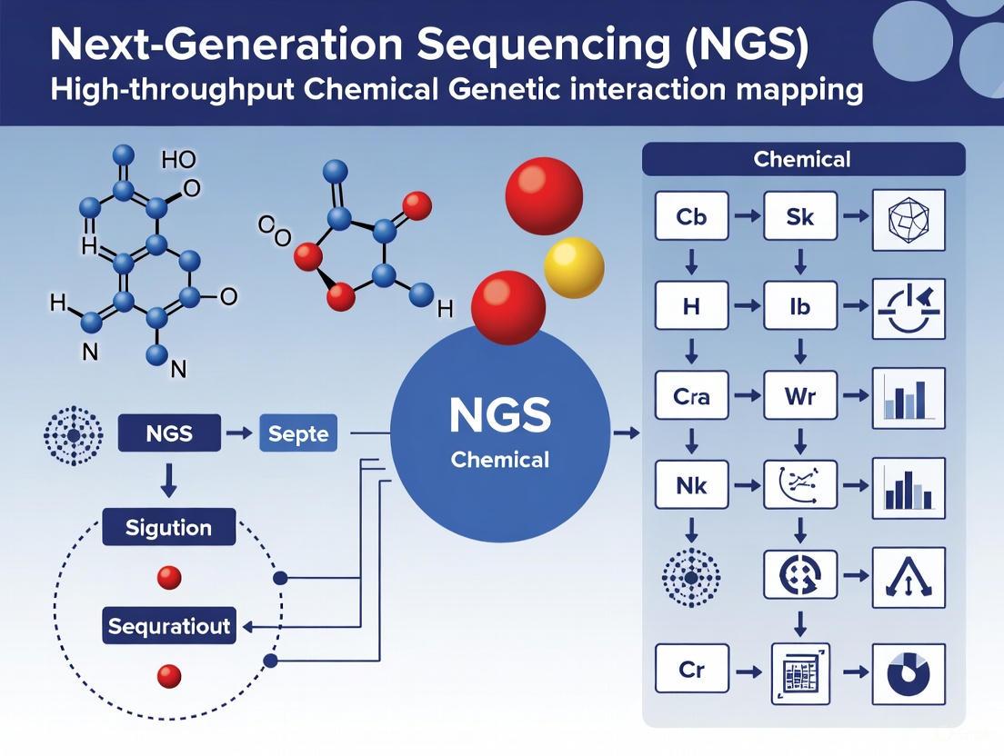
This article explores the transformative role of Next-Generation Sequencing (NGS) in high-throughput chemical genetic interaction mapping, a cornerstone of modern drug discovery.
Mapping the Chemical Genetic Landscape: High-Throughput NGS Strategies for Drug Discovery
Abstract
This article explores the transformative role of Next-Generation Sequencing (NGS) in high-throughput chemical genetic interaction mapping, a cornerstone of modern drug discovery. It provides a comprehensive guide for researchers and drug development professionals, covering foundational NGS principles and their direct application in large-scale screening. The content delves into advanced methodological workflows for identifying drug targets and mechanisms, followed by practical strategies for troubleshooting and optimizing assay sensitivity and reproducibility. Finally, it outlines rigorous analytical validation frameworks and comparative analyses of emerging technologies, offering a holistic perspective on deploying robust, data-driven NGS pipelines to accelerate therapeutic development.
The NGS Revolution: Core Technologies Powering Chemical Genetic Screens
The evolution from Sanger sequencing to Next-Generation Sequencing (NGS) represents a fundamental paradigm shift in genomics, transforming biological research from a targeted, small-scale endeavor to a comprehensive, systems-level science. This transition has been particularly transformative for high-throughput chemical-genetic interaction mapping, a research area essential for understanding gene function and identifying novel therapeutic targets. Where Sanger sequencing provided a precise but narrow snapshot of genetic information, NGS delivers a massively parallelized, panoramic view, enabling researchers to interrogate entire genomes, transcriptomes, and epigenomes in single experiments [1] [2].
The core technological advance lies in parallelism. While Sanger sequencing processes a single DNA fragment per run, NGS simultaneously sequences millions to billions of fragments, creating an unprecedented scale of data output [1] [3]. This democratization has drastically reduced costs and time requirements, moving genome sequencing from a multinational project costing billions to a routine laboratory procedure accessible with standard research funding [3] [4]. The implementation of NGS in chemical-genetic interaction studies, such as the E-MAP (Epistatic Miniarray Profile) and PROSPECT platforms, has empowered researchers to systematically quantify how genetic backgrounds modulate chemical compound effects, rapidly elucidating mechanisms of action for drug discovery [5] [6].
Quantitative Comparison: Sanger Sequencing vs. NGS
The quantitative differences between Sanger and NGS technologies highlight the revolutionary impact of parallelization on genomic research. The following table summarizes key performance metrics that have enabled large-scale genomics.
Table 1: Performance Comparison Between Sanger Sequencing and NGS
| Parameter | Sanger Sequencing | Next-Generation Sequencing |
|---|---|---|
| Sequencing Volume | Single DNA fragment at a time [1] | Millions to billions of fragments simultaneously [1] [3] |
| Throughput | Low (suitable for single genes) [1] [7] | Extremely high (entire genomes or populations) [3] |
| Human Genome Cost | ~$3 billion (Human Genome Project) [3] | Under $1,000 [3] [4] |
| Human Genome Time | 13 years (Human Genome Project) [3] | Hours to days [3] |
| Read Length | 500-1000 base pairs [7] | 50-600 base pairs (short-read); up to millions (long-read) [3] |
| Detection Sensitivity | ~15-20% limit of detection [1] | Down to 1% for low-frequency variants [1] |
| Applications | Single gene analysis, validation [1] [7] | Whole genomes, transcriptomes, epigenomes, metagenomes [1] [2] |
| Data Analysis | Simple chromatogram interpretation [7] | Complex bioinformatics pipelines required [3] |
The cost and time reductions have been particularly dramatic. The first human genome sequence required 13 years and nearly $3 billion to complete using Sanger-based methods [3]. Today, NGS platforms like the Illumina NovaSeq X Plus can sequence more than 20,000 whole genomes per year at a cost of approximately $200 per genome [4]. This efficiency gain of several orders of magnitude has made large-scale genomic studies feasible for individual research institutions, truly democratizing genomic capability.
NGS Workflow for Chemical-Genetic Interaction Mapping
The application of NGS to chemical-genetic interaction profiling follows a standardized workflow that integrates molecular biology, high-throughput screening, and computational analysis. The PROSPECT (PRimary screening Of Strains to Prioritize Expanded Chemistry and Targets) platform exemplifies this approach for antimicrobial discovery [6].
Diagram 1: NGS Chemical-Genetic Screening Workflow. This workflow shows the key steps from library preparation to mechanism of action (MOA) prediction.
Protocol: PROSPECT Platform for Mechanism of Action Prediction
Principle: Identify chemical-genetic interactions (CGIs) by screening compound libraries against pooled hypomorphic mutants of essential genes, using NGS to quantify strain abundance changes and predict mechanisms of action (MOA) through comparative profiling [6].
Materials:
- Pooled hypomorphic Mycobacterium tuberculosis mutant library (each strain with unique DNA barcode)
- Compound library (437 reference compounds with known MOA + experimental compounds)
- NGS library preparation reagents
- Illumina sequencing platform (or equivalent)
- Bioinformatics computational resources
Procedure:
Library Preparation and Compound Treatment
- Grow pooled hypomorphic mutant library to mid-log phase
- Distribute aliquots of pooled library into 96-well plates containing compound treatments (include DMSO vehicle controls)
- Incubate for multiple generations (typically 5-7 population doublings)
- Harvest cells by centrifugation
DNA Extraction and Barcode Amplification
- Extract genomic DNA from harvested cell pellets
- Amplify strain-specific DNA barcodes with primers containing NGS adapter sequences
- Purify amplified libraries using solid-phase reversible immobilization (SPRI) beads
- Quantify library concentration by fluorometry
NGS Sequencing and Data Acquisition
- Pool purified barcode libraries at equimolar concentrations
- Sequence on Illumina platform (minimum 500,000 reads per condition)
- Demultiplex sequences by sample and align to reference barcode database
- Calculate relative abundance of each mutant strain across conditions
Chemical-Genetic Interaction Scoring
- For each compound-mutant pair, calculate CGI score as:
- ε = PABobserved - PABexpected
- Where PABobserved is the observed growth phenotype of the double mutant (hypomorph + compound)
- And PABexpected is the expected phenotype if no interaction exists [5]
- Normalize scores across experiments and replicates
- Generate CGI profile for each compound (vector of all mutant interaction scores)
- For each compound-mutant pair, calculate CGI score as:
Mechanism of Action Prediction
- Compare CGI profiles of experimental compounds to reference set using Perturbagen Class (PCL) analysis
- Calculate similarity metrics (e.g., Pearson correlation) between query and reference profiles
- Assign MOA predictions based on highest similarity references
- Validate predictions through follow-up experiments (e.g., resistance mutation analysis)
Notes: This protocol enables high-throughput MOA prediction with reported sensitivity of 70% and precision of 75% in leave-one-out cross-validation [6]. Include appropriate controls and replicates to ensure statistical robustness.
Essential Research Reagents and Platforms
Successful implementation of NGS-based chemical-genetic interaction mapping requires specific research tools and platforms. The following table details essential components for establishing these workflows.
Table 2: Essential Research Reagent Solutions for NGS Chemical-Genetic Interaction Mapping
| Reagent/Platform | Function | Application Notes |
|---|---|---|
| Hypomorphic Mutant Library | Collection of strains with reduced essential gene function; enables detection of hypersensitivity [6] | Each strain contains unique DNA barcode for NGS quantification; ~400-800 mutants provides optimal coverage [5] [6] |
| NGS Library Prep Kits | Prepare amplified barcode libraries compatible with sequencing platforms [6] | SPRI bead-based cleanup preferred for consistency; incorporate dual index primers for multiplexing |
| Illumina Sequencing Platforms | High-throughput short-read sequencing for barcode quantification [1] [2] | NovaSeq X Series enables 20,000+ genomes annually; MiniSeq suitable for smaller screens [4] |
| TSO 500 Content | Comprehensive genomic profiling for oncology applications; detects variants, TMB, MSI [4] | Uses both DNA and RNA; identifies biomarkers for immunotherapy response |
| TruSight Oncology Comprehensive | In vitro diagnostic kit for cancer biomarker detection in Europe [4] | Companion diagnostic for NTRK fusion cancer therapy (Vitrakvi) |
| Reference Compound Set | Curated compounds with annotated mechanisms of action [6] | 437+ compounds with diverse MOAs essential for training PCL analysis predictions |
NGS Data Analysis Pathway
The computational analysis of NGS data from chemical-genetic interaction studies follows a structured pathway from raw sequence data to biological insight. The PCL (Perturbagen Class) analysis method exemplifies this process for mechanism of action prediction.
Diagram 2: NGS Data Analysis Pathway. This pathway illustrates the computational workflow from sequence data to biological insight.
Protocol: PCL Analysis for MOA Prediction
Principle: Infer compound mechanism of action by comparing its chemical-genetic interaction profile to a curated reference set of profiles from compounds with known targets [6].
Materials:
- CGI profiles for reference compounds (n ≥ 400 recommended)
- CGI profiles for experimental compounds
- Computational environment (R, Python, or specialized software)
- High-performance computing resources for large datasets
Procedure:
Reference Set Curation
- Compile CGI profiles for 400+ compounds with annotated MOAs
- Include established clinical agents, tool compounds, and validated leads
- Ensure representation of diverse target classes and mechanisms
Similarity Metric Calculation
- For each query compound, compute similarity to all reference profiles
- Use Pearson correlation or cosine similarity as distance metric
- Apply appropriate normalization to account for batch effects
MOA Assignment and Confidence Scoring
- Assign MOA based on highest similarity reference matches
- Calculate confidence scores using bootstrap resampling or Bayesian methods
- Apply threshold criteria (e.g., minimum correlation coefficient > 0.4)
Validation and Experimental Follow-up
- For high-confidence predictions, confirm through orthogonal methods
- Test against resistant mutants (e.g., qcrB alleles for QcrB inhibitors)
- Evaluate hypersensitivity in pathway-deficient strains
- Perform chemical optimization for initial hits with weak activity
Notes: In validated studies, PCL analysis achieved 69% sensitivity and 87% precision in MOA prediction for antitubercular compounds [6]. The method successfully identified novel scaffolds targeting QcrB that were subsequently validated experimentally.
The democratization of large-scale genomics through NGS has fundamentally transformed chemical-genetic interaction research, enabling systematic, high-throughput mapping of compound mechanisms of action. The massively parallel nature of NGS provides the scalability required to profile hundreds of compounds against thousands of genetic backgrounds, an undertaking impossible with Sanger sequencing. As NGS technologies continue to advance in accuracy, throughput, and affordability, their integration into drug discovery pipelines will accelerate the identification and validation of novel therapeutic targets, particularly for complex diseases like tuberculosis and cancer. The protocols and methodologies detailed herein provide researchers with practical frameworks for implementing these powerful approaches in their own genomic research programs.
Next-generation sequencing (NGS) has become the cornerstone of high-throughput functional genomics, enabling researchers to decipher complex genetic and chemical-genetic interactions on an unprecedented scale. In the context of chemical genetic interaction mapping—a powerful approach for elucidating small molecule mechanisms of action (MOA) and identifying novel therapeutic targets—the choice between short-read and long-read sequencing technologies represents a critical strategic decision [6]. Each technology offers distinct advantages and limitations that must be carefully considered based on the specific goals of the research, whether focused on comprehensive variant detection, structural variant identification, or resolving complex genomic regions.
Chemical-genetic interaction profiling platforms such as PROSPECT (PRimary screening Of Strains to Prioritize Expanded Chemistry and Targets) generate massive datasets by measuring how chemical perturbations affect pooled mutants depleted of essential proteins [6]. The resulting interaction profiles serve as fingerprints for MOA prediction, but their resolution depends fundamentally on the sequencing methodology employed. Similarly, large-scale genetic interaction studies, such as systematic pairwise gene double knockouts in human cells, require sequencing solutions that can accurately capture complex phenotypic readouts [8]. This application note provides a structured comparison of short-read and long-read sequencing technologies within this context, offering detailed protocols and practical guidance for researchers engaged in high-throughput interaction mapping.
Technology Comparison: Key Characteristics and Performance Metrics
The selection between short-read and long-read sequencing technologies involves balancing multiple factors including read length, accuracy, throughput, and cost. The table below summarizes the core technical characteristics of each approach relevant to interaction mapping applications.
Table 1: Technical Comparison of Short-Read and Long-Recent Sequencing Technologies
| Characteristic | Short-Read Sequencing | Long-Read Sequencing |
|---|---|---|
| Typical Read Length | 50-300 bp [9] | 10 kb to >100 kb; up to hundreds of kilobases for ONT ultra-long reads [10] [11] |
| Primary Platforms | Illumina, Ion Torrent [9] | Pacific Biosciences (PacBio), Oxford Nanopore Technologies (ONT) [10] |
| Accuracy | >99.9% [11] | Varies: PacBio HiFi >99% [11]; ONT 87-98% [11] |
| Key Strengths | High accuracy, low cost per base, established clinical applications [9] | Resolves complex genomic regions, detects structural variants, enables haplotype phasing [12] [10] |
| Limitations for Interaction Mapping | Limited detection of structural variants and repetitive regions [9] | Higher error rates (historically), higher cost per base, more complex data analysis [10] |
| Optimal Use Cases in Interaction Mapping | Variant calling in non-repetitive regions, large-scale screening projects requiring high accuracy at low cost [13] | Resolving complex structural variations, haplotype phasing in regions with high homology, de novo assembly [12] [10] |
Recent benchmarking studies demonstrate that both technologies can be effectively applied to microbial genomics and epidemiology. A 2025 comparison of short-read (Illumina) and long-read (Oxford Nanopore) sequencing for microbial pathogen epidemiology found that long-read assemblies were more complete, while variant calling accuracy depended on the computational approach used [13]. Importantly, the study demonstrated that computationally fragmenting long reads could improve variant calling accuracy, allowing researchers to leverage the assembly advantages of long-read sequencing while maintaining high accuracy in epidemiological analyses [13].
Experimental Protocols for Interaction Mapping Applications
Protocol 1: Chemical-Genetic Interaction Profiling Using Short-Read Sequencing
The PROSPECT platform provides a robust methodology for high-throughput chemical-genetic interaction mapping compatible with short-read sequencing. This protocol enables simultaneous small molecule discovery and MOA identification by screening compounds against pooled hypomorphic mutants of essential genes [6].
Procedure:
- Library Preparation: Generate a pool of hypomorphic Mycobacterium tuberculosis mutants, each engineered to be proteolytically depleted of a different essential protein and tagged with a unique DNA barcode [6].
- Chemical Screening: Expose the pooled mutant library to compound treatments across multiple dose conditions, including controls.
- Sample Collection: Harvest cells after appropriate incubation period and extract genomic DNA.
- Barcode Amplification: Amplify mutant-specific barcodes using PCR with Illumina-compatible adapters.
- Sequencing: Perform short-read sequencing on Illumina platform (typically 2x150 bp) to quantify barcode abundances [6].
- Data Analysis:
- Align sequences to a barcode reference genome using optimized aligners (e.g., MAQ) [14]
- Quantify barcode frequencies across conditions
- Generate chemical-genetic interaction profiles representing each compound's effect on mutant growth
- Apply Perturbagen Class (PCL) analysis to compare interaction profiles to reference compounds with known MOA [6]
Quality Control Considerations:
- Include replicate screens for reproducibility assessment
- Implement randomization schemes to control for batch effects
- Use positive and negative control compounds with established MOA
- Apply neighborhood quality standards (NQS) during variant calling to ensure accuracy [14]
Protocol 2: Genetic Interaction Mapping Using Long-Read Sequencing
This protocol adapts long-read sequencing for large-scale genetic interaction studies, such as systematic pairwise double knockout screens, where comprehensive variant detection and structural variant identification are priorities.
Procedure:
- Library Construction:
- Phenotypic Selection: Culture transduced cells under relevant selective conditions for appropriate duration.
- Genomic DNA Extraction: Use high molecular weight DNA extraction protocols to preserve long fragments.
- Sequencing Library Preparation:
- Sequencing:
- Perform sequencing on appropriate platform (PacBio Sequel/Revio or ONT PromethION)
- Aim for sufficient coverage (typically 50-100x) to detect guide RNA combinations
- Data Analysis:
- Perform base calling and read filtering (e.g., PacBio HiFi read generation)
- Align reads to reference genome using long-read optimized aligners
- Call guide RNA identities and abundances from sequencing data
- Calculate genetic interaction scores based on deviation from expected double mutant phenotypes [8]
Quality Control Considerations:
- Assess DNA integrity prior to library preparation (A260/280 ratio, fragment size distribution)
- Include control guide RNAs with known phenotypes
- Monitor sequencing run metrics (read length distribution, accuracy, throughput)
- Validate key interactions through orthogonal assays
Workflow Visualization: From Experimental Design to Data Analysis
The following workflow diagrams illustrate the key steps in short-read and long-read sequencing approaches for interaction mapping applications, highlighting critical decision points and methodology-specific procedures.
Figure 1: Short-read sequencing workflow for chemical-genetic interaction profiling, adapted from the PROSPECT platform [6]
Figure 2: Long-read sequencing workflow for genetic interaction mapping using combinatorial CRISPR approaches [8]
The Scientist's Toolkit: Essential Research Reagents and Platforms
Successful implementation of interaction mapping studies requires careful selection of reagents, platforms, and computational tools. The following table summarizes key solutions used in the featured protocols and applications.
Table 2: Research Reagent Solutions for Interaction Mapping Studies
| Category | Product/Platform | Specific Application | Key Features |
|---|---|---|---|
| Sequencing Platforms | Illumina NovaSeq 6000 | Short-read sequencing for barcode quantification | High accuracy (>99.9%), high throughput [11] |
| PacBio Sequel II/Revio | HiFi long-read sequencing | >99% accuracy, 15-25 kb read length [10] [11] | |
| Oxford Nanopore PromethION | Ultra-long read sequencing | Reads up to hundreds of kilobases, direct epigenetic detection [10] [11] | |
| CRISPR Systems | Cas12a (Cpf1) | Combinatorial double knockout screens | Processing of two gRNAs from single transcript [8] |
| Cas9 | Standard gene knockout | High efficiency, well-validated guides | |
| Library Prep Kits | SMRTbell Prep Kit | PacBio long-read library preparation | Circular consensus sequencing for high accuracy [11] |
| ONT Ligation Sequencing Kit | Nanopore library preparation | Compatible with ultra-long reads [11] | |
| Analysis Tools | MAQ | Short-read alignment and variant calling | Mapping quality scores, mate-pair utilization [14] |
| DRAGEN | Secondary analysis for mapped reads | Hardware-accelerated, supports constellation mapping [15] | |
| PCL Analysis | Chemical-genetic interaction profiling | Reference-based MOA prediction [6] |
Implementation Guidance: Strategic Technology Selection
When planning interaction mapping studies, researchers should consider the following decision framework to select the most appropriate sequencing technology:
Choose short-read sequencing when:
- Primary research goal involves single nucleotide variant calling or small indel detection
- Studying regions with minimal repeats or structural complexity
- Project requires high sample throughput at minimal cost
- Working with established reference genomes with good annotation
- Applications include barcode quantification in pooled screens [6]
Choose long-read sequencing when:
- Research focuses on structural variant detection or characterization
- Studying regions with high repeat content or segmental duplications
- Haplotype phasing is essential for interpreting genetic interactions
- De novo assembly is required for non-model organisms
- Direct detection of epigenetic modifications is desired [10]
Consider hybrid approaches when:
- Budget allows for combining cost-effective short-read sequencing with targeted long-read sequencing
- Validating structural variants detected in short-read data requires orthogonal confirmation
- Different genomic regions require different resolution approaches
For comprehensive genetic interaction mapping studies, such as the SLC transporter interaction map that utilized both Cas12a and Cas9 systems [8], a hybrid strategy may provide optimal balance between comprehensive variant detection and ability to resolve complex genomic regions.
The strategic selection between short-read and long-read sequencing technologies represents a critical decision point in designing effective interaction mapping studies. Short-read technologies offer established, cost-effective solutions for variant calling and barcode-based screening applications, while long-read platforms provide unparalleled resolution for complex genomic regions and structural variants. As both technologies continue to evolve, with improvements in accuracy, throughput, and cost-effectiveness, their application to chemical and genetic interaction mapping will further expand our understanding of biological systems and accelerate therapeutic discovery.
Researchers should consider their specific biological questions, genomic contexts, and analytical requirements when selecting between these complementary technologies, remaining open to hybrid approaches that leverage the unique strengths of each platform. The continued development of specialized analysis methods, such as PCL analysis for MOA prediction [6] and optimized variant calling pipelines for long-read data [13], will further enhance the utility of both approaches for deciphering complex genetic interactions.
Next-generation sequencing (NGS) has revolutionized functional genomics by enabling the unbiased, systematic profiling of chemical-genetic interactions (CGIs) on a massive scale. In high-throughput chemical-genetic interaction mapping, the fitness of thousands of engineered microbial or human cell mutants is measured simultaneously in response to compound treatment [6] [16]. This approach generates rich CGI profiles—vectors of mutant fitness scores—that reveal a compound's mechanism of action (MOA) by identifying hypersensitive or resistant mutants [6]. The entire paradigm depends critically on a robust NGS workflow to track mutant abundances in pooled screens via DNA barcode sequencing [16]. This application note details the three core technical components—library preparation, cluster generation, and sequencing by synthesis (SBS)—that underpin reliable CGI profiling, providing detailed protocols framed within the context of high-throughput drug discovery research.
Core NGS Workflow Components
Library Preparation
Library preparation converts genomic DNA or cDNA into a sequencing-compatible format by fragmenting samples and adding platform-specific adapters [17] [18] [19]. In CGI screens, this process handles DNA barcodes that uniquely identify each mutant strain in a pooled collection [16].
Protocol: DNA Sequencing Library Preparation for Illumina Systems [19]
- Step 1: Nucleic Acid Extraction and Qualification Isolate genetic material from samples (e.g., bulk tissue, individual cells, biofluids). Assess purity using UV spectrophotometry (A260/A280 ratio ~1.8 for DNA; ~2.0 for RNA) and quantify using fluorometric methods [17].
- Step 2: DNA Fragmentation
Fragment purified DNA to desired sizes (typically 300–600 bp). Choose one of the following methods:
- Mechanical Shearing: Use focused acoustic energy (Covaris) for unbiased, consistent fragmentation with minimal sample loss.
- Enzymatic Digestion: Employ endonuclease cocktails for streamlined, automatable fragmentation requiring lower DNA input.
- Transposon-Based Tagmentation: Simultaneously fragment and tag DNA with adapters in a single reaction, circumventing traditional fragmentation and ligation steps [19].
- Step 3: End Repair and A-Tailing
Convert fragmented DNA's mixed overhangs into blunt ends, phosphorylate 5' ends, and add a single 'A' base to 3' ends to facilitate adapter ligation. This involves:
- Filling in 5' overhangs (5'→3' polymerase activity).
- Removing 3' overhangs (3'→5' exonuclease activity).
- Phosphorylating 5' ends (T4 polynucleotide kinase).
- Adding 'A' to 3' ends (A-tailing using Klenow fragment exo– or Taq polymerase) [19].
- Step 4: Adapter Ligation Ligate duplex oligonucleotide adapters to both ends of the A-tailed fragments. Adapters contain:
- Step 5: Library Amplification and Clean-Up Amplify the adapter-ligated library via limited-cycle PCR to enrich for properly constructed fragments. Purify and quantify the final library, then normalize concentrations before sequencing [19].
Table 1: DNA Fragmentation Methods Comparison
| Method | Principle | Best For | Input DNA | Advantages | Limitations |
|---|---|---|---|---|---|
| Acoustic Shearing | High-frequency sound waves | Unbiased fragmentation, consistent size | Standard input (μg) | Minimal bias, high consistency | Specialized equipment (Covaris) |
| Enzymatic Digestion | Sequence-specific endonucleases | Low-input samples, automation | Low input (ng-μg) | Fast, simple, automatable | Potential sequence bias |
| Tagmentation | Transposase-mediated cut & paste | Ultra-fast library prep | Standard input | Single-tube reaction, fastest | Optimization for complex genomes |
Cluster Generation
Cluster generation amplifies single DNA molecules locally on a flow cell surface to create thousands of identical copies, forming detectable "clusters" that provide sufficient signal intensity for sequencing [18].
Protocol: Bridge Amplification on an Illumina Flow Cell [18]
- Step 1: Flow Cell Priming The flow cell is a glass surface coated with a lawn of two types of oligonucleotides (P5 and P7) that are complementary to the adapters ligated during library preparation [18].
- Step 2: Template Loading and Binding Denature the prepared library into single strands and load it onto the flow cell. Single-stranded DNA fragments bind complementarily to either P5 or P7 oligos on the flow cell surface.
- Step 3: Bridge Amplification
- The bound template is copied by a polymerase, forming a double-stranded bridge.
- The double-stranded molecule is denatured, leaving two single-stranded copies attached to the flow cell.
- This process repeats over ~30 cycles, with each strand bending over to "bridge" to the opposite oligo and serve as a template for copying.
- The result is a dense cluster of ~1,000 identical DNA molecules localized within a sub-micron area [18].
- Step 4: Strand Denaturation and Cleavage After cluster growth, reverse strands are cleaved and washed away, leaving forward strands ready for sequencing. For paired-end reads, the process reverses after the first read to sequence from the opposite end [18].
Sequencing by Synthesis
Sequencing by synthesis is the cyclic process of determining the nucleotide sequence of each cluster through reversible terminator chemistry [18].
Protocol: Illumina's Four-Color SBS Chemistry [18]
- Step 1: Primer Binding and Initialization A sequencing primer binds to the adapter sequence adjacent to the DNA template of each cluster.
- Step 2: Cyclic Nucleotide Incorporation and Imaging
For each cycle, the flow cell is flooded with four fluorescently labeled, reversibly terminated nucleotides.
- Incorporation: DNA polymerase incorporates a single complementary nucleotide onto the growing strand. The reversible terminator blocks further extension.
- Imaging: A high-resolution camera takes four images (one for each laser-excited color channel: A, C, G, T) to determine the identity of the incorporated base at every cluster.
- Cleavage: The fluorescent dye and terminator are chemically cleaved from the nucleotide, enabling the next incorporation cycle.
- Step 3: Base Calling
Instrument software performs base calling, identifying the sequence of nucleotides for each cluster based on the fluorescent signals. The quality of each base call is assessed by a Phred-like Q score:
Q = -10 log₁₀(P), where P is the probability of an incorrect base call. A Q-score of 30 (99.9% accuracy) is standard for high-quality data [20] [18]. - Step 4: Read Completion and Paired-End Sequencing After reading the forward strand, the template can be regenerated in situ to sequence the reverse strand from the opposite end, generating paired-end reads for improved alignment accuracy [18].
Table 2: Key Sequencing Quality Control Metrics
| Metric | Description | Target Value/Range | Significance in CGI Profiling |
|---|---|---|---|
| Q Score | Probability of an incorrect base call [20] | >30 (99.9% accuracy) | Ensures accurate barcode counting for mutant abundance |
| Error Rate | Percentage of incorrectly called bases per cycle [20] | <0.1% | Minimizes false positives/negatives in interaction calls |
| Cluster Density | Clusters per mm² on flow cell | Platform-dependent optimal range | Affects data yield and crosstalk; under/over-clustering harms data |
| % Bases ≥ Q30 | Proportion of bases with QScore≥30 [20] | >75-80% | Indicator of overall run success and data usability |
| Phasing/Prephasing | % clusters falling behind/ahead [20] | <1% per cycle | Reduces signal dephasing, maintains read length and quality |
Application in Chemical-Genetic Interaction Mapping
In high-throughput CGI profiling, the NGS workflow is applied to sequence DNA barcodes that serve as proxies for mutant abundance [6] [16]. The process involves:
- Pooled Screening: A collection of uniquely barcoded mutant strains (e.g., yeast deletion mutants or bacterial hypomorphs) is grown pooled together in the presence of a compound [6] [16].
- DNA Barcode Extraction and Library Prep: Genomic DNA is extracted from the pool pre- and post-compound treatment. The barcode regions are amplified via PCR and prepared for NGS following the library preparation protocol in Section 2.1 [16].
- Sequencing and Analysis: The barcode library is sequenced. The resulting data undergoes primary analysis (base calling), followed by secondary bioinformatics analysis to quantify barcode abundances and calculate fitness scores for each mutant, generating a chemical-genetic interaction profile [6] [16].
The PROSPECT platform for Mycobacterium tuberculosis exemplifies this, using NGS to quantify changes in barcode abundances from a pooled hypomorph library to identify hypersensitive strains and elucidate small molecule mechanism of action [6]. Similarly, high-throughput yeast chemical-genetic screens utilize multiplexed barcode sequencing (e.g., 768-plex) to profile thousands of compounds [16].
The Scientist's Toolkit: Essential Reagents and Materials
Table 3: Key Research Reagent Solutions for NGS-based CGI Screens
| Item | Function | Application Note |
|---|---|---|
| Multiplexed Barcode Library | Collection of mutant strains, each with a unique DNA barcode [16] | Enables pooled fitness assays; yeast (~5000 mutants) or Mtb (hypomorph) libraries are common [6] [16]. |
| NGS Library Prep Kit | Commercial kit for end repair, A-tailing, adapter ligation [19] | Select kit compatible with sequencing platform; ensures high efficiency for low-input barcode PCR products. |
| Indexed Adapters | Oligonucleotides with unique molecular barcodes [18] [19] | Critical for multiplexing many compound screens in one sequencing run, reducing cost per sample. |
| Flow Cell | Glass surface with covalently bound oligos for cluster generation [18] | Platform-specific consumable (e.g., Illumina); cluster density impacts data yield. |
| SBS Kit | Reagent kit containing enzymes and fluorescent nucleotides [18] | Core chemistry for sequencing; newer versions (XLEAP-SBS) offer improved speed/accuracy [17]. |
| Bioinformatics Pipelines | Software for base calling, demultiplexing, and fitness analysis [17] [6] | Essential for translating raw sequence data into chemical-genetic interaction profiles. |
Chemical-genetic interaction mapping represents a powerful functional genomics approach that systematically explores how genetic perturbations modulate cellular responses to chemical compounds. By quantifying the fitness of gene mutants under chemical treatment, this methodology provides deep insights into drug mode-of-action, resistance mechanisms, and functional gene relationships. The integration of next-generation sequencing (NGS) technologies has revolutionized this field, enabling unprecedented scalability and precision in mapping these interactions across entire genomes. This Application Note examines the fundamental principles, methodological frameworks, and practical applications of chemical-genetic interaction mapping, with particular emphasis on NGS-enabled high-throughput screening platforms that are transforming drug discovery and functional genomics.
Chemical-genetic interactions (CGIs) occur when the combination of a genetic mutation and a chemical compound produces an unexpected phenotype that cannot be readily predicted from their individual effects [21]. These interactions are typically measured by assessing cellular fitness—most commonly growth—when mutant strains are exposed to chemical treatments. CGIs manifest as either sensitivity (negative interaction), where the combination of mutation and compound produces a stronger than expected deleterious effect, or resistance (positive interaction), where the mutant exhibits enhanced survival under chemical treatment [22] [21].
The conceptual foundation of CGIs derives from classical genetic interaction studies, where synthetic lethality—a phenomenon where two non-lethal mutations become lethal when combined—demonstrated how functional relationships between genes could be systematically mapped [23]. Chemical-genetic approaches extend this principle by replacing one genetic perturbation with a chemical perturbation, thereby creating a powerful platform for connecting compounds to their cellular targets and mechanisms [21].
In the era of high-throughput genomics, NGS technologies have become indispensable for CGI profiling, enabling the parallel assessment of millions of genetic perturbations under diverse chemical conditions [3] [24]. This technological synergy has transformed CGI mapping from a targeted approach to a comprehensive systems biology tool.
Quantitative Frameworks for Defining Interactions
The accurate quantification of chemical-genetic interactions requires rigorous mathematical frameworks to distinguish meaningful biological interactions from expected additive effects. Multiple definitions have been developed, each with distinct statistical properties and applications.
Alternative Mathematical Definitions
Research by Mani et al. (2008) identified four principal mathematical definitions used for quantifying genetic interactions, each with practical consequences for interpretation [25]:
- Product Definition: Multiplies the individual fitness effects of single perturbations
- Additive Definition: Sums the individual fitness effects
- Log Definition: Utilizes logarithmic transformation of fitness values
- Min Definition: Uses the minimum fitness value of single mutants as reference
Comparative studies in Saccharomyces cerevisiae have demonstrated that while 52% of known synergistic genetic interactions were originally inferred using the Min definition, the Product and Log definitions (shown to be practically equivalent) proved superior for identifying bona fide functional relationships between genes and pathways [25].
Interaction Scoring and Classification
CGIs are quantitatively classified based on the deviation between observed and expected fitness values:
| Interaction Type | Mathematical Relationship | Biological Interpretation |
|---|---|---|
| Synergistic/Negative | Fitness < Expected | Gene mutation enhances compound sensitivity |
| Antagonistic/Positive | Fitness > Expected | Gene mutation confers resistance to compound |
| Neutral/Additive | Fitness ≈ Expected | No functional interaction |
| Suppressive | Double mutant fitter than sickest single mutant | One mutation suppresses effect of other |
Table 1: Classification of chemical-genetic interactions based on fitness deviations.
The quantitative measurement of these interactions enables the construction of chemical-genetic profiles that serve as functional fingerprints for compounds, revealing their cellular targets and mechanisms of action [22] [21].
NGS-Enabled Methodological Frameworks
The integration of NGS technologies has revolutionized CGI profiling through massively parallel sequencing of pooled mutant libraries, enabling genome-wide scalability previously unattainable with arrayed screening formats.
Essential Workflows and Experimental Design
The following diagram illustrates the core workflow for NGS-enabled chemical-genetic interaction screening:
Figure 1: Workflow for NGS-enabled chemical-genetic interaction screening of pooled mutant libraries.
Key Research Reagents and Solutions
Successful implementation of CGI screening requires carefully curated biological and chemical resources:
| Reagent Category | Specific Examples | Function in CGI Studies |
|---|---|---|
| Mutant Libraries | Yeast deletion collection, E. coli Keio collection, CRISPRi libraries | Provides systematic genetic perturbations for screening |
| Chemical Libraries | FDA-approved drugs, natural product libraries, diversity-oriented synthesis compounds | Source of chemical perturbations for profiling |
| Sequencing Platforms | Illumina NovaSeq X, PacBio Sequel, Oxford Nanopore | Enables barcode sequencing and fitness quantification |
| Bioinformatics Tools | CG-TARGET, DeepVariant, Nextflow pipelines | Analyzes NGS data and predicts functional associations |
| Cell Culture Systems | Synthetic genetic array (SGA), TREC, robotic pinning tools | Enables high-throughput manipulation of mutant collections |
Table 2: Essential research reagents for chemical-genetic interaction studies.
Protocol: Genome-Wide Chemical-Genetic Interaction Screening in Yeast
This protocol outlines a robust methodology for systematic CGI profiling in Saccharomyces cerevisiae using NGS-enabled pooled fitness assays.
Library Preparation and Compound Treatment
Materials:
- Yeast knockout deletion collection (
4,800 homozygous diploid mutants~6,000 viable haploid mutants) - Compound of interest dissolved in appropriate vehicle (DMSO typically <1%)
- YPD growth medium and 96-well deep-well plates
- DNA barcodes unique to each mutant strain
Procedure:
- Pool Preparation: Combine equal volumes of all mutant strains from the deletion collection into a single pooled culture. Grow overnight to mid-log phase (OD600 ≈ 0.5-0.8) in rich medium.
- Compound Treatment: Divide the pooled culture into two aliquots. Add compound to treatment condition and vehicle only to control condition. Use multiple concentrations around IC50 when possible.
- Competitive Growth: Incubate cultures with shaking for 12-16 generations to allow fitness differences to manifest. Maintain cultures in mid-log phase through periodic dilution.
- Sample Collection: Harvest approximately 10^8 cells from both treatment and control conditions at multiple time points for time-resolved fitness measurements.
Genomic DNA Extraction and Barcode Amplification
Materials:
- Zymolyase or lyticase for cell wall digestion
- Phenol:chloroform:isoamyl alcohol (25:24:1) and ethanol for DNA precipitation
- Uptag and Dntag specific primers with Illumina adapter sequences
- High-fidelity DNA polymerase for PCR amplification
Procedure:
- Cell Lysis: Digest cell walls with lytic enzymes followed by SDS/proteinase K treatment for complete lysis.
- DNA Purification: Extract genomic DNA using standard phenol-chloroform extraction and ethanol precipitation. Quantify DNA concentration by fluorometry.
- Barcode Amplification: Perform two separate PCR reactions for uptag and dntag barcodes using 1μg genomic DNA as template. Use 18-22 cycles to maintain linear amplification range.
- Library Pooling: Combine uptag and dntag amplifications in equimolar ratios. Purify using solid-phase reversible immobilization (SPRI) beads.
NGS Library Preparation and Sequencing
Materials:
- Illumina platform-specific adapters with dual indices
- Size selection beads (AMPure XP or equivalent)
- Qubit dsDNA HS assay kit for quantification
- Illumina sequencing platform (NovaSeq X preferred for high throughput)
Procedure:
- Library Preparation: Fragment amplified barcode pools to ~300bp and add Illumina sequencing adapters with dual indexing using commercial library preparation kits.
- Quality Control: Validate library fragment size distribution using Bioanalyzer or TapeStation. Quantify by qPCR for accurate cluster generation.
- Sequencing: Pool multiple libraries and sequence on Illumina platform using 75bp single-end reads. Aim for minimum coverage of 200-500 reads per barcode.
- Demultiplexing: Separate sequencing reads by sample index using Illumina bcl2fastq or similar tools.
Bioinformatics Analysis and Interaction Scoring
Materials:
- High-performance computing cluster with ≥16GB RAM
- Barcode-to-mutant mapping file for reference strain collection
- Bioinformatics pipelines (CG-TARGET, established Python/R scripts)
Procedure:
- Sequence Alignment: Map sequencing reads to barcode reference file using exact matching allowing 1-2 mismatches.
- Fitness Calculation: For each mutant, calculate relative abundance in treatment versus control using normalized read counts: Fitness = log2(treatmentreads/controlreads).
- Interaction Scoring: Compute chemical-genetic interaction score (ε) using the equation: ε = WtreatmentAB - (WtreatmentA × WtreatmentB) where W represents fitness values.
- Statistical Analysis: Identify significant interactions using z-score transformation or false discovery rate (FDR) correction for multiple hypothesis testing.
Applications in Drug Discovery and Functional Genomics
CGI mapping provides multifaceted insights that accelerate therapeutic development and functional annotation of genes.
Mode-of-Action Elucidation
Chemical-genetic profiles serve as functional fingerprints that can be compared to reference compounds with known targets through "guilt-by-association" approaches [21]. Machine learning algorithms, including Random Forest and Naïve Bayesian classifiers, have demonstrated strong predictive power for identifying cellular targets based on CGI profiles [26]. For example, CG-TARGET integration of genetic interaction networks with CGI data enabled high-confidence biological process predictions for over 1,500 compounds [22].
Synergistic Combination Prediction
CGI data provides a rational framework for identifying synergistic drug combinations that enhance efficacy while reducing resistance development. Studies have successfully leveraged CGI matrices to predict compound pairs that exhibit species-selective toxicity against human fungal pathogens [26]. The conceptual relationship between genetic and chemical interaction networks for synergy prediction is illustrated below:
Figure 2: Rational prediction of synergistic drug combinations based on synthetic lethal genetic interactions.
Resistance Mechanism Mapping
CGI profiling comprehensively identifies genes involved in drug uptake, efflux, and detoxification—revealing both known and novel resistance determinants [21]. Studies in E. coli have identified dozens of genes with pleiotropic roles in multidrug resistance, highlighting the extensive capacity for intrinsic antibiotic resistance in microbial populations [21]. This knowledge enables predictive models of resistance evolution and strategies to counteract resistance through adjuvant combinations.
Integration with Multi-Omics Technologies
The power of CGI mapping multiplies when integrated with complementary functional genomics approaches:
- Transcriptomic Integration: Correlation with gene expression signatures refines MoA predictions
- Proteomic Overlay: Identifies post-translational regulatory mechanisms
- Structural Bioinformatics: Enables rational design of optimized compounds
- CRISPR Screening Platforms: Extends CGI approaches to mammalian systems
Advanced integration methods like CG-TARGET successfully combine large-scale CGI data with genetic interaction networks to predict biological processes perturbed by compounds with controlled false discovery rates [22].
Chemical-genetic interaction mapping has evolved from a specialized genetic technique to a comprehensive systems biology platform, largely enabled by NGS technologies. The continued advancement of sequencing platforms—with Illumina's NovaSeq X series now capable of sequencing over 20,000 genomes annually at approximately $200 per genome—promises to further democratize and scale CGI profiling [27]. As these technologies converge with artificial intelligence and automated phenotyping, CGI mapping will play an increasingly central role in functional genomics, drug discovery, and personalized medicine, ultimately accelerating the development of novel therapeutic strategies against human diseases.
Building the Pipeline: High-Throughput NGS Workflows for Interaction Mapping
In high-throughput chemical genetic interaction mapping, the ability to systematically screen thousands of compounds against genomic libraries demands precision, reproducibility, and scalability. Next-Generation Sequencing (NGS) has become an indispensable tool in this field, enabling researchers to decipher complex gene-compound interactions at an unprecedented scale. A typical NGS workflow involves four critical steps: sample preparation, library preparation, sequencing, and data analysis [28]. Library preparation, which converts nucleic acids into a sequence-ready format, is particularly crucial as it establishes the foundation for reliable sequencing data [28]. This multi-step process includes DNA fragmentation, adapter ligation, PCR amplification, purification, quantification, and normalization, requiring meticulous attention to detail and precise liquid handling [28].
Manual library preparation methods present significant limitations for large-scale chemical genetic screens, being time-inefficient, labor-intensive, and constrained by limited throughput [28]. Furthermore, manual pipetting is prone to errors, especially when working with small volumes, leading to inconsistent results and challenges in reaction miniaturization [28]. Automated liquid handling systems effectively address these challenges by providing precise and consistent dispensing for complex protocols, particularly for small volumes, thereby reducing processing costs through miniaturization and enhancing reproducibility [28]. For drug development professionals seeking to map chemical-genetic interactions on a large scale, automation is not merely a convenience but a necessity for generating high-quality, statistically powerful datasets.
Key Concepts and Definitions
The Role of Assay Miniaturization in High-Throughput Screening
Assay miniaturization involves scaling down reaction volumes while maintaining accuracy and precision [29]. In the context of NGS library preparation for chemical genetics, this translates to performing reactions in volumes as low as hundreds of nanoliters [28]. The advantages are multifold:
- Cost Reduction: Miniaturization significantly decreases consumption of expensive reagents and precious compounds, making large-scale screening campaigns economically feasible [29]. For example, using induced pluripotent stem cell (iPSC)-derived cells that can cost over $1,000 per vial of 2 million cells, moving from a 96-well to a 384-well format reduces cell consumption approximately fivefold, resulting in substantial savings [30].
- Enhanced Efficiency: Smaller volumes allow for higher well densities (e.g., 384-well or 1536-well plates), amplifying testing scale and efficiency within the same laboratory footprint [29].
- Improved Data Quality: Miniaturization can concentrate targets and reduce diffusion distances, potentially enhancing assay sensitivity and precision [29].
Automated Liquid Handling Technologies
Automated liquid handling (ALH) systems are engineered to deliver precise liquid transfers, enabling both miniaturization and process standardization. These systems generally fall into two categories:
- Non-Contact Dispensers: Instruments like the Mantis and Tempest use patented microfluidic technology to dispense sub-microliter volumes with high precision without tips, minimizing consumable costs and cross-contamination risks [28].
- Liquid Handlers with Tips: Systems such as the F.A.S.T. (96-channel, positive displacement) and FLO i8 PD (8-channel independent spanning, air displacement) are versatile for various volume ranges and protocol steps [28].
The integration of these systems into laboratory workflows is facilitated by features like CSV format file compatibility for sample pooling, normalization, and serial dilution, as well as Application Programming Interfaces (API) for seamless laboratory automation integration [28].
Application Note: Implementing Automated NGS for Compound Screening
Experimental Design for Chemical-Genetic Interaction Mapping
In a typical chemical-genetic interaction mapping study, the goal is to identify how different chemical compounds affect various genetic mutants. The experimental design involves treating an array of yeast deletion mutants or CRISPR-modified human cell lines with a library of compounds, followed by NGS-based readout of mutant abundance to identify genetic sensitivities and resistances.
Key design considerations include:
- Library Complexity: The number of unique barcodes must match the genetic library size, often exceeding 10,000 unique mutants.
- Replication: Appropriate biological and technical replicates are crucial for statistical power in identifying significant interactions.
- Controls: Including untreated controls and reference compounds with known mechanisms is essential for data normalization and quality control.
Automation enables this complex experimental design by ensuring consistent liquid handling across hundreds of plates, precise compound dispensing at nanoliter scales, and reproducible library preparation for accurate sequencing results.
Automated Protocol for NGS Library Preparation from Treated Cell Pools
Table 1: Automated NGS Library Preparation Workflow for Chemical-Genetic Screens
| Step | Process | Automated System | Key Parameters | Volume Range |
|---|---|---|---|---|
| 1 | Genomic DNA Extraction | Agilent Bravo (96 channels) or Biomek NXp (8-channel) | Input: 1-5 million cells; Elution Volume: 50-100 μL | 50-200 μL |
| 2 | DNA Fragmentation | Focused-ultrasonicator (e.g., Covaris LE220) | Target size: 550 bp; Sample Distribution: Automated transfer to microTUBE plates | 50-100 μL |
| 3 | Library Construction | Agilent Bravo, MGI SP-960, or Hamilton NGS STAR | PCR-free or with limited-cycle PCR; Adapter Ligation | 20-50 μL |
| 4 | Library Purification | Magnetic bead-based cleanup on liquid handler | Bead-to-sample ratio: 1.0-1.8X; Elution Volume: 15-30 μL | 15-100 μL |
| 5 | Quality Control | Fragment Analyzer or TapeStation | Size distribution: 300-700 bp; Concentration: ≥ 2 nM | 1-5 μL |
| 6 | Library Normalization & Pooling | Hamilton, Formulatrix FLO i8, or Beckman Biomek i7 | Normalization to 2-4 nM; Equal volume pooling | 5-20 μL |
| 7 | Quantification for Sequencing | qPCR systems (e.g., qMiSeq) | Loading concentration optimization | 2-5 μL |
This protocol, adapted from large-scale sequencing projects [31], can process 96-384 samples in parallel with minimal hands-on time, enabling rapid screening of compound libraries.
Technical Specifications of Automated Liquid Handling Systems
Table 2: Comparison of Automated Liquid Handling Systems for NGS Library Prep
| System | Technology | Precision | Miniaturization Range | Throughput Capacity | Key Features |
|---|---|---|---|---|---|
| Formulatrix Mantis | Non-contact, tipless dispenser | <2% CV at 100 nL | Down to 100 nL | Plates up to 1536 wells; Up to 48 reagents | CSV input, backfill, concentration normalization |
| Formulatrix Tempest | Non-contact, tipless dispenser | <5% CV at 200 nL | Down to 200 nL | Plates up to 1536 wells; 24 plate stacking | 96 nozzles; Serial dilution, pooling, broadcasting |
| Formulatrix F.A.S.T. | 96-channel, positive displacement | <5% CV at 100 nL | Down to 100 nL transfer | Plates up to 384 wells; 6 on-deck positions | Flow Axial Seal Tip technology |
| Formulatrix FLO i8 PD | 8-channel, air displacement | <5% CV at 1 μL | Down to 500 nL transfer | Plates up to 384 wells; 10 on-deck positions | Independent spanning channels; Integrated flow rate sensors |
| Agilent Bravo | 96-channel, adaptable | Protocol-dependent | Down to 1 μL | Plates up to 384 wells | Used with TruSeq DNA PCR-free kits [31] |
| Hamilton NGS STAR | 96-channel or 8-channel | Protocol-dependent | Down to 1 μL | Plates up to 384 wells | Compatible with Illumina DNA Prep [32] |
Research Reagent Solutions for Automated NGS
Table 3: Essential Materials for Automated NGS Library Preparation
| Reagent/Material | Function | Example Products | Automation Considerations |
|---|---|---|---|
| PCR-Free Library Prep Kit | Creates sequencing libraries without PCR bias | Illumina TruSeq DNA PCR-Free HT, MGIEasy PCR-Free DNA Library Prep Set [31] | Compatibility with automated platforms; Dead volume requirements |
| Unique Dual Indexes | Multiplexing samples in sequencing runs | IDT for Illumina TruSeq DNA Unique Dual indexes [31] | Plate-based formatting for automated liquid handlers |
| Magnetic Beads | Library purification and size selection | SPRIselect, AMPure XP | Viscosity and behavior in automated protocols |
| DNA Quantitation Kits | Accurate library quantification | Quant-iT PicoGreen dsDNA kit, Qubit dsDNA HS Assay Kit [31] | Compatibility with automated plate readers |
- Library Preparation Kits: Specialized for automation with reduced dead volumes and pre-formatted reagents.
- Magnetic Beads: Engineered for consistent binding kinetics in small-volume reactions.
- Indexing Primers: Pre-arrayed in plates compatible with automated liquid handlers.
Methods and Protocols
Detailed Protocol: Miniaturized NGS Library Preparation on Automated Systems
Procedure for 384-Well Library Preparation Using PCR-Free Methods
DNA Normalization and Plate Reformatting
- Program the liquid handler (e.g., Biomek NXp) to transfer genomic DNA samples from source plates to a 384-well assay plate, normalizing all samples to 10-20 ng/μL in 50 μL volume using low-EDTA TE buffer [31].
- Centrifuge the plate briefly (1000 × g, 1 minute) to collect liquid at the bottom of wells.
Automated DNA Fragmentation
Library Assembly on Liquid Handler
- Program the system (e.g., Agilent Bravo with 96-channel head) to add:
- 25 μL of fragmented DNA
- 10 μL of End Repair Mix
- 5 μL of Ligation Mix
- 5 μL of Appropriate Adapters with Unique Dual Indexes [31]
- Use the instrument's mixing function to ensure complete homogenization without bubble formation.
- Program the system (e.g., Agilent Bravo with 96-channel head) to add:
Library Purification
- Perform a two-sided size selection using magnetic beads at 0.5X and 0.8X ratios to remove short fragments and adapter dimers.
- Program the liquid handler to:
- Add magnetic beads to each well
- Incubate for 5 minutes
- Engage magnets and wait for solution clarification
- Remove and discard supernatant
- Perform two 80% ethanol washes
- Elute in 25 μL of Resuspension Buffer [31]
Quality Control and Quantification
Library Normalization and Pooling
- Calculate normalization volumes based on QC data.
- Program the liquid handler to transfer calculated volumes of each library to a pooling reservoir.
- Mix the pool thoroughly and transfer to a fresh tube for sequencing.
Protocol: Miniaturized Compound Addition for Chemical Treatment
Procedure for 1536-Well Compound Screening Prior to NGS
Compound Plate Preparation
- Reform compound libraries into 1536-well source plates at appropriate concentrations (typically 1-10 mM in DMSO) using acoustic dispensers or pintool transfer.
- Include control compounds in designated wells.
Miniaturized Compound Transfer
- Using a non-contact dispenser (e.g., Formulatrix Mantis), transfer 20 nL of compound from source plates to 1536-well assay plates containing cells in 2 μL culture medium.
- This results in final compound concentrations of 10-100 μM.
Incubation and Processing
- Incubate plates under appropriate conditions (37°C, 5% CO₂) for the desired treatment period (typically 24-72 hours).
- Process cells for genomic DNA extraction using miniaturized protocols compatible with high-density plates.
Workflow Visualization: Automated NGS Library Preparation
Figure 1: Automated NGS workflow for chemical genetic screens.
System Architecture: Integration of Automation Components
Figure 2: Integration of automation components in NGS workflow.
Results and Discussion
Performance Metrics for Automated NGS Library Preparation
Implementation of automated liquid handling and assay miniaturization in NGS library preparation yields significant improvements in key performance metrics:
- Process Efficiency: Automated systems reduce hands-on time by 50-65% compared to manual methods [32]. For example, the Illumina DNA Prep with Enrichment automated on Hamilton or Beckman systems processes up to 48 DNA libraries with over 65% less hands-on time [32].
- Cost Reduction: Miniaturization of Nextera XT library preparation using the Mantis liquid dispenser demonstrates 75% savings on reagent costs while maintaining high-quality RNA-seq libraries from low-input murine neuronal cells [28].
- Data Quality: Automated systems maintain or improve data quality metrics. The Formulatrix F.A.S.T. instrument enables precise plate-to-plate transfers of single microliter volumes of single cell cDNA, efficiently combining 384 unique indexing primers from separate plates while mitigating evaporation risk [28].
- Reproducibility: Automated liquid handling significantly reduces well-to-well variability, with precision metrics of <2% coefficient of variation (CV) at 100 nL for advanced systems like the Mantis [28].
Addressing Challenges in Miniaturization and Automation
Despite the clear benefits, implementing automated, miniaturized NGS workflows presents challenges that require strategic solutions:
- Evaporation Management: Smaller volumes are more susceptible to evaporation, particularly in edge wells. Strategies to mitigate this include using plate seals, maintaining high humidity in automated environments, and employing non-contact dispensers that minimize well-open time [28] [30].
- Liquid Handling Precision: Transferring nanoliter volumes demands specialized instrumentation. Positive displacement systems and microfluidic-based non-contact dispensers provide the required precision for miniaturized reactions [28].
- Reagent Compatibility: Some reagents may exhibit different behavior in miniaturized formats, requiring optimization of concentrations, incubation times, and mixing parameters.
- Cross-Contamination Risks: As well density increases, the potential for cross-contamination grows. Regular maintenance, proper tip washing protocols, and non-contact dispensing where appropriate minimize this risk.
Future Directions in Automated NGS for Chemical Genetics
The field of automated NGS continues to evolve, with several trends shaping its application in chemical genetic interaction mapping:
- Integration with Multiomics Approaches: Future workflows will likely incorporate simultaneous analysis of genetic interactions with transcriptional, epigenetic, and proteomic changes from the same samples, enabled by automated systems capable of processing multiple analyte types [33].
- AI-Enhanced Experimental Design and Analysis: Artificial intelligence and machine learning are increasingly being applied to optimize screening parameters, predict interactions, and analyze complex datasets [33].
- Further Miniaturization: Ongoing developments in microfluidics and nanodispensing technologies promise continued reduction in reaction volumes, potentially enabling high-density formats beyond 1536-well plates for ultra-high-throughput applications [29].
- Real-Time Process Monitoring: Integration of sensors and real-time quality control checks within automated workflows will enhance process control and reduce failure rates.
For research teams engaged in high-throughput chemical genetic interaction mapping, the strategic implementation of automated liquid handling and assay miniaturization represents a critical capability for scaling screening efforts without compromising data quality or operational efficiency.
The advent of Clustered Regularly Interspaced Short Palindromic Repeats (CRISPR) technology has revolutionized functional genomics, providing an unparalleled toolkit for high-throughput interrogation of gene function. When integrated with Next-Generation Sequencing (NGS), CRISPR-based screens transform genetic mapping from a correlative to a causal science, enabling the systematic deconvolution of complex chemical-genetic interaction networks [34] [35]. This synergy allows researchers to not only identify genes essential for cell viability under specific chemical treatments but also to map entire genetic interaction networks that define drug mechanisms of action and resistance pathways [36]. For drug development professionals, this integrated approach provides a powerful platform for target identification, validation, and mechanism-of-action studies, ultimately accelerating therapeutic discovery.
Experimental Design: Core CRISPR Screening Modalities
The power of CRISPR functional genomics lies in its adaptability. Three primary screening modalities enable either loss-of-function (LOF) or gain-of-function (GOF) studies at scale, each with distinct advantages for specific biological questions.
CRISPR Knockout (CRISPRko)
Mechanism: Utilizes the wild-type Cas9 nuclease to create double-strand breaks (DSBs) in the target DNA. These breaks are repaired by the error-prone non-homologous end joining (NHEJ) pathway, often resulting in insertions or deletions (indels) that disrupt the coding sequence and create gene knockouts [36] [37].
Applications: Identification of essential genes, fitness genes under specific conditions (e.g., drug treatment), and genes involved in pathways governing cellular responses [35].
CRISPR Interference (CRISPRi)
Mechanism: Employs a catalytically "dead" Cas9 (dCas9) fused to a transcriptional repressor domain, such as the KRAB domain. The dCas9-KRAB complex binds to the promoter or transcriptional start site of a target gene without cutting the DNA, leading to targeted epigenetic silencing and reduced gene expression [36].
Applications: Tunable and reversible gene suppression; ideal for studying essential genes where complete knockout is lethal, and for functional characterization of non-coding regulatory elements [36].
CRISPR Activation (CRISPRa)
Mechanism: Uses dCas9 fused to strong transcriptional activation domains, such as the VP64-p65-Rta (VPR) or Synergistic Activation Mediator (SAM) systems. This complex is guided to the promoter regions of target genes to recruit transcriptional machinery and enhance gene expression [36].
Applications: Gain-of-function screens to identify genes that confer resistance to therapeutics, drive cell differentiation, or overcome pathological states.
Table 1: Comparison of Core CRISPR Screening Modalities
| Screening Modality | Core Mechanism | Genetic Outcome | Primary Applications |
|---|---|---|---|
| CRISPRko (Knockout) | Cas9-induced DSB + NHEJ repair | Gene disruption/Loss-of-function | Essential gene discovery, drug-gene interactions, fitness screens [36] [35] |
| CRISPRi (Interference) | dCas9 fused to repressor (e.g., KRAB) | Transcriptional repression/Loss-of-function | Studies of essential genes, non-coding regulatory elements [36] |
| CRISPRa (Activation) | dCas9 fused to activators (e.g., VPR, SAM) | Transcriptional activation/Gain-of-function | Gene suppressor screens, identification of resistance mechanisms [36] |
Detailed Protocols for Key Applications
Protocol: Pooled CRISPRko Screen for Chemical-Genetic Interactions
This protocol outlines the steps for identifying genes that modulate cellular sensitivity to a small molecule compound, a cornerstone of high-throughput chemical genetic interaction mapping [35].
Step 1: Library Design and Selection
- Select a genome-scale or focused sgRNA library (e.g., Brunello, GeCKO).
- Ensure coverage of 3-6 sgRNAs per gene and include non-targeting control sgRNAs.
- Amplify the library plasmid DNA and prepare high-titer lentiviral stock.
Step 2: Cell Transduction and Selection
- Transduce the target cell population (e.g., HAP1, haploid cells) at a low Multiplicity of Infection (MOI ~0.3) to ensure most cells receive a single sgRNA.
- Forty-eight hours post-transduction, select transduced cells with appropriate antibiotics (e.g., Puromycin) for 5-7 days to generate a stable mutant pool.
Step 3: Application of Chemical Challenge
- Split the selected cell pool into two groups: Treatment and Control.
- Treat the Treatment arm with the IC20-IC30 concentration of the compound of interest.
- Culture the Control arm with the compound's vehicle (e.g., DMSO).
- Maintain cultures for 14-21 days, allowing for 10-12 population doublings to enable phenotypic manifestation.
Step 4: Sample Preparation and NGS
- Harvest a minimum of 1,000 cells per sgRNA in the library at both T0 (baseline) and Tfinal (post-treatment/control) time points.
- Extract genomic DNA and amplify the integrated sgRNA sequences using primers containing Illumina adapters and sample barcodes.
- Pool PCR products and perform NGS on an Illumina platform to a depth of 200-500 reads per sgRNA.
Step 5: Data Analysis and Hit Calling
- Process raw FASTQ files to count sgRNA reads for each sample using tools like
MAGeCK count[36]. - Normalize read counts and identify differentially enriched or depleted sgRNAs between Treatment and Control groups using robust statistical models (e.g.,
MAGeCK test). - Aggregate sgRNA-level effects to gene-level scores. Genes whose sgRNAs are significantly depleted in the Treatment arm are "sensitizers" (loss enhances drug effect), while those enriched are "resistors" (loss confers resistance) [36].
Protocol: High-Content Screening with Single-Cell RNA Sequencing (Perturb-Seq)
This advanced protocol couples genetic perturbations with deep phenotypic profiling, enabling the dissection of transcriptional networks and heterogeneous cellular responses at single-cell resolution [36] [35].
Step 1: Library Transduction and Preparation
- Transduce cells with a pooled CRISPRko/i/a library as in Protocol 3.1, but at a higher MOI to ensure widespread perturbation.
- After selection, subject the entire pool of perturbed cells to single-cell RNA sequencing (e.g., using the 10x Genomics platform).
Step 2: Single-Cell Library Construction and Sequencing
- Prepare a single-cell suspension and partition cells into nanoliter droplets along with barcoded beads.
- Generate barcoded cDNA libraries where the transcriptome of each cell is tagged with a unique cellular barcode.
- Include a custom pre-amplification step to also capture and barcode the expressed sgRNAs from each cell.
Step 3: Data Integration and Analysis
- Align sequencing reads to the reference genome and assign transcripts to individual cells using cellular barcodes.
- Demultiplex the perturbations by matching the captured sgRNA sequences to the library manifest.
- Use computational tools like
MIMOSCAorscMAGeCKto regress the single-cell transcriptional profile of each cell against its genetic perturbation [36]. - Identify differentially expressed genes and pathways resulting from each knockout, building a high-resolution map from genetic perturbation to transcriptional outcome.
Advanced Tools: Precision Genome Editing for Variant Functionalization
Beyond gene-level knockout, newer CRISPR-derived technologies enable precise nucleotide-level editing, allowing for the functional characterization of human genetic variants discovered through NGS.
Base Editing
Mechanism: Uses a Cas9 nickase (nCas9) or dCas9 fused to a deaminase enzyme. Cytosine Base Editors (CBEs) convert a C•G base pair to T•A, while Adenine Base Editors (ABEs) convert an A•T base pair to G•C, all without inducing a DSB [34] [37].
Application in Functional Genomics: Saturation mutagenesis of specific codons to assay the functional impact of all possible single-nucleotide variants (SNVs) in a gene region of interest.
Prime Editing
Mechanism: Employs a Cas9 nickase fused to a reverse transcriptase (PE2 system), programmed with a prime editing guide RNA (pegRNA). The pegRNA both specifies the target site and contains the desired edit template. The system nicks the target strand and directly "writes" the new genetic information from the pegRNA template into the genome [34] [38].
Application in Functional Genomics: A recent study demonstrated the power of pooled prime editing to screen over 7,500 pegRNAs targeting tumor suppressor genes like SMARCB1 and MLH1 in HAP1 cells. This approach enabled high-throughput saturation mutagenesis to identify pathogenic loss-of-function variants in both coding and non-coding regions, providing a robust platform for classifying variants of uncertain significance (VUS) identified by clinical NGS [38].
Table 2: Advanced CRISPR-Based Editors for Variant Study
| Editor Type | Key Components | Type of Changes | Advantages for NGS Follow-up |
|---|---|---|---|
| Cytosine Base Editor (CBE) | nCas9/dCas9 + Cytidine Deaminase | C•G to T•A | Clean, efficient installation of specific transition mutations without DSBs [34] |
| Adenine Base Editor (ABE) | nCas9/dCas9 + Adenine Deaminase | A•T to G•C | Installs precise A-to-G changes with minimal indel formation [34] |
| Prime Editor (PE) | nCas9 + Reverse Transcriptase + pegRNA | All 12 base-to-base conversions, small insertions/deletions | Unprecedented precision and versatility for modeling human SNVs and indels [38] |
The Scientist's Toolkit: Essential Research Reagents
Table 3: Key Research Reagent Solutions for CRISPR-Based Functional Genomics
| Reagent / Solution | Function / Description | Example Use Cases |
|---|---|---|
| Cas9 Nucleases | Engineered variants of the Cas9 protein (from S. pyogenes and other species) with different PAM specificities and off-target profiles. | CRISPRko screens; foundation for engineering base and prime editors [37]. |
| dCas9 Effector Fusions | Catalytically inactive Cas9 fused to transcriptional repressors (KRAB for CRISPRi) or activators (VPR/SAM for CRISPRa). | Transcriptional modulation screens; epigenetic editing [36]. |
| Base Editors (BE) | Fusion proteins of nCas9/dCas9 with deaminase enzymes (e.g., BE4 for C->T; ABE8e for A->G). | High-throughput saturation mutagenesis to model SNVs [34] [37]. |
| Prime Editors (PE) | nCas9-reverse transcriptase fusions programmed with pegRNAs. | Installation of precise variants (SNVs, indels) for functional characterization of VUS [38]. |
| sgRNA Libraries | Pooled, barcoded collections of thousands of sgRNAs targeting genes genome-wide or in specific pathways. | Pooled knockout, interference, and activation screens [35]. |
| pegRNA Libraries | Pooled libraries of prime editing guide RNAs designed to install specific variants via prime editing. | Multiplexed Assays of Variant Effect (MAVEs) in the endogenous genomic context [38]. |
| Analysis Software (MAGeCK) | A widely used computational workflow for the robust identification of positively and negatively selected genes from CRISPR screen NGS data. | Statistical analysis of screen results to identify hit genes [36]. |
Enzyme-Coupled Assay Systems and Readout Cascades for Phenotypic Screening
Enzyme-coupled assay systems represent a sophisticated and versatile toolset for phenotypic screening, a drug discovery strategy that has experienced a major resurgence in the past decade. Modern phenotypic drug discovery (PDD) focuses on modulating disease phenotypes or biomarkers rather than pre-specified molecular targets, and has contributed to a disproportionate number of first-in-class medicines [39]. These screens require robust, sensitive readout systems capable of detecting subtle phenotypic changes in realistic disease models. Enzyme-coupled assays fulfill this need by translating molecular events into measurable signals through cascading biochemical reactions, thereby enabling researchers to monitor complex biological processes in high-throughput screening (HTS) environments.
The fundamental principle underlying enzyme-coupled assays involves linking a primary enzymatic reaction of interest to one or more auxiliary enzyme reactions that generate a detectable output signal, typically through absorbance, fluorescence, or luminescence readouts [40]. This signal amplification strategy is particularly valuable for monitoring enzymatic activities where products are not easily measured by available instruments at high-throughput. Within the context of next-generation sequencing (NGS) for chemical-genetic interaction mapping, these assay systems provide the phenotypic data that, when correlated with genetic perturbation information, enables the comprehensive reconstruction of regulatory circuits and drug mechanisms of action [41].
Theoretical Foundations of Enzyme-Coupled Assay Systems
Basic Principles and Kinetic Considerations
Enzyme-coupled assays function on the principle of coupling a primary reaction that generates a product difficult to detect directly to a secondary reaction (or series of reactions) that produces a measurable signal. The most common auxiliary reactions employ enzymes that generate products with distinct absorbance or fluorescence properties [40]. For these coupled systems to accurately report on the primary enzyme's activity, the auxiliary enzymes must be present in excess, ensuring that the initial reaction remains rate-limiting. Under these optimized conditions, the overall molecular flux through the pathway directly correlates with the activity of the target enzyme [40].
The kinetics of coupled enzyme reactions have been extensively characterized, with theoretical frameworks developed to account for scenarios where the second reaction does not follow simple first-order kinetics [42]. A critical consideration in assay design is the transient time – the period required for the coupled system to reach steady state. This lag phase can potentially obscure the true initial velocity measurements if not properly accounted for in experimental design and data interpretation [42]. Properly configured coupled assays allow continuous monitoring of enzyme activity, enabling identification of kinetic deviations such as lag periods or falling-off reaction rates that might indicate complex enzyme behavior or inhibition patterns [40].
Design Considerations for High-Throughput Applications
When adapting enzyme-coupled assays for high-throughput phenotypic screening, several factors require careful optimization. The environmental conditions – particularly temperature and pH – must be compatible with all enzymes in the cascade [40]. Additionally, the signal-to-noise ratio and dynamic range must be sufficient to detect subtle phenotypic changes amid background variability. For HTS compatibility, assays should ideally be homogeneous (mix-and-read format), scalable to 384- or 1536-well formats, and robust enough to maintain consistency across thousands of experimental wells [43].
The Z' factor is a key metric for evaluating HTS assay quality, with values ≥0.7 indicating excellent robustness and suitability for screening campaigns [43]. Furthermore, the assay must demonstrate low false positive and negative rates, minimizing interference from fluorescent compounds or other artifacts that could compromise screening outcomes. Advances in detection chemistries, particularly universal fluorescent approaches that detect common products like ADP, GDP, or SAH across multiple enzyme families, have significantly improved the reliability and efficiency of these systems in drug discovery pipelines [43].
Enzyme-Coupled Assay Formats and Detection Modalities
The selection of an appropriate detection method represents a critical decision point in designing enzyme-coupled assays for phenotypic screening. Each format offers distinct advantages and limitations that must be balanced against experimental requirements, throughput needs, and available instrumentation.
Table 1: Comparison of Enzyme-Coupled Assay Detection Modalities
| Assay Format | Readout Signal | Advantages | Limitations | Optimal Applications |
|---|---|---|---|---|
| Absorbance-Based | Colorimetric change | Simple, inexpensive, robust | Lower sensitivity, not ideal for miniaturized HTS | Early-stage validation, educational assays [43] |
| Fluorescence-Based | Fluorescence intensity or polarization | High sensitivity, HTS compatible, adaptable | Potential fluorescent compound interference | Universal for multiple enzyme classes, primary screening [40] [43] |
| Luminescence-Based | Light emission | High sensitivity, broad dynamic range | Susceptible to luciferase inhibitors | ATP-dependent enzymes, kinase assays [43] |
| Label-Free | Mass, refractive index, or heat changes | No labeling requirements, direct measurement | Low throughput, specialized instrumentation | Mechanistic studies, binding characterization [43] |
Fluorescence-based detection has emerged as particularly valuable for phenotypic screening applications due to its superior sensitivity compared to absorbance-based methods [40]. Recent innovations have focused on creating fluorescent outputs from enzyme-coupled reporter systems with enhanced signal-to-noise ratios. For example, directed evolution of geraniol synthetase was enabled by a coupled assay where enzyme activity generated NADH, which served as a co-substrate for diaphorase, ultimately producing the red fluorescent compound resorufin [40]. Similarly, oxidase-peroxidase couples have been widely employed to generate fluorescent dyes like Amplex UltraRed or resorufin, enabling highly sensitive detection of hydrogen peroxide-producing enzymes [40].
Experimental Protocols for Enzyme-Coupled Assays in Phenotypic Screening
Protocol: Development of a Coupled Enzyme Cascade for Sulfatase Activity Screening
This protocol outlines the procedure for implementing a multi-enzyme cascade system to screen for sulfatase activity, adapted from the approach developed by Ortiz-Tena and colleagues [40].
Reagents and Materials:
- Purified sulfatase enzyme variants (from library screening)
- Sulfatase substrate (appropriate sulfate ester)
- Five-enzyme coupling system: pyruvate phosphate dikinase, pyruvate oxidase, horseradish peroxidase (HRP)
- GDP and reaction cofactors (NAD+, ATP, thiamine pyrophosphate)
- Bindschedler's green dye formation reagents
- 96- or 384-well microplates (clear bottom for absorbance, black for fluorescence)
- Plate reader capable of absorbance/fluorescence detection
Procedure:
- Reaction Mixture Preparation:
- Prepare master mix containing 50 mM buffer (pH optimized for sulfatase), 5 mM MgCl₂, 1 mM ATP, 0.5 mM NAD+, 0.1 mM thiamine pyrophosphate, and coupling enzymes in excess (pyruvate phosphate dikinase: 5 U/mL, pyruvate oxidase: 2 U/mL, HRP: 10 U/mL).
- Add sulfatase substrate at a concentration near its Km value (determined previously).
Enzyme Reaction Initiation:
- Aliquot 90 μL of reaction mixture into each well of the microplate.
- Initiate reaction by adding 10 μL of sulfatase enzyme variants (from lysed cells or purified preparations).
- Include appropriate controls: no enzyme (background), no substrate (enzyme background), and wild-type sulfatase (reference activity).
Signal Detection and Quantification:
- For absorbance-based detection: Monitor increase at 660 nm (Bindschedler's green) continuously for 30-60 minutes at 25-37°C.
- For enhanced sensitivity: Use fluorescence detection with excitation/emission at 355/460 nm if using coupled NADH generation.
- Record data at 30-second intervals to capture linear reaction phase.
Data Analysis:
- Calculate initial velocities from the linear portion of the progress curves.
- Normalize activities to positive control and subtract background signals.
- Apply quality control criteria: Z' factor >0.5, coefficient of variation <15% for replicate controls.
Protocol: Cell Surface Labeling Coupled Assay for FACS-Based Screening
This protocol describes a coupled enzyme system for labeling cells expressing active enzyme variants, enabling fluorescence-activated cell sorting (FACS) of improved variants from library screens [40].
Reagents and Materials:
- Yeast or bacterial cells expressing surface-displayed enzyme variants
- Primary enzyme substrate
- Horseradish peroxidase (HRP)
- Fluorescein tyramide or similar fluorescent substrate for HRP
- FACS buffer (PBS with 1% BSA)
- Microfluidic device or equipment for single-cell encapsulation (optional)
- Flow cytometer with cell sorting capability
Procedure:
- Cell Preparation and Labeling:
- Harvest cells expressing enzyme library variants, wash twice with appropriate buffer.
- Resuspend cells at 1×10⁷ cells/mL in buffer containing primary substrate and HRP (10 U/mL).
Reaction and Labeling:
- Add fluorescein tyramide to final concentration of 10 μM.
- Incubate for 30-60 minutes at room temperature with gentle agitation.
- For microfluidic implementation: Emulsify cells in single water-in-oil microdroplets together with substrate, HRP, and fluorescein tyramide.
Reaction Termination and Cell Sorting:
- Wash cells twice with FACS buffer to remove unbound fluorophore.
- Resuspend in FACS buffer with viability dye (e.g., propidium iodide).
- Sort cells using flow cytometer, gating for high fluorescence and viability.
- Collect sorted populations for regrowth and sequence analysis.
Validation and Hit Confirmation:
- Culture sorted cells and isolate individual clones.
- Re-test enzyme activity using secondary assays.
- Sequence validated hits to identify beneficial mutations.
Integration with Next-Generation Sequencing for Genetic Interaction Mapping
The true power of enzyme-coupled assays in phenotypic screening emerges when these functional readouts are integrated with next-generation sequencing (NGS) technologies. This combination enables systematic mapping between genetic perturbations and phenotypic consequences at unprecedented scale.
NGS technologies have evolved through multiple generations, with second-generation sequencing (Illumina, Ion Torrent) enabling massively parallel sequencing of millions to billions of DNA fragments, while third-generation sequencing (PacBio, Oxford Nanopore) provides long-read capabilities that resolve complex genomic regions [44]. The basic NGS workflow involves template preparation (library preparation and amplification), sequencing and imaging, and data analysis [44]. When applied to phenotypic screening outputs, NGS facilitates the identification of genetic variants associated with desired phenotypic profiles.
Recent innovations like compressed Perturb-seq have dramatically enhanced the efficiency of combining genetic perturbations with phenotypic profiling [41]. This approach leverages the sparse nature of regulatory circuits in cells, measuring multiple random perturbations per cell or multiple cells per droplet, then computationally decompressing these measurements using algorithms that exploit the sparse structure of genetic interactions [41]. Applied to 598 genes in the immune response to bacterial lipopolysaccharide, compressed Perturb-seq achieved the same accuracy as conventional Perturb-seq with an order of magnitude cost reduction and greater power to resolve genetic interactions [41].
The diagram below illustrates the workflow for integrating enzyme-coupled phenotypic assays with NGS in compressed Perturb-seq screening:
The FR-Perturb (Factorize-Recover for Perturb-seq) computational method plays a crucial role in this integrated workflow, employing sparse factorization followed by sparse recovery to infer individual perturbation effects from composite samples [41]. This approach first factorizes the expression count matrix using sparse principal component analysis, then applies LASSO regression on the resulting left factor matrix containing perturbation effects on latent factors, and finally computes perturbation effects on individual genes as the product of the factor matrices [41].
Research Reagent Solutions for Enzyme-Coupled Assay Development
Successful implementation of enzyme-coupled assays for phenotypic screening requires access to specialized reagents and detection systems. The following table outlines essential research tools and their applications in assay development.
Table 2: Essential Research Reagents for Enzyme-Coupled Phenotypic Screening
| Reagent Category | Specific Examples | Function in Assay Development | Representative Applications |
|---|---|---|---|
| Detection Enzymes | Horseradish peroxidase, Glucose oxidase, Diaphorase | Signal generation and amplification through coupled reactions | Hydrogen peroxide detection, NAD(P)H coupling [40] |
| Universal Detection Systems | Transcreener platform, Luciferase-based systems | Detection of common products (ADP, GDP, AMP) across enzyme classes | Kinase, GTPase, methyltransferase screening [43] |
| Fluorescent Probes/Dyes | Resorufin, Amplex UltraRed, Fluorescein tyramide | Generation of measurable fluorescent signals from enzyme activity | Oxidase detection, cell surface labeling [40] |
| Cofactor Regeneration Systems | NAD+/NADH, ATP/ADP, acetyl-CoA | Maintenance of steady-state conditions in coupled systems | Dehydrogenase, kinase, and transferase assays [40] |
| Cell Surface Display Systems | Yeast surface display, Bacterial display | Genotype-phenotype linkage for sorting-based screens | Enzyme evolution, antibody discovery [40] |
| Microfluidic Encapsulation | Droplet generators, Water-in-oil emulsions | Single-cell compartmentalization for high-throughput screening | Directed evolution, single-cell analysis [40] [41] |
Case Studies and Applications in Drug Discovery
Enzyme-coupled assay systems have contributed significantly to recent drug discovery successes, particularly through phenotypic screening approaches. Notable examples include:
Cystic Fibrosis Therapeutics: Target-agnostic compound screens using cell lines expressing disease-associated CFTR variants identified both potentiators (ivacaftor) that improve channel gating and correctors (tezacaftor, elexacaftor) that enhance CFTR folding and membrane insertion – mechanisms that would have been difficult to predict using target-based approaches [39]. The combination therapy (elexacaftor/tezacaftor/ivacaftor) approved in 2019 addresses 90% of the CF patient population [39].
Spinal Muscular Atrophy Treatment: Phenotypic screens identified risdiplam, a small molecule that modulates SMN2 pre-mRNA splicing to increase levels of functional SMN protein [39]. This compound works through an unprecedented mechanism – stabilizing the U1 snRNP complex at specific sites on SMN2 pre-mRNA – and was approved in 2020 as the first oral disease-modifying therapy for SMA [39].
HCV Antiviral Therapy: Phenotypic screening using HCV replicons identified daclatasvir and other modulators of the NS5A protein, which is essential for HCV replication but has no known enzymatic activity [39]. These compounds became key components of direct-acting antiviral combinations that now cure >90% of HCV infections [39].
These successes demonstrate how enzyme-coupled assays in phenotypic screening can expand the "druggable target space" to include unexpected cellular processes such as pre-mRNA splicing, protein folding and trafficking, and novel mechanisms against traditional target classes [39].
Enzyme-coupled assay systems continue to evolve as indispensable tools for phenotypic screening in the era of NGS-driven functional genomics. The integration of sophisticated readout cascades with compressed Perturb-seq and other advanced sequencing methodologies enables researchers to map genetic interactions and regulatory circuits with unprecedented efficiency and scale [41]. As these technologies mature, we anticipate several key developments:
First, the continued refinement of universal detection systems will further streamline assay development, allowing researchers to rapidly deploy standardized platforms across multiple enzyme classes and biological contexts [43]. Second, advances in microfluidic implementation and single-cell analysis will enhance throughput and resolution, enabling more complex genetic interaction studies [40] [41]. Finally, the integration of machine learning approaches with enzyme-coupled phenotypic data will accelerate the prediction of sequence-function relationships and guide more intelligent library design for directed evolution campaigns [40].
Despite these technological advances, enzyme-coupled assays remain fundamentally constrained by the need to carefully optimize reaction conditions, account for kinetic parameters, and validate system performance against biologically relevant standards. The enduring power of these assays lies in their ability to provide direct, quantitative readouts of enzyme function in contexts that increasingly approximate native physiological environments, bridging the critical gap between genetic perturbations and phenotypic outcomes in modern drug discovery.
Multi-omics research represents a paradigm shift in biological science, moving away from siloed analysis of individual molecular layers toward an integrated approach that combines genomics, epigenomics, transcriptomics, and other omics domains [45]. This simultaneous analysis provides a comprehensive view of complex biological systems, enabling researchers to pinpoint biological dysregulation to single reactions and identify actionable therapeutic targets [45]. For high-throughput chemical genetic interaction mapping research, multi-omic integration is particularly valuable as it reveals how chemical perturbations affect interconnected molecular pathways, advancing our understanding of disease mechanisms and therapeutic development [45] [46].
The ability to capture multiple analyte types from the same sample is crucial for eliminating technical variability and confidently linking genotypes to phenotypes [45] [46]. This application note details experimental protocols and analytical frameworks for robust multi-omic integration from single samples, specifically framed within next-generation sequencing (NGS) applications for chemical genetic interaction studies.
Experimental Principles and Significance
The Challenge of Biological Complexity
Complex diseases and chemical perturbation responses originate from interactions across multiple molecular layers [45]. Traditional single-omics approaches provide limited insights because they measure biological molecules in isolation, making it difficult to determine causal relationships between genomic variants, epigenetic regulation, and gene expression changes [45]. Multi-omics integration addresses this limitation by simultaneously capturing data from multiple molecular levels, enabling researchers to connect genetic variants to their functional consequences [46].
Single-Cell Multi-Omic Profiling
Bulk sequencing approaches mask cellular heterogeneity, which is particularly problematic when studying complex tissues or assessing heterogeneous responses to chemical perturbations [45]. Single-cell multi-omics technologies have emerged to address this challenge by allowing investigators to correlate specific genomic, transcriptomic, and epigenomic changes within individual cells [45]. This capability is transforming our understanding of tissue health and disease at single-cell resolution [45].
Protocol: Single-Cell DNA–RNA Sequencing (SDR-Seq)
SDR-seq is a recently developed method that enables simultaneous profiling of up to 480 genomic DNA loci and genes in thousands of single cells [46]. This protocol allows accurate determination of coding and noncoding variant zygosity alongside associated gene expression changes from the same cell, making it particularly valuable for mapping chemical genetic interactions [46].
The diagram below illustrates the complete SDR-seq workflow, from sample preparation to data analysis:
Step-by-Step Methodology
Sample Preparation and Fixation
Begin with a single-cell suspension of your experimental sample (e.g., human induced pluripotent stem cells or primary cells). Fix cells immediately following chemical treatment to capture the molecular state at the time of perturbation [46].
Cell Fixation Options:
- Glyoxal (Recommended): Use at 0.5-1% concentration for 10 minutes at room temperature. Glyoxal does not cross-link nucleic acids, providing more sensitive RNA readout [46].
- Paraformaldehyde (PFA): Traditional fixative at 1-4% concentration for 15 minutes, but can impair gDNA and RNA quality due to cross-linking [46].
Permeabilization: After fixation, permeabilize cells with 0.1-0.5% Triton X-100 for 10 minutes to enable access to intracellular nucleic acids [46].
In Situ Reverse Transcription
Perform in situ reverse transcription to convert mRNA to cDNA while preserving cellular integrity and spatial information [46].
Reaction Mix:
- Custom poly(dT) primers with unique molecular identifiers (UMIs)
- Sample barcode sequences for multiplexing
- Capture sequence for downstream amplification
- Reverse transcriptase enzyme and buffers
Thermal Cycling:
- 42°C for 90 minutes (reverse transcription)
- 70°C for 15 minutes (enzyme inactivation)
Microfluidic Partitioning and Amplification
Load fixed cells containing cDNA onto the Tapestri platform (Mission Bio) or similar microfluidic system for single-cell partitioning [46].
First Droplet Generation: Cells are encapsulated in initial droplets with lysis reagents and proteinase K to release nucleic acids while maintaining cell integrity [46].
Second Droplet Generation:
- Combine with reverse primers for each gDNA or RNA target
- Add forward primers with capture sequence overhangs
- Include PCR reagents and barcoding beads with cell barcode oligonucleotides
Multiplex PCR: Amplify both gDNA and RNA targets within each droplet using the following conditions:
- Initial denaturation: 95°C for 10 minutes
- 25-35 cycles: 95°C for 30s, 60°C for 45s, 72°C for 60s
- Final extension: 72°C for 5 minutes
Library Preparation and Sequencing
After amplification, break emulsions and prepare sequencing libraries [46].
Library Separation: Distinct overhangs on reverse primers (R2N for gDNA, R2 for RNA) enable separation of gDNA and RNA libraries for optimized sequencing [46].
Sequencing Parameters:
- gDNA Libraries: Sequence full-length to cover variant information with cell barcodes
- RNA Libraries: Sequence transcript information with cell barcodes, sample barcodes, and UMIs
Protocol Optimization and Validation
Scalability and Performance
SDR-seq is scalable across different panel sizes while maintaining data quality [46]:
Table 1: SDR-seq Performance Across Panel Sizes
| Parameter | 120-Panel | 240-Panel | 480-Panel |
|---|---|---|---|
| gDNA Targets Detected | >80% | >80% | >80% |
| RNA Targets Detected | >80% | >80% | >80% |
| Cells Recovered | >8,000 | >8,000 | >8,000 |
| Cross-Contamination (gDNA) | <0.16% | <0.16% | <0.16% |
| Cross-Contamination (RNA) | 0.8-1.6% | 0.8-1.6% | 0.8-1.6% |
Species-Mixing Quality Control
Perform species-mixing experiments (e.g., human and mouse cells) to quantify and account for potential cross-contamination [46]. The sample barcode information introduced during in situ RT effectively removes the majority of cross-contaminating RNA from ambient nucleic acids [46].
Data Analysis Framework
Multi-Omic Data Processing Workflow
Implement a standardized data processing workflow to ensure reproducibility and robust integration of multi-omics datasets [47] [48].
Key Analytical Approaches
Network Integration
Map multiple omics datasets onto shared biochemical networks to improve mechanistic understanding [45]. In this approach, analytes (genes, transcripts, proteins, metabolites) are connected based on known interactions, such as transcription factors mapped to the transcripts they regulate or metabolic enzymes mapped to their associated metabolite substrates and products [45].
Statistical Integration for Chemical Genetic Interactions
For chemical genetic interaction studies, integrate omics profiles into a single dataset before conducting statistical analyses [45]. This approach improves the separation of sample groups (e.g., treated vs. untreated, responders vs. non-responders) based on combinations of multiple analyte levels rather than individual molecular changes [45].
Machine Learning Applications
Leverage machine learning and artificial intelligence to extract meaningful insights from multi-omics data [45]. These tools are particularly valuable for building predictive models of disease course, drug efficacy, and chemical perturbation responses in large cohort studies [45].
Research Reagent Solutions
Table 2: Essential Research Reagents for Multi-Omic Studies
| Reagent/Resource | Function | Example Products/Specifications |
|---|---|---|
| Fixation Reagents | Preserve cellular state and nucleic acids | Glyoxal (0.5-1%), Paraformaldehyde (1-4%) |
| Permeabilization Agents | Enable access to intracellular molecules | Triton X-100 (0.1-0.5%), Tween-20 |
| Multiplex PCR Primers | Amplify specific gDNA and RNA targets | Custom panels (120-480 targets) |
| Cell Barcoding Beads | Single-cell indexing | Tapestri Barcoding Beads (Mission Bio) |
| Reverse Transcriptase | cDNA synthesis from fixed cells | Maxima H Minus Reverse Transcriptase |
| Microfluidic System | Single-cell partitioning | Mission Bio Tapestri Platform |
| Analysis Workflows | Data processing and integration | Nextflow-based pipelines, RO-Crate packages |
Implementation Considerations
FAIR Data Principles and Computational Infrastructure
Adopt FAIR (Findable, Accessible, Interoperable, Reusable) principles for research data and computational workflows to ensure reproducibility and facilitate data reuse [47]. Practical implementation includes:
- Workflow Management: Use workflow managers like Nextflow or Snakemake to create reproducible, modular pipelines [47].
- Containerization: Employ software containers (Docker, Apptainer/Singularity) to capture runtime environments and ensure interoperability [47].
- Metadata Standards: Describe workflows with rich semantic metadata and package as Research Object Crates (RO-Crates) for sharing via repositories like WorkflowHub [47].
Computational Requirements
Multi-omics data analysis requires substantial computational resources and specialized tools [45]. Purpose-built analysis tools that can ingest, interrogate, and integrate various omics data types are essential for extracting insights that would be impossible to derive from single-analyte studies [45]. Federated computing infrastructure specifically designed for multi-omic data will be increasingly important as dataset sizes continue to grow [45].
Integrated multi-omic profiling from single samples represents a powerful approach for chemical genetic interaction mapping and therapeutic development. The SDR-seq protocol detailed here enables simultaneous measurement of genomic variants and transcriptomic changes in thousands of single cells, providing unprecedented resolution for connecting genotypes to functional phenotypes [46]. When combined with robust computational integration methods and FAIR data practices, this approach accelerates the discovery of novel biomarkers and therapeutic targets across diverse disease areas [45] [48].
As multi-omics technologies continue to advance, they will increasingly enable researchers to move beyond correlation to causation in understanding how chemical perturbations affect biological systems, ultimately leading to more effective and targeted therapeutic interventions [45] [46].
In the era of high-throughput biology, next-generation sequencing (NGS) has transformed genetic interaction mapping from a small-scale endeavor into a powerful, quantitative discipline capable of systematically interrogating millions of gene pairs. Genetic interactions, defined as the modulation of one mutation's phenotype by a second mutation, provide a powerful lens through which to decipher functional relationships between genes. The emergence of systematic approaches like the Epistatic MiniArray Profile (E-MAP) has enabled the quantitative measurement of genetic interactions on a massive scale, generating complex datasets that require sophisticated bioinformatic strategies for meaningful interpretation [5]. These interactions, which span a spectrum from synthetic sickness/lethality (negative interactions) to suppression and masking effects (positive interactions), reveal functional redundancies and pathway relationships that remain invisible in studies of single genes [5]. Framed within the broader context of NGS for high-throughput chemical genetic interaction mapping research, this article outlines the core bioinformatic methodologies and analytical frameworks required to transform raw genetic data into biological insight, providing application notes and detailed protocols for researchers in genomics and drug development.
Key Concepts and Definitions in Genetic Interaction Mapping
Fundamental Types of Genetic Interactions
Genetic interactions are quantitatively defined by the deviation of a double mutant's observed phenotype (PAB,observed) from an expected value (PAB,expected) under the assumption of non-interaction: εAB = PAB,observed - P_AB,expected [5]. In practical terms, strong genetic interactions manifest as statistical outliers from the broad trends observed across the majority of double-mutant combinations.
Table 1: Classification and Interpretation of Genetic Interactions
| Interaction Type | Mathematical Relationship | Biological Interpretation | Common Example |
|---|---|---|---|
| Negative (Synthetic Sick/Lethal) | ε_AB << 0 | Genes act in complementary or redundant pathways | HIR complex vs. CAF complex mutations [5] |
| Positive (Suppressive/Masking) | ε_AB >> 0 | Genes act in the same pathway or complex | Mutations within the HIR complex [5] |
| Neutral (No Interaction) | ε_AB ≈ 0 | Genes act in functionally unrelated processes | Majority of randomly chosen gene pairs [5] |
The E-MAP Approach: Rationale and Design
The E-MAP methodology is strategically designed to maximize the biological insight gained from high-throughput genetic interaction screening. Its two core strategies are:
- Quantitative Measurement: This allows for the detection of the full spectrum of interaction strengths, which enhances the ability to identify functionally related genes through pattern correlation and makes positive interactions particularly informative for hypothesizing about gene function [5].
- Rationally Selected Gene Sets: By focusing on 400-800 mutations预先选择与特定生物过程相关的突变, the signal-to-noise ratio is increased because genetic interactions occur more frequently between functionally related genes. This approach also provides a rich, contextual background against which to interpret the interaction pattern of a new gene [5].
Bioinformatics Workflow for NGS-Based Genetic Interaction Analysis
The computational analysis of genetic interactions derived from NGS data follows a multi-stage workflow. Each stage transforms the data, bringing it closer to biological interpretation. The following diagram outlines the key steps from raw sequencing data to a functional interaction network.
Primary NGS Data Processing
The initial phase involves processing raw sequencing data into standardized genetic variants.
Sample Processing and Library Preparation: The process begins with nucleic acid extraction from tissue samples (e.g., fresh-frozen or FFPE). The extracted DNA is fragmented, and platform-specific adapter sequences are ligated to create a sequencing library. For targeted sequencing approaches (e.g., exome sequencing or gene panels), an enrichment step is performed using either hybridization capture (e.g., SureSelect) or amplicon-based (e.g., AmpliSeq) methods [49] [50]. Multiplexing, which uses sample-specific barcodes, allows multiple libraries to be pooled and sequenced simultaneously [49].
Alignment and Variant Calling: Raw sequencing reads (FASTQ) are first subjected to quality control. Subsequently, alignment/mapping tools place these reads against a reference genome (e.g., GRCh38/hg38) [49] [50]. The subsequent variant calling process identifies genetic differences (e.g., SNPs, INDELs) relative to the reference. A critical quality metric at this stage is depth, defined as the number of reads covering a particular nucleotide position, which influences confidence in the called variant [49].
Calculating Genetic Interaction Scores from Phenotypic Data
For each mutant and double mutant, a quantitative phenotype (P) must be derived from the NGS data. In yeast E-MAPs, this is often based on organismal growth rate measured by colony size [5]. The core of the analysis is the calculation of the genetic interaction score (ε_AB) for each gene pair, which quantifies the deviation of the observed double mutant phenotype from an empirically defined expectation based on the two single mutants [5]. These scores are then organized into a quantitative genetic interaction matrix.
Computational Methods for Pattern Analysis and Functional Interpretation
From Interaction Scores to Biological Networks
The quantitative interaction matrix is analyzed to reconstruct functional relationships.
Pattern Correlation and Clustering: The pattern of genetic interactions for a given mutation is treated as a multidimensional phenotypic signature. Genes with highly correlated interaction profiles are likely to be functionally related. As demonstrated with the HIR complex, the interaction patterns of its components are more strongly correlated with each other than with genes outside the complex, allowing for accurate functional classification [5]. Hierarchical clustering or other unsupervised learning methods are typically applied to group genes into functional modules.
Network Visualization and Analysis: Genetic interactions can be represented as a network, where genes are nodes and interactions are edges. This network structure can reveal higher-order organization, such as connections between functional modules, providing a systems-level view of cellular processes.
A Protocol for Genetic Interaction Analysis Using an E-MAP Framework
Objective: To quantitatively measure and analyze all pairwise genetic interactions among a defined set of 400-800 genes involved in a specific biological process.
Materials and Reagents: Table 2: Essential Research Reagent Solutions for NGS-Based Genetic Interaction Mapping
| Reagent / Solution | Function / Application in Workflow |
|---|---|
| SureSelect or AmpliSeq Library Prep Kit | Targeted library preparation for enriching genes of interest prior to sequencing [50]. |
| Unique Molecular Identifiers (UMIs) | Short random nucleotide sequences ligated to library fragments to accurately identify and account for PCR duplicates during bioinformatic analysis [50]. |
| Multiplexing Barcodes | Sample-specific oligonucleotide sequences that enable pooling of multiple libraries in a single sequencing run [49]. |
| Illumina or Ion Torrent Sequencing Platform | High-throughput sequencing system for generating raw read data (FASTQ files) [50]. |
Procedure:
Strain Construction: Generate a complete set of single-gene deletion mutants in the chosen model organism (e.g., S. cerevisiae). Create all possible pairwise double mutants within the target gene set through a systematic crossing strategy [5].
Phenotypic Assay and Sequencing: Grow each single and double mutant strain in a pooled or arrayed format. Measure the growth phenotype quantitatively. For NGS-based assays, this may involve tracking strain abundance over time via sequencing of integrated barcodes.
Bioinformatic Processing:
- Data Normalization: Normalize the raw phenotypic measurements (e.g., growth values) across all screens to correct for systematic technical artifacts and plate effects.
- Interaction Score (ε) Calculation: For each double mutant (A,B), compute the expected phenotype, PAB,expected, based on the product or another empirical function of the single mutant phenotypes PA and PB. The genetic interaction score is then calculated as εAB = PAB,observed - PAB,expected [5].
- Statistical Scoring: Assign a statistical confidence value (e.g., p-value or S-score) to each interaction to distinguish true biological signals from background noise.
Data Visualization and Interpretation:
- Generate a clustered interaction matrix, visualizing interaction scores using a color scale (e.g., blue for positive, red for negative interactions).
- Construct a genetic interaction network and use community detection algorithms to identify functional modules.
- Integrate the genetic interaction network with other functional genomics data (e.g., protein-protein interactions, gene expression) to formulate novel biological hypotheses about pathway architecture and gene function.
Principles for Effective Visualization of High-Throughput Genetic Data
Effective communication of results from millions of genetic interactions requires adherence to foundational data visualization principles.
Maximize the Data-Ink Ratio: A core principle is to erase non-data ink and redundant data-ink, ensuring that every graphical element serves the purpose of conveying information [51]. This involves removing heavy gridlines, unnecessary legends, and chartjunk like 3D effects, which can distort perception [51].
Select Geometries Based on the Message: The choice of visual representation should be driven by the type of information being conveyed.
- Relationships and correlations are best shown with scatterplots.
- Distributions of interaction scores should be visualized using box plots or violin plots, which efficiently show median, quartiles, and density [52].
- Comparisons of amounts for a few categories can use bar plots, but note that their data density is low and they must have a zero baseline to avoid misinterpretation [52] [51].
Ensure Accessibility and Clarity:
- Color Contrast: Use sufficient color contrast for legibility. For graphical objects in charts, a minimum contrast ratio of 3:1 against the background is recommended [53]. The following diagram illustrates an accessible color workflow.
- Color Blindness: An estimated 8% of men have color vision deficiency. Avoid problematic color combinations like red-green and use tools like Coblis to test visualizations [51]. Label elements directly instead of relying solely on color.
The integration of high-throughput genetic technologies like E-MAP with robust bioinformatic pipelines provides a powerful, systematic framework for deciphering the complex functional wiring of biological systems. The journey from raw NGS data to biological insight requires careful execution of each analytical step—from alignment and variant calling to the quantitative scoring of interactions and the network-based interpretation of the resulting data. By adhering to these detailed protocols and visualization principles, researchers can effectively map millions of genetic interactions to reveal novel pathway relationships and functional modules, ultimately accelerating discovery in basic research and drug development.
Enhancing Precision and Power: Optimizing NGS Assay Performance
Addressing Library Preparation Bottlenecks with Automated Clean-Up and Normalization
In high-throughput chemical genetic interaction mapping research, next-generation sequencing (NGS) has enabled the systematic interrogation of how chemical perturbations modulate gene-gene networks. However, the scale of these experiments—often encompassing thousands of genetic backgrounds under multiple chemical conditions—creates significant bottlenecks at the library preparation stage. Manual library preparation suffers from critical limitations including pipetting errors, sample variability, and extended hands-on time, which compromise data reproducibility and scalability [54]. These challenges are particularly acute during the clean-up and normalization phases, where precision directly impacts sequencing coverage uniformity and the reliable detection of genetic interactions.
Automated solutions directly address these bottlenecks by standardizing these critical steps. This application note details how integrating automated clean-up and normalization into NGS workflows for chemical genetic screening enhances data quality, reduces manual intervention, and accelerates the path to discovery.
Key Bottlenecks in Manual Library Preparation
Library Clean-Up and Size Selection
Library clean-up is vital for removing unwanted reaction components like adapter dimers, primers, and unincorporated dNTPs that can interfere with downstream sequencing [55]. The most common method for this is Solid Phase Reversible Immobilization (SPRI), which uses silica- or carboxyl-coated magnetic beads to bind nucleic acids in the presence of polyethylene glycol and salt [55]. A key advantage of SPRI beads is their ability to perform size selection; by carefully adjusting the sample-to-bead ratio, researchers can selectively bind and elute DNA fragments within a desired size range, thus refining the library [55].
When performed manually, this process is time-consuming and prone to inconsistency. Inconsistencies in bead resuspension, incubation time, or elution volume can lead to significant sample-to-sample variation, resulting in biased sequencing coverage and reduced inter-experimental reproducibility [54]. This is a major concern in genetic interaction mapping, where subtle interaction signals must be reliably quantified across hundreds of samples.
Library Normalization
Library normalization is the process of adjusting individual library concentrations to the same level before pooling, ensuring even read distribution across all samples during sequencing [56]. Without normalization, libraries of higher concentration will be over-represented (wasting sequencing reads), while lower-concentration libraries will be under-represented, potentially missing crucial biological findings and necessitating costly re-sequencing [57].
The manual normalization process involves quantifying libraries (often via qPCR or fluorometry), calculating dilution factors, and performing a series of dilutions. Pipetting errors at this stage, especially when dealing with sub-microliter volumes, can introduce significant concentration errors and compromise data integrity [56].
Table 1: Impact of Manual vs. Automated Steps on NGS Workflows
| Library Prep Step | Manual Process Challenges | Impact on Genetic Interaction Data |
|---|---|---|
| Post-Ligation Clean-Up | Inconsistent bead binding and elution; sample loss [54] | Increased variability in library yield; biased representation of genetic variants |
| Size Selection | Difficult to reproduce precise fragment size ranges manually [55] | Altered insert size distribution; affects mappability and overlap of sequencing reads |
| Library Normalization | Pipetting inaccuracies, especially with low volumes [56] | Uneven sequencing depth; false negatives/positives in genetic interaction calls |
| Process Tracking | Lack of traceability for troubleshooting [54] | Difficult to pinpoint the source of batch effects across large screens |
Automated Solutions for Clean-Up and Normalization
Automated Magnetic Bead-Based Clean-Up
Automated systems transform the clean-up process by performing all SPRI steps with high precision. Instruments like the G.PURE NGS Clean-Up Device and systems compatible with the KingFisher Automated Purification Systems execute bead binding, washing, and elution in a fully automated manner [55] [58]. This eliminates variability in manual pipetting, ensures consistent incubation times, and minimizes the risk of cross-contamination. The result is higher recovery of target fragments and more effective removal of contaminants and adapter dimers compared to manual protocols [58].
Automated Library Normalization
Automation addresses the pitfalls of manual normalization in two key ways:
- Integrated Quantification and Dilution: Automated workstations can integrate liquid handling with quantification data. They can precisely dilute each library to a target concentration based on pre-determined quantification values, eliminating the volume transfer errors common in manual dilutions [54].
- Bead-Based Normalization: Some automated workflows leverage bead-based normalization chemistries, such as those in specific Illumina kits [56]. These methods normalize libraries based on their mass, effectively bypassing the need for separate quantification and dilution steps. This streamlines the workflow, reducing hands-on time and the potential for human error.
End-to-End Automated Workstations
For the highest levels of throughput and reproducibility, fully integrated systems like the G.STATION NGS Workstation (which includes the I.DOT Liquid Handler and G.PURE Clean-Up Device) automate the entire library prep process from fragmentation to normalized pools [54] [58]. The I.DOT Liquid Handler utilizes non-contact dispensing to transfer nanoliter volumes of reagents with high accuracy, preserving precious enzymes and samples while enabling assay miniaturization [58]. Such walk-away platforms are ideal for large-scale genetic interaction screens, ensuring that every sample is processed identically.
Table 2: Comparison of Automated Solutions for NGS Library Prep
| System / Component | Key Technology | Reported Benefits | Suitable Throughput |
|---|---|---|---|
| G.STATION NGS Workstation [54] [58] | Integrated liquid handling & clean-up | End-to-end automation; traceability; consistent results | High-throughput (96- and 384-well) |
| I.DOT Liquid Handler [54] [58] | Non-contact nanoliter dispensing | Reagent savings (up to 90%); preserves precious samples | Scalable (96-, 384-, 1536-well) |
| KingFisher Systems [55] | Magnetic bead purification | Efficient, high-throughput 30-minute cleanup protocol | High-throughput |
| OT-2 [59] | Flexible robot with protocol library | Low-cost automation; community-driven protocols | Low to medium throughput |
| QIAseq Normalizer Kit [57] | Bead-based normalization without quantification | Saves 30 minutes benchtop time; qPCR-level accuracy | Any throughput |
Application Protocol: Automated Clean-Up and Normalization for a Genetic Interaction Screen
This protocol outlines the use of an automated workstation for the clean-up and normalization of NGS libraries derived from a yeast chemical genetic interaction screen.
Equipment and Reagents
- Automated Workstation: G.STATION NGS Workstation (I.DOT Liquid Handler + G.PURE Clean-Up Device) or Opentrons OT-2 [54] [59]
- Reagents: MagMAX Pure Bind magnetic beads or equivalent (e.g., AMPure XP) [55] [59]
- Labware: 96-well or 384-well PCR plates, low-dead-volume tips
- QC Instruments: Bioanalyzer (Agilent) or Fragment Analyzer for library sizing; qPCR system for quantification [56]
Automated Post-Ligation Clean-Up Protocol
- Post-Ligation Reaction Transfer: The I.DOT Liquid Handler automatically transfers the completed ligation reaction from the thermal cycler plate to a new assay plate.
- Bead Addition: MagMAX Pure Bind beads are dispensed into each well at a 1.8x ratio (or as optimized for your desired size selection) and mixed thoroughly by pipetting [55].
- Incubation: The plate is incubated at room temperature for 5 minutes to allow DNA binding.
- Bead Capture: The plate is moved to the G.PURE magnetic deck, where beads are captured for 2 minutes until the solution clears.
- Washing: The automated system performs two washes with 80% ethanol, carefully removing the supernatant without disturbing the bead pellet.
- Elution: After the beads are air-dried, nuclease-free water or elution buffer is added, mixed to resuspend the beads, and incubated for 2 minutes to elute the purified DNA. The magnets are engaged again, and the purified library is transferred to a new plate.
Automated Library Normalization Protocol
- Quantification: The purified library is quantified via qPCR. The concentration data for each sample is imported into the liquid handler's software.
- Dilution Calculation: The software calculates the volume of each library and dilution buffer required to achieve a target concentration of 4 nM for every sample [56].
- Precision Dilution: The I.DOT Liquid Handler performs all calculated dilutions using non-contact dispensing, ensuring accurate transfers even at volumes as low as 2 µL, which is critical for concentrated samples [56].
- Pooling: The normalized libraries are combined into a single pool by the transfer of equal volumes from each well.
Diagram 1: Automated NGS library clean-up and normalization workflow.
Data Quality and Performance Metrics
Implementation of automated clean-up and normalization yields measurable improvements in data quality. Automated systems demonstrate equivalent or superior performance to manual methods in head-to-head comparisons. For example, MagMAX Pure Bind beads show high recovery (>90%) of amplicons larger than 90bp with efficient removal of primers and primer-dimers, matching the performance of leading competitor beads [55].
Crucially, automated normalization leads to more uniform sequencing coverage. A study on the COVseq protocol, automated using the I.DOT Liquid Handler, demonstrated the ability to process thousands of SARS-CoV-2 samples weekly with a per-sample cost of under $15, highlighting the scalability and cost-effectiveness of automated normalization for large-scale surveillance projects—a principle directly applicable to large-scale genetic screens [58].
Table 3: Performance Outcomes of Automated vs. Manual Processing
| Performance Metric | Manual Processing | Automated Processing |
|---|---|---|
| Hands-on Time per 96 Libraries | ~3 hours [58] | < 15 minutes [58] |
| Inter-sample Variability (CV) | Higher (due to pipetting error) [54] | Significantly Reduced [54] |
| Library Yield Consistency | Variable | High, with less sample loss [54] |
| Adapter Dimer Formation | More common if clean-up is inconsistent | Effectively minimized [55] |
| Sequencing Coverage Uniformity | Can be uneven, requiring over-sequencing | Highly uniform, maximizing data utility [57] |
The Scientist's Toolkit: Essential Research Reagent Solutions
Table 4: Key Reagents and Kits for Automated NGS Library Preparation
| Item | Function | Example Products |
|---|---|---|
| Magnetic Beads | Purification and size selection of DNA fragments; used in clean-up and bead-based normalization. | MagMAX Pure Bind [55], AMPure XP [59] |
| Library Prep Kits | Provide optimized enzymes and buffers for end-to-end library construction, often automation-validated. | NEBNext Ultra II FS Kit [60], Illumina DNA Prep Kit [59], KAPA HyperPrep Kit [59] |
| Normalization Kits | Enable bead-based normalization without pre-quantification, streamlining the pooling workflow. | QIAseq Normalizer Kit [57] |
| Enzymatic Fragmentation Mix | An alternative to mechanical shearing; fragments DNA with minimal bias and is easily automated. | NEBNext Ultra II FS DNA Module [60] |
| Quantification Kits | Accurately measure library concentration (molarity) to inform automated dilution calculations. | NEBNext Library Quant Kit for Illumina [60] |
For high-throughput chemical genetic interaction mapping, the transition from manual to automated library clean-up and normalization is a critical step toward achieving robust, reproducible, and scalable data production. Automation directly tackles the primary sources of variability and inefficiency in the NGS workflow, enabling researchers to pool hundreds of libraries with confidence in their relative representation. By integrating systems like the G.STATION or OT-2, research groups can ensure their sequencing data accurately reflects the underlying biology, paving the way for more reliable discovery of genetic interactions and their modulation by chemical compounds.
Strategies for Conserving Precious Reagents and Samples through Assay Miniaturization
Assay miniaturization is a transformative strategy in genomics, enabling researchers to scale down reaction volumes in molecular biology protocols to a fraction of their original size [61]. Within the context of high-throughput chemical-genetic interaction mapping using next-generation sequencing (NGS), this approach directly addresses critical challenges in modern laboratories. Platforms like PROSPECT (PRimary screening Of Strains to Prioritize Expanded Chemistry and Targets), which identify antibiotic mechanisms of action by screening compound libraries against pooled Mycobacterium tuberculosis mutants, generate immense data requiring efficient resource utilization [6].
Miniaturization allows for substantial conservation of precious reagents and samples, which is paramount for large-scale chemical-genetic studies where thousands of compounds are screened against hundreds of hypomorphic strains [6] [61]. Implementing miniaturized, automated workflows for NGS library preparation and screening not only reduces costs by at least 75% but also maximizes the data yield from limited biological samples, a crucial advantage when working with rare compounds or patient-derived materials [61].
Core Principles and Strategic Advantages
Fundamental Concepts of Miniaturization
Miniaturization involves scaling down the volume of reaction mixtures or assays in molecular biology, typically to one-tenth of the prescribed volume or lower [61]. This process is particularly amenable to additive protocols where reagents are combined without complex mixing steps. At nanoliter (nL) to microliter (μL) volumes, homogenization of reagents occurs through turbulent mixing and diffusion, making many standard NGS and PCR protocols ideal candidates for volume reduction [61].
In chemical-genetic interaction profiling, where PROSPECT platforms measure hypersensitivity patterns of essential gene hypomorphs to small molecules, miniaturization enables researchers to process vastly more chemical-genetic combinations with the same resource investment [6]. This scaling is essential for comprehensive mechanism-of-action studies, as demonstrated by screens of over 5,000 compounds from unbiased libraries while maintaining sensitivity for detecting novel targets [6].
Quantitative Benefits for Research Efficiency
Table 1: Strategic Advantages of Assay Miniaturization in NGS Research
| Advantage Category | Traditional Workflow | Miniaturized Workflow | Impact on Chemical-Genetic Studies |
|---|---|---|---|
| Reagent Consumption | High (standard volumes) | Reduction of at least 75% [61] | Enables larger compound libraries and biological replicates |
| Sample Utilization | Substantial input required | Minimal sample consumption [61] | Permits more screening conditions with rare/limited samples |
| Data Quality | Subject to user variability | Enhanced reproducibility and reliability [61] | Improves confidence in chemical-genetic interaction profiles |
| Throughput Capacity | Limited by resource constraints | Higher throughput with same resources [61] | Expands scale of chemical-genetic screens |
| Plastic Waste Generation | Significant | Substantially reduced [61] | Addresses sustainability in high-throughput laboratories |
The implementation of miniaturized workflows directly enhances key research metrics in chemical-genetic interaction mapping. Laboratories can achieve higher throughput screening without proportional increases in budget or resource consumption, enabling more ambitious research projects. For example, the PCL (Perturbagen CLass) analysis method for determining compound mechanism-of-action relies on comparing chemical-genetic interaction profiles to extensive reference sets—a process greatly enhanced by miniaturized approaches that allow broader reference library development [6].
Implementation Framework for NGS Workflows
NGS Library Preparation Miniaturization
Next-generation sequencing library preparation follows a defined pathway that can be systematically optimized for volume reduction while maintaining library quality and representation. The standard Illumina workflow consists of four key steps that each present miniaturization opportunities [62]:
NGS Workflow with Miniaturization Checkpoints
Successful miniaturization begins with nucleic acid isolation, ensuring maximum yield, purity, and quality even from limited sources such as single cells or archived samples [62]. For chemical-genetic interaction studies involving bacterial mutants like those in PROSPECT, this step is critical for obtaining sufficient material from hypomorphic strains that may have growth limitations [6]. Library preparation then involves fragmenting nucleic acids and ligating platform-specific adapters, with opportunities for volume reduction at each stage [62].
Practical Miniaturization Protocol: NGS Library Preparation
Table 2: Miniaturization Protocol for NGS Library Preparation in 1536-Well Format
| Protocol Step | Traditional Volume (μL) | Miniaturized Volume (μL) | Key Considerations | QC Checkpoint |
|---|---|---|---|---|
| Nucleic Acid Input | 50-100 μL | 5-10 μL | Use high-sensitivity fluorometric quantification | A260/A280 ratio: 1.8-2.0 [62] |
| Fragmentation | 20-50 μL | 2-5 μL | Optimize time/enzyme concentration for desired fragment size | Fragment analyzer: 200-500bp target [62] |
| Adapter Ligation | 15-30 μL | 1.5-3 μL | Ensure adapter concentration appropriate for reduced volumes | qPCR quantification for library yield [62] |
| Library Amplification | 25-50 μL | 2.5-5 μL | Limit PCR cycles to reduce bias; typically 4-10 cycles | Check for over-amplification artifacts [62] |
| Size Selection | 50 μL | 5 μL | Magnetic bead-based cleanups preferred for small volumes | Confirm removal of primer dimers [62] |
| Final Library | 30-50 μL | 3-5 μL | Concentrate if necessary for sequencing input | Final concentration 2-10 nM [62] |
This protocol enables researchers to process significantly more samples with the same reagent volumes, a crucial advantage in chemical-genetic interaction studies where comprehensive coverage of chemical and genetic space is essential. The miniaturized approach is particularly valuable when working with compound libraries like the 437-reference set used in PCL analysis, where multiple replicates and conditions are necessary for robust mechanism-of-action predictions [6].
Automation and Technology Integration
Automated Liquid Handling Systems
Effective miniaturization requires precise liquid handling systems capable of accurately dispensing nanoliter volumes [61]. Automated platforms eliminate the pipetting errors that become increasingly problematic as volumes decrease, with a 0.1 μL variance having minimal impact in a 20 μL reaction but significant consequences in a 2 μL miniaturized protocol [61].
Table 3: Automated Liquid Handling Technologies for Miniaturization
| Technology Type | Volume Range | Advantages | Limitations | Suitable Applications |
|---|---|---|---|---|
| Air Displacement | μL-mL | Familiar technology, wide volume range | Affected by viscosity and air pressure | General liquid handling, bulk reagent addition |
| Positive Displacement | nL-μL | Unaffected by viscosity, low dead volume | Limited mixing capability | Precise reagent dispensing in miniaturized protocols [61] |
| Acoustic Liquid Handlers | nL-μL | Tip-free, minimal waste, high precision | Requires viscosity calibration, limited transfer volume | Compound library reformatting, assay assembly [61] |
Integration of these automated systems enables the execution of complex chemical-genetic interaction screens, such as the PROSPECT platform that identifies chemical-genetic interactions by measuring hypersensitivity patterns in essential gene hypomorphs [6]. The platform's reliance on pooled mutant screening with barcode sequencing necessitates precise miniaturized handling of precious compound libraries and mutant pools.
Essential Research Reagent Solutions
Table 4: Key Research Reagent Solutions for Miniaturized NGS Workflows
| Reagent/Material | Function | Miniaturization-Specific Considerations |
|---|---|---|
| Magnetic Beads | Nucleic acid purification and size selection | Different densities, diameters; optimize binding capacity for small volumes [61] |
| Matrix/Tube Libraries | Compound storage and management | 1536-well plates for high-density storage; critical for large chemical libraries [63] |
| Enzyme Master Mixes | Fragmentation, ligation, amplification | Highly concentrated formulations for small volume reactions; reduce glycerol content [61] |
| Nanoliter-Dispense Tips | Liquid handling | Positive displacement tips for viscous reagents; low dead volume designs [61] |
| Indexed Adapters | Sample multiplexing | Unique dual indexing essential for pooling samples in large chemical-genetic screens [6] |
These specialized reagents and materials form the foundation of successful miniaturization implementation. For chemical-genetic interaction studies, maintaining compound library integrity while working with reduced volumes requires appropriate storage systems and reformatting protocols to ensure consistent concentration and accessibility across screening campaigns [6].
Application to Chemical-Genetic Interaction Mapping
The PROSPECT platform exemplifies the powerful synergy between miniaturization and chemical-genetic interaction mapping. This system identifies antibiotic mechanisms of action by screening compounds against a pool of hypomorphic Mycobacterium tuberculosis mutants, each depleted of a different essential protein [6]. The resulting chemical-genetic interaction profiles serve as fingerprints for predicting mechanisms through comparison to reference compounds.
Chemical-Genetic Interaction Mapping Workflow
Miniaturization makes comprehensive studies like PROSPECT feasible by enabling the screening of thousands of compounds against hundreds of bacterial mutants in a resource-efficient manner [6]. This approach yielded remarkable successes, including the identification of 65 compounds targeting QcrB, a subunit of the cytochrome bcc-aa3 complex, from a GlaxoSmithKline compound collection, and the discovery of a novel QcrB-targeting scaffold from unbiased library screening [6].
The PCL (Perturbagen CLass) analysis method demonstrates the analytical power enabled by miniaturized approaches. By comparing chemical-genetic interaction profiles to a curated reference set of 437 known compounds, this computational method achieves 70% sensitivity and 75% precision in mechanism-of-action prediction [6]. Such comprehensive reference sets are economically feasible only through miniaturized screening approaches that conserve precious compounds and reagents.
Assay miniaturization represents an essential strategic approach for modern genomics research, particularly in high-throughput chemical-genetic interaction mapping. The significant reductions in reagent consumption (≥75%), cost, and plastic waste generation, coupled with enhanced data quality and throughput, make miniaturization indispensable for comprehensive studies of chemical-genetic interactions [61].
As NGS technologies continue to evolve and their applications expand in drug discovery and functional genomics, the implementation of robust miniaturized workflows will become increasingly critical [64]. The integration of miniaturization with automated liquid handling and advanced bioinformatic analysis creates a powerful framework for elucidating compound mechanisms of action, identifying novel antibiotic targets, and accelerating therapeutic development [6]. For research teams working with precious reagents and samples, adopting these strategies provides the pathway to more sustainable, efficient, and impactful scientific discovery.
In high-throughput chemical-genetic interaction (CGI) mapping research, the integrity of sequencing data is paramount. Next-generation sequencing (NGS) technologies enable the massive parallel sequencing required for projects like PRimary screening Of Strains to Prioritize Expanded Chemistry and Targets (PROSPECT), which identifies antibiotic mechanisms of action by profiling sensitivity changes in pooled mutant libraries [6]. However, accurate interpretation of these sophisticated assays can be compromised by specific sequence contexts, particularly homopolymer regions (stretches of identical nucleotides) and GC-rich sequences. These challenging genomic features can induce false insertion/deletion (indel) errors and coverage biases, potentially leading to misinterpretation of chemical-genetic interaction profiles and incorrect mechanism-of-action assignments. This application note details the sources of these errors and provides validated wet-lab and computational protocols to mitigate their impact, ensuring more reliable data for drug discovery pipelines.
Understanding the Error Mechanisms
The Homopolymer Problem
Homopolymer errors originate from the fundamental biochemistry of certain sequencing platforms. In semiconductor-based technologies (e.g., Ion Torrent), sequencing relies on the detection of hydrogen ions released during nucleotide incorporation [65]. Within a homopolymer region, multiple identical nucleotides are incorporated in a single cycle. The electronic signal is theoretically proportional to the number of incorporations, but the relationship is not perfectly linear, leading to miscounting of the homopolymer length. A study analyzing false positive single nucleotide variants (SNVs) in whole-exome sequencing found that nearly all errors were associated with homopolymer regions, manifesting as insertions or deletions that masqueraded as false SNVs [65]. Common error patterns include the apparent transfer of a nucleotide between adjacent homopolymer tracts and the elongation of a homopolymer, which overwrites an adjacent nucleotide [65].
The GC-Rich Sequence Challenge
GC-rich regions present a different set of challenges, primarily related to library preparation and amplification bias. While the provided search results focus more on homopolymers, it is widely documented in genomics that sequences with extremely high GC content can exhibit lower coverage due to inefficient fragmentation and suboptimal behavior during polymerase chain reaction (PCR) amplification steps in library construction. This can lead to under-representation of these regions in the final sequencing data, creating gaps in coverage that hinder variant calling [66].
Quantitative Analysis of Platform Performance
A 2024 study conducted a systematic empirical evaluation of different NGS platforms by sequencing a custom plasmid containing 2- to 8-mer homopolymers of all four nucleotides at known frequencies [67]. The performance was assessed with and without a Unique Molecular Identifier (UMI) correction pipeline. The following table summarizes the key findings on homopolymer detection accuracy.
Table 1: Performance of NGS Platforms in Sequencing Homopolymer Regions Without UMI Correction
| Platform (Technology) | Typical Read Length | Key Homopolymer Limitation | Performance Observation |
|---|---|---|---|
| Ion Torrent (Semiconductor) | 200-400 bp | Signal decompensation in homopolymers leads to indel errors [66] [65]. | High false-positive SNV rate due to homopolymer indels [65]. |
| 454 Pyrosequencing | 400-1000 bp | Inefficient determination of homopolymer length causes insertion/deletion errors [66]. | Accuracy decreases significantly as homopolymer length increases [67]. |
| Illumina (SBS) | 36-300 bp | Overcrowding can spike error rates, but homopolymer errors are less prevalent than in other platforms [66]. | Highly comparable performance to MGISEQ-2000; detected HP frequencies were closer to expected values [67]. |
| MGISEQ-2000 (Tetrachromatic) | Information Missing | Information Missing | Highly comparable performance to Illumina NextSeq 2000 [67]. |
| MGISEQ-200 (Dichromatic) | Information Missing | Information Missing | Demonstrated dramatically decreased rates for poly-G 8-mers [67]. |
The study established a clear negative correlation between the detected frequency of a homopolymer and its length. Significantly decreased detection rates were observed for all 8-mer homopolymers across all tested platforms at expected frequencies of 10%, 30%, and 60%, with the MGISEQ-200 platform showing a particular weakness for poly-G 8-mers [67].
Integrated Experimental Protocol for Error Mitigation
This section outlines a comprehensive workflow designed to minimize the impact of homopolymer and GC-rich region errors in NGS-based CGI profiling, from library preparation to data analysis.
Workflow Diagram
Detailed Protocol Steps
Step 1: Experimental Design and Sample Preparation
- UMI Integration: During the library preparation step, specifically at the reverse transcription stage for the DNA barcodes used in pooled mutant screens (e.g., PROSPECT), incorporate Unique Molecular Identifiers (UMIs). These are short, random nucleotide sequences that uniquely tag each original mRNA/DNA molecule [67] [6].
- PCR Optimization: For GC-rich regions, optimize library amplification by:
- Reducing PCR cycle count to minimize duplication bias.
- Using high-fidelity polymerases specifically engineered for amplifying GC-rich templates.
- Incorporating PCR additives such as betaine or DMSO to lower melting temperatures and facilitate strand separation.
Step 2: Platform Selection and Sequencing
- Based on the quantitative data in Table 1, select a sequencing platform demonstrated to have superior performance in homopolymer contexts, such as Illumina or the MGISEQ-2000, for critical applications [67].
- For the PROSPECT platform, which relies on quantifying the abundance of DNA barcodes attached to each hypomorphic strain, ensure sufficient sequencing depth (e.g., >100x per barcode) to confidently detect changes in strain abundance upon chemical perturbation [6].
Step 3: Bioinformatics and Data Analysis
- UMI Processing: Use a bioinformatics pipeline capable of processing UMIs. This involves:
- Extracting UMIs from raw sequencing reads.
- Grouping reads that originate from the same original molecule (sharing the same UMI).
- Consensus building to create a high-fidelity sequence read from the group, effectively correcting for amplification and sequencing errors [67].
- Advanced Base-Calling and Variant Calling: Employ AI-enhanced tools to improve accuracy. For instance:
- DeepVariant uses a deep neural network to call genetic variants more accurately from sequencing data, outperforming traditional heuristic methods [68].
- Platform-specific error models can be applied to give lower weight to variant calls within known problematic contexts like homopolymers.
Validation and Quality Control
To confirm the effectiveness of the error mitigation strategies, implement the following QC measures:
- Spike-in Controls: Include a control plasmid with known homopolymer sequences and variants at predefined allele frequencies (e.g., the pUC57-homopolymer plasmid described in [67]) in every sequencing run.
- Metric Tracking: Monitor the following metrics pre- and post-UMI application:
- False Positive Rate: The rate at which non-existent variants are called, particularly in homopolymer regions.
- Variant Allele Frequency (VAF) Accuracy: How closely the detected VAF of control variants matches the expected frequency.
Table 2: Impact of UMI Correction on Sequencing Performance (Based on [67])
| Analysis Pipeline | Sensitivity in Homopolymer Regions | Precision in Homopolymer Regions | Key Improvement |
|---|---|---|---|
| Standard Pipeline (No UMI) | Decreased sensitivity, especially for ≥6-mer HPs | Lower precision due to false indels and SNVs | Baseline performance |
| UMI-Aware Pipeline | Restored sensitivity; no difference from expected frequencies for most HPs | High precision; significantly fewer false positives | Corrects amplification and sequencing errors, restoring accurate VAFs |
The empirical data shows that with UMI application, the detected frequencies of homopolymers showed no significant difference from the expected frequencies for all platforms, except for persistent issues with poly-G 8-mers on the MGISEQ-200 platform [67]. This demonstrates that UMIs are a powerful tool for overcoming the inherent homopolymer inaccuracies of NGS systems.
The Scientist's Toolkit: Essential Reagents and Software
Table 3: Key Research Reagent Solutions for Error Mitigation
| Item/Category | Specific Example(s) | Function in Workflow |
|---|---|---|
| UMI Adapter Kits | Commercial UMI-based library prep kits (e.g., from Illumina, Tecan) | Tags each original DNA/RNA molecule with a unique barcode to enable error correction in downstream bioinformatics. |
| PCR Additives | Betaine, DMSO | Destabilizes secondary structures in GC-rich templates, enabling more uniform and efficient amplification. |
| High-Fidelity Polymerases | Q5 Hot Start High-Fidelity DNA Polymerase | Reduces errors introduced during PCR amplification, which is critical for maintaining sequence fidelity. |
| Control Plasmids | Custom pUC57-homopolymer plasmid [67] | Validates platform and pipeline performance by providing known homopolymer sequences and variant sites. |
| AI-Enhanced Software | DeepVariant [68], CRISPResso2 [68] | Uses machine learning models for more accurate variant calling and analysis of editing outcomes, surpassing traditional methods. |
Accurate sequencing through homopolymer and GC-rich regions is not merely a technical challenge but a prerequisite for generating reliable data in high-throughput chemical-genetic interaction mapping. By understanding the root causes of these errors and implementing an integrated strategy—combining wet-lab best practices like UMI incorporation with advanced bioinformatics solutions such as UMI-aware deduplication and AI-powered base-calling—researchers can significantly improve data quality. This robust approach ensures that discoveries in antimicrobial drug discovery and other critical areas of research are built upon a foundation of highly accurate genomic information.
In high-throughput chemical-genetic interaction mapping research, the integrity of the data is paramount. Next-Generation Sequencing (NGS) has become a cornerstone technology for such studies, enabling the systematic identification of gene-compound interactions on a massive scale. However, the complexity and multi-step nature of NGS workflows introduce significant challenges in maintaining consistency and reproducibility. Manual handling in library preparation and other sensitive steps is a major source of human-induced variability, which can compromise data quality and lead to irreproducible findings. This application note details how strategic automation integration is not merely an efficiency gain but a critical component for ensuring reproducible, reliable, and scalable NGS workflows in chemical-genetic screening.
The Impact of Automation on Key NGS Workflow Parameters
Automation directly addresses critical failure points in the NGS workflow, significantly enhancing reproducibility from sample to data. The quantitative benefits of automating a standard NGS library preparation protocol are summarized in the table below.
Table 1: Quantitative Benefits of Automating NGS Library Preparation
| Parameter | Manual Process | Automated Process | Impact on Reproducibility |
|---|---|---|---|
| Hands-on Time | High (Reference) | Over 65% less hands-on time [32] | Frees researcher time for analysis; reduces fatigue-related errors. |
| Throughput per User | ~96 libraries in 24 hours (Reference) | ~1,536 libraries in 24 hours [69] | Enables scalable, parallel processing without sacrificing consistency. |
| Process Consistency | Variable pipetting and reagent mixing | Highly consistent liquid handling [70] | Minimizes batch effects and technical variation between samples and runs. |
| Error Rate | Prone to misplacement, dispensing mistakes [70] | Real-time QC (e.g., pipette tip detection) [68] | In-built controls and error detection ensure an uninterrupted chain of control [70]. |
| Cross-Contamination Risk | Higher due to manual pipetting | Significantly reduced with careful platform design [70] | Protects sample integrity, a prerequisite for accurate variant calling. |
Automation Solutions for Chemical-Genetic Interaction Mapping
The PROSPECT (PRimary screening Of Strains to Prioritize Expanded Chemistry and Targets) platform exemplifies a high-throughput NGS application where automation is indispensable. This platform uses pooled hypomorphic Mycobacterium tuberculosis mutants to identify antibiotic candidates and their mechanisms of action (MOA) by measuring changes in mutant abundance via NGS-based barcode sequencing [71]. The subsequent Perturbagen CLass (PCL) analysis infers a compound's MOA by comparing its chemical-genetic interaction profile to a curated reference set [71]. Automation is critical for:
- Liquid Handling in Library Prep: Automated systems from partners like Hamilton, Beckman Coulter, and Tecan ensure uniform library construction for hundreds of samples, a necessity for generating comparable CGI profiles [32].
- Data Integrity: Consistent NGS library prep is the foundation for generating the high-quality, comparable sequencing data required for computational tools like DeepVariant, which uses deep neural networks for accurate variant calling from sequencing data [68].
- Workflow Integration: Automated liquid handlers, such as the Opentrons OT-2 and Tecan Fluent, can be integrated with AI-powered real-time quality control. For instance, the YOLOv8 model can detect pipette tips and liquid volumes, providing immediate feedback to correct errors [68].
Diagram 1: Automated PROSPECT NGS Workflow. Red arrows highlight critical points of automation integration.
Essential Research Reagent Solutions
Successful automation and reproducible outcomes depend on using reliable, optimized reagents that are compatible with automated platforms.
Table 2: Key Reagent Solutions for Automated NGS Workflows
| Reagent / Kit | Function in Workflow | Suitability for Automation |
|---|---|---|
| seqWell ExpressPlex Kit [69] | Streamlined NGS library preparation from plasmids and PCR products. | Designed for automation; reduces workflow to ~90 minutes on platforms like Tecan Fluent and SPT Labtech firefly. |
| NEBuilder HiFi DNA Assembly [72] | High-fidelity DNA assembly for 2-11 fragments, used in construct or mutant library generation. | Amenable to high-throughput workflows and miniaturization with nanoliter-scale liquid handlers. |
| NEBridge Golden Gate Assembly [72] | Complex DNA assembly, including regions of high GC content or repeats. | Supports miniaturization for automated, high-efficiency assembly reactions. |
| PURExpress Kit [72] | Cell-free protein synthesis for high-throughput protein expression without cellular constraints. | Components are readily dispensable by automated liquid handling devices. |
| NEB 5-alpha Competent E. coli [72] | High-efficiency transformation of assembled DNA constructs. | Compatible with 96-well and 384-well formats for high-throughput screening. |
Detailed Protocol: Automated NGS Library Preparation for PROSPECT-Based Screening
This protocol outlines the automated preparation of NGS libraries from the barcoded cDNA derived from a PROSPECT screen, using the ExpressPlex library prep kit on a Tecan Fluent liquid handling system.
Materials and Equipment
- Source Material: Purified DNA from a PROSPECT screen (hypomorph pool post-compound exposure) [71].
- Library Prep Kit: seqWell ExpressPlex Library Preparation Kit [69].
- Automation System: Tecan Fluent liquid handling platform with integrated thermocycler or plate sealer [69].
- Labware: 96-well or 384-well microplates, low-dead-volume tips.
Automated Workflow Steps
The entire automated process, from fragmented DNA to sequence-ready libraries, is completed in approximately 90 minutes of hands-off instrument time [69].
System Startup and Prime (5 minutes)
- Power on the Tecan Fluent system and initialize the FluentControl software.
- Load the required method file ("ExpressPlex96Sample.mfo").
- Prime the liquid handling lines with appropriate buffers.
Reagent and Plate Setup (10 minutes, manual)
- Dispense the purified, fragmented DNA samples into a 96-well microplate.
- Load the seqWell ExpressPlex reagents onto the designated chilled reagent positions on the deck according to the method's layout.
- Load a fresh 96-well microplate for the final library and a box of clean tips.
Automated Library Construction (75 minutes, automated)
- Run the "ExpressPlex" method. The system will automatically:
- Tagmentation and Adapter Ligation: Precisely transfer the tagmentation enzyme and barcoded adapters to each sample well. The method includes mixing steps and incubation at 55°C for 15 minutes.
- Reaction Stop: Add the stop solution to halt the tagmentation reaction.
- Library Amplification: Add the PCR master mix with index primers to the reaction plate. The method will then seal the plate (if an on-deck sealer is available) and transfer it to an integrated or off-deck thermocycler. The thermal cycling conditions (e.g., 72°C for 5 min; 98°C for 2 min; 12 cycles of 98°C for 30s, 60°C for 30s, 72°C for 1 min; 72°C for 5 min) will be executed.
- Library Pooling: Following amplification, the method will combine a small, equal volume from each well into a single collection tube, creating the final pooled library for sequencing.
- Run the "ExpressPlex" method. The system will automatically:
Post-Processing and QC (Manual)
- Manually retrieve the pooled library and purify it using magnetic beads (e.g., SPRIselect).
- Quantify the final library using a fluorometric method (e.g., Qubit) and assess the size distribution using a Fragment Analyzer or similar system [70].
Diagram 2: Automation Strategy for Reproducibility. Green nodes highlight strategic advantages of using pre-developed protocols.
For high-throughput chemical-genetic interaction mapping, the transition from manual to automated NGS workflows is a critical step toward achieving scientific rigor and reproducibility. Automation systematically reduces human variability at its source, ensuring that the complex data generated by platforms like PROSPECT are reliable and actionable. By implementing the application notes and protocols detailed herein—leveraging robust automation platforms, optimized reagent kits, and standardized workflows—research teams can confidently scale their operations, accelerate drug discovery, and generate the high-quality data necessary for predicting compound mechanism of action with precision.
Bioinformatic Filtering to Distinguish True Signal from Artifact in Large Datasets
In high-throughput chemical-genetic interaction mapping, Next-Generation Sequencing (NGS) has enabled the systematic profiling of how chemical perturbations affect thousands of genetic backgrounds in parallel [71] [73]. However, the immense volume and complexity of data generated present a significant analytical challenge: distinguishing true biological signals from technical artifacts. Artifacts arising from sequencing errors, mapping biases, or platform-specific technical noise can obscure true chemical-genetic interactions, leading to both false positives and false negatives in mechanism-of-action (MOA) studies [74]. Effective bioinformatic filtering strategies are therefore indispensable for ensuring data integrity and drawing biologically accurate conclusions in drug discovery pipelines.
This Application Note outlines standardized protocols for artifact identification and filtering within large-scale chemical-genetic datasets. We focus on practical, actionable strategies that maintain sensitivity while enhancing specificity, enabling researchers to prioritize genuine hits with greater confidence. The methods described are particularly critical for reference-based MOA prediction platforms like PROSPECT, where the accurate quantification of chemical-genetic interaction (CGI) profiles directly impacts target identification and hit prioritization [71].
Key Filtering Methodologies and Applications
Advanced filtering approaches combine platform-specific artifact removal with biological context to distinguish true signals. The table below summarizes the primary strategies used in modern chemical-genetic studies.
Table 1: Bioinformatic Filtering Strategies for Chemical-Genetic Datasets
| Filtering Strategy | Primary Function | Application Context | Key Advantage |
|---|---|---|---|
| Reference-Based Filtering (e.g., FAVR) [74] | Filters variants/patterns seen in control datasets | Rare variant analysis; Platform-specific artifact removal | Uses empirical data from comparator samples to identify non-reproducible signals |
| Signature-Based MOA Prediction (e.g., PCL Analysis) [71] | Compounds CGI profile to curated reference set of known MOAs | MOA identification and prioritization | Enables "guilt-by-association" analysis without prior knowledge of specific biology |
| Adaptive Common Average Reference (ACAR) [75] | Removes spatially correlated noise from multi-channel data | Signal preprocessing for pooled screening data | Automatically adapts to noise amplitude/polarity changes across channels |
| Paired-End Imbalance Filtering (e.g., PE Bias Detector) [74] | Removes artifacts from imbalanced paired-end sequencing | SOLiD platform data analysis; Library preparation artifacts | Targets a specific, common technical artifact source |
| Spike-In Normalization (e.g., QMAP-Seq) [73] | Quantifies cell abundance in pooled screens using spike-in standards | Multiplexed chemical-genetic phenotyping in mammalian cells | Converts sequencing reads into quantitative cell numbers, correcting for PCR bias |
Reference-Based Profiling with PCL Analysis
The Perturbagen Class (PCL) analysis method infers a compound's mechanism of action by comparing its chemical-genetic interaction profile to a curated reference set of compounds with known MOAs [71]. This "guilt-by-association" approach relies on high-quality, artifact-filtered profiles for accurate prediction.
Diagram: Workflow for PROSPECT and PCL Analysis
Multiplexed Phenotyping with QMAP-Seq
QMAP-Seq (Quantitative and Multiplexed Analysis of Phenotype by Sequencing) enables pooled chemical-genetic profiling in mammalian cells by combining cell barcoding with spike-in normalization [73]. This methodology is particularly valuable for identifying synthetic lethal interactions in cancer research.
Diagram: QMAP-Seq Experimental and Computational Workflow
Quantitative Performance Metrics
Rigorous validation is essential for evaluating filtering efficacy. The following tables present performance metrics from published studies implementing the described methodologies.
Table 2: Performance of PCL Analysis in MOA Prediction
| Validation Method | Sensitivity | Precision | Dataset | Result |
|---|---|---|---|---|
| Leave-One-Out Cross-Validation | 70% | 75% | Curated Reference Set (437 compounds) | Accurate MOA prediction for majority of reference compounds |
| Independent Test Set | 69% | 87% | 75 GSK compounds with known MOA | High precision in real-world validation |
| Prospective Prediction | N/A | N/A | 98 GSK compounds with unknown MOA | 60 compounds assigned putative MOA; 29 validated as QcrB inhibitors |
Table 3: Impact of FAVR Filtering on Specificity in Rare Variant Analysis
| Analysis Metric | Pre-FAVR Processing | Post-FAVR Processing | Improvement |
|---|---|---|---|
| Rare SNV Shortlist Size | Baseline | 3-fold smaller | Significant reduction in false positives |
| Sensitivity (Sanger Validation) | No reduction | Maintained | Specificity gained without sensitivity loss |
| Expected vs. Observed Shared Variants in Cousin Pairs | Significant deviation | Matched expected 12.5% sharing | Improved biological accuracy |
Experimental Protocols
Protocol: PROSPECT/PCL Analysis for MOA Identification
This protocol outlines the procedure for identifying a compound's mechanism of action using chemical-genetic interaction profiling in Mycobacterium tuberculosis [71].
Materials
- Pooled hypomorph library of M. tuberculosis (each strain depleted of a different essential protein)
- Compound(s) of interest and DMSO vehicle control
- NGS library preparation kit
- Curated reference set of compounds with known MOAs
- Bioinformatic pipelines for PROSPECT analysis
Procedure
- Compound Screening: Treat the pooled hypomorph library with the compound of interest across a range of concentrations, including a DMSO negative control.
- NGS Library Preparation: Harvest cells, extract genomic DNA, and amplify strain-specific barcodes via PCR for sequencing.
- Sequencing and Demultiplexing: Sequence amplified barcodes on an NGS platform. Demultiplex reads based on sample-specific indices.
- Fitness Profile Generation: For each compound concentration, calculate the relative abundance (fitness) of each hypomorph strain compared to the DMSO control.
- Chemical-Genetic Interaction (CGI) Profile Creation: Combine fitness values across all hypomorph strains and concentrations into a single quantitative vector (the CGI profile) for the compound.
- PCL Analysis: Compare the compound's CGI profile to all profiles in the curated reference set using a similarity metric.
- MOA Prediction: Assign a putative MOA based on the highest similarity match to reference compounds. Predictions with high confidence can be prioritized for validation.
Protocol: QMAP-Seq for Multiplexed Chemical-Genetic Profiling
This protocol describes QMAP-Seq for quantitative chemical-genetic phenotyping in mammalian cells [73].
Materials
- Barcoded cell lines (e.g., MDA-MB-231 with inducible Cas9 and sgRNAs)
- 293T cells for spike-in standards
- Compound library in DMSO
- Doxycycline for Cas9 induction
- Cell lysis buffer
- PCR reagents and indexed primers
- NGS platform
Procedure
- Cell Pool Preparation: Combine all barcoded cell lines into a single pool. Induce Cas9 expression with doxycycline to initiate gene knockout.
- Compound Treatment: Aliquot the cell pool into 384-well plates. Treat with compounds at multiple doses in duplicate, including DMSO controls.
- Spike-In Addition: After 72 hours of treatment, add a predetermined number of 293T spike-in cells (carrying unique barcodes) to each well. The number should cover the expected range of cell numbers for any perturbation.
- Cell Lysis and Pooling: Lyse cells and pool samples from the same plate.
- Library Preparation and Sequencing: Amplify barcodes via PCR using indexed primers, pool PCR products, and sequence on an NGS platform.
- Bioinformatic Analysis:
- Demultiplexing: Assign reads to samples based on i5 and i7 indices.
- Barcode Counting: Count reads for each cell line barcode and sgRNA barcode.
- Spike-In Normalization: Generate a sample-specific standard curve from spike-in reads and interpolate cell numbers for each barcode.
- Viability Calculation: Calculate relative cell number for each genetic perturbation in compound-treated wells versus DMSO controls.
The Scientist's Toolkit
Table 4: Essential Research Reagents and Computational Tools
| Item Name | Type | Function/Application | Example/Reference |
|---|---|---|---|
| Pooled Hypomorph Library | Biological Reagent | Enables sensitive detection of chemical-genetic interactions via targeted protein depletion. | M. tuberculosis hypomorph library with depleted essential genes [71] |
| Lentiviral sgRNA Libraries | Biological Reagent | Enables scalable genetic perturbation (CRISPR) in mammalian cells for loss-of-function screens. | lentiGuide-Puro plasmid with cell line barcodes [73] |
| Cell Spike-In Standards | Biological Reagent | Provides internal control for quantitative normalization in pooled screening. | 293T cells with unique barcodes for QMAP-Seq [73] |
| FAVR Suite | Computational Tool | Filters sequencing artefacts and common genetic variants using signatures in comparator BAM files. | Rare and True Filter, PE Bias Detector [74] |
| QCI Interpret | Computational Tool | Clinical decision support software for variant annotation, filtering, and interpretation. | Enhanced variant filtering in 2025 release [76] |
| DeepVariant | Computational Tool | Uses deep learning for accurate variant calling from NGS data, surpassing heuristic methods. | AI-based variant caller [68] |
| Adaptive Common Average Reference (ACAR) | Computational Algorithm | Removes spatially correlated noise from multi-channel recordings by combining CAR and adaptive filtering. | Artefact removal in physiological recordings [75] |
Establishing Confidence: Validation Frameworks and Technology Comparisons
In high-throughput chemical-genetic interaction mapping research, establishing robust analytical validation for Next-Generation Sequencing (NGS) workflows is paramount for generating reliable, reproducible data that drives drug discovery. Analytical validation formally establishes that a test performance is sufficient to detect the specific analytes it claims to measure, providing researchers and drug development professionals with confidence in their experimental outcomes [77]. In the context of chemical-genetic interaction profiling platforms like PROSPECT (PRimary screening Of Strains to Prioritize Expanded Chemistry and Targets), which utilizes NGS to quantify how chemical perturbations affect pools of bacterial hypomorphs, proper validation ensures accurate mechanism-of-action (MOA) predictions for novel compounds [71]. This protocol outlines comprehensive guidelines for establishing sensitivity, specificity, and positive predictive value (PPV) specifically tailored to NGS-based chemical-genetic interaction studies, providing a framework that balances rigorous statistical standards with practical implementation in a high-throughput research environment.
The transition from traditional screening methods to NGS-based approaches in chemical genetics has introduced both unprecedented scalability and new computational challenges. As noted in recent research, "Without early MOA information, not only are subsequent extensive chemistry campaigns more challenging because of the lack of insight from structural target engagement, but they also often result in frustration when much later target identification reveals an MOA of little interest" [71]. Thus, establishing rigorous analytical validation standards upfront is crucial for efficient resource allocation and accelerating the drug discovery pipeline. The recommendations herein align with emerging best practices in clinical bioinformatics [78] while addressing the specific needs of chemical-genetic interaction research.
Defining Validation Parameters for NGS Assays
Key Performance Metrics
For NGS-based chemical-genetic interaction studies, three core metrics form the foundation of analytical validation: sensitivity, specificity, and positive predictive value. Sensitivity (also called recall) measures the test's ability to correctly identify true positive interactions, calculated as TP/(TP+FN), where TP represents true positives and FN represents false negatives. Specificity measures the test's ability to correctly exclude non-interactions, calculated as TN/(TN+FP), where TN represents true negatives and FP represents false positives. Positive Predictive Value indicates the probability that a positive interaction call truly represents a real biological effect, calculated as TP/(TP+FP) [77] [79].
In chemical-genetic interaction mapping, these metrics must be evaluated across multiple dimensions of the assay performance. As demonstrated in PROSPECT platform development, validation should encompass not only variant calling accuracy but also the detection of chemical-genetic interactions (CGIs) that reveal a compound's mechanism of action [71]. The complex genetic interactions within a cell mean that it is rare to identify the target directly based only on a single, most sensitized hypomorphic strain, necessitating comprehensive validation of the entire CGI profile [71].
Establishing Truth Sets for Validation
Robust validation requires appropriate reference materials with known characteristics. For chemical-genetic interaction studies, this typically involves:
- Reference compounds with well-annotated mechanisms of action (e.g., 437 compounds curated for PROSPECT validation) [71]
- Control strains with known genetic perturbations and expected interaction profiles
- Synthetic spike-ins with predefined variant allele frequencies for limit of detection studies
As emphasized in clinical NGS guidelines, "Standard truth sets such as GIAB and SEQC2 for germline and somatic variant calling, respectively, should be supplemented by recall testing of real human samples that have been previously tested using a validated method" [78]. In chemical-genetic research, this translates to using previously characterized compound-strain interactions as benchmark truth sets.
Experimental Protocols for Validation
Protocol 1: Determining Limit of Detection (LOD) and Sensitivity
Purpose: To establish the minimum level at which a chemical-genetic interaction can be reliably detected in the NGS assay.
Materials:
- Dilution series of reference compounds with known targets
- Bacterial hypomorph pool with DNA barcodes
- NGS library preparation reagents
- Sequencing platform (e.g., Illumina, Oxford Nanopore)
- Bioinformatics pipeline for chemical-genetic interaction analysis
Procedure:
- Prepare a dilution series of reference compounds covering a range of concentrations (e.g., 0.001×MIC to 10×MIC)
- Expose the pooled hypomorph strains to each compound concentration in triplicate
- Extract genomic DNA and prepare NGS libraries using standardized protocols
- Sequence libraries to appropriate depth (typically 100-400× coverage based on application) [80]
- Process data through bioinformatics pipeline to quantify hypomorph abundance changes
- For each compound concentration, calculate sensitivity as: TP/(TP+FN), where true positives are known interactions detected and false negatives are known interactions not detected
- Plot sensitivity versus compound concentration to determine the LOD as the lowest concentration where sensitivity ≥95%
Validation Acceptance Criteria: LOD should demonstrate ≥95% sensitivity for detecting known interactions at the established threshold. As demonstrated in ctDNA assay validation, LODs can reach 0.11% for single nucleotide variants and 0.21% for fusions with appropriate input DNA [79].
Protocol 2: Establishing Specificity and PPV
Purpose: To determine the assay's ability to correctly exclude non-interactions and validate positive calls.
Materials:
- Reference compounds with annotated MOAs
- Negative control compounds with no expected interactions
- Validation strain set
Procedure:
- Screen reference compounds against hypomorph pool in biological triplicate
- Include negative control compounds with no expected interactions
- Process samples through complete NGS workflow
- For each compound, generate chemical-genetic interaction (CGI) profiles
- Compare CGI profiles to reference database of known interactions
- Calculate specificity as: TN/(TN+FP), where true negatives are confirmed non-interactions and false positives are incorrect interaction calls
- Calculate PPV as: TP/(TP+FP), where true positives are verified interactions
Validation Acceptance Criteria: Specificity should be ≥99% for variant calling [77], and PPV should be ≥95% for high-confidence mechanism-of-action predictions [71].
Protocol 3: Assessing Precision (Repeatability and Reproducibility)
Purpose: To evaluate the assay consistency under defined conditions.
Materials:
- Reference compounds with intermediate effect strengths
- Standardized hypomorph pool
- Multiple operators, instruments, and days for testing
Procedure:
- Within-run precision (repeatability):
- Process identical samples in triplicate within the same sequencing run
- Calculate coefficient of variation for interaction strength measurements
- Between-run precision (reproducibility):
- Process identical samples across three different sequencing runs
- Use different operators and instruments when possible
- Calculate intraclass correlation coefficient for interaction scores
- Data analysis:
- For k-mer based workflows, demonstrated precision should be ≥99.39% repeatability and ≥99.09% reproducibility [77]
Validation Acceptance Criteria: Coefficient of variation <15% for quantitative interaction metrics; intraclass correlation coefficient ≥0.9 for cross-run comparisons.
Quantitative Validation Standards
Table 1: Analytical Validation Performance Targets for NGS-Based Chemical-Genetic Interaction Studies
| Parameter | Target Performance | Experimental Approach | Key Considerations |
|---|---|---|---|
| Sensitivity | ≥95% at LOD | Dilution series of reference compounds; known interaction truth set | Varies by variant type; higher coverage increases sensitivity [80] |
| Specificity | ≥99% | Negative control compounds; non-interacting strain pairs | Per-base specificity should approach 100% [77] [79] |
| PPV | ≥95% | Reference set with annotated MOAs; orthogonal validation | Dependent on prevalence of true interactions in screen |
| LOD | ≤0.1% VAF for SNVs/indels | Serial dilutions of known interactions | Function of input DNA, coverage, and bioinformatics pipeline [79] |
| Repeatability | CV <15% | Intra-run triplicates | k-mer workflows show ≥99.39% repeatability [77] |
| Reproducibility | ICC ≥0.9 | Inter-run, inter-operator, inter-instrument | k-mer workflows show ≥99.09% reproducibility [77] |
Table 2: Validation Requirements Across NGS Applications in Chemical Genetics
| Application | Recommended Coverage | Key Validation Metrics | Special Considerations |
|---|---|---|---|
| Chemical-Genetic Interaction Profiling | 100-400× [80] | Sensitivity, PPV for MOA prediction | Reference-based approaches require curated compound libraries [71] |
| Variant Calling | 20-30× minimum [77] | Per-base sensitivity/specificity | Accuracy depends on bioinformatics tools; k-mer vs alignment-based |
| Structural Variation | 100-400× [80] | Balanced/unbalanced SV detection | Long-read technologies improve detection [81] [24] |
| Copy Number Variation | 100-200× | Limit of detection for CNAs | ctDNA assays can detect 2.13 copies for CNAs [79] |
Research Reagent Solutions
Table 3: Essential Research Reagents for NGS Validation Studies
| Reagent/Category | Function in Validation | Examples/Specifications |
|---|---|---|
| Reference Compounds | Truth set for sensitivity/specificity | 437 compounds with annotated MOAs; positive/negative controls [71] |
| Barcoded Hypomorph Pools | Enable multiplexed screening | Mycobacterium tuberculosis mutants depleted of essential proteins [71] |
| DNA Extraction Kits | Ensure high-quality input material | QIAsymphony DSP DNA Kit with Gram-positive/negative modifications [77] |
| Library Preparation Kits | Generate sequencing libraries | Illumina-compatible kits with unique dual indexing |
| Bioinformatics Tools | Data analysis and variant calling | Kraken2 (taxonomic), CARD RGI (AMR), custom CGI profiling [71] [77] |
| Validation Software | Accuracy and precision assessment | Custom scripts for sensitivity/specificity; GIAB tools for benchmarking [78] |
Workflow Visualization
Diagram 1: Analytical Validation Workflow for NGS Methods. This workflow outlines the key stages in establishing validated NGS protocols for chemical-genetic interaction studies, from initial planning through final documentation.
Diagram 2: PROSPECT Platform Workflow for Chemical-Genetic Interaction Mapping. This diagram illustrates the key steps in generating and validating chemical-genetic interaction data using the PROSPECT platform, from compound treatment through mechanism of action prediction [71].
Establishing comprehensive analytical validation for NGS-based chemical-genetic interaction mapping requires meticulous attention to sensitivity, specificity, and positive predictive value across all stages of the workflow. By implementing the protocols and standards outlined in this document, researchers can ensure their high-throughput screening results are sufficiently robust to drive target identification and drug discovery decisions. The integration of standardized reference materials, rigorous statistical frameworks, and systematic validation protocols creates a foundation for reproducible, reliable chemical-genetic research that accelerates the development of novel therapeutic agents.
As the field advances, validation practices must evolve to address emerging technologies including long-read sequencing, single-cell approaches, and artificial intelligence-driven analysis methods [81] [24]. Maintaining rigorous validation standards while adapting to technological innovations will ensure that chemical-genetic interaction mapping continues to provide meaningful insights into compound mechanism of action and potential therapeutic applications.
Within high-throughput chemical-genetic interaction mapping, next-generation sequencing (NGS) has become an indispensable tool for unraveling complex biological responses to chemical perturbations. The reliability of these datasets is paramount, as they form the basis for identifying novel drug targets, understanding mechanisms of action, and discovering synthetic lethal interactions for cancer therapy [73]. Establishing rigorous benchmarking protocols for NGS performance using orthogonal methods provides the foundation for data integrity in these expansive studies. This framework ensures that the genetic variants and expression changes identified through NGS truly represent biological phenomena rather than technical artifacts, thereby increasing confidence in subsequent conclusions about chemical-genetic interactions.
The critical importance of validation is exemplified by a chemical-genetic interaction study that utilized high-content imaging, where confirming the integrity of the genetic models and the precision of the measured phenotypes was essential for accurate interpretation of the drug-gene relationships [82]. Similarly, in the development of QMAP-Seq—a multiplexed sequencing-based platform for chemical-genetic phenotyping—researchers employed orthogonal cell viability assays to validate their sequencing-derived results, confirming the accuracy of their quantitative measurements [73]. This application note outlines standardized protocols and metrics for establishing NGS performance benchmarks through concordance studies with orthogonal methods, providing a rigorous framework applicable to chemical-genetic interaction research.
Key Performance Metrics for Targeted NGS
Targeted NGS panels, commonly used in chemical-genetic studies for their cost-effectiveness and depth of coverage, require monitoring of specific quality metrics to ensure data reliability. These metrics provide crucial insights into the efficiency and specificity of hybridization-based target enrichment experiments [83].
Table 1: Essential Performance Metrics for Targeted NGS Experiments
| Metric | Definition | Interpretation | Optimal Range |
|---|---|---|---|
| Depth of Coverage | Number of times a base is sequenced | Higher coverage increases confidence in variant calling, especially for rare variants | Varies by application; typically >100X for rare variants |
| On-target Rate | Percentage of sequenced bases or reads mapping to target regions | Indicates probe specificity and enrichment efficiency; higher values preferred | Maximize through well-designed probes and optimized protocols |
| GC-bias | Disproportionate coverage in regions of high or low GC content | Can be introduced during library prep, hybrid capture, or sequencing | Minimal bias; normalized coverage should mirror %GC in reference |
| Fold-80 Base Penalty | Measure of coverage uniformity; additional sequencing needed for 80% of bases to reach mean coverage | Perfect uniformity = 1; higher values indicate uneven coverage | Closer to 1.0 indicates better uniformity |
| Duplicate Rate | Fraction of mapped reads that are exact duplicates | High rates indicate PCR over-amplification or low library complexity | Minimize through adequate input DNA and reduced PCR cycles |
These metrics collectively enable researchers to evaluate the success of target enrichment experiments, troubleshoot issues, and optimize workflows to conserve resources while improving data quality [83]. Monitoring these parameters is particularly crucial in chemical-genetic interaction studies where consistent performance across multiple experimental conditions ensures comparable results.
Reference Materials for NGS Benchmarking
The National Institute of Standards and Technology (NIST) has developed well-characterized reference materials that enable standardized benchmarking of NGS performance across laboratories. The Genome in a Bottle (GIAB) consortium provides reference materials for five human genomes, with DNA aliquots available for purchase and high-confidence variant calls freely accessible [84] [85]. These resources include:
- RM 8398: DNA from GM12878 cell line
- RM 8392: DNA from an Ashkenazi Jewish trio (mother-father-son)
- RM 8393: DNA from an individual of Chinese ancestry
These reference materials are invaluable for benchmarking targeted sequencing panels commonly used in clinical and research settings [84]. The GIAB samples have been sequenced with multiple technologies to generate benchmark variant calls that laboratories can use to assess the performance of their own NGS methods and bioinformatics pipelines. The availability of these characterized genomes enables quantitative assessment of sensitivity, specificity, and accuracy for variant detection across different platforms and laboratory protocols.
Establishing NGS Performance Through Orthogonal Confirmation
The Need for Orthogonal Confirmation
Orthogonal confirmation of NGS-detected variants has been standard practice in clinical genetic testing to ensure maximum specificity, though the necessity of confirming all variants has been questioned as NGS technologies have improved [86]. A rigorous interlaboratory examination demonstrated that carefully designed criteria can identify which NGS calls require orthogonal confirmation while maintaining clinical accuracy [86]. This approach is equally valuable in research settings, where balancing data quality with operational efficiency is essential for large-scale chemical-genetic studies.
The convergence of evidence from multiple independent studies suggests that NGS accuracy for certain variant types has improved substantially. One study examining concordance between two comprehensive NGS assays (PGDx elio tissue complete and FoundationOne) reported >95% positive percentage agreement for single-nucleotide variants and insertions/deletions in clinically actionable genes [87]. Copy number alterations and gene translocations showed slightly lower agreement (80-83%), highlighting the continued importance of validation for these complex variant types [87].
Machine Learning Approaches for Variant Prioritization
Advanced computational methods now enable more sophisticated approaches to variant validation. Machine learning models can be trained to classify single nucleotide variants (SNVs) into high or low-confidence categories with high precision, significantly reducing the need for confirmatory testing [88]. These models utilize sequencing quality metrics such as:
- Allele frequency
- Read depth and quality
- Mapping quality
- Read position probability
- Sequence context (e.g., homopolymer regions)
In one implementation, a two-tiered confirmation bypass pipeline incorporating gradient boosting machine learning models achieved 99.9% precision and 98% specificity in identifying true positive heterozygous SNVs within benchmark regions [88]. This approach demonstrates how laboratories can develop test-specific criteria to minimize confirmation burden without compromising data quality.
Table 2: Performance of Orthogonal Confirmation Across Variant Types
| Variant Type | Concordance Rate | Considerations for Chemical-Genetic Studies |
|---|---|---|
| Single Nucleotide Variants (SNVs) | >95% [87] | High confidence; limited confirmation needed with quality metrics |
| Insertions/Deletions (Indels) | >95% [87] | Moderate confidence; confirmation beneficial in homopolymer regions |
| Copy Number Alterations (CNAs) | 80-83% [87] | Lower confidence; orthogonal confirmation recommended |
| Gene Fusions/Translocations | 80-83% [87] | Lower confidence; orthogonal confirmation recommended |
| Complex Structural Variants | Variable | Highly dependent on methodology; confirmation essential |
Experimental Protocols for Benchmarking NGS Performance
Protocol 1: Establishing Baseline Performance Using GIAB Reference Materials
Purpose: To determine sensitivity and specificity of targeted NGS panels using characterized reference materials.
Materials:
- GIAB reference DNA (e.g., NA12878, NA24385, NA24149, NA24143, NA24631)
- Targeted sequencing panel (hybrid capture or amplicon-based)
- Library preparation reagents
- Sequencing platform
- Bioinformatics pipeline for variant calling
Procedure:
- Extract gDNA from GIAB reference samples using standard protocols.
- Prepare sequencing libraries according to manufacturer's instructions. For hybrid capture: Use TruSight Rapid Capture kit or similar following manufacturer's protocol [84]. For amplicon-based: Use Ion AmpliSeq Library Kit 2.0 or similar [84].
- Sequence libraries to appropriate depth (typically >100X mean coverage).
- Process sequencing data through standard bioinformatics pipeline for variant calling.
- Compare variant calls to GIAB benchmark variants using GA4GH benchmarking tools on precisionFDA or similar platform [84].
- Calculate performance metrics: Sensitivity = TP/(TP+FN); Specificity = TN/(TN+FP) [84].
Data Analysis:
- Stratify performance by variant type (SNVs, indels), size, and genomic context
- Identify systematic errors or problematic genomic regions
- Establish quality thresholds for variant calling parameters
- Determine coverage requirements for different variant types
Protocol 2: Orthogonal Confirmation of NGS-Detected Variants
Purpose: To validate NGS-detected variants using an independent method.
Materials:
- Nucleic acids from original specimen
- Orthogonal validation platform (Sanger sequencing, digital PCR, etc.)
- PCR reagents and thermocycler
- Capillary electrophoresis system (for Sanger sequencing)
Procedure:
- Identify variants requiring confirmation based on established criteria (e.g., novel findings, complex variants, low-quality scores).
- Design orthogonal assays:
- For Sanger sequencing: Design primers flanking variant (amplicon size 400-600 bp)
- Verify primer specificity using in silico PCR tools [88]
- Perform orthogonal testing:
- For Sanger: Amplify target region, purify PCR products, sequence by capillary electrophoresis [88]
- For digital PCR: Prepare reaction mix, partition samples, amplify, analyze
- Compare results between NGS and orthogonal method.
- Resolve discrepancies through additional testing or manual review.
Interpretation:
- Calculate concordance rates by variant type and quality metrics
- Use discrepancies to refine variant filtering criteria
- Establish laboratory-specific validation requirements based on error profiles
Workflow Visualization
NGS Benchmarking Workflow
The Scientist's Toolkit: Essential Research Reagents and Materials
Table 3: Essential Research Reagents for NGS Benchmarking Studies
| Category | Specific Examples | Application in Benchmarking |
|---|---|---|
| Reference Materials | GIAB cell lines (NA12878, NA24385, etc.) [84] [88] | Provides ground truth for variant calling performance |
| Library Prep Kits | TruSight Rapid Capture [84], Ion AmpliSeq [84], KAPA HyperPlus [88] | Sample preparation for targeted sequencing |
| Target Enrichment | TruSight Inherited Disease Panel [84], Custom panels [73] | Capture of genomic regions of interest |
| Sequencing Platforms | Illumina MiSeq/NextSeq [87] [84], Ion Torrent PGM/S5 [84] | DNA sequencing with different technology principles |
| Orthogonal Methods | Sanger sequencing [88], Digital PCR [87] | Independent confirmation of NGS findings |
| Analysis Tools | GA4GH Benchmarking [84], BEDTools [84], CLCBio [88] | Bioinformatics analysis of sequencing data |
| Quality Metrics | Coverage depth, On-target rate, GC-bias [83] | Monitoring technical performance of experiments |
Application to Chemical-Genetic Interaction Mapping
The benchmarking approaches described herein directly support the reliability of chemical-genetic interaction studies. In one application, researchers developed Quantitative and Multiplexed Analysis of Phenotype by Sequencing (QMAP-Seq) to measure how cellular stress response factors affect therapeutic response in cancer [73]. This method involved treating pools of 60 cell types—comprising 12 genetic perturbations in five cell lines—with 1,440 compound-dose combinations, generating 86,400 chemical-genetic measurements [73]. The robustness of the NGS readout was confirmed through comparison with gold standard assays, demonstrating comparable accuracy at increased throughput and lower cost [73].
Similarly, in a study of pediatric acute lymphoblastic leukemia, researchers benchmarked emerging genomic approaches including RNA sequencing and targeted NGS against standard-of-care methods [89]. They demonstrated that combining digital multiplex ligation-dependent probe amplification (dMLPA) and RNA-seq detected clinically relevant alterations in 95% of cases compared to 46.7% with standard techniques [89]. This significant improvement in detection capability highlights how properly validated NGS approaches can enhance the resolution of genetic characterization in disease models.
Chemical-Genetic Study Validation
Rigorous benchmarking of NGS performance through concordance studies with orthogonal methods establishes the foundation for reliable chemical-genetic interaction mapping. The integration of standardized reference materials, comprehensive quality metrics, and strategic validation protocols enables researchers to produce high-quality data while optimizing resource utilization. As chemical-genetic approaches continue to reveal novel biological insights and therapeutic opportunities, maintained vigilance in NGS performance monitoring will ensure these findings withstand scientific scrutiny and effectively translate to clinical applications. The protocols and frameworks presented herein provide a pathway to achieving this essential standard of evidence in high-throughput genomic research.
High-throughput screening technologies are pivotal in modern drug discovery and functional genomics, enabling the systematic identification of chemical-genetic interactions (CGIs) that illuminate small molecule mechanisms of action (MoA) [71] [90]. These technologies primarily fall into two categories: empirical laboratory methods, which rely on physical screening of compounds against biological systems, and in silico prediction platforms, which use computational models to forecast biological activity. Next-Generation Sequencing (NGS) has become a cornerstone technology for empirical screening, providing the high-throughput data acquisition necessary for large-scale CGI profiling [91]. Concurrently, advances in machine learning and artificial intelligence have refined in silico methods, allowing for the prediction of variant effects and compound MoAs from sequence and chemical structure data [92] [93]. This application note provides a comparative analysis of these complementary approaches, detailing their workflows, performance, and applications within chemical-genetic interaction research.
Empirical Screening Platforms
Empirical screening involves direct experimental testing of compounds against genetic libraries. NGS-based methods have revolutionized this field by enabling highly parallel analysis.
PROSPECT: PRimary Screening Of Strains to Prioritize Expanded Chemistry and Targets
The PROSPECT platform is designed for antibacterial discovery, specifically against Mycobacterium tuberculosis (Mtb). It identifies whole-cell active compounds with high sensitivity while simultaneously providing mechanistic insight for hit prioritization [71] [6].
Experimental Protocol
- Step 1: Library Preparation. Generate a pooled library of hypomorphic Mtb mutants, each engineered to be proteolytically depleted of a different essential protein. Each strain contains a unique DNA barcode.
- Step 2: Compound Screening. Screen small molecule compounds against the pooled mutant library across multiple dose concentrations.
- Step 3: NGS Sample Processing. After incubation, harvest cells and extract genomic DNA. Amplify barcode regions via PCR and prepare libraries for NGS.
- Step 4: Data Analysis. Sequence barcodes using NGS platforms (e.g., Illumina). Quantify the abundance of each mutant strain in treated versus control conditions to generate chemical-genetic interaction (CGI) profiles [71].
The resulting CGI profile is a vector representing the growth response of each hypomorph to a compound, serving as a fingerprint for its biological activity [71] [6].
Scalable CRISPR-Cas9 Chemical-Genetic Screens
This platform uses CRISPR-Cas9 gene editing in human cell lines for MoA profiling, particularly for DNA damage-inducing compounds [90].
Experimental Protocol
- Step 1: Library Design and Cell Line Engineering. Create a targeted single-guide RNA (sgRNA) library focusing on relevant gene categories (e.g., DNA damage response). The library used by [90] targeted 1011 genes with 3033 sgRNAs. Generate a human cell line (e.g., RPE-1) stably expressing Cas9 nuclease, ideally in a TP53-knockout background to prevent p53-mediated cell cycle arrest upon DNA cleavage by Cas9.
- Step 2: Genetic Perturbation. Transduce the cell population with the sgRNA library at a low multiplicity of infection (MOI) to ensure most cells receive a single guide. Select for successfully transduced cells using antibiotics (e.g., puromycin).
- Step 3: Compound Treatment. Treat the transduced cell population with the test compound at a predetermined inhibitory concentration (e.g., IC20). Maintain a separate vehicle-treated (e.g., DMSO) control population.
- Step 4: Sample Collection and NGS. Passage cells over multiple population doublings, collecting samples at various time points (e.g., T0, T6, T12). Extract genomic DNA and amplify the integrated sgRNA cassette via PCR for NGS.
- Step 5: CGI Scoring. Map NGS reads to the sgRNA library to calculate relative guide abundances. Compute guide and gene-level log2 fold changes (LFCs) in treated versus control samples. CGI scores are quantified as a corrected differential LFC, with negative scores indicating knockout-induced sensitivity and positive scores indicating resistance [90].
In Silico Prediction Platforms
In silico methods leverage computational models to predict the biological impact of genetic variants or small molecules, offering a rapid and resource-efficient alternative to empirical screening.
Sequence-Based AI Models for Variant Effect Prediction (VEP)
These models predict the functional consequences of genetic variants in coding and non-coding regions, which is crucial for interpreting variants of uncertain significance (VUS) in disease contexts [92] [94].
Computational Protocol
- Step 1: Data Curation and Preprocessing. Collect and preprocess large-scale genomic datasets, including reference genomes, multiple sequence alignments across species, and functional genomic data (e.g., from epigenomic assays).
- Step 2: Model Selection and Training. Employ self-supervised or supervised deep learning architectures. Unsupervised models (e.g., protein language models) learn evolutionary constraints from sequence alignments. Supervised models train on labeled datasets linking genotypes to molecular or macroscopic phenotypes [92].
- Step 3: Variant Effect Scoring. Input the wild-type and mutant sequences into the trained model. The model outputs a quantitative score predicting the functional impact of the variant, often interpreted as a deleteriousness score or a predicted change in molecular activity [92].
- Step 4: Validation. Correlate in silico predictions with experimental data, such as functional assays or known pathogenic/benign variant classifications, to assess model accuracy and generalizability [92] [94].
Machine Learning Models for Chemical Genotoxicity Prediction
This approach predicts compound toxicity based on chemical structure, aiding in the early prioritization of drug candidates [93].
Computational Protocol
- Step 1: Dataset Curation. Compile a dataset of chemicals with known experimental outcomes (e.g., in vivo micronucleus assay results). Preprocess structures by removing duplicates, neutralizing salts, and standardizing representations.
- Step 2: Molecular Featurization. Represent each molecule using molecular fingerprints (e.g., Pubchem, MACCS) or molecular descriptors that encode structural and physicochemical properties.
- Step 3: Model Training. Train binary classification models using various machine learning algorithms, such as Support Vector Machine (SVM), Random Forest (RF), or Naïve Bayes (NB). Optimize hyperparameters via cross-validation.
- Step 4: Prediction and Domain Applicability. Use the trained model to predict the genotoxicity of novel compounds. Define an applicability domain using similarity metrics (e.g., Tanimoto coefficient) to identify predictions made with high confidence [93].
Comparative Performance Analysis
The table below summarizes the key characteristics of the discussed platforms.
Table 1: Quantitative Comparison of Screening and Prediction Platforms
| Platform | Throughput | Key Performance Metrics | Key Advantages | Key Limitations |
|---|---|---|---|---|
| PROSPECT (Empirical) [71] [6] | High | • 70% Sensitivity• 75% Precision (LOOCV) | • Provides direct MoA insight• 10x more sensitive than wild-type screening | • Constrained by reference set availability• Complex experimental workflow |
| CRISPR Screens (Empirical) [90] | High (Scalable) | • High replicate correlation (PCC r=0.8)• ~20x cost reduction vs. genome-wide | • Directly interrogates gene function in human cells• Targeted library reduces cost and complexity | • Requires p53 KO background• Off-target effects of Cas9 |
| In Silico Variant Effect [92] | Very High | • Accuracy dependent on training data and validation | • Generalizes across genomic contexts• Unifies model across loci | • Accuracy depends on training data• Requires experimental validation |
| In Silico Genotoxicity [93] | Very High | • Best Model Accuracy: 0.846 - 0.938 (External Validation) | • Rapid and low-cost initial screening• Defined applicability domain | • Limited to pre-defined endpoints• Relies on quality/balance of training data |
Research Reagent Solutions
The table below lists essential reagents and resources for implementing these platforms.
Table 2: Key Research Reagents and Resources
| Item | Function/Description | Example Application/Note |
|---|---|---|
| Hypomorphic Mutant Pool | A pooled library of bacterial strains, each underproducing an essential gene product. | Core of the PROSPECT platform; enables detection of chemical-genetic interactions [71]. |
| Targeted sgRNA Library | A compressed CRISPR library targeting biologically informative genes (e.g., DDR, frequent interactors). | Enables scalable chemical-genetic screens in human cells; reduces cost by >20-fold [90]. |
| NGS Platform (e.g., Illumina) | Technology for high-throughput, parallel sequencing of barcodes or sgRNA cassettes. | Provides the digital readout for quantifying genetic perturbations in empirical screens [91]. |
| Curated Reference Set | A collection of compounds with known, annotated mechanisms of action. | Essential for reference-based MoA prediction in methods like PCL analysis [71] [6]. |
| Molecular Fingerprints/Descriptors | Numerical representations of chemical structure used as input for machine learning models. | Examples: Pubchem, MACCS fingerprints; used for in silico genotoxicity prediction [93]. |
Workflow and Pathway Visualizations
The following diagrams illustrate the core workflows for the primary platforms discussed.
PROSPECT Platform Workflow
Scalable CRISPR-Cas9 Screening Workflow
In Silico Prediction Workflow
Empirical NGS-based screening and in silico prediction platforms represent powerful, complementary paradigms for high-throughput chemical-genetic interaction mapping. Empirical methods like PROSPECT and scalable CRISPR screens provide direct, experimentally grounded insights into MoA with high sensitivity, making them indispensable for validation and novel discovery [71] [90]. In contrast, in silico methods offer unparalleled speed and scalability for initial prioritization and hazard assessment, continuously improving with advances in AI [92] [93]. The optimal strategy for modern drug development and functional genomics involves an integrated approach, leveraging the predictive power of computational models to guide the design of focused, informative empirical screens, thereby accelerating the journey from hit identification to a mechanistically understood therapeutic candidate.
Liquid biopsy, the analysis of tumor-derived material from blood, is transforming precision oncology by providing a minimally invasive alternative to traditional tissue biopsies [95]. These assays screen for tumor-specific genetic alterations in circulating tumor DNA (ctDNA), a component of circulating free DNA (cfDNA) that typically comprises only 0.1% to 1.0% of the total cfDNA in cancer patients [96]. Detecting these trace amounts requires technological approaches of exceptional sensitivity and specificity. Next-Generation Sequencing (NGS) has emerged as a cornerstone technology for this purpose, as it can read millions of DNA fragments simultaneously, making it thousands of times faster and cheaper than traditional methods [3]. The convergence of liquid biopsy and NGS technologies enables real-time snapshots of tumor burden and genomic evolution, which is crucial for clinical decision-making in areas such as therapy selection, response monitoring, and the detection of resistance mechanisms [97].
This case study analyzes the international, multicenter analytical validation of the Hedera Profiling 2 (HP2) circulating tumor DNA test panel, a hybrid capture-based NGS assay [95] [98]. The validation of such pan-cancer assays is a critical step in translating genomic research into clinically actionable tools. Furthermore, the analytical frameworks and high-throughput capabilities of these assays are directly relevant to high-throughput chemical-genetic interaction mapping research, a powerful approach for understanding drug mechanisms of action (MOA). Studies like the PROSPECT platform for Mycobacterium tuberculosis demonstrate how profiling a pool of hypomorphic mutants against chemical perturbations can reveal a compound's MOA through its unique interaction fingerprint [71]. The robust, sensitive NGS methodologies validated for liquid biopsy are thus equally essential for generating the high-quality, large-scale genetic interaction data required to advance drug discovery.
The analytical performance of the HP2 assay was evaluated using reference standards and a diverse cohort of 137 clinical samples that had been pre-characterized by orthogonal methods [95] [98]. The assay covers 32 genes and detects multiple variant types—single-nucleotide variants (SNVs), insertions and deletions (Indels), fusions, copy number variations (CNVs), and microsatellite instability (MSI) status—from a single DNA-only workflow [95].
Table 1: Key Analytical Performance Metrics of the HP2 Assay from Reference Standards
| Performance Measure | Variant Type | Result | Test Condition |
|---|---|---|---|
| Sensitivity | SNVs/Indels | 96.92% | 0.5% Allele Frequency |
| Specificity | SNVs/Indels | 99.67% | 0.5% Allele Frequency |
| Sensitivity | Fusions | 100% | 0.5% Allele Frequency |
| Clinical Concordance | Tier I SNVs/Indels | 94% | 137 Clinical Samples |
In clinical samples, the assay demonstrated high concordance with orthogonal testing methods, particularly for variants with the highest level of clinical actionability (94% for European Society for Medical Oncology (ESMO) Scale of Clinical Actionability for Molecular Targets level I variants) [95]. The study also found evidence for solid sensitivity in CNV detection and MSI status determination [95].
For context, other commercial liquid biopsy assays have been developed with a focus on ultra-high sensitivity. For instance, the Northstar Select assay, an 84-gene panel, reported a 95% Limit of Detection (LOD) of 0.15% variant allele frequency (VAF) for SNVs/Indels, outperforming on-market comprehensive genomic profiling (CGP) assays by identifying 51% more pathogenic SNVs/indels and 109% more CNVs [99]. Another study on the AVENIO ctDNA platform demonstrated 100% sensitivity for detecting SNVs at ≥0.5% allele frequency with a 20-40 ng sample input [100].
Table 2: Comparative Assay Performance Overview
| Assay / Platform | Gene Coverage | Key Analytical Performance Highlights |
|---|---|---|
| Hedera Profiling 2 (HP2) | 32 genes | 96.92% sensitivity (SNV/Indel @ 0.5% AF); Integrated DNA-only workflow for SNV, Indel, Fusion, CNV, MSI [95]. |
| Northstar Select | 84 genes | 95% LOD of 0.15% VAF for SNV/Indels; Detected 51% more pathogenic SNV/indels and 109% more CNVs vs. other CGP assays [99]. |
| AVENIO ctDNA Platform | 17-197 genes | 100% sensitivity for SNVs at ≥0.5% AF; Specific bioinformatics pipeline with digital error suppression (iDES) [100]. |
| Tempus 33-gene ctDNA Panel | 33 genes | 76% sensitivity for Tier I variants vs. matched tissue; Actionable variants found in 65.0% of patients in a real-world cohort [101]. |
Experimental Protocols and Methodologies
Sample Acquisition and Circulating Free DNA (cfDNA) Extraction
The foundational step for a reliable liquid biopsy assay is the standardized collection and extraction of cfDNA.
- Plasma Collection: Whole blood is collected in K2-EDTA tubes. Plasma separation must be performed within a strict timeframe (e.g., within 4 hours of collection) via double centrifugation (e.g., 1500 x g for 10 minutes) to prevent genomic DNA contamination from lysed white blood cells. The separated plasma is stored frozen at -70°C to -80°C until DNA isolation [100].
- cfDNA Extraction: The Avenio cfDNA Extraction Kit (Roche) provides a representative workflow. Thawed plasma is centrifuged to remove debris, then incubated with Proteinase K and binding buffer. The mixture is processed through a High Pure Extender Assembly, where cfDNA binds to a silica membrane. After washing, the purified cfDNA is eluted in a low-volume buffer [100].
- Quality and Quantity Control: The extracted cfDNA is quantified using a fluorescence-based method like the Qubit dsDNA High Sensitivity Assay. Fragment size distribution and quality are assessed using an Agilent Bioanalyzer with a High Sensitivity DNA kit, confirming an expected peak at ~167 base pairs [100].
NGS Library Preparation and Target Enrichment
The HP2 assay utilizes a hybrid capture-based target enrichment strategy [95]. The following protocol is synthesized from common practices for such assays, including the Avenio library prep kit [100].
- Library Preparation: Isolated cfDNA (typically 10-40 ng) undergoes end-repair, A-tailing, and ligation of unique dual-indexed sequencing adapters. This step is critical for sample multiplexing and compatibility with the sequencing platform. Ligation reactions are often performed overnight at 16°C for maximum efficiency [100].
- Hybrid Capture: The adapter-ligated library is incubated with biotinylated DNA or RNA oligonucleotide "baits" that are complementary to the 32-gene panel of interest. The bait-target complexes are then captured using streptavidin-coated magnetic beads. This process enriches the sequencing library for genomic regions of clinical interest, increasing the depth of coverage and the assay's ability to detect low-frequency variants [95].
- Post-Capture Amplification and Pooling: The captured DNA is amplified via a limited-cycle PCR to generate sufficient material for sequencing. The final libraries from multiple samples are pooled in equimolar ratios.
Sequencing, Data Analysis, and Variant Calling
- Sequencing: The pooled library is sequenced on an Illumina-based platform (e.g., NextSeq) to a high read depth (often >10,000x) to confidently identify variants present at very low allele frequencies [95] [100].
- Primary Data Analysis: Base calling and demultiplexing generate FASTQ files for each sample.
- Variant Calling and Digital Error Suppression: The HP2 assay and other sensitive platforms like the Avenio kit incorporate sophisticated bioinformatics pipelines. A key feature is the use of molecular barcodes (unique molecular identifiers, or UMIs) attached during library prep. These barcodes allow bioinformatics tools to group reads originating from the same original DNA fragment. This facilitates digital error suppression, which distinguishes true low-frequency variants from sequencing artifacts by requiring a mutation to be present in multiple independent DNA fragments [100]. The final output is a list of high-confidence somatic variants across all covered genomic alterations.
The Scientist's Toolkit: Essential Research Reagents and Materials
The successful implementation of a validated liquid biopsy assay relies on a suite of specialized reagents and tools.
Table 3: Key Research Reagent Solutions for Liquid Biopsy Assay Validation
| Reagent / Material | Function | Example Product / Note |
|---|---|---|
| Reference Standards | Assess assay accuracy, sensitivity, and LOD. Contains predefined mutations at known allele frequencies. | Horizon Discovery Multiplex cfDNA; SeraCare Seraseq ctDNA Mutation Mix [100]. |
| cfDNA Extraction Kit | Isulate high-quality, ultra-pure cfDNA from plasma samples. | Avenio cfDNA Extraction Kit (Roche); kits optimized for low DNA concentrations are critical [100]. |
| NGS Library Prep Kit | Prepare sequencing libraries from low-input cfDNA. Includes end-repair, adapter ligation, and PCR reagents. | Avenio cfDNA Library Prep Kit; HP2 uses a custom hybrid capture workflow [95] [100]. |
| Hybrid Capture Probes | Enrich sequencing libraries for specific genomic targets (e.g., 32-gene panel). | Custom biotinylated probe sets designed for the gene panel of interest [95]. |
| Sequenceing Platform | Perform high-throughput, massively parallel sequencing of prepared libraries. | Illumina NextSeq / NovaSeq series are industry standards for this application [3] [100]. |
| Bioinformatics Pipeline | Analyze NGS data, perform alignment, variant calling, and error suppression. | Integrated software with digital error suppression (e.g., iDES in Avenio pipeline) [100]. |
The analytical validation of the HP2 assay demonstrates that sensitive, accurate, and multifunctional pan-cancer liquid biopsy testing is feasible in a decentralized laboratory setting [95]. The high concordance with orthogonal methods and robust performance across variant types underscore the maturity of NGS-based liquid biopsy as a clinical tool. For oncologists, this provides a less invasive means to guide treatment, especially when tissue is unavailable, as evidenced by a study where a 33-gene ctDNA panel detected actionable variants in 65% of patients and, when used concurrently with tissue testing, increased actionable variant detection by 14.3% [101].
For the field of high-throughput chemical-genetic interaction mapping, the methodologies refined in liquid biopsy—particularly ultra-sensitive variant detection, robust NGS workflow design, and sophisticated bioinformatic error suppression—are directly transferable. The PROSPECT platform for antibiotic discovery, which relies on NGS to quantify chemical-genetic interaction profiles, is a prime example [71]. The ability to confidently detect subtle genetic interactions in pooled mutant screens is analogous to detecting low-frequency ctDNA variants against a background of normal DNA. As such, the continued advancement and validation of pan-cancer NGS assays for liquid biopsy not only propel precision oncology forward but also provide a proven technological foundation for accelerating systematic, genome-scale drug discovery and mechanism-of-action research.
The Rise of Multi-Modal Data Integration and AI for Enhanced Predictive Modeling
Application Note: Multi-Modal AI in Chemical-Genetic Interaction Research
The integration of multi-modal artificial intelligence (MMAI) with Next-Generation Sequencing (NGS) is revolutionizing high-throughput chemical genetic interaction mapping. This approach synergistically combines diverse biological data types—genomic, transcriptomic, proteomic, imaging, and clinical information—into unified analytical models to uncover complex relationships between chemical compounds and genetic perturbations [68] [102]. This paradigm shift enables researchers to move beyond single-biomarker analyses toward a holistic understanding of compound mechanisms and cellular responses.
Key Applications and Quantitative Benchmarks
MMAI enhances predictive accuracy across multiple stages of drug discovery. The following table summarizes core applications and performance metrics relevant to chemical-genetic interaction studies.
Table 1: Performance Metrics of Multi-Modal AI in Drug Discovery Applications
| Application Area | Specific Task | Reported Performance | Significance for Chemical-Genetic Mapping |
|---|---|---|---|
| Therapy Response Prediction | Anti-HER2 therapy response prediction | AUC = 0.91 [103] | Demonstrates superior predictive power for patient/compound stratification. |
| Hit Identification | ML-assisted iterative HTS for SIK2 inhibitors | Identified 43.3% of primary actives by screening only 5.9% of a 2M compound library [104]. | Dramatically increases screening efficiency and reduces experimental cost. |
| Tumor Microenvironment Characterization | Integration of single-cell and spatial transcriptomics | Revealed immunotherapy-relevant heterogeneity in NSCLC TME [103]. | Enables mapping of compound effects on complex cellular ecosystems. |
| Reaction Outcome Prediction | FlowER model for chemical reaction prediction | Matches or outperforms existing approaches with massive increase in validity and conservation [105]. | Predicts biochemical feasibility of proposed compounds or pathways. |
MMAI Platforms and Frameworks in Oncology
Several specialized platforms demonstrate the operationalization of MMAI, offering frameworks adaptable to chemical-genetic interaction research:
- ABACO and TRIDENT ML Models: AstraZeneca's platforms integrate multimodal data to identify predictive biomarkers for targeted treatment selection, optimize therapy response predictions, and improve patient stratification [102].
- Pathomic Fusion: This model integrates pathological image features with genomic data, demonstrating superior performance in predicting cancer progression and therapy response compared to single-modality approaches [102].
- DREAM Challenge and TransNEO/ARTemis Studies: In breast cancer research, multimodal models consistently outperformed unimodal benchmarks in predicting treatment response, validating the MMAI approach [102].
Protocol: Implementing a Multi-Modal AI Workflow for NGS-Based Interaction Mapping
This protocol details a methodology for integrating multi-modal data to predict chemical compound effects using a combination of wet-lab and computational approaches.
Stage 1: Pre-Wet-Lab Experimental Design and In Silico Planning
Objective: Strategically plan experiments using AI to predict outcomes, optimize protocols, and anticipate challenges [68].
Materials and Reagents:
- AI Design Tools: Benchling (for experiment design and data management), DeepGene (for predicting gene expression under various conditions) [68].
- Virtual Lab Platforms: Labster for simulating experimental setups and troubleshooting [68].
- Generative AI Assistants: LabGPT or Indigo AI for automated protocol generation and experimental planning [68].
Procedure:
- Hypothesis Formulation: Define the chemical-genetic interaction to be tested (e.g., "Compound X induces synthetic lethality in cell lines with Y genetic background").
- In Silico Simulation:
- Resource Optimization: Based on simulations, predict the necessary scale of the wet-lab experiment (e.g., number of cell lines, replicates, sequencing depth) to achieve statistical power.
Stage 2: Automated Wet-Lab NGS Library Preparation
Objective: Execute the NGS workflow for chemical-treated samples with high reproducibility and scalability through AI-driven automation [68].
Materials and Reagents:
- Cell Lines and Compounds: Relevant biological models and chemical libraries.
- NGS Library Prep Kits: (e.g., Illumina). The specific kit depends on the application: RNA-Seq for transcriptomics, ChIP-Seq for epigenomics.
- Automated Liquid Handling Systems: Tecan Fluent or Opentrons OT-2 workstations [68].
- Real-Time QC System: Integration of the YOLOv8 AI model for real-time detection of pipette tips and liquid volumes on the Opentrons platform [68].
Procedure:
- Cell Treatment and Nucleic Acid Extraction: Treat cells with the compound library and appropriate controls. Extract high-quality DNA or RNA using automated protocols on the liquid handling robot.
- Automated NGS Library Preparation:
- Fragmentation: Fragment nucleic acids to the appropriate size (e.g., 200-500bp) using sonication or enzymatic digestion [106].
- Library Construction: Automate the steps of end-repair, adapter ligation (including barcodes for multiplexing), and PCR amplification using the liquid handling system [106] [68].
- Real-Time Quality Control: The integrated YOLOv8 model provides immediate feedback on liquid handling steps, correcting errors like missing tips or incorrect volumes to ensure experimental accuracy [68].
- Sequencing: Pool the barcoded libraries and load onto a production-scale (e.g., Illumina NovaSeq) or benchtop (e.g., Illumina MiSeq) NGS platform, depending on the required throughput [106].
Stage 3: Post-Wet-Lab Multi-Modal Data Integration and AI Analysis
Objective: Process and integrate the generated NGS data with other data modalities to build predictive models of chemical-genetic interactions.
Materials and Software:
- Computing Infrastructure: High-performance computing cluster or cloud computing environment (e.g., Google Cloud, AWS).
- Bioinformatics Platforms: Illumina BaseSpace Sequence Hub or DNAnexus for user-friendly, cloud-based NGS data analysis [68].
- AI/ML Tools: DeepVariant for accurate variant calling [68], PyTorch/TensorFlow for building custom deep learning models.
- Data Types for Integration:
Procedure:
- Primary NGS Data Processing:
- Quality Control: Assess raw read quality (FASTQ) using tools like FastQC. Trim adapters and low-quality bases [106] [107].
- Alignment: Map cleaned reads to a reference genome using aligners like BWA or STAR, generating SAM/BAM files [107].
- Feature Generation: Convert alignments into analysis-ready formats (e.g., generate read depth coverage in Wiggle format, call genetic variants, quantify gene expression) [107].
- Multi-Modal Data Fusion:
- Feature Extraction: Use dedicated AI models to extract features from each data modality: a CNN for pathology images, a deep neural network for genomic/transcriptomic data [102] [103].
- Data Integration: Fuse the extracted features into a unified representation using a fusion model architecture (e.g., Pathomic Fusion) [102] [103].
- Predictive Modeling and Interpretation:
- Model Training: Train a machine learning model (e.g., a hybrid CNN-RNN) on the fused multi-modal data to predict the endpoint of interest (e.g., compound sensitivity, mechanism of action) [68].
- Model Interpretation: Use explainable AI (XAI) techniques to interpret the model's predictions, identifying the key genomic, transcriptomic, and image-based features driving the outcome [102] [108].
Workflow Visualization
AI-MMAI Workflow
Research Reagent Solutions
Table 2: Essential Research Reagents and Platforms for Multi-Modal NGS Studies
| Item | Function/Description | Example Use Case |
|---|---|---|
| NGS Library Prep Kits | Convert extracted nucleic acids into sequencing-ready libraries via fragmentation, adapter ligation, and amplification [106]. | Preparing RNA-Seq libraries from compound-treated cells to profile transcriptomic changes. |
| Multiplexed Barcodes | Short, unique DNA sequences ligated to fragments from individual samples, enabling sample pooling (multiplexing) [106]. | Running dozens of different cell line or compound treatment conditions in a single sequencing run to reduce cost and batch effects. |
| AI-Driven Liquid Handlers | Automated workstations (e.g., Tecan Fluent, Opentrons OT-2) that use AI for real-time QC and error correction in liquid handling [68]. | Automating the high-throughput NGS library preparation process to ensure reproducibility and scalability. |
| NGS Platforms | High-throughput sequencers (e.g., Illumina, PacBio) that generate massive volumes of short- or long-read sequence data [106] [66]. | Generating the primary genomic or transcriptomic data for multi-modal integration. |
| Cloud Bioinformatic Platforms | User-friendly, cloud-based environments (e.g., DNAnexus, BaseSpace) with integrated, AI-powered bioinformatics tools [68]. | Providing a centralized, scalable compute environment for researchers without advanced programming skills to analyze complex NGS data. |
| AI Model Architectures | Computational frameworks like CNNs for images and RNNs/Transformers for sequence data, used to build predictive models [68] [102]. | Creating the core fusion model that integrates different data types to predict chemical-genetic interactions. |
Conclusion
The integration of high-throughput NGS into chemical genetic interaction mapping has fundamentally reshaped the landscape of drug discovery, enabling the systematic deconvolution of complex biological mechanisms. The foundational technologies of massively parallel sequencing, combined with robust methodological pipelines for CRISPR screening and multi-omic integration, provide an unparalleled ability to generate vast, informative datasets. Success hinges on meticulous troubleshooting and optimization to ensure data quality and reproducibility, which in turn must be backed by rigorous analytical validation frameworks. As we look to the future, the convergence of ever-more accessible sequencing, direct molecular interrogation, and sophisticated AI-powered analytics promises to unlock deeper biological insights. This progression will move the field beyond simple variant discovery towards a holistic, systems-level understanding of disease, ultimately accelerating the development of personalized and highly effective therapeutics.
References
- 1. NGS vs Sanger Sequencing [https://www.illumina.com/science/technology/next-generation-sequencing/beginners/advantages/ngs-vs-sanger.html]
- 2. Historical Perspective, Development and Applications of Next ... [https://pmc.ncbi.nlm.nih.gov/articles/PMC3917434/]
- 3. Next-generation sequencing: 2025 Revolution [https://lifebit.ai/blog/next-generation-sequencing/]
- 4. 25 greatest impacts in 25 years: Illumina and the evolution ... [https://www.illumina.com/company/news-center/feature-articles/25-greatest-impacts-in-25-years--a-look-back-at-illumina-and-the.html]
- 5. Quantitative Genetic Interaction Mapping Using the E- ... [https://pmc.ncbi.nlm.nih.gov/articles/PMC2957675/]
- 6. Reference-based chemical-genetic interaction profiling to ... [https://www.nature.com/articles/s41467-025-64662-x]
- 7. Sanger Sequencing vs. Next-Generation Sequencing (NGS) [https://www.genscript.com/gene-news/sanger-sequencing-vs-next-generation-sequencing.html]
- 8. The genetic interaction map of the human solute carrier ... [https://pmc.ncbi.nlm.nih.gov/articles/PMC12130552/]
- 9. Short-read sequencing — Knowledge Hub [https://www.genomicseducation.hee.nhs.uk/genotes/knowledge-hub/short-read-sequencing/]
- 10. Benefits and Applications of Long-Read Sequencing [https://geneviatechnologies.com/blog/benefits-and-applications-of-long-read-sequencing/]
- 11. Long-read human genome sequencing and its applications [https://pmc.ncbi.nlm.nih.gov/articles/PMC7877196/]
- 12. Long-Read Sequencing Technology | For challenging ... [https://www.illumina.com/science/technology/next-generation-sequencing/long-read-sequencing.html]
- 13. and long-read whole-genome sequencing for microbial ... [https://pubmed.ncbi.nlm.nih.gov/41222256/]
- 14. Mapping short DNA sequencing reads and calling variants ... [https://pmc.ncbi.nlm.nih.gov/articles/PMC2577856/]
- 15. Mapped read technology | For hard-to-map genomic regions [https://www.illumina.com/techniques/sequencing/dna-sequencing/mapped-reads.html]
- 16. Functional Annotation of Chemical Libraries across ... [https://pmc.ncbi.nlm.nih.gov/articles/PMC6056180/]
- 17. NGS Workflow Steps - Next-Generation Sequencing [https://www.illumina.com/science/technology/next-generation-sequencing/beginners/ngs-workflow.html]
- 18. Illumina Next-Generation Sequencing (NGS): Principles ... [https://www.cd-genomics.com/resource-illumina-ngs-principles-and-workflow.html]
- 19. Preparation of DNA Sequencing Libraries for Illumina ... [https://www.thermofisher.com/us/en/home/life-science/cloning/cloning-learning-center/invitrogen-school-of-molecular-biology/next-generation-sequencing/dna-sequencing-preparation-illumina.html]
- 20. How-to: NGS Quality Control [https://frontlinegenomics.com/how-to-ngs-quality-control/]
- 21. Chemical genetics in drug discovery - PMC [https://pmc.ncbi.nlm.nih.gov/articles/PMC7371212/]
- 22. Predicting bioprocess targets of chemical compounds through ... [https://journals.plos.org/ploscompbiol/article?id=10.1371/journal.pcbi.1006532]
- 23. Chemical-Genetic Interactions as a Means to Characterize ... [https://pubmed.ncbi.nlm.nih.gov/34590281/]
- 24. 2025 and Beyond: The Future of Genomic Data Analysis ... [https://blog.crownbio.com/2025-and-beyond-the-future-of-genomic-data-analysis-and-innovations-in-genomics-services]
- 25. Defining genetic interaction - PubMed - NIH [https://pubmed.ncbi.nlm.nih.gov/18305163/]
- 26. Prediction of Synergism from Chemical-Genetic ... [https://www.sciencedirect.com/science/article/pii/S2405471215002173]
- 27. United States Next Generation Sequencing (NGS) Market ... [https://crisprmedicinenews.com/press-release-service/card/united-states-next-generation-sequencing-ngs-market-research-2025-2033-personalized-medicine-res/]
- 28. Automated Liquid Handling for NGS Library Prep [https://formulatrix.com/automated-liquid-handling-for-ngs-library-prep/]
- 29. The Ultimate Guide to Assay Miniaturization [https://dispendix.com/blog/the-ultimate-guide-to-assay-miniaturization]
- 30. High-Throughput Screening Assay Miniaturization [https://focus.gbo.com/blog/hts/the-power-of-small-high-throughput-screening-assay-miniaturization]
- 31. Advancements in Whole-Genome Sequencing Protocols [https://pmc.ncbi.nlm.nih.gov/articles/PMC12598290/]
- 32. Library prep automation | Automated NGS protocols [https://www.illumina.com/techniques/sequencing/ngs-library-prep/automation.html]
- 33. 2025 Trends: NGS [https://www.genengnews.com/topics/genome-editing/2025-trends-ngs/]
- 34. CRISPR-based genetic tools for the study of host-microbe ... [https://pmc.ncbi.nlm.nih.gov/articles/PMC12418746/]
- 35. High-content CRISPR screening [https://www.nature.com/articles/s43586-021-00093-4]
- 36. Bioinformatics approaches to analyzing CRISPR screen data [https://pmc.ncbi.nlm.nih.gov/articles/PMC10019185/]
- 37. CRISPR-based functional genomics tools in vertebrate ... [https://www.nature.com/articles/s12276-025-01514-0]
- 38. High-throughput screening of human genetic variants by ... [https://www.sciencedirect.com/science/article/pii/S2666979X25000709]
- 39. Phenotypic Drug Discovery: Recent successes, lessons ... [https://pmc.ncbi.nlm.nih.gov/articles/PMC9708951/]
- 40. High-throughput screening, next generation sequencing ... [https://pmc.ncbi.nlm.nih.gov/articles/PMC8851469/]
- 41. Scalable genetic screening for regulatory circuits using ... [https://www.nature.com/articles/s41587-023-01964-9]
- 42. The kinetics of coupled enzyme reactions. Applications to ... [https://pmc.ncbi.nlm.nih.gov/articles/PMC1168067/]
- 43. Enzyme Assays: The Foundation of Modern Drug Discovery [https://bellbrooklabs.com/enzyme-assays-and-drug-discovery/?srsltid=AfmBOooNddn-jB0RlOE1tebccSJTctlBv2oQmlWAviPtxa5cgIKj_IwX]
- 44. Next-Generation Sequencing (NGS): Platforms and ... [https://pmc.ncbi.nlm.nih.gov/articles/PMC11001031/]
- 45. 2025 Trends: Multiomics [https://www.genengnews.com/topics/omics/2025-trends-multiomics/]
- 46. Functional phenotyping of genomic variants using joint ... [https://www.nature.com/articles/s41592-025-02805-0]
- 47. A multi-omics data analysis workflow packaged as a FAIR ... [https://pmc.ncbi.nlm.nih.gov/articles/PMC10787363/]
- 48. A practical data processing workflow for multi-OMICS projects [https://www.sciencedirect.com/science/article/pii/S1570963913000927]
- 49. A clinician's guide to bioinformatics for next-generation ... [https://pmc.ncbi.nlm.nih.gov/articles/PMC9870988/]
- 50. Bioinformatics and Computational Tools for Next-Generation Sequencing Analysis in Clinical Genetics - PMC [https://pmc.ncbi.nlm.nih.gov/articles/PMC7019349/]
- 51. 16 Guidelines for Effective Data Visualizations in ... - Luís Cruz [https://luiscruz.github.io/2021/03/01/effective-visualizations.html]
- 52. Principles of Effective Data Visualization - PMC [https://pmc.ncbi.nlm.nih.gov/articles/PMC7733875/]
- 53. Color contrast - Accessibility - MDN Web Docs - Mozilla [https://developer.mozilla.org/en-US/docs/Web/Accessibility/Guides/Understanding_WCAG/Perceivable/Color_contrast]
- 54. 5 Best Practices for NGS Library Prep Success [https://dispendix.com/blog/5-best-practices-for-ngs-library-prep-success]
- 55. Next-Generation Sequencing DNA Cleanup and PCR ... [https://www.thermofisher.com/us/en/home/life-science/sequencing/next-generation-sequencing/ion-torrent-next-generation-sequencing-workflow/library-cleanup-pcr-purification.html]
- 56. Best practices for manually normalizing library concentrations [https://knowledge.illumina.com/library-preparation/general/library-preparation-general-reference_material-list/000001252]
- 57. NGS library normalization [https://www.qiagen.com/us/applications/next-generation-sequencing/normalization]
- 58. 3 Innovations in Next-Generation Sequencing Sample Prep [https://dispendix.com/blog/3-innovations-in-next-generation-sequencing-sample-prep]
- 59. Automating NGS [https://insights.opentrons.com/ngs-automation]
- 60. Improving Enzymatic DNA Fragmentation for Next ... [https://www.neb.com/en-us/tools-and-resources/feature-articles/improving-enzymatic-dna-fragmentation-for-next-generation-sequencing-library-construction?srsltid=AfmBOoqCKKnKX4skNNVxvAAc8JhEftMyear6-KEEU850JQRDPTPB2Xtg]
- 61. Miniaturization in Genomics, NGS and PCR [In-Depth Guide] [https://www.sptlabtech.com/miniaturization-genomics-ngs-pcr-complete-guide]
- 62. Next-Generation Sequencing Illumina Workflow–4 Key Steps [https://www.thermofisher.com/us/en/home/life-science/cloning/cloning-learning-center/invitrogen-school-of-molecular-biology/next-generation-sequencing/illumina-workflow.html]
- 63. High-Throughput Screening - Drug Discovery [https://www.technologynetworks.com/drug-discovery/articles/high-throughput-screening-principles-applications-and-advancements-405121]
- 64. Next Generation Sequencing Market Size, Top Share And ... [https://straitsresearch.com/report/next-generation-sequencing-market]
- 65. Single nucleotide variant sequencing errors in whole exome... [https://www.spandidos-publications.com/10.3892/br.2017.911]
- 66. Next-Generation Sequencing Technology: Current Trends ... [https://pmc.ncbi.nlm.nih.gov/articles/PMC10376292/]
- 67. The performance of homopolymer detection using dichromatic and... [https://pmc.ncbi.nlm.nih.gov/articles/PMC11140927/]
- 68. Integrating Artificial Intelligence in Next-Generation Sequencing [https://pmc.ncbi.nlm.nih.gov/articles/PMC12191491/]
- 69. Optimizing Next Generation Sequencing with Automation ... [https://seqwell.com/optimizing-next-generation-sequencing-with-automation-and-one-step-workflows/]
- 70. 7 secrets for success when automating NGS for clinical ... [https://www.tecan.com/blog/next-generation-sequencing-for-clinical-diagnostics-automation]
- 71. Reference-based chemical-genetic interaction profiling to ... [https://pmc.ncbi.nlm.nih.gov/articles/PMC12583738/]
- 72. High-throughput cloning and automation solutions [https://www.neb.com/en-us/applications/cloning-and-synthetic-biology/high-throughput-cloning-and-automation-solutions?srsltid=AfmBOooeyPc5wXqp8XdPptqcwq3J-zHbueoxNodIL2vMWb9m_QQufNvP]
- 73. Quantitative and multiplexed chemical-genetic ... [https://www.nature.com/articles/s41467-020-19553-8]
- 74. FAVR (Filtering and Annotation of Variants that are Rare ... [https://bmcbioinformatics.biomedcentral.com/articles/10.1186/1471-2105-14-65]
- 75. Automated Filtering of Common Mode Artifacts in Multi- ... [https://pmc.ncbi.nlm.nih.gov/articles/PMC4084824/]
- 76. New QCI Interpret 2025 Release enhances variant filtering, ... [https://digitalinsights.qiagen.com/news/blog/clinical/qci-interpret-2025-release/]
- 77. Analytical Performance Validation of Next-Generation ... [https://www.frontiersin.org/journals/microbiology/articles/10.3389/fmicb.2020.01883/full]
- 78. Recommendations for bioinformatics in clinical practice [https://genomemedicine.biomedcentral.com/articles/10.1186/s13073-025-01543-4]
- 79. Analytical and Clinical Validation of a Highly Sensitive ... [https://www.e-crt.org/journal/view.php?number=3579]
- 80. Optical Genome Mapping: A New Tool for Cytogenomic ... [https://pmc.ncbi.nlm.nih.gov/articles/PMC12385997/]
- 81. Next-generation sequencing in breast cancer [https://pmc.ncbi.nlm.nih.gov/articles/PMC12502106/]
- 82. A chemical-genetic interaction map of small molecules ... [https://pubmed.ncbi.nlm.nih.gov/26700849/]
- 83. Making the Most of Your NGS Data: Understanding Metrics ... [https://sequencing.roche.com/us/en/article-listing/metrics-for-target-enriched-ngs.html]
- 84. Determining Performance Metrics for Targeted Next ... [https://pmc.ncbi.nlm.nih.gov/articles/PMC6172655/]
- 85. Determining Performance Metrics for Targeted Next ... [https://www.sciencedirect.com/science/article/pii/S1525157818300576]
- 86. A Rigorous Interlaboratory Examination of the Need to ... [https://www.sciencedirect.com/science/article/pii/S1525157818302915]
- 87. Next-Generation Sequencing Concordance Analysis of ... [https://pmc.ncbi.nlm.nih.gov/articles/PMC8567158/]
- 88. Determination of high-confidence germline genetic variants ... [https://pmc.ncbi.nlm.nih.gov/articles/PMC12326712/]
- 89. Benchmarking standard-of-care and emerging genomic ... [https://www.nature.com/articles/s41416-025-03204-0]
- 90. A scalable platform for efficient CRISPR-Cas9 chemical ... [https://www.nature.com/articles/s41598-024-51735-y]
- 91. NGS Approaches in Clinical Diagnostics: From Workflow to ... [https://www.mdpi.com/1422-0067/26/19/9597]
- 92. In silico prediction of variant effects: promises and limitations ... [https://pmc.ncbi.nlm.nih.gov/articles/PMC12304032/]
- 93. In silico prediction of chemical genotoxicity using machine ... [https://pmc.ncbi.nlm.nih.gov/articles/PMC6062245/]
- 94. In Silico Deciphering of the Potential Impact of Variants ... [https://www.mdpi.com/2073-4409/13/16/1314]
- 95. Analytical Validation of a Pan-Cancer NGS Assay for In- ... [https://www.sciencedirect.com/science/article/pii/S152515782500220X]
- 96. Liquid biopsy in cancer: current status, challenges and ... [https://www.nature.com/articles/s41392-024-02021-w]
- 97. Quantitative and Qualitative Analysis of Blood-based Liquid ... [https://pmc.ncbi.nlm.nih.gov/articles/PMC8941682/]
- 98. Analytical Validation of a Pan-Cancer NGS Assay for In- ... [https://pubmed.ncbi.nlm.nih.gov/41015181/]
- 99. Validation of a liquid biopsy assay with increased ... [https://www.sciencedirect.com/science/article/pii/S2950195425000384]
- 100. Analytical performance evaluation of a commercial next ... [https://bmccancer.biomedcentral.com/articles/10.1186/s12885-020-07445-5]
- 101. Validation and Clinical Utility of a Pan-Cancer Circulating ... [https://www.tempus.com/publications/validation-and-clinical-utility-of-a-pan-cancer-circulating-tumor-dna-assay-as-a-first-approach-test/?srsltid=AfmBOorlTWteFx8GYYA7MSKI-dbfXHn4JgYF_JjkFzFi36_1lGkKZ9wf]
- 102. The AI Revolution: How Multimodal Intelligence Will ... [https://www.sophiagenetics.com/resource/the-ai-revolution-how-multimodal-intelligence-will-reshape-the-oncology-ecosystem/]
- 103. Multimodal Integration in Health Care: Development With ... [https://www.jmir.org/2025/1/e76557]
- 104. Machine Learning-Assisted Iterative Screening for Efficient ... [https://pubmed.ncbi.nlm.nih.gov/40658043/]
- 105. A new generative AI approach to predicting chemical ... [https://news.mit.edu/2025/generative-ai-approach-to-predicting-chemical-reactions-0903]
- 106. Next Generation Sequencing Platform: Ultimate Guide 2025 [https://lifebit.ai/blog/next-generation-sequencing-platform/]
- 107. Next-generation sequencing data format and visualization ... [https://www.slideshare.net/slideshow/nextgeneration-sequencing-data-format-and-visualization-with-ngsplot-2015/52538782]
- 108. AI-driven High Throughput Screening for Targeted Drug ... [https://oxfordglobal.com/discovery-development/resources/ai-driven-high-throughput-screening-for-targeted-drug-discovery]