From Data to Drugs: The Critical Role of Bioinformatics in Analyzing NGS Data for Chemogenomics
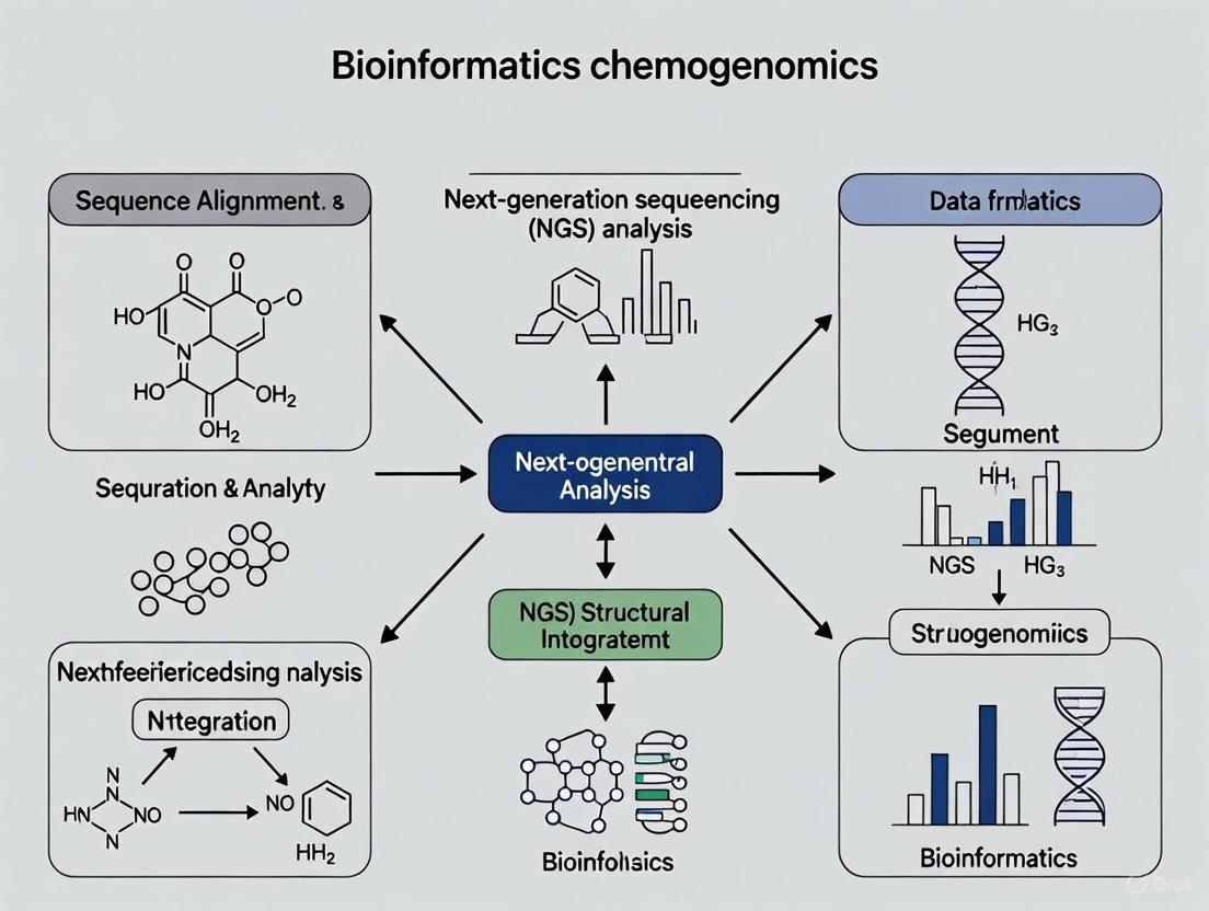
This article explores the indispensable role of bioinformatics in transforming next-generation sequencing (NGS) data into actionable insights for chemogenomics and drug discovery.
From Data to Drugs: The Critical Role of Bioinformatics in Analyzing NGS Data for Chemogenomics
Abstract
This article explores the indispensable role of bioinformatics in transforming next-generation sequencing (NGS) data into actionable insights for chemogenomics and drug discovery. Aimed at researchers, scientists, and drug development professionals, it provides a comprehensive overview of how advanced computational tools, including AI and multi-omics integration, are used to decode complex biological data, identify novel drug targets, and accelerate the development of personalized therapeutics. The content covers foundational concepts, methodological applications, common troubleshooting strategies, and essential validation frameworks, offering a complete guide for leveraging NGS in modern pharmaceutical research.
The Bioinformatics Bridge: Connecting NGS Data to Chemogenomic Insights
The fields of bioinformatics, Next-Generation Sequencing (NGS), and chemogenomics represent a powerful triad that is fundamentally reshaping the landscape of modern drug discovery and development. This synergy provides researchers with an unprecedented capacity to navigate the vast complexity of biological systems and chemical space. Bioinformatics offers the computational framework for extracting meaningful patterns from large-scale biological data. NGS technologies generate comprehensive genomic, transcriptomic, and epigenomic profiles at an astonishing scale and resolution. Chemogenomics systematically investigates the interactions between chemical compounds and biological targets on a genome-wide scale, thereby linking chemical space to biological space [1] [2]. The integration of these disciplines is critical for addressing the inherent challenges in the drug discovery pipeline, a process traditionally characterized by high costs, extensive timelines, and significant attrition rates [1]. By leveraging NGS data within a chemogenomic framework, researchers can now identify novel drug targets, predict drug-target interactions (DTIs), and identify synergistic drug combinations with greater speed and accuracy, ultimately paving the way for more effective therapies and the advancement of personalized medicine [1] [3].
Core Conceptual Foundations
Bioinformatics: The Computational Engine
Bioinformatics is the indispensable discipline that develops and applies computational tools for organizing, analyzing, and interpreting complex biological data. Its role is central to the interpretation and application of biological data generated by modern high-throughput technologies [4]. In the context of NGS and chemogenomics, bioinformatics provides the essential algorithms and statistical methods for tasks such as sequence alignment, variant calling, structural annotation, and functional enrichment analysis. It transforms raw data into biologically meaningful insights, enabling researchers to formulate and test hypotheses about gene function, disease mechanisms, and drug action [4]. The field relies on a robust technology stack, often utilizing high-performance computing clusters and a vast ecosystem of open-source software to provide statistically robust and biologically relevant analyses [4].
Next-Generation Sequencing (NGS): The Data Generation Powerhouse
NGS technologies are high-throughput platforms that determine the precise order of nucleotides within DNA or RNA molecules rapidly and accurately [4]. Common NGS applications that feed directly into chemogenomic studies include:
- DNAseq: Used for whole-genome sequencing, targeted re-sequencing (e.g., of specific gene panels), and de novo assembly to identify genetic variations like single nucleotide polymorphisms (SNPs) and insertions/deletions (indels) [5] [4].
- RNAseq (Transcriptomics): Profiles the transcriptome to quantify gene expression levels, identify differentially expressed genes between conditions (e.g., diseased vs. healthy), and discover novel splice variants [6] [4].
- Metagenomics: Characterizes the composition and functional potential of microbial communities, often via 16S rRNA sequencing, which is crucial for understanding the human microbiome's role in drug metabolism and response [7] [4].
The primary output of an NGS run is data in the FASTQ format, which contains both the nucleotide sequences and their corresponding quality scores [8]. However, this raw data requires significant computational preprocessing and quality control before it can be used for downstream analysis.
Chemogenomics: The Integrative Framework
Chemogenomics is a systematic approach that studies the interactions between chemical compounds (drugs, ligands) and biological targets (proteins, genes) on a genomic scale [1] [2]. Its primary goal is to link chemical space to biological function, thereby accelerating the identification and validation of new drug targets and lead compounds. A central application of chemogenomics is the prediction of Drug-Target Interactions (DTIs), which can be framed as a classification problem to determine whether a given drug and target will interact [1]. Chemogenomic approaches are also extensively used to predict synergistic drug combinations, where the combined effect of two or more drugs is greater than the sum of their individual effects, a phenomenon critical for treating complex diseases like cancer and overcoming drug resistance [9] [3] [10].
The Synergistic Workflow: From Raw Data to Biological Insight
The practical integration of NGS and chemogenomics involves a multi-stage analytical workflow where bioinformatics tools are applied at each step to transform raw sequencing data into actionable chemogenomic insights.
NGS Data Processing: The Foundational Steps
Before NGS data can inform chemogenomic models, it must undergo rigorous preprocessing to ensure its quality and reliability.
Experimental Protocol: NGS Data Preprocessing and QC This protocol details the critical steps for preparing raw NGS data for downstream analysis [8].
- Quality Control (QC) of Raw Reads: The initial step involves assessing the quality of sequences in the FASTQ files using tools like FastQC. This evaluation provides information on per-base sequence quality, adapter contamination, and other potential issues [8].
- Adapter Trimming and Quality Filtering: If QC reveals adapter contamination or low-quality bases, tools like Trimmomatic are used to remove adapter sequences and trim low-quality regions. This step is crucial as contaminants can interfere with subsequent mapping and analysis.
- Command Example (using Trimmomatic on a high-performance computing cluster): This command runs Trimmomatic in parallel on multiple samples, removing adapters specified in a reference file and discarding any reads shorter than 25 bases after trimming [8].
- Post-Trimming QC: After trimming, FastQC and MultiQC are run again on the cleaned FASTQ files to confirm that the data quality is sufficient for further analysis.
- Command Example: MultiQC aggregates results from multiple FastQC runs into a single report for efficient inspection [8].
Diagram: NGS Data Preprocessing Workflow
NGS data undergoes quality control, with trimming and filtering if needed, to produce analysis-ready data.
Building Chemogenomic Models from Processed NGS Data
Processed NGS data is used to construct features that train chemogenomic models for DTI and synergy prediction.
Experimental Protocol: Constructing a Multi-Omics Synergy Prediction Model This protocol outlines the methodology for developing a computational model, such as MultiSyn, to predict synergistic drug combinations by integrating multi-omics data from NGS [3].
Data Collection and Feature Extraction:
- Cell Line Features: Utilize processed NGS and other omics data to represent cancer cell lines.
- Transcriptomics: Use normalized gene expression profiles from RNAseq (e.g., from the Cancer Cell Line Encyclopedia - CCLE) [3].
- Genomics: Incorporate features like gene mutations (from COSMIC database) and copy number variations [3] [10].
- Biological Networks: Integrate Protein-Protein Interaction (PPI) networks (e.g., from STRING database) using Graph Neural Networks (GNNs) to provide functional context and capture complex cellular relationships [3].
- Drug Features: Represent the chemical structure of drugs.
- Molecular Graphs: Decompose drugs into atoms and pharmacophore-containing fragments (key functional groups responsible for drug activity) to create a heterogeneous molecular graph [3].
- Learning Representations: Use a Heterogeneous Graph Transformer to learn comprehensive representations of the drug's structure from this graph [3].
- Cell Line Features: Utilize processed NGS and other omics data to represent cancer cell lines.
Model Integration and Training:
- Fuse the cell line features (multi-omics + PPI) and drug features (molecular graph) into a unified representation.
- Train a machine learning predictor (e.g., a deep learning model) on a benchmark dataset of known drug combination synergy scores (e.g., the O'Neil dataset) to learn the complex mapping from features to synergy outcomes [3].
Validation and Evaluation:
- Evaluate model performance using rigorous cross-validation strategies (e.g., 5-fold CV) and leave-one-out protocols (leaving out specific drugs, drug pairs, or tissue types) to assess generalizability [3].
- Quantify predictive accuracy using metrics like the Bliss Independence Synergy Score or the Combination Index (CI) to compare predicted synergy with experimental results [10].
Diagram: Chemogenomic Model Integration
Processed NGS data and drug structures are transformed into features and integrated by a model to predict drug synergy.
Quantitative Frameworks: Classifying Chemogenomic Approaches
The field of chemogenomics encompasses a diverse set of computational strategies for predicting drug-target interactions and synergistic combinations. The table below summarizes the key categories, their principles, advantages, and limitations.
Table 1: Classification and Comparison of Chemogenomic Approaches for Drug-Target Interaction Prediction
| Chemogenomic Category | Core Principle | Advantages | Disadvantages |
|---|---|---|---|
| Network-Based Inference (NBI) | Uses topology of drug-target bipartite networks for prediction [1]. | Does not require 3D target structures or negative samples [1]. | Suffers from "cold start" problem for new drugs; biased towards highly connected nodes [1]. |
| Similarity Inference | Applies "guilt-by-association": similar drugs likely hit similar targets and vice-versa [1]. | Highly interpretable; leverages "wisdom of the crowd" [1]. | May miss serendipitous discoveries; often ignores continuous binding affinity data [1]. |
| Feature-Based Machine Learning | Treats DTI as a classification/regression problem using features from drugs and targets [1]. | Can handle new drugs/targets via their features; no need for similar neighbors [1]. | Feature selection is critical and difficult; class imbalance can be an issue [1]. |
| Matrix Factorization | Decomposes the drug-target interaction matrix into lower-dimensional latent features [1]. | Does not require negative samples [1]. | Primarily models linear relationships; may struggle with complex non-linearities [1]. |
| Deep Learning (e.g., MultiSyn) | Uses deep neural networks to automatically learn complex features from raw data (e.g., molecular graphs, omics) [3]. | Surpasses manual feature extraction; can model highly non-linear relationships [3]. | "Black box" nature reduces interpretability; reliability of learned features can be a concern [1] [3]. |
Successful integration of NGS and chemogenomics relies on a curated set of computational tools, databases, and reagents.
Table 2: Essential Resources for NGS and Chemogenomics Research
| Resource Type | Name | Primary Function / Application |
|---|---|---|
| NGS Analysis Tools | FastQC | Quality control tool for high throughput sequencing data [8]. |
| Trimmomatic | Flexible tool for trimming and removing adapters from NGS reads [8]. | |
| BWA | Read-mapping algorithm for aligning sequencing reads to a reference genome [5]. | |
| samtools | Suite of programs for manipulating and viewing alignments in SAM/BAM format [5]. | |
| Galaxy | Web-based, user-friendly platform for accessible NGS data analysis [6]. | |
| Key Databases | NCBI SRA | Public repository for raw sequencing data from NGS studies [6]. |
| CCLE | Catalogues genomic and transcriptomic data from a large panel of human cancer cell lines [3]. | |
| DrugBank | Database containing drug and drug-target information, including SMILES structures [3]. | |
| STRING | Database of known and predicted Protein-Protein Interactions (PPIs) [3]. | |
| Chemogenomic Models | MAGENTA | Predicts antibiotic combination efficacy under different metabolic environments using chemogenomic profiles [9]. |
| MultiSyn | Predicts synergistic anti-cancer drug combinations by integrating multi-omics data and drug pharmacophore features [3]. |
The strategic synergy between bioinformatics, NGS, and chemogenomics is creating a powerful, data-driven paradigm for biological discovery and therapeutic development. This integrated framework allows researchers to move beyond a one-drug-one-target mindset and instead view drug action and interaction within the complex, interconnected system of the cell. As these fields continue to evolve, future progress will be driven by several key trends: the move towards even deeper multi-omics data integration (including proteomics and metabolomics), a strong emphasis on improving the interpretability of "black box" deep learning models, and the rigorous clinical validation of computational predictions to bridge the gap between in silico findings and patient outcomes [3] [10]. By continuing to refine this collaborative approach, the scientific community is poised to accelerate the discovery of novel therapeutics and usher in a new era of precision medicine tailored to individual genetic and molecular profiles.
Next-generation sequencing (NGS) has revolutionized chemogenomics, enabling researchers to understand the complex interplay between genetic variation and drug response at an unprecedented scale. This field leverages high-throughput sequencing technologies to uncover how genomic variations influence individual responses to pharmaceuticals, thereby facilitating the development of personalized medicine strategies [11]. The versatility of NGS platforms provides researchers with a powerful toolkit for analyzing DNA and RNA molecules in a high-throughput and cost-effective manner, swiftly propelling genomics advancements across diverse domains [12]. The integration of sophisticated bioinformatics is fundamental to this process, transforming raw sequencing data into actionable biological insights that can guide drug discovery and clinical application.
In chemogenomics, the strategic selection of NGS approach—whether whole genome sequencing (WGS), whole exome sequencing (WES), or targeted panels—directly influences the scope and resolution of pharmacogenomic insights that can be obtained. Each method offers distinct advantages in breadth of genomic coverage, depth of sequencing, cost-effectiveness, and analytical complexity [13]. This technical guide examines these core NGS technologies, their experimental protocols, and their specific applications within chemogenomics research, with particular emphasis on the indispensable role of bioinformatics in processing, interpreting, and contextualizing the resulting data.
Core NGS Technologies in Chemogenomics
Whole Genome Sequencing (WGS)
Whole genome sequencing (WGS) represents the most comprehensive approach for analyzing entire genomes, providing a complete view of both coding and non-coding regions [14] [15]. This technology delivers a high-resolution, base-by-base view of the genome, capturing both large and small variants that might be missed with more targeted approaches [15]. In chemogenomics, this comprehensive view is particularly valuable for identifying potential causative variants for further follow-up studies of gene expression and regulation mechanisms that underlie differential drug responses [15].
WGS employs various technical approaches, with sequencing by synthesis being a commonly used method. This approach sequences a DNA sample by attaching it to a solid support, producing single-stranded DNA, followed by synthesis of the complementary copy where each incorporated nucleotide is detected [14]. Two main techniques are utilized:
- Short-read sequencing provides reads of approximately 150bp, offering cost-effective and highly accurate (>99.9%) sequencing [14].
- Long-read sequencing provides substantially longer reads (10kb to >1 Mb) and circumvents the use of PCR amplification, enabling better resolution of complex genomic regions [14].
For chemogenomics applications, WGS is particularly valuable because it enables the identification of genetic variations throughout the entire genome, including single nucleotide polymorphisms (SNPs), insertions, deletions, and copy number variations by comparing sequences with an internationally approved reference genome [14]. This is crucial for pharmacogenomics, as drug response variants do not necessarily occur only within coding regions and may reside in regulatory elements that influence gene expression [11].
Table 1: Key Whole Genome Sequencing Methods in Chemogenomics
| Method | Primary Use | Key Advantages | Chemogenomics Applications |
|---|---|---|---|
| Large WGS (>5 Mb) | Plant, animal, or human genomes | Comprehensive variant detection | Identifying novel pharmacogenomic markers across populations |
| Small WGS (≤5 Mb) | Bacteria, viruses, microbes | Culture-independent analysis | Antimicrobial resistance profiling and drug target discovery |
| De novo sequencing | Novel genomes without reference | Complete genomic characterization | Model organism development for drug screening |
| Phased sequencing | Haplotype resolution | Allele-specific assignment on homologous chromosomes | Understanding allele-specific drug metabolism |
| Single-cell WGS | Cellular heterogeneity | Resolution at individual cell level | Characterizing tumor heterogeneity in drug response |
Targeted Sequencing Panels
Targeted sequencing panels represent a focused approach that uses NGS to target specific genes, coding regions of the genome, or chromosomal segments for rapid identification and analysis of genetic mutations relevant to drug response [16]. This method is particularly useful for studying gene variants in selected genes or specific regions of the genome, as it can rapidly and cost-effectively target a large diversity of genetic regions [16]. In chemogenomics, targeted sequencing is employed to examine gene interactions in specific pharmacological pathways and is generally faster and more cost-effective than whole genome sequencing (WGS) because it analyzes a smaller, more relevant set of nucleotides rather than broadly sequencing the entire genome [16].
Targeted panels can be either predesigned, containing important genes or gene regions associated with specific diseases or drug responses selected from publications and expert guidance, or custom-designed, allowing researchers to target regions of the genome relevant to their specific research interests [17]. Illumina supports two primary methods for custom targeted gene sequencing:
- Target enrichment: Regions of interest are captured by hybridization to biotinylated probes and then isolated by magnetic pulldown. This method is suitable for larger gene content, typically >50 genes, and provides more comprehensive profiling for all variant types [17].
- Amplicon sequencing: Regions of interest are amplified and purified using highly multiplexed oligo pools. This approach is ideal for smaller gene content, typically <50 genes, and is optimized for analyzing single nucleotide variants and insertions/deletions (indels) [17].
The advantages of targeted sequencing in chemogenomics include the ability to sequence key pharmacogenes of interest to high depth (500–1000× or higher), allowing identification of rare variants; cost-effective findings for studies of disease-related genes; accurate, easy-to-interpret results that identify gene variants at low allele frequencies (down to 0.2%); and confident identification of causative novel or inherited mutations in a single assay [17].
Table 2: Comparison of Targeted Sequencing Approaches in Chemogenomics
| Parameter | Target Enrichment | Amplicon Sequencing |
|---|---|---|
| Ideal Gene Content | Larger panels (>50 genes) | Smaller panels (<50 genes) |
| Variant Detection | Comprehensive for all variant types | Optimal for SNVs and indels |
| Hands-on Time | Longer | Shorter |
| Cost | Higher | More affordable |
| Workflow Complexity | More complex | Streamlined |
| Turnaround Time | Longer | Faster |
Whole Exome Sequencing (WES)
Whole exome sequencing occupies a middle ground between comprehensive WGS and focused targeted panels, specifically targeting the exon regions that comprise only 1-2% of the genome but harbor approximately 85% of known pathogenic variants [13] [14]. This approach is typically more cost-effective than WGS and provides more extensive information than targeted sequencing, making it an ideal first-tier test for cases involving severe, nonspecific symptoms or conditions where multiple genetic factors may influence drug response [13].
However, WES presents certain limitations for chemogenomics applications. Not all exonic regions can be equally evaluated, and critical noncoding regions are not sequenced, making it impossible to detect functional variants outside the exonic areas that may regulate drug metabolism genes [13]. Additionally, except for a few cases of copy number variations (CNVs), WES shows low sensitivity to structural variants (SVs) that can affect pharmacogene function [13]. The results of WES can also vary depending on the test kit used, as the targeted regions and probe manufacturing methods differ between commercial kits, potentially leading to variations in data quality and coverage of key pharmacogenes [13].
Experimental Protocols and Methodologies
Protocol for Targeted NGS Panel Development and Validation
A recent study demonstrated the development and validation of a targeted NGS panel for clinically relevant mutation profiles in solid tumours, providing an exemplary protocol for chemogenomics research [18]. The researchers developed an oncopanel targeting 61 cancer-associated genes and validated its efficacy by performing NGS on 43 unique samples including clinical tissues, external quality assessment samples, and reference controls.
Experimental Workflow:
- Library Preparation: Applied a hybridization-capture based DNA target enrichment method using library kits compatible with an automated library preparation system to ensure consistency and reduce contamination risk.
- Sequencing: Utilized the DNBSEQ-G50RS sequencer with cPAS sequencing technology for precise sequencing with high SNP and Indel detection accuracy.
- Bioinformatics Analysis: Implemented specialized software using machine learning for rapid variant analysis and visualization of mutated and wild type hotspot positions, connecting molecular profiles to clinical insights through a clinical classification portal.
Performance Validation: The assay's analytical performance was rigorously validated through several parameters:
- DNA Input Optimization: Titration experiments determined that ≥50 ng of DNA input was necessary for reliable detection of all expected mutations in the reference standard.
- Limit of Detection: The assay demonstrated 100% sensitivity for variants at >3.0% variant allele frequency (VAF), with the minimum reliable detection threshold established at 2.9% VAF for both SNVs and INDELs.
- Precision: Replicate analyses showed 99.99% repeatability (intra-run precision) and 99.98% reproducibility (inter-run precision) at 95% confidence intervals.
- Concordance: The method detected 794 mutations including all 92 known variants from orthogonal methods, demonstrating 100% concordance with external genomic data.
This validation protocol highlights the rigorous approach required for implementing targeted NGS panels in clinical chemogenomics applications, where reliable detection of pharmacogenomic variants is essential for treatment decisions.
Bioinformatics Analysis Workflow for Chemogenomics
The bioinformatics workflow for processing NGS data in chemogenomics involves multiple critical steps that transform raw sequencing data into clinically actionable information. This process requires sophisticated computational tools and analytical expertise to derive meaningful insights from the vast amounts of data generated by NGS technologies [19].
Diagram 1: NGS Data Analysis Workflow
Key Steps in the Bioinformatics Pipeline:
Quality Control and Trimming: Initial assessment of sequencing quality using tools like FastQC, followed by trimming of adapter sequences and low-quality bases to ensure data integrity.
Alignment to Reference Genome: Processed reads are aligned to a reference genome using aligners such as BWA or Bowtie2, generating BAM files that represent the genomic landscape of the sample.
Variant Calling: Specialized algorithms identify genetic variants relative to the reference genome. Tools like DeepVariant and Strelka2 are particularly effective for detecting SNPs, indels, and other variants in pharmacogenes [19].
Variant Annotation and Functional Prediction: Identified variants are annotated using comprehensive genomic databases (e.g., Ensembl, NCBI) to determine their functional impact, population frequency, and prior evidence of clinical significance [19].
Clinical Interpretation: Annotated variants are interpreted in the context of pharmacogenomic knowledge bases, which curate evidence linking specific genetic variants to drug response phenotypes. This step often utilizes machine learning models to predict disease risk, drug response, and other complex phenotypes, with Explainable AI (XAI) being crucial for understanding the basis of these predictions [19].
The integration of these bioinformatics processes enables researchers to bridge the gap between genomic findings and clinical application, ultimately supporting personalized treatment recommendations based on an individual's genetic profile.
The Scientist's Toolkit: Essential Research Reagents and Materials
Successful implementation of NGS technologies in chemogenomics requires access to specialized reagents, kits, and computational resources. The following table outlines essential components of the chemogenomics research toolkit.
Table 3: Essential Research Reagents and Solutions for Chemogenomics NGS
| Category | Specific Products/Solutions | Primary Function | Application in Chemogenomics |
|---|---|---|---|
| Library Preparation | Illumina DNA Prep with Enrichment; Sophia Genetics Library Kit [18] | Prepares DNA fragments for sequencing by adding adapters and indices | Target enrichment for pharmacogene panels; whole genome library construction |
| Target Capture | Illumina Custom Enrichment Panel v2; AmpliSeq for Illumina Custom Panels [17] | Enriches for specific genomic regions of interest | Focusing sequencing on known pharmacogenes and regulatory regions |
| Sequencing Platforms | MGI DNBSEQ-G50RS; Illumina MiSeq [18] | Generates sequence data from prepared libraries | Producing high-quality sequencing data for variant discovery |
| Bioinformatics Tools | Sophia DDM; DeepVariant; Strelka2 [19] [18] | Analyzes sequencing data for variant calling and interpretation | Identifying and annotating pharmacogenomically relevant variants |
| Reference Materials | HD701 Reference Standard; External Quality Assessment samples [18] | Provides quality control and assay validation | Ensuring analytical validity and reproducibility of NGS assays |
| Data Analysis Platforms | Nextflow; Snakemake; Docker [19] | Workflow management and containerization | Enabling reproducible analysis pipelines across computing environments |
The strategic selection and implementation of NGS technologies are critical for advancing chemogenomics research and clinical application. Whole genome sequencing offers the most comprehensive approach for discovery-phase research, while targeted panels provide cost-effective, deep coverage for focused investigation of known pharmacogenes. Whole exome sequencing represents a balanced approach for many clinical applications. The future of NGS in chemogenomics will be shaped by emerging trends including increased adoption of long-read sequencing, multi-omics integration, workflow automation, cloud-based computing, and real-time data analysis [19].
As these technologies continue to evolve, the role of bioinformatics becomes increasingly central to extracting meaningful insights from complex genomic datasets. Advanced computational methods, including machine learning and artificial intelligence, are enhancing our ability to predict drug response and identify novel pharmacogenomic biomarkers [19] [20]. By strategically leveraging the appropriate NGS technology for specific research questions and clinical applications, scientists can continue to advance personalized medicine and optimize therapeutic outcomes based on individual genetic profiles.
Next-generation sequencing (NGS) has revolutionized genomics research, enabling the rapid sequencing of millions of DNA fragments simultaneously to provide comprehensive insights into genome structure, genetic variations, and gene expression profiles [12]. This transformative technology has become a fundamental tool across diverse domains, from basic biology to clinical diagnostics, particularly in the field of chemogenomics where understanding the interaction between chemical compounds and biological systems is paramount [12]. However, the unprecedented scale and complexity of data generated by NGS technologies present a formidable challenge that can overwhelm conventional computational infrastructure and analytical workflows. The sheer volume of data, combined with intricate processing requirements, creates a significant bottleneck that researchers must navigate to extract meaningful biological insights relevant to drug discovery and development [21]. This technical guide examines the core challenges associated with NGS data management and provides structured frameworks and methodologies to address them effectively within chemogenomics research.
The Data Deluge: Quantifying NGS Data Challenges
Exponential Growth of Genomic Data
The dramatic reduction in sequencing costs has catalyzed an explosive growth in genomic data generation. Conventional integrative analysis techniques and computational methods that worked well with traditional genomics data are ill-equipped to deal with the unique data characteristics and overwhelming volumes of NGS data [21]. This data explosion presents significant challenges, both in terms of crunching raw data at scale and in analysing and interpreting complex datasets [21].
Table 1: Global Population Genomics Initiatives Contributing to NGS Data Growth
| Initiative Name | Region/Country | Scale | Primary Focus |
|---|---|---|---|
| All of Us | USA | 1 million genomes | Personalized medicine, health disparities |
| Genomics England | United Kingdom | 100,000 genomes | NHS integration, rare diseases, cancer |
| IndiGen | India | 1,029 genomes (initial phase) | India-centric genomic variations |
| 1+ Million Genomes | European Union | 1+ million genomes | Cross-border healthcare, research |
| Saudi Human Genome Program | Saudi Arabia | 100,000+ genomes | Regional genetic disorders, population genetics |
Table 2: NGS Data Generation Metrics by Sequencing Type
| Sequencing Approach | Typical Data Volume per Sample | Primary Applications in Chemogenomics | Key Challenges |
|---|---|---|---|
| Whole Genome Sequencing (WGS) | 100-200 GB | Pharmacogenomics, variant discovery, personalized therapy | Storage costs, processing time, data transfer |
| Whole Exome Sequencing (WES) | 5-15 GB | Target identification, Mendelian disorders, cancer genomics | Coverage uniformity, variant interpretation |
| RNA Sequencing | 10-30 GB | Transcriptional profiling, drug mechanism of action, biomarker discovery | Normalization, batch effects, complex analysis |
| Targeted Panels | 1-5 GB | Clinical diagnostics, therapeutic monitoring, pharmacogenetics | Panel design, limited discovery power |
| Single-cell RNAseq | 20-50 GB | Cellular heterogeneity, tumor microenvironment, drug resistance | Computational intensity, specialized tools |
Technical and Infrastructural Hurdles
The data exploration and analysis already lag data generation by a significant order of magnitude – and this deficit will only be exacerbated as we transition from NGS to third-generation sequencing technologies [21]. Most large institutions are already heavily invested in hardware/software infrastructure and in standardized workflows for genomic data analysis. A wholesale remapping of these investments to integrate agility, flexibility, and versatility features required for big data genomics is often impractical [21].
A critical challenge emerges from the specialized workforce requirements for NGS data analysis. Retaining proficient personnel can be a substantial obstacle because of the unique and specialized knowledge required, which in turn increases costs for adequate staff compensation [22]. In 2021, the Association of Public Health Laboratories (APHL) reported that 30% of surveyed public health laboratory staff indicated an intent to leave the workforce within the next 5 years [22]. This talent gap significantly restricts the pace of progress in genomics research [21].
Quality Control Frameworks: Essential Protocols for Reliable NGS Data
Comprehensive QC Metrics and Standards
Quality control (QC) is the process of assessing the quality of raw sequencing data to identify any potential problems that may affect downstream analyses [23]. QC involves several steps, including the assessment of data quality metrics, the detection of adapter contamination, and the removal of low-quality reads [23]. To ensure that high-quality data is generated, researchers must perform QC at various stages of the NGS workflow, including after sample preparation, library preparation, and sequencing [23].
Protocol 1: Pre-alignment Quality Control Assessment
Quality Metric Evaluation: Assess raw sequencing data using tools such as FastQC to generate comprehensive reports on read length, sequencing depth, base quality, and GC content [23].
Adapter Contamination Detection: Identify residual adapter sequences using specialized tools like Trimmomatic or Cutadapt. Adapter contamination occurs when adapter sequences used in library preparation are not fully removed from the sequencing data, leading to false positives and reduced accuracy in downstream analyses [23].
Low-Quality Read Filtering: Remove reads containing sequencing errors (base-calling errors, phasing errors, and insertion-deletion errors) using quality score thresholds implemented in tools such as Trimmomatic or Cutadapt [23].
Sample-Level Validation: Verify sample identity and quality through methods including sex chromosome concordance checks, contamination estimation, and comparison of expected versus observed variant frequencies.
Protocol 2: Post-alignment Quality Control Measures
Alignment Metric Quantification: Evaluate mapping quality using metrics including total reads aligned, percentage of properly paired reads, duplication rates, and coverage uniformity across target regions.
Variant Calling Quality Assessment: Implement multiple calling algorithms with concordance analysis, strand bias evaluation, and genotype quality metrics.
Experimental Concordance Verification: Compare technical replicates, cross-validate with orthogonal technologies (e.g., Sanger sequencing, microarrays), and assess inheritance patterns in family-based studies.
The following workflow diagram illustrates the comprehensive quality control process for NGS data:
Bioinformatics Pipelines: Analytical Frameworks for NGS Data
Core Computational Workflows
The analysis of NGS data requires sophisticated computational methods and bioinformatics expertise [24]. The sheer amount and variety of data generated by NGS assays require sophisticated computational resources and specialized bioinformatics software to yield informative and actionable results [24]. The primary bioinformatics procedures include alignment, variant calling, and annotation [24].
Table 3: Essential Bioinformatics Tools for NGS Data Analysis
| Analytical Step | Tool Options | Key Functionality | Considerations for Chemogenomics |
|---|---|---|---|
| Read Alignment | BWA, STAR, Bowtie | Maps sequencing reads to reference genome | Impact on variant calling accuracy for pharmacogenes |
| Variant Calling | GATK, Samtools, FreeBayes | Identifies genetic variants relative to reference | Sensitivity for detecting rare variants with clinical significance |
| Variant Annotation | ANNOVAR, SnpEff, VEP | Functional prediction of variant consequences | Drug metabolism pathway gene prioritization |
| Expression Analysis | DESeq2, edgeR, limma | Quantifies differential gene expression | Identification of drug response signatures |
| Copy Number Analysis | CNVkit, Control-FREEC | Detects genomic amplifications/deletions | Association with drug resistance mechanisms |
Specialized Methodologies for Chemogenomics Applications
Protocol 3: Transcriptomic Profiling for Drug Response Assessment
Library Preparation Considerations: Isolate RNA and convert to complementary DNA (cDNA) for sequencing library construction. Evaluate RNA integrity numbers (RIN) to ensure sample quality, with minimum thresholds of 7.0 for bulk RNA-seq and 8.0 for single-cell applications [24].
Sequencing Depth Determination: Target 20-50 million reads per sample for standard differential expression analysis. Increase to 50-100 million reads for isoform-level quantification or novel transcript discovery.
Expression Quantification: Utilize alignment-based (STAR/RSEM) or alignment-free (Kallisto/Salmon) approaches to estimate transcript abundance [23].
Differential Expression Analysis: Apply statistical models (DESeq2, edgeR, limma) to identify genes significantly altered between treatment conditions [23]. Implement multiple testing correction with false discovery rate (FDR) control.
Pathway Enrichment Analysis: Integrate expression changes with chemical-target interactions using databases such as CHEMBL, DrugBank, or STITCH to identify affected biological processes and potential mechanisms of action.
Protocol 4: Somatic Variant Detection in Preclinical Models
Tumor Purity Assessment: Estimate tumor cell content through pathological review or computational methods (e.g., ESTIMATE, ABSOLUTE). Higher purity samples (>30%) generally yield more reliable variant calls [24].
Matched Normal Sequencing: Sequence normal tissue from the same organism to distinguish somatic from germline variants. This is critical for identifying acquired mutations relevant to drug sensitivity and resistance.
Variant Calling Parameters: Optimize minimum depth thresholds (typically 50-100x for tumor, 30-50x for normal) and variant allele frequency cutoffs based on tumor purity and ploidy characteristics.
Variant Annotation and Prioritization: Filter variants based on population frequency (e.g., gnomAD), functional impact (e.g., SIFT, PolyPhen), and relevance to drug targets or resistance mechanisms.
The following diagram illustrates the comprehensive bioinformatics workflow for NGS data analysis in chemogenomics:
Research Reagent Solutions for NGS Experiments
Table 4: Essential Research Reagents and Platforms for NGS Workflows
| Reagent/Platform Type | Specific Examples | Function in NGS Workflow | Considerations for Selection |
|---|---|---|---|
| Library Preparation Kits | Illumina DNA Prep | Fragments DNA and adds adapters for sequencing | Compatibility with input material, hands-on time |
| Target Enrichment | Illumina Nextera Flex | Enriches specific genomic regions of interest | Coverage uniformity, off-target rates |
| Sequencing Platforms | Illumina MiSeq, NextSeq | Performs actual sequencing reaction | Throughput, read length, cost per sample |
| Multiplexing Barcodes | Dual Index Barcodes | Allows sample pooling for efficient sequencing | Index hopping rates, complexity |
| Quality Control Kits | Bioanalyzer, TapeStation | Assesses library quality before sequencing | Sensitivity, required equipment |
| Enzymatic Mixes | Polymerases, Ligases | Facilitates library construction reactions | Fidelity, efficiency with damaged DNA |
Integrated Solutions and Future Directions
Consolidated Bioinformatics Platforms
To address the specialized talent gap in bioinformatics, several integrated platforms have emerged that consolidate analysis workflows into more accessible interfaces. These solutions aim to provide end-to-end, self-service platforms that unify all components of the genomics analysis and research workflow into comprehensive solutions [21]. Such platforms precompute and index numerous sequences from public databases into proprietary knowledge bases that are continuously updated, allowing researchers to search through volumes of sequence data and retrieve pertinent information about alignments, similarities, and differences rapidly [21].
Regulatory and Quality Considerations
For laboratories implementing NGS, particularly in regulated environments, the Next-Generation Sequencing Quality Initiative (NGS QI) provides tools and resources to build a robust quality management system [22]. This initiative addresses challenges associated with personnel management, equipment management, and process management across NGS laboratories [22]. Their resources include QMS Assessment Tools, SOPs for Identifying and Monitoring NGS Key Performance Indicators, NGS Method Validation Plans, and NGS Method Validation SOPs [22].
Emerging Technologies and Methodologies
The NGS landscape continues to evolve with the introduction of new platforms and improved chemistries. For example, new kit chemistries from Oxford Nanopore Technologies that use CRISPR for targeted sequencing and improved basecaller algorithms using artificial intelligence and machine learning lead to increased accuracy [22]. Other emerging platforms, such as Element Biosciences, also show increasing accuracies with lower costs, which might encourage transition from older platforms to new platforms and chemistries [22].
To keep up with evolving practices, organizations are implementing cyclic review processes and performing regular updates to their analytical frameworks. However, the rapid pace of changes in policy and technology means that regular updates do not always resolve challenges [22]. Despite these difficulties, maintaining validated, locked-down workflows while simultaneously evaluating technological advancements remains essential for producing high-quality, reproducible, and reliable results [22].
The management of NGS data volume and complexity represents a central challenge in modern chemogenomics research. As sequencing technologies continue to advance and data generation accelerates, the implementation of robust quality control frameworks, standardized bioinformatics pipelines, and integrated analytical platforms becomes increasingly critical. By adopting the structured approaches and methodologies outlined in this technical guide, researchers can more effectively navigate the complexities of NGS data, transform raw sequence information into actionable biological insights, and accelerate the discovery and development of novel therapeutic compounds. The continuous evolution of computational infrastructure, analytical algorithms, and workforce expertise will remain essential to fully harness the potential of NGS technologies in advancing chemogenomics and personalized medicine.
The field of bioinformatics has undergone a revolutionary transformation, evolving from a discipline focused primarily on managing and analyzing basic sequencing data into a sophisticated, AI-powered engine for scientific discovery. This evolution is particularly impactful in chemogenomics, which explores the complex interactions between chemical compounds and biological systems. The advent of Next-Generation Sequencing (NGS) has been a cornerstone of this shift, generating unprecedented volumes of genomic, transcriptomic, and epigenomic data [12]. Initially, bioinformatics provided the essential tools for processing this data. However, the integration of Artificial Intelligence (AI) and machine learning (ML) has fundamentally altered the landscape, enabling the extraction of deeper insights and the prediction of complex biological outcomes [25] [26]. This whitepaper details this technological evolution, framing it within the context of chemogenomics research, where these advanced bioinformatics strategies are accelerating the identification and validation of novel therapeutic targets and biomarkers.
The Next-Generation Sequencing (NGS) Revolution
Next-Generation Sequencing technologies have democratized genomic analysis by providing high-throughput, cost-effective methods for sequencing DNA and RNA molecules [12]. Unlike first-generation Sanger sequencing, NGS allows for the parallel sequencing of millions to billions of DNA fragments, providing comprehensive insights into genome structure, genetic variations, and gene expression profiles [12] [27].
Evolution and Key Sequencing Technologies
Sequencing technologies have rapidly advanced, leading to the development of multiple platforms, each with distinct strengths and applications, as summarized in Table 1.
Table 1: Key Characteristics of Major Sequencing Platforms
| Platform | Sequencing Technology | Amplification Type | Read Length (bp) | Primary Applications & Limitations |
|---|---|---|---|---|
| Illumina [12] | Sequencing-by-Synthesis | Bridge PCR | 36-300 | Applications: Whole-genome sequencing, transcriptomics, targeted sequencing. Limitations: Potential signal overlap and ~1% error rate with sample overloading. |
| Ion Torrent [12] | Sequencing-by-Synthesis (Semiconductor) | Emulsion PCR | 200-400 | Applications: Rapid targeted sequencing, diagnostic panels. Limitations: Signal degradation with homopolymer sequences. |
| PacBio SMRT [12] | Single-Molecule Real-Time Sequencing | Without PCR | 10,000-25,000 (average) | Applications: De novo genome assembly, resolving complex genomic regions, full-length transcript sequencing. Limitations: Higher cost per run. |
| Oxford Nanopore [12] | Electrical Impedance Detection | Without PCR | 10,000-30,000 (average) | Applications: Real-time sequencing, direct RNA sequencing, field sequencing. Limitations: Error rate can be as high as 15%. |
| SOLiD [12] | Sequencing-by-Ligation | Emulsion PCR | 75 | Applications: Originally used for whole-genome and transcriptome sequencing. Limitations: Short reads limit applications; under-represents GC-rich regions. |
A Standard NGS Data Analysis Workflow
The bioinformatics analysis of NGS data follows a multi-step workflow to transform raw sequencing data into biological insights. The following diagram illustrates this pipeline, highlighting key quality control checkpoints.
Title: Core NGS Bioinformatics Data Pipeline
Detailed Methodologies for Key Workflow Steps:
- Quality Control (QC): Using tools like FastQC, raw sequence data (in FASTQ format) is assessed for per-base sequence quality, GC content, overrepresented sequences, and adapter contamination. This step determines if data meets the minimum threshold for further analysis (e.g., Q-score > 30 for most bases) [28].
- Preprocessing: Based on QC results, tools like Trimmomatic or Cutadapt are used to remove adapter sequences, trim low-quality bases from the ends of reads, and discard reads that fall below a minimum length threshold.
- Alignment/Mapping: Processed reads are aligned to a reference genome using aligners such as BWA (for DNA) or STAR (for RNA-seq). This step generates SAM/BAM files, which contain the genomic coordinates for each read.
- Post-Alignment QC: Tools like Samtools flagstat and Picard Tools CollectMultipleMetrics are used to assess mapping rates, insert sizes, and coverage uniformity. For variant calling, the Genome Analysis Toolkit (GATK) Best Practices pipeline includes base quality score recalibration and local realignment around indels.
- Variant/Expression Calling:
- For genomics: Variant callers like GATK HaplotypeCaller identify single nucleotide polymorphisms (SNPs) and insertions/deletions (indels) from the aligned reads.
- For transcriptomics: Tools like HTSeq or featureCounts quantify gene expression levels, which are then used for differential expression analysis with packages like DESeq2 or edgeR in R.
- Biological Interpretation: The final list of variants or differentially expressed genes is annotated and analyzed in the context of pathways (e.g., using KEGG, Reactome), gene ontologies, and protein-protein interaction networks to derive biological meaning.
The AI Integration: Transforming Data into Discovery
The massive, complex datasets generated by NGS have rendered traditional computational approaches insufficient for many tasks [25]. The integration of AI, particularly machine learning (ML) and deep learning (DL), has created a paradigm shift, enhancing every stage of the bioinformatics workflow [26].
AI-Enhanced NGS Workflows
AI's impact spans the entire research lifecycle, from initial planning to final data interpretation. The following diagram maps AI applications onto the key phases of an NGS-based study.
Title: AI Applications in NGS Research Phases
Key AI Applications and Experimental Protocols:
Pre-Wet-Lab Phase:
- AI-Powered Experimental Design: Platforms like Benchling and LabGPT use AI to help researchers optimize protocols and plan experiments by drawing on vast databases of published protocols and outcomes [25].
- Outcome Simulation: Tools like DeepGene use deep neural networks to predict gene expression levels under different experimental conditions, allowing researchers to prioritize promising hypotheses before any wet-lab work begins [25].
Wet-Lab Phase:
- Laboratory Automation and QC: AI-driven robotic systems like the Tecan Fluent automate liquid handling and NGS library preparation. Integrated AI models (e.g., YOLOv8) can provide real-time quality control by detecting pipette tips and verifying liquid volumes, drastically reducing human error [25].
- gRNA Design for Functional Validation: In chemogenomics, CRISPR is used to validate gene targets. AI tools like DeepCRISPR and R-CRISPR use convolutional and recurrent neural networks (CNNs/RNNs) to predict gRNA efficacy and minimize off-target effects, streamlining the experimental workflow [25].
Post-Wet-Lab Phase:
- Variant Calling: Google's DeepVariant employs a CNN, treating the aligned sequencing data as an image to identify genetic variants with significantly higher accuracy than traditional heuristic methods [25] [26].
- Single-Cell and Multi-Omics Data Integration: AI is crucial for analyzing high-dimensional data from single-cell RNA sequencing (scRNA-seq). Deep learning models can integrate this data with genomic and proteomic information to identify novel cell subtypes and regulatory networks, which is central to understanding drug mechanisms [25] [29].
Application in Chemogenomics NGS Data Research
The synergy of NGS and AI provides a powerful framework for chemogenomics, which aims to link chemical compounds to genomic and phenotypic responses.
An AI-Driven Chemogenomics Workflow
This integrated workflow leverages NGS data and AI to streamline the target identification and validation process in drug discovery.
Title: AI-Driven Chemogenomics Discovery Pipeline
Detailed Experimental Protocol for a Chemogenomics Study:
NGS Data Generation:
- Experimental Design: Treat a cell line or model organism with a compound of interest and include a DMSO/vehicle control. Use at least three biological replicates per condition.
- Library Preparation and Sequencing: Extract total RNA. Use an NGS library prep kit (e.g., Illumina TruSeq) to generate RNA-seq libraries. Sequence the libraries on an Illumina platform to a depth of at least 25-30 million reads per sample.
AI-Based Bioinformatic Analysis:
- Differential Expression: Process RNA-seq data through the standard workflow (Section 2.2). Use an AI-enhanced tool like DESeq2 (which uses statistical ML) to identify significantly differentially expressed genes (adjusted p-value < 0.05 and |log2 fold change| > 1).
- Pathway and Network Analysis: Input the list of significant genes into a pathway analysis tool like IPA (Ingenuity Pathway Analysis) or GSEA (Gene Set Enrichment Analysis). Use AI-driven network analysis tools (e.g., graph neural networks) to identify key hub genes and dysregulated pathways that represent potential therapeutic targets [26].
Target Prioritization and Compound Screening:
- Variant Annotation: If working with genomic data from patient samples, use ML tools like CADD (Combined Annotation Dependent Depletion) to score and prioritize identified genetic variants based on their predicted deleteriousness [26].
- Generative AI for Compound Design: Use generative adversarial networks (GANs) or variational autoencoders (VAEs) to design novel molecular structures that are predicted to interact with the prioritized target. Alternatively, use AI for virtual screening of large compound libraries to identify potential hits [26].
The Scientist's Toolkit: Essential Research Reagents and Materials
Successful execution of NGS and AI-driven chemogenomics research relies on a suite of wet-lab and computational tools.
Table 2: Essential Research Reagent Solutions and Computational Tools
| Category | Item/Reagent | Function in Chemogenomics Research |
|---|---|---|
| Wet-Lab Reagents | NGS Library Prep Kits (e.g., Illumina TruSeq) | Prepare fragmented and adapter-ligated DNA/cDNA libraries for sequencing. |
| CRISPR-Cas9 Reagents (e.g., Synthego) | Validate candidate gene targets by performing gene knock-out or knock-in experiments. | |
| Single-Cell RNA-seq Kits (e.g., 10x Genomics) | Profile gene expression at single-cell resolution to uncover cellular heterogeneity in response to compounds. | |
| Computational Tools & Databases | AI/ML Frameworks (e.g., TensorFlow, PyTorch) | Build and train custom deep learning models for predictive tasks. |
| Bioinformatics Platforms (e.g., Galaxy, DNAnexus) | Provide user-friendly, cloud-based environments for building and running analysis pipelines without advanced coding. | |
| Chemical & Genomic Databases (e.g., ChEMBL, TCGA) | Provide annotated data on chemical compounds and cancer genomes for model training and validation. | |
| Workflow Managers (e.g., Nextflow, Snakemake) | Ensure reproducible, scalable, and automated execution of complex bioinformatics pipelines [28]. |
The evolution of bioinformatics from a supportive role in basic sequencing analysis to a central role in AI-driven discovery marks a new era in life sciences. For researchers in chemogenomics, this transition is pivotal. The integration of high-throughput NGS technologies with sophisticated AI and ML models provides an unparalleled capability to decode the complex interactions between chemicals and biological systems. This powerful synergy is accelerating the entire drug development pipeline, from the initial identification of novel targets to the design of optimized lead compounds, ultimately paving the way for more effective and personalized therapeutic strategies.
From Raw Sequences to Drug Candidates: Bioinformatics Workflows in Action
Next-generation sequencing (NGS) has revolutionized chemogenomics, enabling the systematic study of how chemical compounds interact with biological systems at a genomic level. A standardized bioinformatics pipeline is crucial for transforming raw sequencing data into reliable, actionable insights for drug discovery and development. This guide details the core steps, from raw data to variant calling, providing a framework for robust and reproducible research.
In chemogenomics research, scientists screen chemical compounds against biological targets to understand their mechanisms of action and identify potential therapeutics. NGS technologies allow for the genome-wide assessment of how these compounds affect cellular processes, gene expression, and genetic stability. A standardized data analysis pipeline ensures that the genetic variants identified—such as single nucleotide polymorphisms (SNPs) and insertions/deletions (indels)—are detected with high accuracy and consistency. This is paramount for linking compound-induced cellular responses to specific genomic alterations, thereby guiding the development of targeted therapies [30] [31].
The journey from a biological sample to a final list of genetic variants involves multiple computational steps, typically grouped into three stages: primary, secondary, and tertiary analysis [32] [33]. This guide will focus on the transition from the raw sequence data (FASTQ) to the variant call format (VCF) file, which encapsulates the secondary analysis phase. Adherence to joint recommendations from professional bodies like the Association for Molecular Pathology (AMP) and the College of American Pathologists (CAP) is essential for validating these pipelines in a clinical or translational research context [34] [35].
The NGS Data Analysis Workflow: A Three-Stage Process
The pathway from raw sequencing output to identifiable genetic variants is a multi-stage process. The following diagram illustrates the complete workflow from initial sample preparation to the final variant call file.
Primary Analysis: From Signal to Sequence
Primary analysis is the first computational step, performed by the sequencer's onboard software. It converts raw signal data (e.g., fluorescence or electrical current) into nucleotide sequences with associated quality scores [32] [33].
- Input: Binary files specific to the sequencing platform (e.g.,
.bclfiles for Illumina) [32]. - Core Process: Base calling identifies the sequence of nucleotides for each read. Demultiplexing then sorts the sequenced reads into separate files based on their unique molecular barcodes (indexes) if multiple samples were pooled in a single run [32] [33]. The CDC's Nex-StoCT II workgroup recommends discarding reads with mismatched indexes and validating de-multiplexing fidelity to ensure samples are not cross-contaminated [33].
- Output: FASTQ files, which contain the nucleotide sequences for each read and a corresponding string of Phred quality scores (Q scores) for every base call [32] [36]. A Q score of 30 (Q30) indicates a 99.9% base call accuracy and is a common quality threshold [32].
Secondary Analysis: Alignment and Variant Discovery
Secondary analysis is the most computationally intensive phase, where sequences are refined, mapped to a reference genome, and analyzed for variations. This guide details the key steps within this stage in the following diagram.
Read Cleanup and Quality Control (QC)
The initial step involves curating the raw sequencing reads to ensure data quality before alignment.
- Purpose: To remove technical artifacts that could hinder accurate alignment or variant calling [32].
- Common Tools: FastQC is widely used for initial quality assessment, providing an overview of per-base quality scores, GC content, adapter contamination, and overrepresented sequences [32].
- Key Actions:
- Trimming: Removing adapter sequences, barcodes, and low-quality bases from the ends of reads.
- Filtering: Discarding entire reads that fall below a defined quality threshold or are too short.
- Deduplication: Identifying and flagging PCR duplicates—identical reads arising from the amplification of a single original molecule—to prevent bias in variant calling [32]. The use of Unique Molecular Identifiers (UMIs) can correct for such amplification errors [32].
Sequence Alignment (Mapping)
Alignment, or mapping, is the process of determining the position of each sequenced read within a reference genome.
- Purpose: To establish the genomic origin of every read, which is foundational for identifying variations [32].
- Common Tools: BWA (Burrows-Wheeler Aligner) and Bowtie 2 are industry-standard aligners that offer a good balance of speed and accuracy [32]. The Nex-StoCT II workgroup recommends evaluating different aligners or settings to optimize for the specific variants of interest [33].
- Reference Genome: The choice of reference is critical. For human samples, the current standard is GRCh38/hg38, though GRCh37/hg19 is still commonly used. The assembly accession and version number must be documented for traceability [32] [33].
- Output: BAM file (Binary Alignment Map), a compressed and efficient binary version of the human-readable SAM file. BAM files are sorted by genomic coordinate and indexed (producing a
.baifile) to allow for rapid retrieval of reads from specific regions [32] [36]. Visualization tools like the Integrative Genomic Viewer (IGV) can then be used to inspect the alignments [32].
Variant Calling
Variant calling is the process of identifying positions in the sequenced genome that differ from the reference genome.
- Purpose: To discover genetic variants such as SNPs, indels, and larger structural variations [32].
- Methodology: The variant caller analyzes the pileup of aligned reads in the BAM file at each genomic position, considering the base calls, quality scores, and alignment statistics to distinguish true biological variants from sequencing or alignment errors [32].
- Common Tools: Traditional statistical methods are implemented in tools like GATK. Recently, AI-powered tools like DeepVariant have demonstrated superior accuracy by using deep learning models to identify variants [30]. Best practices suggest evaluating more than one variant caller during pipeline optimization [33].
- Output: VCF file (Variant Call Format), a standardized, text-based file that lists the genomic coordinates of variants, the reference and alternate alleles, and quality metrics for each call [36]. This file marks the end of the secondary analysis phase and is the primary input for downstream interpretation.
Quality Control Metrics and Standards
Ensuring data quality throughout the pipeline is non-negotiable for reliable results. Multiple organizations provide guidelines for quality control (QC) in clinical NGS [35]. The following table summarizes key QC metrics and the standards that govern them.
Table 1: Essential Quality Control Metrics in the NGS Pipeline
| QC Parameter | Analysis Stage | Description | Recommended Threshold | Governing Standards |
|---|---|---|---|---|
| Q30 Score [32] | Primary | Percentage of bases with a Phred quality score ≥30 (0.1% error rate). | >80% | CAP, CLIA |
| Cluster Density [32] | Primary | Density of clusters on the flow cell. | Optimal range per instrument | - |
| % Reads Aligned [32] | Primary/Alignment | Percentage of reads that successfully map to the reference genome. | Varies by application | EuroGentest |
| Depth of Coverage [35] | Alignment | Average number of reads covering a genomic base. | Varies by application (e.g., 30x for WGS) | CAP, CLIA, ACMG |
| DNA/RNA Integrity [35] | Pre-Analysis | Quality of the input nucleic acid material. | Sample-dependent | CAP, CLIA, ACMG |
A successful NGS experiment relies on a combination of wet-lab reagents and dry-lab computational resources.
Table 2: Research Reagent and Resource Solutions
| Item / Solution | Function / Purpose | Example Products / Tools |
|---|---|---|
| NGS Library Prep Kit | Prepares DNA/RNA samples for sequencing by fragmenting, adding adapters, and amplifying. | Illumina Nextera, KAPA HyperPrep |
| Indexing Barcodes | Unique oligonucleotide sequences used to tag individual samples for multiplexing. | Illumina Dual Indexes, IDT for Illumina |
| Reference Genome | A standardized, assembled genomic sequence used as a baseline for read alignment. | GRCh38 from GENCODE, GRCm39 from ENSEMBL |
| Alignment Software | Maps sequencing reads to their correct location in the reference genome. | BWA-MEM, Bowtie 2, STAR (for RNA-seq) |
| Variant Caller | Identifies genetic variants by comparing the aligned sequence to the reference. | GATK, DeepVariant, SAMtools mpileup |
The field of NGS data analysis is dynamic, with several trends shaping its future. The integration of Artificial Intelligence (AI) and Machine Learning (ML), as seen in tools like DeepVariant, is increasing the accuracy of variant calling and functional annotation [30] [31]. Furthermore, the move toward multi-omics integration—combining genomic data with transcriptomic, proteomic, and epigenomic data—is providing a more holistic view of biological systems and disease mechanisms, which is particularly powerful in chemogenomics for understanding the full impact of compound treatments [30] [31]. Finally, cloud computing platforms like AWS and Google Cloud are becoming the standard for handling the massive computational and storage demands of NGS data, enabling scalability and collaboration [30].
In conclusion, a standardized and rigorously validated NGS pipeline from FASTQ to VCF is the backbone of modern, data-driven chemogenomics research. By adhering to established guidelines and continuously integrating technological advancements, researchers can ensure the generation of high-quality, reliable genomic data. This robustness is fundamental for uncovering novel drug-target interactions, understanding mechanisms of drug action and resistance, and ultimately accelerating the journey of therapeutics from the lab to the clinic.
The integration of bioinformatics into chemogenomics has revolutionized modern pharmaceutical research, creating a powerful paradigm for linking genomic variations with therapeutic interventions. Next-Generation Sequencing (NGS) technologies have enabled rapid, cost-effective sequencing of large amounts of DNA and RNA, generating vast genomic datasets that require sophisticated computational analysis [37]. Within this context, variant calling and annotation represent critical bioinformatics processes that transform raw sequencing data into biologically meaningful information, ultimately identifying actionable genetic alterations that can guide targeted therapy development.
The fundamental challenge in chemogenomics lies in connecting the complex landscape of genomic variations with potential chemical modulators. Bioinformatics bridges this gap through computational tools that process, analyze, and interpret complex biological data, enabling researchers to prioritize genetic alterations based on their potential druggability [31]. This approach has been particularly transformative in oncology, where identifying actionable mutations has directly impacted personalized cancer treatment strategies and clinical outcomes [38].
Technical Foundations of Variant Calling
Core Concepts and Definitions
Variant calling refers to the bioinformatics process of identifying differences between sequenced DNA or RNA fragments and a reference genome. These differences, or variants, can be broadly categorized into several types:
- Single Nucleotide Variants (SNVs): Changes in a single DNA base pair
- Insertions-Deletions (Indels): Small sequences added or removed from the genome
- Copy Number Alterations (CNAs): Changes in the number of copies of genomic regions
- Structural Variants (SVs): Large-scale genomic rearrangements
The accurate detection of these variants forms the foundation for subsequent analysis of actionable alterations in chemogenomics research [37].
Bioinformatics Workflows for Variant Detection
A standardized bioinformatics workflow for variant calling typically involves multiple computational steps that ensure accurate variant identification:
- Sequence Alignment: Mapping raw sequencing reads to a reference genome using tools like BWA or Bowtie2
- Post-Alignment Processing: Quality control, duplicate marking, and base quality recalibration
- Variant Calling: Application of specialized algorithms to identify genomic variants
- Variant Filtering: Removal of false positives based on quality metrics
This workflow must be optimized based on the specific application, distinguishing between germline variant calling (inherited mutations) and somatic variant calling (acquired mutations in cancer cells) [37]. For circulating cell-free DNA (cfDNA) analysis—a noninvasive approach gaining traction in clinical oncology—specialized considerations are needed to address lower tumor DNA fraction in blood samples [38].
Annotation: From Raw Variants to Biological Meaning
Annotation Pipelines and Tools
Variant annotation represents the process of adding biological context and functional information to identified genetic variants. The GATK VariantAnnotator tool provides a comprehensive framework for this process, allowing researchers to augment variant calls with critical contextual information [39]. This tool accepts VCF format files and can incorporate diverse annotation modules based on research needs.
Annotation pipelines typically add multiple layers of information to each variant:
- Functional Impact: Predicting whether variants affect protein function (e.g., missense, nonsense, frameshift)
- Population Frequency: Determining how common variants are in general populations
- Conservation Scores: Assessing evolutionary conservation of genomic regions
- Regulatory Elements: Identifying effects on regulatory regions
- Clinical Associations: Linking variants to known disease associations
These annotation layers collectively enable researchers to filter and prioritize variants based on their potential functional and clinical significance [37].
Clinical Actionability Assessment
A critical step in annotation for chemogenomics is assessing clinical actionability, which determines whether identified variants have potential therapeutic implications. In advanced cancer studies, this involves categorizing variants based on their potential for clinical action using specific criteria [38]:
- Functional Significance: Classifying variants as activating, inactivating, or of unknown function
- Actionable Variant Call: Determining level of evidence (literature-based, functional genomics, or inferred)
- Therapeutic Context: Establishing actionability for specific tumor types
- Final Categorization: Classifying variants as having high potential for clinical action (HPCA), low potential, or not recommended for action
Table 1: Actionable Alteration Detection Rates in Advanced Cancers
| Study Population | Patients with ≥1 Alteration | Patients with HPCA Alterations | Commonly Altered Actionable Genes |
|---|---|---|---|
| 575 patients with advanced cancer [38] | 438 (76.2%) | 205 (35.7%) | EGFR, ERBB2, MET, KRAS, BRAF |
| Breast cancer subtypes [40] | >30% across subtypes | Variable by subtype | Genes in mTOR pathway, immune checkpoints, estrogen signaling |
Methodologies for Identifying Actionable Alterations
Experimental Design Considerations
Identifying actionable alterations requires careful experimental design. For comprehensive genomic profiling in cancer research, two primary approaches have emerged:
Tissue-based Genomic Profiling
- Utilizes DNA extracted from formalin-fixed paraffin-embedded (FFPE) tumor tissue
- Provides comprehensive genomic information from a specific lesion
- Limited by tumor heterogeneity and sample accessibility
Liquid Biopsy Approaches
- Analyzes circulating cell-free DNA (cfDNA) from blood samples
- Captures tumor heterogeneity across multiple metastatic sites
- Enables noninvasive, serial monitoring of genomic evolution
- Particularly valuable when tissue biopsy is infeasible or when monitoring treatment resistance [38]
Studies implementing cfDNA testing have demonstrated that 76.2% of patients with advanced cancers have at least one alteration detected, with 35.7% harboring alterations with high potential for clinical action [38].
Bioinformatics Protocols for Actionable Alteration Detection
Protocol 1: Comprehensive Variant Annotation Pipeline
- Input Preparation: Generate a VCF file containing variant calls from any variant caller
- Resource Configuration: Compile necessary annotation resources including dbSNP, population databases, and clinical variant databases
- Variant Annotation Execution:
- External Resource Integration: Incorporate allele frequency data from external resources using the
--resource-allele-concordanceflag [39]
Protocol 2: Clinical Actionability Assessment
- Actionable Gene Definition: Define genes with established biological roles in cancer for which clinically available drugs exist
- Variant Functional Annotation: Classify variants by functional significance (activating, inactivating, etc.)
- Evidence-Based Categorization: Assign actionable variant calls based on levels of evidence (literature-based, functional genomics, or inferred)
- Therapeutic Actionability Determination: Combine functional significance and actionable variant calls to determine final clinical action potential (HPCA, low potential, or not recommended) [38]
Table 2: Classification Framework for Actionable Variants
| Parameter | Categories | Description | Application in Therapy |
|---|---|---|---|
| Functional Significance | Activating, Inactivating, Unknown, Likely Benign | Biological effect of the variant | Determines drug sensitivity/resistance |
| Actionable Variant Call | Literature-based, Functional Genomics, Inferred, Potentially, Unknown, No | Level of evidence supporting actionability | Informs confidence in therapeutic matching |
| Potential for Clinical Action | High, Low, Not Recommended | Composite score guiding clinical utility | Supports treatment decision-making |
Research Reagent Solutions
Table 3: Essential Research Reagents and Platforms for Variant Analysis
| Reagent/Platform | Function | Application in Variant Analysis |
|---|---|---|
| Guardant360 cfDNA Panel [38] | Detection of genomic alterations in circulating tumor DNA | Identifies point mutations, indels, copy number amplifications, and fusions in 70+ cancer-related genes from blood samples |
| Bionano Solve [41] | Structural variant detection and analysis | Provides improved sensitivity, specificity and resolution for structural variant detection with expanded background variant database |
| Bionano VIA [41] | AI-powered variant interpretation | Utilizes laboratory historical data and significance-associated phenotype scoring to streamline interpretation decisions |
| GATK VariantAnnotator [39] | Functional annotation of variant calls | Adds contextual information to VCF files including dbSNP IDs, coverage metrics, and external resource integration |
| Stratys Compute Platform [41] | High-performance computing for genomic analysis | Leverages GPU acceleration to double sample processing throughput for cancer genomic analyses |
Data Integration and Clinical Translation
Multi-Omics Integration for Comprehensive Profiling
The identification of truly actionable alterations increasingly requires multi-omics integration, combining data from genomics, transcriptomics, proteomics, and epigenomics [31]. This approach provides:
- Holistic Biological Insights: A multi-dimensional view of cellular processes by linking genetic information with gene expression, protein activity, and metabolic pathways
- Advanced Biomarker Discovery: Identification of complex biomarker signatures across different molecular layers
- Enhanced Disease Mechanism Understanding: Detailed insights into disease pathogenesis by connecting molecular changes across omics levels
- Improved Personalized Medicine: More accurate disease diagnosis, prognosis, and therapy selection by considering multiple molecular factors
In breast cancer research, integrated analysis has revealed that copy number alterations in 69% of genes and mutations in 26% of genes were significantly associated with gene expression, validating copy number events as a dominant oncogenic mechanism [40].
Clinical Trial Matching and Therapeutic Implications
The ultimate goal of identifying actionable alterations is to match patients with targeted therapies, either through approved drugs or clinical trials. Studies implementing comprehensive annotation and actionability assessment have demonstrated that clinical trials can be identified for 80% of patients with any alteration and 92% of patients with HPCA alterations [38]. However, real-world implementation faces challenges, including poor patient performance status at treatment decision points, which was the primary reason for not acting on alterations in 28.1% of cases [38].
Visualizing Variant Analysis Workflows
Comprehensive Variant Calling and Annotation Workflow
Actionability Assessment Decision Framework
Emerging Trends and Technologies
The field of variant calling and annotation continues to evolve rapidly, driven by several technological innovations:
Artificial Intelligence and Machine Learning AI and ML play increasingly crucial roles in analyzing complex biological data, with applications in genome analysis, protein structure prediction, gene expression analysis, and drug discovery [31]. These technologies automate labor-intensive tasks, improve analytical accuracy, and enhance scalability for handling large-scale datasets generated by modern high-throughput technologies.
Advanced Sequencing Technologies Long-read sequencing technologies provide more comprehensive genomic characterization, enabling improved detection of complex structural variants and repetitive regions [37]. The integration of optical genome mapping (OGM) with NGS data provides a more complete picture of genomic alterations, with recent software upgrades incorporating AI to enhance interpretation in both constitutional genetic disorders and hematological malignancies [41].
Integrated Data Analysis Platforms Future platforms will continue to enhance the integration of computational and experimental data, creating iterative feedback loops that ensure insights gained from experimental research directly inform and improve computational workflows [42]. The rise of cloud computing and advanced GPU-accelerated processing, as demonstrated in the Stratys Compute Platform, significantly increases analytical throughput for cancer samples [41].
Variant calling and annotation represent fundamental bioinformatics processes that transform raw sequencing data into clinically actionable information. Through sophisticated computational pipelines that identify, annotate, and prioritize genomic alterations, researchers can bridge the gap between genomic discoveries and therapeutic applications in chemogenomics. The continued refinement of these methodologies, coupled with emerging technologies in artificial intelligence and multi-omics integration, promises to enhance our ability to identify actionable alterations and develop precisely targeted therapeutics, ultimately improving clinical outcomes across diverse disease areas.
As the field advances, the integration of bioinformatics approaches across the drug discovery pipeline will be essential for realizing the full potential of precision medicine, enabling the development of more effective, targeted therapies based on comprehensive understanding of genomic alterations and their functional implications.
AI and Machine Learning for Drug-Target Interaction Prediction
The process of drug discovery and development is notoriously protracted and expensive, often spanning over 12 years with cumulative costs exceeding $2.5 billion [43]. A significant bottleneck in this pipeline is the identification and validation of interactions between potential drug compounds and their biological targets, a foundational step in understanding therapeutic efficacy and safety [44]. Traditionally, this has relied on experimental methods that are time-consuming, resource-intensive, and low-throughput. The emergence of artificial intelligence (AI), particularly machine learning (ML) and deep learning (DL), has catalyzed a paradigm shift, enabling the rapid, accurate, and large-scale prediction of drug-target interactions (DTIs) [43] [45]. By effectively extracting molecular structural features and modeling the complex relationships between drugs, targets, and diseases, AI approaches improve prediction accuracy, accelerate discovery timelines, and reduce the high failure rates associated with conventional trial-and-error methods [43]. This technical guide explores the core AI methodologies for DTI prediction, situating them within the broader context of bioinformatics and chemogenomics, where the analysis of next-generation sequencing (NGS) data provides critical insights for target identification and validation.
The Drug Discovery Landscape and the Role of DTI Prediction
Challenges in Traditional Drug Development
The traditional drug discovery paradigm faces formidable challenges characterized by lengthy development cycles and a high preclinical trial failure rate [43]. The probability of success declines precipitously from Phase I (52%) to Phase II (28.9%), culminating in an overall likelihood of regulatory approval of merely 8.1% [43]. This inefficiency has global efforts intensifying to diversify therapeutic targets and reduce the preclinical attrition rate of candidate drugs.
DTI Prediction as a Computational Solution
Accurate prediction of how drugs interact with their targets is a crucial step with immense potential to speed up the drug discovery process [44]. DTI prediction can be framed as two primary computational tasks:
- Binary DTI Prediction: A classification problem to predict whether an interaction exists between a drug and a target [46].
- Drug-Target Affinity (DTA) Prediction: A regression problem to predict the strength of the binding interaction, quantified by measures such as Kd, Ki, or IC50 [44] [46].
Understanding these interactions not only facilitates the identification of new therapeutic agents but also plays a vital role in drug repurposing—finding new therapeutic uses for existing or abandoned drugs, which significantly reduces development time and costs by leveraging known safety profiles [44].
AI and Machine Learning Methodologies for DTI Prediction
AI develops systems capable of human-like reasoning and decision-making, with contemporary systems integrating ML and DL to address pharmaceutical challenges [43]. These methods can be broadly categorized into several paradigms.
Machine Learning Approaches
ML employs algorithmic frameworks to analyze high-dimensional datasets and construct predictive models [43].
Table 1: Key Machine Learning Paradigms in Drug-Target Interaction Prediction
| Paradigm | Key Characteristics | Common Algorithms | Typical Applications in DTI |
|---|---|---|---|
| Supervised Learning | Uses labeled datasets to train models for prediction. | Support Vector Machines (SVM), Random Forests (RF), Support Vector Regression (SVR) [43]. | Binary DTI classification, DTA regression [46]. |
| Unsupervised Learning | Identifies latent structures in unlabeled data. | Principal Component Analysis (PCA), K-means Clustering [43]. | Dimensionality reduction, revealing underlying pharmacological patterns. |
| Semi-Supervised Learning | Leverages both labeled and unlabeled data. | Autoencoders, weighted SVM [43] [47]. | Boosting DTI prediction when labeled data is scarce. |
| Reinforcement Learning | Agents learn optimal policies through reward-driven interaction with an environment. | Policy Gradient Methods [43] [48]. | De novo molecular design and optimization. |
Early ML approaches for DTI often relied on drug similarity matrices, effectively incorporated into SVM kernels, to infer interactions [47]. To address data scarcity, semi-supervised methods like autoencoders combined with weighted SVM were developed [47]. Furthermore, models like three-step kernel ridge regression were designed to tackle the "cold-start" problem—predicting interactions for novel drugs or targets with no known interactions [47].
Deep Learning and Advanced Architectures
Deep learning models, with their ability to automatically learn hierarchical features from raw data, have emerged as a powerful alternative, often achieving state-of-the-art performance [44] [46].
- Convolutional Neural Networks (CNNs): Can learn representations from the Simplified Molecular-Input Line-Entry System (SMILES) strings of compounds and amino acid sequences of proteins for affinity prediction, as demonstrated by the DeepDTA model [46].
- Recurrent Neural Networks (RNNs): Suitable for processing sequential data like protein sequences. Models like DeepAffinity unify RNNs and CNNs to jointly encode molecular and protein representations [46].
- Graph Neural Networks (GNNs): Directly operate on the molecular graph structure of drugs, capturing rich information about atomic bonds and functional groups [44] [48]. They are highly relevant for tasks like molecular property prediction [48].
- Transformers and Self-Supervised Learning: Modern frameworks, such as DTIAM, use Transformer-based architectures and multi-task self-supervised pre-training on large amounts of unlabeled molecular graphs and protein sequences [46]. This approach learns meaningful representations of substructures and contextual information, which significantly improves generalization performance, especially in cold-start scenarios [46].
- Knowledge Graphs and Graph Embeddings: Integrate heterogeneous data (e.g., chemical, genomic, pharmacological) into a network structure, enabling the prediction of novel DTIs through relational learning [48]. These methods are also widely used for drug repurposing and predicting adverse drug effects [48] [47].
The following diagram illustrates the typical workflow of an advanced, pre-training-based DTI prediction framework like DTIAM.
A Unified Framework: The Case of DTIAM
DTIAM is a representative state-of-the-art framework that unifies the prediction of DTI, DTA, and the mechanism of action (MoA)—whether a drug activates or inhibits its target [46]. Its architecture comprises three modules:
- Drug Pre-training: A module that takes molecular graphs as input, segments them into substructures, and learns representations via self-supervised tasks like Masked Language Modeling and Molecular Descriptor Prediction using a Transformer encoder.
- Target Pre-training: A module that uses Transformer attention maps to learn representations and contacts of proteins from primary sequences via unsupervised language modeling.
- Drug-Target Prediction: A module that integrates the learned drug and target representations using machine learning models (e.g., neural networks) within an automated ML framework to produce final predictions [46].
Independent validation on targets like EGFR and CDK4/6 has demonstrated DTIAM's strong generalization ability, suggesting its utility as a practical tool for predicting novel DTIs and deciphering action mechanisms [46].
Experimental Protocols and Methodologies
Implementing and evaluating AI models for DTI prediction requires a structured approach, from data preparation to model training and validation.
Data Sourcing and Preprocessing
The first step involves acquiring and curating high-quality benchmark datasets.
Table 2: Key Datasets and Databases for DTI/DTA Prediction
| Dataset/Database | Content Description | Primary Use Case |
|---|---|---|
| BindingDB | A public database of measured binding affinities for drug-like molecules and proteins [44]. | DTA Prediction, Virtual Screening |
| Davis | Contains quantitative binding affinities (Kd) for kinases and inhibitors [44]. | DTA Prediction |
| KIBA | Provides kinase inhibitor bioactivity scores integrating Ki, Kd, and IC50 measurements [44]. | DTA Prediction |
| DrugBank | A comprehensive database containing drug, target, and interaction information [47]. | Binary DTI Prediction, Feature Extraction |
| SIDER | Contains information on marketed medicines and their recorded adverse drug reactions [44]. | Side effect prediction, Polypharmacology |
| TWOSIDES | A dataset of drug-side effect associations [47]. | Adverse effect prediction |
Preprocessing Steps:
- For Drug Molecules: SMILES strings are typically standardized and canonicalized. For graph-based models, molecules are converted into graph representations where atoms are nodes and bonds are edges.
- For Target Proteins: Amino acid sequences are retrieved and may be encoded as one-hot vectors or using more advanced embeddings (e.g., from protein language models).
- For Interaction Data: Continuous affinity values (e.g., Kd, Ki) may be transformed (e.g., log-transformed) to normalize their distribution for regression tasks [44].
Model Training and Evaluation Protocols
A critical aspect of developing DTI models is their rigorous evaluation under realistic conditions.
Evaluation Metrics:
- For Binary DTI Prediction: Area Under the Receiver Operating Characteristic Curve (AUC-ROC), Area Under the Precision-Recall Curve (AUPR), F1-score, and accuracy [44].
- For DTA Prediction: Mean Squared Error (MSE), Concordance Index (CI), and Pearson Correlation Coefficient (r) between predicted and experimental binding affinities [44] [46].
Cross-Validation Settings: It is essential to evaluate models under different validation splits to assess their real-world applicability, particularly for new drugs or targets.
- Warm Start: Drugs and targets in the test set are present in the training set. This is the simplest and most common setting.
- Drug Cold Start: Drugs in the test set are completely unseen during training. This tests the model's ability to generalize to novel compounds.
- Target Cold Start: Targets in the test set are completely unseen during training. This tests the model's ability to generalize to novel targets [46].
State-of-the-art models like DTIAM have shown substantial performance improvements over baseline methods across all tasks, particularly in the challenging cold-start scenarios [46].
Successful implementation of AI-driven DTI prediction relies on a suite of computational tools and data resources.
Table 3: Key Research Reagent Solutions for AI-Driven DTI Prediction
| Tool/Resource | Type | Function and Application |
|---|---|---|
| Therapeutics Data Commons (TDC) | Software Framework | Provides a collection of datasets, tools, and functions for a systematic machine-learning approach in therapeutics [48]. |
| DeepPurpose | Software Library | A deep learning toolkit for DTA prediction that allows easy benchmarking and customization of various model architectures [48]. |
| MolDesigner | Software Tool | An interactive user interface that provides support for the design of efficacious drugs with deep learning [48]. |
| PyTorch / TensorFlow | Deep Learning Frameworks | Open-source libraries used to build and train complex neural network models, including CNNs, RNNs, and GNNs. |
| Open Targets | Data Platform | A platform for therapeutic target identification and validation, integrating public domain data on genetics, genomics, and drugs [45]. |
| PDBbind | Database | Provides experimentally measured binding affinity data for biomolecular complexes in the Protein Data Bank (PDB) [44]. |
Integration with Bioinformatics and Chemogenomics NGS Data
The power of AI for DTI prediction is magnified when integrated with the broader field of bioinformatics, particularly the analysis of NGS data within chemogenomics—the study of the interaction of chemical compounds with genomes and proteomes.
- Target Identification and Validation: NGS technologies (e.g., RNA-Seq, Whole Genome Sequencing) enable the identification of disease-associated genes and pathways. ML can be applied to human transcriptomic data for biomarker discovery and tissue-specific drug target identification [45]. Platforms like Open Targets integrate genetic evidence to prioritize and validate novel therapeutic targets [45].
- Linking Genotype to Phenotype: Bioinformatics pipelines process NGS data to call genetic variants (e.g., SNPs, indels) that may influence drug response (pharmacogenomics) or serve as therapeutic targets themselves [49]. AI models can then incorporate this genomic information to predict how genetic variations affect drug-target binding and efficacy.
- Multi-Omics Data Integration: AI models, especially knowledge graphs and GNNs, are adept at integrating diverse data types. They can combine NGS-derived genomic data with transcriptomic, proteomic, and chemical data to build a more comprehensive model of drug action, leading to more accurate DTI predictions and better understanding of polypharmacology [45] [47].
The following diagram illustrates how AI for DTI functions within a larger bioinformatics-driven drug discovery workflow.
AI and machine learning have fundamentally transformed the landscape of drug-target interaction prediction. From early machine learning models to advanced, self-supervised deep learning frameworks like DTIAM, these computational approaches are enabling faster, more accurate, and more generalizable predictions. Their integration with bioinformatics and the vast datasets generated by NGS technologies is crucial for placing DTI prediction into a meaningful biological and clinical context, thereby accelerating the journey from genomic insights to effective therapeutics. While challenges remain—including model interpretability, data standardization, and the need for high-quality negative samples—the continued advancement of AI promises to further streamline drug discovery, reduce costs, and ultimately improve success rates in developing new medicines.
The comprehensive understanding of human health and diseases requires the interpretation of molecular intricacy and variations at multiple levels, including the genome, epigenome, transcriptome, and proteome [50]. Multi-omics integration represents a transformative approach in bioinformatics that combines data from these complementary biological layers to provide a holistic perspective of cellular functions and disease mechanisms. This paradigm shift from single-omics analysis to integrated approaches has revolutionized medicine and biology by creating avenues for integrated system-level approaches that can bridge the gap from genotype to phenotype [50].
The fundamental premise of multi-omics integration lies in its ability to assess the flow of biological information from one omics level to another, thereby revealing the complex interplay of biomolecules that would remain obscured when examining individual layers in isolation [50]. By virtue of this holistic approach, multi-omics integration significantly improves the prognostic and predictive accuracy of disease phenotypes, ultimately contributing to better treatment strategies and preventive medicine [50]. For drug development professionals and researchers, this integrated framework provides unprecedented opportunities to identify novel therapeutic targets, understand drug mechanisms of action, and develop personalized treatment strategies.
Key Multi-Omics Data Types and Repositories
Primary Omics Data Types
Multi-omics investigations encompass several core molecular layers, each providing unique insights into biological systems:
- Genomics: Focuses on DNA sequence analysis, including genetic variations such as single-nucleotide polymorphisms (SNPs), copy number variations (CNVs), and structural variants. Next-generation sequencing technologies have revolutionized this field through massively parallel sequencing approaches [51].
- Transcriptomics: Examines RNA expression patterns, including messenger RNA (mRNA), microRNA (miRNA), and alternative splicing variants through techniques like RNA sequencing (RNA-Seq) [50] [52].
- Proteomics: Analyzes protein expression, post-translational modifications, and protein-protein interactions, typically using mass spectrometry-based approaches such as LC-MS/MS [50] [53].
- Epigenomics: Studies heritable changes in gene expression that do not involve changes to the underlying DNA sequence, including DNA methylation, histone modifications, and chromatin accessibility [50] [52].
Public Data Repositories
Several comprehensive repositories provide curated multi-omics datasets that are indispensable for research:
Table 1: Major Public Multi-Omics Data Repositories
| Repository Name | Disease Focus | Available Data Types | Access Link |
|---|---|---|---|
| The Cancer Genome Atlas (TCGA) | Cancer | RNA-Seq, DNA-Seq, miRNA-Seq, SNV, CNV, DNA methylation, RPPA | https://cancergenome.nih.gov/ |
| International Cancer Genomics Consortium (ICGC) | Cancer | Whole genome sequencing, somatic and germline mutation data | https://icgc.org/ |
| Clinical Proteomic Tumor Analysis Consortium (CPTAC) | Cancer | Proteomics data corresponding to TCGA cohorts | https://cptac-data-portal.georgetown.edu/ |
| Cancer Cell Line Encyclopedia (CCLE) | Cancer cell lines | Gene expression, copy number, sequencing data, drug profiles | https://portals.broadinstitute.org/ccle |
| METABRIC | Breast cancer | Clinical traits, gene expression, SNP, CNV | http://molonc.bccrc.ca/aparicio-lab/research/metabric/ |
| Omics Discovery Index | Consolidated data from multiple diseases | Genomics, transcriptomics, proteomics, metabolomics | https://www.omicsdi.org/ |
These repositories provide standardized datasets essential for benchmarking analytical methods, validating biological findings, and conducting large-scale integrative analyses across different research groups and consortia.
Computational Integration Strategies and Methodologies
Approaches to Data Integration
Multi-omics data integration strategies can be classified based on the relationship between the samples and the omics measurements:
- Matched (Vertical) Integration: Integrates data from different omics modalities profiled from the same set of cells or samples. The cell itself serves as an anchor to bring these omics together [54].
- Unmatched (Diagonal) Integration: Combines different omics data from different cells or different studies. This approach requires creating a co-embedded space to find commonality between cells since they cannot be directly linked by the same cellular origin [54].
- Mosaic Integration: Employed when experimental designs feature various combinations of omics that create sufficient overlap across samples, such as when different sample subsets have different omics measurements [54].
- Horizontal Integration: Merges the same omic type across multiple datasets, which while technically a form of integration, does not constitute true multi-omics integration [54] [53].
Computational Tools and Methods
A wide array of computational tools has been developed to address the challenges of multi-omics integration:
Table 2: Multi-Omics Integration Tools and Their Applications
| Tool Name | Year | Methodology | Integration Capacity | Data Requirements |
|---|---|---|---|---|
| MOFA+ | 2020 | Factor analysis | mRNA, DNA methylation, chromatin accessibility | Matched data |
| Seurat v4/v5 | 2020/2022 | Weighted nearest-neighbor, Bridge integration | mRNA, spatial coordinates, protein, chromatin accessibility | Matched and unmatched |
| totalVI | 2020 | Deep generative model | mRNA, protein | Matched data |
| GLUE | 2022 | Graph-linked unified embedding (variational autoencoder) | Chromatin accessibility, DNA methylation, mRNA | Unmatched data |
| LIGER | 2019 | Integrative non-negative matrix factorization | mRNA, DNA methylation | Unmatched data |
| MultiVI | 2022 | Probabilistic modeling | mRNA, chromatin accessibility | Mosaic data |
| StabMap | 2022 | Mosaic data integration | mRNA, chromatin accessibility | Mosaic data |
The choice of integration method depends on multiple factors, including data modality, sample relationships, study objectives, and computational resources. Matrix factorization methods like MOFA+ decompose multi-omics data into a set of latent factors that capture the shared and specific variations across omics layers [54]. Neural network-based approaches, such as variational autoencoders, learn non-linear representations that can integrate complex multi-omics relationships [54]. Network-based methods leverage biological knowledge graphs to guide the integration process [54].
Experimental Design and Reference Materials
The Quartet Project Framework
The Quartet Project addresses a critical challenge in multi-omics research: the lack of ground truth for validating integration methodologies [53]. This initiative provides comprehensive reference materials derived from B-lymphoblastoid cell lines from a family quartet (parents and monozygotic twin daughters), establishing built-in truth defined by:
- Mendelian relationships among family members
- The central dogma of information flow from DNA to RNA to protein
- Expected similarities between monozygotic twins
The project offers suites of publicly available multi-omics reference materials (DNA, RNA, protein, and metabolites) simultaneously established from the same immortalized cell lines, enabling robust quality control and method validation [53].
Ratio-Based Profiling Approach
The Quartet Project advocates for a paradigm shift from absolute to ratio-based quantitative profiling to address irreproducibility in multi-omics measurement and data integration [53]. This approach involves scaling the absolute feature values of study samples relative to those of a concurrently measured common reference sample:
This ratio-based method produces reproducible and comparable data suitable for integration across batches, laboratories, and analytical platforms, effectively mitigating technical variations that often confound biological signals [53].
Applications in Drug Discovery and Chemogenomics
Target Identification and Validation
Multi-omics integration plays a pivotal role in modern drug discovery pipelines, particularly in identifying and validating novel therapeutic targets:
- Kinase Prioritization for Neglected Tropical Diseases: Bioinformatics and chemogenomics approaches enable systematic identification of protein kinases as drug targets for neglected tropical diseases by analyzing chemical-biological interactions and screening ligands against selected target families [55].
- Cancer Driver Gene Identification: Integrated analysis of genomic, transcriptomic, and proteomic data has proven invaluable for prioritizing driver genes in cancers. For example, in colorectal cancer, proteomics integration helped identify potential candidates on chromosome 20q, including HNF4A, TOMM34, and SRC, that were associated with global changes at both mRNA and protein levels [50].
- Mechanism of Action Studies: Integration of transcriptomics and proteomics facilitates understanding of drug mechanisms by revealing discrepancies between mRNA expression and protein abundance, providing insights into post-transcriptional regulation and protein turnover [56].
Biomarker Discovery and Disease Subtyping
Multi-omics approaches significantly enhance biomarker prediction and disease classification:
- Prostate Cancer Biomarkers: Integrated analysis of metabolomics and transcriptomics identified sphingosine as a metabolite with high specificity and sensitivity for distinguishing prostate cancer from benign prostatic hyperplasia, revealing impaired sphingosine-1-phosphate receptor 2 signaling as a potential therapeutic target [50].
- Breast Cancer Subtyping: The METABRIC project utilized integrated analysis of clinical traits, gene expression, SNPs, and CNVs to identify 10 molecular subgroups of breast cancer with distinct clinical outcomes and therapeutic vulnerabilities [50].
- Heart Disease Classification: Ensemble models and brute-force feature selection methodologies applied to multi-omics data have achieved high accuracy rates for heart disease classification, demonstrating the clinical utility of integrated approaches [57].
Technical Protocols and Workflows
Standardized Multi-Omics Integration Workflow
A robust multi-omics integration protocol involves several critical stages:
Quality Control Metrics
The Quartet Project proposes specific quality control metrics for assessing multi-omics data integration performance:
- Mendelian Concordance Rate: For genomic variant calls in family-based designs
- Signal-to-Noise Ratio (SNR): For quantitative omics profiling
- Sample Classification Accuracy: Ability to correctly classify samples based on known relationships
- Central Dogma Consistency: Ability to identify cross-omics feature relationships that follow the information flow from DNA to RNA to protein [53]
Research Reagent Solutions
Successful multi-omics integration requires carefully selected reagents and reference materials:
Table 3: Essential Research Reagents for Multi-Omics Studies
| Reagent/Material | Function | Example Applications |
|---|---|---|
| Quartet Reference Materials | Provides ground truth with built-in biological relationships for QC and method validation | Evaluating multi-omics technologies, benchmarking integration methods [53] |
| Cell Line Encyclopedia | Standardized models for perturbation studies and drug screening | CCLE for pharmacological profiling across cancer cell lines [50] |
| National Institute for Biological Standards (NIBSC) References | Quality assurance for sequencing and omics technologies | Proficiency testing, method validation [4] |
| Targeted Panel Generation | Custom-designed capture reagents for specific genomic regions | Focused analysis of disease-relevant genes and pathways [52] |
| Mass Spectrometry Standards | Quantitative standards for proteomic and metabolomic analyses | Absolute quantification of proteins and metabolites [53] |
Challenges and Future Directions
Despite significant advances, multi-omics integration faces several technical and analytical challenges:
- Data Heterogeneity: Different omics technologies produce data with varying scales, noise structures, and statistical properties, making integration computationally challenging [54] [53].
- Missing Data: Comprehensive multi-omics profiling is often limited by practical constraints, resulting in missing modalities for some samples [54].
- Interpretation Complexity: Biological interpretation of integrated results requires sophisticated methods to distinguish causal relationships from correlative patterns [51].
- Technical Variability: Batch effects and platform-specific biases can confound biological signals, necessitating careful experimental design and normalization strategies [53].
Future developments in multi-omics integration will likely focus on single-cell multi-omics, spatial integration methods, and dynamic modeling of biological processes across time. Additionally, artificial intelligence and machine learning approaches are expected to play an increasingly important role in extracting biologically meaningful patterns from these complex, high-dimensional datasets.
For researchers in chemogenomics and drug development, multi-omics integration represents a powerful framework for understanding the complex relationships between chemical compounds and biological systems, ultimately accelerating the discovery of novel therapeutics and personalized treatment strategies.
Pharmacogenomics (PGx) stands as a cornerstone of precision medicine, fundamentally shifting therapeutic strategies from a universal "one-size-fits-all" approach to a personalized paradigm that accounts for individual genetic makeup. This discipline examines how inherited genetic variations influence inter-individual variability in drug efficacy and toxicity, discovering predictive and prognostic biomarkers to guide therapeutic decisions [58]. The clinical significance of PGx is substantial, with studies indicating that over 90% of the general population carries at least one genetic variant that could significantly affect drug therapy [59]. Furthermore, approximately one-third of serious adverse drug reactions (ADRs) involve medications with known pharmacogenetic associations, highlighting the immense potential for PGx to improve medication safety [59].
The integration of bioinformatics into PGx has revolutionized our ability to translate genetic data into clinically actionable insights. Within the context of chemogenomics and Next-Generation Sequencing (NGS) data research, bioinformatics provides the essential computational framework for managing, analyzing, and interpreting the vast and complex datasets generated by modern genomic technologies [58]. This synergy is particularly crucial for resolving complex pharmacogenes, integrating multi-omics data, and developing algorithms that can predict drug response phenotypes from genotype information, thereby enabling the realization of personalized therapeutic strategies.
Bioinformatics Foundations of Pharmacogenomics
Key Pharmacogenes and Functional Consequences
Variations in genes involved in pharmacodynamics (what the drug does to the body) and pharmacokinetics (what the body does to the drug) pathways are primary contributors to variability in drug response. These pharmacogenes can be functionally categorized into:
- Drug Metabolizing Enzymes: These include phase I cytochrome P450 enzymes (e.g., CYP2D6, CYP2C19, CYP2C9) and phase II enzymes such as Uridine 5′-diphospho-glucuronosyltransferases (UGTs) and N-acetyltransferases (NATs). Genetic polymorphisms in these enzymes can lead to altered drug metabolism, classified into poor, intermediate, extensive, and ultrarapid metabolizer phenotypes [58] [60].
- Drug Transporters: Genes encoding proteins like the organic anion transporting polypeptides (OATPs) and ATP-binding cassette (ABC) transporters affect drug absorption, distribution, and excretion. For example, the SLCO1B1 c.521T>C variant is associated with an increased risk of simvastatin-induced myopathy [58].
- Drug Targets: These include genes encoding proteins that serve as drug targets, such as VKORC1 for warfarin and CFTR for ivacaftor. Variants in these genes can directly modulate drug efficacy [58].
- Human Leukocyte Antigen (HLA) Genes: Specific alleles, such as HLA-B15:02 and HLA-B58:01, are linked to severe cutaneous adverse reactions to drugs like carbamazepine and allopurinol, respectively [61] [59].
The types of genetic variations with clinical significance in PGx include single nucleotide polymorphisms (SNPs), copy number variations (CNVs), insertions, deletions, and a variable number of tandem repeats. These variations can result in a complete loss of function, reduced function, enhanced function, or altered substrate specificity of the encoded proteins [58].
Robust clinical interpretation of PGx findings relies heavily on curated knowledge bases that aggregate evidence-based guidelines and variant annotations. The table below summarizes key bioinformatics resources essential for PGx research and implementation.
Table 1: Key Bioinformatics Databases for Pharmacogenomics
| Database | Focus | Key Features | Utility in PGx |
|---|---|---|---|
| PharmGKB (Pharmacogenomics Knowledge Base) | PGx knowledge aggregation | Drug-gene pairs, clinical guidelines, pathway maps, curated literature | Comprehensive resource for evidence-based drug-gene interactions [58] |
| CPIC (Clinical Pharmacogenetics Implementation Consortium) | Clinical implementation | Evidence-based, peer-reviewed dosing guidelines based on genetic variants | Provides actionable clinical recommendations for gene-drug pairs [58] [59] |
| DPWG (Dutch Pharmacogenetics Working Group) | Clinical guideline development | Dosing guidelines based on genetic variants | Offers alternative clinical guidelines, widely used in Europe [58] |
| PharmVar (Pharmacogene Variation Consortium) | Allele nomenclature | Standardized star (*) allele nomenclature for pharmacogenes | Authoritative resource for allele naming and sequence definitions [58] |
| dbSNP (Database of Single Nucleotide Polymorphisms) | Genetic variant catalog | Comprehensive repository of SNPs and other genetic variations | Provides reference information for specific genetic variants [58] |
| DrugBank | Drug data | Detailed drug profiles, including mechanisms, targets, and PGx interactions | Contextualizes drugs within PGx frameworks [58] |
Analytical Workflows and Computational Methodologies
The transformation of raw NGS data into clinically actionable PGx insights requires a sophisticated bioinformatics pipeline. This process involves multiple computational steps, each with specific methodological considerations.
Next-Generation Sequencing and Targeted Approaches
While whole-genome sequencing provides comprehensive data, targeted sequencing approaches offer a cost-effective strategy for focusing on clinically relevant pharmacogenes. Targeted Adaptive Sampling with Long-Read Sequencing (TAS-LRS), implemented on platforms like Oxford Nanopore Technologies, represents a significant advancement [59]. This method enriches predefined genomic regions during sequencing, generating high-quality, haplotype-resolved data for complex pharmacogenes while also producing low-coverage off-target data for potential genome-wide analyses. A typical TAS-LRS workflow for PGx involves:
- Library Preparation: Using 1,000 ng of input DNA, which is fragmented and prepared for sequencing.
- Target Enrichment via Adaptive Sampling: During a sequencing run, initial sequence reads are basecalled and aligned in real-time to a predefined set of target regions (e.g., 35 key pharmacogenes). Reads matching the targets are sequenced to completion, while non-matching fragments are ejected from the pores, effectively enriching the data for targets.
- Multiplexing: Multiple samples (e.g., 3-plex) can be sequenced on a single flow cell to improve cost efficiency, achieving consistent on-target coverage (e.g., >25x) required for accurate variant calling [59].
This workflow has demonstrated high accuracy, with concordance rates of 99.9% for small variants and >95% for structural variants, making it suitable for clinical application [59].
Computational Tools for Data Analysis
The bioinformatics pipeline for PGx data integrates various computational approaches to derive meaningful biological and clinical insights from genetic variants.
Statistical and Machine Learning Approaches: Machine learning (ML) and artificial intelligence (AI) are increasingly integral to PGx for analyzing complex datasets and predicting drug responses [62] [31]. Supervised ML models can be trained on known gene-drug interactions to predict phenotypes such as drug efficacy or risk of adverse events. AI also powers in-silico prediction tools for assessing the functional impact of novel or rare variants in pharmacogenes, which is crucial given the limitations of conventional tools like SIFT and PolyPhen-2 that were designed for disease-associated mutations [61]. AI-driven platforms like AlphaFold have also revolutionized protein structure prediction, aiding in understanding how genetic variations affect drug-target interactions [31] [42].
Network Analysis and Pathway Enrichment: Understanding the broader context of how pharmacogenes interact within biological systems is essential. Network analysis constructs interaction networks between genes, proteins, and drugs, helping to identify key regulatory nodes and polypharmacology (a drug's ability to interact with multiple targets) [58] [42]. Pathway enrichment analysis tools determine if certain biological pathways (e.g., drug metabolism, signaling pathways) are over-represented in a set of genetic variants, providing a systems biology perspective on drug response mechanisms [58].
Table 2: Core Computational Methodologies in Pharmacogenomics
| Methodology | Primary Function | Examples/Tools | Application in PGx |
|---|---|---|---|
| Variant Calling & Haplotype Phasing | Identify genetic variants and determine their phase on chromosomes | CYP2D6 caller for TAS-LRS [59], GATK | Essential for accurate star allele assignment (e.g., distinguishing 3A from *3B+3C) |
| Machine Learning / AI | Predict drug response phenotypes and variant functional impact | Random Forest, Deep Learning models, AlphaFold [31] [42] | Predicting drug efficacy/toxicity; analyzing gene expression; protein structure prediction |
| Network Analysis | Model complex interactions within drug response pathways | Cytoscape, in-house pipelines [58] | Identifying polypharmacology and biomarker discovery |
| Pathway Enrichment Analysis | Identify biologically relevant pathways from variant data | GSEA, Enrichr [58] | Placing PGx findings in the context of metabolic or signaling pathways |
Experimental Protocols for Key Clinical Applications
Protocol: Preemptive PGx Testing Using Targeted Adaptive Sampling
The following protocol outlines a validated end-to-end workflow for clinical preemptive PGx testing [59].
Objective: To accurately genotype a panel of 35 pharmacogenes for preemptive clinical use, providing haplotype-resolved data to guide future prescribing decisions.
Materials and Reagents:
- DNA Sample: 1,000 ng of high-quality genomic DNA.
- Sequencing Platform: Oxford Nanopore PromethION flow cell.
- Library Prep Kit: Ligation Sequencing Kit (e.g., SQK-LSK114).
- Bioinformatics Tools: Custom pipeline including aligners (minimap2), variant callers, and a specialized CYP2D6 caller.
Methodology:
- Pre-Test Phase: (Optional) Conduct patient consultation. Obtain informed consent.
- Library Preparation and Sequencing:
- Fragment genomic DNA to a target size.
- Prepare the sequencing library using the ligation kit.
- Load the library onto the PromethION flow cell.
- Perform sequencing with Targeted Adaptive Sampling enabled, using a bed file defining the coordinates of the 35 target pharmacogenes.
- Multiplex up to three samples per flow cell to maximize cost-efficiency.
- Bioinformatics Analysis:
- Basecalling and Demultiplexing: Perform real-time basecalling and assign reads to each sample.
- Alignment: Align sequences to the human reference genome (GRCh38).
- Variant Calling and Phasing: Call SNPs, indels, and structural variants. Perform haplotype phasing to determine star alleles.
- Phenotype Prediction: Translate diplotypes into predicted metabolizer phenotypes (e.g., Poor Metabolizer, Ultrarapid Metabolizer) based on CPIC or DPWG definitions.
- Reporting: Generate a clinical report highlighting actionable gene-drug interactions.
- Post-Test Phase: Review the report with a healthcare provider or PGx expert to integrate findings into the patient's care plan.
Quality Control: Monitor sequencing metrics: mean on-target coverage should be >25x, with high concordance (>99.9%) for small variants against reference materials [59].
Case Study: Pharmacogenomics-Guided Antidepressant Therapy
Background: A 27-year-old female with depression experienced relapse and adverse drug reactions with empiric antidepressant treatment [63].
Methodology:
- PGx Testing: The patient underwent PGx testing, likely using a microarray or NGS panel covering key pharmacogenes involved in antidepressant metabolism (e.g., CYP2D6, CYP2C19, CYP2C9).
- Bioinformatics Analysis: The raw genetic data was processed to identify variants and assign diplotypes and phenotypes for the relevant genes.
- Clinical Interpretation: The bioinformatics report was interpreted by the clinical team. It revealed a specific genetic profile indicating altered metabolism for the previously prescribed drugs.
Intervention and Outcome: The antidepressant regimen was optimized based on the PGx results, selecting a medication and dose aligned with the patient's metabolic capacity. This led to rapid symptom remission without further adverse reactions, demonstrating the clinical utility of PGx in avoiding iterative, ineffective trials [63].
Implementation Challenges and Bioinformatics Solutions
Despite its potential, the integration of PGx into routine clinical practice faces several hurdles, many of which can be addressed through advanced bioinformatics strategies.
Table 3: Key Challenges in Clinical PGx Implementation and Bioinformatics Responses
| Challenge | Impact on Implementation | Bioinformatics Solutions |
|---|---|---|
| Data Complexity & Interpretation | Difficulty in translating genetic variants into actionable clinical recommendations [61] [58] | Clinical Decision Support (CDS) tools integrated into Electronic Health Records (EHRs); standardized reporting pipelines [64] [65] |
| EHR Integration & Data Portability | Poor accessibility of PGx results across different health systems, hindering preemptive use [64] [60] | Development of standards (e.g., HL7 FHIR) for structured data storage and sharing; blockchain for secure data provenance [62] |
| Rare and Novel Variants | Uncertainty in the functional and clinical impact of uncharacterized variants [61] | Gene-specific in-silico prediction tools and AI models trained on PGx data; functional annotation pipelines [61] |
| Multi-omics Integration | Incomplete picture of drug response, which is influenced by more than just genomics [61] | Bioinformatics platforms for integrating genomic, transcriptomic, and epigenomic data (pharmaco-epigenomics) [31] [61] [58] |
| Health Disparities and Population Bias | PGx tests derived from limited populations may not perform equitably across diverse ethnic groups [61] [60] | Population-specific algorithms and curation of diverse genomic datasets in resources like PharmGKB [61] |
The Scientist's Toolkit: Essential Research Reagents and Materials
Successful execution of PGx research and clinical testing requires a suite of well-characterized reagents, computational tools, and reference materials. The following table details key components of the modern PGx toolkit.
Table 4: Essential Research Reagents and Materials for PGx Studies
| Tool / Reagent | Function / Purpose | Specific Examples | Critical Parameters / Notes |
|---|---|---|---|
| Reference Genomic DNA | Analytical validation and quality control | Coriell Institute cell lines, CAP EQA samples [59] | Well-characterized diplotype for key pharmacogenes |
| Targeted Sequencing Panel | Enrichment of pharmacogenes prior to sequencing | Custom TAS-LRS panel (35 genes) [59], DMET Plus microarray [58] | Panel content should cover VIPs from PharmGKB and relevant HLA genes |
| Bioinformatics Pipeline | Data analysis, variant calling, and reporting | In-house pipelines incorporating tools for alignment, variant calling (e.g., CYP2D6 caller), and phasing [59] | Must be clinically validated for accuracy, precision, and LOD |
| Clinical Decision Support (CDS) Software | Integrates PGx results into EHR and provides alerts at point-of-care | CDS systems integrated with CPIC guidelines [64] [65] | Requires seamless EHR integration and regular updates to guidelines |
| Curated Knowledgebase | Evidence-based clinical interpretation of variants | PharmGKB, CPIC Guidelines, PharmVar [58] | Must be frequently updated to reflect latest clinical evidence |
Pharmacogenomics, powerfully enabled by bioinformatics, is fundamentally advancing our approach to drug therapy. The integration of sophisticated computational tools—from AI-driven predictive models and long-read sequencing technologies to curated knowledge bases—is transforming raw NGS data into personalized therapeutic guidance. This synergy is critical for tackling complex challenges in drug metabolism and response, moving the field beyond single-gene testing toward a holistic, multi-omics informed future.
The trajectory of PGx points toward several key advancements. The rise of preemptive, panel-based testing using scalable technologies like TAS-LRS will make comprehensive genotyping more accessible [59]. Artificial intelligence will play an increasingly dominant role, not only in predicting variant pathogenicity and drug response but also in de novo drug design and identifying novel gene-drug interactions from real-world data [62] [42]. Furthermore, the integration of pharmaco-epigenomics and other omics data will provide a more dynamic and complete understanding of individual drug response profiles [61]. For the full potential of PGx to be realized, continued efforts in standardizing bioinformatics pipelines, improving EHR integration, and ensuring equitable access across diverse populations will be paramount. Through these advancements, bioinformatics will continue to solidify its role as the indispensable engine driving pharmacogenomics from a promising concept into a foundational component of modern, personalized healthcare.
Cancer remains one of the most pressing global health challenges, characterized by profound molecular, genetic, and phenotypic heterogeneity. This heterogeneity manifests not only across different patients but also within individual tumors and even among distinct cellular components of the tumor microenvironment (TME). Such complexity underlies key obstacles in cancer treatment, including therapeutic resistance, metastatic progression, and inter-patient variability in clinical outcomes [66]. Conventional bulk-tissue sequencing approaches, due to signal averaging across heterogeneous cell populations, often fail to resolve clinically relevant rare cellular subsets, thereby limiting the advancement of personalized cancer therapies [66].
The advent of single-cell and spatial transcriptomics technologies has revolutionized our ability to dissect tumor complexity with unprecedented resolution, offering novel insights into cancer biology. These approaches enable multi-dimensional single-cell omics analyses—including genomics, transcriptomics, epigenomics, proteomics, and spatial transcriptomics—allowing researchers to construct high-resolution cellular atlases of tumors, delineate tumor evolutionary trajectories, and unravel the intricate regulatory networks within the TME [66]. Within the broader context of bioinformatics in chemogenomics NGS data research, these technologies provide the critical resolution needed to connect molecular alterations with their functional consequences in the tumor ecosystem, ultimately enabling more targeted therapeutic interventions.
Technical Foundations: Methodological Approaches and Platforms
Single-Cell RNA Sequencing (scRNA-seq) Technologies
Single-cell RNA sequencing enables unbiased characterization of gene expression programs at cellular resolution. Due to the low RNA content of individual cells, optimized workflows incorporate efficient mRNA reverse transcription, cDNA amplification, and the use of unique molecular identifiers (UMIs) and cell-specific barcodes to minimize technical noise and enable high-throughput analysis [66]. These technical optimizations have enabled the detection of rare cell types, characterization of intermediate cell states, and reconstruction of developmental trajectories across diverse biological contexts.
Advanced platforms such as 10x Genomics Chromium X and BD Rhapsody HT-Xpress now enable profiling of over one million cells per run with improved sensitivity and multimodal compatibility [66]. The key experimental workflow involves: (1) tissue dissociation into single-cell suspensions, (2) single-cell isolation through microfluidic technologies or droplet-based systems, (3) cell lysis and reverse transcription with barcoded primers, (4) cDNA amplification and library preparation, and (5) high-throughput sequencing and bioinformatic analysis [66].
Spatial Transcriptomics Platforms
Spatial transcriptomics has emerged as a transformative technology that enables gene expression analysis while preserving tissue spatial architecture, providing unprecedented insights into tumor heterogeneity, cellular interactions, and disease mechanisms [67]. Several commercial technologies are currently available, with Visium HD Spatial Gene Expression representing a significant advancement with single-cell-scale resolution compatible with formalin-fixed paraffin-embedded (FFPE) samples [68].
The Visium HD platform provides a dramatically increased oligonucleotide barcode density (~11,000,000 continuous 2-µm features in a 6.5 × 6.5-mm capture area, compared to ~5,000 55-µm features with gaps in earlier versions) [68]. This technology uses the CytAssist instrument to control reagent flow, allowing target molecules from the tissue to be captured upon release while preventing free diffusion of transcripts and ensuring accurate transfer of analytes from tissues to capture arrays [68]. Spatial fidelity assessments demonstrate that 98.3-99% of transcripts are localized in their expected morphological locations, confirming the technology's precision [68].
Integrated Single-Cell and Spatial Approaches
The integration of single-cell and spatial transcriptomics provides complementary advantages—single-cell technologies offer superior cellular resolution while spatial technologies maintain architectural context. Computational integration strategies include spot-level deconvolution, non-negative matrix factorization (NMF), label transfer, and reference mapping, which collectively enable precise cell-type identification within spatial contexts [69] [70]. These approaches have been successfully applied to map cellular composition, lineage dynamics, and spatial organization across various cancer types, revealing critical cancer-immune-stromal interactions in situ [70].
Table 1: Comparison of Major Transcriptomic Profiling Platforms
| Technology Type | Resolution | Key Advantages | Limitations | Example Platforms |
|---|---|---|---|---|
| Bulk RNA-seq | Tissue-level | Cost-effective; well-established protocols | Averages signals across cell populations; masks heterogeneity | Standard Illumina, Ion Torrent |
| Single-cell RNA-seq | Single-cell | Reveals cellular heterogeneity; identifies rare populations | Loses spatial context; requires tissue dissociation | 10x Genomics, BD Rhapsody |
| Spatial Transcriptomics | 2-55 µm (depending on platform) | Preserves spatial architecture; enables cell localization | Lower resolution than pure scRNA-seq; higher cost | Visium HD, STOmics, DBiT-seq |
| Integrated Approaches | Single-cell + spatial | Combines cellular resolution with spatial context | Computationally complex; requires advanced bioinformatics | Combined scRNA-seq + Visium HD |
The scaling of single-cell and spatial genomics is evidenced by the development of comprehensive databases that aggregate data across numerous studies. CellResDB, for instance, represents a patient-derived platform comprising nearly 4.7 million cells from 1391 patient samples across 24 cancer types, providing comprehensive annotations of TME features linked to therapy resistance [71]. This resource documents patient samples classified based on treatment response: 787 samples (56.58%) as responders, 541 (38.89%) as non-responders, and 63 samples (4.53%) as untreated [71].
In terms of cancer type representation, skin cancer datasets are the most represented in CellResDB with 22 datasets (30.56%), followed by lung and colorectal cancer, each with 9 datasets [71]. Colorectal cancer contributes the largest number of samples, comprising 435 (31.27%), followed by hepatocellular carcinoma with 268 samples (19.27%) [71]. The database spans various treatment modalities, with immunotherapy being the most prevalent, frequently used in combination with chemotherapy or targeted therapies [71].
The high-definition Visium HD technology has been successfully applied to profile colorectal cancer samples, generating a highly refined whole-transcriptome spatial profile that identified 23 clusters grouped into nine major cell types (tumor, intestinal epithelial, endothelial, smooth muscle, T cells, fibroblasts, B cells, myeloid, neuronal) aligning with expected morphological features [68]. This refined resolution enables the mapping of distinct immune cell populations, specifically macrophage subpopulations in different spatial niches with potential pro-tumor and anti-tumor functions via interactions with tumor and T cells [68].
Table 2: Scale and Composition of Major Cancer Transcriptomics Databases
| Database | Technology Focus | Scale | Cancer Types Covered | Clinical Annotations |
|---|---|---|---|---|
| CellResDB | scRNA-seq + therapy response | ~4.7 million cells, 1391 samples, 24 cancer types | Skin (30.56%), Lung, Colorectal (9 each) | Treatment response (Responder/Non-responder) |
| TISCH2 | scRNA-seq | >6 million cells | Multiple cancer types | Limited therapy annotation |
| CancerSCEM 2.0 | scRNA-seq | 41,900 cells | Multiple cancer types | Limited clinical annotations |
| Curated Cancer Cell Atlas | scRNA-seq | 2.5 million cells | Multiple cancer types | Limited therapy annotation |
| ICBatlas | Bulk RNA-seq + immunotherapy | N/A | Focused on immunotherapy | Immune checkpoint blockade response |
| DRMref | scRNA-seq + treatment response | 42 datasets (22 from patients) | Multiple cancer types | Treatment response focus |
Experimental Protocols: Detailed Methodologies for Key Applications
Integrated Single-Cell and Spatial Atlas Construction
The construction of an integrated molecular atlas of human tissues, as demonstrated in hippocampal research with direct relevance to cancer neuroscience applications, involves a systematic protocol [69]:
Tissue Acquisition and Preparation: Source postmortem tissue specimens with well-defined neuroanatomy that systematically encompasses all subfields. For cancer applications, this translates to acquiring tumor samples with appropriate normal adjacent tissue controls.
Paired Spatial and Single-Nucleus Profiling: Perform Visium Spatial Gene Expression and 3' Single Cell Gene Expression experiments on adjacent sections from the same donors. For spatial transcriptomics, use multiple capture areas per donor to encompass all major tissue regions.
Quality Control Implementation: Apply rigorous quality control metrics. For spatial data, retain spots based on established QC thresholds (e.g., 150,917 spots from 36 capture areas in the hippocampal study) [69]. For snRNA-seq, retain high-quality nuclei (e.g., 75,411 nuclei across ten donors after QC) [69].
Spatial Domain Identification: Leverage spatially aware feature selection (nnSVG) and clustering (PRECAST) methods to identify spatial domains. Evaluate clustering resolutions using Akaike Information Criterion, marker gene expression, and comparison with histological annotations.
Differential Gene Expression Analysis: Employ 'layer-enriched' linear mixed-effects modeling strategy performed on pseudobulked spatial data to identify differentially expressed genes across spatial domains.
Multi-Modal Data Integration: Use spot-level deconvolution and non-negative matrix factorization to integrate spatial and single-nucleus datasets, enabling biological insights about molecular organization of cell types, cell states, and spatial domains.
High-Definition Spatial Mapping of Tumor Microenvironment
The protocol for high-definition spatial transcriptomic profiling of immune populations in colorectal cancer represents a cutting-edge methodology applicable across cancer types [68]:
Sample Processing: Profile tumor biopsies from multiple patients, in addition to normal adjacent tissue from the same patients when available. Use serial sections of FFPE tissues for technology benchmarking and TME exploration.
Visium HD Processing: Utilize the continuous lawn of capture oligonucleotides (2×2-µm squares) on Visium HD slides. Process through CytAssist instrument to control reagent flow and ensure accurate transfer of analytes.
Data Processing and Binning: Process raw data through the Space Ranger pipeline, which outputs raw 2-µm data and data binned at 8- and 16-µm resolution. Use 8-µm binned data for most analyses unless higher resolution is required.
Single-Cell Reference Atlas Generation: Generate a complementary single-cell reference atlas from serial FFPE sections of normal and cancerous tissues. Use this dataset as a reference to deconvolve the HD data, yielding a highly resolved map of cell types within the tissue.
Spatial Validation: Validate spatial findings using orthogonal technologies such as Xenium In Situ Gene Expression to confirm cell population localizations and identify clonally expanded T cell populations within specific microenvironments.
Neural Invasion Mapping in Pancreatic Cancer
For investigating specialized microenvironments such as neural invasion in pancreatic ductal adenocarcinoma, the following integrated protocol has been developed [70]:
Comprehensive Sample Collection: Perform single-cell/single-nucleus RNA sequencing and spatial transcriptomics on multiple samples (e.g., 62 samples from 25 patients) representing varying neural invasion statuses.
Comparative Analysis Framework: Map cellular composition, lineage dynamics, and spatial organization across low-NI versus high-NI tissues.
Specialized Cell Population Identification: Characterize unique stromal and neural cell populations, such as endoneurial NRP2+ fibroblasts and distinct Schwann cell subsets, using differential gene expression analysis and trajectory inference.
Spatial Correlation Assessment: Identify spatial relationships between specific structures (e.g., tertiary lymphoid structures with non-invaded nerves; NLRP3+ macrophages and cancer-associated myofibroblasts surrounding invaded nerves).
Functional Validation: Correlate identified cell populations with clinical outcomes and validate functional roles through in vitro and in vivo models where possible.
Visualization: Experimental Workflows and Signaling Pathways
Integrated Single-Cell and Spatial Analysis Workflow
Integrated Analysis Workflow: This diagram illustrates the complementary workflow for integrating single-cell and spatial transcriptomics data to generate comprehensive spatial maps of cell types within intact tissue architecture.
Tumor Microenvironment Signaling Network
TME Signaling Network: This diagram illustrates key signaling pathways and cellular interactions within the tumor microenvironment that drive therapy resistance and metastatic progression, as revealed by single-cell and spatial transcriptomics.
The Scientist's Toolkit: Essential Research Reagents and Platforms
Table 3: Essential Research Reagents and Platforms for Single-Cell and Spatial Transcriptomics
| Category | Specific Tools/Reagents | Function | Application Context |
|---|---|---|---|
| Cell Isolation Technologies | FACS, MACS, Microfluidic platforms | Efficient isolation of individual cells from tumor tissues | Pre-sequencing cell preparation; population enrichment |
| Single-Cell Platforms | 10x Genomics Chromium, BD Rhapsody | High-throughput scRNA-seq processing | Cellular heterogeneity mapping; rare population identification |
| Spatial Transcriptomics Platforms | Visium HD, STOmics, DBiT-seq | Gene expression analysis with spatial context | Tissue architecture preservation; cellular localization |
| Single-Cell Assays | scATAC-seq, scCUT&Tag | Epigenomic profiling at single-cell resolution | Chromatin accessibility; histone modification mapping |
| Analysis Pipelines | CellRanger, Space Ranger, Seurat, Scanpy | Data processing, normalization, and basic analysis | Primary data analysis; quality control |
| Integration Tools | NMF, Label Transfer, Harmony | Multi-modal data integration | Combining scRNA-seq and spatial data |
| Visualization Platforms | CellResDB, TISCH2 | Data sharing, exploration, and visualization | Community resource access; data mining |
| AI-Enhanced Analysis | CellResDB-Robot, DeepVariant | Intelligent data retrieval; variant calling | Natural language querying; mutation identification |
Clinical Translation: Applications in Therapy Development and Resistance
Deciphering Therapy Resistance Mechanisms
Single-cell and spatial transcriptomics have proven invaluable for deciphering cancer therapy resistance mechanisms, which remain a major challenge in clinical oncology. The CellResDB resource exemplifies how systematic analysis of nearly 4.7 million cells from 1391 patient samples across 24 cancer types can provide insights into TME features linked to therapy resistance [71]. This resource enables researchers to explore alterations in cell type proportions under specific treatment conditions and investigate gene expression changes across distinct cell types after therapy [71].
These approaches have revealed that the tumor microenvironment plays a pivotal role in therapy resistance through interactions between therapeutic approaches and treatment response, often involving communication between T and B lymphocytes [71]. By combining longitudinal sampling with single-cell profiling, researchers can track dynamic changes over time, revealing potential mechanisms of resistance and novel therapeutic targets [71].
Immunotherapy Response Biomarkers
In the context of immunotherapy, single-cell and spatial technologies have identified critical biomarkers and mechanisms of response and resistance. Spatial transcriptomics has enabled the identification of transcriptomically distinct macrophage subpopulations in different spatial niches with potential pro-tumor and anti-tumor functions via interactions with tumor and T cells [68]. In colorectal cancer, high-definition spatial profiling has localized clonally expanded T cell populations close to macrophages with anti-tumor features, providing insights into the immune contexture determining therapeutic outcomes [68].
Studies in melanoma brain metastases have revealed that immunotherapy-treated tumors exhibit immune activation signatures, while untreated tumors show cold tumor microenvironments [72]. Specifically, immunotherapy-treated patients showed enriched pathways related to epithelial-mesenchymal transition, interferon-gamma signaling, oxidative phosphorylation, T-cell signaling, inflammation, and DNA damage, which aligned with distinct cellular compositions observed in spatial analysis [72].
Neural Invasion and Cancer Neuroscience
Recent applications of integrated single-cell and spatial transcriptomics have uncovered the critical role of neural invasion in cancer progression, particularly in pancreatic ductal adenocarcinoma. These approaches have identified a unique TGFBI+ Schwann cell subset that locates at the leading edge of neural invasion, can be induced by TGF-β signaling, promotes tumor cell migration, and correlates with poor survival [70]. Additionally, researchers have identified basal-like and neural-reactive malignant subpopulations with distinct morphologies and heightened neural invasion potential [70].
Spatial analysis has revealed that tertiary lymphoid structures are abundant in low-neural invasion tumor tissues and co-localize with non-invaded nerves, while NLRP3+ macrophages and cancer-associated myofibroblasts surround invaded nerves in high-neural invasion tissues [70]. This emerging field of cancer neuroscience highlights how transcriptomic technologies are uncovering previously underappreciated mechanisms of cancer progression.
Single-cell and spatial transcriptomics technologies have fundamentally transformed our understanding of tumor heterogeneity and its implications for targeted therapy. By enabling comprehensive dissection of the tumor microenvironment at cellular resolution while preserving spatial context, these approaches provide unprecedented insights into the cellular composition, molecular signatures, and cellular interactions that drive cancer progression and therapeutic resistance.
Within the broader context of bioinformatics in chemogenomics NGS data research, these technologies represent a paradigm shift from bulk tissue analysis to high-resolution molecular profiling that can capture the full complexity of tumor ecosystems. The integration of computational methods with advanced molecular profiling is essential for translating these complex datasets into clinically actionable insights.
As these technologies continue to evolve—with improvements in resolution, multiplexing capability, and computational integration—they hold the promise of enabling truly personalized cancer therapeutic strategies based on the specific cellular and spatial composition of individual patients' tumors. This approach will ultimately facilitate the development of more effective targeted therapies and combination strategies that address the complex heterogeneity of cancer ecosystems.
Navigating the Bottlenecks: Strategies for Robust and Efficient NGS Analysis
Next-Generation Sequencing (NGS) has transformed chemogenomics, enabling unprecedented insights into how chemical compounds modulate biological systems. However, the path from raw sequencing data to biologically meaningful conclusions is fraught with technical challenges that can compromise data integrity and interpretation. In chemogenomics research, where understanding the precise mechanisms of compound-genome interactions is paramount, two pitfalls stand out as particularly consequential: sequencing errors and tool variability [73]. These issues introduce uncertainty in variant identification, complicate the distinction between true biological signals and technical artifacts, and ultimately threaten the reproducibility of chemogenomics studies [73] [74]. This technical guide examines the sources and impacts of these pitfalls while providing proven strategies to overcome them, thereby strengthening the bioinformatics foundation of modern drug discovery pipelines.
Origins and Types of Sequencing Errors
Sequencing errors are incorrect base calls introduced during various stages of the NGS workflow, from initial library preparation to final base calling. In chemogenomics studies, where detecting chemically-induced mutations is a key objective, distinguishing these technical errors from true biological variants is especially challenging [74].
Library Preparation Artifacts: The initial phase of converting nucleic acid samples into sequence-ready libraries introduces multiple potential error sources. PCR amplification during library prep can create duplicates and introduce mutations, particularly in GC-rich regions [75]. Contamination from other samples or adapter dimers formed by ligation of free adapters also generates false sequences. The quality of starting material significantly influences error rates; degraded RNA or cross-contaminated samples produce misleading transcript abundance measurements in compound-treated versus untreated cells [75].
Platform-Specific Error Profiles: Different NGS technologies exhibit characteristic error patterns. Short-read platforms like Illumina predominantly produce substitution errors, with quality scores typically decreasing toward read ends [12]. Long-read technologies such as Oxford Nanopore and PacBio traditionally had higher error rates (up to 15%), though recent improvements have substantially enhanced their accuracy [12]. Each platform's distinct error profile must be considered when interpreting variants in chemogenomics experiments.
Table 1: Common NGS Platform Error Profiles and Characteristics
| Sequencing Platform | Primary Error Type | Typical Read Length | Common Quality Control Metrics |
|---|---|---|---|
| Illumina | Substitution errors | 50-300 bp | Q-score >30, Cluster density optimization |
| Ion Torrent | Homopolymer indels | 200-400 bp | Read length uniformity, Signal purity |
| PacBio SMRT | Random insertions/deletions | 10,000-25,000 bp | Read length distribution, Consensus accuracy |
| Oxford Nanopore | Random substitutions | 10,000-30,000 bp | Q-score, Adapter contamination check |
Experimental Design and Quality Control Protocols
Robust quality control (QC) protocols are essential for identifying and mitigating sequencing errors. Implementing comprehensive QC checks at multiple stages of the NGS workflow dramatically improves data reliability for chemogenomics applications.
Pre-Sequencing Quality Assessment: Before sequencing, evaluate nucleic acid quality using appropriate instrumentation. For DNA, measure sample concentration and purity via spectrophotometry (e.g., NanoDrop), targeting A260/A280 ratios of ~1.8 [75]. For RNA samples, use systems like the Agilent TapeStation to generate RNA Integrity Numbers (RIN), with values ≥8 indicating high-quality RNA suitable for transcriptomic studies in compound-treated cells [75]. Assess library fragment size distribution and adapter contamination before sequencing to prevent systematic errors.
Post-Sequencing Quality Control: After sequencing, process raw data through quality assessment pipelines. FastQC provides comprehensive quality metrics including per-base sequence quality, adapter contamination, and GC content [75]. Key thresholds for clinical-grade sequencing include Q-scores >30 (indicating <0.1% error probability) and minimal adapter contamination [75]. For long-read data, specialized tools like NanoPlot or PycoQC generate quality reports tailored to platform-specific characteristics [75].
Read Trimming and Filtering: Remove low-quality sequences before alignment using tools such as Trimmomatic or CutAdapt [76]. Standard parameters include: trimming read ends with quality scores below Q20; removing adapter sequences using platform-specific adapter sequences; and discarding reads shorter than 50 bases after trimming [75]. For specialized chemogenomics applications like error-corrected NGS (ecNGS), implement additional filtering to eliminate duplicates and low-complexity reads that interfere with rare variant detection [74].
Figure 1: NGS Data Quality Control and Trimming Workflow. This flowchart outlines the sequential steps for evaluating and improving raw sequencing data quality before downstream analysis.
Tool Variability: Navigating Bioinformatics Method Selection
Bioinformatics tool variability presents a significant challenge in chemogenomics, where different algorithms applied to the same dataset can yield conflicting biological interpretations [73]. This variability stems from multiple sources within the analysis pipeline.
Algorithmic Differences: Variant callers employ distinct statistical models and assumptions. For instance, some tools use Bayesian approaches while others rely on machine learning, each with different sensitivities to sequencing artifacts [73]. Alignment algorithms also vary in how they handle gaps, mismatches, and splice junctions, directly impacting mutation identification in chemogenomics datasets [76].
Parameter Configuration: Most bioinformatics tools offer numerous adjustable parameters that significantly impact results. Parameters such as mapping quality thresholds, base quality recalibration settings, and variant filtering criteria can dramatically alter the final variant set identified [73]. In chemogenomics, where detecting subtle mutation patterns reveals a compound's mechanism of action, inconsistent parameter settings across studies hinder reproducibility and comparison.
Standardization Strategies for Reproducible Analysis
Implementing standardized workflows and validation protocols minimizes variability and enhances result reproducibility across chemogenomics studies.
Workflow Standardization: Containerization platforms such as Docker or Singularity encapsulate complete analysis environments, ensuring consistent tool versions and dependencies [76]. Workflow management systems like Nextflow or Snakemake provide structured frameworks for executing multi-step NGS analyses, enabling precise reproduction of analytical methods across different computing environments [77]. The NGS Quality Initiative (NGS QI) offers standardized operating procedures specifically designed to improve consistency in clinical and public health NGS applications [22].
Benchmarking and Validation: Establish performance benchmarks using well-characterized reference materials with known variants. The Genome in a Bottle Consortium provides reference genomes with extensively validated variant calls suitable for benchmarking chemogenomics pipelines [76]. For targeted applications, implement positive and negative controls in each sequencing run, such as synthetic spike-in controls with predetermined mutation frequencies [74]. Cross-validate findings using multiple analysis approaches or orthogonal experimental methods to confirm biologically relevant results.
Table 2: Key Bioinformatics Tools for NGS Analysis with Application Context
| Analysis Step | Tool Options | Strengths | Chemogenomics Application Notes |
|---|---|---|---|
| Read Quality Control | FastQC, NanoPlot | Comprehensive metrics, visualization | Essential for detecting compound-induced degradation |
| Read Alignment | BWA, STAR, Bowtie2 | Speed, accuracy, splice junction awareness | Choice depends on reference complexity and read type |
| Variant Calling | GATK, DeepVariant, FreeBayes | Sensitivity/specificity balance | DeepVariant uses AI for improved accuracy [25] |
| Variant Annotation | ANNOVAR, SnpEff, VEP | Functional prediction, database integration | Critical for interpreting mutation functional impact |
Figure 2: Tool Selection and Validation Workflow. A decision process for selecting appropriate bioinformatics tools based on data type and required validation steps.
Advanced Methodologies for Error-Robust Chemogenomics
Error-Corrected NGS (ecNGS) in Mutagenicity Assessment
Error-corrected NGS methodologies enable unprecedented sensitivity in detecting rare mutations induced by chemical compounds, addressing fundamental limitations of conventional sequencing approaches.
Duplex Sequencing Methodology: Duplex sequencing, a prominent ecNGS approach, physically tags both strands of each DNA duplex before amplification [74]. This strand-specific bracing allows genuine mutations present in both strands to be distinguished from PCR errors or sequencing artifacts appearing in only one strand. The protocol involves: extracting genomic DNA from compound-exposed cells (e.g., human HepaRG cells); ligating dual-stranded adapters with unique molecular identifiers; performing PCR amplification; sequencing both strands independently; and bioinformatically comparing strands to identify consensus mutations [74].
Application in Genetic Toxicology: In practice, HepaRG cells are exposed to genotoxic agents like ethyl methanesulfonate (EMS) or benzo[a]pyrene (BAP) for 24 hours, followed by a 7-day expression period to fix mutations [74]. DNA is then extracted, prepared with duplex adapters, and sequenced. Bioinformatic analysis identifies mutation frequencies and characteristic substitution patterns (e.g., C>A transversions for BAP), providing mechanistic insights into compound mutagenicity while filtering technical errors [74].
Artificial Intelligence-Enhanced Variant Detection
Machine learning approaches increasingly address both sequencing errors and tool variability by learning to distinguish true biological variants from technical artifacts.
Deep Learning-Based Variant Calling: Tools like Google's DeepVariant apply convolutional neural networks to convert alignment data into images, then classify each potential variant based on learned patterns from training data [25] [30]. This approach achieves superior accuracy compared to traditional statistical methods, particularly in complex genomic regions problematic for conventional callers [25].
AI-Powered Basecalling: For long-read sequencing, AI-enhanced basecallers like Bonito (Nanopore) continuously improve raw read accuracy by learning from large training datasets [25]. These tools integrate signal processing and sequence interpretation, significantly reducing indel and substitution errors that complicate structural variant detection in chemogenomics studies [22].
Table 3: Research Reagent Solutions for Error-Reduced NGS in Chemogenomics
| Reagent/Kit | Manufacturer | Primary Function | Application in Error Reduction |
|---|---|---|---|
| Duplex Sequencing Kit | Integrated DNA Technologies | Dual-strand barcoding | Distinguishes true mutations from amplification artifacts |
| ThruPLEX Plasma-Seq Kit | Takara Bio | Cell-free DNA library prep | Maintains mutation detection accuracy in liquid biopsies |
| KAPA HyperPrep Kit | Roche | High-throughput library construction | Minimizes PCR duplicates and base incorporation errors |
| QIAseq Methyl Library Kit | QIAGEN | Methylation-aware library prep | Reduces bias in epigenetic modification detection |
Integrated Quality Management for Clinical-Grade NGS
Implementing systematic quality management throughout the NGS workflow ensures data integrity essential for chemogenomics research and potential regulatory submissions.
Quality Management Systems (QMS): The NGS Quality Initiative provides frameworks for developing robust QMS specific to NGS workflows [22]. Key components include: documented standard operating procedures (SOPs) for each process step; personnel competency assessment protocols; equipment performance verification; and method validation requirements [22]. These systems establish quality control checkpoints that proactively identify deviations before they compromise data integrity.
Method Validation Protocols: Thorough validation demonstrates that NGS methods consistently produce accurate, reliable results. The NGS QI Validation Plan template guides laboratories through essential validation studies including: accuracy assessment using reference materials; precision evaluation through replicate testing; sensitivity/specificity determination against orthogonal methods; and reproducibility testing across operators, instruments, and days [22]. For chemogenomics applications, establish limit of detection studies specifically for variant frequencies relevant to chemical exposure scenarios.
Sequencing errors and tool variability represent significant challenges in NGS-based chemogenomics research, but systematic approaches exist to mitigate their impact. Through rigorous quality control, workflow standardization, advanced error-correction methods, and comprehensive quality management, researchers can significantly enhance the reliability and reproducibility of their genomic analyses. As AI-integrated tools and third-generation sequencing technologies continue to evolve, they promise further improvements in accuracy and consistency [25] [30]. By implementing the strategies outlined in this guide, chemogenomics researchers can strengthen the bioinformatics foundation of their compound mechanism studies, leading to more robust conclusions and accelerated therapeutic discovery.
Implementing Rigorous Quality Control (QC) at Every Stage
In chemogenomics, which explores the complex interplay between chemical compounds and biological systems, the integrity of Next-Generation Sequencing (NGS) data is paramount. Rigorous quality control (QC) forms the foundational pillar for deriving accurate, reproducible insights that can connect molecular signatures to drug response phenotypes. The global NGS market is projected to grow at a compound annual growth rate (CAGR) of 15-20%, reaching USD 27 billion by 2032, underscoring the critical need for standardized QC practices to manage this data deluge [78] [35]. Within chemogenomics research, implementing end-to-end QC is not merely a preliminary step but a continuous process that ensures the identification of biologically relevant, high-confidence targets and biomarkers from massive genomic datasets.
Failures in QC can lead to inaccurate variant calls, misinterpretation of compound mechanisms, and ultimately, costly failures in drug development pipelines. This guide provides a comprehensive technical framework for implementing rigorous QC at every stage of the NGS workflow, tailored to the unique demands of chemogenomics data research.
The End-to-End QC Workflow: From Sample to Insight
A robust QC protocol spans the entire NGS journey, from initial sample handling to final data interpretation. The following workflow provides a visual overview of this multi-stage process, highlighting key checkpoints and decision points critical for chemogenomics applications.
Figure 1: Comprehensive QC Workflow for NGS in Chemogenomics. This end-to-end process ensures data integrity from sample collection to final analysis, with critical checkpoints at each stage.
Pre-Analytical QC: Foundation of Reliable Data
Sample Quality Assessment
The quality of sequencing data is fundamentally limited by the integrity of the input biological material. In chemogenomics, where experiments often involve treated cell lines or tissue samples, rigorous sample QC is essential.
- Nucleic Acid Quantification and Purity: Use spectrophotometers (e.g., Thermo Scientific NanoDrop) to measure sample concentration and purity via A260/A280 ratios. Target ratios are ~1.8 for DNA and ~2.0 for RNA [75]. Deviations indicate potential contamination that can interfere with library preparation and downstream analysis.
- RNA Integrity Assessment: For transcriptomics studies in compound treatment experiments, use electrophoresis instruments (e.g., Agilent TapeStation) to generate RNA Integrity Numbers (RIN). A RIN of ≥8 is generally required for reliable gene expression analysis [75].
- Sample-Specific Considerations: When working with precious clinical samples or rare cell populations in chemogenomics screens, implement techniques like Unique Molecular Identifiers (UMIs) to account for low input amounts and mitigate amplification biases [79].
Library Preparation QC
The library preparation process converts nucleic acids into sequences ready for sequencing, and its quality directly impacts data uniformity and complexity.
- Size Selection and Distribution: Use fragment analyzers to verify library size distributions appropriate for your sequencing platform. For Illumina short-read systems, typical insert sizes range from 200-500bp, while long-read platforms accommodate larger fragments [79].
- Quantification and Normalization: Accurately quantify final libraries using qPCR-based methods (e.g., KAPA Library Quantification) rather than fluorometric approaches alone, as this ensures equimolar pooling of multiplexed samples and optimal cluster density on the flow cell.
- Method-Specific QC: For hybrid capture-based approaches (common in targeted chemogenomics panels), ensure capture efficiency meets minimum thresholds (>80%); for amplicon-based approaches, monitor for primer dimer formation and uniform amplification across targets [79].
Analytical QC: Monitoring Sequencing Performance
Run Metrics and Real-Time Monitoring
During the sequencing run itself, multiple metrics provide real-time feedback on performance and potential issues.
Table 1: Key Sequencing Performance Metrics and Their Quality Thresholds
| Metric | Description | Quality Threshold | Clinical Guideline Source |
|---|---|---|---|
| Q Score | Probability of incorrect base call; Q30 = 99.9% base call accuracy | ≥ Q30 for >75% of bases | [75] [35] |
| Cluster Density | Number of clusters per mm² on flow cell | Platform-dependent optimal range (e.g., 170-220K for Illumina) | [75] |
| % Bases ≥ Q30 | Percentage of bases with quality score of 30 or higher | > 70-80% | [75] [35] |
| Error Rate | Percentage of incorrectly identified bases | < 0.1% per cycle | [75] |
| Phasing/Prephasing | Signal loss from out-of-sync clusters | < 0.5% per cycle for Illumina | [75] |
| % Aligned | Percentage of reads aligned to reference | > 90% for WGS, > 70% for exome | [80] [35] |
Raw Data QC and Preprocessing
Once sequencing is complete, raw data in FASTQ format must undergo comprehensive QC before analysis.
- Per-Base Sequence Quality: Assess using FastQC to identify degradation of quality towards read ends. Sharp drops in quality may indicate technical issues with sequencing chemistry or library preparation [75].
- Adapter Contamination: Screen for adapter sequences using tools like CutAdapt or Trimmomatic. High adapter content suggests fragment sizes shorter than read length, requiring trimming to prevent misalignment [75].
- GC Content and Sequence Bias: Examine GC distribution across reads. Unusual GC profiles may indicate contamination or PCR artifacts, particularly relevant in chemogenomics where compound treatments can alter transcriptome composition [75].
- Read Trimming and Filtering: Implement quality-based trimming to remove low-quality bases (typically quality threshold <20) and filter out short reads (<25bp) using tools like CutAdapt or FASTQ Quality Trimmer [75].
Post-Analytical QC: Ensuring Analytical Integrity
Alignment and Variant Calling QC
After raw data processing, QC focuses on the accuracy of alignment and variant identification, crucial for connecting genetic variations to compound response.
Table 2: Post-Analytical QC Metrics for Variant Detection
| QC Metric | Description | Target Value | Relevance to Chemogenomics |
|---|---|---|---|
| Mean Coverage Depth | Average number of reads covering genomic positions | >30x for WGS, >100x for targeted panels | Ensures statistical power to detect somatic mutations in compound-treated samples |
| Uniformity of Coverage | Percentage of target bases covered at ≥10% of mean depth | >95% for exomes, >80% for genomes | Identifies regions with poor coverage that might miss key variants in drug targets |
| Mapping Quality | Phred-scaled probability of incorrect alignment | Mean MAPQ > 30 | High confidence in read placement, critical for structural variant detection |
| Transition/Transversion Ratio (Ts/Tv) | Ratio of transition to transversion mutations | ~2.0-2.1 for WGS, ~3.0-3.3 for exomes | Quality indicator for variant calling; deviations suggest technical artifacts |
| Variant Call Quality | FILTER field status in VCF files | PASS for high-confidence calls | Ensures only reliable variants proceed to association analysis with compound response |
- Reference Genome Considerations: For clinical-grade analysis, the hg38 genome build is now recommended as it provides improved representation of complex regions relevant to pharmacogenes [80].
- Variant Calling Validation: Utilize benchmark sets such as Genome in a Bottle (GIAB) for germline variants and SEQC2 for somatic variants to validate calling accuracy [80]. In chemogenomics, spiked-in control samples with known mutations can further verify detection sensitivity for specific drug target regions.
Advanced QC for Chemogenomics Applications
- Tumor Mutational Burden (TMB) and Microsatellite Instability (MSI): When working with oncology-focused compound screens, standardized QC for TMB and MSI calculation is essential. Define panel sizes and normalization methods consistently across samples [80].
- Mitochondrial DNA Variant Calling: For compounds with potential mitochondrial toxicity, implement specialized QC for mtDNA variant calling, including metrics for heteroplasmy detection thresholds [80].
- Multi-omics Integration QC: In advanced chemogenomics, correlate genomic findings with transcriptomic or proteomic data. Ensure consistent sample tracking and implement genetic fingerprinting (using common SNP sets) to verify sample identity across different data modalities [80] [81].
Table 3: Essential Bioinformatics Tools for NGS Quality Control
| Tool/Resource | Function | Application in QC Workflow | Key Features |
|---|---|---|---|
| FastQC | Quality control analysis of raw sequencing data | Initial assessment of FASTQ files | Generates comprehensive HTML reports with multiple QC metrics [75] |
| MultiQC | Aggregate results from multiple tools and samples | Compile QC metrics across entire project batch | Parses output from various tools (FastQC, samtools, etc.) into single report [82] |
| CutAdapt/Trimmomatic | Read trimming and adapter removal | Preprocessing of raw sequencing data | Removes low-quality bases, adapter sequences, and filters short reads [75] |
| NanoPlot | Quality assessment for long-read sequencing | QC for Oxford Nanopore or PacBio data | Generates statistics and plots for read quality and length distributions [75] |
| samtools stats | Alignment statistics from BAM files | Post-alignment QC | Provides metrics on mapping quality, insert sizes, and coverage distribution [80] |
| GIAB Reference Materials | Benchmark variants for validation | Pipeline performance assessment | Provides high-confidence call sets for evaluating variant calling accuracy [80] [35] |
| nf-core Pipelines | Standardized, versioned analysis workflows | Reproducible processing and QC | Community-built pipelines with built-in QC reporting and portability [82] |
Regulatory and Reproducibility Frameworks
Compliance with Evolving Standards
Clinical application of chemogenomics findings requires adherence to established regulatory frameworks and quality management systems.
- Quality Management Systems (QMS): Implement the CDC/APHL Next Generation Sequencing Quality Initiative guidelines, which provide over 100 free guidance documents and SOPs for NGS workflows [35].
- Professional Guidelines: Follow technical standards from the American College of Medical Genetics and Genomics (ACMG) and the Association for Molecular Pathology (AMP) for variant interpretation and reporting [80] [35].
- International Standards: Adopt Global Alliance for Genomics and Health (GA4GH) standards for data sharing and interoperability, particularly important for multi-institutional chemogenomics collaborations [35].
Ensuring Computational Reproducibility
- Containerization and Version Control: Use Docker or Singularity containers to encapsulate software environments, ensuring consistent tool versions and dependencies across analyses [80] [82].
- Workflow Management Systems: Implement pipelines using Nextflow or similar systems that support version pinning, detailed logging, and automatic provenance tracking [82].
- Comprehensive Documentation: Maintain rigorous documentation following CLSI MM09 guidelines for analytical validation, including unit, integration, and end-to-end testing of bioinformatics pipelines [83].
Emerging Trends and Future Directions
The NGS QC landscape continues to evolve with several emerging trends particularly relevant to chemogenomics:
- AI-Enhanced QC: Artificial intelligence tools are increasingly being integrated into QC pipelines, with systems like DeepVariant demonstrating up to 30% improvement in variant calling accuracy compared to traditional methods [78] [30].
- Automated QC Monitoring: Modern bioinformatics platforms now offer real-time QC dashboards that automatically flag deviations from expected metrics, enabling rapid intervention during sequencing runs [82].
- Multi-omics QC Frameworks: As chemogenomics embraces integrated omics approaches, new QC standards are emerging for correlating data quality across genomic, transcriptomic, and epigenomic modalities [81] [30].
- Cloud-Based QC Implementations: Cloud platforms provide scalable solutions for standardized QC across distributed research teams, with technologies like AWS HealthOmics offering pre-configured workflow templates with built-in QC checks [82] [30].
Implementing rigorous, multi-stage quality control is non-negotiable for deriving biologically meaningful and reproducible insights from NGS data in chemogenomics research. From initial sample evaluation to final variant interpretation, each QC checkpoint serves as a critical gatekeeper for data integrity. By adopting the comprehensive framework outlined in this guide—leveraging standardized metrics, robust computational tools, and evolving best practices—researchers can ensure their chemogenomics findings provide a reliable foundation for target discovery, mechanism of action studies, and therapeutic development. In an era of increasingly complex multi-omics investigations, such rigorous QC practices will separate robust, translatable discoveries from mere computational artifacts.
Overcoming Computational Limits with Cloud and High-Performance Computing (HPC)
The advent of next-generation sequencing (NGS) has revolutionized chemogenomics and drug discovery, generating unprecedented volumes of biological data that demand transformative computational solutions [84]. The unprecedented generation of biological data and the computational intensity of modern biomedical research have created a significant gap between data production and analytical capabilities [85]. High-Performance Computing (HPC) and cloud computing have emerged as pivotal technologies driving innovation in bioinformatics, enabling researchers to overcome these computational barriers [84]. The combination of AI and HPC has particularly revolutionized genomics, drug discovery, and precision medicine, making large-scale chemogenomics research feasible [84].
In chemogenomics NGS data research, the computational challenges are multifaceted. Whole-genome sequencing (WGS) of a single human genome at 30× coverage produces approximately 100 gigabytes of nucleotide bases, with corresponding FASTQ files reaching about 250 GB [85]. For a typical study involving 400 subjects, this translates to 100 terabytes of disk space required for raw data alone, with additional space needed for intermediate files generated during analysis [85]. Traditional computational infrastructures in many research institutions are ill-equipped to handle these massive datasets, creating a pressing need for scalable solutions that cloud and HPC environments provide.
Cloud Computing Models for NGS Data Analysis
Cloud computing has emerged as a viable solution to address the computational challenges of working with very large volumes of data generated by NGS technology [85]. Cloud computing refers to the on-demand delivery of IT resources and applications via the Internet with pay-as-you-go pricing, providing ubiquitous, on-demand access to a shared pool of configurable computing resources [85]. This model offers several essential characteristics that make it particularly suitable for bioinformatics research:
- Rapid elasticity: Computational resources can be dynamically scaled up and down as analytical needs change over time [85]
- Cost efficiency: Instead of large upfront investments in hardware, researchers pay only for the resources they actually use [86]
- Access to specialized hardware: Cloud platforms provide access to advanced computational resources like GPUs that may be unavailable in local infrastructures [86]
The table below summarizes the key advantages of cloud computing for NGS data analysis in chemogenomics research:
Table 1: Advantages of Cloud Computing for NGS Data Analysis
| Advantage | Description | Impact on Research |
|---|---|---|
| Scalability | Dynamically allocate resources based on workload demands | Handle variable computational needs without over-provisioning |
| Cost Effectiveness | Pay-per-use model eliminates large capital expenditures | Convert CAPEX to OPEX, making projects more financially manageable |
| Access to Advanced Tools | Pre-configured bioinformatics platforms and pipelines | Reduce setup time and technical barriers to advanced analyses |
| Collaboration | Centralized data and analysis pipelines | Facilitate multi-institutional research projects |
| Flexibility | Wide selection of virtual machine configurations | Tailor computational resources to specific analytical tasks |
Cloud Platform Implementations for NGS
Several cloud platforms have been specifically developed or adapted to handle NGS data analysis. These platforms provide specialized environments that simplify the computational challenges associated with large-scale genomic data:
- Globus Genomics: An enhanced Galaxy workflow system implemented on Amazon's cloud computing infrastructure that takes advantage of elastic scaling of compute resources to run multiple workflows in parallel [87]
- Closha 2.0: A cloud computing service that provides a user-friendly platform for analyzing massive genomic datasets, featuring a workflow manager that uses container orchestration with Podman to efficiently manage resources [88]
- Google Cloud Platform (GCP): Used for deploying ultra-rapid NGS analysis pipelines like Sentieon DNASeq and Clara Parabricks Germline, demonstrating the practical application of cloud resources in healthcare settings [86]
HPC Architectures for Bioinformatics Applications
While cloud computing offers flexibility, traditional HPC systems remain essential for many computationally intensive bioinformatics tasks. HPC clusters, typically comprising thousands of compute cores connected by high-speed interconnects, provide the raw computational power needed for the most demanding NGS analyses. The integration of GPUs (Graphics Processing Units) has been particularly transformative, enabling massive parallelism for specific bioinformatics algorithms [86].
In chemogenomics research, HPC systems facilitate:
- Virtual drug screening against large compound libraries
- Molecular dynamics simulations of drug-target interactions
- Large-scale genome-wide association studies (GWAS)
- Phylogenetic analysis across extensive genomic datasets
- Multi-omics data integration for systems biology approaches
The Center for High Performance Computing at Shenzhen Institutes of Advanced Technology, Chinese Academy of Sciences, exemplifies how dedicated HPC resources can drive computational biology research, supporting the analysis of large-scale sequence datasets and data mining tasks [84]. Similarly, the High-performance Computing Lab at Shandong University has developed tools and algorithms for data processing and computational science using parallel computing technologies including CUDA-enabled GPUs, CPU or GPU clusters, and supercomputers [84].
Benchmarking Cloud Performance for NGS Analysis
Recent benchmarking studies provide quantitative insights into the performance of cloud-based NGS analysis pipelines. A 2025 study evaluated two widely used pipelines for ultra-rapid NGS analysis—Sentieon DNASeq and Clara Parabricks Germline—on Google Cloud Platform, measuring runtime, cost, and resource utilization for both whole-exome sequencing (WES) and whole-genome sequencing (WGS) data [86].
The experimental design utilized five publicly available WES samples and five WGS samples, processing raw FASTQ files to VCF using standardized parameters. The study employed distinct virtual machine configurations optimized for each pipeline:
- Sentieon DNASeq: CPU-based VM with 64 vCPUs and 57GB memory
- Clara Parabricks Germline: GPU-accelerated VM with 48 vCPUs, 58GB memory, and 1 T4 NVIDIA GPU
Table 2: Performance Benchmarking of Ultra-Rapid NGS Pipelines on Google Cloud Platform [86]
| Pipeline | VM Configuration | Hardware Focus | Cost/Hour | Best Application |
|---|---|---|---|---|
| Sentieon DNASeq | 64 vCPUs, 57GB memory | CPU-optimized | $1.79 | Institutions with standardized CPU-based infrastructure |
| Clara Parabricks Germline | 48 vCPUs, 58GB memory, 1 T4 GPU | GPU-accelerated | $1.65 | Time-sensitive analyses requiring maximum speed |
The results demonstrated that both pipelines are viable options for rapid, cloud-based NGS analysis, enabling healthcare providers and researchers to access advanced genomic tools without extensive local infrastructure [86]. The comparable performance highlights how cloud implementations can be tailored to specific analytical needs and budget constraints.
Experimental Protocol: Deploying NGS Pipelines on Cloud Infrastructure
For researchers implementing cloud-based NGS analysis, the following step-by-step protocol provides a practical guide to deployment:
1. Prerequisites and Requirements
- A cloud provider account (Google Cloud Platform, AWS, or Azure) with billing enabled
- Basic familiarity with bash shell and command-line interfaces
- Valid software licenses for commercial pipelines (if required) [86]
2. Virtual Machine Configuration
- Select appropriate VM series based on computational needs (e.g., N1 series on GCP)
- Choose machine type balancing vCPUs, memory, and specialized hardware (GPUs)
- Configure region and zone based on geographical location to minimize latency [86]
3. Software Installation and Setup
- Download and transfer software licenses using Secure Copy Protocol (SCP)
- Install bioinformatics pipelines following provider-specific instructions
- Configure environment variables and path settings [86]
4. Data Transfer and Management
- Upload FASTQ or BAM files to cloud storage
- Implement encryption for data in transit and at rest
- Set appropriate access controls to maintain data security [86]
5. Pipeline Execution and Monitoring
- Execute with default or customized parameters
- Monitor resource utilization through cloud provider dashboard
- Implement checkpointing to resume interrupted analyses [86]
6. Results Download and Storage
- Transfer result files (VCF, BAM) to local storage for long-term archiving
- Terminate cloud instances to avoid unnecessary charges
- Document all parameters for reproducibility [86]
Bioinformatics Workflow Management Systems
Effective utilization of cloud and HPC resources requires robust workflow management systems that can orchestrate complex multi-step analyses. These systems provide the abstraction layer that enables researchers to execute sophisticated pipelines without deep computational expertise:
- Galaxy: One of the most widely used open-source platforms, particularly strong for genomic analyses and accessible to users with limited programming experience [87]
- Closha 2.0: Features a graphical workflow canvas with drag-and-drop functionality, container-based execution for stability, and reentrancy capabilities that allow workflows to resume from the last successfully completed step [88]
- Snakemake and Nextflow: Commonly used workflow management systems that reduce complexity by providing reproducible and scalable data analysis pipelines [88]
The following diagram illustrates a generalized computational workflow for NGS data analysis in chemogenomics:
NGS Data Analysis Workflow: A generalized bioinformatics pipeline for processing next-generation sequencing data, from raw reads to interpreted results.
The Scientist's Toolkit: Essential Research Reagents and Computational Solutions
Successful implementation of cloud and HPC solutions for chemogenomics research requires both computational tools and biological reagents. The table below summarizes key resources mentioned in the literature:
Table 3: Essential Research Reagents and Computational Solutions for NGS-Based Chemogenomics
| Resource Type | Specific Examples | Function/Application |
|---|---|---|
| Sequencing Platforms | Illumina, Element Biosciences, 10X Genomics | Generate raw NGS data for analysis [89] |
| Analysis Pipelines | Sentieon DNASeq, Clara Parabricks Germline | Ultra-rapid processing of NGS data from FASTQ to VCF [86] |
| Workflow Systems | Galaxy, Closha 2.0, Snakemake, Nextflow | Orchestrate complex multi-step analyses [87] [88] |
| Cloud Platforms | Google Cloud Platform, AWS, Microsoft Azure | Provide scalable computational infrastructure [86] [88] |
| Pharmacogenomic Databases | PharmGKB, CPIC, dbSNP, PharmVar, DrugBank | Curate gene-drug interactions and clinical guidelines [90] |
| Chemical Libraries | DNA-encoded chemical libraries (DEL) | Identify potential drug candidates and targets [91] |
Applications in Drug Discovery and Development
The integration of cloud and HPC resources has enabled several critical applications in chemogenomics and drug discovery:
Target Identification and Validation
Bioinformatic analysis accelerates drug target identification and drug candidate screening by leveraging high-throughput molecular data [92]. Multi-omics approaches integrate genomic, epigenomic, transcriptomic, and proteomic data to identify clinically relevant targets and establish target-disease associations [93]. Cloud computing provides the computational infrastructure necessary to perform these integrative analyses across large patient cohorts.
Pharmacogenomics and Personalized Medicine
Pharmacogenomics (PGx) studies how inherited genetic backgrounds influence inter-individual variability in drug response [90]. The identification of genetic variants in drug metabolism enzymes and transporters (ADME genes) helps explain differences in drug efficacy and toxicity [90]. HPC resources enable the analysis of large PGx datasets, facilitating the discovery of biomarkers that guide personalized treatment decisions.
Drug Repurposing
NGS technologies enable drug repurposing by identifying new therapeutic applications for existing compounds [91]. For example, a study investigating 10 million single nucleotide polymorphisms (SNPs) in over 100,000 subjects identified three cancer drugs that could potentially be repurposed for rheumatoid arthritis treatment [91]. Cloud-based analysis of large-scale genomic datasets makes such discoveries feasible by providing access to extensive computational resources.
Implementation Considerations and Best Practices
When implementing cloud or HPC solutions for chemogenomics research, several practical considerations emerge:
Data Transfer and Security
Transferring large NGS datasets to cloud environments presents significant logistical challenges. Solutions like GBox in Closha 2.0 facilitate rapid transfer of large datasets [88]. Data security remains paramount, particularly for clinical genomics data containing patient information [85]. Secure protocols like sFTP should be used for data transfer, and encryption should be applied to data both in transit and at rest [89].
Cost Management
While cloud computing can be cost-effective, expenses must be actively managed:
- Shutdown policies: Implement automated shutdown of instances when not in use
- Resource tagging: Track costs by project, researcher, or department
- Preemptible instances: Use lower-cost interruptible instances for fault-tolerant workloads [86]
Computational Efficiency
Maximize computational efficiency through:
- Pipeline optimization: Select efficient algorithms and tools
- Containerization: Use Docker or Singularity for reproducible environments
- Parallelization: Design workflows to maximize parallel execution [88]
The following diagram illustrates the architecture of a cloud-based bioinformatics system, showing how various components interact:
Cloud Bioinformatics Architecture: The interaction between local workstations and cloud resources in a typical bioinformatics analysis system.
Cloud and High-Performance Computing have fundamentally transformed the landscape of bioinformatics, particularly in the field of chemogenomics NGS data research. These technologies have enabled researchers to overcome previously insurmountable computational barriers, facilitating the analysis of massive genomic datasets and accelerating drug discovery pipelines. As NGS technologies continue to evolve and generate ever-larger datasets, the strategic implementation of cloud and HPC solutions will become increasingly critical to extracting meaningful biological insights. The integration of these computational approaches with advanced AI and machine learning methods promises to further revolutionize chemogenomics, enabling more personalized and effective therapeutic interventions.
Standardizing Workflows for Reproducibility and Consistency
In modern chemogenomics research, which utilizes Next-Generation Sequencing (NGS) to investigate the interactions between chemical compounds and biological systems, the volume and complexity of data present significant challenges. Standardized workflows are not merely a best practice but a fundamental requirement for achieving reproducible and consistent results. The integration of bioinformatics across the entire NGS pipeline is critical for transforming raw data into reliable biological insights that can drive drug discovery and development [94]. This guide provides a comprehensive framework for implementing standardized, reproducible workflows tailored for chemogenomics applications, ensuring data integrity from initial library preparation through final bioinformatic analysis.
Establishing a Standardization Framework
A robust standardization framework encompasses the entire NGS workflow, from wet-lab procedures to computational analysis. The core principle is the implementation of consistent, documented processes that minimize variability and enable the verification of results at every stage.
Core Principles for Standardized NGS Workflows
The following principles form the foundation of any standardized NGS operation in a production environment, including clinical diagnostics and high-throughput chemogenomics screening [95]:
- Adoption of Standard References: Use the current hg38 human genome build as the primary reference for alignment to ensure consistency and compatibility with public data resources [95].
- Comprehensive Analysis Set: Implement a standard set of variant calls and analyses, including single nucleotide variants (SNVs), copy number variants (CNVs), and structural variants (SVs). For cancer chemogenomics, this should be extended to tumor mutational burden (TMB) and microsatellite instability (MSI) [95].
- Computational Rigor: Operate clinical-grade, high-performance computing (HPC) systems, preferably air-gapped for security. All production code must be under strict version control (e.g., git), and software should be encapsulated in containers (e.g., Docker, Singularity) to guarantee reproducibility across computing environments [95].
- Data and Sample Integrity: Verify data integrity using file hashing (e.g., MD5, SHA-1) and confirm sample identity through genetic fingerprinting and checks for relatedness between samples [95].
Quality Management and Accreditation
For laboratories aiming to implement diagnostic-level reproducibility, adherence to established quality management systems is essential. Clinical bioinformatics production should operate under standards similar to ISO 15189 [95]. Furthermore, the College of American Pathologists (CAP) NGS Work Group has developed 18 laboratory accreditation checklist requirements that provide a detailed framework for quality documentation, assay validation, quality assurance, and data management [96].
Table 1: Key Performance Indicators for NGS Workflow Validation. Based on guidelines from the New York State Department of Health and CLIA [96].
| Validation Parameter | Recommended Minimum Standard | Description |
|---|---|---|
| Accuracy | 50 samples of different material types | Concordance of results with a known reference or gold standard method. |
| Analytical Sensitivity | Determined by coverage depth | The probability of a positive result when the variant is present (true positive rate). |
| Analytical Specificity | Determined by coverage depth | The probability of a negative result when the variant is absent (true negative rate). |
| Precision (Repeatability) | 3 positive samples per variant type | The ability to return identical results under identical conditions. |
| Precision (Reproducibility) | Testing under changed conditions | The ability to return identical results under changed conditions (e.g., different labs, operators). |
| Robustness | Likelihood of assay success | The capacity of the workflow to remain unaffected by small, deliberate variations in parameters. |
Standardized Experimental Protocols
Standardization begins at the laboratory bench. Inconsistencies introduced during the initial wet-lab phases can propagate and amplify through subsequent bioinformatic analyses, leading to irreproducible results.
Best Practices for NGS Library Preparation
Library preparation is a critical source of variability. Implementing the following best practices is crucial for success [97]:
- Optimize Adapter Ligation Conditions: Use freshly prepared adapters and control ligation temperature and duration. For blunt-end ligations, use room temperature with high enzyme concentrations for 15–30 minutes; for cohesive ends, use 12–16°C overnight. Ensure correct molar ratios to reduce adapter dimer formation [97].
- Handle Enzymes with Care: Maintain enzyme stability by avoiding repeated freeze-thaw cycles and storing at recommended temperatures. Precise pipetting is essential to maintain consistent enzyme activity [97].
- Accurate Library Normalization: Normalize libraries precisely before pooling to ensure each one contributes equally to the final sequencing pool. This prevents over- or under-representation, which biases sequencing depth and compromises data quality [97].
- Implement Quality Control Checkpoints: Establish QC checkpoints at post-ligation, post-PCR, and post-normalization stages. Use methods like fragment analysis, qPCR, and fluorometry to assess library quality and detect issues early [97].
The Role of Automation in Standardization
Automation is a powerful tool for enforcing standardized protocols and minimizing human error [97] [98].
- Reduced Variability: Automated liquid handling systems eliminate pipetting inaccuracies and cross-contamination risks, ensuring precise reagent dispensing across all samples [98].
- Enhanced Reproducibility: Standardized automated protocols eliminate subtle differences in reagent handling or incubation times that occur with manual workflows, producing uniform library quality across experiments and batches [98].
- Improved Traceability: Integration with Laboratory Information Management Systems (LIMS) enables real-time tracking of samples and reagents, ensuring complete traceability and compliance with regulatory frameworks like IVDR [98].
- Real-Time Quality Monitoring: Automated systems can be integrated with QC tools that provide real-time monitoring of genomic samples, flagging those that do not meet pre-defined quality thresholds before they progress to costly sequencing runs [98].
Standardized Bioinformatics and Data Analysis
The bioinformatics pipeline is where data is transformed into information. Standardization here is non-negotiable for reproducibility.
The Three Stages of NGS Data Analysis
The bioinformatics workflow can be broken down into three main stages [94]:
- Primary Analysis: This involves the conversion of raw binary data files (BCL) from the sequencer into text-based FASTQ files, which contain the sequence data and quality scores. This step also includes demultiplexing, sorting sequencing reads by their sample indexes [94].
- Secondary Analysis: This stage begins with quality control (QC) checks on the FASTQ files. If the data passes QC, subsequent steps include adapter trimming, alignment of reads to a reference genome (producing BAM files), and variant calling (producing VCF files). For target-enriched sequencing, this includes analyzing coverage depth, uniformity, and on-target rates [94].
- Tertiary Analysis: This final stage involves the biological interpretation of the data, such as identifying disease-associated variants, annotating variants in curated databases, and performing pathway analysis to understand the chemogenomic effects of compounds [94].
The following diagram illustrates the key decision points in a standardized NGS bioinformatics workflow.
Pipeline Testing, Validation, and Containerization
To ensure the bioinformatics pipeline itself produces accurate and reproducible results, a rigorous testing and validation protocol must be followed [95].
- Pipeline Testing: Pipelines must be tested at multiple levels: unit tests for individual software components, integration tests for groups of tools, system tests for the entire pipeline, and end-to-end tests that run the complete workflow on a known dataset [95].
- Validation with Reference Materials: Validation should use standard truth sets such as Genome in a Bottle (GIAB) for germline variant calling and SEQC2 for somatic variant calling. This must be supplemented by "recall testing," which involves re-sequencing and re-analyzing real human samples that were previously characterized using a validated method [95].
- Containerized Software Environments: The use of container solutions (e.g., Docker, Singularity) is critical for reproducibility. Containerization encapsulates all software dependencies and versions, ensuring that the pipeline runs identically regardless of the underlying computing infrastructure [95] [94]. This simplifies maintenance, sharing, and alignment of results across different research groups.
The Scientist's Toolkit: Essential Reagents and Materials
Successful implementation of a standardized NGS workflow requires careful selection of reagents and materials. The following table details key components.
Table 2: Key Research Reagent Solutions for Standardized NGS Workflows.
| Item | Function | Standardization Consideration |
|---|---|---|
| Validated Adapter Kits | Ligation of platform-specific sequences to DNA/ fragments for sequencing. | Use freshly prepared lots and control molar ratios to prevent adapter dimer formation and ensure uniform sample representation [97]. |
| Enzyme Master Mixes | Fragmentation, end-repair, A-tailing, and PCR amplification during library prep. | Avoid repeated freeze-thaw cycles; use automated pipetting to ensure consistent volume and activity across all samples [97]. |
| Quantification Standards | Accurate quantification of library concentration (e.g., via qPCR, fluorometry). | Essential for precise library normalization before pooling, preventing biased sequencing depth [97]. |
| Reference Standard Materials | Validated control samples with known variants (e.g., from GIAB, SEQC2). | Used for initial pipeline validation, ongoing quality control, and proficiency testing to ensure analytical accuracy [95] [96]. |
| Quality Control Kits | Assessment of library size distribution and integrity (e.g., Fragment Analyzer, Bioanalyzer). | Applied at post-ligation and post-amplification steps to flag libraries that do not meet pre-defined quality thresholds [97]. |
For researchers and drug development professionals in chemogenomics, the path to reliable and impactful discoveries is paved with standardized workflows. By integrating rigorous wet-lab practices, automated systems, and a robust, validated bioinformatics pipeline, laboratories can generate NGS data that is both reproducible and consistent. This foundation of quality and reliability is indispensable for translating chemogenomic insights into validated therapeutic targets and ultimately, new medicines.
Addressing High Host DNA Contamination in Clinical Samples
In the context of chemogenomics, which seeks to understand the complex interactions between chemical compounds and biological systems, next-generation sequencing (NGS) provides powerful insights into microbial drug targets, resistance mechanisms, and host-pathogen dynamics. However, the efficacy of this approach is significantly compromised when applied to clinical samples plagued by high levels of host DNA contamination. This overwhelming abundance of human DNA can consume over 99% of sequencing reads, drastically reducing the sensitivity for detecting pathogenic microorganisms and their genomic signatures [99] [100]. The resulting data scarcity for microbial content impedes critical chemogenomic analyses, including the identification of potential drug targets within pathogens, the discovery of resistance genes, and the understanding of how host chemistry influences microbial persistence. This technical guide explores advanced wet-lab and computational strategies to overcome this bottleneck, thereby enhancing the value of NGS in drug discovery and development pipelines.
Host Depletion Methods: Mechanisms and Comparisons
Host depletion techniques can be broadly categorized into pre-extraction and post-extraction methods. Pre-extraction methods physically separate or lyse host cells before DNA extraction, while post-extraction methods selectively remove or degrade host DNA after nucleic acid extraction.
Pre-extraction Methods
- Cell Filtration and Size Selection: Techniques like the Zwitterionic Interface Ultra-Self-assemble Coating (ZISC)-based filtration device exploit size differences between host and microbial cells. This novel filter achieves >99% removal of white blood cells while allowing the unimpeded passage of bacteria and viruses, significantly enriching microbial DNA for sequencing [99]. Another method, F_ase, uses a 10 μm filter followed by nuclease digestion to deplete host cells [100].
- Differential Lysis: Methods such as saponin lysis (Sase) use detergents at low concentrations (e.g., 0.025%) to selectively lyse mammalian cells without damaging microbial cells with more robust cell walls. The released host DNA is then digested with nucleases [100]. Commercial kits like the QIAamp DNA Microbiome Kit (Kqia) and HostZERO Microbial DNA Kit (K_zym) also operate on this principle.
- Osmotic Lysis: Techniques like Opma and Oase use hypotonic conditions to burst human cells, followed by either propidium monoazide (PMA) treatment and light exposure to cross-link and prevent the amplification of free host DNA, or by nuclease digestion to degrade it [100].
Post-extraction Methods
- Methylation-Based Enrichment: The NEBNext Microbiome DNA Enrichment Kit targets the CpG-methylation pattern prevalent in the human genome. It uses methyl-binding proteins to capture and remove host DNA, leaving microbial DNA enriched in the solution [99]. However, studies note its performance can be poor in respiratory samples and other sample types [100].
Table 1: Performance Comparison of Host Depletion Methods in Respiratory Samples
| Method | Category | Key Principle | Host DNA Removal Efficiency | Reported Microbial Read Increase (Fold) |
|---|---|---|---|---|
| ZISC-based Filtration [99] | Pre-extraction | Coated filter for host cell binding | >99% WBC removal | >10-fold (gDNA from blood) |
| S_ase [100] | Pre-extraction | Saponin lysis + nuclease | ~99.99% (1.1‱ of original in BALF) | 55.8-fold (BALF) |
| K_zym (HostZERO) [100] | Pre-extraction | Differential lysis + nuclease | ~99.99% (0.9‱ of original in BALF) | 100.3-fold (BALF) |
| F_ase [100] | Pre-extraction | 10μm filtration + nuclease | Significantly decreased | 65.6-fold (BALF) |
| K_qia (QIAamp) [100] | Pre-extraction | Differential lysis + nuclease | Significantly decreased | 55.3-fold (BALF) |
| R_ase [100] | Pre-extraction | Nuclease digestion only | Significantly decreased | 16.2-fold (BALF) |
| O_pma [100] | Pre-extraction | Osmotic lysis + PMA | Significantly decreased | 2.5-fold (BALF) |
| Methylation-Based [99] | Post-extraction | CpG-methylated DNA removal | Varies; can be inefficient | Not specified |
Diagram 1: Host DNA depletion method workflow overview.
Detailed Experimental Protocols
Protocol 1: ZISC-based Filtration for Blood Samples
This protocol is designed for enriching microbial cells from whole blood, making it particularly suitable for sepsis diagnostics [99].
- Sample Preparation: Collect whole blood in appropriate anticoagulant tubes. For validation, samples can be spiked with known microbial communities (e.g., ZymoBIOMICS reference materials).
- Filtration Setup: Securely connect the novel ZISC-based fractionation filter (e.g., Devin filter from Micronbrane) to a sterile syringe.
- Filtration: Transfer approximately 4 mL of whole blood into the syringe. Gently depress the plunger to push the blood sample through the filter into a clean 15 mL collection tube.
- Plasma Separation: Centrifuge the filtered blood at 400g for 15 minutes at room temperature to isolate the plasma.
- Microbial Pellet Isolation: Transfer the plasma to a new tube and perform high-speed centrifugation at 16,000g to pellet microbial cells and any residual debris.
- DNA Extraction: Proceed with DNA extraction from the pellet using a microbial DNA enrichment kit, incorporating an internal control like the ZymoBIOMICS Spike-in Control.
Protocol 2: Saponin Lysis with Nuclease (S_ase) for Respiratory Samples
This protocol is optimized for complex respiratory samples like BronchoAlveolar Lavage Fluid (BALF) [100].
- Sample Treatment: Mix the respiratory sample (e.g., BALF) with a saponin solution to a final concentration of 0.025%. Incubate on ice for 15 minutes to lyse host cells.
- Nuclease Digestion: Add a nuclease enzyme (e.g., Benzonase) along with MgCl₂ to a final concentration of 2 mM. Incubate at 37°C for 30 minutes to degrade free host DNA.
- Reaction Stopping: Add EDTA to a final concentration of 5 mM to chelate Mg²⁺ and stop the nuclease reaction.
- Microbial Collection: Centrifuge the sample to pellet intact microbial cells. Wash the pellet with a suitable buffer to remove residual saponin and nucleotides.
- DNA Extraction: Extract DNA from the microbial pellet using a standard DNA extraction kit.
Table 2: Reagent Kits and Their Applications in Host Depletion
| Research Reagent / Kit | Provider | Function / Principle | Recommended Sample Types |
|---|---|---|---|
| ZISC-based Filtration Device | Micronbrane | Coated filter for physical retention of host leukocytes | Whole Blood [99] |
| QIAamp DNA Microbiome Kit | Qiagen | Differential lysis of human cells and nuclease digestion | Respiratory samples, BALF [99] [100] |
| HostZERO Microbial DNA Kit | Zymo Research | Differential lysis of human cells and nuclease digestion | Respiratory samples, BALF [100] |
| NEBNext Microbiome DNA Enrichment Kit | New England Biolabs | Magnetic bead-based capture of methylated host DNA | Various (with variable efficiency) [99] [100] |
| ZymoBIOMICS Spike-in Control | Zymo Research | Internal reference control for monitoring microbial detection efficiency | All sample types [99] |
Bioinformatics Strategies for Contamination Management
Following laboratory-based host depletion, robust bioinformatics pipelines are essential to identify and filter out any residual host reads, detect potential contaminants, and ensure the reliability of results.
Primary Quality Control and Preprocessing
The initial bioinformatics step involves assessing the quality of raw sequencing data.
- Quality Control Analysis: Tools like FastQC generate reports on key metrics such as per-base sequence quality, GC content, and adapter contamination [75]. A Phred quality score (Q score) above 30 is generally considered good, indicating a base call accuracy of 99.9% [75].
- Read Trimming and Filtering: Low-quality bases, sequencing adapters, and other artifacts must be removed using tools like CutAdapt or Trimmomatic [75]. This step is crucial to maximize the number of reads that can be accurately aligned in subsequent steps.
Alignment and Host Read Subtraction
The core of computational host depletion involves mapping reads to reference genomes.
- Alignment to Host Genome: Initially, all reads are aligned to a human reference genome (e.g., GRCh38) using efficient aligners like BWA or STAR [24] [101].
- Read Classification: Reads that map confidently to the human genome are segregated and excluded from downstream microbial analysis. The remaining unmapped reads are considered non-host and are carried forward.
Detection of Contamination in Unmapped Reads
The unmapped read fraction requires careful examination, as it may contain microbial sequences, but also potential laboratory contaminants or poorly characterized sequences.
- Tool: DecontaMiner is a specialized tool designed to detect contamination in unmapped NGS data [102].
- Workflow: It uses a subtraction approach, aligning unmapped reads against databases of bacterial, fungal, and viral genomes. This helps identify if the sample is contaminated with exogenous nucleic acids, which could be from the laboratory environment or the biological source [102].
- Cross-Contamination Detection: For targeted NGS panels with limited variants, specialized tools like MICon (Microhaplotype Contamination detection) have been developed. MICon uses variant allele frequencies at microhaplotype sites to detect sample-to-sample cross-contamination with high accuracy (AUC > 0.995) [103].
Diagram 2: Bioinformatics workflow for host sequence removal.
Effectively addressing high host DNA contamination in clinical samples requires an integrated "wet-lab-informatics" strategy. The choice between advanced pre-extraction methods like ZISC-filtration or saponin lysis and post-extraction methods must be guided by the sample type, the required sensitivity, and available resources. Coupling these laboratory techniques with a robust bioinformatics pipeline for quality control, host read subtraction, and contamination screening is paramount. For chemogenomics research, this integrated approach ensures the generation of high-quality, reliable microbial genomic data. This data is fundamental for accelerating the identification and validation of novel drug targets, understanding mechanisms of antibiotic resistance, and ultimately guiding the development of more precise anti-infective therapies.
Ensuring Accuracy: Validation Frameworks and Comparative Analysis of Bioinformatics Tools
Establishing Analytical Validation Best Practices for Clinical NGS
In the field of chemogenomics, where the relationship between chemical compounds and biological systems is explored through genomic approaches, the reliability of Next-Generation Sequencing (NGS) data is paramount. Analytical validation establishes the performance characteristics of an NGS test, ensuring its accuracy, precision, sensitivity, and specificity for its intended use [104]. For drug development professionals, a rigorously validated NGS pipeline is not merely a quality control step; it is the foundation upon which credible target identification, biomarker discovery, and pharmacogenomic insights are built. As clinical NGS increasingly replaces traditional methods like chromosomal microarrays and whole-exome sequencing as a first-tier diagnostic test, standardized best practices for its validation become critical to generating reproducible and clinically actionable data [104]. This guide outlines the core principles and detailed methodologies for establishing these best practices within a chemogenomics research context.
Defining the Test and Establishing Performance Benchmarks
Test Definition and Scope
A clearly defined test scope is the cornerstone of analytical validation. For a clinical NGS test, this definition must explicitly state the variant types it will report and the genomic regions it will interrogate [104].
- Core Variant Types: A clinical NGS test should, at a minimum, aim to validate and report single nucleotide variants (SNVs), small insertions and deletions (indels), and copy number variations (CNVs) [104].
- Expanded Variant Types: Laboratories are increasingly encouraged to extend validation to more complex variant types, including structural variants (SVs), short tandem repeats (STRs), and mitochondrial (MT) DNA variants, acknowledging that performance for these may initially be lower than for established methods [104] [80].
- Intended Use and Limitations: The test definition must align with its intended use in a specific patient population. Any known performance gaps compared to reference standard tests must be clearly documented and communicated to end-users, such as the potential for reduced sensitivity in detecting low-level mosaicism [104].
Performance Metrics and Acceptance Criteria
Validation requires establishing performance metrics against pre-defined acceptance criteria. These criteria should demonstrate that the NGS test meets or exceeds the performance of any existing tests it is intended to replace [104].
Table 1: Key Analytical Performance Metrics and Recommended Thresholds for Clinical NGS
| Performance Metric | Definition | Recommended Threshold | Validation Consideration |
|---|---|---|---|
| Analytical Sensitivity | The ability to correctly identify true positive variants | >99% for SNVs/Indels in well-covered regions [104] | Assess separately for each variant type (SNV, indel, CNV, SV). |
| Analytical Specificity | The ability to correctly identify true negative variants | >99% for SNVs/Indels [104] | High specificity minimizes false positives and unnecessary follow-up. |
| Precision | The reproducibility of the test result | 100% concordance for replicate samples [104] | Includes both repeatability (same run) and reproducibility (different runs, operators, instruments). |
| Coverage Uniformity | The consistency of sequencing depth across targeted bases | >95% of target bases ≥20x coverage for WGS; higher for panels [104] | Critical for ensuring all regions of interest are interrogated adequately. |
The Validation Workflow: A Step-by-Step Methodology
The analytical validation of a clinical NGS test is a multi-stage process, from initial test development to ongoing quality management. The following workflow diagrams the key stages and decision points.
Test Development and Optimization Workflow
The initial phase involves defining the test's purpose and optimizing its components. Key steps include:
- Test Definition: Clearly define the intended use, target patient population, and specific variant types to be reported [104].
- Wet-Lab Protocol Selection: Choose appropriate methods for nucleic acid extraction and library preparation. The choice between hybrid capture and amplicon-based methods is critical, as hybrid capture provides more uniform coverage, while amplicon-based approaches are faster and less expensive but can suffer from PCR bias [79].
- Bioinformatics Pipeline Design: Establish the secondary analysis pipeline, including read alignment to a reference genome (e.g., hg38) and variant calling using tools optimized for different variant types [80]. The use of unique molecular identifiers (UMIs) during library preparation should be considered to control for PCR duplicates and improve quantitative accuracy [79].
Test Validation and Ongoing Quality Management Workflow
The formal validation and quality management phase ensures the test performs reliably in a production environment.
Reference Materials and Sample Selection: Validation requires a well-characterized set of samples. This should include:
- Commercial Reference Standards: Such as those from the Genome in a Bottle (GIAB) consortium for germline variants or SEQC2 for somatic variants [80].
- In-house Samples: Previously tested clinical samples that reflect the test's intended use and genetic diversity [80].
- Sample Types: The set should encompass a range of variant types (SNVs, indels, CNVs, SVs) and allelic fractions to thoroughly challenge the pipeline.
Execution and Analysis: Process the validation sample set through the entire NGS workflow, from nucleic acid extraction to variant calling. Calculate all pre-defined performance metrics (Table 1) and compare them against the acceptance criteria.
Ongoing Quality Management: Once deployed, continuous monitoring is essential.
- Quality Control Metrics: Track metrics like coverage uniformity, duplication rates, and mean insert size for every sample [104].
- Sample Identity Verification: Use genetic fingerprinting and genetically inferred sex to confirm sample identity throughout the process [80].
- Data Integrity: Ensure data integrity using file hashing and version-controlled, containerized software environments to guarantee reproducibility [80].
A successful clinical NGS validation relies on a suite of validated reagents, reference materials, and computational tools.
Table 2: Key Research Reagent Solutions for Clinical NGS Validation
| Item | Function in Validation | Examples & Notes |
|---|---|---|
| Reference Standard Materials | Provides a truth set for benchmarking variant calls; essential for establishing accuracy. | Genome in a Bottle (GIAB) for germline [80]; SEQC2 for somatic; characterized cell lines. |
| Library Preparation Kits | Converts genomic DNA into a sequenceable library; method impacts performance. | Hybrid-capture (e.g., SureSelect, SeqCap) or amplicon-based (e.g., AmpliSeq) [79]. |
| Unique Molecular Identifiers (UMIs) | Short random sequences ligated to fragments to tag and track unique molecules, correcting for PCR duplicates and improving quantitative accuracy. | Integrated into modern library prep kits; critical for detecting low-frequency variants [79]. |
| Bioinformatics Software | Tools for secondary analysis (alignment, variant calling) and tertiary analysis (annotation). | BWA, GATK, DeepVariant; use multiple SV callers; containerize (Docker/Singularity) for reproducibility [80] [30]. |
| High-Performance Computing (HPC) | Provides the computational power for processing large NGS datasets. | Off-grid, clinical-grade HPC systems or scalable cloud platforms (AWS, Google Cloud) [80] [30]. |
Advanced Considerations for Chemogenomics Applications
In chemogenomics, NGS data often informs critical decisions in drug discovery and development. Therefore, validation practices must extend beyond routine germline variant detection.
- Integration with Multi-Omics Data: The true power in chemogenomics emerges from integrating genomic data with other molecular layers, such as transcriptomics, proteomics, and metabolomics [31] [30]. Validated NGS data provides the foundational layer for these integrative models, which can reveal complex biomarker signatures and disease mechanisms [31].
- Pharmacogenomic Variants: Validation should pay special attention to variants in genes known to influence drug metabolism (e.g., CYP450 family) and response. This ensures reliable pharmacogenomic profiling to predict individual drug responses and optimize dosage [31] [30].
- Somatic Variant Detection for Oncology: While focused on germline disease, the principles of rigorous validation directly apply to somatic testing in cancer. This includes validating the detection of tumor mutational burden (TMB), microsatellite instability (MSI), and homologous recombination deficiency (HRD), which are crucial biomarkers for guiding immunotherapy and targeted therapies [80].
- Artificial Intelligence and Machine Learning: AI/ML tools are increasingly used for variant calling (e.g., DeepVariant) and analyzing complex genomic patterns [31] [30]. Validating a pipeline that incorporates AI models requires robust training and independent testing datasets to prevent overfitting and ensure generalizability.
Establishing robust analytical validation best practices for clinical NGS is a non-negotiable prerequisite for generating reliable data in chemogenomics research and drug development. By defining the test scope, setting rigorous performance benchmarks, following a structured validation workflow, and implementing ongoing quality management, laboratories can ensure their NGS pipelines produce accurate, precise, and reproducible results. As the field evolves with trends like AI integration and multi-omics, the framework of analytical validation will continue to be the bedrock of scientific credibility and clinical utility, ultimately enabling more precise and effective therapeutic interventions.
The expansion of next-generation sequencing (NGS) within chemogenomics, which integrates chemical and genomic data for drug discovery, necessitates rigorous benchmarking of bioinformatic tools. The reliability of insights into genetic variations, transcriptomics, and spatial biology depends fundamentally on the sensitivity, specificity, and reproducibility of the computational methods employed. This whitepaper provides an in-depth technical guide to benchmarking methodologies, drawing on recent systematic evaluations. We summarize quantitative performance data across various tool categories, detail experimental protocols for conducting robust benchmarks, and establish a framework for selecting optimal tools to advance precision oncology and therapeutic development.
Chemogenomics utilizes large-scale genomic and chemical data to identify novel drug targets and understand compound mechanisms of action. The analysis of NGS data is central to this endeavor, from identifying disease-associated variants to characterizing tumor microenvironments. However, the analytical pipelines used to interpret this data can produce markedly different results, potentially leading to divergent biological conclusions and clinical recommendations. For instance, in copy number variation (CNV) detection, a critical task in cancer genomics, different tools show low concordance, and their performance is highly dependent on sample purity and preparation [105]. Similarly, in long noncoding RNA (lncRNA) identification, no single tool performs optimally across all species or data quality conditions [106]. These discrepancies underscore that the choice of bioinformatic tool is not merely a technical detail but a fundamental variable in research outcomes. Systematic benchmarking is, therefore, an essential practice to ensure that computational methods are fit-for-purpose, providing reliable and reproducible results that can confidently guide drug discovery and development efforts.
Theoretical Foundations of Benchmarking Metrics
Benchmarking requires a clear conceptual framework to evaluate tool performance for a given task, typically involving a definition of correctness or ground truth [107]. The core metrics used in this evaluation can be categorized by the type of machine learning task the tool performs, such as classification, regression, or clustering.
Metrics for Classification and Detection Tasks
In tasks like variant calling or classifying transcripts as coding/non-coding, outcomes for each genomic element can be categorized as follows:
- True Positive (TP): A real variant or signal correctly identified.
- False Positive (FP): A non-existent variant or signal incorrectly reported (Type I error).
- True Negative (TN): The absence of a variant correctly acknowledged.
- False Negative (FN): A real variant or signal that is missed (Type II error).
From these counts, standard metrics are derived [108] [109]:
- Sensitivity (Recall/True Positive Rate):
TP / (TP + FN). Measures the proportion of true signals that are correctly detected. Crucial in medical diagnostics to minimize missed findings. - Specificity:
TN / (TN + FP). Measures the proportion of true negative signals correctly identified. Important for reducing false alarms. - Precision:
TP / (TP + FP). Measures the reliability of positive predictions. - F1-Score:
2 * (Precision * Sensitivity) / (Precision + Sensitivity). The harmonic mean of precision and sensitivity, useful for imbalanced datasets. - Accuracy:
(TP + TN) / (TP + FP + TN + FN). The overall correctness, which can be misleading for imbalanced datasets. - Area Under the Receiver Operating Characteristic Curve (AUC-ROC): Plots the true positive rate against the false positive rate at various thresholds, providing a aggregate measure of performance.
Metrics for Clustering and Unsupervised Tasks
For tasks like cell type identification from single-cell RNA sequencing, where known labels may not exist, metrics are either extrinsic or intrinsic [108].
- Extrinsic Metrics (Require Ground Truth):
- Adjusted Rand Index (ARI): Measures the similarity between two clusterings (e.g., computed vs. known), accounting for chance. An ARI of 1 indicates perfect agreement, 0 indicates random agreement, and -1 indicates complete disagreement [108].
- Adjusted Mutual Information (AMI): A normalized measure of the mutual information between two clusterings, also adjusted for chance.
- Intrinsic Metrics (No Ground Truth):
- Silhouette Index: Measures how similar an object is to its own cluster compared to other clusters.
- Davies-Bouldin Index: Evaluates intra-cluster similarity and inter-cluster differences, with lower values indicating better clustering.
Benchmarking in Practice: Case Studies and Quantitative Comparisons
Systematic evaluations provide critical, data-driven guidance for tool selection. The following case studies highlight how benchmarking is conducted and its concrete findings.
Case Study 1: Benchmarking CNV Detection Tools in Low-Coverage WGS
Low-coverage whole-genome sequencing (lcWGS) is a cost-effective method for genome-wide CNV profiling in large cohorts, but its technical limitations require careful tool selection. A 2025 study benchmarked five CNV detection tools using simulated and real-world datasets, focusing on sequencing depth, tumor purity, and sample type (e.g., FFPE artifacts) [105].
Table 1: Performance of CNV Detection Tools in lcWGS (Adapted from [105])
| Tool | Optimal Purity | Performance at High Purity (≥50%) | Performance at Low Purity (<50%) | Runtime Efficiency | Key Limitations |
|---|---|---|---|---|---|
| ichorCNA | High | High precision | Lower sensitivity | Fast | Optimal only at high purity |
| ACE | - | - | - | - | - |
| ASCAT.sc | - | - | - | - | - |
| CNVkit | - | - | - | - | - |
| Control-FREEC | - | - | - | - | - |
Key findings from this benchmark include:
- Tool Performance: ichorCNA outperformed other tools in precision and runtime at high tumor purity (≥50%), making it the optimal choice for lcWGS-based workflows where this condition is met [105].
- Sample Quality: Prolonged FFPE fixation induced artifactual short-segment CNVs due to DNA fragmentation, a bias none of the evaluated tools could computationally correct. This necessitates strict fixation time control or prioritization of fresh-frozen samples [105].
- Reproducibility: Multi-center analysis revealed high reproducibility for the same tool across different sequencing facilities, but comparisons between different tools showed low concordance, highlighting a significant challenge for data integration and meta-analyses [105].
Case Study 2: Benchmarking Tools for Long Noncoding RNA Identification
The accurate identification of lncRNAs is a key step in functional genomics, with dozens of tools developed for this purpose. A 2021 study systematically evaluated 41 analysis models based on 14 software packages using high-quality data, low-quality data, and data from 33 species [106].
Table 2: Performance of Selected lncRNA Identification Tools (Adapted from [106])
| Tool | Best For | Key Strength | Key Consideration |
|---|---|---|---|
| FEELncallcl | General use across most species | Robust performance | - |
| CPC | General use across most species | Robust performance | - |
| CPAT_mouse | General use across most species | Robust performance | - |
| COME | Model organisms | High accuracy | Requires genome annotation file |
| CNCI | Model organisms | High accuracy | - |
| lncScore | Model organisms | High accuracy | Requires genome annotation file |
The study concluded that no single model was superior under all test conditions. Performance relied heavily on the source of transcripts and the quality of assemblies [106]. As a practical guidance:
- For general applications, especially in non-model species, FEELncallcl, CPC, and CPAT_mouse were recommended.
- For model organisms, COME, CNCI, and lncScore were good choices.
- The study also found that a joint prediction approach, combining properly selected models, could perform better than any single model [106].
Case Study 3: Benchmarking High-Throughput Subcellular Spatial Transcriptomics Platforms
Spatial transcriptomics (ST) bridges single-cell RNA sequencing with tissue architecture, and recent platforms offer subcellular resolution. A 2025 study systematically benchmarked four high-throughput ST platforms—Stereo-seq v1.3, Visium HD FFPE, CosMx 6K, and Xenium 5K—using serial sections from the same tumor samples [110]. Ground truth was established via CODEX protein profiling and scRNA-seq on adjacent sections.
Key metrics and findings included:
- Sensitivity: Xenium 5K demonstrated superior sensitivity for multiple marker genes. Among sequencing-based platforms, Visium HD FFPE outperformed Stereo-seq v1.3 in shared tissue regions [110].
- Gene Expression Correlation: Stereo-seq v1.3, Visium HD FFPE, and Xenium 5K showed high gene-wise correlation with matched scRNA-seq profiles, whereas CosMx 6K showed substantial deviation despite high total transcript counts [110].
- Specificity and Diffusion: All platforms showed high specificity (>99%) when assessed against negative control probes. CosMx 6K exhibited the lowest transcript diffusion, indicating superior spatial fidelity [110].
Experimental Protocols for Robust Benchmarking
A rigorous benchmarking study requires careful design and execution. The following protocol outlines the key steps, drawing from the methodologies of the cited case studies.
Protocol: Designing a Benchmarking Study for a Bioinformatics Tool
Objective: To systematically evaluate the sensitivity, specificity, and reproducibility of a bioinformatics tool (or a set of tools) for a specific genomic task (e.g., variant calling, differential expression).
Step 1: Define Benchmark Components and Ground Truth
- Task Definition: Precisely define the computational task (e.g., "detection of CNVs >50kbp from 5x WGS data") [107].
- Ground Truth: Establish a trusted reference for evaluating correctness. This can be:
- Simulated Data: In silico generated datasets where alterations are known [105].
- Orthogonal Validation Data: Using a different, highly trusted technology as a reference (e.g., using third-generation PacBio sequencing data to validate CNVs called from short-read data) [105].
- Expert-Curated Public Datasets: Using high-quality, manually annotated datasets like GENCODE [106].
Step 2: Assay Design and Data Collection
- Dataset Selection: Curate datasets that reflect real-world variability. Include:
- Data Preprocessing: Process all raw data through a uniform pipeline (e.g., consistent read alignment using the same aligner and reference genome) to minimize confounding variables [110].
Step 3: Tool Execution and Parameter Configuration
- Software Environment: Use containerized or virtualized environments (e.g., Docker, Singularity) to ensure software version and dependency consistency [107].
- Parameter Settings: Run tools with both default and optimized parameters, clearly documenting all configurations. For fairness, parameters should not be tuned on the final test set [105].
- Computational Resources: Record runtime and memory usage for each tool under standardized hardware conditions [105].
Step 4: Performance Evaluation and Metric Calculation
- Output Standardization: Convert all tool outputs to a standard format for comparison.
- Metric Calculation: Calculate the relevant metrics described in Section 2 (Sensitivity, Specificity, ARI, etc.) by comparing tool outputs against the ground truth.
- Statistical Analysis: Perform statistical significance testing (e.g., paired t-tests, confidence intervals via bootstrapping) to determine if observed performance differences are meaningful [109].
Step 5: Data Synthesis and Visualization
- Aggregate Results: Summarize results across all datasets and conditions in structured tables.
- Visualize: Generate plots such as ROC curves, precision-recall curves, and bar charts of key metrics to facilitate comparison.
- Report: Document findings, including tool rankings under different scenarios, identified limitations, and best-practice recommendations.
Diagram 1: Benchmarking workflow
The Scientist's Toolkit: Essential Reagents and Materials
Successful benchmarking and application of bioinformatic tools rely on a foundation of high-quality data and computational resources. The following table details key "research reagent solutions" in this context.
Table 3: Essential Research Reagents and Resources for Bioinformatics Benchmarking
| Item Name | Function/Description | Example in Use |
|---|---|---|
| Reference Genomes | Standardized, high-quality genome sequences used as a coordinate system for read alignment and variant calling. | GRCh38 (human), GRCm39 (mouse). |
| Curated Gold-Standard Datasets | Public datasets with trusted, often manually curated, annotations used for training and as ground truth for benchmarking. | GENCODE for lncRNAs [106], NA12878 genome for variants [105]. |
| Biobanked Samples | Well-characterized biological samples with matched multi-omics data, enabling cross-platform validation. | FFPE and fresh-frozen tumor blocks with matched scRNA-seq and proteomics data [110]. |
| In Silico Simulators | Software that generates synthetic NGS data with known characteristics, providing a controlled ground truth. | Used to simulate lcWGS data at different depths and tumor purities [105]. |
| Containerized Software | Pre-configured computational environments that ensure tool version and dependency consistency. | Docker or Singularity images for tools like ichorCNA or CNVkit [107]. |
| High-Performance Computing (HPC) Cluster | Infrastructure combining on-site and cloud resources to run computationally intensive analyses. | Used for running multiple tools in parallel and managing large datasets [4]. |
Benchmarking is not a one-time exercise but a continuous, integral part of the scientific method in bioinformatics [107]. The case studies presented demonstrate that tool performance is highly context-dependent, influenced by data quality, biological sample, and technical parameters. To ensure sensitivity, specificity, and reproducibility in chemogenomics research, we recommend the following best practices:
- Define the Benchmark Upfront: Before evaluation, formally define the task, ground truth, metrics, and all components in a structured manner, ideally as a configuration file [107].
- Context is Critical: Select tools based on the specific research context. A tool that excels in one scenario (e.g., ichorCNA for high-purity tumors) may be suboptimal in another (e.g., low-purity samples) [105].
- Prioritize Reproducibility: Use containerized software environments and workflow management systems to ensure that analyses can be exactly repeated by other researchers [107].
- Embrace Multi-Factor Evaluation: Assess tools not only on accuracy but also on runtime, computational resource requirements, usability, and robustness to technical variables like sequencing depth and sample type [105] [106].
- Leverage Joint Predictions: When no single tool is definitively best, consider a consensus approach from multiple high-performing tools to improve overall accuracy and reliability [106].
By adopting a rigorous and systematic approach to benchmarking, researchers and drug developers can make informed, evidence-based choices about bioinformatic tools, thereby strengthening the foundation of genomic discoveries and their translation into new therapeutics.
Adhering to Regulatory Standards and Quality Management Systems (QMS)
In the specialized field of chemogenomics, where researchers utilize Next-Generation Sequencing (NGS) to understand compound-genome interactions for drug discovery, adherence to robust Regulatory Standards and Quality Management Systems (QMS) is not merely a regulatory formality but a scientific necessity. The integration of bioinformatics into this process introduces unique challenges, as the data analysis pipeline itself becomes a critical component of the experimental system, requiring validation and control on par with wet-lab procedures. The convergence of high-throughput sequencing, complex bioinformatic analyses, and stringent regulatory requirements creates an environment where quality management becomes the foundation for reproducible, reliable research that can successfully transition to clinical applications [22] [95]. The global NGS market's projected growth to USD 27 billion by 2032 further underscores the importance of establishing standardized, quality-focused practices to ensure data integrity across the expanding landscape of genomic applications [35].
Within chemogenomics research, where the ultimate goal often includes identifying novel therapeutic targets and biomarkers, the implementation of a comprehensive QMS ensures that NGS data generated across different experiments, platforms, and time points maintains consistency, accuracy, and reliability—attributes essential for making high-confidence decisions in drug development pipelines. This technical guide provides researchers, scientists, and drug development professionals with a comprehensive framework for implementing regulatory standards and QMS specifically within the context of bioinformatics-driven chemogenomics research using NGS technologies.
Regulatory Landscape for Clinical NGS
The regulatory environment governing NGS applications is complex and multifaceted, involving numerous international organizations that provide guidelines, standards, and accreditation requirements. For chemogenomics research with potential clinical translation, understanding this landscape is paramount for designing studies that can successfully transition from research to clinical application.
Table 1: Core Regulatory and Standards Organizations for NGS Applications
| Organization | Key Focus Areas | Relevance to Chemogenomics |
|---|---|---|
| FDA (US Food and Drug Administration) | Analytical validation, bioinformatics pipelines, clinical application of NGS-based diagnostics [35]. | Critical for companion diagnostic development and drug validation studies. |
| EMA (European Medicines Agency) | Validation and use of NGS in clinical trials and pharmaceutical development [35]. | Essential for EU-based clinical trials and drug registration. |
| ICH (International Council for Harmonisation) | Harmonizing technical requirements for pharmaceuticals (e.g., Q5A(R2) for viral safety) [111]. | Provides international standards for drug safety assessment using NGS. |
| ISO (International Organization for Standardization) | Biobanking (ISO 20387:2018), quality management systems (ISO 15189) [35]. | Standardizes sample handling and laboratory quality systems. |
| CLIA (Clinical Laboratory Improvement Amendments) | Standards for sample quality, test validation, and proficiency testing in US clinical labs [22] [35]. | Framework for clinical test validation and quality assurance. |
| CAP (College of American Pathologists) | Comprehensive QC metrics for clinical diagnostics; emphasis on pre-analytical, analytical, and post-analytical validation [35]. | Laboratory accreditation standards for clinical testing. |
| GA4GH (Global Alliance for Genomics and Health) | Data sharing, privacy, and interoperability in genomic research [35]. | Enables collaborative research while maintaining data security. |
Recent regulatory developments have significantly impacted NGS applications in drug development. The implementation of ICH Q5A(R2) guidelines, which recommend NGS for evaluating viral safety of biotechnology products, represents a shift toward NGS as a standard regulatory tool [111]. Similarly, the FDA's 21 CFR Part 11 requirements for electronic records and signatures establish critical framework for bioinformatic systems handling NGS data in GxP environments [111]. For chemogenomics researchers, these regulations translate to specific technical requirements for data integrity, including time-stamped audit trails, access controls, and data provenance tracking throughout the analytical pipeline [111] [112].
The regulatory landscape exhibits both convergence and regional variation. While international harmonization efforts through organizations like ICH and ISO provide common frameworks, regional implementation through agencies like FDA (US), EMA (EU), and country-specific bodies creates a complex compliance environment for global drug development programs [35]. The 2025 recommendations from the Nordic Alliance for Clinical Genomics (NACG) emphasize adopting hg38 as the standard reference genome, using multiple tools for structural variant calling, and implementing containerized software environments to ensure reproducibility—all critical considerations for chemogenomics pipelines [95].
Implementing a QMS in NGS Bioinformatics
A robust Quality Management System (QMS) provides the structural framework for ensuring quality throughout the entire NGS bioinformatics workflow. For chemogenomics applications, this extends beyond basic compliance to encompass scientific rigor and reproducibility essential for drug discovery decisions.
Core QMS Components
The Centers for Disease Control and Prevention (CDC), in collaboration with the Association of Public Health Laboratories (APHL), established the Next-Generation Sequencing Quality Initiative (NGS QI) to address challenges in implementing NGS in clinical and public health settings [22]. This initiative provides over 100 free guidance documents and Standard Operating Procedures (SOPs) based on the Clinical & Laboratory Standards Institute's (CLSI) 12 Quality System Essentials (QSEs) [35]. The most widely used documents from this initiative include:
- QMS Assessment Tool
- Identifying and Monitoring NGS Key Performance Indicators SOP
- NGS Method Validation Plan
- NGS Method Validation SOP [22]
These resources help laboratories navigate complex regulatory environments while implementing NGS effectively in an evolving technological landscape [22]. For chemogenomics researchers, adapting these clinical tools to research settings provides a strong foundation for quality data generation.
Personnel and Training
The implementation of NGS requires an experienced workforce capable of generating high-quality results. Retaining proficient personnel presents a substantial challenge due to the specialized knowledge required, which in turn increases costs for adequate staff compensation [22]. A 2021 survey by the Association of Public Health Laboratories (APHL) indicated that 30% of surveyed public health laboratory staff planned to leave the workforce within 5 years, highlighting retention challenges [22]. The NGS QI addresses these challenges through tools for personnel management, including 25 tools for personnel management (e.g., Bioinformatics Employee Training SOP) and 4 tools for assessment (e.g., Bioinformatician Competency Assessment SOP) [22].
Documentation and Change Control
A fundamental aspect of QMS implementation involves comprehensive documentation and controlled evolution of analytical processes. The NGS QI recommends that all documents undergo a review period every 3 years to ensure they remain current with technology, standard practices, and regulatory changes [22]. This cyclic review process is particularly important in chemogenomics, where analytical methods must evolve with scientific understanding while maintaining traceability and reproducibility for regulatory submissions.
NGS Workflow: Quality Control Points
The NGS workflow consists of multiple interconnected stages, each requiring specific quality control measures. The complexity of this workflow necessitates a systematic approach to quality assessment at each transition point.
NGS Workflow with Critical Quality Control Points
Pre-Analytical Phase Quality Control
The pre-analytical phase establishes the foundation for quality NGS data. Key quality checkpoints include:
Sample Quality: Assessment of DNA/RNA integrity, purity, and quantity using methods such as fluorometry and spectrophotometry [35]. For chemogenomics applications involving compound treatments, ensuring minimal degradation is particularly important for accurate gene expression measurement.
Library QC: Evaluation of insert size distribution and library concentration using capillary electrophoresis or microfluidic approaches [35]. Proper library quality directly impacts sequencing efficiency and data quality.
Sequencing QC: Monitoring of real-time metrics including Q30 scores (percentage of bases with quality score ≥30, indicating 1 in 1000 error probability) and cluster density optimization on the flow cell [113] [35]. The quality score is calculated as: Q = -10 × log₁₀(P_error), where P_error is the probability of an incorrect base call [113].
Bioinformatics Pipeline Quality Control
The bioinformatics phase requires rigorous quality assessment at multiple points:
FASTQ QC: Comprehensive quality assessment of raw sequencing data using tools such as FastQC, which evaluates per-base sequence quality, adapter contamination, and other parameters [113]. The "Per base sequence quality" plot visualizes error likelihood at each base position averaged over all sequences, with quality scores categorized as reliable (28-40, green), less reliable (20-28, yellow), or error-prone (1-20, red) [113].
Alignment QC: Assessment of mapping metrics including mapping rate, coverage uniformity, and duplicate reads using tools like SAMstat or QualiMap [35]. For chemogenomics studies, uniform coverage across gene regions is particularly important for accurate quantification of expression changes in response to compounds.
Variant Calling QC: Evaluation of variant detection accuracy using metrics such as sensitivity, specificity, and F-score compared to reference materials [95] [35]. The 2025 NACG recommendations emphasize using multiple tools for structural variant calling and filtering against in-house datasets to reduce false positives [95].
Validation and Proficiency Testing
Validation of NGS bioinformatics pipelines requires a multi-layered approach to ensure analytical accuracy and clinical validity. For chemogenomics applications, this validation must address both technical performance and biological relevance.
Table 2: Validation Testing Framework for NGS Bioinformatics Pipelines
| Validation Type | Description | Recommended Materials/Approaches |
|---|---|---|
| Unit Testing | Verification of individual pipeline components and algorithms [95]. | Synthetic datasets with known variants; component-level verification. |
| Integration Testing | Validation of data flow between pipeline components [95]. | Intermediate file format validation; data integrity checks between steps. |
| System Testing | Comprehensive testing of the complete pipeline [95]. | Reference materials (GIAB for germline; SEQC2 for somatic) [95]. |
| Performance Testing | Evaluation of computational efficiency and resource utilization [95]. | Runtime analysis; memory usage; scalability assessment with large datasets. |
| End-to-End Testing | Full validation from FASTQ to final variants/expression data [95]. | Recall testing of real human samples previously tested with validated methods [95]. |
Reference Materials and Benchmarking
The use of well-characterized reference materials provides the foundation for pipeline validation. The National Institute of Standards and Technology (NIST)/Genome in a Bottle (GIAB) consortium provides benchmark variants for germline analysis, while the SEQC2 consortium offers reference materials for somatic variant calling [95] [35]. These materials enable quantitative assessment of pipeline performance using metrics such as sensitivity, specificity, and precision for different variant types.
For chemogenomics applications, standard reference materials should be supplemented with in-house datasets relevant to specific research contexts. The 2025 NACG recommendations emphasize that "validation using standard truth sets should be accompanied by a recall-test of previous real human clinical cases from validated—preferably from orthogonal—methods" [95]. This approach ensures that pipelines perform optimally with the specific sample types and variant profiles relevant to drug discovery programs.
Proficiency Testing and Continuous Monitoring
Ongoing proficiency testing ensures sustained pipeline performance following initial validation. External Quality Assessment (EQA) schemes provide inter-laboratory comparison, while internal proficiency testing using control samples with known variants monitors day-to-day performance [35]. The Association for Molecular Pathology (AMP) recommends monitoring key performance indicators (KPIs) through the pipeline lifecycle, with established thresholds for investigation and corrective action [112].
For chemogenomics applications, establishing sample identity verification through genetic fingerprinting and checks for relatedness between samples is particularly important when analyzing large compound screens with multiple replicates over time [95]. Data integrity verification using file hashing (e.g., MD5 or SHA-1) ensures that data files have not been corrupted or altered during processing [95].
The Scientist's Toolkit: Essential Research Reagents and Materials
Implementing quality-focused NGS workflows in chemogenomics requires specific reagents, materials, and computational resources. The following toolkit outlines essential components for establishing and maintaining a robust NGS bioinformatics pipeline.
Table 3: Essential Research Reagents and Computational Solutions for NGS Bioinformatics
| Category | Specific Tools/Resources | Function/Purpose |
|---|---|---|
| Reference Materials | GIAB (Genome in a Bottle) references [95] [35] | Benchmarking germline variant calling performance |
| SEQC2 reference materials [95] | Validation of somatic variant detection pipelines | |
| Bioinformatics Platforms | QIAGEN CLC Genomics Server [111] | GxP-compliant NGS data analysis with audit trails |
| DNAnexus [114] | Cloud-native genomic collaboration platform | |
| Workflow Management | Nextflow, Snakemake, Cromwell [19] | Reproducible, scalable analysis pipeline execution |
| Containerization | Docker, Singularity [95] [19] | Software environment consistency and reproducibility |
| Variant Calling | DeepVariant, Strelka2 [19] | High-accuracy variant detection using ML approaches |
| Quality Control Tools | FastQC [113] | Comprehensive quality assessment of FASTQ files |
| MultiQC [35] | Aggregation of QC results from multiple tools | |
| Genomic Databases | Ensembl, NCBI [19] | Variant annotation and functional interpretation |
| Visualization Tools | Integrated Genome Viewer (IGV) [19] | Interactive exploration of genomic data |
Computational Infrastructure Requirements
The 2025 NACG recommendations emphasize the importance of "reliable air-gapped clinical production-grade HPC and IT systems" for clinical bioinformatics operations [95]. While research environments may not require complete air-gapping, dedicated computational resources with appropriate security controls are essential for handling sensitive genomic data. Containerization technologies (Docker, Singularity) and workflow management systems (Nextflow, Snakemake) enable reproducible analyses across different computing environments [95] [19].
Cloud-based platforms such as DNAnexus offer GxP-compliant environments for genomic data analysis, providing scalability while maintaining regulatory compliance [114]. These platforms typically include features for automated audit trail generation, access controls, and data integrity verification—essential elements for regulated research environments [114] [111].
The integration of comprehensive Regulatory Standards and Quality Management Systems into chemogenomics NGS research represents both a challenge and an opportunity for drug development professionals. The rapidly evolving landscape of sequencing technologies, bioinformatics tools, and regulatory expectations requires an agile yet rigorous approach to quality management. By implementing the frameworks, validation strategies, and tools outlined in this technical guide, researchers can establish NGS bioinformatics workflows that generate reliable, reproducible data capable of supporting high-confidence decisions in drug discovery and development.
As NGS technologies continue to advance—with emerging applications in long-read sequencing, single-cell analysis, and multi-omics integration—the foundational principles of quality management, documentation, and validation will remain essential for translating genomic discoveries into clinical applications. The organizations and resources highlighted throughout this guide provide ongoing support for maintaining compliance and quality in this dynamic technological environment, enabling researchers to leverage the full potential of NGS in advancing chemogenomics and personalized medicine.
Comparative Analysis of Variant Callers and Algorithm Performance
In the field of chemogenomics, where the interactions between chemical compounds and biological systems are studied on a genomic scale, next-generation sequencing (NGS) has become an indispensable tool. The accurate identification of genetic variants—from single nucleotide changes to large structural rearrangements—is crucial for understanding drug responses, identifying novel therapeutic targets, and uncovering resistance mechanisms. This genomic analysis relies fundamentally on bioinformatics pipelines whose performance varies considerably based on the algorithms employed, sequencing technologies used, and genomic contexts investigated [115].
The evolution of variant calling has progressed from conventional statistical methods to modern artificial intelligence (AI)-based approaches, with each offering distinct advantages and limitations. As precision medicine advances, the choice of variant calling tools directly impacts the reliability of genomic data that informs drug discovery and development pipelines. This technical guide provides an in-depth analysis of current variant calling methodologies, their performance characteristics, and practical implementation strategies tailored for chemogenomics research.
Variant Calling Fundamentals in Chemogenomics
Variant Types and Their Clinical Relevance
Genetic variations exist across multiple scales, each with distinct implications for chemogenomics research:
- Single Nucleotide Variants (SNVs) and small Insertions/Deletions (Indels): These small-scale variants can directly alter drug binding sites, metabolic enzyme activity, or regulatory elements. For example, SNVs in cytochrome P450 genes significantly influence drug metabolism rates and dosing requirements [115].
- Structural Variants (SVs): Defined as genomic alterations ≥50 base pairs, SVs include deletions, duplications, insertions, inversions, and translocations. These larger variants can result in gene fusions—a key target for many cancer therapeutics—or copy number variations that affect gene dosage and drug sensitivity [116] [117].
- Complex Biomarkers: Composite metrics like Tumor Mutational Burden (TMB), Microsatellite Instability (MSI), and Homologous Recombination Deficiency (HRD) are derived from patterns of variants across the genome and have emerged as critical biomarkers for immunotherapy response and targeted therapy selection [115].
Sequencing Technologies and Their Applications
Different sequencing approaches offer complementary strengths for chemogenomics applications:
- Whole Genome Sequencing (WGS): Provides comprehensive coverage of the entire genome, enabling discovery of variants in both coding and non-coding regulatory regions that may influence drug response [115].
- Whole Exome Sequencing (WES): Focuses on protein-coding regions, offering a cost-effective approach for identifying variants with direct functional consequences on protein structure [118] [119].
- Targeted Sequencing: Allows for ultra-deep sequencing of specific genomic regions known to be relevant to drug targets or metabolism, ideal for clinical applications with limited sample material [115].
The choice of sequencing technology also significantly impacts variant detection accuracy. Short-read technologies (e.g., Illumina) provide high base-level accuracy but struggle with repetitive regions and structural variant detection. Long-read technologies (PacBio HiFi and Oxford Nanopore) overcome these limitations but have historically had higher error rates, though recent improvements have made them increasingly competitive [120] [116].
Performance Benchmarking of Variant Callers
SNV and Indel Calling Performance
Recent comprehensive benchmarking studies have evaluated the performance of various variant callers across different sequencing technologies. The table below summarizes the performance of widely used tools for SNV and Indel detection:
Table 1: Performance comparison of variant callers for SNV and Indel detection across sequencing technologies
| Variant Caller | Type | Sequencing Tech | SNV F1-Score | Indel F1-Score | Key Strengths |
|---|---|---|---|---|---|
| DeepVariant | AI-based | Illumina | 96.07% | 81.41% | Best overall performance for Illumina [120] |
| DNAscope | AI-based | Illumina | ~95%* | 57.53% | High SNV recall [120] |
| BCFTools | Conventional | Illumina | 95.67% | 81.21% | Memory-efficient [120] |
| GATK4 | Conventional | Illumina | ~95%* | ~80%* | Well-established [120] |
| Platypus | Conventional | Illumina | 91.19% | ~75%* | Fast execution [120] |
| DeepVariant | AI-based | PacBio HiFi | >99.9% | >99.5% | Exceptional accuracy with long reads [120] |
| DNAscope | AI-based | PacBio HiFi | >99.9% | >99.5% | Excellent for long reads [120] |
| DeepVariant | AI-based | ONT | High | 80.40% | Best ONT performance [120] |
| BCFTools | Conventional | ONT | Moderate | 0% | Failed to detect INDELs [120] |
| Clair3 | AI-based | ONT | 99.99% | 99.53% | Superior bacterial variant calling [121] |
Note: Values approximated from performance data in source material
The performance differential between conventional and AI-based callers is most pronounced in challenging genomic contexts. AI-based tools like DeepVariant and DNAscope demonstrate remarkable accuracy with both short and long-read technologies, consistently outperforming conventional methods [120]. For Oxford Nanopore data, AI-based approaches show particular promise, with Clair3 achieving F1-scores of 99.99% for SNPs and 99.53% for indels in bacterial genomes [121].
Structural Variant Calling Performance
Structural variant detection presents distinct challenges, with performance highly dependent on both the calling algorithm and sequencing technology:
Table 2: Performance comparison of structural variant callers
| SV Caller | Sequencing Tech | Precision | Recall | F1-Score | Best For |
|---|---|---|---|---|---|
| DRAGEN v4.2 | Short-read | High | High | Best overall | Commercial solution [116] |
| Manta + Minimap2 | Short-read | High | High | Comparable to DRAGEN | Open-source combination [116] |
| Sniffles2 | PacBio long-read | High | High | Best performer | PacBio data [116] |
| Union approach | Short-read | Moderate | High | Enhanced detection | Multiple SV types [117] |
| DELLY | Short-read | Moderate | Moderate | Established method | RP and SR integration [117] |
For short-read data, DRAGEN v4.2 demonstrates the highest accuracy among SV callers, though combining Minimap2 with Manta achieves comparable performance [116]. A union strategy that integrates calls from multiple algorithms can enhance detection capabilities for deletions and insertions, achieving performance similar to commercial software [117]. With long-read technologies, Sniffles2 outperforms other tools for PacBio data, while alignment software choice significantly impacts SV calling accuracy for Oxford Nanopore data [116].
Computational Resource Requirements
Computational efficiency varies substantially across variant callers, an important consideration for large-scale chemogenomics studies:
Table 3: Computational resource requirements of variant callers
| Variant Caller | Sequencing Data | Runtime | Memory Usage | Computational Notes |
|---|---|---|---|---|
| Platypus | Illumina | 0.34 hours | Low | Fastest for Illumina [120] |
| BCFTools | Illumina | ~1 hour | 0.49 GB | Most memory-efficient [120] |
| DNAscope | PacBio HiFi | 11.66 hours | Moderate | Balanced performance [120] |
| GATK4 | PacBio HiFi | 102.83 hours | High | Highest memory usage [120] |
| DeepVariant | ONT | 105.22 hours | High | Slowest for ONT [120] |
| CLC Genomics | WES | 6-25 minutes | Moderate | Fast WES processing [118] |
| Illumina DRAGEN | WES | 29-36 minutes | Moderate | Fast commercial solution [118] |
| Partek Flow | WES | 3.6-29.7 hours | Variable | Slowest WES processing [118] |
BCFTools consistently demonstrates the most efficient memory utilization, while AI-based methods like DeepVariant and GATK4 require substantially more computational resources, particularly for long-read data [120]. For whole-exome sequencing, commercial solutions like CLC Genomics and Illumina DRAGEN offer significantly faster processing times compared to other approaches [118].
Methodologies for Benchmarking Experiments
Reference Datasets and Validation Standards
Robust benchmarking relies on well-characterized reference datasets:
- Genome in a Bottle (GIAB) Consortium: Provides high-confidence variant calls for a set of samples (including HG001-HG007) derived from multiple sequencing technologies and validation methods [120] [118] [119]. These reference materials enable standardized performance assessment across different variant calling pipelines.
- Stratified Performance Evaluation: The GA4GH benchmarking tool, hap.py, enables stratified performance analysis across different genomic regions (e.g., coding sequences, repetitive regions) and variant types [119]. This approach reveals how tool performance varies across genomic contexts with different technical challenges.
- Variant Calling Assessment Tool (VCAT): Used in commercial software evaluations to compare variant calls against truth sets, providing metrics including precision, recall, and F1-scores [118].
Experimental Design Considerations
Comprehensive variant caller evaluation incorporates multiple factors:
- Sequencing Technology Comparison: Performance should be assessed across Illumina, PacBio HiFi, and Oxford Nanopore technologies using the same sample to isolate technology-specific effects [120].
- Coverage Depth Assessment: Evaluating performance at different coverage levels (e.g., 10x, 30x, 50x) determines optimal sequencing depth for cost-effective study design [116] [122].
- Region-Specific Analysis: Stratifying performance in high-confidence regions versus challenging areas (e.g., low-complexity regions, segmental duplications) reveals systematic biases [119] [122].
- Variant Type and Size Distribution: Assessing performance separately for SNVs, indels, and different classes of structural variants provides granular insights into caller strengths and limitations [116] [117].
Variant calling workflow from raw data to biological interpretation
Essential Research Reagents and Tools
Table 4: Essential research reagents and computational tools for variant calling
| Resource Type | Specific Examples | Function/Purpose |
|---|---|---|
| Reference Samples | GIAB samples (HG001-HG007) | Gold standard for validation [120] [118] [119] |
| Alignment Tools | BWA-MEM, Minimap2, Bowtie2 | Map reads to reference genome [116] [119] |
| Conventional Variant Callers | GATK, BCFTools, FreeBayes | Established statistical approaches [120] [119] |
| AI-Based Variant Callers | DeepVariant, DNAscope, Clair3 | Deep learning approaches [120] [123] [121] |
| Structural Variant Callers | DRAGEN, Manta, DELLY, Sniffles2 | Detect large-scale variants [116] [117] |
| Benchmarking Tools | hap.py, VCAT, vcfdist | Performance assessment [118] [119] [121] |
| Quality Control Tools | FastQC, Picard, Mosdepth | Data quality assessment [115] |
Implementation Considerations for Chemogenomics
Selecting appropriate variant calling strategies for chemogenomics applications requires consideration of several factors:
- Sample Type and Quality: Tumor samples with low purity or formalin-fixed paraffin-embedded (FFPE) material may require specialized approaches with enhanced sensitivity for low-frequency variants [122].
- Variant Frequency Spectrum: The detection of subclonal variants in heterogeneous samples requires deeper sequencing and specialized algorithms optimized for low variant allele frequencies [122].
- Reference Genome Selection: Emerging evidence suggests that graph-based genome references or the human pangenome reference can improve variant calling accuracy, particularly in complex genomic regions [116].
- Multi-Caller Approaches: Combining results from multiple callers can increase sensitivity, though at the cost of increased computational requirements and more complex analysis pipelines [119] [117].
Decision framework for selecting variant calling strategies in chemogenomics
The field of variant calling continues to evolve rapidly, with AI-based methods increasingly establishing new standards for accuracy across diverse sequencing technologies. For chemogenomics applications, where reliable variant detection forms the foundation for understanding drug-gene interactions, selecting appropriate calling algorithms is paramount.
The benchmarking data presented in this analysis demonstrates that AI-based callers like DeepVariant, DNAscope, and Clair3 consistently outperform conventional statistical approaches, particularly for challenging variant types and with long-read sequencing technologies. However, conventional methods still offer advantages in computational efficiency and established best practices.
Future developments in variant calling will likely focus on improved accuracy in complex genomic regions, enhanced efficiency for large-scale studies, and specialized approaches for detecting rare and subclonal variants in heterogeneous samples. As chemogenomics continues to integrate genomic data into drug discovery and development pipelines, maintaining awareness of these advancing methodologies will be essential for generating biologically meaningful and clinically actionable results.
For researchers designing chemogenomics studies, a hybrid approach—leveraging AI-based callers for maximum accuracy in critical regions while employing optimized conventional methods for large-scale screening—may offer the most practical balance of performance and efficiency. Regular re-evaluation of tools against emerging benchmarks will ensure that variant detection pipelines remain at the cutting edge of genomic science.
In the field of chemogenomics and clinical diagnostics, next-generation sequencing (NGS) has evolved from a research tool to a cornerstone of precision medicine. This transition demands that bioinformatics workflows graduate from flexible research pipelines to locked-down, monitored production systems. The core challenge lies in implementing processes that are both reproducible and robust enough for clinical decision-making and drug development, while remaining traceable for regulatory audits [95] [124].
The principle of "garbage in, garbage out" is particularly salient in clinical bioinformatics; even the most sophisticated analysis cannot compensate for fundamental flaws in data quality or workflow inconsistency [125]. Furthermore, with studies indicating that up to 30% of published research contains errors traceable to data quality issues, the economic and clinical stakes are immense [125]. Locking down and monitoring workflows is therefore not merely a technical exercise but a fundamental requirement for ensuring patient safety, regulatory compliance, and the reliability of the scientific insights that drive drug discovery [126] [76].
Locking Down the Validated Workflow
"Locking down" a bioinformatics pipeline refers to the process of formalizing and fixing every component and parameter of the workflow after rigorous validation. This creates an immutable analytical process that ensures every sample processed yields consistent, reproducible results.
Core Components of a Locked-Down Workflow
A clinically implemented workflow requires standardization across several dimensions, as detailed in the following table.
Table 1: Core Components of a Locked-Down Clinical Bioinformatics Workflow
| Component | Description | Clinical Implementation Standard |
|---|---|---|
| Reference Genome | The standard genomic sequence used for read alignment. | Hg38 is recommended as the current reference build for clinical whole-genome sequencing [95]. |
| Software & Dependencies | The specific tools and libraries used for each analysis step. | All software must be encapsulated in containers (e.g., Docker, Singularity) or managed environments (e.g., Conda) to ensure immutable execution environments [95] [82]. |
| Pipeline Code | The core scripted workflow that orchestrates the analysis. | Code must be managed under strict version control (e.g., Git), with a single, tagged version deployed for clinical production [95] [124]. |
| Parameters & Configuration | All settings and thresholds for alignment, variant calling, and filtering. | All command-line parameters and configuration settings must be documented and locked prior to validation [95] [124]. |
| Analysis Types | The standard set of variant classes and analyses reported. | A standard set is recommended: SNV, CNV, SV, STR, LOH, and variant annotation. For cancer, TMB, HRD, and MSI are also advised [95]. |
Implementation Protocols: From Code to Clinic
Transforming a developed pipeline into a locked-down clinical system involves several key protocols:
- Version Control and Semantic Versioning: All pipeline code, configuration files, and documentation must be managed in a version-controlled system. Each deployment to clinical production should have a unique semantic version (e.g., v2.1.0). This allows for precise tracking of what code was used to generate any clinical result and facilitates rollbacks if needed [124].
- Containerization for Reproducibility: Software dependencies represent a major source of irreproducibility. Containerization technology packages a tool and all its dependencies into a single, immutable image. This eliminates the "it works on my machine" problem and ensures that the same software environment is used throughout the pipeline's lifecycle [95] [82].
- Comprehensive Documentation and Standard Operating Procedures (SOPs): Detailed documentation is required for both the operational use of the pipeline (the SOP) and its technical specifications. This should include the rationale for tool selection, parameter settings, and detailed descriptions of all quality control metrics and their acceptable ranges [126] [125].
Figure 1: Technical Pathway for Locking Down a Clinical Bioinformatics Pipeline. This workflow illustrates the integration of version control and containerization to create an immutable, validated production environment.
Monitoring and Quality Assurance in Production
Once a workflow is locked down and deployed, continuous monitoring is essential to ensure its ongoing performance, detect drift, and maintain data integrity.
Establishing a Quality Assurance Framework
A robust Quality Assurance (QA) framework in bioinformatics is a proactive, systematic process for evaluating data throughout its lifecycle to ensure accuracy, completeness, and consistency [126]. This goes beyond simple quality control (QC) by aiming to prevent errors before they occur.
Key components of this framework include:
- Data Integrity Verification: Using cryptographic file hashing (e.g., MD5, SHA-1) to verify that data files have not been corrupted or altered during processing or transfer [95].
- Sample Identity Monitoring: Confirming sample identity through genetically inferred markers such as sex, ancestry-informative markers, and checks for relatedness between samples. This prevents sample mix-ups, a critical error in clinical diagnostics [95] [125].
- Performance Metric Tracking: Continuously monitoring a defined set of QC metrics across all processed samples. This allows for the establishment of baselines and the detection of deviations that may indicate emerging problems.
Key Performance Indicators and Metrics for Continuous Monitoring
Tracking the right metrics is crucial for operational monitoring. The following table outlines essential metrics for NGS workflows.
Table 2: Essential Quality Metrics for Monitoring Clinical NGS Workflows
| Workflow Stage | Key Metric | Purpose & Clinical Significance |
|---|---|---|
| Raw Data | Mean Base Quality (Phred Score), GC Content, Adapter Content | Assesses the technical quality of the sequencing run itself. Low scores can indicate sequencing chemistry issues [126] [125]. |
| Alignment | Alignment Rate, Mean Coverage, Coverage Uniformity | Ensures reads are mapping correctly and the target region is sequenced sufficiently and evenly. Low coverage can lead to false negatives [76] [124]. |
| Variant Calling | Transition/Transversion (Ti/Tv) Ratio, Variant Quality Score | Acts as a sanity check for variant calls (Ti/Tv ratio has a known value in human genomes) and helps filter false positives [76]. |
| Sample Identity | Genetically Inferred Sex, Relatedness | Verifies sample identity and detects potential swaps by comparing genetic data to provided metadata [95]. |
Implementing automated systems to track these metrics over time, using tools like MultiQC for visualization, enables laboratories to establish Levy-Jennings style control charts. This makes it possible to observe trends and identify when a process is moving out of its validated state, triggering investigation and preventive action [82].
Figure 2: Automated Quality Monitoring and Alert System. This workflow demonstrates a continuous monitoring feedback loop, from data ingestion to automated alerts for out-of-spec results, ensuring ongoing pipeline performance.
Validation and Change Control
A Multi-Faceted Validation Strategy
Before a workflow can be locked down for clinical use, it must undergo a rigorous validation to establish its performance characteristics. A comprehensive strategy incorporates multiple approaches [95]:
- Validation with Standard Truth Sets: Using well-characterized reference samples, such as those from the Genome in a Bottle (GIAB) consortium for germline variants or SEQC2 for somatic variant calling, to benchmark the pipeline's accuracy, sensitivity, and specificity [95].
- In-House Recall Testing: Re-analyzing a set of previous clinical samples that were tested using a validated (often orthogonal) method. This tests the pipeline's performance on real-world samples and against the laboratory's established standards [95].
- Tiered Pipeline Testing: The pipeline software itself should be tested at multiple levels:
- Unit Tests: Validate individual components or software functions.
- Integration Tests: Ensure components work together correctly.
- End-to-End Tests: Verify the entire pipeline produces the expected output from a known input [95].
Managing Pipeline Updates
A locked-down pipeline is not frozen forever. Bug fixes, new reference databases, and the need to detect new variant types necessitate updates. All modifications must be governed by a formal change control process [124]. This process requires:
- Documentation: A formal proposal detailing the reason for the change, the components affected, and a validation plan.
- Validation: The updated pipeline must undergo a re-validation, the scope of which is determined by the nature of the changes. A major version change (e.g., v1.0 to v2.0) will likely require a full re-validation, while a minor patch might only need limited testing.
- Version Control: The new version is tagged and deployed following the same locking-down procedures as the original.
- Communication: Clinical teams and stakeholders must be informed of the update, especially if it changes the content or format of clinical reports [124].
Implementing and maintaining a clinical bioinformatics workflow requires both computational tools and curated biological data resources.
Table 3: Essential Resources for Clinical Bioinformatics Workflows
| Resource Category | Example | Function in the Workflow |
|---|---|---|
| Reference Standards | Genome in a Bottle (GIAB), SEQC2 | Provide a ground-truth set of variants for pipeline validation and benchmarking to establish sensitivity and specificity [95]. |
| Variant Databases | dbSNP, gnomAD, COSMIC | Provide population frequency and clinical context for filtering and interpreting variants, distinguishing common polymorphisms from rare, potentially pathogenic mutations [76]. |
| Clinical Interpretation Tools | ANNOVAR, ClinVar | Functional annotation of variants and aggregation of clinical assertions to aid in pathogenicity classification [76]. |
| Workflow Orchestrators | Nextflow, Snakemake | Define, execute, and manage complex, multi-step bioinformatics pipelines across different computing environments, ensuring reproducibility and scalability [82] [127]. |
| Container Platforms | Docker, Singularity | Package software and all its dependencies into a portable, immutable unit, guaranteeing consistent execution regardless of the underlying operating system [95] [82]. |
The path to clinical implementation for a bioinformatics workflow is a deliberate journey from flexible research code to a locked-down, monitored, and managed clinical system. This transition, guided by the principles of standardization, validation, and continuous monitoring, is fundamental to bridging the gap between chemogenomics research and clinical application. By implementing the rigorous practices outlined in this guide—from containerization and version control to automated quality monitoring and formal change management—research organizations can build the foundational infrastructure necessary to deliver reproducible, reliable, and auditable genomic analyses. This robust bioinformatics foundation is not merely an operational requirement; it is the bedrock upon which trustworthy precision medicine and successful drug development are built.
Conclusion
Bioinformatics has evolved from a supportive discipline to the central engine driving chemogenomics and modern drug discovery. By providing the methodologies to process vast NGS datasets, integrate multi-omics layers, and extract biologically meaningful patterns—often through AI—bioinformatics directly enables the identification of novel drug targets and the development of personalized therapies. Future progress hinges on overcoming key challenges, including the management of ever-larger datasets, improving the accessibility and interoperability of computational tools, and establishing even more robust global standards for clinical validation. The continued convergence of AI, multi-omics, and scalable computing promises to further refine predictive models, deconvolute complex disease mechanisms, and ultimately accelerate the delivery of precision medicines to patients, solidifying the role of bioinformatics as an indispensable pillar of 21st-century biomedical research.
References
- 1. Chemogenomic Approaches for Revealing Drug Target ... [https://pmc.ncbi.nlm.nih.gov/articles/PMC8844939/]
- 2. The Use of Bioinformatics and Chemogenomics in Drug ... [https://link.springer.com/content/pdf/10.1007/978-1-59745-532-9_3]
- 3. Accurate prediction of synergistic drug combination using a ... [https://bmcbiol.biomedcentral.com/articles/10.1186/s12915-025-02302-y]
- 4. NIBSC - Bioinformatics and NGS [https://nibsc.org/science_and_research/analytical_sciences/bioinformatics.aspx]
- 5. Analysis Course - NGS Workshop Data NGS 2025 [https://www.ecseq.com/workshops/workshop_2025-03-NGS-Next-Generation-Sequencing-Data-Analysis-A-Practical-Introduction]
- 6. INBIOSIS Workshop Series 2025 – NGS Informatics: Processing... [https://www.ukm.my/inbiosis/en/ngs-informatics-workshop2025/]
- 7. Integrating Bioinformatics Tools with Amplicon Sequencing [https://www.paragongenomics.com/integrating-bioinformatics-tools-with-amplicon-sequencing/]
- 8. Lesson 8: Cleaning and Preparing Next Generation Sequencing ( NGS )... [https://bioinformatics.ccr.cancer.gov/docs/bioinformatics-for-beginners-2025/Module2_RNA_Sequencing/Lesson8/]
- 9. Chemogenomic model identifies synergistic drug ... [https://pmc.ncbi.nlm.nih.gov/articles/PMC6329523/]
- 10. Computational frameworks transform antagonism to synergy ... [https://pmc.ncbi.nlm.nih.gov/articles/PMC11743742/]
- 11. Whole genome sequencing in pharmacogenomics - PMC [https://pmc.ncbi.nlm.nih.gov/articles/PMC4374451/]
- 12. Next-Generation Sequencing Technology: Current Trends ... [https://pmc.ncbi.nlm.nih.gov/articles/PMC10376292/]
- 13. Panels, WES, or WGS: Which is Best for Rare Disease ... [https://3billion.io/blog/which-is-the-best-ngs-approach-for-rare-disease-diagnosis-panels-wes-or-wgs]
- 14. Whole genome sequencing: Types and applications [https://www.abcam.com/en-us/knowledge-center/dna-and-rna/whole-genome-sequencing]
- 15. Whole-Genome Sequencing (WGS) [https://www.illumina.com/techniques/sequencing/dna-sequencing/whole-genome-sequencing.html]
- 16. Targeted Sequencing | GENEWIZ from Azenta [https://www.genewiz.com/public/services/next-generation-sequencing/targeted-resequencing-panels]
- 17. Targeted Gene Sequencing Panels [https://www.illumina.com/techniques/sequencing/dna-sequencing/targeted-resequencing/targeted-panels.html]
- 18. A targeted next-generation sequencing panel for ... [https://www.nature.com/articles/s41598-025-08039-6]
- 19. Top NGS Data Analysis Tools of 2025 [https://www.dromicsedu.com/blog-details/revolutionizing-genomics-top-ngs-data-analysis-tools-of-2025]
- 20. Bioinformatics Now 2025 Trends & Top Job Roles [https://www.linkedin.com/pulse/bioinformatics-now-2025-trends-top-job-h6ppe]
- 21. Data science challenges in population genomics [https://www.mindwalkai.com/blog/data-science-challenges-in-population-genomics]
- 22. The Next-Generation Sequencing Quality Initiative and ... [https://wwwnc.cdc.gov/eid/article/31/13/24-1175_article]
- 23. Best Practices for Quality Control of NGS Data [https://www.basepairtech.com/blog/best-practices-for-quality-control-and-pre-processing-of-ngs-data-a-guide-for-academic-researchers/]
- 24. A clinician's guide to bioinformatics for next-generation ... [https://pmc.ncbi.nlm.nih.gov/articles/PMC9870988/]
- 25. Integrating Artificial Intelligence in Next-Generation Sequencing [https://pmc.ncbi.nlm.nih.gov/articles/PMC12191491/]
- 26. How Is AI Used in Bioinformatics? [https://ranchobiosciences.com/2025/05/artificial-intelligence-and-bioinformatics/]
- 27. Evolution and applications of Next Generation Sequencing ... [https://www.sciencedirect.com/science/article/pii/S2772391724000082]
- 28. Bioinformatics Pipeline For Data Visualization [https://www.meegle.com/en_us/topics/bioinformatics-pipeline/bioinformatics-pipeline-for-data-visualization]
- 29. Generative artificial intelligence, integrative bioinformatics ... [https://www.sciencedirect.com/science/article/pii/S2162253125001003]
- 30. 2025 and Beyond: The Future of Genomic Data Analysis ... [https://blog.crownbio.com/2025-and-beyond-the-future-of-genomic-data-analysis-and-innovations-in-genomics-services]
- 31. Top Trends in Bioinformatics: Expert Insights for 2025 [https://bdglifesciences.com/blogs/top-trends-in-bioinformatics-expert-insights-for-2025]
- 32. NGS Data Analysis for Illumina Platform—Overview and ... [https://www.thermofisher.com/us/en/home/life-science/cloning/cloning-learning-center/invitrogen-school-of-molecular-biology/next-generation-sequencing/ngs-data-analysis-illumina.html]
- 33. Good laboratory practice for clinical next-generation ... [https://pmc.ncbi.nlm.nih.gov/articles/PMC6504172/]
- 34. Standards and Guidelines for Validating Next-Generation ... [https://www.sciencedirect.com/science/article/pii/S1525157817303732]
- 35. Evolving Standards in Clinical NGS (updated) [https://www.euformatics.com/blog-post/evolving-standards-in-clinical-ngs]
- 36. The Complete Guide to NGS Data Types and Formats [https://ngs101.com/the-complete-guide-to-ngs-data-types-and-formats-from-raw-reads-to-analysis-ready-files/]
- 37. Variant Calling, Annotation, and Prioritization [https://pubmed.ncbi.nlm.nih.gov/39745615/]
- 38. Identification of Actionable Genomic Alterations Using ... [https://pmc.ncbi.nlm.nih.gov/articles/PMC7448805/]
- 39. VariantAnnotator – GATK [https://gatk.broadinstitute.org/hc/en-us/articles/30331971691035-VariantAnnotator]
- 40. Identification of putative actionable alterations in clinically ... [https://www.nature.com/articles/s41416-021-01522-7]
- 41. Bionano Announces Upgrades to its Software and Compute ... [https://ir.bionano.com/news-releases/news-release-details/bionano-announces-upgrades-its-software-and-compute-platforms]
- 42. Unlocking the Potential of Bioinformatics and ... [https://www.desertsci.com/2025/05/19/unlocking-the-potential-of-bioinformatics-and-cheminformatics-in-structure-based-drug-design/]
- 43. Progress of AI-Driven Drug–Target Interaction Prediction and ... [https://pmc.ncbi.nlm.nih.gov/articles/PMC12563295/]
- 44. Drug-target interaction/affinity prediction: Deep learning ... [https://www.sciencedirect.com/science/article/abs/pii/S0010482525007899]
- 45. Applications of machine learning in drug discovery and ... [https://www.nature.com/articles/s41573-019-0024-5]
- 46. DTIAM: a unified framework for predicting drug-target ... [https://www.nature.com/articles/s41467-025-57828-0]
- 47. A comprehensive landscape of AI applications in broad ... [https://jcheminf.biomedcentral.com/articles/10.1186/s13321-025-01093-2]
- 48. Machine Learning for Drug Development - Zitnik Lab [https://zitniklab.hms.harvard.edu/drugml/]
- 49. Genome bioinformatics: from short- to long-read sequencing [https://www.ebi.ac.uk/training/events/genome-bioinformatics-2025]
- 50. Multi-omics Data Integration, Interpretation, and Its Application [https://pmc.ncbi.nlm.nih.gov/articles/PMC7003173/]
- 51. From Omics to Multi-Omics: A Review of Advantages and ... [https://pmc.ncbi.nlm.nih.gov/articles/PMC11675490/]
- 52. Bioinformatics & Data Solutions [https://researchdx.com/bioinformatics/]
- 53. Multi-omics data integration using ratio-based quantitative ... [https://www.nature.com/articles/s41587-023-01934-1]
- 54. A Guide to Multi-omics Integration Strategies [https://frontlinegenomics.com/a-guide-to-multi-omics-integration-strategies/]
- 55. Chemogenomics and bioinformatics approaches for ... [https://pubmed.ncbi.nlm.nih.gov/33632465/]
- 56. The role and application of bioinformatics techniques and ... [https://www.frontiersin.org/journals/pharmacology/articles/10.3389/fphar.2025.1547131/full]
- 57. Advances in the Applications of Bioinformatics and ... [https://pmc.ncbi.nlm.nih.gov/articles/PMC10384252/]
- 58. Bioinformatic challenges for pharmacogenomic study [https://pmc.ncbi.nlm.nih.gov/articles/PMC12022492/]
- 59. Targeted adaptive sampling enables clinical ... [https://www.nature.com/articles/s41598-025-22941-z]
- 60. Challenges and considerations in implementing clinical ... [https://jlpm.amegroups.org/article/view/9678/html]
- 61. Challenges in Clinical Pharmacogenomics Implementation [https://esmed.org/challenges-in-clinical-pharmacogenomics-implementation/]
- 62. Bioinformatics in 2025: Key Innovations and Trends ... [https://bioinformy.com/blog/bioinformatics-in-2025-key-innovations-and-trends]
- 63. Optimization of antidepressant treatment by ... [https://pubmed.ncbi.nlm.nih.gov/39794795/]
- 64. the challenges of integrating pharmacogenomic testing into ... [https://pubmed.ncbi.nlm.nih.gov/41017595/]
- 65. AAN 2025: Precision Medicine and Pharmacogenomics in ... [https://www.pharmacytimes.com/view/aan-2025-precision-medicine-and-pharmacogenomics-in-neurological-care]
- 66. Single-cell multi-omics in cancer immunotherapy: from tumor ... [https://pmc.ncbi.nlm.nih.gov/articles/PMC12376342/]
- 67. Spatial Transcriptomics Decodes Breast Cancer ... [https://pubmed.ncbi.nlm.nih.gov/40867511/]
- 68. High-definition spatial transcriptomic profiling of immune ... [https://www.nature.com/articles/s41588-025-02193-3]
- 69. An integrated single-nucleus and spatial transcriptomics ... [https://www.nature.com/articles/s41593-025-02022-0]
- 70. Integrated single-cell and spatial transcriptomics uncover ... [https://www.sciencedirect.com/science/article/pii/S1535610825002703]
- 71. Deciphering cancer therapy resistance via patient-level ... [https://www.nature.com/articles/s42003-025-08457-2]
- 72. In-depth patient-specific analysis of tumor heterogeneity ... [https://www.sciencedirect.com/science/article/pii/S1936523325001998]
- 73. NGS Data Analysis Bottlenecks: Common Pitfalls & Proven ... [https://www.genomebeans.com/blog/ngs-data-analysis-bottlenecks-common-pitfalls-and-how-to-overcome-them]
- 74. Error-corrected next-generation sequencing mutagenicity ... [https://pmc.ncbi.nlm.nih.gov/articles/PMC12477553/]
- 75. How-to: NGS Quality Control [https://frontlinegenomics.com/how-to-ngs-quality-control/]
- 76. Next generation DNA Sequencing data analysis and its ... [https://www.sciencedirect.com/science/article/abs/pii/S034403382500473X]
- 77. Next-generation sequencing: 2025 Revolution [https://lifebit.ai/blog/next-generation-sequencing/]
- 78. The 2025 Bioinformatics Toolkit: From Sample Prep to AI- ... [https://blog.omni-inc.com/blog/the-2025-bioinformatics-toolkit-from-sample-prep-to-ai-driven-analysis]
- 79. Bioinformatics and Computational Tools for Next- ... [https://pmc.ncbi.nlm.nih.gov/articles/PMC7019349/]
- 80. Recommendations for bioinformatics in clinical practice [https://genomemedicine.biomedcentral.com/articles/10.1186/s13073-025-01543-4]
- 81. 2025 Trends: NGS [https://www.genengnews.com/topics/genome-editing/2025-trends-ngs/]
- 82. Bioinformatics Platform: Unlock Insights 2025 [https://lifebit.ai/blog/bioinformatics-platform-best-practices-2025/]
- 83. Next Generation Sequencing (NGS) Worksheets [https://www.cap.org/member-resources/precision-medicine/next-generation-sequencing-ngs-worksheets]
- 84. HPC and AI in bioinformatics [https://www.sciencedirect.com/science/article/abs/pii/S0167739X25003140]
- 85. Cloud Computing for Next-Generation Sequencing Data ... [https://www.intechopen.com/chapters/53334]
- 86. Rapid NGS Analysis on Google Cloud Platform [https://pmc.ncbi.nlm.nih.gov/articles/PMC12627752/]
- 87. A case study for cloud based high throughput analysis of ... [https://www.sciencedirect.com/science/article/pii/S2001037014000427]
- 88. Closha 2.0: a bio-workflow design system for massive genome ... [https://bmcbioinformatics.biomedcentral.com/articles/10.1186/s12859-024-05963-8]
- 89. The Ultimate Guide to NGS Service Providers: Your Path ... [https://www.lexogen.com/the-ultimate-guide-to-ngs-service-providers-your-path-to-drug-discovery-breakthroughs/]
- 90. Bioinformatic challenges for pharmacogenomic study: tools ... [https://www.frontiersin.org/journals/pharmacology/articles/10.3389/fphar.2025.1548991/full]
- 91. Applications of NGS in Drug Research [https://www.psomagen.com/blog/next-generation-sequencing-revolutionizes-drug-discovery-and-development]
- 92. Bioinformatics and Drug Discovery - PMC - PubMed Central [https://pmc.ncbi.nlm.nih.gov/articles/PMC5421137/]
- 93. A guide for bioinformaticians: 'omics-based drug discovery ... [https://www.sciencedirect.com/science/article/abs/pii/S1359644620303354]
- 94. Bioinformatics in NGS workflows: An overview [https://www.labroots.com/trending/genetics-and-genomics/28123/bioinformatics-ngs-workflows-overview?srsltid=AfmBOoq_861aX2x7MCKOua9E23XQQr-pPViAXXcVGxoxjer5NoqtWX6f]
- 95. Recommendations for bioinformatics in clinical practice - PMC [https://pmc.ncbi.nlm.nih.gov/articles/PMC12535132/]
- 96. Standardization and quality management in next-generation ... [https://pmc.ncbi.nlm.nih.gov/articles/PMC5025460/]
- 97. 5 Best Practices for NGS Library Prep Success [https://dispendix.com/blog/5-best-practices-for-ngs-library-prep-success]
- 98. How NGS Automation Improves Library Preparation ... [https://www.euformatics.com/blog-post/how-ngs-automation-improves-library-preparation-efficiency-and-accuracy]
- 99. Optimization of Metagenomic Next-Generation Sequencing ... [https://pmc.ncbi.nlm.nih.gov/articles/PMC12436562/]
- 100. Benefits and challenges of host depletion methods in ... [https://www.nature.com/articles/s41522-025-00762-2]
- 101. Getting started with NGS data analysis: An overview [https://www.ecseq.com/support/ngs/getting-started-with-ngs-data-analysis-overview]
- 102. detecting unexpected contamination in unmapped NGS data [https://bmcbioinformatics.biomedcentral.com/articles/10.1186/s12859-019-2684-x]
- 103. MICon Contamination Detection Workflow for Next- ... [https://www.sciencedirect.com/science/article/pii/S1525157823001058]
- 104. Best practices for the analytical validation of clinical whole ... [https://www.nature.com/articles/s41525-020-00154-9]
- 105. Benchmarking copy number variation detection with low- ... [https://pmc.ncbi.nlm.nih.gov/articles/PMC12481693/]
- 106. A systematic evaluation of bioinformatics tools for ... [https://pmc.ncbi.nlm.nih.gov/articles/PMC7749630/]
- 107. Building a continuous benchmarking ecosystem in ... [https://journals.plos.org/ploscompbiol/article?id=10.1371/journal.pcbi.1013658]
- 108. A review of model evaluation metrics for machine learning ... [https://www.frontiersin.org/journals/bioinformatics/articles/10.3389/fbinf.2024.1457619/full]
- 109. Model evaluation and validation | Bioinformatics Class Notes [https://fiveable.me/bioinformatics/unit-8/model-evaluation-validation/study-guide/pYMJJKRVG6shJlYE]
- 110. Systematic benchmarking of high-throughput subcellular ... [https://www.nature.com/articles/s41467-025-64292-3]
- 111. NGS is changing GxP compliance. Will you adapt? [https://digitalinsights.qiagen.com/news/blog/discovery/ngs-is-changing-gxp-compliance-will-you-adapt/]
- 112. Clinical Laboratory Regulation and Data Security Core: A ... [https://www.sciencedirect.com/science/article/abs/pii/S1525157825001096]
- 113. Introduction to Next Generation Sequencing (NGS) Data and ... [https://bioinformatics.ccr.cancer.gov/docs/bioinformatics-for-beginners-2025/Module2_RNA_Sequencing/Lesson7/]
- 114. Top 10 Bioinformatics and Biotechnology Innovators of 2025 [https://clival.com/blog/top-10-bioinformatics-and-biotechnology-innovators-of-2025]
- 115. Bioinformatics: From NGS Data to Biological Complexity in ... [https://pmc.ncbi.nlm.nih.gov/articles/PMC9495893/]
- 116. A Hitchhiker Guide to Structural Variant Calling [https://pmc.ncbi.nlm.nih.gov/articles/PMC12383524/]
- 117. Comparisons of performances of structural variants detection ... [https://journals.plos.org/plosone/article?id=10.1371/journal.pone.0314982]
- 118. Benchmarking of variant calling software for whole-exome ... [https://www.nature.com/articles/s41598-025-97047-7]
- 119. Systematic benchmark of state-of-the-art variant calling ... [https://bmcgenomics.biomedcentral.com/articles/10.1186/s12864-022-08365-3]
- 120. Performance analysis of conventional and AI-based variant ... [https://bmcbioinformatics.biomedcentral.com/articles/10.1186/s12859-023-05596-3]
- 121. Benchmarking reveals superiority of deep learning variant ... [https://elifesciences.org/reviewed-preprints/98300]
- 122. Population-based metrics to stratify somatic variant calling ... [https://www.illumina.com/science/genomics-research/articles/population-metrics-stratify-somatic-variant.html]
- 123. Artificial intelligence in variant calling: a review [https://www.frontiersin.org/journals/bioinformatics/articles/10.3389/fbinf.2025.1574359/full]
- 124. Next-Generation Sequencing Bioinformatics Pipelines [https://myadlm.org/cln/articles/2020/march/next-generation-sequencing-bioinformatics-pipelines]
- 125. Garbage In, Garbage Out: Dealing with Data Errors ... [https://blog.omni-inc.com/blog/garbage-in-garbage-out-dealing-with-data-errors-bioinformatics]
- 126. What Is Data Quality Assurance in Bioinformatics? [https://ranchobiosciences.com/2025/04/bioinformatics-and-quality-assurance-data/]
- 127. Optimizing Bioinformatics Workflows: Why It Matters & ... [https://ardigen.com/optimizing-bioinformatics-workflows-why-it-matters-and-when-to-start-thinking-about-it/]