Ensuring Data Integrity: A Comprehensive Guide to Quality Control for Chemogenomic NGS Libraries
This article provides a comprehensive framework for implementing robust quality control (QC) protocols in chemogenomic Next-Generation Sequencing (NGS) workflows.
Ensuring Data Integrity: A Comprehensive Guide to Quality Control for Chemogenomic NGS Libraries
Abstract
This article provides a comprehensive framework for implementing robust quality control (QC) protocols in chemogenomic Next-Generation Sequencing (NGS) workflows. Aimed at researchers, scientists, and drug development professionals, it bridges the gap between foundational QC principles and their specific application in studying drug-genome interactions. The content systematically guides the reader from establishing foundational knowledge and methodological applications to advanced troubleshooting and rigorous validation strategies. By synthesizing current best practices, regulatory considerations, and comparative analyses of NGS methods, this guide empowers scientists to generate high-quality, reproducible data crucial for target discovery, resistance mechanism identification, and biomarker development.
The Critical Role of Quality Control in Chemogenomic NGS
Defining Chemogenomic NGS and Its Unique QC Challenges
Chemogenomics is a strategic approach in drug discovery that involves the systematic screening of libraries of small molecules against families of biologically related drug targets (such as GPCRs, kinases, or proteases) to identify novel drugs and drug targets [1]. The ultimate goal is to study the intersection of all possible drugs on all potential therapeutic targets derived from the human genome [1].
Chemogenomic NGS applies next-generation sequencing to this paradigm, expediting the discovery of therapeutically relevant targets from complex phenotypic screens [2]. This powerful combination allows researchers to analyze the vast interactions between chemical compounds and the genome on an unprecedented scale. However, the fusion of these fields introduces unique quality control (QC) challenges that are critical for generating reliable, actionable data.
Frequently Asked Questions (FAQs)
1. What exactly is a "chemogenomic NGS library" and how does it differ from a standard NGS library? A chemogenomic NGS library is prepared from biological samples that have been perturbed by small molecule compounds from a targeted chemical library [2] [1]. Unlike standard NGS libraries which often sequence a static genome or transcriptome, chemogenomic libraries are designed to capture the dynamic molecular changes—in genes, transcripts, or epigenetic marks—induced by these chemical probes. The uniqueness lies in the experimental design and the subsequent need to accurately link observed phenotypic changes to specific molecular targets.
2. Why is library quantification so critical in chemogenomic NGS, and which method is best? Accurate quantification is the key to a successful sequencing run because it directly impacts cluster generation on the flow cell [3] [4]. Underestimation of amplifiable molecules leads to mixed signals and poor data quality, while overestimation results in poor cluster yield and wasted sequencing capacity [3]. For most applications, qPCR-based quantification is recommended as it selectively quantifies only DNA fragments that have the required adapter sequences on both ends and are therefore capable of amplification during sequencing [3] [4].
3. What are the most common sources of bias in a chemogenomic NGS experiment? Bias can be introduced at multiple points:
- Library Preparation: Protocols can introduce biases in gene body coverage evenness, GC content, and insert size [5].
- Compound Interference: The small molecules used to perturb the biological system can sometimes interfere with enzymatic reactions during library construction.
- Quantification Inaccuracy: Using non-specific quantification methods (like spectrophotometry) that measure total nucleic acids instead of just adapter-ligated, amplifiable fragments can lead to loading inaccuracies [3] [4].
4. My chemogenomic screen yielded a high number of unexplained hits. Could this be a QC issue? Potentially, yes. Inconsistent library quality or concentration across different compound screens in a panel can create false positives or negatives. A common culprit is the use of non-specific quantification methods, which provide an inaccurate measure of usable library fragments. This can cause some samples to be under-sequenced (missing real hits) while others are over-sequenced (increasing background noise). Implementing qPCR or digital PCR for precise, amplifiable-specific quantification is crucial for normalizing sequencing power across all samples in a screen [3].
Troubleshooting Guides
Problem 1: Low Library Diversity & High Duplicate Rates
Potential Cause: Inaccurate library quantification leading to over-clustering [3]. When too many amplifiable library molecules are loaded onto the flow cell, multiple identical molecules form clusters in close proximity, which the sequencer cannot resolve as distinct reads.
Solution:
- Validate Quantification Method: Switch from fluorometric methods to qPCR for accurate quantification of adapter-ligated, amplifiable fragments [3] [4].
- Titrate Load: Perform a loading calibration experiment using qPCR-quantified libraries to determine the optimal loading concentration for your specific system.
Problem 2: High Adapter-Dimer Formation
Potential Cause: Inefficient purification after adapter ligation during library prep, leaving an excess of free adapters that ligate to each other.
Solution:
- Improve Size Selection: Use bead-based cleanups with optimized sample-to-bead ratios to remove short fragments effectively.
- Implement Rigorous QC: Use a microfluidics-based electrophoresis system (e.g., Bioanalyzer or TapeStation) before sequencing to visually inspect the library profile for a clean peak and the presence of a low-molecular-weight adapter-dimer peak [3] [4]. Do not sequence if adapter-dimer content is high.
Problem 3: Inconsistent Results Across Multi-Plate Chemogenomic Screens
Potential Cause: Inter-plate variability in library quality and concentration, making it difficult to compare phenotypic outcomes fairly.
Solution:
- Standardize QC Pipeline: Implement a uniform, sensitive QC protocol for every library in the screen. The table below compares common methods.
Table 1: Comparison of NGS Library QC and Quantification Methods
| Method | What It Measures | Key Advantage | Key Disadvantage | Recommendation for Chemogenomics |
|---|---|---|---|---|
| UV Spectrophotometry | Total nucleic acid concentration | Fast, easy | Cannot distinguish adapter-ligated fragments; inaccurate [4] | Not Recommended [4] |
| Fluorometry (e.g., Qubit) | Total dsDNA or ssDNA concentration | More specific for DNA than UV | Cannot distinguish adapter-ligated fragments [3] [4] | Use for rough pre-qPCR assessment |
| qPCR | Concentration of amplifiable, adapter-ligated fragments | High accuracy; specific to sequencer-compatible molecules [3] [4] | Requires standard curve | Highly Recommended [3] [4] |
| Digital PCR | Absolute concentration of amplifiable, adapter-ligated fragments | Ultimate accuracy; no standard curve needed; single-molecule sensitivity [3] | Expensive equipment; not yet widespread [3] | Gold standard for critical assays |
| Electropherogram (e.g., Bioanalyzer) | Library fragment size distribution and qualitative assessment | Excellent for visualizing adapter-dimer contamination and size profile [3] [4] | Not recommended as a primary quantification method [4] | Essential for quality assessment |
The Scientist's Toolkit: Research Reagent Solutions
Table 2: Essential Materials for Chemogenomic NGS Library QC
| Item | Function | Brief Explanation |
|---|---|---|
| Targeted Chemical Library | Small molecule probes | A collection of compounds designed to interact with specific protein target families (e.g., kinases), used to perturb the biological system [2] [1]. |
| qPCR Quantification Kit | Library quantification | Selectively amplifies and quantifies only DNA fragments that have the required sequencing adapters, ensuring accurate loading [3]. |
| Microfluidics-based Electrophoresis Kit | Library quality control | Provides a sensitive, automated assessment of library average fragment size and distribution, and detects contaminants like adapter dimers [3]. |
| Size Selection Beads | Library purification | Magnetic beads used to purify and select for DNA fragments within a desired size range, removing unwanted short fragments and reaction components. |
| NGS Library Prep Kit | Library construction | A ready-to-use kit containing the necessary enzymes and buffers for the end-to-end process of converting sample DNA or RNA into a sequencer-compatible library [6]. |
Experimental Workflow & Protocol
The following diagram outlines the core workflow for a reverse chemogenomics approach, a common strategy in the field, and highlights the critical QC checkpoints.
Detailed Protocol for Key QC Steps:
1. Nucleic Acid Extraction and QC (Post-Step D)
- Methodology: Extract DNA/RNA using standard kits appropriate for your sample type (e.g., cell lines, tissues).
- Purity Check: Use UV spectrophotometry (e.g., Nanodrop) to assess protein or solvent contamination. Acceptable 260/280 ratios are ~1.8 for DNA and ~2.0 for RNA.
- Quantity Check: Use fluorometry (e.g., Qubit with dsDNA HS or RNA HS assay) for accurate concentration measurement [6].
2. Library Profiling (Post-Step E)
- Methodology: Use a microfluidics-based electrophoresis instrument (e.g., Agilent Bioanalyzer or Fragment Analyzer).
- Procedure: Load 1 µL of the undiluted library. The instrument will provide an electropherogram and data table.
- Interpretation: Look for a tight, single peak in the expected size range (e.g., 300-500 bp for DNA-seq). A clean library will have no or a very small peak below 150 bp, indicating minimal adapter-dimer contamination [3].
3. Quantification of Amplifiable Fragments (Critical QC Step F)
- Methodology: Use a qPCR-based kit designed for NGS library quantification.
- Procedure:
- Dilute the library appropriately (e.g., 1:10,000 to 1:100,000) in a low-EDTA TE buffer.
- Prepare a standard curve using the provided standards.
- Run the qPCR reaction according to the manufacturer's instructions.
- Calculation: The qPCR software will provide a concentration (in nM) based on the standard curve. This is the concentration you will use to dilute your library for sequencing [3] [4].
In the high-stakes field of chemogenomic NGS library research, quality control is the fundamental barrier between reliable discovery and costly misdirection. For researchers and drug development professionals, robust QC protocols ensure that the data underlying critical decisions is complete, accurate, and trustworthy. Neglecting data integrity can invalidate years of research, lead to regulatory penalties, and ultimately compromise patient safety [7]. This technical support center provides the foundational principles and practical tools to embed uncompromising quality into every step of your NGS workflow.
The Data Integrity Foundation: ALCOA+
Regulatory bodies like the FDA and EMA enforce strict data integrity standards, often defined by the ALCOA+ framework. Adherence to these principles is non-negotiable for GMP compliance and regulatory audits [8].
ALCOA+ Principles for QC in NGS Research
| Principle | Description | Application in NGS Library Prep |
|---|---|---|
| Attributable | Clearly record who did what and when. | Electronic signatures in LIMS, user-specific login for instruments. |
| Legible | Ensure all data is readable for its entire lifecycle. | Permanent, secure data storage; no handwritten notes as primary records. |
| Contemporaneous | Document at the time of the activity. | Direct data capture from instruments; use of Electronic Lab Notebooks (ELN). |
| Original | Maintain original records or certified copies. | Storage of raw sequencing data files; certified copies of analysis reports. |
| Accurate | No errors or undocumented edits. | Automated data capture; audit trails that log all changes. |
| + Complete | Capture all data including deviations and re-tests. | Documenting all QC runs, including failures and repeated experiments. |
| + Consistent | Follow chronological order and standardized formats. | Using standardized SOPs and data formats for all library preps. |
| + Enduring | Protect data from loss or damage. | Secure, backed-up, and validated data storage systems. |
| + Available | Make data accessible for review or audit. | Data archiving in searchable, retrievable formats for the required lifetime. |
NGS Library Preparation: Market and Technology Trends
Understanding the evolving landscape of tools and technologies is crucial for selecting the right QC strategies. The market is rapidly advancing towards automation and higher throughput.
Global NGS Library Preparation Market Overview
| Metric | Value |
|---|---|
| Market Size in 2025 | USD 2.07 Billion [9] |
| Market Size in 2026 | USD 2.34 Billion [9] |
| Forecasted Market Size by 2034 | USD 6.44 Billion [9] |
| CAGR (2025-2034) | 13.47% [9] |
| Dominating Region (2024) | North America (44% share) [9] |
| Fastest Growing Region | Asia Pacific [9] |
| Dominating Product Type | Library Preparation Kits (50% share in 2024) [9] |
| Fastest Growing Product Type | Automation & Library Prep Instruments (13% CAGR) [9] |
Key Technological Shifts Influencing QC [9]:
- Automation of Workflows: Reduces manual intervention, increases throughput and reproducibility.
- Integration of Microfluidics Technology: Enables precise microscale control of samples and reagents.
- Advancement in Single-Cell and Low-Input Kits: Allows high-quality sequencing from minimal DNA/RNA, expanding applications in oncology and personalized medicine.
Troubleshooting QC Failures in NGS Library Prep
A structured approach to troubleshooting is essential. Do not automatically re-run or recalibrate; instead, follow a logical process to identify the root cause [10].
Troubleshooting QC Failures Flowchart
Systematic Error Investigation Protocol
When initial checks don't resolve the issue, a detailed investigation is required.
Methodology:
- Review Instrument Performance:
- Check calibration records and status.
- Verify that all scheduled maintenance has been performed and documented.
- Look for any unusual patterns in internal instrument QC data.
- Audit Reagents and Consumables:
- Confirm that all reagents are within their expiration dates.
- Document the lot numbers of all reagents used in the failed QC run.
- Check for any prior issues associated with specific reagent lots.
- Evaluate QC Materials:
- Verify the preparation and storage of the QC samples themselves.
- Ensure the QC materials are appropriate for the test and have not degraded.
- Analyze the Failure Pattern:
- Determine if the error is systematic (affecting all samples in a predictable way, often due to calibration or reagent issues) or random (sporadic, often due to pipetting error or sample-specific problems) [10].
- Apply Westgard Rules or other multi-rule QC procedures to interpret control data and identify the type of error [11].
Post-Failure Action Protocol
Once the root cause is identified and corrected, specific actions must be taken.
Methodology:
- Assay Correction: Perform necessary actions such as recalibration, reagent replacement, or instrument maintenance.
- Re-run Validation: After correction, re-run the failed QC sample to confirm the process is back in control.
- Patient/Result Impact Assessment: Crucially, evaluate all patient or research sample data generated since the last acceptable QC. This data may need to be invalidated and the samples re-run [10].
- Documentation: Record the failure, the investigation process, the root cause, corrective actions taken, and the impact on sample results in the laboratory's deviation management system.
Implementing a Risk-Based QC Strategy with Westgard Rules
Applying the correct QC rules based on the performance of your method is a best practice that moves beyond one-size-fits-all compliance to true quality assurance [11].
Risk-Based QC Strategy Selection
Sigma-Metric Calculation Protocol
The Sigma-metric is a powerful tool for quantifying the performance of your testing process.
Methodology:
- Define the Quality Requirement (TEa): Establish the total allowable error (TEa) for your NGS assay. This can be derived from:
- CLIA proficiency testing criteria.
- Clinical decision intervals based on physician input.
- Biological variation data [11].
- Determine Method Bias: Calculate the systematic error of your method compared to a reference method or peer group performance in a proficiency testing scheme. Bias (%) = (Your Method's Mean - Reference Mean) / Reference Mean * 100.
- Determine Method Imprecision (CV): Calculate the coefficient of variation of your method from internal QC results. CV (%) = (Standard Deviation / Mean) * 100.
- Calculate Sigma: Sigma = (TEa - |Bias|) / CV [11].
The Scientist's Toolkit: Essential Research Reagent Solutions
Key Research Reagent Solutions for Chemogenomic NGS Libraries [9]
| Item | Function in NGS Library Prep |
|---|---|
| Library Preparation Kits | Provides all necessary enzymes, buffers, and master mixes for end-repair, adapter ligation, and PCR amplification in a standardized, optimized format. |
| Automated Library Prep Instruments | Reduces manual intervention and human error, enabling high-throughput, reproducible processing of hundreds of samples. |
| Single-Cell/Low-Input Kits | Allows for the generation of sequencing libraries from minimal starting material, crucial for rare cell populations or limited clinical samples. |
| Lyophilized (Dry) Kits | Removes cold-chain shipping and storage constraints, improving reagent stability and accessibility in labs with limited freezer capacity. |
| Platform-Specific Kits | Kits optimized for compatibility with major sequencing platforms (e.g., Illumina, Oxford Nanopore), ensuring optimal performance and data output. |
Frequently Asked Questions (FAQs)
Data Integrity and Compliance
Q1: What are the real-world consequences of poor data integrity in a research lab? The consequences are severe and multifaceted. They include regulatory penalties (FDA warning letters, fines, lab shutdowns), loss of research credibility, invalidation of clinical trials, and most critically, compromised patient safety if erroneous data leads to misdiagnosis or unsafe therapeutics [7]. In fiscal year 2023, the FDA issued 180 warning letters, with a significant portion involving data integrity issues [7].
Q2: What is the difference between compliance and quality? Compliance means meeting the minimum regulatory standards—it's retrospective and about proving what was done. Quality is proactive; it's about ensuring processes are capable, controlled, and consistently produce reliable results. You can be compliant without having high quality, but you cannot have sustainable quality without compliance [12].
QC Protocols and Troubleshooting
Q3: What is the most common mistake labs make when QC fails? The most common mistake is to automatically re-run the controls or recalibrate without first performing a structured investigation to find the root cause. This can mask underlying problems with instruments, reagents, or processes, allowing them to persist and affect future results [10].
Q4: How do I choose the right QC rules for my NGS assay? Avoid a one-size-fits-all approach. Instead, calculate the Sigma-metric for your assay. Use simple single rules (e.g., 1:3s) for high Sigma performance (≥5.5) and multi-rules (e.g., 1:3s/2:2s/R:4s) for methods with moderate to low Sigma performance (<5.5) to increase error detection [11].
Technology and Market
Q5: Is automating NGS library preparation worth the investment? For labs focused on scalability, reproducibility, and minimizing human error, yes. The automation segment is the fastest-growing product type in the NGS library prep market (CAGR of 13%). Automation increases throughput, standardizes workflows, and frees up highly skilled personnel for data analysis and other complex tasks [9].
Q6: What are the emerging trends in NGS library preparation? Key trends include the move towards automation, the integration of microfluidics for precise miniaturization of reactions, and the development of advanced kits for single-cell and low-input samples, which are expanding applications in oncology and personalized medicine [9].
A robust Quality Management System (QMS) is foundational for any Next-Generation Sequencing (NGS) laboratory, ensuring the generation of reliable, accurate, and reproducible data. For chemogenomic research, where NGS libraries are used to explore compound-genome interactions, a QMS directly impacts the validity of scientific conclusions and drug development decisions. Adherence to established QMS standards, such as ISO 17025, provides a framework for laboratories to demonstrate their technical competence and the validity of their results [13]. This system encompasses all stages of the NGS workflow, from sample reception to data reporting, and is critical for meeting the rigorous demands of regulatory compliance and high-quality scientific research.
Core Components of a QMS for NGS
A comprehensive QMS for NGS laboratories is built on several interconnected pillars. The following diagram illustrates the logical structure and relationships between these core components.
Personnel and Training
Laboratory personnel must be competent, impartial, and have the appropriate education, training, and skills for their assigned activities [13]. The laboratory must maintain records of all personnel's competency, including the requirements for each position and the qualifications fulfilling those requirements. For NGS laboratories, this includes specific training on sequencing platforms, library preparation protocols, and bioinformatics analysis. A key QMS requirement is the clear definition of roles and responsibilities for all staff, with access to systems restricted based on user-level controls to ensure data integrity [13].
Equipment and Infrastructure Management
NGS relies on sophisticated instrumentation, from sequencers to bioinformatics servers. The QMS must ensure all equipment is suitable for its purpose and properly maintained.
- Calibration and Maintenance: Equipment requires regular calibration and maintenance, with records retained for each piece of equipment, including a unique ID, calibration dates, and service history [13]. Equipment marked for repair should not be used for generating results.
- Environmental Monitoring: Laboratory conditions must not compromise the validity of results. This includes monitoring and recording environmental conditions such as temperature and humidity, which is critical for sensitive NGS reactions and server hardware [13] [14].
- Infrastructure for Bioinformatics: Local bioinformatics platforms require robust hardware. The choice between a high-performance server or a multi-node computer cluster should be based on local clinical analysis needs, report turnaround times, and sample volume [14]. A dedicated server room with an Uninterruptible Power Supply (UPS) is recommended for stability [14].
Process Control and Standard Operating Procedures (SOPs)
Every critical step in the NGS workflow must be governed by a detailed Standard Operating Procedure (SOP). SOPs ensure consistency and reproducibility, which are vital for chemogenomic studies where experimental conditions must be tightly controlled.
Data Management and Security
Data integrity and security are paramount in clinical NGS. The QMS must enforce strict data handling protocols.
- Data Confidentiality: The laboratory must ensure the confidentiality of all client and patient information [13]. This is often managed through role-based access controls within laboratory information systems.
- Data Storage and Backup: NGS data storage must be planned for the long term. It is recommended that original data backups be stored for at least 15 years [14]. A multi-layered backup strategy (e.g., Grandfather-Father-Son) should be implemented, and data integrity should be verified using checksum tools like SHA-256 [14].
- Audit Trails: The QMS must include an audit trail system that logs all changes to data, allowing for the tracking of any modifications, who made them, and when [13].
Documentation and Record Control
A QMS runs on its documentation. This includes the controlled management of SOPs, forms, and results. All testing and calibration activities must be recorded in a way that allows for full traceability from the final result back to the original sample [13].
Implementing QMS Across the NGS Workflow
Quality control must be integrated into every stage of the NGS process. The following workflow diagram maps key QMS activities and QC checkpoints to the primary NGS steps.
Nucleic Acid Extraction and QC
The first critical QC checkpoint occurs after nucleic acid extraction. For DNA intended for NGS, the following quality parameters are essential [15]:
- Purity: Assessed by A260/A280 ratio, which should be between 1.6 and 2.2.
- Quantity: Measured by fluorometric methods (e.g., Qubit) for greater accuracy than UV spectrophotometry.
- Integrity: Checked by gel electrophoresis to confirm the DNA is high molecular weight and not degraded.
Extracted DNA should be stored appropriately: 4°C for 4 weeks, -20°C for 1 year, or -80°C for long-term storage (up to 7 years), with fewer than 3 freeze-thaw cycles [15].
Library Preparation and QC
Library construction is a complex step where quality control is vital. Key QMS considerations include [15]:
- Establishing a Validated Protocol: The laboratory must determine the minimum input DNA required for a reliable library (e.g., 200 ng for hybrid capture, 10 ng for amplicon-based approaches) and validate all reagents.
- Use of Controls: Each library preparation batch should include a blank negative control and a positive control (e.g., a commercial reference standard or a previously validated sample) to monitor for contamination and assess batch-to-batch reproducibility.
- Library QC: The final library must be quantified and its fragment size distribution analyzed (e.g., via Bioanalyzer) before sequencing.
Sequencing Run and QC
During the sequencing run, several quality metrics are monitored to assess performance. Key metrics include [15]:
- Q30 Score: The percentage of bases with a base call accuracy of 99.9% or higher. A common threshold is ≥70%.
- Cluster Density: Must be within the optimal range specified by the sequencer manufacturer.
- Error Rates and Intensity: Monitored in real-time on some platforms.
- Instrument and Reagent Monitoring: It is recommended to use standard品libraries at fixed intervals or with new reagent batches to monitor the stability of the sequencer and reagents over time [15].
Bioinformatic Analysis and Data Management
The bioinformatic pipeline is a critical part of the NGS workflow and must be rigorously controlled.
- Pipeline Validation: The entire analysis流程, from raw data (FASTQ) to variant calls (VCF), must be validated for accuracy and precision using established reference materials [14].
- Data Storage and Naming: Files should follow a standard naming convention including sample ID, date, and data type. A structured directory system is mandatory for organization [14]. FASTQ files are recommended to be stored for at least 5 years, while原始数据备份 should be kept for 15 years or more [14].
- Data Security: Data should be encrypted both in transit and at rest using Advanced Encryption Standard (AES). Access should be governed by a "least privilege" model, and data should be de-identified to protect patient privacy [14].
Troubleshooting Common NGS Issues within a QMS Framework
When problems arise, a QMS provides a structured approach for investigation and resolution, known as Corrective and Preventive Actions (CAPA).
FAQ: Frequently Asked Troubleshooting Questions
Q1: Our sequencing run yield is low. What are the primary causes and how do we investigate? A: A low yield can stem from multiple sources. Follow a systematic investigation:
- Library QC: Re-check the library concentration and size profile. A poorly constructed library is a common cause.
- Cluster Generation: Verify that the cluster generation step on the flow cell was successful. Check for optimal cluster density.
- Sequencing Reagents: Ensure reagents were loaded correctly, are within expiration dates, and were handled properly.
- Image Analysis: Check the instrument's real-time analysis reports for any errors in fluorescence detection or base calling. A documented investigation using this checklist helps identify the root cause.
Q2: We are observing a high rate of duplicate reads in our data. What does this indicate and how can it be mitigated? A: A high duplication rate often indicates a lack of library complexity, meaning there was insufficient starting material or the amplification during library prep was excessive.
- Mitigation: Optimize the input DNA quantity to the validated level. If working with limited DNA, use library prep kits designed for low input to reduce PCR cycles. Ensure accurate quantification of the library before loading onto the sequencer to avoid overloading the flow cell.
Q3: Our positive control is failing in the library prep batch. What is the immediate action and long-term solution? A: This is a critical QC failure.
- Immediate Action: Do not process patient or research samples from the failed batch. Initiate a non-conformance report. Repeat the library preparation for the control and a test sample if possible.
- Long-Term Solution (CAPA): Investigate root causes: Were reagents stored and handled correctly? Was the protocol followed exactly? Has the control material degraded? Validate new lots of critical reagents. Retrain staff if a procedural error is identified. This CAPA process is a core tenet of QMS.
Q4: The instrument reports a fluidics or initialization error during a run. What are the first steps? A: As per manufacturer guidelines, initial steps often include [16]:
- Check Consumables: Confirm that all solutions (e.g., wash buffers) are present in sufficient volumes and that bottles are properly seated.
- Restart the Process: Soft-restart the initialization or calibration process. Power cycling the instrument and associated server can resolve connectivity or software glitches [16].
- Inspect for Physical Issues: Check for loose cables, chip clamps not being fully closed, or visible leaks or bubbles in the fluidics system [16]. If simple checks fail, contact technical support and document the issue and all actions taken in the equipment log.
Essential Reagents and Materials for Quality-Assured NGS
The following table details key reagents and materials used in NGS workflows, along with their critical quality attributes and functions from a QMS perspective.
Research Reagent Solutions for NGS
| Item | Function in NGS Workflow | Key Quality Attributes & QMS Considerations |
|---|---|---|
| Nucleic Acid Extraction Kits | Isolation and purification of DNA/RNA from sample types (tissue, blood, cells). | Purity and Yield: Validated for specific sample types. Inhibitor Removal: Critical for downstream PCR efficiency. Traceability: Lot number must be recorded. |
| Library Preparation Kits | Fragmentation, end-repair, adapter ligation, and amplification of DNA/RNA to create sequencer-compatible libraries. | Conversion Efficiency: Ratio of input DNA to final library. Bias: Representation of original genome. Validation: Kit must be fully validated for its intended use (e.g., whole genome, targeted). |
| Hybridization Capture Probes | For targeted sequencing, these probes (e.g., biotinylated oligonucleotides) enrich specific genomic regions of interest. | Specificity: Ability to bind intended targets with minimal off-target capture. Coverage Uniformity: Evenness of sequencing depth across targets. Lot-to-Lot Consistency. |
| Sequencing Primers & Adapters | Universal and index adapter sequences are essential for proper cluster amplification on the flow cell and sample multiplexing [17]. | Sequence Fidelity: Oligos must have the correct sequence. Purity: Free from truncated products or contaminants. Compatibility: Must match the sequencing platform and library prep kit. |
| Control Materials | Used for quality control and validation. Includes positive controls (e.g., reference standards) and negative controls (e.g., blank, non-template control). | Characterization: Well-defined variant spectrum (for positive controls). Stability: Must be stable over time. Commutable: Should behave like a real patient sample. QMS Use: Essential for monitoring process stability [15]. |
| Quantitation Kits | Fluorometric-based quantification of DNA, RNA, and final libraries (e.g., Qubit assays). | Specificity: Ability to distinguish DNA from RNA or single vs. double-stranded DNA. Accuracy and Precision: Compared to a standard curve. Dynamic Range: Must cover expected sample concentrations. |
Quality Evaluation and Continuous Improvement
A QMS is not static; it requires ongoing evaluation and improvement.
- Internal Audits: Regular internal audits must be conducted to assess compliance with the QMS and identify areas for improvement.
- External Quality Assessment (EQA)/Proficiency Testing (PT): Laboratories should participate in EQA/PT programs, such as those organized by the College of American Pathologists (CAP) or national health authorities, at least annually or biennially [15]. A failed EQA must trigger a thorough investigation and CAPA.
- Management Review: Laboratory management must periodically review the QMS, including audit results, EQA outcomes, customer feedback, and non-conformities, to ensure its continuing suitability, adequacy, and effectiveness.
By implementing and adhering to these core principles, an NGS laboratory can build a culture of quality that ensures the reliability of its data, which is the ultimate foundation for robust chemogenomic research and confident drug development.
For clinical laboratories in the United States, the primary regulatory framework is established by the Clinical Laboratory Improvement Amendments (CLIA) of 1988 [18]. CLIA regulations apply to all facilities that test human specimens for health assessment, diagnosis, prevention, or treatment of disease [18]. The program is jointly administered by three federal agencies: the Centers for Medicare & Medicaid Services (CMS), the Food and Drug Administration (FDA), and the Centers for Disease Control and Prevention (CDC), each with distinct responsibilities [19] [18].
A significant recent development occurred on March 31, 2025, when a U.S. District Court vacated the FDA's Final Rule on Laboratory Developed Tests (LDTs) [20] [21]. This ruling concluded that LDTs constitute professional services rather than manufactured devices, placing them outside FDA's regulatory jurisdiction under the Food, Drug, and Cosmetic Act [21]. Consequently, CLIA remains the principal regulatory framework for laboratories developing and performing their own tests, including chemogenomic NGS libraries for clinical use [20].
Agency Roles and Responsibilities
Table: CLIA Program Responsibilities by Agency
| Agency | Primary Responsibilities |
|---|---|
| Centers for Medicare & Medicaid Services (CMS) | Issues laboratory certificates, collects user fees, conducts inspections, enforces regulatory compliance, approves accreditation organizations [19]. |
| Food and Drug Administration (FDA) | Categorizes tests based on complexity, reviews requests for CLIA waivers, develops rules for CLIA complexity categorization [19]. |
| Centers for Disease Control and Prevention (CDC) | Provides analysis, research, and technical assistance; develops technical standards and laboratory practice guidelines; monitors proficiency testing practices [19] [18]. |
Frequently Asked Questions (FAQs)
What type of CLIA certificate does our lab need for NGS-based testing?
All laboratories performing non-waived testing on human specimens must have an appropriate CLIA certificate before accepting samples [19]. For NGS-based tests, which are classified as high-complexity, your laboratory typically needs a Certificate of Compliance or Certificate of Accreditation [22]. A Certificate of Compliance is issued after a successful state survey, while a Certificate of Accreditation is granted to laboratories accredited by a CMS-approved organization like the College of American Pathologists (CAP) [22].
We perform both RUO and clinical NGS. How does the recent LDT ruling affect us?
The March 2025 court decision vacating the FDA's LDT Rule means that laboratories offering LDTs are no longer subject to FDA medical device regulations for those tests [20] [21]. This means for your clinical LDTs:
- No FDA premarket review is required
- Test registration and listing with the FDA is not mandatory
- FDA quality system regulations do not apply
- Focus returns to compliance with CLIA requirements for high-complexity testing [20]
Your Research Use Only (RUO) tests remain outside CLIA jurisdiction as long as no patient-specific results are reported for clinical decision-making [18].
What is the most common reason for NGS library preparation failure?
The most common points of failure in NGS library preparation include:
- Insufficient or degraded input DNA/RNA: Starting material with low quantity, purity, or integrity produces poor libraries [23]
- Inefficient library construction: Characterized by a low percentage of fragments with correct adapters, leading to decreased data output and increased chimeric fragments [24]
- Excessive PCR amplification bias: Over-amplification introduces duplicates and uneven coverage [24]
Our NGS results are inconsistent across runs. Where should we look?
Inconsistent results typically stem from pre-analytical or analytical variables:
- Review nucleic acid quality control metrics: Ensure consistent DNA/RNA quantity, purity (A260/280 ratios), and integrity (intact bands on gel) [23]
- Standardize library preparation protocols: Implement calibrated pipetting, consistent reagent lots, and standardized fragmentation methods [25]
- Enhance personnel competency assessments: Ensure all staff demonstrate semiannual competency in all testing phases [26]
Troubleshooting Guides
Problem: Poor Library Complexity in NGS Libraries
Potential Causes and Solutions
Table: Troubleshooting Poor NGS Library Complexity
| Symptoms | Potential Causes | Corrective Actions |
|---|---|---|
| High PCR duplicate rates, uneven coverage [24] | Insufficient starting material | Increase input DNA/RNA within kit specifications; use specialized low-input protocols [24] |
| Over-amplification during PCR | Optimize PCR cycle number; use high-fidelity polymerases that minimize bias [24] | |
| DNA degradation | Verify DNA integrity via gel electrophoresis; use fresh, properly stored samples [23] | |
| Low library yield with good input DNA [24] | Inefficient adapter ligation | Verify A-tailing efficiency; use fresh ligation reagents; optimize adapter concentration [24] |
| Size selection too stringent | Widen size selection range; verify fragment size distribution pre- and post-cleanup [24] |
Step-by-Step Protocol: Library QC Assessment
- Quantity Assessment: Using a DNA binding dye (e.g., Qubit dsDNA HS Assay), measure library concentration. Ensure values exceed minimum threshold (typically > 10 nM) [23]
- Fragment Size Analysis: Using automated electrophoresis (e.g., Agilent Bioanalyzer/TapeStation), verify library fragment distribution meets platform specifications (e.g., 350-430 bp for Illumina) [23]
- Adapter Validation: Confirm successful adapter ligation through qPCR with adapter-specific primers if needed [25]
- Functional QC: For clinical tests, include positive control samples with known variants to confirm library functionality [26]
Problem: CLIA Compliance Issues for NGS Assays
Potential Causes and Solutions
Table: Addressing Common CLIA Compliance Gaps
| Compliance Area | Common Deficiencies | Remedial Actions |
|---|---|---|
| Test Validation | Incomplete verification of performance specifications [26] | Document accuracy, precision, reportable range, and reference ranges using ≥20 specimens spanning reportable range [26] |
| Quality Control | Inadequate daily QC procedures [26] | Establish and document daily QC with at least two levels of controls; define explicit acceptance criteria [26] |
| Proficiency Testing | Failure to enroll in approved PT programs [20] | Enroll in CMS-approved PT programs for each analyte; investigate and document corrective actions for unsatisfactory results [20] |
| Personnel Competency | Incomplete competency assessment documentation [26] | Implement semiannual (first year) then annual assessments for all testing personnel across all 6 CLIA-required components [26] |
Step-by-Step Protocol: Method Verification for Clinical NGS Assays
- Accuracy Assessment: Compare results from 20-30 clinical samples with a validated reference method or certified reference materials [26]
- Precision Evaluation: Perform within-run and day-to-day replication (minimum 20 replicates each) to determine standard deviation and CV [26]
- Reportable Range Verification: Establish the range of reliable results using samples spanning clinical decision points [26]
- Reference Range Determination: Establish normal values for your laboratory's patient population using appropriate statistical methods [26]
- Documentation: Compile comprehensive verification report signed and dated by the Laboratory Director [26]
Experimental Protocols and Workflows
NGS Library Preparation Quality Control Protocol
Purpose: To ensure consistent production of high-quality sequencing libraries for chemogenomic applications
Reagents and Equipment:
- Extracted genomic DNA/RNA (meeting QC specifications)
- Library preparation kit (e.g., Illumina, Thermo Fisher)
- Magnetic bead-based cleanup reagents
- DNA binding dye (Qubit assay or equivalent)
- Automated electrophoresis system (Bioanalyzer, TapeStation)
- Real-time PCR instrument (for quantification)
- Adapter-specific primers (if performing qPCR QC)
Procedure:
- Input Material QC:
- Quantify nucleic acids using fluorometric method (e.g., Qubit)
- Assess purity via spectrophotometry (A260/280 ratio: 1.8-2.0)
- Verify integrity via gel electrophoresis or automated electrophoresis (DNA Integrity Number >7 for DNA; RIN >8 for RNA) [23]
- Acceptance Criteria: DNA concentration ≥15 ng/μL, no degradation, minimal contamination
Library Construction:
- Fragment DNA to target size (e.g., 200-500bp) via acoustic shearing or enzymatic fragmentation
- Perform end-repair, A-tailing, and adapter ligation according to manufacturer protocols
- Clean up using magnetic bead-based purification (0.8-1.0X ratio typically)
- Amplify library with optimal PCR cycles (determined empirically to minimize bias) [24]
Library QC:
- Quantify library yield using fluorometric methods
- Assess fragment size distribution using automated electrophoresis
- Verify adapter ligation efficiency through qPCR if needed
- Acceptance Criteria: Library concentration ≥10 nM, fragment size within platform specifications, minimal adapter dimer [23]
Functional Validation (For Clinical Assays):
- Sequence control samples with known variants
- Assess sensitivity, specificity, and reproducibility
- Document all validation data for regulatory compliance [26]
NGS Quality Control Workflow: This diagram illustrates the complete quality control pathway for NGS testing, highlighting critical checkpoints and decision points where CLIA compliance is essential for clinical applications.
The Scientist's Toolkit: Research Reagent Solutions
Table: Essential Reagents for Quality NGS Library Preparation
| Reagent/Category | Function | Key Considerations |
|---|---|---|
| Nucleic Acid Extraction Kits | Isolate high-quality DNA/RNA from diverse sample types | Select based on sample type (blood, tissue, cells); verify yield and purity specifications [24] |
| Library Preparation Kits | Convert nucleic acids to sequencer-compatible format | Choose platform-specific kits; consider input requirements and application needs [9] |
| Quality Control Assays | Verify quantity, size, and integrity of nucleic acids and libraries | Fluorometric quantification (Qubit), spectrophotometry (NanoDrop), automated electrophoresis (Bioanalyzer) [23] |
| Adapter/Oligo Sets | Enable sample multiplexing and platform recognition | Ensure unique dual indexing to prevent cross-contamination; verify compatibility with sequencing platform [25] |
| Enzymatic Mixes | Perform fragmentation, ligation, and amplification | Use high-fidelity polymerases to minimize errors; optimize enzyme-to-template ratios [24] |
| Purification Beads | Clean up reactions and select size ranges | Magnetic bead-based systems offer reproducibility; optimize bead-to-sample ratios [24] |
Regulatory Compliance Checklist
Pre-Analytical Phase
- Verify CLIA certificate appropriate for test complexity [22]
- Document specimen acceptance/rejection criteria [26]
- Establish nucleic acid extraction and QC protocols [23]
- Validate sample storage conditions to preserve integrity [26]
Analytical Phase
- Perform and document method validation/verification [26]
- Establish quality control procedures with acceptance criteria [26]
- Implement calibrated instrument maintenance schedules [26]
- Document all procedures in approved manual [26]
Post-Analytical Phase
- Establish result reporting protocols including critical values [26]
- Implement data storage and retention systems [20]
- Ensure patient access to test results as required by HIPAA [20]
- Maintain systems for result interpretation and consultation [26]
Quality Systems
- Perform semiannual/annual personnel competency assessments [26]
- Participate in approved proficiency testing programs [20]
- Establish comprehensive quality assessment program [26]
- Maintain documentation for all regulatory requirements [26]
Regulatory Relationships: This diagram shows the key components of CLIA compliance and how the recent LDT ruling reinforces CLIA as the primary regulatory framework for laboratory-developed tests.
Glossary of Essential QC Terminology
Table 1: Core QC Terminology for NGS Libraries
| Term | Definition | Importance in QC |
|---|---|---|
| Library Complexity | The number of unique DNA fragments in a library prior to amplification [27]. | High complexity ensures greater sequencing coverage and reduces the need for high redundancy, which is critical for detecting rare variants [27]. |
| Adapter Dimers | Artifacts formed by the ligation of two adapter molecules without an insert DNA fragment [28] [29]. | They consume sequencing throughput and can significantly reduce the quality of data. Their presence indicates inefficient library purification [28] [3]. |
| Duplication Rate | The fraction of mapped sequencing reads that are exact duplicates of another read (same start and end coordinates) [30]. | A high rate indicates low library complexity and potential over-amplification during PCR, which can bias variant calling [30]. |
| On-target Rate | The percentage of sequencing reads or bases that map to the intended target regions [30]. | Measures the efficiency and specificity of target enrichment (e.g., hybrid capture); a low rate signifies wasted sequencing capacity [30]. |
| Fold-80 Base Penalty | A metric for coverage uniformity, indicating how much more sequencing is required to bring 80% of target bases to the mean coverage [30]. | A score of 1 indicates perfect uniformity. Higher scores reveal uneven coverage, often due to probe design or capture issues [30]. |
| Depth of Coverage | The average number of times a given nucleotide in the target region is sequenced [30]. | Critical for variant calling confidence; lower coverage increases the chance of missing true variants (false negatives) [30]. |
| GC Bias | The non-uniform representation of genomic regions with high or low GC content in the sequencing data [30]. | Can lead to gaps in coverage and missed variants. Often introduced during library amplification [30]. |
| Key Performance Indicators (KPIs) | Measurable values that demonstrate how effectively a process, like an NGS workflow, is achieving key objectives [31] [32]. | Allow labs to track performance, identify bottlenecks, and ensure consistent, high-quality results over time [31]. |
Troubleshooting Guides & FAQs
Frequently Asked Questions
What are the most critical checkpoints for QC in an NGS library prep workflow? Implementing QC at multiple stages is crucial for success. The key checkpoints are [29]:
- Sample QC: Assess the quantity, purity (A260/A280 and A260/230 ratios), and integrity (e.g., RIN for RNA) of the starting material [33] [29].
- Fragmentation QC: Verify that the fragmentation process yielded the desired fragment size distribution [29].
- Final Library QC: Analyze the library for size distribution, concentration, and the absence of adapter dimers before sequencing [29] [3].
My final library yield is low. What are the most likely causes? Low yield can stem from issues at several steps. The primary causes and their fixes are summarized in the table below [28]:
Table 2: Troubleshooting Low Library Yield
| Root Cause | Mechanism of Yield Loss | Corrective Action |
|---|---|---|
| Poor Input Quality | Enzyme inhibition from contaminants like phenol, salts, or EDTA [28]. | Re-purify the input sample using clean columns or beads. Ensure high purity (260/230 > 1.8, 260/280 ~1.8) [28]. |
| Inaccurate Quantification | Over- or under-estimating input concentration leads to suboptimal enzyme stoichiometry [28]. | Use fluorometric methods (Qubit) over UV spectrophotometry for template quantification [4] [29]. |
| Inefficient Adapter Ligation | Poor ligase performance or an incorrect adapter-to-insert molar ratio [28]. | Titrate adapter concentrations, ensure fresh ligase and buffer, and maintain optimal reaction conditions [28]. |
My sequencing data shows a high duplication rate. What does this mean and how can I prevent it? A high duplication rate indicates that many of your sequencing reads are not unique, which reduces the effective coverage of your genome. This is often a result of low library complexity [30]. To prevent it [27] [30]:
- Avoid Over-amplification: Use the minimum number of PCR cycles necessary during library prep.
- Use Adequate Input: Ensure you are using sufficient input DNA to capture the original diversity of fragments.
- Employ Unique Molecular Barcodes: For ultrasensitive applications (e.g., ctDNA detection), use barcoding to label individual molecules before amplification, allowing bioinformatic removal of PCR duplicates [27].
NGS Library Preparation and QC Workflow
The Scientist's Toolkit: Essential Research Reagents & Materials
Table 3: Key Reagents and Tools for NGS Library QC
| Item | Function/Brief Explanation |
|---|---|
| Fluorometric Dyes (Qubit) | Accurately quantifies double-stranded DNA (dsDNA) or RNA without interference from contaminants, unlike UV spectrophotometry [4] [29]. |
| qPCR Quantification Kits | Specifically quantifies only DNA molecules that have adapters ligated to both ends, providing a count of "amplifiable" library molecules for accurate cluster generation [4] [3]. |
| Microfluidics-based Electrophoresis (Bioanalyzer/TapeStation) | Provides high-sensitivity analysis of library fragment size distribution and identifies contaminants like adapter dimers [33] [29] [3]. |
| Library Preparation Kit | A collection of enzymes (ligases, polymerases), buffers, and adapters optimized for a specific sequencing platform and application [33]. |
| Molecular Barcodes (UMIs) | Short random nucleotide sequences used to uniquely tag individual molecules before amplification, allowing bioinformatic correction of PCR duplicates and errors [27]. |
| Target Enrichment Probes | Biotinylated oligonucleotides designed to capture genomic regions of interest via hybridization, crucial for targeted sequencing panels and exome sequencing [30]. |
Key Performance Indicators (KPIs) for the NGS Laboratory
Monitoring KPIs helps transform a reactive lab into a proactive, continuously improving operation.
Table 4: Key Performance Indicators for an NGS Lab
| KPI Category | Example KPIs | Why It Matters |
|---|---|---|
| Process & Data Quality | Assay-specific precision/accuracy; Library conversion efficiency; First-pass yield success rate [27] [31]. | Tracks the technical robustness of your workflows and the reliability of the final data [31]. |
| Operational & Business | Turn-around time per library; Consumable cost per analysis; Device uptime (e.g., sequencer, Bioanalyzer) [31]. | Measures efficiency, cost-effectiveness, and resource utilization to ensure project timelines and budgets are met [31]. |
| Inventory & Environment | Amount of wasted consumables; Free space in critical storage; Temperature in refrigerators/freezers [31]. | Prevents workflow interruptions and protects valuable samples and reagents by ensuring stable storage conditions [31]. |
Building a Robust QC Workflow: From Sample to Sequence
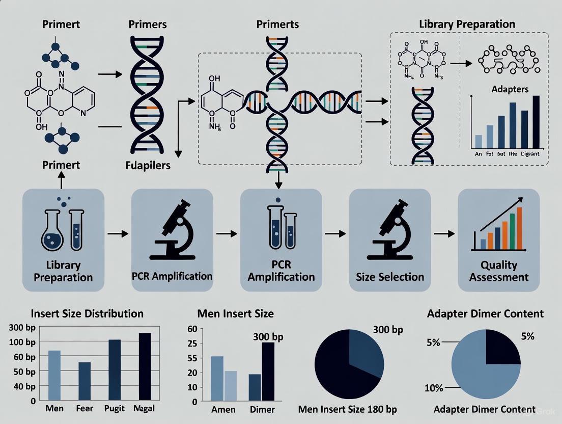
In the field of chemogenomics research, where understanding the complex interactions between small molecules and biological systems is paramount, the quality of next-generation sequencing (NGS) data is critical. The library preparation phase serves as the foundation for all subsequent analysis, and rigorous quality control (QC) at specific checkpoints is essential for generating reliable, reproducible data. Effective QC minimizes costly errors, reduces sequencing artifacts, and ensures that the resulting data accurately represents the biological system under investigation [29]. This guide provides a structured, step-by-step framework for implementing robust QC protocols throughout the NGS library preparation workflow, specifically tailored to support high-quality chemogenomic research.
The NGS Library Preparation Workflow: A Visual Guide
The following diagram illustrates the complete NGS library preparation workflow with its integrated quality control checkpoints, showing the sequence of steps and where critical QC interventions should occur.
Essential QC Checkpoints and Parameters
Implementing QC at the following critical junctures ensures the integrity of your NGS library throughout the preparation process.
Checkpoint 1: Starting Material QC
Purpose: To verify that the input nucleic acids (DNA or RNA) are of sufficient quality and quantity to proceed with library construction. High-quality starting material is the foundation for successful library preparation [29].
Critical Parameters and Methods:
Table: QC Parameters for Starting Material
| Parameter | Acceptance Criteria | Assessment Methods | Impact of Deviation |
|---|---|---|---|
| Quantity | Meets kit requirements (typically 1-1000 ng) | Fluorometry (Qubit), spectrophotometry (NanoDrop) | Low yield: insufficient material for library prep; High yield: potential over-representation |
| Purity | A260/A280 ~1.8 (DNA), ~2.0 (RNA); A260/A230 ~2.0 | Spectrophotometry (NanoDrop) | Contaminants (phenol, salts) inhibit enzymatic reactions in downstream steps [29] [33] |
| Integrity | High RIN/RQN >7 (RNA); intact genomic DNA | Capillary electrophoresis (Bioanalyzer, TapeStation) | Degraded samples yield biased, fragmented libraries with reduced complexity [29] [33] |
Protocol:
- Quantification: Use fluorometric methods (e.g., Qubit) for accurate concentration measurement of double-stranded DNA. Avoid relying solely on UV spectrophotometry, which can overestimate concentration due to contamination [34].
- Purity Assessment: Measure absorbance at 230nm, 260nm, and 280nm. Calculate A260/A280 and A260/A230 ratios.
- Integrity Check: For RNA, use the Bioanalyzer or TapeStation to generate an RNA Integrity Number (RIN). For DNA, examine the electrophoregram for a tight, high-molecular-weight distribution.
Checkpoint 2: Fragmentation QC
Purpose: To confirm successful fragmentation and verify that the fragment size distribution aligns with the requirements of your sequencing platform and application.
Critical Parameters:
- Fragment Size Distribution: Should match the expected range for your application (e.g., 200-500bp for many Illumina applications).
- Fragment Homogeneity: Fragments should be uniformly sized without significant smearing or multiple peaks.
Protocol:
- After fragmentation, run a small aliquot (1 μL) on a Bioanalyzer, TapeStation, or agarose gel.
- Analyze the resulting electrophoregram for the primary peak size and distribution.
- Adjust fragmentation parameters (time, enzyme concentration) if the size distribution is not optimal.
Checkpoint 3: Post-Ligation QC
Purpose: To validate efficient adapter ligation and detect the formation of adapter dimers, which can compete with library fragments during sequencing and significantly reduce useful data output [29].
Critical Parameters:
- Adapter Ligation Efficiency: The majority of fragments should have adapters successfully ligated.
- Adapter Dimer Formation: Minimal to no adapter dimer peaks (~70-90bp) should be visible.
Protocol:
- Post-ligation, analyze the library using a high-sensitivity DNA assay on the Bioanalyzer or TapeStation.
- Look for a shift in fragment size corresponding to the addition of adapters.
- Check for a small peak around 70-90bp, indicating adapter dimers. If present, perform additional cleanup or size selection.
Checkpoint 4: Amplification QC
Purpose: To verify that PCR amplification was efficient without introducing significant bias or duplicates. Over-amplification can result in increased duplicates and biases, while under-amplification can lead to insufficient yield [29].
Critical Parameters:
- Amplification Efficiency: Library concentration should increase appropriately after PCR.
- PCR Bias: The size distribution and complexity should remain similar to pre-amplification profiles.
- Minimal Duplication: Avoid excessive PCR cycles that increase duplicate rate.
Protocol:
- Quantify the library before and after amplification using fluorometry.
- Check the size profile post-amplification to ensure it hasn't shifted significantly.
- Limit PCR cycles to the minimum necessary (typically 4-15 cycles) to maintain library complexity.
Checkpoint 5: Final Library QC
Purpose: To comprehensively assess the quality, quantity, and size distribution of the final library before sequencing. This is the last opportunity to identify issues that could compromise the entire sequencing run [29] [3].
Critical Parameters and Methods:
Table: QC Methods for Final NGS Libraries
| Method | What It Measures | Key Outputs | Considerations |
|---|---|---|---|
| qPCR | Concentration of amplifiable molecules (with both adapters) | Molarity (nM) for accurate pooling | Most accurate for clustering; required for patterned flow cells [34] [3] |
| Fluorometry (Qubit) | Total double-stranded DNA concentration | Mass concentration (ng/μL) | Overestimates functional library if adapter dimers present [34] |
| Electrophoresis (Bioanalyzer) | Size distribution and profile | Fragment size, adapter dimer contamination, profile quality | Essential for visual quality assessment; not ideal for quantification of broad distributions [29] [34] |
Protocol:
- Quantification: Use qPCR for the most accurate quantification of amplifiable library fragments, especially when pooling multiple libraries [34] [3].
- Size Profiling: Run the final library on a Bioanalyzer or TapeStation to confirm the expected size distribution and check for adapter dimers or other contaminants.
- Normalization: Precisely normalize libraries based on qPCR-derived molarity to ensure equal representation in multiplexed sequencing.
Essential Research Reagent Solutions
The following reagents and kits are fundamental to successful NGS library preparation and quality control.
Table: Essential Research Reagent Solutions for NGS Library Preparation
| Reagent/Kits | Function | Application Notes |
|---|---|---|
| Nucleic Acid Extraction Kits | Isolate DNA/RNA from various sample types | Choose based on sample source (e.g., tissue, cells, FFPE) |
| Library Preparation Kits | Fragment, end-repair, A-tail, and ligate adapters | Select platform-specific (Illumina, MGI) and application-specific kits [35] [36] |
| High-Fidelity DNA Polymerase | Amplify library fragments with minimal errors | Essential for maintaining sequence accuracy and reducing bias [35] |
| Magnetic Beads | Purify and size-select fragments between steps | Bead-to-sample ratio is critical for optimal size selection [35] |
| QC Assay Kits (Bioanalyzer, TapeStation) | Analyze size distribution and integrity | Use high-sensitivity assays for final library QC [29] |
| Quantification Kits (Qubit, qPCR) | Accurately measure concentration | qPCR provides most accurate quantification for pooling [34] |
Frequently Asked Questions (FAQs)
Q1: Why is my final library yield low, and how can I fix this? A: Low library yield can result from several issues:
- Poor input quality: Degraded DNA/RNA or contaminants (phenol, salts) inhibit enzymes. Re-purify input material and verify purity ratios (A260/280 ~1.8, A260/230 ~2.0) [29] [28].
- Inefficient ligation: Ensure fresh ligase, optimal adapter-to-insert ratio, and proper reaction conditions (temperature, time) [37] [28].
- Overly aggressive cleanup: Magnetic bead ratios that are too high can exclude desired fragments. Precisely follow recommended bead:sample ratios [28].
Q2: How can I prevent adapter dimers in my library? A: Adapter dimers form when excess adapters ligate to each other instead of library fragments. Prevent them by:
- Optimizing adapter concentration using correct molar ratios during ligation [37].
- Incorporating dual-size selection bead cleanups to remove small fragments before and after amplification [35].
- Using fresh, properly prepared adapters and ensuring they are not degraded [37].
- Validating each preparation with a high-sensitivity electrophoresis assay to detect dimers early [29].
Q3: What is the most accurate method for quantifying my final library before sequencing? A: qPCR is the gold standard for final library quantification because it specifically measures amplifiable molecules containing both adapter sequences [34] [3]. This is crucial for determining optimal cluster density on the flow cell. Fluorometric methods (e.g., Qubit) measure total dsDNA, including non-functional fragments, and can lead to overestimation, while UV spectrophotometry should be avoided for final library quantification due to poor sensitivity and specificity [34].
Q4: My Bioanalyzer trace shows a broad size distribution. Is this acceptable? A: It depends on your application. For amplicon or small RNA sequencing, a tight size distribution is expected. For whole genome or transcriptome sequencing, a broader distribution (e.g., 200-500bp) is normal. However, an abnormally broad distribution with multiple peaks could indicate uneven fragmentation, contamination, or poor size selection, which may require protocol optimization [34].
Q5: How does automation improve NGS library preparation QC? A: Automation significantly enhances reproducibility and reduces human error by:
- Standardizing pipetting volumes and reaction setups, minimizing technician-to-technician variability [37].
- Providing precise temperature control for enzymatic steps (ligation, amplification).
- Reducing cross-contamination through non-contact dispensing.
- Enabling detailed audit trails for troubleshooting and regulatory compliance [37].
Why is starting material QC critical for NGS success?
The quality of your DNA or RNA starting material is the foundational step upon which your entire Next-Generation Sequencing (NGS) experiment is built. Success in NGS heavily relies on the quality of the starting materials used in the library preparation process [29]. High-quality starting materials ensure accurate and representative sequencing data, while compromised samples can lead to biased results, loss of valuable sequencing material, and compromised library complexity, ultimately wasting reagents, sequencing cycles, and research time [28] [29].
The core parameters you must assess for any starting material are Quantity, Purity, and Integrity. Failure to properly evaluate these can lead to a cascade of problems in downstream library preparation, including enzyme inhibition during fragmentation or ligation, biased representation of your sample, and ultimately, failed or unreliable sequencing runs [28].
How do I quantify DNA/RNA, and what methods should I avoid?
Accurate quantification is essential to determine the appropriate amount of starting material for your specific NGS library prep kit. Using too little DNA or RNA can lead to low library yield and poor coverage, while too much can cause over-amplification artifacts and bias [28].
The table below summarizes the common quantification methods and their recommended use cases.
Table: Comparison of Nucleic Acid Quantification Methods for NGS
| Method | Principle | What It Measures | Recommended for NGS? |
|---|---|---|---|
| UV Spectrophotometry (e.g., NanoDrop) | Measures UV absorbance at 260 nm [33]. | Total nucleic acid concentration; also assesses purity via 260/280 and 260/230 ratios [33]. | Not recommended for final library quantification. Can overestimate usable material by counting non-template background like contaminants or free nucleotides [28] [4]. |
| Fluorometry (e.g., Qubit) | Fluorescent dye binding specifically to DNA or RNA [4]. | Concentration of specific nucleic acid type (e.g., dsDNA, ssDNA, RNA) [4]. | Yes, highly recommended. Provides more accurate quantification of the target nucleic acid than spectrophotometry [28] [4]. |
| qPCR-based Methods | Amplification of adapter-ligated sequences using real-time PCR [3]. | Concentration of amplifiable library molecules with adapters on both ends [3]. | Yes, essential for final library quantification. Specifically quantifies the molecules that will actually cluster on the flow cell [3] [4]. |
Key Takeaway: For starting material QC, use fluorometric methods (Qubit) for accurate concentration measurement. Avoid relying solely on NanoDrop for quantification, though it is useful for a quick purity check. For the final library, qPCR-based quantification is considered the gold standard for most Illumina-based workflows [3] [4].
How do I assess sample purity, and what are the acceptable values?
Purity assessment ensures your sample is free of contaminants that can inhibit the enzymes (e.g., polymerases, ligases) used in library preparation. This is typically done using UV spectrophotometry [33] [29].
Table: Interpreting Spectrophotometric Ratios for Sample Purity
| Absorbance Ratio | Target Value | What It Indicates | Common Contaminants |
|---|---|---|---|
| A260/A280 | ~1.8 (DNA)~2.0 (RNA) [33] [29] | Protein contamination. | Residual phenol or protein from the extraction process [28]. |
| A260/A230 | >2.0 [29] | Chemical contamination. | Salts, EDTA, guanidine, carbohydrates, or organic solvents [28]. |
Troubleshooting Purity Issues: If your ratios are outside the ideal ranges, it is recommended to re-purify your input sample using clean columns or beads to remove inhibitors before proceeding with library preparation [28].
How is sample integrity measured, and why does it matter?
Integrity refers to the degree of degradation of your nucleic acids. Using degraded starting material is a primary cause of low library complexity and yield, as it provides fragmented templates for library construction [28] [29].
- DNA Integrity: Visually assessed using gel electrophoresis (agarose) or automated electrophoresis systems (e.g., Agilent TapeStation, Bioanalyzer). High-quality genomic DNA should appear as a single, high-molecular-weight band with minimal smearing below it [28].
- RNA Integrity: Quantified using capillary electrophoresis (e.g., Agilent Bioanalyzer or TapeStation), which generates an RNA Integrity Number (RIN) or (RQN). This score ranges from 1 (completely degraded) to 10 (perfectly intact) [33]. A high RIN/RQN score indicates intact RNA molecules, which is crucial for representative transcriptome data [33] [29].
FAQ: Troubleshooting Common Starting Material QC Issues
Q1: My Bioanalyzer trace shows a smear instead of a sharp band. What should I do? This indicates sample degradation [28]. If the smear is severe, the sample should not be used for NGS as it will result in a low-complexity library. For RNA, a low RIN score (e.g., below 7) confirms degradation. It is best to repeat the nucleic acid extraction, paying close attention to RNase-free techniques for RNA and avoiding repeated freeze-thaw cycles.
Q2: My sample has good concentration but poor 260/230 ratio. Can I still use it? A low 260/230 ratio suggests chemical contamination that can inhibit enzymatic reactions [28]. Do not proceed without cleaning up the sample. Re-purify the DNA or RNA using column-based or bead-based clean-up protocols to remove salts and other chemical contaminants. After clean-up, re-quantify and re-assess the purity ratios [28].
Q3: I have a limited amount of a precious sample with low concentration. How can I proceed? For low-input protocols, quantification and QC become even more critical. Use highly sensitive fluorometric assays (e.g., Qubit dsDNA HS Assay). For RNA, consider a qPCR assay during library generation to assess the quality and quantity of the input prior to final library preparation, as traditional QC methods may not be sensitive enough [38].
The Scientist's Toolkit: Essential QC Instruments & Reagents
Table: Key Equipment and Reagents for Starting Material QC
| Tool / Reagent | Primary Function | Key Consideration |
|---|---|---|
| Fluorometer (e.g., Qubit) | Accurate quantification of specific nucleic acid types (dsDNA, RNA). | More specific than spectrophotometry; requires specific assay kits for different nucleic acids. |
| Spectrophotometer (e.g., NanoDrop) | Rapid assessment of sample concentration and purity (A260/A280 & A260/230). | Useful for initial screening but can overestimate concentration; not suitable for low-concentration samples. |
| Automated Electrophoresis System (e.g., Agilent Bioanalyzer/TapeStation, Fragment Analyzer) | Gold-standard for assessing nucleic acid integrity and size distribution. | Provides a RIN for RNA and visual profile for DNA; higher throughput systems (e.g., Fragment Analyzer) are available for large-scale projects [38]. |
| qPCR Instrument | Accurate quantification of amplifiable library molecules; can be used for QC during low-input library generation [38]. | Essential for final library quantification; targets adapter sequences to count only functional molecules [3]. |
Workflow Diagram: Starting Material QC for NGS
The following diagram summarizes the decision-making workflow for assessing DNA and RNA starting material quality before NGS library preparation.
In the construction of high-quality chemogenomic NGS libraries, Quality Control (QC) following fragmentation and adapter ligation is not merely a recommended step—it is a fundamental determinant of experimental success. These checkpoints serve to validate that the library molecules have been properly prepared for the subsequent sequencing process, ensuring that the resulting data is both reliable and reproducible. Efficient post-fragmentation and post-ligation QC directly mitigates the risk of costly sequencing failures, biased data, and inconclusive results in downstream drug discovery analyses [29].
The core objective at this stage is to confirm two key parameters: that the nucleic acid fragments fall within the optimal size range for your specific sequencing platform and application, and that the adapter ligation step has been efficient, with minimal formation of by-products like adapter dimers that can drastically reduce usable sequencing output [28] [3]. This guide provides a structured troubleshooting framework and detailed protocols to diagnose and rectify common issues encountered after fragmentation and ligation.
Troubleshooting Guide: Common Issues and Solutions
The table below outlines frequent problems, their root causes, and corrective actions based on established laboratory practices and guidelines [28].
Table 1: Troubleshooting Common Post-Fragmentation and Post-Ligation Issues
| Problem & Symptoms | Potential Root Cause | Corrective Action & Solution |
|---|---|---|
| Unexpected Fragment Size Distribution [28]► Overly short or long fragments► High size heterogeneity (smeared profile) | ► Fragmentation Inefficiency: Over- or under-shearing due to miscalibrated equipment or suboptimal enzymatic reaction conditions [28]. | ► Optimize Fragmentation: Re-calibrate sonication/covaris settings or titrate enzymatic fragmentation mix concentrations. Run a fragmentation optimization gradient [28]. |
| High Adapter Dimer Peak (~70-90 bp) [28]► Sharp, dominant peak in electropherogram at ~70-90 bp, crowding out the library peak. | ► Suboptimal Adapter Ligation: Excess adapters in the reaction promote self-ligation [28].► Inefficient Cleanup: Incomplete removal of un-ligated adapters after the ligation step [28]. | ► Titrate Adapter Ratio: Optimize the adapter-to-insert molar ratio to find the ideal balance for your input DNA [28].► Optimize Cleanup: Increase bead-to-sample ratio during post-ligation cleanup to more efficiently remove short fragments and adapter dimers [28]. |
| Low Library Yield Post-Ligation [28]► Low concentration after ligation and cleanup, despite sufficient input. | ► Poor Ligation Efficiency: Caused by inhibited ligase, degraded reagents, or improper reaction buffer conditions [28].► Overly Aggressive Cleanup: Sample loss during bead-based size selection or purification [28]. | ► Check Reagents: Use fresh ligase and buffer, ensure correct reaction temperature.► Review Cleanup Protocol: Avoid over-drying magnetic beads and ensure accurate pipetting to prevent sample loss [28]. |
Experimental Protocols for Key QC Analyses
Protocol: Assessing Fragment Size and Distribution
Principle: Microfluidics-based capillary electrophoresis (e.g., Agilent Bioanalyzer/TapeStation) provides a digital, high-resolution profile of fragment size distribution, replacing traditional, time-consuming agarose gel methods [3].
Methodology:
- Sample Preparation: Dilute 1 µL of the post-fragmentation or post-ligation library according to the manufacturer's specifications for the relevant assay (e.g., High Sensitivity DNA kit).
- Instrument Operation: Load the sample onto the designated chip or cartridge. The system automatically separates fragments via electrophoresis and detects them with an intercalating fluorescent dye.
- Data Analysis: The software generates an electropherogram and a virtual gel image. Key data outputs include:
- Peak Profile: A sharp, single peak indicates a tight size distribution. A broad or multi-peak profile suggests uneven fragmentation [28] [29].
- Average Fragment Size: Confirms the library is within the optimal range for your sequencing platform (e.g., 300-500 bp for many Illumina systems).
- Presence of Adapter Dimers: A sharp peak at ~70-90 bp indicates significant adapter-dimer formation [28].
Protocol: Quantifying Amplifiable Libraries via qPCR
Principle: While fluorometry (Qubit) measures total DNA concentration, quantitative PCR (qPCR) specifically quantifies only library fragments that have adapters ligated to both ends—the "amplifiable" molecules that will actually cluster on the flow cell [3]. This is critical for accurate loading and optimal cluster density.
Methodology:
- Standard Curve: Prepare a dilution series of a library standard with known concentration.
- Reaction Setup: Mix diluted, unknown library samples with a qPCR master mix containing primers that bind to the adapter sequences.
- Amplification & Quantification: Run the qPCR program. The cycle threshold (Ct) values for unknown samples are interpolated from the standard curve to determine the molar concentration of amplifiable molecules [3].
- Interpretation: A significant discrepancy between Qubit (ng/µL) and qPCR (nM) concentrations suggests a high proportion of molecules are not properly ligated or are adapter dimers, requiring further optimization.
Visual Guide to the QC Workflow
The following diagram illustrates the logical sequence of checks and decisions for post-fragmentation and post-ligation QC.
The Scientist's Toolkit: Essential Research Reagents & Instruments
Table 2: Key Materials and Instruments for Post-Fragmentation and Post-Ligation QC
| Item | Function/Brief Explanation | Example Products/Brands |
|---|---|---|
| Microfluidics Electrophoresis System | Provides high-sensitivity, automated analysis of library fragment size distribution and detects adapter dimers [29] [3]. | Agilent Bioanalyzer, Agilent TapeStation, PerkinElmer LabChip GX |
| Fluorometer | Accurately quantifies total double-stranded DNA concentration, but cannot distinguish between adapter-ligated and non-ligated fragments [28] [3]. | Thermo Fisher Qubit, Promega QuantiFluor |
| qPCR Instrument | Specifically quantifies the concentration of amplifiable library molecules that have adapters on both ends, which is critical for optimal sequencing cluster density [3]. | Applied Biosystems QuantStudio, Bio-Rad CFX, Roche LightCycler |
| Magnetic Beads | Used for post-ligation cleanup and size selection to remove reaction components, salts, and undesired short fragments (like adapter dimers) [28]. | SPRIselect beads, AMPure XP beads |
| Library Quantification Kits | qPCR-ready kits containing primers specific to common adapter sequences (e.g., Illumina P5/P7) and standards for absolute quantification of amplifiable libraries [3]. | Kapa Library Quantification Kit (Roche), Illumina Library Quantification Kit |
Frequently Asked Questions (FAQs)
Q1: My Bioanalyzer trace shows a perfect library peak but also a significant adapter dimer peak at ~80 bp. Should I proceed to sequencing? It is highly recommended to not proceed without addressing the adapter dimer issue. Adapter dimers will compete for sequencing reagents and generate a high proportion of useless reads, drastically reducing the yield of meaningful data from your library. A post-ligation cleanup with optimized bead ratios is necessary to remove these dimers before sequencing [28].
Q2: Why is there a large difference between my library concentration measured by Qubit versus qPCR? This discrepancy is a key diagnostic. Qubit measures all double-stranded DNA present, including properly ligated fragments, un-ligated fragments, and adapter dimers. qPCR, however, only amplifies and detects fragments that have adapters on both ends. A significantly lower qPCR concentration indicates that a large portion of your DNA is not competent for sequencing, often due to inefficient ligation or a high degree of adapter-dimer formation [3].
Q3: What is an acceptable adapter dimer threshold for a library to be sequenced? While the acceptable level can vary, a general best-practice guideline is to ensure that the molar concentration of adapter dimers is below 1-5% of the total molar concentration of the target library. Most capillary electrophoresis software can provide this molar percentage. Visually on a Bioanalyzer trace, the library peak should be the dominant feature, with the adapter dimer peak being a minor or non-existent shoulder [28].
Accurate Final Library Quality Control (QC) is a critical determinant of success in chemogenomic Next-Generation Sequencing (NGS) research. It ensures that sequencing resources are used efficiently and that the resulting data are reliable and reproducible. For researchers and drug development professionals, failures at the sequencing stage represent significant costs in time, budget, and precious samples. This guide details the core principles and troubleshooting procedures for the three pillars of final library QC: accurate quantification, size distribution analysis, and adapter dimer detection, providing a framework to optimize your NGS workflow for chemogenomic applications.
Key Concepts in Library QC
Why Accurate Quantification is Non-Negotiable
The chemistry of NGS sequencing, particularly on Illumina platforms, requires loading a very precise amount of sequencing-ready library onto the flow cell [39] [40].
- Over-clustering: If the library concentration is too high, clusters on the flow cell become too dense. This leads to poor resolution, difficulties in base calling, and can ultimately cause run failure [39] [40].
- Under-clustering: If the library concentration is too low, the flow cell is sparsely populated. This results in inefficient use of sequencing capacity and increased costs per sample due to inadequate data yield [39] [40].
- Multiplexing Normalization: In chemogenomic studies, where multiplexing dozens or hundreds of samples is standard, accurate quantification is essential to ensure equivalent representation of each library in the pool. Inaccurate pooling leads to some samples being over-represented (wasting sequencing depth) while others are under-represented, potentially requiring re-sequencing [40].
The Critical Role of Size Distribution
Assessing the size profile of your library confirms that the preparation steps, particularly fragmentation and size selection, were successful [41].
- Uniformity: A tight, single peak indicates a library with uniform fragment sizes, which leads to more consistent and interpretable sequencing data [41].
- Informativeness: Size selection removes non-informative fragments, ensuring that sequencing reads are derived from target molecules of the expected length, thereby increasing the yield of usable data per run [41].
- Application-Specific Requirements: Certain downstream analyses, such as the detection of structural variants, benefit from a defined and optimized fragment size range.
The Adapter Dimer Problem
Adapter dimers are short, artifactual molecules formed by the ligation of two adapter sequences without an intervening genomic insert [42]. They are a common and serious QC issue because:
- They Cluster Efficiently: Due to their small size (~120-170 bp), adapter dimers cluster on the flow cell more efficiently than the intended, larger library fragments [42].
- They Consume Sequencing Reads: A significant portion of the sequencing output can be "stolen" by these dimers, drastically reducing the coverage on your target sequences [42].
- They Can Cause Run Failure: The presence of adapter dimers can generate specific base-calling error patterns and may even cause a sequencing run to stop prematurely, especially on advanced patterned flow cells where Illumina recommends limiting adapter dimers to 0.5% or lower [42].
Frequently Asked Questions (FAQs)
Q1: Why is qPCR considered the gold standard for NGS library quantification? qPCR is highly valued because it specifically quantifies only molecules that are competent for sequencing—those containing the full adapter sequences required for cluster amplification on the flow cell [4] [40]. Unlike fluorometric methods that measure all dsDNA (including non-sequenceable molecules like adapter dimers), qPCR uses primers binding to the adapters, ensuring that the quantified concentration directly correlates with the potential cluster-forming molecules [39] [40].
Q2: My Bioanalyzer trace shows a small peak at ~125 bp. What is it and why is it a problem? This peak is almost certainly an adapter dimer [42]. It is problematic because these dimer molecules contain full-length adapters and will efficiently bind to the flow cell and generate clusters. Since they are small, they cluster very efficiently and can consume a large portion of your sequencing reads, significantly reducing the data output for your actual library. Any level of adapter dimer is undesirable, but levels above 0.5% can severely impact run performance on modern sequencers [42].
Q3: Can I use a NanoDrop for final library quantification? It is strongly discouraged to use UV spectrophotometry (e.g., NanoDrop) for final library QC [4] [40]. This method cannot distinguish between DNA, RNA, free nucleotides, or protein contaminants, leading to highly inaccurate concentration readings [40]. Furthermore, it provides no information about library size distribution or the presence of contaminants like adapter dimers, making it unsuitable for ensuring library quality before a costly sequencing run [4].
Q4: What is the single biggest improvement I can make to my library QC workflow? Implementing a dual-method approach is the most significant upgrade. Use an electrophoresis-based instrument (e.g., Bioanalyzer, TapeStation) to visually confirm the correct size profile and check for adapter dimers. Then, use a quantification method specific for adapter-ligated fragments (e.g., qPCR) to obtain the accurate molarity needed for precise normalization and pooling [4] [40]. This combination directly addresses all three critical aspects of final library QC.
Troubleshooting Guides
Troubleshooting Library Quantification
| Symptom | Potential Cause | Recommended Solution |
|---|---|---|
| Low sequencing cluster density | Inaccurate quantification (underestimation) leading to underloading [40]. | Verify quantification method. Use qPCR for accurate molar concentration. Re-quantify and re-pool libraries. |
| High sequencing cluster density/run failure | Inaccurate quantification (overestimation) leading to overloading [40]. | Verify quantification method. Ensure you are not using spectrophotometry. Use qPCR and re-quantify. |
| Uneven sample representation in multiplexed run | Improper normalization due to inaccurate quantification or use of non-specific methods (e.g., fluorometry alone) [40]. | Normalize libraries based on qPCR-derived molarity. Avoid using only fluorometric values (ng/µL) for pooling. |
| High variability between technical replicates | User-user variability and error in manual, multi-step qPCR protocols [39]. | Automate dilution steps where possible. Switch to a more consistent quantification method, such as integrated fluorometric assays (e.g., NuQuant [39]). |
Troubleshooting Size Distribution & Adapter Dimers
| Symptom | Potential Cause | Recommended Solution |
|---|---|---|
| Adapter dimer peak (~125 bp) | Insufficient starting material [42]. | Accurately quantify input DNA/RNA using a fluorometric method before library prep. |
| Poor quality or degraded input material [42]. | Use high-integrity input material. Check RNA Integrity Number (RIN) for RNA-seq [33]. | |
| Inefficient size selection or bead clean-up [42]. | Perform an additional clean-up with magnetic beads (e.g., AMPure XP) at a 0.8x-1.0x ratio to remove small fragments [42]. | |
| Broader than expected size distribution | Over-fragmentation or inconsistent fragmentation. | Optimize fragmentation conditions (time, enzyme concentration, sonication settings). |
| Inefficient size selection. | Ensure precise ratios for bead-based size selection or excise the correct region from a gel [41]. | |
| Multiple large, unexpected peaks | PCR artifacts or contamination. | Optimize PCR cycle number to minimize artifacts. Use clean, dedicated pre-PCR areas. |
Experimental Protocols for Key QC Experiments
Protocol: Accurate Quantification via qPCR
Principle: Using primers complementary to the adapter sequences, qPCR selectively amplifies and quantifies only library fragments that contain both adapters, providing a precise molar concentration of sequenceable molecules [40].
Materials:
- Library samples
- Commercial library quantification qPCR kit (e.g., KAPA Library Quantification Kit)
- DNA standards provided in the kit
- qPCR instrument and compatible plates/tubes
- Nuclease-free water
Method:
- Dilute Library: Dilute your library to a predicted concentration within the linear range of the assay (e.g., a 1:10,000 or 1:100,000 dilution in nuclease-free water is a common starting point) [40].
- Prepare Standards: Serially dilute the DNA standards provided in the kit as per the manufacturer's instructions.
- Prepare Master Mix: Create a qPCR master mix containing the SYBR Green dye, primers, and polymerase. Aliquot into the qPCR plate.
- Add DNA: Add the diluted standards and library samples to the respective wells in triplicate.
- Run qPCR Program: Run the plate using the standard qPCR cycling conditions recommended by the kit manufacturer.
- Analyze Data: The qPCR software will generate a standard curve from the known standards. Use this curve to determine the initial concentration of your unknown library samples.
Protocol: Assessing Size Distribution with the Bioanalyzer
Principle: Microfluidic capillary electrophoresis separates DNA fragments by size. An intercalating dye fluoresces upon binding DNA, generating an electropherogram that visualizes the library's size distribution and integrity [40] [43].
Materials:
- Agilent Bioanalyzer instrument
- Agilent High Sensitivity DNA Kit
- Library samples and High Sensitivity DNA marker
- Heater, magnetic stirrer, and vortex
Method:
- Prepare Gel-Dye Mix: Pipette the filtered gel-dye mix into the spin filter and centrifuge. Aliquot the gel-dye matrix into the appropriate wells of the DNA chip.
- Load Marker and Samples: Pipette the DNA marker into the ladder and sample wells. Then, add your library samples to the remaining sample wells.
- Run the Chip: Place the chip in the Bioanalyzer adapter and run the assay. The instrument software will automatically control the electrophoresis and data collection.
- Interpret Results: Review the resulting electropherogram and gel-like image.
- Ideal Library: A single, sharp peak at the expected size (e.g., 300-500 bp).
- Adapter Dimer: A distinct smaller peak at ~125 bp [42].
- Degraded DNA: A smear of small fragments.
- High-Molecular-Weight Contamination: A peak or smear at a very large size.
The Scientist's Toolkit: Essential Research Reagents & Solutions
| Item | Function/Benefit |
|---|---|
| qPCR Library Quantification Kits | Provide the specific primers, standards, and optimized mix for accurate molar quantification of adapter-ligated fragments [40]. |
| Fluorometric Dyes (e.g., Qubit dsDNA HS Assay) | Accurately measure the mass concentration (ng/µL) of dsDNA in a sample without interference from RNA or free nucleotides, useful for input quantification [4] [40]. |
| Microfluidic Capillary Electrophoresis Kits | Enable precise analysis of library fragment size distribution and detection of contaminants like adapter dimers [4] [43]. |
| Magnetic Beads (e.g., AMPure XP) | Used for post-library preparation clean-up and size selection to remove unwanted enzymes, salts, and short fragments like adapter dimers [41] [42]. |
| Integrated Quantification Kits (e.g., NuQuant) | Novel methods that incorporate fluorescent labels during library prep, allowing direct fluorometric measurement of molar concentration in minutes, saving time and reducing variability [39]. |
NGS Final Library QC Workflow
The following diagram illustrates the logical decision-making process for performing final library quality control, integrating quantification, size analysis, and troubleshooting for adapter dimers.
Rigorous final library QC is the cornerstone of a successful and cost-effective chemogenomic NGS experiment. By systematically addressing accurate quantification, precise size distribution analysis, and vigilant adapter dimer detection, researchers can dramatically increase their sequencing success rates and data quality. Integrating the troubleshooting guides and standardized protocols outlined in this document will ensure that your libraries are of the highest standard, providing a solid foundation for robust and reproducible scientific discovery.
In the context of chemogenomic Next-Generation Sequencing (NGS) research, the quality of sequencing libraries is paramount. Automated systems for NGS library preparation directly enhance data quality and reliability by minimizing human-induced variability, standardizing complex protocols, and increasing processing throughput. This technical support center provides targeted troubleshooting guides and FAQs to help researchers and drug development professionals identify and resolve common issues in automated NGS workflows, thereby supporting the broader thesis of rigorous quality control in chemogenomic library generation.
FAQs: Core Benefits and Implementation
1. How does automation specifically improve reproducibility in chemogenomic library prep? Automation enhances reproducibility by executing predefined protocols with high precision, eliminating the subtle variations in technique that occur between different users. Key mechanisms include:
- Standardized Liquid Handling: Automated liquid handling systems precisely dispense reagents, ensuring each sample receives identical volumes, which is critical for consistent enzymatic reactions in steps like end-repair and adapter ligation [44].
- Protocol Enforcement: Automated systems run standardized, validated protocols that strictly control incubation times, temperatures, and reaction sequences, eliminating batch-to-batch variations common in manual workflows [44].
- Integrated Quality Control: Advanced automated systems can incorporate real-time quality control checks, flagging samples that do not meet pre-defined quality thresholds before they proceed to sequencing, thus ensuring only high-quality libraries are advanced [44] [45].
2. What throughput gains can I realistically expect from automating my library prep? Throughput improvements are significant and are achieved through:
- Reduced Hands-On Time: Automation can reduce hands-on time by over 50% to 65%, freeing personnel for other tasks [46].
- High-Throughput Processing: Systems can process up to 96 DNA or 48 DNA and 48 RNA libraries in a single run [46].
- Faster Overall Workflow: With full automation, including integrated QC, libraries can be prepared and quantified in less than four hours, compared to seven hours or more for other methods [45].
3. My lab handles low-to-medium sample volumes. Are there cost-effective automation options? Yes, for labs that don't require high-throughput liquid handlers, alternative solutions are emerging. Lab-on-a-chip (LoC) platforms provide an alternative automation strategy. These compact, microfluidic systems are designed for low-to-medium throughput requirements and offer a lower initial investment while maintaining the benefits of a standardized, automated workflow from sample input to a ready-to-sequence library [47].
4. How does automation assist with regulatory compliance (e.g., IVDR, ISO 13485) in diagnostic development? Automated systems support regulatory adherence by ensuring complete traceability and standardizing processes. When integrated with a Laboratory Information Management System (LIMS), they enable real-time tracking of samples, reagents, and process steps, which is mandatory for compliance with frameworks like IVDR. Furthermore, standardized automated protocols are fundamental for meeting the quality management system requirements of ISO 13485 [44].
Troubleshooting Guides
Problem 1: Low Library Yield After Automated Preparation
Low library yield can result from issues at various stages of the preparation process. The following table outlines common causes and solutions.
| Cause Category | Specific Cause | Corrective Action |
|---|---|---|
| Sample Input & Quality | Input DNA/RNA is degraded or contaminated with inhibitors. | Re-purify input sample; check purity via spectrophotometry (260/280 ~1.8). Fluorometric quantification (e.g., Qubit) is superior to UV absorbance for detecting contaminants [28]. |
| Pipetting & Quantification | Inaccurate initial sample quantification or pipetting error. | Calibrate pipettes; use fluorometric quantification for input samples; employ master mixes to reduce pipetting steps [28]. |
| Fragmentation & Ligation | Suboptimal fragmentation or inefficient adapter ligation. | Verify fragmentation profile pre-proceeding; titrate adapter-to-insert molar ratio; ensure fresh ligase and optimal reaction conditions [28]. |
| Purification & Size Selection | Overly aggressive purification leading to sample loss. | Optimize bead-to-sample ratios; avoid over-drying magnetic beads during purification steps [28]. |
Problem 2: High Rate of Adapter Dimer Contamination
Adapter dimers appear as a sharp peak around 70-90 bp in an electropherogram and compete with your library during sequencing.
- Root Cause 1: Suboptimal Adapter-to-Insert Ratio. An excess of adapters in the ligation reaction promotes adapter-Adapter ligation [28].
- Solution: Precisely quantify your fragmented DNA input and titrate the adapter concentration to find the optimal molar ratio. Automated liquid handlers excel at delivering the precision required for this step [44].
- Root Cause 2: Inefficient Purification. The cleanup steps post-ligation failed to remove excess adapters and dimer products [28].
- Solution: Re-optimize your magnetic bead-based purification protocol. Ensure the bead-to-sample ratio is correct for selecting your desired fragment size and that washing steps are performed thoroughly. Automated systems with integrated QC can flag libraries with high adapter-dimer content before sequencing [45].
Problem 3: Inconsistent Results Between Runs or Operators
This is a classic sign of a reproducibility issue, which automation is designed to solve.
- Root Cause: Protocol Deviations. In manual workflows, subtle differences in pipetting technique, mixing, or incubation timing between operators can cause inconsistency. With automation, the cause often lies in worktable setup variability or unoptimized scripts [48].
- Solution:
- Standardize Worktable Setup: Ensure all labware, tips, and reagents are placed in the exact same locations on the deck every time. Use a detailed checklist [28].
- Use Pre-Validated Protocols: Whenever possible, use vendor-developed and qualified protocols (e.g., Illumina-ready protocols) rather than building scripts from scratch [46] [48].
- Implement Rigorous Training: Train all personnel on the exact same procedures for worktable setup, instrument operation, and routine maintenance [44].
Workflow and Troubleshooting Logic
The following diagram illustrates the logical pathway for diagnosing and resolving common NGS library preparation issues.
Research Reagent Solutions Toolkit
This table details key reagents and kits used in automated NGS library preparation, as cited in recent literature and commercial offerings.
| Item | Function in Workflow | Example/Application Notes |
|---|---|---|
| NEBnext Ultra II Library Kit | Provides reagents for end-repair, adapter ligation, and library amplification. | Used in a proof-of-concept automated lab-on-a-chip workflow for classical ligation-based library prep, ideal for cfDNA and other short fragments [47]. |
| Illumina DNA Prep Kit | Streamlined library preparation chemistry for whole-genome sequencing. | Features protocols automated on platforms from Beckman Coulter, Hamilton, Eppendorf, and others for flexible throughput [46]. |
| AmpliSeq for Illumina Panels | Targeted sequencing panels for cancer hotspots or other defined gene sets. | Requires consideration of dead volume; automated protocols are available for specific liquid handlers [46]. |
| NuQuant Technology | Integrated direct fluorometric assay for library quantification. | Enables efficient, real-time QC within an automated workflow (e.g., NGS DreamPrep), preventing concentration variation and saving time [45]. |
| Magnetic Beads (SPRI) | Solid-phase reversible immobilization for nucleic acid purification and size selection. | Used in multiple automated systems for cleanup between enzymatic steps; the bead-to-sample ratio is a critical optimization parameter [47] [28]. |
Diagnosing and Solving Common Chemogenomic Library Preparation Failures
What does a "low library yield" indicate, and how can I fix it?
Low library yield, a final library concentration significantly below expectations, can stem from issues at multiple stages of preparation. A systematic approach to diagnosis and correction is essential [28].
Primary Causes and Corrective Actions
| Cause of Low Yield | Mechanism of Yield Loss | Corrective Action |
|---|---|---|
| Poor Input Quality / Contaminants | Enzyme inhibition by residual salts, phenol, EDTA, or polysaccharides [28]. | Re-purify input sample; ensure wash buffers are fresh; target high purity (e.g., 260/230 > 1.8) [28]. |
| Inaccurate Quantification | Over- or under-estimating input concentration leads to suboptimal enzyme stoichiometry [28]. | Use fluorometric methods (Qubit, PicoGreen) over UV absorbance for template quantification; calibrate pipettes [28] [49]. |
| Suboptimal Adapter Ligation | Poor ligase performance, wrong molar ratio, or reaction conditions reduce adapter incorporation [28]. | Titrate adapter-to-insert molar ratios; ensure fresh ligase and buffer; maintain optimal temperature [28]. |
| Overly Aggressive Purification | Desired fragments are excluded or lost during bead-based cleanup or size selection [28]. | Optimize bead-to-sample ratio; avoid over-drying beads; use a more selective size selection method [28] [50]. |
Special Case: Low-Yield DNA Samples
For samples with concentrations below the recommended threshold (e.g., from FFPE tissue or needle biopsies), vacuum centrifugation can concentrate DNA to sufficient levels without significantly compromising integrity or the mutational profile, enabling successful NGS analysis [49].
Why is my sequencing data showing a high duplication rate?
A high read duplication rate occurs when multiple sequencing reads are assigned to the same genomic location. This can be either a natural phenomenon due to highly abundant fragments (e.g., from a highly expressed gene or a specific genomic region) or an artificial artifact, most commonly from over-amplification during library preparation [28] [51].
Differentiating Between Natural and Artificial Duplicates
| Feature | Natural Duplicates | Artificial (PCR) Duplicates |
|---|---|---|
| Origin | Biological over-representation of a fragment [51]. | Over-amplification of a single molecule during library PCR [28] [51]. |
| Read Distribution | Smooth distribution of reads around a site; roughly equal numbers on both strands [51]. | "Spiky" enrichment at a single location; heavy strand imbalances with most reads being identical [51]. |
| Common Cause | Highly expressed genes in RNA-Seq; binding sites in ChIP-Seq [51]. | Too many PCR cycles; low starting input material leading to excessive amplification [28] [51]. |
How to Fix High Duplication Rates
- Reduce PCR Cycles: Optimize the number of amplification cycles during library prep to the minimum required for sufficient yield [28] [50].
- Increase Input Material: Use the recommended amount of high-quality input DNA/RNA to improve library complexity and reduce the need for amplification [28] [52].
- Use High-Fidelity Polymerases: Select PCR enzymes known to minimize amplification bias [24].
- Bioinformatic Removal: Use tools like Picard MarkDuplicates or SAMTools to remove duplicates after sequencing, though this is more common for variant calling than for differential gene expression analysis [51] [24].
What does a sharp peak at ~70 bp or ~90 bp on my Bioanalyzer trace mean?
A sharp peak at approximately 70 bp (for non-barcoded libraries) or ~90 bp (for barcoded libraries) is a classic signature of adapter dimers [28] [50]. These are short artifacts formed when sequencing adapters ligate to each other instead of to your target DNA fragments.
Why Adapter Dimers Are a Problem
Adapter dimers are efficiently amplified during library preparation and can consume a significant portion of your sequencing throughput, leading to a lower yield of useful data for your target of interest [50].
Solutions to Remove and Prevent Adapter Dimers
| Solution | Description |
|---|---|
| Optimize Ligation | Titrate the adapter-to-insert molar ratio to avoid excess adapters. Ensure fresh ligase and optimal reaction conditions [28]. |
| Perform Size Selection | Use bead-based cleanup (e.g., with adjusted bead-to-sample ratio) or gel extraction after ligation to selectively remove short fragments like adapter dimers [28] [50]. |
| Additional Clean-up | If a Bioanalyzer trace indicates adapter dimers are present, perform an additional round of purification and size selection before proceeding to template preparation [50]. |
How can I improve the signal-to-noise ratio and consistency of my NGS data?
Improving the signal-to-noise ratio (distinguishing true variants from errors) and achieving consistent results requires a holistic approach focusing on standardization and quality control [53].
Key Strategies for Robust NGS Data
- Standardize and Automate Protocols: Manual library preparation is prone to human error and variability. Using liquid handling automation for steps like DNA extraction and library prep minimizes pipetting inaccuracies, reduces cross-contamination risk, and dramatically improves run-to-run consistency [53] [52].
- Implement Rigorous QC Measures:
- Minimize PCR Artifacts:
- Use Multiplexing with Caution: Be aware that index misassignment can occur during multiplexing. Use kits with robust barcode systems and follow protocols precisely to prevent cross-talk between samples [52].
Research Reagent Solutions for Quality Control
| Reagent / Kit | Function in NGS QC | Key Consideration |
|---|---|---|
| Qubit dsDNA HS/BR Assay Kits | Accurate, dye-based fluorometric quantification of double-stranded DNA; ignores free nucleotides and RNA [28] [49]. | Essential for measuring usable input material; superior to UV absorbance for library prep. |
| BioAnalyzer / TapeStation | Microfluidics/capillary electrophoresis for sizing and quantifying DNA libraries; detects adapter dimers and size deviations [28] [50]. | Critical for visualizing library profile and identifying common failure signals before sequencing. |
| Uracil-DNA Glycosylase (UDG) | Enzyme that removes uracil bases from DNA, counteracting cytosine deamination artifacts common in FFPE-derived DNA [49]. | Reduces false positive C:G>T:A transitions, improving variant calling accuracy. |
| Library Quantification Kits (qPCR-based) | Accurately quantifies only "amplifiable" library fragments for pooling and loading onto sequencers [50]. | Does not differentiate between target library fragments and adapter dimers; requires prior size analysis. |
Key Quantitative Metrics for NGS Library QC
For a reliable chemogenomic NGS experiment, your library should meet the following benchmarks before sequencing.
| Metric | Target / Acceptable Range | Method of Assessment |
|---|---|---|
| DNA Input Quantity | Platform-specific (e.g., 1-50 ng); use fluorometry for accuracy [49] [52]. | Qubit Fluorometer |
| DNA Purity | 260/280 ~1.8; 260/230 >1.8 [28]. | NanoDrop Spectrophotometer |
| Library Size Distribution | Platform-specific (e.g., 200-500 bp); a tight, single peak is ideal. | BioAnalyzer / TapeStation |
| Adapter Dimer Presence | Minimal to none (sharp peak at ~70-90 bp is problematic) [28] [50]. | BioAnalyzer / TapeStation |
| Final Library Concentration | Varies by platform; requires accurate quantification for pooling. | qPCR-based Library Quant Kit |
In chemogenomic research, where you are often profiling the effects of chemical compounds on biological systems, the integrity of your sequencing data is non-negotiable. The journey from a cellular sample to a chemogenomic library is fraught with potential pitfalls, and the quality of your initial sample input sets the stage for everything that follows. Issues like nucleic acid degradation, contamination from chemicals or the compound libraries themselves, and inaccuracies in quantification can introduce profound biases, obscuring the true biological signal and compromising the validity of your findings. This guide provides a targeted, troubleshooting-focused resource to help you identify, diagnose, and resolve the most common sample input issues, ensuring the generation of robust and reliable NGS libraries for your drug discovery and development pipelines.
Identifying the Problem: A Diagnostic Flowchart
Follow this logical pathway to diagnose the root cause of your sample input issues.
Deep Dive into Common Issues & Solutions
Problem: Nucleic Acid Degradation
- Failure Signals: Low library complexity, high duplicate rates, smeared electrophoretic profile (e.g., Bioanalyzer), and overall low sequencing yield [28].
- Root Causes: Degradation can occur through several pathways, including oxidative damage, hydrolytic depurination, and enzymatic breakdown by nucleases [54]. In chemogenomic settings, extended incubation times or harsh compound treatments can exacerbate these processes.
- Solutions:
- Optimized Homogenization: Use instruments like the Bead Ruptor Elite which provide precise control over speed and cycle duration to ensure effective lysis while minimizing DNA shearing [54].
- Proper Preservation: For non-immediate processing, flash-freezing in liquid nitrogen and storage at -80°C is the gold standard. Use nucleic acid stabilizers if freezing is not feasible [54].
- Quality Control: Routinely use fragment analysis systems (e.g., Agilent Bioanalyzer/TapeStation) to generate RNA Integrity Numbers (RIN) or DNA Integrity Numbers (DIN) to objectively assess degradation before proceeding to library prep [33].
Problem: Sample Contaminants
- Failure Signals: Enzyme inhibition during library preparation (e.g., inefficient ligation or amplification), abnormal A260/A230 and A260/A280 ratios, and low library yield [28].
- Root Causes: Common contaminants include residual phenol, salts, guanidine, EDTA, and polysaccharides from the extraction process [28]. In chemogenomics, carryover of small-molecule compounds from screening plates is a frequent and specific concern.
- Solutions:
- Re-purification: Re-purify the sample using clean column- or bead-based methods. Ensure wash buffers are fresh and used in correct volumes [28].
- Purity Assessment: Always use spectrophotometric (e.g., NanoDrop) and fluorometric methods in tandem. Target purity ratios of A260/A280 ~1.8 and A260/A230 >1.8 for DNA [33] [29].
- Inhibition Tests: Perform a pilot qPCR reaction on a small aliquot of the sample. A significantly higher Ct value than expected or compared to a control sample indicates the presence of polymerase inhibitors.
Problem: Quantification Errors
- Failure Signals: Library preparation fails entirely or yields a library of suboptimal concentration. Sequencing results show uneven coverage or low cluster density on the flow cell [28].
- Root Causes: Over-reliance on absorbance-based methods (e.g., NanoDrop), which cannot distinguish between intact nucleic acids and degraded fragments, free nucleotides, or other contaminants that also absorb at 260 nm [28] [29]. Pipetting inaccuracies are another common source of error.
- Solutions:
- Use Fluorometric Quantification: Switch to dye-based assays like Qubit (Thermo Fisher) or PicoGreen, which specifically bind to double-stranded DNA and provide a more accurate measure of usable nucleic acid concentration [28] [29].
- Employ qPCR for Library Quant: For final library quantification, use qPCR-based methods, as they only quantify amplifiable fragments, which is exactly what is required for successful cluster generation on Illumina platforms [29].
- Regular Pipette Calibration: Ensure all pipettes are regularly calibrated and that technicians are trained in proper pipetting technique.
Experimental Protocols for Diagnosis and Resolution
Protocol: Comprehensive Sample QC Workflow
This protocol should be performed on all samples prior to committing them to library preparation.
Spectrophotometric Purity Check:
Fluorometric Quantification:
- Method: Follow the manufacturer's protocol for the Qubit dsDNA HS or RNA HS Assay. This provides the true concentration of nucleic acids for library preparation input [29].
Integrity Analysis:
- Method: Use an Agilent Bioanalyzer or TapeStation with the appropriate RNA or DNA kit.
- Data Interpretation: For RNA, an RNA Integrity Number (RIN) > 8.0 is generally considered high-quality. For DNA, look for a tight, high-molecular-weight peak and avoid samples with a smeared profile indicating fragmentation [33].
Protocol: Sample Re-purification for Contaminant Removal
If contaminants are suspected, this column-based cleanup protocol can be applied.
- Select a Kit: Use a commercial cleanup kit (e.g., Zymo Research's Clean & Concentrator kits, Qiagen's MinElute kits).
- Adjust Binding Conditions: Ensure the sample is in the recommended binding buffer. This often involves adding a specific volume of a binding solution or ethanol.
- Wash: Pass the sample through the column. Perform two wash steps with the provided wash buffer to thoroughly remove salts and other inhibitors.
- Elute: Elute the purified nucleic acid in nuclease-free water or a low-EDTA TE buffer to avoid interfering with downstream enzymatic steps.
- Re-quantify: Repeat the QC workflow (Section 4.1) to confirm improved purity and determine the new, accurate concentration.
The Scientist's Toolkit: Essential Research Reagent Solutions
Table 1: Key reagents and kits for troubleshooting sample input issues.
| Item | Function/Benefit | Example Use Case |
|---|---|---|
| Qubit Fluorometer & Assay Kits | Provides highly accurate, specific quantification of DNA, RNA, or proteins by using fluorescent dyes that bind specifically to the target molecule. | Differentiating between intact DNA and contaminating RNA or nucleotides, which is crucial for accurate library input [28] [29]. |
| Agilent Bioanalyzer/TapeStation | Provides an electrophoretic profile of the sample, assigning numerical integrity scores (RIN/DIN) and visually revealing degradation, adapter dimers, or fragment size distribution. | Objectively determining if a sample is too degraded for standard library prep protocols or requires a specialized approach for low-input/degraded samples [33]. |
| Bead-Based Cleanup Kits (e.g., SPRIselect) | Used for efficient size selection and purification of nucleic acids from contaminants like salts, enzymes, and other inhibitors. | Removing adapter dimers after ligation or performing precise size selection to enrich for a specific insert size range [28]. |
| DNase/RNase Inactivation Reagents | Prevents enzymatic degradation of nucleic acids during storage and handling. | Adding RNase inhibitors to RNA samples during extraction and storage to maintain integrity [54]. |
| High-Fidelity DNA Polymerases | Enzymes with proofreading activity that reduce errors introduced during PCR amplification steps of library preparation. | Generating highly complex and accurate libraries for sensitive applications like variant calling in chemogenomic screens [55]. |
Frequently Asked Questions (FAQs)
Q1: My DNA sample has a good concentration per Qubit but a low A260/A280 ratio on the NanoDrop. Should I proceed with library prep? A1: No. A low A260/A280 ratio (significantly below 1.8) suggests protein contamination, which can inhibit enzymes like ligases and polymerases used in library construction. You should re-purify the sample before proceeding [28] [29].
Q2: My RNA sample has a RIN of 7.0. Is it still usable for transcriptomic analysis in my chemogenomic screen? A2: A RIN of 7.0 indicates moderate degradation. While it may be usable, it will likely result in 3' bias and lower library complexity. It is recommended to use a library prep kit specifically designed for degraded RNA (e.g., those employing random priming) and to be cautious in interpreting data, especially for 5' transcript ends. For critical experiments, ideally use samples with RIN > 8.0 [33].
Q3: I suspect my sample is contaminated with a small molecule from my compound library. How can I confirm and fix this? A3: The most direct symptom is inhibition of enzymatic reactions in library prep. Run a pilot qPCR or a test ligation reaction. If inhibited, perform a bead-based clean-up (see Protocol 4.2). The significant dilution and wash steps involved often effectively reduce small-molecule contaminants to sub-inhibitory concentrations [28] [54].
Q4: How can I prevent quantification errors in a high-throughput setting? A4: Automate the process. Implement automated liquid handling systems for both sample QC (e.g., dispensing into Qubit assays) and library preparation itself. This minimizes pipetting errors and improves reproducibility across a large number of samples, which is common in chemogenomic studies [44].
Addressing Fragmentation and Ligation Inefficiencies
How can I identify if my library has inefficient ligation?
Inefficient ligation during Next-Generation Sequencing (NGS) library preparation can manifest through several specific indicators in your quality control data. Recognizing these signs early is crucial for troubleshooting.
The most common failure signal is the presence of a sharp peak at approximately 70-90 base pairs (bp) in your electropherogram trace, which indicates adapter-dimer formation [28]. This occurs when adapters ligate to each other instead of to your DNA fragments, often due to an imbalance in the adapter-to-insert ratio or poor ligase performance [28].
Other key indicators include:
- Unexpectedly low final library yield, which can result from poor ligase activity, suboptimal reaction conditions, or inaccurate quantification of the input DNA prior to ligation [28].
- Low library complexity, leading to high duplication rates in the sequencing data, as an insufficient number of unique molecules were successfully captured [28].
What causes uneven coverage, particularly in high-GC regions, and how is it related to fragmentation?
Uneven coverage, especially in regions with extreme GC content, is frequently a direct consequence of the fragmentation method and subsequent amplification.
- Fragmentation Bias: Enzymatic fragmentation methods, including tagmentation, can exhibit sequence-specific biases. Some enzymes preferentially cleave in lower-GC regions, leading to under-representation of high-GC areas in the final library [56]. This creates coverage imbalances that can obscure clinically relevant variants [56].
- Amplification Bias: During PCR, AT-rich regions are generally amplified more efficiently than GC-rich regions. This is because the double-stranded DNA in GC-rich regions is harder to denature, leading to a skewed representation in the amplified library [57]. This effect is exacerbated with high numbers of PCR cycles.
Comparison of Fragmentation Methods and Their Impact on Coverage
| Fragmentation Method | Typical Coverage Uniformity | Key Advantages | Key Limitations |
|---|---|---|---|
| Mechanical Shearing (e.g., Acoustic shearing) | More uniform across GC spectrum [56] | Reduced sequence bias; consistent performance across sample types [56] | Requires specialized equipment; can involve more hands-on time and sample loss [58] [59] |
| Enzymatic Fragmentation | More prone to coverage drops in high-GC regions [56] | Quick, easy, no special equipment; amenable to high-throughput and automation [58] [59] | Potential for sequence-specific bias impacting variant detection sensitivity [56] |
What are the best practices to minimize adapter dimers and improve ligation efficiency?
Preventing adapter dimers and optimizing ligation is a multi-step process focusing on reaction components and cleanup.
- Optimize Adapter Concentration: Titrate the adapter-to-insert molar ratio. Too much adapter promotes dimer formation, while too little reduces ligation yield of your target fragments [28].
- Ensure Enzyme and Buffer Integrity: Use fresh, high-quality ligase and ensure the reaction buffer has not been degraded by freeze-thaw cycles. Always mix reagents thoroughly and maintain the optimal ligation temperature [28] [57].
- Implement rigorous purification: Use bead-based cleanup with the correct bead-to-sample ratio to efficiently remove unligated adapters and small dimer products before sequencing [28]. It is critical to use freshly prepared 70% ethanol for washes, as ethanol concentration can decrease over time due to evaporation, leading to inefficient washing and sample loss [57].
My library yield is low after fragmentation and ligation. What steps should I take?
Low yield can stem from issues at multiple points in the workflow. A systematic diagnostic approach is recommended.
- Trace Backward Through the Workflow:
- Check Input DNA Quality: Re-purify your sample if contaminants like phenol, salts, or guanidine are suspected, as they inhibit enzymatic steps. Verify purity using spectrophotometric ratios (260/280 ~1.8, 260/230 >1.8) [28] [60].
- Verify Quantification Method: Avoid relying solely on UV absorbance (e.g., NanoDrop), which can overestimate concentration by counting non-template molecules. Use fluorometric methods (e.g., Qubit) for accurate DNA quantification [28] [57].
- Assess Fragmentation Efficiency: Over- or under-fragmentation can reduce the number of fragments in the desired size range. Check the fragmentation profile on an electropherogram before proceeding to ligation [28].
- Review Ligation Conditions: Ensure the ligation reaction is set up with the correct temperature and incubation time, and that reagents are fresh [28].
How do I reduce bias introduced during library amplification?
Reducing amplification bias is key for generating libraries that accurately represent the original sample complexity.
- Minimize PCR Cycles: The most effective strategy is to use as few PCR cycles as possible. This can be achieved by increasing the amount of starting material and using a library preparation kit with high-efficiency enzymes for end repair, A-tailing, and adapter ligation [57].
- Choose a Robust Enrichment Strategy: For targeted sequencing, selecting hybridization capture over amplicon-based approaches typically requires fewer PCR cycles and results in better uniformity of coverage and fewer false positives [57].
- Utilize Unique Molecular Identifiers (UMIs): Incorporating UMIs allows for bioinformatic correction of PCR errors and duplicates, enabling the identification of true variants versus those introduced during amplification [57].
Research Reagent Solutions
The following table lists key reagents and their critical functions in optimizing fragmentation and ligation.
| Reagent / Kit | Primary Function | Considerations for Fragmentation/Ligation |
|---|---|---|
| High-Fidelity DNA Ligase | Covalently attaches adapters to fragmented DNA | Ensure high activity and freshness; sensitive to contaminants [28]. |
| Magnetic Beads (e.g., AMPure XP) | Purification and size selection | Correct bead-to-sample ratio is critical for removing adapter dimers and minimizing sample loss [28] [57]. |
| NEBNext Ultra II FS DNA Library Prep Kit | Integrated enzymatic fragmentation & library prep | Designed to reduce GC-bias and simplify workflow by combining fragmentation, end repair, and A-tailing [58]. |
| Covaris truCOVER PCR-free Library Prep Kit | Library prep with mechanical shearing | Utilizes Adaptive Focused Acoustics (AFA) for uniform fragmentation, minimizing GC-bias [56]. |
| xGen DNA Library Prep EZ Kit | Enzymatic fragmentation & library prep | Optimized for consistent coverage and reduced GC bias in a simple workflow [59]. |
| Universal NGS Complete Workflow | Streamlined library preparation | Combines fragmentation and end-repair steps to minimize handling and potential for error [57]. |
Correcting Amplification Artifacts and PCR Bias
In chemogenomic Next-Generation Sequencing (NGS) research, the integrity of your data is paramount. Amplification artifacts and PCR bias introduced during library preparation can significantly skew results, leading to false conclusions in variant calling, gene expression analysis, and compound mechanism-of-action studies. This guide provides targeted troubleshooting and solutions to identify, correct, and prevent these common technical challenges, ensuring your sequencing data accurately reflects the underlying biology.
Troubleshooting FAQs
What causes high duplication rates in my sequencing data and how can I resolve this?
High duplication rates, where multiple reads have identical start and end positions, typically indicate over-amplification during library preparation. This occurs when too many PCR cycles are used, exponentially amplifying a limited number of original DNA fragments [28].
Solutions:
- Reduce PCR Cycles: Optimize your protocol to use the fewest number of PCR cycles necessary, ideally fewer than 15, to minimize duplicate reads [61].
- Increase Input DNA: Use higher quantities of high-quality input DNA to increase initial library complexity, reducing the need for excessive amplification [52].
- Utilize UMIs: Incorporate Unique Molecular Identifiers (UMIs) during adapter ligation. These short random nucleotide sequences tag each original molecule, allowing bioinformatic tools to distinguish between true biological duplicates and PCR-generated duplicates during analysis [61].
My library QC shows a large peak at ~2x the expected size. Are these "PCR bubbles" and can I sequence this library?
Yes, this large peak is likely caused by "PCR bubbles" or "over-amplification artifacts." These structures form when PCR reagents, especially primers, are exhausted. Instead of binding to primers, amplified products anneal to each other via their complementary adapter sequences, creating partially single-stranded, slow-migrating molecules [62].
Solutions:
- Reconditioning PCR: You can sequence the library, but for accurate quantification and analysis, perform a single-cycle "Reconditioning PCR" with standard Illumina P5 and P7 primers. This converts the bubbles back into standard double-stranded libraries [62].
- Protocol: Mix 1-4 µL of the PCR product with 2 µL of a 10 µM primer mix (P5 and P7) and Kapa HiFi master mix. Cycle once: 98°C for 45 sec, 98°C for 15 sec, 60°C for 30 sec, 72°C for 30 sec, followed by a final 72°C extension for 1 minute. Clean up with SPRI beads [62].
- Preventive Optimization: The best solution is to prevent bubbles by optimizing your library prep protocol to use a lower number of PCR cycles [62].
How can I minimize bias against high-GC or high-AT genomic regions during amplification?
Regions with extreme GC content are often underrepresented due to inefficient polymerase binding and amplification [63].
Solutions:
- Use GC-Rich Polymerases: Switch to a high-fidelity polymerase specifically engineered for improved performance on GC-rich templates, such as PrimeSTAR GXL or Q5 [64] [61].
- Consider PCR-Free Protocols: For sufficient input DNA, use PCR-free library preparation workflows. This eliminates amplification bias entirely, ensuring uniform coverage across all genomic regions [63].
- Optimize Buffer Systems: Use the polymerase's recommended high-salt buffer, which can help denature stable secondary structures in GC-rich regions [61].
I see nonspecific bands or smearing on my agarose gel. What is the source of this contamination?
Nonspecific amplification or smearing can result from several factors, including suboptimal PCR conditions or contaminating DNA [64].
Solutions:
- Increase Annealing Temperature: Raise the temperature in increments of 2°C to promote stricter primer binding [64].
- Check Primer Specificity: Use BLAST alignment to ensure your primers are specific to the target and redesign them if necessary [64].
- Eliminate Contamination: If a negative control (no template) also shows smearing, your reagents are contaminated.
Comparison of PCR Enzyme Error Rates
Selecting the right polymerase is critical for minimizing introduced errors. The following table compares common enzymes [61].
| Enzyme | Error Rate (per base) | Proofreading Activity | Max Amplicon Length | GC-Rich Tolerance |
|---|---|---|---|---|
| Standard Taq | ~1 x 10⁻⁴ | No | Varies | Low |
| Phusion | ~4.4 x 10⁻⁷ | Yes | ~10 kb | Moderate |
| Q5 Hot Start | ~1 x 10⁻⁶ | Yes | ~20 kb | High |
| KAPA HiFi | ~1 x 10⁻⁶ | Yes | ~15 kb | Moderate |
| PrimeSTAR GXL | ~1 x 10⁻⁶ | Yes | ~30 kb | Excellent |
Common PCR Artifacts and Their Identifiers
Recognizing artifacts early is key to troubleshooting. This table summarizes key indicators [28] [62] [63].
| Artifact / Bias | Primary Failure Signal | Common Root Cause |
|---|---|---|
| Over-amplification/Duplicates | High duplicate read rate; PCR bubbles in QC | Excessive PCR cycles; low input DNA complexity |
| GC Bias | Uneven coverage; drop-outs in GC-rich regions | Polymerase inefficiency with stable secondary structures |
| Chimeric Reads | Reads from non-adjacent genomic regions | Template switching during amplification; enzyme error |
| Adapter Dimers | Sharp peak at ~70-90 bp in electropherogram | Inefficient cleanup; suboptimal adapter-to-insert ratio |
| Misincorporation Errors | False-positive variant calls | Low-fidelity polymerase; overcycling; high Mg²⁺ |
Experimental Protocols
Protocol 1: Reconditioning PCR to Remove "PCR Bubbles"
This protocol rescues over-amplified libraries for sequencing [62].
- Primer Mix: Create a 10x concentrated primer mix with 20 µM each of the standard Illumina P5 (
AATGATACGGCGACCACCGAGATCT) and P7 (CAAGCAGAAGACGGCATACGAGAT) primers. - Reaction Setup: In a PCR tube, combine:
- 1-4 µL of the previous PCR product (containing bubbles)
- 2 µL of the 10x primer mix
- 10 µL of Kapa HiFi 2x Hotstart PCR master mix
- Nuclease-free water to a final volume of 20 µL
- Thermal Cycling:
- Initial Denaturation: 98°C for 45 seconds.
- Cycling (1 cycle only): 98°C for 15 seconds (denaturation), 60°C for 30 seconds (annealing), 72°C for 30 seconds (extension).
- Final Extension: 72°C for 1 minute.
- Cleanup: Perform a standard SPRI bead cleanup (e.g., with a 1.2x bead-to-sample ratio) to purify the final library before quantification and sequencing.
Protocol 2: Thermal-Bias PCR for Balanced Amplicon Libraries
This advanced protocol uses non-degenerate primers and temperature control to proportionally amplify targets, including those with primer-binding mismatches, minimizing bias from degenerate primer pools [65].
- Primer Design: Use only two non-degenerate primers targeting your region of interest, instead of a complex degenerate primer pool.
- Reaction Setup: Prepare a standard PCR mix with a high-fidelity polymerase, template DNA, and the non-degenerate primers.
- Thermal Cycling:
- The protocol exploits a large difference between a low-temperature targeting phase and a high-temperature amplification phase.
- The initial low-temperature annealing stage facilitates primer binding even to mismatched templates.
- Subsequent high-temperature cycling ensures efficient and specific amplification of all bound targets.
- Outcome: This method allows for the reproducible production of amplicon libraries that maintain the proportional representation of both rare and abundant targets, providing a more accurate picture of community structure [65].
Workflow Visualization
Diagram: Strategies to Mitigate PCR Bias in NGS
Diagram: Thermal-Bias PCR Workflow
The Scientist's Toolkit: Research Reagent Solutions
Essential Reagents for Minimizing Amplification Artifacts
| Reagent / Tool | Function in Bias Mitigation |
|---|---|
| High-Fidelity DNA Polymerase (e.g., Q5, KAPA HiFi) | Reduces misincorporation errors through 3'→5' proofreading activity, crucial for accurate variant calling [61]. |
| PCR Enzyme for GC-Rich Templates (e.g., PrimeSTAR GXL) | Specialized enzyme formulations that improve amplification efficiency in difficult sequences, mitigating GC bias [64] [61]. |
| UMI Adapter Kits | Provides unique barcodes to label each original molecule, enabling computational correction of PCR duplicates and amplification bias [61]. |
| PCR-Free Library Prep Kits | Eliminates amplification bias entirely by avoiding PCR, ideal for whole-genome sequencing when input DNA is sufficient [63]. |
| Automated Liquid Handling Systems | Increases reproducibility and reduces human error (e.g., pipetting inaccuracies) during repetitive library prep steps, standardizing results [52] [9]. |
Optimizing Purification and Size Selection to Maximize Library Complexity
This technical support center provides targeted troubleshooting guides and FAQs to help researchers overcome common challenges in the purification and size selection steps of Next-Generation Sequencing (NGS) library preparation, with the goal of maximizing library complexity for robust chemogenomic research.
Troubleshooting Guides
Low Library Yield After Purification and Size Selection
Problem: The final library concentration is unexpectedly low following purification and size selection steps.
| Root Cause | Diagnostic Clues | Corrective Action |
|---|---|---|
| Overly aggressive size selection [28] | Electropherogram shows loss of target fragment size range; low concentration post-cleanup. | Re-optimize and precisely follow bead-to-sample ratio; for gel-based methods, excise a wider region around the target size [66] [28]. |
| Bead over-drying [50] [28] | Bead pellet appears matte or cracked, leading to inefficient elution and low DNA recovery. | Ensure bead pellet remains glossy during the drying step; do not over-dry [28]. |
| Inaccurate sample quantification [28] [29] | Fluorometric (Qubit) and qPCR values disagree with UV absorbance (NanoDrop); yield loss starts with poor input. | Use fluorometry (e.g., Qubit) or qPCR for input quantification instead of UV absorbance to accurately measure usable material [28] [29]. |
| Carryover of contaminants [28] | Residual salts or ethanol inhibit downstream enzymes; poor 260/230 ratios in initial QC [29]. | Ensure wash buffers are fresh and use proper pipetting technique to remove all ethanol in cleanup steps [50] [28]. |
Presence of Adapter Dimers and Small Artifacts
Problem: A sharp peak at ~70 bp (non-barcoded) or ~90 bp (barcoded) appears in the final library trace, indicating adapter-dimer contamination [50] [28].
| Root Cause | Diagnostic Clues | Corrective Action |
|---|---|---|
| Suboptimal adapter-to-insert ratio [28] | Adapter dimer peak is visible even after cleanup; ligation efficiency is low. | Titrate the adapter:insert molar ratio to find the optimum, typically around 10:1 [66] [28]. |
| Inefficient size selection [66] [28] | Cleanup step fails to resolve and remove the small dimer products. | For small RNAs or when bead-only cleanup fails, use agarose gel electrophoresis for higher-resolution size selection [66]. |
| Over-amplification [28] | High duplication rates in sequencing data; adapter dimers are amplified in PCR. | Reduce the number of PCR cycles; amplify from leftover ligation product rather than overcycling a weak product [28]. |
Reduced Library Complexity and High Duplication Rates
Problem: Sequencing data shows high levels of PCR duplicates, indicating a low-diversity library that does not adequately represent the original sample.
| Root Cause | Diagnostic Clues | Corrective Action |
|---|---|---|
| Excessive PCR cycles [28] | Data shows high duplicate rates; overamplification artifacts are present. | Use the minimum number of PCR cycles necessary; re-amplify from ligation product if yield is low [28]. |
| Insufficient starting material [66] | Low library yield from the beginning; associated with input degradation. | Use high-quality, intact input DNA/RNA. Increase input material where possible to minimize the required amplification [66] [29]. |
| Overly narrow size selection [66] | A very tight fragment distribution is selected, limiting the diversity of fragments in the library. | For applications like de novo assembly, a more uniform insert size is beneficial, but for standard sequencing, ensure the size selection window is not unnecessarily restrictive [66]. |
Frequently Asked Questions (FAQs)
Q1: What is the most critical parameter for accurate size selection? The most critical parameter is the precise ratio of purification beads to sample volume. An incorrect ratio will systematically exclude desired fragments or fail to remove unwanted small fragments like adapter dimers [28]. Always use well-mixed beads and calibrated pipettes for this step [50].
Q2: How can I prevent the loss of valuable library material during cleanup? To minimize sample loss, avoid over-drying beads, use master mixes to reduce pipetting steps, and consider methods like the NuQuant QC workflow that read the library directly from the plate, eliminating a cleanup and transfer step [39] [28]. For critical low-input samples, combining bead-based with gel-based purification may be necessary [66].
Q3: My input DNA is limited. How can I optimize my workflow for low-input samples? With low-input samples, the risk of generating adapter dimers increases [66]. Use high-fidelity polymerases, perform careful titration of adapters, and employ gel-based size selection to rigorously remove dimers that bead-based methods might not fully eliminate [66] [28]. Additionally, ensure accurate quantification with fluorescent methods to make the most of available material [29].
Q4: Why does my final library show multiple peaks or a smear on the Bioanalyzer? A smear or multiple peaks can indicate several issues:
- Degraded input material: Results in a smear starting from the desired size down to smaller fragments [28].
- Inefficient fragmentation: Yields a heterogeneous fragment size distribution [28].
- Incomplete cleanup: Leaves behind primers, adapter dimers, or other reaction components, creating extra peaks [28] [29]. Troubleshoot by checking the input DNA/RNA quality and ensuring all purification steps are performed correctly.
Research Reagent Solutions
The following table lists key reagents and kits used in optimizing NGS library purification and size selection.
| Item | Primary Function | Key Application Note |
|---|---|---|
| SPRIselect Beads (or equivalent) | Size-selective purification using magnetic beads. | The precise bead-to-sample ratio defines the size cutoff. Optimize the ratio for your target insert size to maximize yield and remove dimers [28]. |
| Agilent Bioanalyzer or TapeStation | Fragment size analysis via capillary electrophoresis. | Essential for QC before and after size selection to visually confirm fragment distribution and detect adapter dimers (~70-90 bp) [50] [29]. |
| High-Sensitivity DNA Assay Kits (Qubit) | Accurate fluorometric quantification of double-stranded DNA. | Used for quantifying input DNA and final libraries. More accurate than UV spectrophotometry for measuring usable nucleic acid concentration without contaminant interference [39] [29]. |
| Library Quantification Kit for qPCR | Precise quantification of amplifiable library molecules. | The gold standard for normalizing libraries before pooling and sequencing, as it only quantifies fragments competent for sequencing [39] [50]. |
| NuQuant Reagents | Fluorometric-based direct molar quantification. | Integrated into some kits, this method allows rapid, accurate library quantification without a separate fragment analysis step, reducing workflow time and sample loss [39]. |
Experimental Workflow Diagram
The following diagram illustrates the key decision points and optimization strategies in the purification and size selection workflow.
Purification Optimization Logic
This diagram outlines the systematic thought process for diagnosing and resolving common purification issues.
Validating Performance and Comparing NGS Approaches for Clinical Readiness
Designing a Rigorous NGS Method Validation Plan
This technical support center provides troubleshooting guides and FAQs to help researchers address specific issues encountered during Next-Generation Sequencing (NGS) method validation, framed within the context of quality control for chemogenomic library research.
Frequently Asked Questions
What are the critical metrics to establish during NGS assay validation?
A rigorous NGS validation plan must establish specific performance metrics to ensure analytical accuracy and reliability. The key metrics, along with their definitions and target values, are summarized in the table below.
Table 1: Essential Analytical Performance Metrics for NGS Validation
| Performance Metric | Definition | Common Target/Calculation |
|---|---|---|
| Analytical Sensitivity | The ability to correctly detect a variant when it is present [67]. | Limit of Detection (LoD) determined via probit analysis; often 439-706 copies/mL for viral targets [67]. |
| Positive Percent Agreement (PPA) | The proportion of known positive samples that are correctly identified as positive [68]. | 100% for SNVs; 79%-91.7% for CNVs of different sizes [68]. |
| Negative Percent Agreement (NPA) | The proportion of known negative samples that are correctly identified as negative [68]. | 100% concordance for clinically relevant SNVs [68]. |
| Precision | The reproducibility of results across repeated runs [67]. | Intra-assay: <10% CV; Inter-assay: <30% log-transformed CV [67]. |
| Linearity | The ability to provide results that are directly proportional to the analyte concentration [67]. | 100% linearity across serial dilutions; log10 deviation <0.52 [67]. |
Why is my assay failing to detect variants present in the reference material?
Non-detection of expected variants can be frustrating and often points to specific, correctable issues in your workflow. The following troubleshooting guide outlines common root causes and their solutions.
Table 2: Troubleshooting Guide for Failed Variant Detection
| Problem | Potential Root Cause | Corrective Action |
|---|---|---|
| Variant Not Detected | Assay and variant incompatibility; the genomic region may not be covered by your panel [69]. | Perform a deep dive into the assay's product literature (e.g., Target Region GTF file) to confirm the variant is within the targeted regions [69]. |
| Low/Variable Allele Frequency | Low sequencing depth or insufficient library complexity [69]. | Increase sequencing coverage per manufacturer's recommendations. Ensure sufficient input DNA/RNA to generate a complex library and avoid over-amplification [69]. |
| Poor Yield & High Duplication | Degraded input DNA/RNA or contaminants inhibiting enzymes [28]. | Re-purify input sample; use fluorometric quantification (e.g., Qubit) instead of UV absorbance; optimize fragmentation [28]. |
| Adapter Dimer Contamination | Suboptimal adapter ligation conditions or inefficient purification [28]. | Titrate adapter-to-insert molar ratios; use fresh ligase; optimize bead-based cleanup ratios to remove short fragments [28] [37]. |
The following flowchart provides a systematic diagnostic strategy for this issue:
How can I ensure my NGS library prep is reproducible and high-quality?
Consistent library preparation is foundational to a successful NGS assay. Adhering to best practices minimizes variability and ensures high-quality data.
- Optimize Adapter Ligation: Use freshly prepared adapters and control ligation temperature and duration. For blunt-end ligations, use room temperature with high enzyme concentrations for 15–30 minutes. For cohesive ends, use 12–16°C for longer durations, even overnight, especially for low-input samples [37].
- Handle Enzymes with Care: Maintain enzyme stability by avoiding repeated freeze-thaw cycles and storing at recommended temperatures. Accurate pipetting is crucial [37].
- Normalize Libraries Accurately: Precise normalization before pooling ensures each library is equally represented, preventing bias in sequencing depth. Automated systems reduce variability introduced by manual quantification and dilution [37].
- Minimize Human Error with Automation: Manual pipetting is a major source of variability. Automated liquid handlers standardize workflows, improve reproducibility, and reduce hands-on time [37].
- Validate Every Step with Quality Control (QC): Implement QC checkpoints post-ligation, post-amplification, and post-normalization. Use fragment analysis, qPCR, and fluorometry to assess library quality and detect issues early [37].
The Scientist's Toolkit: Key Research Reagent Solutions
The following reagents and materials are essential for developing and validating a robust NGS method.
Table 3: Essential Research Reagents for NGS Validation
| Reagent / Material | Function in Validation |
|---|---|
| Multiplexed Reference Materials | Contains multiple variants at defined allele frequencies to evaluate assay performance across different variant types and contexts during development and optimization [69]. |
| External RNA Controls Consortium (ERCC) Spike-In Mix | Used as a quantitative internal control for RNA sequencing to generate a standard curve, enabling absolute quantification and assessment of technical variability [67]. |
| Characterized Somatic Reference Samples | Community-validated reference samples (e.g., from the SRS Initiative) containing clinically relevant cancer variants for benchmarking and standardizing NGS-based cancer diagnostics [70]. |
| AccuPlex / Verification Panels | Commercially available panels of quantified viruses or other targets used as external positive controls to monitor assay performance, determine LoD, and assess linearity [67]. |
| MS2 Phage | An internal qualitative control spiked into each sample to evaluate the background level and overall sequencing success [67]. |
| FDA-ARGOS Database Sequences | Curated, high-quality reference genome sequences incorporated into bioinformatics pipelines to improve the accuracy of pathogen identification and variant calling [67]. |
Experimental Protocol: A Framework for NGS Method Validation
This protocol, synthesized from published validation studies, provides a methodological framework for establishing analytical accuracy [68] [67] [71].
Step 1: Test Familiarization and Design
Before validation, define the test's intended use, target regions, and clinical requirements. Utilize structured worksheets (e.g., from CAP/CLSI MM09 guideline) to assemble critical information on genes, disorders, and key variants [71].
Step 2: Assay Optimization and Validation Study Design
Translate design requirements into an initial assay. Define parameters like coverage depth and sequencing methodology. Design a validation study that includes a sufficient number of positive and negative samples, as well as reference materials, to statistically power the evaluation of sensitivity, specificity, and precision [71].
Step 3: Wet-Lab Analytical Validation
- Sample Preparation: Extract nucleic acids from clinical or reference samples. For the library preparation step, use a validated protocol, whether manual or automated. Automated library preparation on systems like the Hamilton Microlab STAR can demonstrate equivalence to CE-IVD certified methods [68].
- Sequencing: Process validation samples on the designated NGS platform (e.g., Illumina NovaSeq6000).
- Data Collection: Record raw sequencing data and quality metrics (e.g., >5 million preprocessed reads per sample, >75% bases with quality score >30) [67].
Step 4: Bioinformatics and Data Analysis
- Variant Calling: Process raw data using the established bioinformatics pipeline.
- Performance Calculation: Calculate analytical sensitivity (LoD, PPA), specificity (NPA), precision, and linearity by comparing the assay's results to the known truth set or reference standard [68] [67] [71].
The overall workflow for a rigorous validation plan integrates these steps systematically:
NGS Method Selection Guide
The table below compares the core characteristics of metagenomic and targeted Next-Generation Sequencing to guide your selection.
| Parameter | Metagenomic NGS (mNGS) | Targeted NGS (tNGS) |
|---|---|---|
| Primary Principle | Untargeted, shotgun sequencing of all nucleic acids in a sample [72] | Targeted enrichment of specific pathogens or genetic regions via probes or primers [73] [74] |
| Typical Pathogens Detected | Broad spectrum: bacteria, viruses, fungi, parasites (known, rare, and novel) [75] [72] | Pre-defined panel of pathogens (e.g., 198 targets in respiratory panels) [73] [74] |
| Diagnostic Sensitivity | 95.08% (for fungal infections) [74] | 95.08% (amplification-based, for fungal infections); can exceed 99% (capture-based) [73] [74] |
| Diagnostic Specificity | 90.74% (for fungal infections) [74] | 85.19% (amplification-based, for fungal infections) [74] |
| Turnaround Time (TAT) | ~20 hours [73] | Shorter than mNGS; suited for rapid results [73] |
| Cost (USD) | ~$840 per sample [73] | Generally more cost-effective than mNGS [73] [74] |
| Key Advantage | Hypothesis-free; ideal for rare, novel, or unexpected pathogens [75] [72] | High sensitivity for targeted pathogens; detects resistance/virulence genes; cost-effective [73] [74] |
| Major Limitation | High host background; complex data analysis; higher cost [73] [75] [72] | Limited to pre-defined targets; may miss co-infections with non-panel pathogens [73] [75] |
Frequently Asked Questions & Troubleshooting
FAQ 1: When should I choose mNGS over tNGS for my infection diagnostics?
Choose mNGS when:
- Conventional tests have failed to identify a pathogen despite a strong clinical suspicion of infection [72].
- You suspect a rare, novel, or unexpected pathogen that would not be on a standard targeted panel [75].
- The clinical scenario is complex, and you need a broad, hypothesis-free approach to identify potential polymicrobial or co-infections [75] [72].
Choose tNGS when:
- You have a specific clinical hypothesis (e.g., community-acquired pneumonia) and want a rapid, cost-effective test for a defined set of common pathogens [73].
- Your resources are limited, as tNGS is more cost-effective than mNGS [73] [74].
- You require genotypic data on antimicrobial resistance or virulence factors for targeted therapeutic interventions [73].
FAQ 2: My NGS library yield is low. What are the common causes and solutions?
Low library yield is a common bottleneck. The table below outlines frequent causes and corrective actions.
| Root Cause | Mechanism of Yield Loss | Corrective Action |
|---|---|---|
| Poor Input Quality | Degraded DNA/RNA or contaminants (phenol, salts) inhibit enzymatic reactions [28] [76]. | Re-purify input; check purity via absorbance ratios (A260/A280 ~1.8); use fluorometric quantification (e.g., Qubit) [29] [28]. |
| Fragmentation Issues | Over- or under-fragmentation produces fragments outside the optimal size range for adapter ligation [28]. | Optimize fragmentation parameters (time, energy); verify fragment size distribution post-shearing [66] [28]. |
| Inefficient Ligation | Suboptimal adapter-to-insert ratio, inactive ligase, or poor reaction conditions [28]. | Titrate adapter:insert ratio; ensure fresh ligase and buffer; maintain optimal reaction temperature [28]. |
| Overly Aggressive Cleanup | Desired library fragments are accidentally removed during bead-based or column-based purification [28]. | Precisely follow bead-to-sample ratios; avoid over-drying beads; consider gel-based size selection for critical applications [66] [28]. |
FAQ 3: How can I minimize false positives in my mNGS results?
False positives in mNGS can arise from contamination or background noise. Key strategies to minimize them include:
- Rigorous Controls: Always process negative controls (e.g., no-template controls, healthy donor samples) in parallel with your clinical samples. Use these to establish background thresholds [73].
- Bioinformatic Thresholding: Apply stringent bioinformatic filters. A common method is to use a Reads-Per-Million (RPM) ratio, where a pathogen is only reported if
(RPM_sample / RPM_negative_control) ≥ 10[73] [74]. - Host Depletion: Use benzonase or similar treatments to remove host nucleic acids, which increases the relative proportion of microbial reads and improves signal-to-noise ratio [73].
- Clinical Correlation: Always interpret mNGS findings in the context of the patient's clinical symptoms, other laboratory results, and imaging findings [75] [74].
Experimental Protocols for Method Comparison
The following protocol, adapted from recent studies, provides a framework for a direct, head-to-head comparison of mNGS and tNGS.
Sample Collection and Processing
- Sample Type: Collect bronchoalveolar lavage fluid (BALF) from patients with suspected lower respiratory tract infections [73] [74].
- Aliquoting: Divide 5-10 mL of BALF equally into three sterile cryovials for mNGS, tNGS, and conventional microbiological tests (CMT) [73].
- Storage: Store samples at ≤ -20°C during transport and until processing [73].
Metagenomic NGS (mNGS) Workflow
- Nucleic Acid Extraction:
- Library Construction:
- Reverse-transcribe RNA and amplify using the Ovation RNA-Seq system.
- Fragment the combined DNA/cDNA and construct libraries using the Ovation Ultralow System V2 [73].
- Sequencing: Sequence on an Illumina NextSeq 550 platform to generate at least 20 million single-end 75-bp reads per sample [73].
- Bioinformatic Analysis:
- Remove low-quality reads and human sequences (by aligning to hg38).
- Align microbial reads to a curated pathogen database using SNAP.
- Positive Call Threshold: For a given species, report as positive if the RPM ratio (RPMsample / RPMnegative control) is ≥10, or if RPM ≥0.05 for pathogens absent from the negative control [73] [74].
Targeted NGS (tNGS) Workflow
- Nucleic Acid Extraction:
- Library Construction (Amplification-based):
- Use the Respiratory Pathogen Detection Kit.
- Perform two rounds of ultra-multiplex PCR with 198 pathogen-specific primers to enrich target sequences.
- Purify PCR products and amplify with barcoded primers containing sequencing adapters [73].
- Sequencing: Sequence on an Illumina MiniSeq platform, aiming for approximately 0.1 million reads per library [73].
- Bioinformatic Analysis:
- Perform quality filtering (e.g., retain reads with Q30 > 75%).
- Align reads to a self-building clinical pathogen database to determine read counts for each target [73].
Workflow Visualization
The following diagram illustrates the key steps and decision points in the combined mNGS and tNGS experimental protocol.
Diagram Title: mNGS vs tNGS Comparative Workflow
The Scientist's Toolkit: Essential Research Reagents
The table below lists key reagents and kits used in the protocols above, which are essential for reproducing this comparative analysis.
| Reagent / Kit | Function / Application | Specific Use Case |
|---|---|---|
| QIAamp UCP Pathogen DNA Kit (Qiagen) | Extraction of high-quality microbial DNA from clinical samples. | DNA extraction for mNGS; includes steps for host DNA depletion [73] [74]. |
| Ovation Ultralow System V2 (NuGEN) | Library preparation for low-input or challenging samples. | Construction of mNGS sequencing libraries from fragmented nucleic acids [73]. |
| Respiratory Pathogen Detection Kit (KingCreate) | Targeted enrichment of respiratory pathogens via multiplex PCR. | Amplification-based tNGS library construction for a defined panel of 198 pathogens [73] [74]. |
| MagPure Pathogen DNA/RNA Kit (Magen) | Simultaneous co-extraction of DNA and RNA. | Preparation of total nucleic acids for tNGS analysis [73] [74]. |
| Benzonase (Qiagen) | Enzyme that degrades all forms of DNA and RNA. | Critical for host nucleic acid depletion in mNGS sample prep to increase microbial sequencing depth [73]. |
| Dithiothreitol (DTT) | Mucolytic agent that breaks down disulfide bonds in mucus. | Liquefaction of viscous samples like BALF or sputum prior to nucleic acid extraction [73] [74]. |
Frequently Asked Questions (FAQs)
Technology Selection & Performance
Q1: What are the primary technical differences between short-read and long-read sequencing for resistance detection?
The core differences lie in read length, underlying chemistry, and the types of genetic variations each platform best resolves.
- Short-Read Sequencing (e.g., Illumina, Element Biosciences AVITI): Generates highly accurate reads of 50-300 base pairs. It excels in detecting single nucleotide variants (SNVs) and small insertions/deletions (indels) with high precision and recall, comparable to long-read data in non-repetitive regions [77] [78]. However, it struggles with long insertions (>10 bp), structural variations (SVs), and resolving complex, repetitive genomic regions due to the limited read length [77].
- Long-Read Sequencing (e.g., PacBio HiFi, Oxford Nanopore): Produces reads thousands to tens of thousands of base pairs long. This allows them to span repetitive elements and large structural variations, providing a clear advantage for detecting complex resistance mechanisms, gene duplications, and variations in GC-rich regions [79] [77]. PacBio HiFi reads, in particular, now offer accuracy exceeding 99.9% (Q30), making them highly reliable for variant calling [80].
Q2: When should I choose short-read sequencing for antimicrobial resistance (AMR) studies?
Short-read sequencing is an excellent choice when your research goals and resources align with its strengths [78]:
- High-Throughput, Cost-Effective Screening: For projects involving hundreds or thousands of samples, such as surveillance studies, where cost per sample is a primary concern.
- Focus on Known SNVs and Small Indels: When the resistance mechanisms of interest are well-characterized and primarily involve single nucleotide polymorphisms (SNPs) or small insertions/deletions, such as specific SNPs in genes like gyrA and parC conferring fluoroquinolone resistance [81].
- Adequate Target Coverage: When you can ensure sufficient sequencing depth. For example, a minimum of 15x coverage of the target genome (e.g., ~300,000 Illumina reads for an E. coli genome) is required for reliable ARG detection [81].
Q3: In what scenarios is long-read sequencing essential for resistance gene detection?
Long-read technologies are critical when the genetic context of resistance is complex [79]:
- Resolving Complex Pharmacogenes: For genes with pseudogenes, high homology, or complex structural variations (e.g., CYP2D6, CYP2B6, HLA), long reads are necessary for accurate haplotyping and variant calling [79].
- Detecting Structural Variations and Copy Number Variations: To identify large insertions, deletions, duplications, and gene amplifications that are often missed by short reads [77].
- De Novo Assembly and Metagenomics: For assembling complete genomes or plasmids from scratch or characterizing resistomes in complex microbial communities without a reference genome, long reads provide superior continuity by spanning repetitive regions [80].
Q4: What are the key quantitative performance metrics I should compare?
The following table summarizes critical performance characteristics from recent studies:
| Performance Metric | Short-Read Sequencing | Long-Read Sequencing |
|---|---|---|
| Typical Read Length | 50-300 bp [78] | 5,000 - 30,000+ bp [80] [78] |
| Raw Read Accuracy | Very High (>99.9%) [80] | High (PacBio HiFi: >99.9%; ONT: >98%) [80] [77] |
| SNV Detection Recall/Precision | High in non-repetitive regions [77] | High in non-repetitive regions [77] |
| Indel Detection (>10 bp) Recall | Lower, especially for insertions [77] | High [77] |
| SV Detection Recall in Repetitive Regions | Significantly lower [77] | High [77] |
| Minimum Coverage for ARG Detection | ~15x (e.g., 300,000 reads for E. coli) [81] | Protocol-dependent; generally lower coverage may be sufficient due to longer contigs. |
Troubleshooting Experimental Issues
Q5: Our short-read data is failing to detect large insertions or structural variants known to be present. What should we do?
This is a common limitation. Your options are:
- Utilize Specialized SV Callers: Employ multiple structural variant detection algorithms (e.g., Manta, DELLY) that use read-pair, split-read, or read-depth signals and then merge the calls to improve recall [77].
- Hybrid Sequencing Approach: Use long-read sequencing to confirm or identify the suspected SVs. Even low-coverage long-read data can be used to scaffold and verify variations in problematic regions [77].
- Switch to Long-Read Sequencing: For projects where SVs are a primary focus, transitioning to a long-read platform is the most robust solution [79] [77].
Q6: We are getting inconsistent results in repetitive genomic regions. How can we improve accuracy?
Repetitive regions are challenging for short reads due to ambiguous mapping.
- Increase Sequencing Depth: Higher coverage can help distinguish true variants from mapping errors, but it is not a complete solution [81].
- Use Long-Read Data for Phasing: Leverage long-read sequencing to create a phased reference for your sample, which can then improve the accuracy of short-read mapping in subsequent experiments [79].
- Opt for Long-Read Sequencing: The most effective strategy is to use a technology where reads are long enough to span the entire repetitive element, providing unambiguous alignment [77].
Q7: What is the minimum sequencing depth required for reliable resistance gene detection in a metagenomic sample?
The required depth is a function of the target organism's abundance and the desired coverage of its genome.
- For a target genome in a mix: To detect ARGs in an E. coli strain present at 1% relative abundance in a synthetic community with 15x coverage, approximately 30 million Illumina reads were required during metagenome assembly [81].
- Key Consideration: This is highly dependent on community complexity and the abundance of the resistant pathogen. For low-abundance targets, very high sequencing depths are necessary [81].
Experimental Protocols for Performance Comparison
Protocol 1: Benchmarking ARG Detection Sensitivity with Short-Read Sequencing
This protocol outlines a method to determine the minimum sequencing depth required for antimicrobial resistance gene (ARG) detection using Illumina short-read technology, as described in [81].
1. Sample Preparation:
- DNA Source: Use a well-characterized, multidrug-resistant bacterial isolate (e.g., E. coli ST38 used in [81]).
- Spike-In Community: For metagenomic sensitivity, spike the isolate at varying abundances (e.g., 1%, 0.1%) into a synthetic microbial community with a defined composition.
2. Library Preparation & Sequencing:
- Library Prep: Construct sequencing libraries using a standard Illumina-compatible kit (e.g., Illumina DNA Prep). Accurate library preparation is critical for unbiased representation [25].
- Sequencing: Sequence on an Illumina platform (e.g., NovaSeq) using a 2x150 bp paired-end configuration to generate very high coverage (~6,800x) for the isolate [81].
3. In silico Subsampling & Bioinformatic Analysis:
- Subsampling: Bioinformatically subsample the high-coverage data to simulate lower sequencing depths (e.g., 5M, 1M, 300k, 100k, 50k read pairs) [81].
- Assembly & ARG Prediction:
- Assemble reads from each subsample into contigs using a de novo assembler.
- Predict ARGs from the contigs using an assembly-based tool like the Resistance Gene Identifier (RGI) with the Comprehensive Antibiotic Resistance Database (CARD) [81].
- Calculation of Metrics:
- Sensitivity: Calculate as (True Positives) / (True Positives + False Negatives). Use the highest depth results as the truth set.
- Positive Predictive Value (PPV): Calculate as (True Positives) / (True Positives + False Positives).
- Detection Frequency: For each ARG, calculate the percentage of subsamples at a given depth where it was detected.
4. Expected Outcome: The experiment will establish a benchmark, such as 300,000 reads (~15x coverage) being sufficient for high-confidence detection of most ARGs in a pure isolate, and will quantify the millions of reads needed for detection in complex metagenomes at specific abundances [81].
Protocol 2: Evaluating Performance in Complex Pharmacogenes Using Long-Read Sequencing
This protocol is designed to assess the ability of long-read sequencing to resolve variants in complex genes like CYP2D6, which are challenging for short-read technologies [79].
1. Sample Selection:
- Select samples known or suspected to harbor structural variations or complex diplotypes in target pharmacogenes (e.g., CYP2D6, CYP2B6, HLA).
2. Library Preparation & Sequencing:
- DNA Quality: Use high-molecular-weight (HMW) DNA. Avoid excessive fragmentation.
- Library Prep: Prepare libraries using a platform-specific kit (e.g., PacBio HiFi or ONT Ligation Sequencing Kit).
- Sequencing: Sequence on the appropriate platform (e.g., PacBio Sequel IIe/Revio or ONT PromethION) to achieve sufficient coverage (>20x) for confident variant calling and phasing.
3. Bioinformatic Analysis:
- Variant Calling: Use long-read-aware variant callers. For SNVs/indels, tools like DeepVariant or Pepper-Margin-DeepVariant are recommended [77]. For SVs, use tools like cuteSV, Sniffles, or pbsv [77].
- Phasing & Haplotyping: Perform haplotype phasing using the long reads to determine the specific combination of variants on each chromosome, which is essential for diplotype assignment in pharmacogenes [79].
- Comparison: Compare the long-read-derived variants and diplotypes with calls from a short-read dataset from the same sample. Manually inspect discordant calls in a genome browser (e.g., IGV) [77].
4. Expected Outcome: Long-read sequencing is expected to provide a more complete and accurate picture of the gene's structure, correctly identifying star alleles, resolving copy number variations, and detecting hybrid genes caused by structural variations that short-read methods often miss [79].
Workflow Visualization
Short-Read ARG Detection Workflow
Long-Read SV Detection Workflow
The Scientist's Toolkit: Essential Research Reagents & Materials
| Item | Function / Application |
|---|---|
| Comprehensive Antibiotic Resistance Database (CARD) | A curated resource of ARGs and associated polymorphisms used for annotating resistance determinants from sequence data [81]. |
| Resistance Gene Identifier (RGI) | A bioinformatic software tool, often used with CARD, for predicting ARGs from assembled contigs or reads [81]. |
| High-Molecular-Weight (HMW) DNA Extraction Kit | Essential for long-read sequencing to obtain intact, high-quality DNA fragments tens of kilobases in length. |
| PacBio SMRTbell or ONT Ligation Sequencing Kit | Library preparation kits specifically designed for their respective long-read sequencing platforms. |
| Illumina DNA Prep Kit | A standard library preparation kit for Illumina short-read sequencers. |
| Automated Liquid Handling System | Robotics to improve precision, reduce human error, and ensure reproducibility in library preparation steps [44]. |
| DeepVariant | A deep learning-based variant caller that shows high accuracy for both short-read and long-read data for SNVs and indels [77]. |
| Sniffles / cuteSV | Specialized variant callers for detecting structural variations from long-read sequencing data [77]. |
Benchmarking Sensitivity and Specificity Against Gold-Standard Methods
Foundational Concepts: Sensitivity and Specificity
Definitions and Calculations
In the context of chemogenomic NGS library quality control, sensitivity and specificity are essential metrics for validating new diagnostic or analytical tests against established gold-standard (reference standard) methods [82] [83].
- Sensitivity (True Positive Rate): The ability of a test to correctly identify positive results. In NGS library QC, this translates to correctly detecting true library preparation failures, contaminants, or quality issues [82] [84].
- Specificity (True Negative Rate): The ability of a test to correctly identify negative results. This means correctly identifying libraries that are truly free of specific quality issues [82] [84].
These metrics are often inversely related; increasing sensitivity typically decreases specificity, and vice versa [82] [83]. They are intrinsic to the test itself and are not directly influenced by the prevalence of the issue in the population [84].
Related Performance Metrics
While sensitivity and specificity describe the test's characteristics, Predictive Values describe the probability that a test result is correct in a given population [82] [84].
- Positive Predictive Value (PPV): The probability that a positive test result truly indicates a problem. PPV is heavily influenced by the prevalence of the issue [82].
- Formula: PPV = True Positives (TP) / (True Positives + False Positives (FP)) [82]
- Negative Predictive Value (NPV): The probability that a negative test result truly indicates the absence of a problem [82].
- Formula: NPV = True Negatives (TN) / (True Negatives + False Negatives (FN)) [82]
For datasets with imbalanced classes (e.g., a rare issue in a large set of libraries), Precision (synonymous with PPV) and Recall (synonymous with Sensitivity) can provide more insightful performance metrics than sensitivity and specificity alone [85].
Table 1: Key Performance Metrics for Diagnostic and QC Tests
| Metric | Definition | Interpretation in NGS QC | Formula |
|---|---|---|---|
| Sensitivity | Proportion of true positives correctly identified | How well the test finds actual library failures | TP / (TP + FN) |
| Specificity | Proportion of true negatives correctly identified | How well the test confirms good libraries | TN / (TN + FP) |
| PPV/Precision | Proportion of positive test results that are true positives | Probability a failed QC call is correct | TP / (TP + FP) |
| NPV | Proportion of negative test results that are true negatives | Probability a passed QC call is correct | TN / (TN + FN) |
Experimental Protocols for Benchmarking
Establishing the Validation Framework
To benchmark a new QC method, you must compare its results to those from a trusted reference standard.
Protocol: Conducting a Validation Study
- Define the Reference Standard: Establish the definitive method for determining the true status of an NGS library. This could be an orthogonal, well-validated technology (e.g., Bioanalyzer for fragment sizing) or a composite of multiple QC measures [84].
- Prepare Sample Cohort: Assemble a set of NGS libraries that represent the expected spectrum of quality—from high-quality to known failures (e.g., degraded, contaminated, or with adapter dimers) [28]. The sample size should provide sufficient statistical power.
- Run Tests in Parallel: Subject all libraries in the cohort to both the new test and the reference standard method. Ensure blinding to prevent bias in interpretation.
- Construct a 2x2 Contingency Table: Tally the results into four categories [82]:
- True Positive (TP): The new test flags an issue, and the reference standard confirms it.
- False Positive (FP): The new test flags an issue, but the reference standard does not confirm it.
- True Negative (TN): The new test indicates no issue, and the reference standard confirms the library is acceptable.
- False Negative (FN): The new test indicates no issue, but the reference standard identifies a problem.
- Calculate Metrics: Use the values from the contingency table to compute sensitivity, specificity, PPV, and NPV using the formulas provided in Section 1.1 [82].
The following workflow outlines the experimental and calculation process:
Interpreting the Results
- High Sensitivity is critical when the cost of missing a true problem (false negative) is high. For example, a test to detect adapter dimers that could ruin a sequencing run should be highly sensitive to avoid false negatives [83].
- High Specificity is important when the cost of a false alarm (false positive) is high, such as incorrectly rejecting expensive, rare, or patient samples [83].
- Consider the Prevalence of the issue. A test with high specificity will have a higher Positive Predictive Value (more reliable positive results) when the prevalence of the problem is low [82] [84].
Frequently Asked Questions (FAQs)
Q1: My new QC method has high sensitivity but low specificity. What are the implications for my NGS workflow? A1: This means your test is excellent at catching true problems but has a high rate of false alarms. While you are unlikely to sequence a poor-quality library, you may unnecessarily re-prepare or re-process many good libraries, increasing time and reagent costs. To mitigate this, you could use this test as an initial sensitive screen, with positive results confirmed by a more specific (and perhaps more costly or time-consuming) secondary test [82] [83].
Q2: When should I use precision and recall instead of sensitivity and specificity for benchmarking? A2: Precision (PPV) and recall (sensitivity) are particularly useful when your dataset is imbalanced [85]. In chemogenomics, if you are screening for a rare artifact (e.g., a specific contamination that only appears in 1% of libraries), sensitivity and specificity can be misleadingly high. A focus on precision will tell you how much you can trust a positive result from your test, and recall will tell you how many of the true rare events you are capturing [85].
Q3: I've benchmarked my method and get a lot of false positives. What could be the root cause? A3: In the context of NGS library QC, false positives can stem from:
- Contamination: Reagent or amplicon contamination leading to incorrect positive signals [28].
- Over-amplification: PCR artifacts being misclassified as library integrity issues [28] [38].
- Inadequate Specificity: The test may cross-react with structurally similar but non-problematic library components [82]. Review your purification and cleanup steps, ensure optimal PCR cycle numbers to prevent over-amplification, and verify that your assay conditions are stringent enough [28] [50].
Q4: How does the choice of a "gold standard" impact my benchmarking results? A4: The validity of your sensitivity and specificity metrics is entirely dependent on the accuracy of your reference standard [84]. If the gold standard itself is imperfect, your calculated metrics will be biased. This is known as "work-up" or "verification" bias. Always use the most accurate and reliable method available as your reference and acknowledge any limitations in your reporting [84].
Troubleshooting Common Benchmarking Problems
Table 2: Troubleshooting Guide for Benchmarking Experiments
| Problem | Potential Causes | Corrective Actions |
|---|---|---|
| Low Sensitivity (High FN) | Test is not detecting true problems. | 1. Re-optimize assay detection parameters (e.g., threshold levels).2. Check for reagent degradation or suboptimal reaction conditions.3. Verify that the test can detect all known variants of the target issue [28]. |
| Low Specificity (High FP) | Test is generating false alarms. | 1. Increase assay stringency to reduce cross-reactivity.2. Ensure thorough cleanup of libraries to remove contaminants that may interfere [28] [50].3. Re-evaluate the threshold used to define a "positive" result [82]. |
| Low PPV (Many false positives) | This can occur even with good specificity if the prevalence of the issue is very low. | 1. Use the test in populations where the issue is more common.2. Implement a two-step testing strategy where positive results are confirmed with a different, highly specific test [84]. |
| Inconsistent Results | Technical variability in the test or reference method. | 1. Standardize protocols across operators and reagent batches [52].2. Implement master mixes to reduce pipetting error [28].3. Use automated platforms to improve reproducibility [52]. |
The Scientist's Toolkit: Essential Reagents and Materials
Table 3: Key Research Reagent Solutions for Benchmarking QC Methods
| Item | Function in Benchmarking |
|---|---|
| High-Quality Reference Standard | Provides the "ground truth" against which the new test is measured (e.g., Bioanalyzer/Fragment Analyzer for size distribution, Qubit for accurate quantification, or qPCR for amplifiable library concentration) [29] [38]. |
| Control Libraries | Characterized libraries of known quality (both good and flawed) used to validate the test's performance and for ongoing quality control of the benchmarking process itself. |
| qPCR Quantification Kits | Accurately measure the concentration of amplifiable library fragments, which is critical for normalizing inputs and ensuring a fair comparison between tests [38]. |
| Size Selection Beads | Used to clean up libraries and remove artifacts like adapter dimers, which can be a source of false positives or negatives if not properly controlled [28] [50]. |
| Automated Liquid Handling System | Reduces pipetting errors and operator-to-operator variability, increasing the reproducibility and reliability of both the test and reference method results [52]. |
Troubleshooting Guide: Common NGS QC Failures and Solutions
This guide addresses frequent bioinformatic quality control challenges in chemogenomic NGS library research, helping you diagnose and resolve data quality issues.
FAQ: Why is my raw sequencing data quality poor, with low Q-scores?
- Problem: Per-base sequence quality scores are below the recommended threshold (Q30), particularly at the ends of reads.
- Diagnosis Steps:
- Solutions:
FAQ: Why is my data contaminated with adapter sequences?
- Problem: Adapter sequences used in library preparation are present in the final sequencing reads, leading to misalignment and analysis errors.
- Diagnosis Steps:
- Use FastQC to check the "Overrepresented sequences" module [33].
- Manually inspect reads for adapter sequences at the 3' end.
- Solutions:
FAQ: Why is my coverage uneven or depth insufficient?
- Problem: The sequencing depth varies significantly across target regions, or the overall depth is too low for confident variant calling.
- Diagnosis Steps:
- Solutions:
- Ensure high-quality starting material with accurate quantification using fluorometric methods (e.g., Qubit) [28] [60].
- For hybrid capture methods, optimize hybridization conditions. For amplicon-based methods, consider using Unique Molecular Identifiers (UMIs) to correct for PCR amplification bias and duplicates [87].
FAQ: How do I know if my bioinformatics pipeline is producing accurate results?
- Problem: Uncertainty about the analytical validity of variant calls from your bioinformatic workflow.
- Diagnosis Steps:
- Solutions:
Key Performance Indicators (KPIs) for Bioinformatics QC
Systematically monitor these KPIs to ensure the ongoing quality of your NGS data and bioinformatic processes.
Table 1: Essential KPIs for Bioinformatic QC Monitoring
| Metric | Target | Assessment Method | Significance |
|---|---|---|---|
| Q-score / Phred Score | ≥ Q30 (≥ 99.9% base call accuracy) [33] | FastQC, sequencing platform software | Probability of an incorrect base call; fundamental for data reliability. |
| Total Reads/Yield | Platform and application-dependent | FASTQ file analysis, platform output | Total data output; impacts statistical power and coverage depth. |
| Error Rate | Platform-dependent (typically < 0.1%) [33] | Sequencing platform software | Percentage of bases incorrectly called during a cycle. |
| % Adapter Content | < 1-5% | FastQC, Cutadapt reports | Indicates inefficient adapter removal during library prep. |
| % Duplication | Application-dependent; lower is better | FastQC, MarkDuplicates (GATK) | High rates suggest low library complexity or PCR over-amplification [28]. |
| Mean Depth of Coverage | Varies by application (e.g., >100x for somatic) | Alignment file (BAM) analysis | Average number of times a base is sequenced; critical for sensitivity. |
| % Uniformity of Coverage | > 80% of targets at 0.2x mean depth | Bedtools, custom scripts | Evenness of coverage across all target regions; vital for avoiding drop-outs. |
Methodologies for Analytical Validation
A robust analytical validation ensures your bioinformatic pipeline produces accurate, reproducible, and reliable results suitable for research and development.
1. Validation Study Design
- Use Reference Materials: Employ well-characterized reference samples, such as cell lines or synthetic controls, with known variants across different genomic contexts [71] [88].
- Coverage of Variant Types: Ensure your validation set includes a range of variant types relevant to chemogenomics: Single Nucleotide Variants (SNVs), small Insertions/Deletions (Indels), and Copy Number Variations (CNVs) [88].
- Precision and Reproducibility: Test samples across multiple runs, days, and operators (if applicable) to assess reproducibility. Re-analyze a subset of samples to determine repeatability [89].
2. Accuracy and Sensitivity/Ppecificity Calculations
Calculate key performance metrics for each variant type using a confusion matrix approach against your truth set.
- Sensitivity (Recall): = True Positives / (True Positives + False Negatives)
- Specificity: = True Negatives / (True Negatives + False Positives)
- Positive Predictive Value (Precision): = True Positives / (True Positives + False Positives)
3. Implementation of a Quality Management System (QMS)
Adopt a framework for continual improvement, as recommended by initiatives like the CDC's NGS Quality Initiative (NGS QI) [89].
- Standard Operating Procedures (SOPs): Develop and lock down SOPs for all bioinformatic workflows after validation [89].
- Competency Assessment: Regularly assess the competency of bioinformaticians and personnel [89].
- Version Control and Reproducibility: Use strict version control for all scripts and pipelines. Ensure reproducibility through containerized software environments (e.g., Docker, Singularity) [88].
The diagram below illustrates the core workflow for establishing a validated bioinformatics pipeline.
The Scientist's Toolkit: Essential Research Reagent Solutions
Table 2: Key Materials and Tools for NGS Bioinformatics QC
| Item / Solution | Function / Explanation | Example Tools / Sources |
|---|---|---|
| Reference Materials | Provides a ground truth for validating variant calls and pipeline accuracy. | Genome in a Bottle (GIAB), SEQC2, commercial reference cell lines [88]. |
| Quality Control Software | Assesses raw and processed sequencing data for quality metrics and potential issues. | FastQC, Nanoplot (for long reads), MultiQC [33] [86]. |
| Read Trimming & Filtering Tools | Removes low-quality bases, adapter sequences, and contaminated reads. | Trimmomatic, Cutadapt, Filtlong [33] [86]. |
| Alignment Algorithms | Maps sequencing reads to a reference genome to determine their genomic origin. | BWA, Bowtie2, STAR (for RNA-seq), Minimap2 (for long reads) [87] [86]. |
| Variant Callers | Identifies genetic variants (SNVs, Indels, SVs) from aligned sequencing data. | GATK, FreeBayes, DeepVariant, Manta [88]. |
| Containerization Platforms | Packages software and dependencies into isolated units to ensure computational reproducibility. | Docker, Singularity [88]. |
| High-Performance Computing (HPC) | Off-grid clinical-grade computing systems for secure and efficient data processing [88]. | On-premise servers or secure cloud computing infrastructure. |
The following diagram outlines the critical path for validating a bioinformatics pipeline, from initial testing to final implementation.
Conclusion
A stringent, multi-stage quality control protocol is the cornerstone of successful chemogenomic NGS, directly influencing the reliability of data used for critical decisions in drug discovery and development. By integrating foundational principles, methodological rigor, proactive troubleshooting, and comprehensive validation, laboratories can overcome the inherent complexities of these assays. The future of chemogenomics lies in the adoption of automated workflows, advanced bioinformatics powered by AI, and the seamless integration of multiomic data. Adherence to evolving regulatory standards and a commitment to continuous improvement will be paramount in translating high-quality sequencing data into actionable therapeutic insights, ultimately accelerating the pace of precision medicine.
References
- 1. Chemogenomics - Wikipedia [https://en.wikipedia.org/wiki/Chemogenomics]
- 2. Chemogenomics - an overview | ScienceDirect Topics [https://www.sciencedirect.com/topics/biochemistry-genetics-and-molecular-biology/chemogenomics]
- 3. Library QC for NGS [https://www.qiagen.com/us/knowledge-and-support/knowledge-hub/bench-guide/ngs/dna-sequencing/library-qc-for-ngs]
- 4. Library quantification and quality control quick reference ... [https://knowledge.illumina.com/library-preparation/general/library-preparation-general-reference_material-list/000001247]
- 5. Clinical Genomics: Challenges and Opportunities - PMC [https://pmc.ncbi.nlm.nih.gov/articles/PMC5470591/]
- 6. NGS Workflow Steps - Next-Generation Sequencing [https://www.illumina.com/science/technology/next-generation-sequencing/beginners/ngs-workflow.html]
- 7. Lab Data Integrity: The Hidden Risk That Could Cost Your ... [https://www.scispot.com/blog/lab-data-integrity-the-hidden-risk-that-could-cost-your-lab-everything]
- 8. ALCOA+ Principles for Pharma Data Integrity: A Guide ... [https://www.linkedin.com/posts/pankaj-kumar-2a2349234_data-integrity-alcoa-activity-7351069618410565633-Fovz]
- 9. Next-generation Sequencing Library Preparation Market ... [https://www.precedenceresearch.com/next-generation-sequencing-library-preparation-market]
- 10. Troubleshooting QC Failures Guide | PDF [https://www.scribd.com/document/375236597/D-Chesher-Troubleshooting-Failed-Quality-Control-pdf]
- 11. Best Practices for "Westgard Rules" [https://www.westgard.com/lessons/westgard-rules/westgard-rules/lesson74.html]
- 12. Compliance ≠ Quality: why “meeting the standard” isn't ... [https://manufacturingchemist.com/compliance-quality-why-meeting-the-standard-isn-t]
- 13. ISO 17025 合规性要求 [https://labcollector.com/zh-CN/support/knowledge-base/iso17025-compliancy-requirements/]
- 14. [J]. 中国癌症防治杂志, 2024, 16(6): 638-649. [http://www.caca.org.cn/system/2025/01/09/030124432.shtml]
- 15. 新闻- 指南解读| 临床NGS全流程共识之实验室检测 [https://www.gene-well.com/information/show/65]
- 16. NGS Sequencing Support-Troubleshooting - 赛默飞 [https://www.thermofisher.cn/ao/en/home/technical-resources/technical-reference-library/next-generation-sequencing-support-center/ngs-support/ngs-support-troubleshooting.html]
- 17. Ngs sequencing [https://www.sigmaaldrich.com/HK/zh/search/ngs-sequencing?focus=sitecontent&page=1&perpage=30&sort=relevance&term=ngs%20sequencing&type=site_content&srsltid=AfmBOopC0d58p2jLfFLFLsmtQM6kO_9FWZl4-CAx622HCN4v77jfp73V]
- 18. Clinical Laboratory Improvement Amendments (CLIA) [https://www.cdc.gov/clia/php/about/index.html]
- 19. Clinical Laboratory Improvement Amendments (CLIA) | FDA [https://www.fda.gov/medical-devices/ivd-regulatory-assistance/clinical-laboratory-improvement-amendments-clia]
- 20. What happens next for labs after the ACLA vs. FDA decision? [https://www.paubox.com/blog/what-happens-next-for-labs-after-the-acla-vs.-fda-decision]
- 21. District Court Embraces Loper Bright and Overturns FDA ... [https://www.ropesgray.com/en/insights/alerts/2025/04/district-court-embraces-loper-bright-and-overturns-fda-laboratory-developed-test-rule]
- 22. eCFR :: 42 CFR Part 493 -- Laboratory Requirements [https://www.ecfr.gov/current/title-42/chapter-IV/subchapter-G/part-493]
- 23. Why quality control (QC) is performed for NGS testing [https://3billion.io/blog/why-quality-control-qc-is-performed-for-ngs-testing]
- 24. Sample Preparation for NGS – A Comprehensive Guide [https://frontlinegenomics.com/sample-preparation-for-ngs-a-comprehensive-guide/]
- 25. Accurate Library Preparation in NGS Workflows - Uncoded [https://uncoded.in/the-importance-of-accurate-library-preparation-in-ngs-workflows/]
- 26. Clinical Laboratory Improvement Amendments (CLIA) | AAFP [https://www.aafp.org/family-physician/practice-and-career/managing-your-practice/clia.html]
- 27. Understanding NGS Library Complexity: Sources of ... [https://seqonce.com/blog/understanding-ngs-library-complexity-sources-of-sequencing-errors-limits-of-detection/]
- 28. How to Troubleshoot Sequencing Preparation Errors (NGS ... [https://www.cd-genomics.com/resource-sequencing-preparation-troubleshooting.html]
- 29. Quality Control in NGS Library Preparation Workflow [https://www.cd-genomics.com/resource-ngs-library-preparation-quality-control.html]
- 30. Making the Most of Your NGS Data: Understanding Metrics ... [https://sequencing.roche.com/us/en/article-listing/metrics-for-target-enriched-ngs.html]
- 31. Laboratory Key Performance Indicators (KPIs) [https://biosistemika.com/blog/laboratory-key-performance-indicators-kpis/]
- 32. KPIs (Key Performance Indicators) - (Intro to Chemical ... [https://fiveable.me/key-terms/introduction-chemical-engineering/kpis-key-performance-indicators]
- 33. How-to: NGS Quality Control [https://frontlinegenomics.com/how-to-ngs-quality-control/]
- 34. Best practices for library quantification [https://knowledge.illumina.com/library-preparation/general/library-preparation-general-reference_material-list/000003750]
- 35. Comprehensive Overview of NGS Library Preparation Workflow [https://www.yeasenbio.com/blogs/ngs/comprehensive-overview-of-ngs-library-preparation-workflow]
- 36. NGS library preparation [https://www.qiagen.com/us/knowledge-and-support/knowledge-hub/bench-guide/ngs/dna-sequencing/library-preparation]
- 37. 5 Best Practices for NGS Library Prep Success [https://dispendix.com/blog/5-best-practices-for-ngs-library-prep-success]
- 38. Library Preparation Quality Control and Quantification [https://www.lexogen.com/blog/rna-lexicon-library-preparation-quality-control-and-quantification/]
- 39. How to optimize NGS library QC to improve your ... [https://www.tecan.com/blog/the-benefits-of-optimizing-ngs-library-qc]
- 40. The Quantitation Question: How does accurate library ... [https://www.neb.com/en-us/tools-and-resources/feature-articles/the-quantitation-question-how-does-accurate-library-quantitation-influence-sequencing?srsltid=AfmBOorXMA6DB7O57PwOd-6LI9lAwVgo6yJX4UBZckLll-HagIQ6GusR]
- 41. Size Selection for NGS [https://nordicbiosite.com/blog/size-selection-for-ngs]
- 42. Adapter dimers causes, effects, and how to remove them [https://knowledge.illumina.com/library-preparation/general/library-preparation-general-troubleshooting-list/000001911]
- 43. Library QC and Troubleshooting with the BioAnalyzer ... [https://www.illumina.com/company/video-hub/uie_UYNjFSk.html]
- 44. How NGS Automation Improves Library Preparation Efficiency and Accuracy - Euformatics [https://www.euformatics.com/blog-post/how-ngs-automation-improves-library-preparation-efficiency-and-accuracy]
- 45. Improve your workflows with automated NGS library preparation with integrated QC - The Blog - Tecan [https://www.tecan.com/blog/the-benefits-of-ngs-library-prep-automation]
- 46. Library prep automation | Automated NGS protocols [https://www.illumina.com/techniques/sequencing/ngs-library-prep/automation.html]
- 47. Automation of customizable library preparation for next-generation sequencing into an open microfluidic platform | Scientific Reports [https://www.nature.com/articles/s41598-024-67950-6]
- 48. How to tackle 3 common challenges when automating ... [https://www.tecan.com/blog/3-common-challenges-when-automating-sample-prep-in-genomics]
- 49. Rescue of Low-Yield DNA Samples for Next-Generation ... [https://pmc.ncbi.nlm.nih.gov/articles/PMC12225422/]
- 50. NGS Library Preparation Support—Troubleshooting [https://www.thermofisher.com/us/en/home/technical-resources/technical-reference-library/next-generation-sequencing-support-center/ngs-library-preparation-support/ngs-library-preparation-support-troubleshooting.html]
- 51. Read Duplicates [https://www.biostars.org/p/159597/]
- 52. Avoid Costly Errors in NGS Sample Handling and Library ... [https://seqwell.com/avoid-costly-errors-in-ngs-sample-handling-and-library-prep/]
- 53. From Bench to Breakthrough: Streamlining NGS Assay ... [https://dispendix.com/blog/ngs-assay-development-challenges]
- 54. How to Work with Challenging or Limited Genomic ... [https://blog.omni-inc.com/blog/how-to-work-with-challenging-or-limited-genomic-samples-updated-for-2025]
- 55. Correcting PCR amplification errors in unique molecular ... [https://www.nature.com/articles/s41592-024-02168-y]
- 56. Optimization of DNA Fragmentation Techniques to Maximize ... [https://pmc.ncbi.nlm.nih.gov/articles/PMC12468493/]
- 57. NGS: Six top tips to improve your sequencing library [https://www.ogt.com/ca/resources/ngs-resources-and-support/ngs-resources/ngs-six-top-tips-to-improve-your-sequencing-library/]
- 58. Improving Enzymatic DNA Fragmentation for Next ... [https://www.neb.com/en-us/tools-and-resources/feature-articles/improving-enzymatic-dna-fragmentation-for-next-generation-sequencing-library-construction?srsltid=AfmBOor6qtC1JBJQ3Vbeowsp1s1EgEQYnCRLi721NAr2pxX_CAloVDao]
- 59. Ligation based library preparation [https://eu.idtdna.com/pages/technology/next-generation-sequencing/library-preparation/ligation-based-library-prep]
- 60. What's wrong with my NGS? [https://www.ogt.com/resources/ngs-resources-and-support/ngs-support/whats-wrong-with-my-ngs/]
- 61. Minimizing PCR-Induced Errors in Next-Generation ... [https://thearkdb.org/minimizing-pcr-induced-errors-in-next-generation-sequencing-library-prep-the-role-of-high-fidelity-enzymes/]
- 62. My libraries show peaks larger than expected. Can I still ... [https://dnatech.ucdavis.edu/faqs/my-libraries-show-peaks-larger-than-expected-can-i-still-sequence-these-pcr-bubbles]
- 63. Reducing GC & PCR Bias in Whole-Genome Sequencing [https://www.revvity.com/blog/understanding-impact-gc-and-pcr-biases-whole-genome-sequencing]
- 64. Troubleshooting your PCR [https://www.takarabio.com/learning-centers/pcr/faq/troubleshooting?srsltid=AfmBOor5tk2Npl3qvaveO89wHtOTXqNQDH8SfGs_xPgx7CkHxsVtFK0i]
- 65. Thermal-bias PCR: generation of amplicon libraries without ... [https://pmc.ncbi.nlm.nih.gov/articles/PMC12558157/]
- 66. Library construction for next-generation sequencing [https://pmc.ncbi.nlm.nih.gov/articles/PMC4351865/]
- 67. Laboratory validation of a clinical metagenomic next ... [https://www.nature.com/articles/s41467-024-51470-y]
- 68. Validation of the NovaSeq6000 Platform and automated ... [https://www.sciencedirect.com/science/article/pii/S2001037025004556]
- 69. FAQ: What to do when your NGS assay fails to detect a ... [https://blog.seracare.com/ngs/faq-what-to-do-when-your-ngs-assay-fails-to-detect-a-variant-contained-in-seraseq-reference-materials]
- 70. Transforming Validation of NGS-Based Cancer Diagnostics ... [https://www.clinicallab.com/transforming-validation-of-ngs-based-cancer-diagnostics-through-collaboration-27453]
- 71. Next Generation Sequencing (NGS) Worksheets [https://www.cap.org/member-resources/precision-medicine/next-generation-sequencing-ngs-worksheets]
- 72. Application of metagenomic next-generation sequencing in ... [https://pmc.ncbi.nlm.nih.gov/articles/PMC11604630/]
- 73. Comparative diagnostic performance of metagenomic and ... [https://www.nature.com/articles/s41598-025-11834-w]
- 74. Comparative of metagenomic and targeted next-generation ... [https://www.frontiersin.org/journals/cellular-and-infection-microbiology/articles/10.3389/fcimb.2025.1534519/full]
- 75. Advantages and challenges of metagenomic sequencing ... [https://pmc.ncbi.nlm.nih.gov/articles/PMC9527156/]
- 76. Methods to improve the accuracy of next-generation sequencing [https://pmc.ncbi.nlm.nih.gov/articles/PMC9895957/]
- 77. Comparative evaluation of SNVs, indels, and structural ... [https://www.nature.com/articles/s41439-024-00276-x]
- 78. Short read vs long read sequencing - INTEGRA Biosciences [https://www.integra-biosciences.com/united-states/en/blog/article/short-read-vs-long-read-sequencing]
- 79. Leveraging long-read sequencing technologies for ... [https://pmc.ncbi.nlm.nih.gov/articles/PMC12075302/]
- 80. Long-read sequencing vs short-read sequencing [https://frontlinegenomics.com/long-read-sequencing-vs-short-read-sequencing/]
- 81. Performance Characteristics of Next-Generation ... [https://pmc.ncbi.nlm.nih.gov/articles/PMC9238399/]
- 82. Diagnostic Testing Accuracy: Sensitivity, Specificity, Predictive ... [https://www.ncbi.nlm.nih.gov/books/NBK557491/]
- 83. Sensitivity and specificity [https://en.wikipedia.org/wiki/Sensitivity_and_specificity]
- 84. Sensitivity, Specificity, and Predictive Values: Foundations, ... [https://www.frontiersin.org/journals/public-health/articles/10.3389/fpubh.2017.00307/full]
- 85. Benchmarking performance with sensitivity, specificity, ... [https://epi2me.nanoporetech.com/benchmarking-performance/]
- 86. Best Practices for Quality Control of NGS Data [https://www.basepairtech.com/blog/best-practices-for-quality-control-and-pre-processing-of-ngs-data-a-guide-for-academic-researchers/]
- 87. Bioinformatics and Computational Tools for Next- ... [https://pmc.ncbi.nlm.nih.gov/articles/PMC7019349/]
- 88. Recommendations for bioinformatics in clinical practice [https://genomemedicine.biomedcentral.com/articles/10.1186/s13073-025-01543-4]
- 89. The Next-Generation Sequencing Quality Initiative and ... [https://wwwnc.cdc.gov/eid/article/31/13/24-1175_article]