A Practical Framework for Orthogonal Validation of NGS-Derived Chemogenomic Signatures in Drug Discovery
This article provides a comprehensive roadmap for researchers and drug development professionals to rigorously validate next-generation sequencing (NGS)-derived chemogenomic signatures.
A Practical Framework for Orthogonal Validation of NGS-Derived Chemogenomic Signatures in Drug Discovery
Abstract
This article provides a comprehensive roadmap for researchers and drug development professionals to rigorously validate next-generation sequencing (NGS)-derived chemogenomic signatures. It covers the foundational principles of chemogenomics and NGS technology, explores integrated methodological approaches for signature discovery, addresses critical troubleshooting and optimization challenges, and establishes a robust framework for validation using orthogonal techniques. By synthesizing current best practices and validation strategies, this guide aims to enhance the reliability and clinical translatability of chemogenomic data, ultimately accelerating targeted therapeutic development.
Laying the Groundwork: Core Principles of NGS and Chemogenomics
Chemogenomics represents an emerging, interdisciplinary field that has prompted a fundamental paradigm shift within pharmaceutical research, moving from traditional receptor-specific studies to a comprehensive cross-receptor view [1]. This approach systematically explores biological interactions by attempting to fully map the pharmacological space between chemical compounds and macromolecular targets, fundamentally operating on the principle that "similar receptors bind similar ligands" [1] [2]. The primary objective of chemogenomics is to establish predictive links between the chemical structures of bioactive molecules and the receptors with which these molecules interact, thereby accelerating the modern drug discovery process [1].
This strategic reorientation addresses a critical pharmacological reality: while the human genome encodes approximately 3000 druggable targets, only about 800 have been seriously investigated by the pharmaceutical industry [2]. Similarly, of the millions of known chemical structures, only a minute fraction has been tested against this limited target space [2]. Chemogenomics aims to bridge this gap by systematically matching target space and ligand space through high-throughput miniaturization of chemical synthesis and biological evaluation, ultimately seeking to identify all ligands for all potential targets [2].
Core Principles and Conceptual Framework
Fundamental Hypotheses and Operational Assumptions
The chemogenomic approach rests on two foundational hypotheses that guide its methodology and experimental design. First, compounds sharing chemical similarity should share biological targets, allowing for prediction of novel targets based on structural resemblance to known active compounds [2]. Second, targets sharing similar ligands should share similar binding site patterns, enabling the extrapolation of ligand information across related protein families [2]. These principles facilitate the systematic compilation of the theoretical chemogenomic matrix—a comprehensive two-dimensional grid mapping all possible compounds against all potential targets [2].
The practical implementation of these principles occurs through three primary methodological frameworks: ligand-based approaches (comparing known ligands to predict their most probable targets), target-based approaches (comparing targets or ligand-binding sites to predict their most likely ligands), and integrated target-ligand approaches (using experimental and predicted binding affinity matrices) [1] [2]. This multi-faceted strategy enables researchers to fill knowledge gaps in the chemogenomic matrix by inferring data for "unliganded" targets from the closest "liganded" neighboring targets, and information for "untargeted" ligands from the closest "targeted" ligands [2].
Key Methodological Approaches in Chemogenomics
Table 1: Comparative Analysis of Chemogenomic Methodological Approaches
| Approach Type | Fundamental Principle | Primary Applications | Key Advantages |
|---|---|---|---|
| Ligand-Based | "Similar compounds bind similar targets" [2] | GPCR-focused library design [1]; Target prediction | Applicable when target structure is unknown |
| Target-Based | "Similar targets bind similar ligands" [1] | Target hopping between receptor families [1]; Binding site comparison | Leverages protein sequence/structure data |
| Target-Ligand | Integrated analysis of compound-target pairs [1] | Machine learning prediction of orphan receptor ligands [1] | Holistic view of chemical-biological space |
Experimental Applications and Workflows
Chemogenomic Profiling in Infectious Disease Research
Chemogenomic profiling has demonstrated significant utility in antimicrobial drug discovery, particularly for pathogens like Plasmodium falciparum, the parasite responsible for malaria. This approach enables the functional classification of drugs with similar mechanisms of action by comparing drug fitness profiles across a collection of mutants [3]. The experimental workflow involves creating a library of single-insertion mutants via piggyBac transposon mutagenesis, followed by quantitative dose-response assessment (IC50 values) of each mutant against a library of antimalarial drugs and metabolic inhibitors [3].
The resulting chemogenomic profiles enable researchers to visualize complex genotype-phenotype associations through two-dimensional hierarchical clustering, grouping genes with similar chemogenomic signatures horizontally and compounds displaying similar phenotypic patterns vertically [3]. This methodology successfully identified that drugs targeting the same pathway exhibit significantly more similar profiles than those targeting different pathways (correlation of r = 0.33 versus r = 0.24; Wilcoxon rank sum test, P = 0.01) [3]. Furthermore, this approach confirmed known antimalarial drug pairs with similar activity while revealing unexpected associations, such as the positive correlation between responses to the mitochondrial inhibitors rotenone and atovaquone with lumefantrine, suggesting potential novel mitochondrial interactions for the latter drug [3].
Figure 1: Chemogenomic profiling workflow for antimalarial drug discovery, showing the process from mutant library creation to mechanism of action prediction [3].
Integrated RNA and DNA Sequencing for Signature Validation
The validation of chemogenomic signatures increasingly relies on advanced genomic technologies, particularly integrated RNA sequencing (RNA-seq) with whole exome sequencing (WES). This combined approach substantially improves detection of clinically relevant alterations in cancer by enabling direct correlation of somatic alterations with gene expression, recovery of variants missed by DNA-only testing, and enhanced detection of gene fusions [4]. When applied to 2230 clinical tumor samples, this integrated assay demonstrated the capability to uncover clinically actionable alterations in 98% of cases, while also revealing complex genomic rearrangements that would likely have remained undetected without RNA data [4].
The analytical validation of such integrated assays requires a rigorous multi-step process: (1) analytical validation using custom reference samples containing thousands of SNVs and CNVs; (2) orthogonal testing in patient samples; and (3) assessment of clinical utility in real-world cases [4]. This comprehensive validation framework ensures that chemogenomic signatures derived from these platforms meet the stringent requirements for clinical application and therapeutic decision-making.
In Silico Repositioning Strategies for Parasitic Diseases
Chemogenomic approaches have proven particularly valuable for drug repositioning in neglected tropical diseases, as demonstrated in schistosomiasis research. This strategy involves the systematic screening of a parasite proteome (2114 proteins in the case of Schistosoma mansoni) against databases of approved drugs to identify potential drug-target interactions [5]. The methodology employs a combination of pairwise alignment, conservation state of functional regions, and chemical space analysis to refine predicted drug-target interactions [5].
This computational repositioning strategy successfully identified 115 drugs that had not been experimentally tested against schistosomes but showed potential activity based on target similarity [5]. The approach correctly predicted several drugs previously known to be active against Schistosoma species, including clonazepam, auranofin, nifedipine, and artesunate, thereby validating the methodology before its application to novel compound discovery [5].
Research Toolkit: Essential Reagents and Platforms
Table 2: Essential Research Reagents and Platforms for Chemogenomic Studies
| Reagent/Platform Category | Specific Examples | Primary Function | Application Context |
|---|---|---|---|
| Compound Libraries | GPCR-focused library [1]; Purinergic GPCR-targeted library [1]; Pfizer/GSK compound sets [6] | Provide diverse chemical matter for screening | Phenotypic screening; Target-based screening |
| Bioinformatic Databases | ChEMBL [6]; TTD [5]; DrugBank [5]; STITCH [5] | Store drug-target interaction data | In silico prediction; Target identification |
| Pathway Resources | KEGG [6]; Gene Ontology [6] | Annotate protein function and pathways | Mechanism of action studies |
| Genomic Tools | Whole exome sequencing [4]; RNA-seq [4] | Detect genetic variants and expression | Signature validation; Biomarker discovery |
| Screening Technologies | Cell Painting [6]; High-content imaging [6] | Generate morphological profiles | Phenotypic screening; Mechanism analysis |
Data Analysis and Machine Learning Integration
Machine Learning for Variant Confidence Prediction
Modern chemogenomics increasingly incorporates machine learning algorithms to enhance the prediction and validation of genomic signatures. In next-generation sequencing applications, supervised machine learning models including random forest, logistic regression, gradient boosting, AdaBoost, and Easy Ensemble methods have been employed to classify single nucleotide variants (SNVs) into high or low-confidence categories [7]. These models utilize features such as allele frequency, read count metrics, coverage, quality scores, read position probability, homopolymer presence, and overlap with low-complexity sequences to differentiate true positive from false positive variants [7].
The implementation of a two-tiered confirmation bypass pipeline incorporating these models has demonstrated exceptional performance, achieving 99.9% precision and 98% specificity in identifying true positive heterozygous SNVs within benchmark regions [7]. This approach significantly reduces the need for orthogonal confirmation of high-confidence variants while maintaining rigorous accuracy standards, thereby streamlining the analytical workflow for chemogenomic signature validation.
Figure 2: Machine learning workflow for variant classification, showing the process from training data to high/low confidence categorization [7].
Chemogenomic Data Integration and Network Analysis
The integration of heterogeneous data sources represents a critical component of modern chemogenomics. Network pharmacology platforms that integrate drug-target-pathway-disease relationships have been developed using graph database technologies (e.g., Neo4j), enabling sophisticated analysis of the complex relationships between chemical compounds, their protein targets, and associated biological pathways [6]. These platforms facilitate the identification of proteins modulated by chemicals that correlate with morphological perturbations at the cellular level, potentially leading to identifiable phenotypes or disease states [6].
The development of specialized chemogenomic libraries comprising 5000 small molecules representing diverse drug targets involved in multiple biological effects and diseases further enhances these network-based approaches [6]. Such libraries, when combined with morphological profiling data from high-content imaging assays like Cell Painting, create powerful systems for target identification and mechanism deconvolution in phenotypic screening campaigns [6].
Comparative Performance of Methodological Approaches
Cross-Technology Validation Frameworks
The validation of chemogenomic signatures requires rigorous orthogonal methods to ensure analytical and clinical accuracy. For integrated RNA and DNA sequencing assays, this involves a comprehensive framework including: (1) analytical validation using reference samples containing 3042 SNVs and 47,466 CNVs; (2) orthogonal testing in patient samples; and (3) assessment of clinical utility in real-world applications [4]. This multi-layered approach ensures that detected alterations, including gene expression changes, fusions, and alternative splicing events, meet stringent clinical standards [4].
For machine learning-based variant classification, performance metrics across different algorithms demonstrate that while logistic regression and random forest models exhibit the highest false positive capture rates, gradient boosting achieves the optimal balance between false positive capture rates and true positive flag rates [7]. These quantitative comparisons inform the selection of appropriate analytical methods for specific chemogenomic applications.
Practical Applications in Drug Discovery
The practical impact of chemogenomic approaches is evidenced by multiple successful applications in drug discovery programs. For instance, the design and knowledge-based synthesis of chemical libraries targeting the purinergic GPCR subfamily at Sanofi-Aventis resulted in the identification of three novel adenosine A1 receptor antagonist series from screening libraries comprising 2400 compounds built around 5 chemical scaffolds [1]. Similarly, "target hopping" approaches leveraging binding site similarities have enabled the identification of potent antagonists for the prostaglandin D2-binding GPCR (CRTH2) by screening compounds based on angiotensin II antagonists, despite low overall sequence homology between these receptors [1].
These successes underscore the transformative potential of chemogenomics to accelerate lead identification and optimization by leveraging the fundamental principles of receptor similarity and ligand promiscuity across target families, ultimately expanding the druggable genome and enabling more efficient therapeutic development.
Next-generation sequencing (NGS) has revolutionized genomics by enabling massively parallel sequencing of millions to billions of DNA fragments simultaneously, dramatically reducing the cost and time required for genetic analysis compared to first-generation Sanger sequencing [8]. This transformation began with second-generation short-read technologies and has expanded to include third-generation long-read platforms, each with distinct advantages for specific applications in research and clinical diagnostics [9] [10].
The evolution of NGS technologies represents a fundamental shift from sequential to parallel processing of genetic information. First-generation methods like Sanger sequencing provided accurate but low-throughput readouts, while contemporary NGS platforms now deliver unprecedented volumes of genetic data, making large-scale projects like whole-genome sequencing accessible to individual laboratories [9]. This technological progression has been characterized by continuous improvements in read length, accuracy, throughput, and cost-effectiveness, enabling increasingly sophisticated applications across diverse fields including oncology, infectious diseases, agrigenomics, and personalized medicine [11] [8].
Comparative Analysis of Major NGS Platforms
Platform Specifications and Performance Metrics
The current NGS landscape features diverse platforms with specialized capabilities. Table 1 summarizes the key technical specifications of major sequencing systems, highlighting their distinct approaches to nucleic acid sequencing.
Table 1: Comparison of Major NGS Platforms and Technologies
| Platform/Company | Sequencing Technology | Read Length | Key Applications | Strengths | Limitations |
|---|---|---|---|---|---|
| Illumina [8] | Sequencing-by-Synthesis (SBS) with reversible dye terminators | Short-read (36-300 bp) | Whole-genome sequencing, targeted sequencing, gene expression | High accuracy, high throughput, established workflows | Potential signal crowding in overloaded samples |
| Pacific Biosciences (PacBio) [10] [8] | Single-Molecule Real-Time (SMRT) sequencing | Long-read (avg. 10,000-25,000 bp) | De novo genome assembly, full-length isoform sequencing, structural variant detection | Very long reads, high consensus accuracy (HiFi reads: Q30-Q40) | Higher cost per sample, complex data analysis |
| Oxford Nanopore Technologies (ONT) [10] [8] | Nanopore detection of electrical signal changes | Long-read (avg. 10,000-30,000 bp) | Real-time sequencing, field sequencing, metagenomics, epigenetic modifications | Ultra-long reads, portability, direct RNA sequencing | Higher error rates (~15% for simplex), though duplex reads now achieve >Q30 |
| MGI Tech [12] [13] | DNA Nanoball sequencing with combinatorial probe anchor synthesis | Short-read (50-150 bp) | Whole exome sequencing, whole genome sequencing | Cost-effective, high throughput | Multiple PCR cycles required |
| Element Biosciences [13] | Avidity sequencing | Short-read | Transcriptomics, chromatin profiling | Lower cost, high data quality | Relatively new platform |
| Ultima Genomics [13] | Sequencing on silicon wafers | Short-read | Large-scale genomic studies | Ultra-low cost ($80/genome) | Emerging technology |
Experimental Validation of Platform Performance
Rigorous validation studies provide critical performance data for platform selection. A 2025 comparative assessment of four whole exome sequencing (WES) platforms on the DNBSEQ-T7 sequencer demonstrated that platforms from BOKE, IDT, Nanodigmbio, and Twist Bioscience exhibited comparable reproducibility and superior technical stability with high variant detection accuracy [12]. The study established a robust workflow for probe hybridization capture compatible across all four commercial exome kits, enhancing interoperability regardless of probe brand [12].
For combined RNA and DNA analysis, a 2025 validated assay integrating RNA-seq with WES demonstrated substantially improved detection of clinically relevant alterations in cancer compared to DNA-only approaches [4]. Applied to 2230 clinical tumor samples, this integrated approach enabled direct correlation of somatic alterations with gene expression, recovered variants missed by DNA-only testing, and improved fusion detection, uncovering clinically actionable alterations in 98% of cases [4].
Advanced Applications in Research and Clinical Settings
Multi-Omics Integration and Single-Cell Analysis
The integration of multiple data modalities represents a frontier in NGS applications. Multi-omics approaches combine genomics with transcriptomics, proteomics, metabolomics, and epigenomics to provide a comprehensive view of biological systems [11]. This integrative strategy has proven particularly valuable in cancer research, where it helps dissect the tumor microenvironment and reveal interactions between cancer cells and their surroundings [11].
PacBio's recently launched SPRQ chemistry exemplifies this trend toward multi-omics by enabling simultaneous extraction of DNA sequence and regulatory information from the same molecule [10]. This approach uses a transposase enzyme to insert special adapters into open chromatin regions, preserving long, native DNA molecules while capturing accessibility information that reflects regulatory activity [10].
Pharmacogenomics and Complex Gene Analysis
Long-read sequencing technologies have emerged as particularly valuable for pharmacogenomics applications, where they resolve challenges posed by complex genomic regions in key pharmacogenes. Table 2 highlights specific pharmacogenomic applications where long-read sequencing provides unique advantages.
Table 2: Long-Read Sequencing Applications in Pharmacogenomics
| Gene | Challenging Features | LRS Advantage |
|---|---|---|
| CYP2D6 [14] | Structural variants, copy number variations, pseudogenes (CYP2D7, CYP2D8) | Resolves complex diplotypes, detects structural variants and hybrid genes |
| CYP2B6 [14] | Structural variants (CYP2B6*29, *30), pseudogene (CYP2B7) | Accurate variant calling in repetitive regions and pseudogene-homologous areas |
| HLA genes [14] | Extreme polymorphism, structural variants | Provides complete phasing and accurate allele determination |
| UGT2B17 [14] | Gene deletion polymorphisms, copy number variations | Direct detection of gene presence/absence and precise CNV characterization |
Long-read sequencing platforms from PacBio and Oxford Nanopore enable accurate genotyping in analytically challenging pharmacogenes without specialized DNA treatment, performing full phasing and resolving complex diplotypes while reducing false-negative results in a single assay [14]. This capability is particularly valuable for clinical implementation of pharmacogenomic testing where accurate haplotype determination directly impacts phenotype prediction and drug response stratification [14].
Experimental Design and Methodological Considerations
NGS Workflow and Quality Control
The fundamental NGS workflow comprises three critical stages: (1) template preparation, (2) sequencing and imaging, and (3) data analysis [9]. Each stage requires rigorous quality control to ensure reliable results. The following diagram illustrates a generalized NGS workflow with key quality checkpoints:
For WES, specifically, the hybridization capture process requires careful optimization. A 2025 study established a robust protocol using the MGIEasy Fast Hybridization and Wash Kit that demonstrated uniform performance across four different commercial exome capture platforms [12]. This protocol utilized:
- 50-200 ng of fragmented genomic DNA (100-700 bp fragments)
- MGIEasy UDB Universal Library Prep Set for library construction
- 1-plex (1000 ng input) or 8-plex (250 ng per library) hybridization
- 1-hour standardized hybridization incubation
- 12 cycles of post-capture PCR amplification [12]
Integrated DNA-RNA Sequencing Protocol
For comprehensive genomic characterization, particularly in oncology, integrated DNA-RNA sequencing approaches provide complementary information. A validated combined assay utilizes the following methodology:
Wet Lab Procedures:
- Nucleic acid isolation from tumor samples using Qiagen AllPrep DNA/RNA kits
- Library construction with TruSeq stranded mRNA kit (RNA) and SureSelect XTHS2 kits (DNA)
- Exome capture using SureSelect Human All Exon V7 + UTR (RNA) and V7 (DNA) probes
- Sequencing on Illumina NovaSeq 6000 with Q30 > 90% and PF > 80% thresholds [4]
Bioinformatics Analysis:
- WES data mapped to hg38 using BWA aligner v.0.7.17
- RNA-seq data mapped to hg38 using STAR aligner v2.4.2
- Gene expression quantification with Kallisto v0.43.0
- Somatic variant calling with Strelka v2.9.10
- Variant filtration using depth (tumor ≥10 reads, normal ≥20 reads) and VAF (tumor ≥0.05, normal ≤0.05) thresholds [4]
Essential Research Reagents and Solutions
Successful NGS experimentation requires carefully selected reagents and solutions at each workflow stage. Table 3 catalogizes key research reagents with their specific functions in NGS protocols.
Table 3: Essential Research Reagent Solutions for NGS Workflows
| Reagent/Solution | Manufacturer | Function | Application Notes |
|---|---|---|---|
| MGIEasy UDB Universal Library Prep Set [12] | MGI | Library preparation for NGS | Used in comparative WES study, provides uniform performance across platforms |
| SureSelect XTHS2 DNA/RNA Kit [4] | Agilent Technologies | Library construction from FFPE samples | Enables integrated DNA-RNA sequencing from challenging samples |
| TruSeq Stranded mRNA Kit [4] | Illumina | RNA library preparation | Maintains strand specificity for transcriptome analysis |
| SureSelect Human All Exon V7 + UTR [4] | Agilent Technologies | Exome capture probe | Captures exonic regions and untranslated regions for comprehensive analysis |
| TargetCap Core Exome Panel v3.0 [12] | BOKE Bioscience | Exome capture | One of four platforms showing comparable performance on DNBSEQ-T7 |
| xGen Exome Hyb Panel v2 [12] | Integrated DNA Technologies | Exome capture | Demonstrated high technical stability in comparative evaluation |
| MGIEasy Fast Hybridization and Wash Kit [12] | MGI | Hybridization and wash steps | Enabled uniform performance across different probe brands |
| Qubit dsDNA HS Assay [12] | Thermo Fisher Scientific | DNA quantification | Provides accurate concentration measurements for library normalization |
Future Directions and Concluding Remarks
The NGS technology landscape continues to evolve rapidly, with several convergent trends shaping its future trajectory. Accuracy improvements represent a key focus, with Oxford Nanopore's duplex sequencing now achieving Q30 accuracy (>99.9%) and PacBio's HiFi reads reaching Q30-Q40 precision [10]. The integration of multi-omic data from a single experiment is becoming increasingly feasible, as demonstrated by PacBio's SPRQ chemistry which captures both DNA sequence and chromatin accessibility information [10].
The NGS market is projected to grow significantly, with estimates suggesting expansion from $3.88 billion in 2024 to $16.57 billion by 2033, representing a 17.5% compound annual growth rate [15]. This growth is fueled by rising adoption in clinical diagnostics, particularly oncology, and expanding applications in personalized medicine [15] [16].
Emerging technologies like Roche's SBX (Sequencing by Expansion) promise to further transform the landscape by encoding DNA into surrogate Xpandomer molecules 50 times longer than target DNA, enabling highly accurate single-molecule nanopore sequencing [13]. Simultaneously, the continued reduction in sequencing costs - with Ultima Genomics now offering a $80 genome - is democratizing access to genomic technologies [13].
For researchers validating NGS-derived chemogenomic signatures, the current technology landscape offers multiple orthogonal validation pathways, including platform cross-comparison, integrated DNA-RNA sequencing, and long-read verification of complex genomic regions. As these technologies continue to mature and converge, they will further enhance the precision and comprehensiveness of genomic analyses across basic research, drug development, and clinical applications.
Mutational signatures, which are specific patterns of somatic mutations left in the genome by various DNA damage and repair processes, have emerged as powerful tools for understanding cancer development and therapeutic opportunities [17]. These signatures provide insights into the mutational processes a tumor has undergone, revealing its molecular history and potential vulnerabilities [18]. The critical link between these signatures and drug response lies in their ability to identify specific DNA repair deficiencies and other molecular alterations that can be therapeutically exploited, enabling more precise treatment strategies and improved patient outcomes [18]. This guide compares approaches for identifying and validating these signatures, with a focus on their application in predicting drug response and target vulnerability.
Comparative Analysis of Mutational Signature Detection Methodologies
Comparison of Sequencing Approaches for Mutational Signature Analysis
| Sequencing Method | Key Characteristics | Advantages | Limitations | Best Applications in Drug Development |
|---|---|---|---|---|
| Whole Genome Sequencing (WGS) | Sequences entire genome; detects mutations in coding and non-coding regions [8]. | Comprehensive mutational landscape; ideal for de novo signature discovery [18]. | Higher cost and computational burden; larger data storage needs [8]. | Research applications, discovery of novel signatures, biomarker identification. |
| Whole Exome Sequencing (WES) | Targets protein-coding regions (exons) only [8]. | Cost-effective; focuses on functionally relevant regions [17]. | May miss clinically relevant non-coding mutations; less comprehensive than WGS [17]. | Large-scale cohort studies, validating known signatures in clinical contexts. |
| Targeted Sequencing Panels | Focuses on curated sets of cancer-related genes (e.g., 50-500 genes) [17]. | Clinical feasibility; cost-effective for known biomarkers; faster turnaround [17]. | Limited gene coverage; may not capture full signature complexity [17]. | Clinical diagnostics, therapy selection, patient stratification in trials. |
Targeted sequencing panels, despite their limited scope, can effectively reflect WES-level mutational signatures, making them suitable for many clinical applications. Research shows that panels targeting 200-400 cancer-related genes can achieve high similarity to WES-level signatures, though the optimal number varies by cancer type [17].
Clinically Actionable Mutational Signatures and Their Therapeutic Implications
| Mutational Signature | Associated Process/Deficiency | Therapeutic Implications | Cancer Types with Prevalence | Clinical Evidence Strength |
|---|---|---|---|---|
| Homologous Recombination Deficiency (HRd) - SBS3 | Defective DNA double-strand break repair [18]. | Sensitivity to PARP inhibitors (e.g., olaparib) and platinum chemotherapy [18]. | Ovarian, breast, pancreatic, prostate [18]. | Strong; validated predictive biomarker in clinical trials. |
| Mismatch Repair Deficiency (MMRd) | Defective DNA mismatch repair [19]. | Sensitivity to immune checkpoint inhibitors (e.g., anti-PD-1/PD-L1) [19]. | Colorectal, endometrial, gastric [19]. | Strong; FDA-approved for immunotherapy selection. |
| APOBEC Hypermutation | Activity of APOBEC cytidine deaminases [18]. | Emerging target for APOBEC inhibitors; potential biomarker for immunotherapy [18]. | Bladder, breast, lung, head/neck [18]. | Preclinical and early clinical investigation. |
| Polymerase Epsilon Mutation | Ultramutated phenotype [18]. | Prognostic implications; potential implications for immunotherapy [18]. | Endometrial, colorectal [18]. | Clinical observation, ongoing studies. |
Experimental Protocols for Signature Identification and Validation
Multimodal Mutational Signature Analysis Workflow
Diagram: Multimodal Mutational Signature Analysis. This workflow integrates multiple mutation types (SBS, Indel, Structural Variants) for improved signature resolution and clinical prediction.
Protocol Details:
- Sample Preparation: Process tumor and matched normal samples (FFPE or fresh frozen) according to standard WGS protocols [18].
- Sequencing: Perform WGS to a minimum coverage of 60x for tumor and 30x for normal samples using platforms such as Illumina NovaSeq or PacBio Revio [8].
- Variant Calling: Identify somatic single-nucleotide variants (SNVs), small insertions and deletions (indels), and structural variants using callers like Mutect2 and Manta [18].
- Spectrum Generation: Categorize SNVs into 96 trinucleotide contexts and indels into 83 categories according to COSMIC standards [17] [18].
- Signature Analysis: Extract mutational signatures using non-negative matrix factorization (NMF) with tools like SigProfiler, then refit using COSMIC reference signatures [18].
- Multimodal Integration: Jointly analyze SNV and indel signatures to improve resolution of featureless signatures like SBS3, enabling identification of subtypes with distinct clinical outcomes [18].
Orthogonal Functional Validation of Signature-Directed Therapies
Diagram: Orthogonal Validation Workflow. This approach combines multiple experimental methods to validate therapeutic hypotheses generated from mutational signatures.
Validation Protocol:
- Genome-wide CRISPR Screens: Conduct positive and negative selection screens using libraries like Brunello (4 sgRNAs/gene) in models with specific mutational signatures. Identify genes whose knockout sensitizes to signature-relevant drugs (e.g., Prexasertib in HR-deficient models) [20].
- Chemical-Genetic Interaction Profiling: Treat parasite RNAi libraries (RIT-seq) with drugs to identify resistance mechanisms, applying this principle to cancer models to map genetic networks underlying signature-associated therapeutic vulnerabilities [21].
- Proteogenomic Integration: Combine transcriptional signatures with proteomic analysis of chromatin complexes to identify druggable mediators of therapy response, as demonstrated in the PA2G4-MYC axis in 3q26 AML [22].
- Patient-Derived Xenografts: Validate signature-directed therapeutic efficacy in PDX models with defined mutational signatures, assessing tumor regression and biomarker modulation [22].
Essential Research Reagents and Platforms for Signature Analysis
Research Reagent Solutions for Mutational Signature Studies
| Category | Specific Product/Platform | Key Function | Application Notes |
|---|---|---|---|
| Sequencing Platforms | Illumina NovaSeq X Plus [8] | High-throughput WGS/WES | Enables large-scale cohort sequencing for signature discovery. |
| PacBio Revio [8] | Long-read sequencing | Resolves complex genomic regions and structural variants. | |
| Oxford Nanopore MinION [23] | Portable real-time sequencing | Rapid signature assessment in clinical settings. | |
| Signature Analysis Tools | SigProfiler [18] | De novo signature extraction | Gold-standard for COSMIC-compliant signature analysis. |
| deconstructSigs [17] | Signature refitting | Assigns known signatures to individual samples. | |
| Functional Validation | Brunello CRISPR Library [20] | Genome-wide knockout | Identifies genes modulating signature-associated drug response. |
| MSK-IMPACT Panel [17] | Targeted sequencing (468 genes) | Validates signatures in clinical-grade targeted sequencing. | |
| Bioinformatics | Enrichr/Reactome [19] | Pathway analysis | Maps signature-associated mutations to biological pathways. |
| CMap/L1000 [22] | Connectivity mapping | Identifies signature-targeting small molecules. |
Mutational signatures provide a critical link between tumor genomics and therapeutic strategy, moving beyond single-gene biomarkers to capture the complex molecular history of malignancies. The comparative data presented demonstrates that while targeted sequencing offers clinical utility for known signatures, WGS-based multimodal approaches provide superior resolution for identifying novel therapeutic vulnerabilities. The experimental protocols and reagents detailed enable robust identification and validation of these signatures, supporting their integration into drug development pipelines and clinical trial design. As these approaches mature, mutational signatures are poised to become standard biomarkers for therapy selection, fundamentally enhancing precision oncology.
Next-generation sequencing (NGS) has revolutionized biological research by enabling the reading of DNA, RNA, and epigenetic modifications at an unprecedented scale, transforming sequencers into general-purpose molecular readout devices [10]. In chemogenomics, which studies the complex interactions between cellular networks and chemical compounds, extracting robust biological meaning from NGS data is paramount for identifying novel drug targets and biomarkers. This process involves multiple data transformations, each producing specific file types and requiring specialized analytical approaches [24]. The path from biological sample to scientific insight begins with sequencing instruments that generate raw electrical signals and base calls, proceeds through quality control and alignment where reads are mapped to reference genomes, and culminates in quantification, variant calling, and biological annotation [24]. In the context of chemogenomic signature validation, each step must be rigorously optimized and validated to ensure that the resulting insights accurately reflect true biological responses to chemical perturbations rather than technical artifacts.
The scale of NGS data presents significant computational challenges, with experiments generating massive datasets containing millions to billions of sequencing reads [24]. This data volume necessitates efficient compression methods, sophisticated indexing schemes for random access to specific genomic regions, standardized formats for interoperability between analysis tools, and rich metadata annotation for complex experimental designs [24]. The analytical workflow generally follows three core stages: primary analysis assessing raw sequencing data for quality, secondary analysis converting data to aligned results, and tertiary analysis where conclusions are made about genetic features or mutations of interest [25]. For chemogenomic applications, this workflow must be specifically tailored to detect subtle, chemically-induced genomic changes and distinguish them from background biological noise, often requiring specialized statistical methods and validation frameworks.
Comparative Analysis of NGS Technologies and Performance
Technology Platforms and Specifications
The NGS landscape in 2025 features diverse technologies from multiple companies, each with distinct strengths and limitations for chemogenomic applications [10]. Understanding these platform characteristics is essential for selecting appropriate sequencing methods for specific research questions. Illumina's sequencing-by-synthesis (SBS) technology dominates the market due to its high accuracy and throughput, with the latest NovaSeq X series capable of outputting up to 16 terabases of data (26 billion reads) per flow cell [10]. This platform excels in applications requiring high base-level accuracy, such as variant calling and gene expression quantification. In contrast, third-generation technologies from Pacific Biosciences (PacBio) and Oxford Nanopore Technologies (ONT) enable long-read sequencing, with PacBio's HiFi reads combining length advantages (10-25 kb) with high accuracy (Q30-Q40, or 99.9-99.99%), and ONT providing the unique capability of ultra-long reads (up to 2 Mb) with recent duplex chemistry achieving Q30 (>99.9%) accuracy [10]. Each technology exhibits distinct error profiles: Illumina has low substitution error rates, while Nanopore has higher indel rates particularly in homopolymer regions, and PacBio errors are random and thus correctable through consensus approaches [10].
For chemogenomic studies, technology selection depends on the specific analytical goals. Illumina platforms are ideal for detecting single nucleotide variants and quantifying gene expression changes in response to compound treatment, while long-read technologies enable resolution of complex genomic rearrangements, full-length isoform sequencing to detect alternative splicing events induced by chemical perturbations, and direct detection of epigenetic modifications that may be influenced by drug treatment [10]. The emergence of multi-omics platforms, such as PacBio's SPRQ chemistry which simultaneously extracts DNA sequence and chromatin accessibility information from the same molecule, provides particularly powerful tools for understanding the multidimensional effects of chemical compounds on biological systems [10].
Table 1: Comparison of Major NGS Platforms for Chemogenomic Applications
| Platform | Primary Technology | Read Length | Accuracy | Error Profile | Ideal Chemogenomic Applications |
|---|---|---|---|---|---|
| Illumina NovaSeq X | Sequencing-by-synthesis | 50-300 bp | >99.9% (Q30) | Low substitution errors | Variant detection, gene expression profiling, high-throughput compound screening |
| PacBio Revio | Single Molecule Real-Time (SMRT) | 10-25 kb (HiFi) | 99.9-99.99% (Q30-Q40) | Random errors | Structural variant detection, isoform sequencing, epigenetic modification analysis |
| Oxford Nanopore | Nanopore sensing | 1 kb-2 Mb | >99.9% (Q30 duplex) | Indels, homopolymer errors | Real-time sequencing, direct RNA sequencing, large structural variant detection |
| PacBio SPRQ | SMRT with transposase labeling | 10-25 kb | 99.9-99.99% (Q30-Q40) | Random errors | Integrated genome sequence and chromatin accessibility analysis |
Analytical Performance Benchmarks
Rigorous analytical validation is essential for establishing the reliability of NGS-based chemogenomic insights. The NCI-MATCH (Molecular Analysis for Therapy Choice) trial provides a comprehensive framework for NGS assay validation that can be adapted for chemogenomic applications [26]. This validation approach demonstrated that a properly optimized NGS assay can achieve an overall sensitivity of 96.98% for detecting 265 known mutations with 99.99% specificity across multiple clinical laboratories [26]. The validation established distinct limits of detection for different variant types: 2.8% for single-nucleotide variants (SNVs), 10.5% for small insertion/deletions (indels), 6.8% for large indels (gap ≥4 bp), and four copies for gene amplification [26]. These performance characteristics are particularly relevant for chemogenomic studies that aim to detect rare mutant subpopulations emerging under chemical selection pressure.
The reproducibility of NGS assays is another critical performance parameter, especially when evaluating compound-induced genomic changes across multiple experimental batches. The NCI-MATCH validation demonstrated that high reproducibility is achievable, with a 99.99% mean interoperator pairwise concordance across four independent laboratories [26]. This level of reproducibility provides confidence that observed genomic changes truly reflect biological responses to chemical perturbations rather than technical variability. For chemogenomic applications, establishing similar reproducibility metrics through inter-laboratory validation studies is essential, particularly when identifying signatures for drug development decisions. The use of formalin-fixed, paraffin-embedded (FFPE) clinical specimens in the validation approach further enhances its relevance to real-world chemogenomic studies that often utilize archived samples [26].
Table 2: Analytical Performance Metrics from NCI-MATCH NGS Validation Study
| Performance Parameter | Performance Value | Implication for Chemogenomics |
|---|---|---|
| Overall Sensitivity | 96.98% | High confidence in detecting true compound-induced mutations |
| Specificity | 99.99% | Minimal false positives in chemogenomic signature identification |
| SNV Limit of Detection | 2.8% | Ability to detect minor mutant subpopulations emerging under treatment |
| Indel Limit of Detection | 10.5% | Sensitivity to frame-shift mutations and small insertions/deletions |
| Large Indel Limit of Detection | 6.8% | Detection of larger structural variations induced by compound treatment |
| Interoperator Reproducibility | 99.99% mean concordance | Reliable signature identification across different laboratories and operators |
Experimental Protocols for NGS Data Generation and Analysis
Sample Processing and Library Preparation
The foundation of reliable chemogenomic insights begins with robust sample processing and library preparation methods. In the NCI-MATCH trial framework, clinical biopsy samples underwent rigorous preanalytical histologic assessment by board-certified pathologists to evaluate tumor content, a critical step for ensuring adequate cellular material for subsequent analysis [26]. For chemogenomic studies investigating compound effects on cell lines or patient-derived models, similar quality assessment is essential, including evaluation of cell viability, potential contamination, and morphological features. Following pathological assessment, nucleic acids (both DNA and RNA) are extracted using standardized protocols optimized for the specific sample type, whether fresh frozen, formalin-fixed paraffin-embedded (FFPE), or other preservation methods [26]. The NCI-MATCH protocol utilized FFPE clinical tumor specimens with various histopathologic diagnoses to include a wide variety of known somatic variants, demonstrating the applicability of this approach to diverse sample types relevant to chemogenomics [26].
Library preparation represents a crucial gateway in the NGS workflow where significant technical bias can be introduced if not carefully controlled. The NCI-MATCH assay employed the Oncomine Cancer Panel using AmpliSeq chemistry, a targeted approach focusing on 143 genes with clinical relevance [26]. For comprehensive chemogenomic studies, library preparation must be tailored to the specific research question—whether whole genome sequencing for unbiased mutation discovery, whole transcriptome sequencing for gene expression profiling, or targeted sequencing for focused investigation of specific pathways. The use of unique molecular identifiers (UMIs) during library preparation is particularly valuable for chemogenomic applications, as these molecular barcodes enable correction for amplification biases and more accurate quantification of transcript abundance or mutation frequency in response to compound treatment [25]. For RNA sequencing applications, the selection of stranded RNA sequencing kits preserves information about the transcriptional strand origin, enabling more accurate annotation of antisense transcription and overlapping genes that may be regulated by chemical compounds [25].
Sequencing and Primary Data Analysis
Following library preparation, sequencing execution and primary data analysis form the next critical phase. The NCI-MATCH trial utilized the Personal Genome Machine (PGM) sequencer with locked standard operating procedures across four networked CLIA-certified laboratories [26]. For chemogenomic studies, consistent sequencing depth and coverage must be maintained across all samples in a comparative experiment to ensure equitable detection power. The primary analysis begins with the conversion of raw sequencing data from platform-specific formats (such as Illumina's BCL files or Nanopore's FAST5/POD5 files) into the standardized FASTQ format [25] [24]. This conversion is typically managed by instrument software (e.g., bcl2fastq for Illumina), which also performs demultiplexing to separate pooled samples based on their unique index sequences [25].
Quality assessment of the raw sequencing data is then performed using multiple metrics, including total yield (number of base reads), cluster density (measure of purity of base call signals), phasing/prephasing (percentage of base signal lost in each cycle), and alignment rates [25]. A critical quality metric is the Phred quality score (Q score), which measures the probability of an incorrect base call using the equation Q = -10 log10 P, where P is the error probability [25] [27]. A Q score >30, representing a <0.1% base call error rate, is generally considered acceptable for most applications [25]. Tools like FastQC provide comprehensive quality assessment through visualization of per-base and per-sequence quality scores, sequence content, GC content, and duplicate sequences [25] [27]. For chemogenomic studies, careful attention to these quality metrics at the primary analysis stage is essential for identifying potential technical batch effects that could confound the identification of compound-induced biological signatures.
Secondary Analysis: Alignment and Variant Calling
Secondary analysis transforms quality-assessed sequencing reads into biologically interpretable data through alignment to reference genomes and identification of genomic features. The process begins with read cleanup, which involves removing adapter sequences, trimming low-quality bases (typically using a Phred score cutoff of 30), and potentially merging paired-end reads [25]. For chemogenomic studies utilizing degraded samples or those with specific characteristics, additional cleanup steps may be necessary, such as removing reads shorter than a certain length or correcting sequence biases introduced during library preparation [25]. For RNA sequencing data, additional quality assessment may include quantification of ribosomal RNA contaminants and determination of strandedness if a directional RNA sequencing kit was used [25].
Sequence alignment represents one of the most computationally intensive steps in the NGS workflow, where cleaned reads in FASTQ format are mapped to a reference genome using specialized algorithms [25]. Common alignment tools include BWA and Bowtie 2, which offer a reliable balance between computational efficiency and mapping quality [25]. The choice of reference genome is critical—for human studies, the current standard is GRCh38 (hg38), though the previous GRCh37 (hg19) is still widely used [25]. The output of alignment is typically stored in Binary Alignment Map (BAM) format, a compressed, efficient representation of the mapping results [24]. For chemogenomic time-course experiments or dose-response studies, consistent alignment parameters and reference genomes across all samples are essential for comparative analysis.
Following alignment, variant calling identifies mutations and other genomic features that differ from the reference genome. The NCI-MATCH assay was designed to detect and report 4,066 predefined genomic variations across 143 genes, including single-nucleotide variants, insertions/deletions, copy number variants, and gene fusions [26]. For chemogenomic studies, variant calling must be optimized based on the specific experimental design—somatic mutation detection in chemically-treated versus control samples, identification of allele-specific expression changes, or detection of fusion genes induced by compound treatment. The variant calling output is typically stored in Variant Call Format (VCF) files, which catalog all identified variants along with quality metrics and supporting evidence [25]. For RNA sequencing experiments, gene expression quantification produces count matrices that tabulate reads mapping to each gene across all samples, enabling subsequent differential expression analysis [25] [24].
Tertiary Analysis: Extracting Chemogenomic Insights
Tertiary analysis represents the transition from genomic observations to biological insights, where aligned sequencing data and identified variants are interpreted in the context of chemogenomic questions. This stage begins with comprehensive annotation of genomic features, connecting identified variants to functional consequences (e.g., missense, nonsense, splice site variants), population frequency databases, predicted pathogenicity scores, and known drug-gene interactions [26]. For chemogenomic applications, this annotation is particularly important for distinguishing driver mutations that may mediate compound sensitivity from passenger mutations with minimal functional impact.
Pathway and functional analysis then places individually significant genes into broader biological context, identifying networks and processes significantly enriched among compound-induced genomic changes. For gene expression data, this typically involves gene set enrichment analysis (GSEA) or overrepresentation analysis of Gene Ontology terms, KEGG pathways, or other curated gene sets relevant to the mechanism of action of the tested compounds [27]. For mutation data, pathway analysis may identify biological processes with significant mutational burden following chemical treatment. The development of chemogenomic signatures often involves integrating multiple data types—such as mutation status, gene expression changes, and copy number alterations—into multi-parameter models that predict compound sensitivity or resistance.
The final stage of tertiary analysis focuses on validation of chemogenomic signatures using orthogonal methods, a critical requirement for establishing robust, actionable insights [26]. This validation may include functional assays using RNA interference or CRISPR-based approaches to confirm putative targets, direct measurement of compound-target engagement using cellular thermal shift assays or drug affinity responsiveness, or correlation of genomic signatures with compound sensitivity across large panels of cell line models. The NCI-MATCH trial established a framework for classifying genomic alterations based on levels of evidence, ranging from variants credentialed for FDA-approved drugs to those supported by preclinical inferential data [26]. Similar evidence-based classification should be applied to chemogenomic signatures to prioritize those with the strongest support for guiding drug development decisions.
Visualization Methods for NGS Data Interpretation
Quality Control and Exploratory Visualization
Effective visualization is essential for interpreting the massive datasets generated in NGS-based chemogenomic studies. Quality control visualization begins with tools like FastQC, which provides graphs representing quality scores across all bases, sequence content, GC distribution, and adapter contamination [25] [27]. These visualizations enable rapid assessment of potential technical issues that could compromise downstream analysis, such as declining quality toward read ends, biased nucleotide composition, or overrepresented sequences indicating contamination [27]. For chemogenomic studies comparing multiple compounds or doses, quality metrics should be visualized across all samples simultaneously to identify batch effects or sample-specific outliers that might confound biological interpretation.
Following quality assessment, exploratory data analysis visualization techniques like Principal Component Analysis (PCA) reduce the dimensionality of complex NGS data, enabling visualization of sample relationships in two-dimensional space [27]. In PCA plots, samples with similar genomic profiles cluster together, allowing researchers to identify patterns related to experimental conditions, such as separation of compound-treated versus control samples, dose-dependent trends, or time-course trajectories [27]. For chemogenomic applications, PCA and similar techniques (t-SNE, UMAP) are invaluable for assessing overall data quality, identifying potential confounding factors, and generating initial hypotheses about compound-specific effects based on global genomic profiles.
Genomic Feature Visualization
Visualization of genomic features in their chromosomal context provides critical biological insights that may be missed in tabular data summaries. Genome browsers such as the Integrative Genomic Viewer (IGV), University of California Santa Cruz (UCSC) Genome Browser, or Tablet enable navigation across genomic regions with simultaneous display of multiple data types [25] [28]. These tools visualize read alignments (BAM files), variant calls (VCF files), gene annotations, and other genomic features in coordinated views, allowing researchers to assess the validity of specific variants, examine read support for mutation calls, visualize splice junctions in RNA-seq data, and identify potential artifacts [25] [28]. For chemogenomic studies, genome browser visualization is particularly valuable for examining variants in genes of interest, assessing compound-induced changes in splicing patterns, and validating structural variations suggested by analytical algorithms.
Specialized visualization approaches have been developed for specific NGS applications. For gene expression data, heatmaps effectively display expression patterns across multiple samples and genes, highlighting coordinated transcriptional responses to compound treatment [27]. Circular layouts are commonly used in whole genome sequencing to display overall genomic features and structural variations [27]. Network graphs visualize co-expression relationships or functional interactions between genes modulated by chemical compounds [27]. For epigenomic studies such as ChIP-seq or methylation analyses, heatmaps and histograms effectively display enrichment patterns or methylation rates across genomic regions [27]. The selection of appropriate visualization techniques should be guided by the specific research question and data type, with the goal of making complex chemogenomic data accessible and interpretable.
Programmatic Visualization with R and Bioconductor
Programmatic visualization using R and Bioconductor provides flexible, reproducible approaches for creating publication-quality figures from NGS data. The GenomicAlignments and GenomicRanges packages enable efficient handling of aligned sequencing data, allowing researchers to calculate and visualize coverage across genomic regions of interest [29]. For example, base-pair coverage can be computed from BAM files and plotted to visualize read density across genes or regulatory elements, revealing compound-induced changes in transcription or chromatin accessibility [29]. Visualization of exon-level data can be achieved by extracting genomic coordinates from transcript databases (TxDb objects) and plotting exon structures as annotated arrows, indicating strand orientation and exon boundaries [29].
Advanced genomic visualization packages like Gviz provide specialized frameworks for creating sophisticated multi-track figures that integrate diverse data types [29]. These tools enable simultaneous visualization of genome axis tracks, gene model annotations, coverage plots from multiple samples, variant positions, and other genomic features in coordinated views [29]. For chemogenomic studies, such integrated visualizations are invaluable for correlating compound-induced changes across different molecular layers—such as connecting mutations in specific genes to changes in their expression or splicing patterns. The reproducibility of programmatic approaches ensures that visualizations can be consistently regenerated as data is updated, facilitating iterative analysis and refinement of chemogenomic insights throughout the research process.
Core Analysis Tools and Software
The computational analysis of NGS data for chemogenomic applications requires a sophisticated toolkit of bioinformatic software and programming resources. The core analysis typically involves three primary stages—primary, secondary, and tertiary analysis—each with specialized tools [25]. Primary analysis, which assesses raw sequencing data quality, is often performed by instrument-embedded software like bcl2fastq for Illumina platforms, generating FASTQ files with base calls and quality scores [25]. Secondary analysis, comprising read cleanup, alignment, and variant calling, utilizes tools such as FastQC for quality assessment, BWA and Bowtie 2 for alignment, and variant callers like GATK or SAMtools for identifying genomic variations [25] [27]. Tertiary analysis focuses on biological interpretation using tools for annotation (e.g., SnpEff, VEP), pathway analysis (e.g., GSEA, clusterProfiler), and specialized chemogenomic databases connecting genomic features to compound sensitivity.
A critical consideration in NGS data analysis is the computational infrastructure required to handle massive datasets, which often necessitates access to advanced computing resources through private networks or cloud platforms [25]. Programming skills in Python, Perl, R, and Bash scripting are highly valuable, typically performed within Linux/Unix-like operating systems and command-line environments [25]. For researchers without extensive computational backgrounds, user-friendly platforms like the CSI NGS Portal provide online environments for automated NGS data analysis and sharing, lowering the barrier to sophisticated genomic analysis [27]. The selection of specific tools should be guided by the experimental design, with different software packages optimized for whole genome sequencing, RNA sequencing, methylation analyses, or exome sequencing applications [27].
Table 3: Essential Computational Tools for NGS-Based Chemogenomic Analysis
| Analysis Stage | Tool Category | Representative Tools | Primary Function |
|---|---|---|---|
| Primary Analysis | Base Calling | bcl2fastq | Convert raw data to FASTQ format |
| Quality Assessment | FastQC | Comprehensive quality control reports | |
| Secondary Analysis | Read Cleanup | Trimmomatic, Cutadapt | Remove adapters, quality trimming |
| Alignment | BWA, Bowtie 2, HISAT2 | Map reads to reference genomes | |
| Variant Calling | GATK, SAMtools, FreeBayes | Identify SNPs, indels, structural variants | |
| Expression Quantification | featureCounts, HTSeq | Generate gene expression count matrices | |
| Tertiary Analysis | Variant Annotation | SnpEff, VEP | Functional consequence prediction |
| Differential Expression | DESeq2, edgeR, limma | Identify statistically significant expression changes | |
| Pathway Analysis | GSEA, clusterProfiler | Functional enrichment analysis | |
| Visualization | IGV, Gviz, Tablet | Genomic data visualization |
Research Reagent Solutions
Experimental validation of NGS-derived chemogenomic insights requires specialized research reagents and assay systems. Cell line models represent fundamental reagents, with well-characterized cancer cell lines (e.g., NCI-60 panel) or primary cell models providing biologically relevant systems for testing compound responses. The NCI-MATCH trial utilized formalin-fixed, paraffin-embedded (FFPE) clinical specimens with pathologist-assessed tumor content, highlighting the importance of well-characterized biological materials [26]. For nucleic acid extraction, standardized kits from commercial providers ensure high-quality DNA and RNA suitable for NGS library preparation, with specific protocols optimized for different sample types including FFPE tissue [26].
Targeted sequencing panels, such as the Oncomine Cancer Panel used in the NCI-MATCH trial, provide focused content for efficient assessment of clinically relevant genomic regions [26]. These panels typically employ AmpliSeq or similar technologies to amplify targeted regions across key genes, enabling sensitive detection of mutations with known or potential therapeutic implications [26]. For functional validation, CRISPR/Cas9 reagents enable genomic editing to confirm the functional role of putative resistance or sensitivity genes, while RNA interference tools (siRNA, shRNA) provide alternative approaches for gene knockdown studies. High-content screening assays, including cellular viability assays, apoptosis detection, and pathway-specific reporters, provide phenotypic readouts that connect genomic features to functional compound responses, completing the cycle from NGS discovery to biological validation.
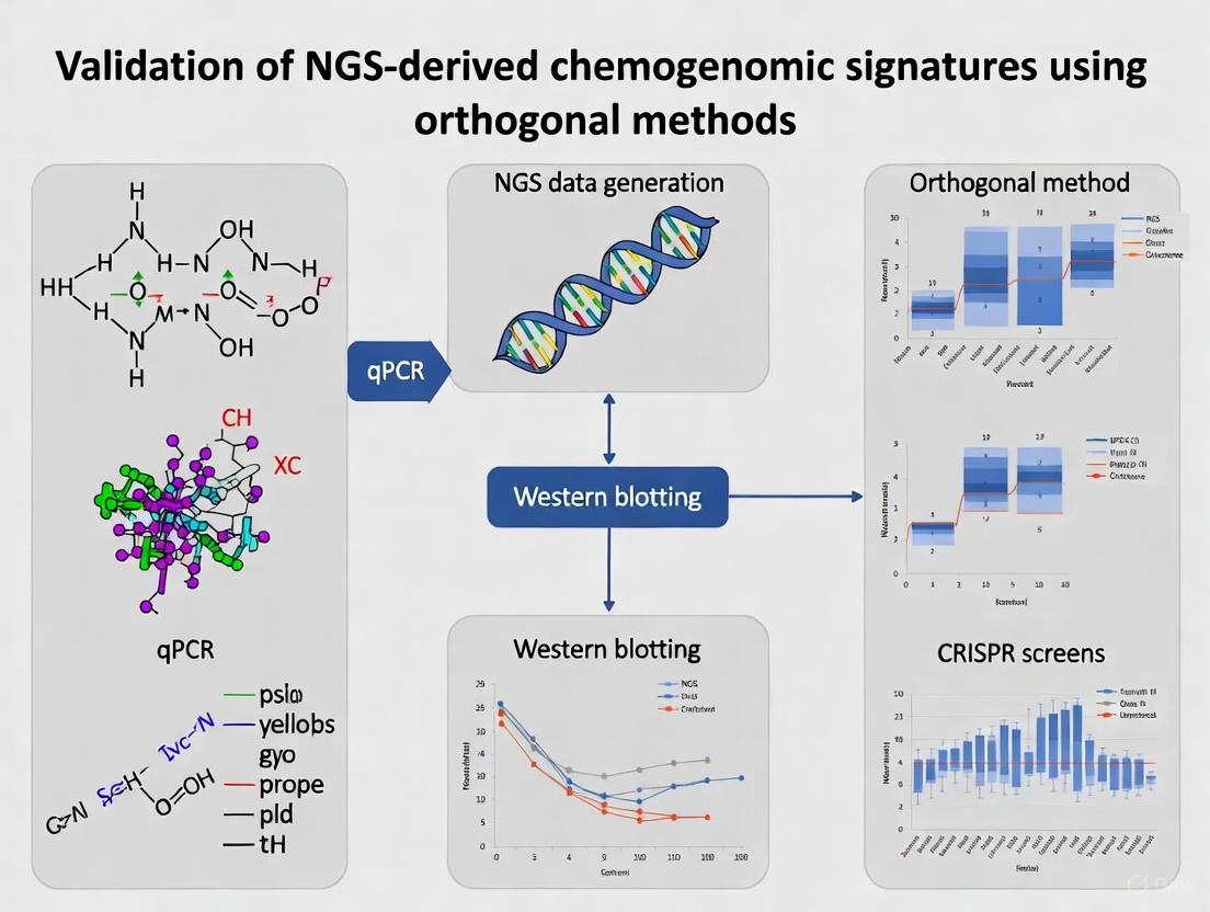
Experimental Protocols for Orthogonal Validation
Orthogonal validation of NGS-derived chemogenomic signatures requires carefully designed experimental protocols that confirm findings using independent methodological approaches. The NCI-MATCH trial established a framework for classifying genomic alterations based on levels of evidence, with Level 1 representing variants credentialed for FDA-approved drugs and Level 3 based on preclinical inferential data [26]. This evidence-based classification can be adapted for chemogenomic signature validation, beginning with computational prediction and progressing through experimental confirmation.
Functional validation typically begins with genetic perturbation experiments using CRISPR-based gene knockout or RNA interference-mediated knockdown in model cell lines, assessing how these manipulations alter compound sensitivity [26]. For signatures suggesting direct compound-target interactions, biochemical assays such as cellular thermal shift assays (CETSA) or drug affinity responsiveness target stability (DARTS) can confirm physical engagement between compounds and their putative protein targets. Proteomic approaches using mass spectrometry-based quantification provide orthogonal confirmation of protein-level changes corresponding to transcriptomic alterations identified by RNA sequencing. For signatures with potential clinical translation, validation in patient-derived xenograft models or correlation with clinical response data in appropriate patient cohorts provides the highest level of evidence for actionable chemogenomic insights.
Table 4: Orthogonal Validation Methods for NGS-Derived Chemogenomic Signatures
| Validation Method | Experimental Approach | Information Gained | Level of Evidence |
|---|---|---|---|
| Genetic Perturbation | CRISPR knockout, RNAi knockdown | Causal relationship between gene and compound response | Medium-High |
| Biochemical Binding | CETSA, DARTS, SPR | Direct physical interaction between compound and target | High |
| Proteomic Analysis | Mass spectrometry, Western blot | Protein-level confirmation of transcriptomic changes | Medium |
| Cellular Phenotyping | High-content imaging, viability assays | Functional consequences of genomic alterations | Medium |
| Preclinical Models | PDX models, organoids | Relevance in more physiological systems | High |
| Clinical Correlation | Retrospective analysis of patient responses | Direct clinical translatability | Highest |
From Data to Discovery: Methodologies for Deriving and Applying Signatures
Integrated multi-omic profiling represents a transformative approach in biomedical research, enabling a comprehensive understanding of biological systems by simultaneously analyzing multiple molecular layers. The convergence of whole-exome sequencing (WES), RNA sequencing (RNA-Seq), and epigenetic profiling technologies provides unprecedented insights into the complex interplay between genetic predispositions, transcriptional regulation, and epigenetic modifications that drive disease pathogenesis and therapeutic responses [30]. This integrated approach is particularly vital for validating next-generation sequencing (NGS)-derived chemogenomic signatures, as it allows researchers to bridge the gap between identified genomic variants and their functional consequences across molecular layers.
The analytical validation of NGS assays, as demonstrated in large-scale precision medicine trials like NCI-MATCH, requires rigorous benchmarking to ensure reliability across multiple clinical laboratories [26]. Such validation establishes critical performance parameters including sensitivity, specificity, and reproducibility that are essential for generating clinically actionable insights. As the field advances, integrated multi-omics approaches are increasingly being applied to unravel the biological and clinical insights of complex diseases, particularly in cancer research where molecular heterogeneity remains a fundamental challenge [31].
Performance Benchmarking of Multi-Omics Integration Methods
Method Comparison and Evaluation Frameworks
The computational integration of multi-omics data presents significant challenges, necessitating rigorous benchmarking of different integration strategies. A comprehensive evaluation of joint dimensionality reduction (jDR) approaches revealed that method performance varies substantially across different analytical contexts [32]. Integrative Non-negative Matrix Factorization (intNMF) demonstrated superior performance in sample clustering tasks, while Multiple co-inertia analysis (MCIA) offered consistently effective behavior across multiple analysis contexts [32].
Benchmarking studies have systematically evaluated integration methods using three complementary approaches: (1) performance in retrieving ground-truth sample clustering from simulated multi-omics datasets, (2) prediction accuracy for survival, clinical annotations, and known pathways using TCGA cancer data, and (3) classification accuracy for multi-omics single-cell data [32]. These evaluations consistently demonstrate that no single method universally outperforms all others across every metric, highlighting the importance of selecting integration approaches based on specific research objectives.
Table 1: Performance Benchmarking of Multi-Omics Integration Methods for Cancer Subtyping
| Integration Method | Mathematical Foundation | Clustering Accuracy | Survival Prediction | Biological Interpretability | Best Use Cases |
|---|---|---|---|---|---|
| intNMF | Non-negative Matrix Factorization | High | Moderate | High | Sample clustering, distinct subtypes |
| MCIA | Principal Component Analysis | Moderate | High | High | Multi-context analysis, visualization |
| MOFA | Factor Analysis | Moderate | High | High | Capturing shared and unique variation |
| SNF | Similarity Network Fusion | Moderate | Moderate | Moderate | Network-based integration |
| iCluster | Factor Analysis | Moderate | High | Moderate | Genomic data integration |
Impact of Data Type Combinations on Integration Performance
Contrary to intuitive expectations, simply incorporating more omics data types does not always improve integration performance. Systematic evaluation of eleven different combinations of four primary omics data types (genomics, epigenomics, transcriptomics, and proteomics) revealed situations where integrating additional data types negatively impacts method performance [33]. This counterintuitive finding underscores the importance of strategic data selection rather than exhaustive data inclusion.
Research has identified particularly effective combinations for specific cancer types. For example, in breast cancer (BRCA), integrating gene expression with DNA methylation data frequently yields superior subtyping results, while in kidney cancer (KIRC), combining gene expression with miRNA expression proves most effective [33]. These findings emphasize that the optimal multi-omics combination is context-dependent and should be informed by biological knowledge of the disease system under investigation.
Table 2: Analytical Performance of NGS Assays in Clinical Validation
| Performance Metric | SNVs | Small Indels | Large Indels (≥4 bp) | Gene Amplifications | Overall Performance |
|---|---|---|---|---|---|
| Sensitivity | >99.9% | >99.9% | >99.9% | >99.9% | 96.98% |
| Specificity | >99.9% | >99.9% | >99.9% | >99.9% | 99.99% |
| Limit of Detection | 2.8% | 10.5% | 6.8% | 4 copies | Variant-dependent |
| Reproducibility | >99.9% | >99.9% | >99.9% | >99.9% | 99.99% |
Experimental Designs and Methodological Protocols
Integrated Multi-Omics Workflow for Disease Mechanism Elucidation
A comprehensive multi-omics study on post-operative recurrence in stage I non-small cell lung cancer (NSCLC) exemplifies a robust integrated profiling approach [31]. This research combined whole-exome sequencing, nanopore sequencing, RNA-seq, and single-cell RNA sequencing on samples from 122 stage I NSCLC patients (57 with recurrence, 65 without recurrence) to identify molecular determinants of disease recurrence. The experimental workflow incorporated matched tumor and adjacent normal tissues from fresh frozen (FF) and formalin-fixed paraffin-embedded (FFPE) specimens to maximize analytical robustness while addressing practical clinical constraints.
The analytical approach implemented in this study exemplifies best practices for integrated multi-omics analysis: (1) genomic characterization of somatic mutations, copy number variations, and structural variants; (2) epigenomic profiling of differentially methylated regions using nanopore sequencing; (3) transcriptomic analysis of gene expression patterns; and (4) single-cell resolution decomposition of the tumor microenvironment [31]. This layered analytical strategy enabled the identification of coordinated molecular events across biological layers that would remain undetectable in single-omics analyses.
Diagram 1: Integrated multi-omics workflow for NSCLC recurrence analysis. This workflow demonstrates the parallel processing of multi-omics data streams and their integration for clinical stratification.
Quality Control Protocols for Multi-Omics Data
Rigorous quality control is paramount for generating reliable multi-omics data, particularly given the technical variability across different assay platforms. A comprehensive quality control framework for epigenomics and transcriptomics data outlines specific metrics and mitigation strategies for eleven different assay types [34]. For WES data, essential quality metrics include sequencing depth (typically >100x for somatic variants), coverage uniformity, base quality scores, and contamination estimates. For RNA-seq data, critical parameters include ribosomal RNA content, library complexity, transcript integrity numbers, and gene body coverage. For epigenetic profiling methods such as bisulfite sequencing or ChIP-seq, key metrics include bisulfite conversion efficiency, CpG coverage, enrichment efficiency, and peak distribution patterns.
The NCI-MATCH trial established a robust framework for analytical validation of NGS assays across multiple clinical laboratories, achieving an overall sensitivity of 96.98% for 265 known mutations and 99.99% specificity [26]. This validation approach incorporated formalin-fixed paraffin-embedded (FFPE) clinical specimens and cell lines to assess reproducibility across variant types, with a 99.99% mean inter-operator pairwise concordance across four independent laboratories [26]. The establishment of such rigorous quality standards is essential for generating clinically actionable insights from multi-omics profiling.
Biological Insights from Integrated Multi-Omics Studies
Molecular Determinants of Cancer Recurrence
The integrated analysis of WES, RNA-seq, and epigenetic data in stage I NSCLC identified distinct molecular features associated with post-operative recurrence [31]. Genomic characterization revealed that recurrent tumors exhibited significantly higher homologous recombination deficiency (HRD) scores and enriched APOBEC-related mutational signatures, indicating increased genomic instability. Furthermore, specific TP53 missense mutations in the DNA-binding domain were associated with significantly shorter time to recurrence, highlighting their potential prognostic value.
Epigenomic profiling through nanopore sequencing identified pronounced DNA hypomethylation in recurrent NSCLC, with PRAME identified as a significantly hypomethylated and overexpressed gene in recurrent lung adenocarcinoma [31]. Mechanistically, hypomethylation at the TEAD1 binding site was shown to facilitate transcriptional activation of PRAME, and functional validation demonstrated that PRAME inhibition restrains tumor metastasis through downregulation of epithelial-mesenchymal transition-related genes. This finding exemplifies how multi-omics integration can identify epigenetically dysregulated oncogenic drivers with potential therapeutic implications.
Tumor Ecosystem Characterization
Single-cell RNA sequencing integrated with bulk multi-omics data revealed essential ecosystem features associated with NSCLC recurrence [31]. The analysis identified enrichment of AT2 cells with higher copy number variation burden, exhausted CD8+ T cells, and Macro_SPP1 macrophages in recurrent LUAD, along with reduced interaction between AT2 and immune cells. This comprehensive ecosystem characterization provides insights into the immunosuppressive microenvironment that facilitates disease recurrence despite surgical resection.
Multi-omics clustering stratified NSCLC patients into four distinct subclusters with varying recurrence risk and subcluster-specific therapeutic vulnerabilities [31]. This stratification demonstrated superior prognostic performance compared to single-omics approaches, highlighting the clinical value of integrated molecular profiling for precision oncology applications.
Diagram 2: Molecular mechanisms of NSCLC recurrence identified through multi-omics profiling. This diagram illustrates the coordinated molecular events across biological layers that drive disease recurrence.
The Scientist's Toolkit: Essential Research Reagent Solutions
Table 3: Essential Research Reagents and Platforms for Multi-Omics Profiling
| Reagent/Platform | Function | Application Notes | Technical Considerations |
|---|---|---|---|
| Oncomine Cancer Panel | Targeted NGS panel | Detects 4066 predefined variants across 143 genes | Optimized for FFPE samples; validated in CLIA labs |
| Ion Torrent PGM | Next-generation sequencer | Medium-throughput sequencing | Used in NCI-MATCH with locked analysis pipeline |
| Thermo Fisher AmpliSeq | Library preparation | RNA and DNA library construction | Integrated with Oncomine panel |
| 10X Genomics Chromium | Single-cell partitioning | High-throughput single-cell sequencing | Utilizes gel bead-in-emulsion technology |
| Pacific Biosciences SMRT | Long-read sequencing | Epigenetic modification detection | Identifies base modifications without bisulfite treatment |
| Oxford Nanopore | Long-read sequencing | Direct DNA/RNA sequencing | Enables simultaneous sequence and modification detection |
| PyClone-VI | Clonal decomposition | Phylogenetic analysis | Infers clonal architecture from multi-omics data |
| MOFA+ | Multi-omics factor analysis | Dimensionality reduction | Identifies shared and unique sources of variation |
Integrated multi-omic profiling combining WES, RNA-seq, and epigenetic data represents a powerful approach for elucidating complex biological mechanisms and validating NGS-derived chemogenomic signatures. The rigorous benchmarking of integration methods and comprehensive quality control frameworks established in recent studies provide robust analytical foundations for extracting biologically and clinically meaningful insights from these complex datasets. As the field advances, emerging technologies including long-read sequencing, single-cell multi-omics, and spatial transcriptomics are further enhancing the resolution and comprehensiveness of integrated molecular profiling [35].
The future of multi-omics research lies in the development of increasingly sophisticated integration algorithms that can simultaneously accommodate diverse data types while accounting for technical artifacts and biological heterogeneity. Furthermore, the translation of multi-omics insights into clinically actionable biomarkers requires standardized validation frameworks across multiple laboratories and patient cohorts [26]. As these technologies become more accessible and analytical methods more refined, integrated multi-omic profiling is poised to become an indispensable tool for precision medicine, fundamentally advancing our ability to understand, diagnose, and treat complex diseases.
Computational Frameworks for Signature Extraction and Analysis
The validation of next-generation sequencing (NGS)-derived chemogenomic signatures represents a critical frontier in precision oncology, bridging the gap between computational prediction and clinical application. As cancer treatment increasingly shifts from a one-size-fits-all approach to personalized strategies, the ability to accurately extract and analyze gene expression signatures that predict drug response has become paramount [36]. These signatures enable oncologists to simulate therapeutic efficacy computationally, bypassing the time-consuming and costly process of in vitro drug screening [36].
Current computational frameworks leverage diverse methodologies including independent component analysis, discretization algorithms, and multi-omics integration to transform raw transcriptomic data into clinically actionable insights. The emerging "chemogram" concept—inspired by clinical antibiograms used in infectious disease—aims to rank chemotherapeutic sensitivity for individual tumors using only gene expression data [36]. However, the translational potential of these approaches hinges on rigorous validation using orthogonal methods to ensure reliability and clinical utility.
This comparison guide provides an objective assessment of leading computational frameworks for signature extraction and analysis, focusing on their methodological approaches, performance characteristics, and validation requirements to inform researchers, scientists, and drug development professionals.
Comparative Analysis of Computational Frameworks
Table 1: Computational Frameworks for Signature Extraction and Analysis
| Framework | Primary Methodology | Input Data | Key Features | Validation Approaches |
|---|---|---|---|---|
| ICARus [37] | Independent Component Analysis (ICA) with robustness assessment | Normalized gene expression matrix (genes × samples) | Identifies near-optimal parameters; evaluates signature reproducibility across parameters; outputs gene contributions and sample signature scores | Internal stability indices (>0.75 threshold); reproducibility across parameter values; gene set enrichment analysis |
| gdGSE [38] | Discretization of gene expression values | Bulk or single-cell transcriptomes | Binarizes gene expression matrix; converts to gene set enrichment matrix; mitigates data distribution discrepancies | Concordance with experimental drug mechanisms (>90%); cancer stemness quantification; cell type identification accuracy |
| Chemogram [36] | Pre-derived predictive gene signatures | Transcriptomic data from tumor samples | Ranks relative drug sensitivity across multiple therapeutics; pan-cancer application; inspired by clinical antibiograms | Comparison against observed drug response in cell lines; benchmarking against random signatures and differential expression |
| mSigSDK [39] | Mutational signature analysis | Mutation Annotation Format (MAF) files | Browser-based computation without downloads; privacy-preserving analysis; integrates with mSigPortal APIs | Orthogonal validation against established mutational catalogs; compatibility with COSMIC and SIGNAL resources |
| Drug Combination Predictors [40] | Deep learning (AuDNNsynergy), multi-omics integration | Multi-omics data (genomics, transcriptomics, proteomics) | Predicts synergistic/antagonistic drug interactions; uses Bliss Independence and Combination Index scores | Experimental validation of predicted combinations; correlation with observed drug responses |
Performance Metrics and Experimental Validation
Table 2: Performance Metrics and Validation Data for Signature Analysis Frameworks
| Framework | Reported Performance Metrics | Experimental Validation Methods | Therapeutic Concordance | Limitations and Considerations |
|---|---|---|---|---|
| ICARus [37] | Stability index >0.75 for robust signatures; reproducibility across parameter values | Gene Set Enrichment Analysis (GSEA); association with sample phenotypes and temporal patterns | Not explicitly reported | Sensitive to normalization methods; requires pre-filtering of sparsely expressed genes |
| gdGSE [38] | >90% concordance with experimental drug mechanisms; enhanced clustering performance | Patient-derived xenografts; estrogen receptor-positive breast cancer cell lines; cancer stemness quantification | High concordance with validated drug mechanisms | Discretization may lose subtle expression information |
| Chemogram [36] | More accurate than random signatures; comparable to established prediction methods | GDSC and TCGA data; novel muscle-invasive bladder cancer dataset; provisional patent application | Accurate rank order of drug sensitivity in multiple cancer types | Limited to pre-derived signature database; requires further clinical validation |
| mSigSDK [39] | Compatible with established mutational signature resources | Comparison against COSMIC mutational signatures; integration with NCI/DCEG resources | Not explicitly reported | Computational limitations for de novo extraction in browser environment |
| Drug Combination Predictors [40] | DeepSynergy: Pearson correlation 0.73, AUC 0.90; 7.2% improvement in MSE | Bliss Independence score; Combination Index; experimental validation in preclinical models | Predicts synergistic and antagonistic interactions | Limited mechanistic explanation; dependency on comprehensive omics data |
Experimental Protocols and Methodologies
Signature Extraction and Robustness Assessment
The ICARus pipeline implements a rigorous approach for extracting robust gene expression signatures from transcriptomic datasets [37]. The methodology begins with a normalized gene expression matrix (genes × samples) using appropriate normalization methods such as Counts-per-Million (CPM) or Ratio of median. Principal Component Analysis (PCA) is then performed to determine the near-optimal parameter set for Independent Component Analysis (ICA). The Kneedle Algorithm identifies the critical elbow/knee point in the standard deviation or cumulative variance plot, establishing the minimum number (n) for the near-optimal parameter set.
For intra-parameter robustness assessment, ICA is performed 100 times for each n value within the determined range. Resulting signatures undergo sign correction and hierarchical clustering. The stability index for each cluster is calculated using the Icasso method, which measures similarities between signatures from different runs using the absolute value of the Pearson correlation coefficient [37]. Signatures with stability indices exceeding 0.75 are considered robust. For inter-parameter reproducibility, robust signatures are clustered across different n values, with reproducible signatures identified as those clustering together across multiple parameter values within the near-optimal set.
Figure 1: ICARus Signature Extraction Workflow
Discretization-Based Pathway Enrichment Analysis
The gdGSE algorithm introduces a novel approach to gene set enrichment analysis by discretizing gene expression values [38]. The methodology consists of two primary steps. First, statistical thresholds are applied to binarize the gene expression matrix, converting continuous expression values into discrete categories (e.g., high/low expression). This discretization process mitigates discrepancies caused by data distributions and technical variations. Second, the binarized gene expression matrix is transformed into a gene set enrichment matrix, where pathway activity is quantified based on the discrete expression patterns of member genes.
Validation of gdGSE involves multiple approaches including precise quantification of cancer stemness with prognostic relevance, enhanced clustering performance for tumor subtype stratification, and accurate identification of cell types in single-cell data. Most notably, concordance with experimentally validated drug mechanisms is assessed using patient-derived xenografts and estrogen receptor-positive breast cancer cell lines, with reported concordance exceeding 90% [38].
Transcriptomic Chemogram Development
The chemogram framework utilizes pre-derived predictive gene signatures to rank drug sensitivity across multiple therapeutics [36]. Signature derivation follows the methodology established by Scarborough et al., which identifies gene expression patterns in genomically disparate tumors exhibiting sensitivity to the same chemotherapeutic agent. This approach exploits convergent evolution by identifying co-expression patterns in sensitive tumors regardless of cancer type.
Validation involves applying predictive signatures to rank sensitivity among drugs within cancer cell lines and comparing the rank order of predicted and observed response. Performance is assessed against negative controls including randomly generated gene signatures and signatures derived from differential expression alone. The framework is tested across hundreds of cancer cell lines from resources such as The Genomics of Drug Sensitivity in Cancer (GDSC) and The Cancer Genome Atlas (TCGA) [36].
Figure 2: Chemogram Development and Validation
Table 3: Key Research Reagent Solutions for Signature Validation
| Resource Category | Specific Examples | Function and Application | Access Information |
|---|---|---|---|
| Reference Datasets | GDSC (Genomics of Drug Sensitivity in Cancer) [36] | Provides drug sensitivity data and molecular profiles of cancer cell lines for signature development and validation | Publicly available |
| TCGA (The Cancer Genome Atlas) [36] | Offers comprehensive molecular characterization of primary tumors for signature validation | Publicly available | |
| GIAB (Genome in a Bottle) [7] | Provides benchmark variant calls for assessing sequencing accuracy and variant calling performance | Publicly available through NIST | |
| Analysis Platforms | mSigPortal [39] | Web-based platform for exploring curated mutational signature databases and performing analysis | https://analysistools.cancer.gov/mutational-signatures/ |
| UCSC Genome Browser [39] | Genome visualization and conversion of MAF files into mutational spectra | Publicly available | |
| Software Libraries | ICARus R Package [37] | Implements robust signature extraction pipeline using Independent Component Analysis | https://github.com/Zha0rong/ICArus |
| gdGSE R Package [38] | Performs gene set enrichment analysis using discretized gene expression values | https://github.com/WangX-Lab/gdGSE | |
| Experimental Validation Resources | Patient-derived xenografts [38] | In vivo models for validating signature-predicted drug mechanisms | Institutional core facilities |
| Cell line panels [36] [38] | In vitro systems for testing signature-predicted drug sensitivity | Commercial providers (ATCC) |
Computational frameworks for signature extraction and analysis show tremendous potential for advancing personalized cancer therapy, yet their clinical implementation requires rigorous validation using orthogonal methods. The featured frameworks—ICARus, gdGSE, Chemogram, mSigSDK, and drug combination predictors—each offer distinct methodological advantages for different research contexts.
ICARus provides exceptional robustness for signature extraction through its multi-parameter reproducibility assessment, while gdGSE offers innovative discretization approaches that show remarkable concordance with experimental drug mechanisms. The chemogram framework presents a clinically intuitive model for ranking therapeutic options, though it requires further validation in clinical settings. Across all platforms, the integration of multi-omics data and adherence to FAIR principles (Findability, Accessibility, Interoperability, and Reusability) will be essential for advancing the field [39].
Future development should focus on improving model interpretability, standardization of validation protocols, and demonstration of clinical utility through prospective trials. As next-generation sequencing technologies continue to evolve and multi-omics integration becomes more sophisticated [41], these computational frameworks will play an increasingly vital role in translating genomic discoveries into personalized therapeutic strategies.
Leveraging Chemogenomic Signatures for Target Identification and Lead Compound Optimization
Chemogenomics represents a systematic framework for screening targeted chemical libraries against families of biological targets, with the ultimate goal of identifying novel drugs and drug targets [42]. This approach integrates target and drug discovery by using active compounds as probes to characterize proteome functions, creating a powerful bridge between chemical space and biological response [42]. The field has evolved significantly with the advent of advanced screening technologies and computational methods, enabling researchers to navigate the complex landscape of drug-target interactions more efficiently. The completion of the human genome project provided an abundance of potential targets for therapeutic intervention, and chemogenomics strives to study the intersection of all possible drugs on these potential targets [42].
Two primary experimental paradigms dominate chemogenomic research: forward (classical) and reverse approaches [42]. Forward chemogenomics begins with a specific phenotype and identifies small molecules that induce this phenotype, subsequently determining the molecular targets responsible. Conversely, reverse chemogenomics starts with specific protein targets, identifies compounds that modulate their activity, and then characterizes the resulting phenotypic effects [42]. Both approaches have contributed significantly to drug discovery, including the identification of novel antibacterial agents [42] and the elucidation of previously unknown genes in biological pathways [42].
Comparative Analysis of Screening Methodologies
Phenotypic vs. Target-Based Screening Approaches
Table 1: Comparison of Small Molecule and Genetic Screening Approaches in Phenotypic Discovery
| Parameter | Small Molecule Screening | Genetic Screening |
|---|---|---|
| Scope of Targets | Limited to ~1,000-2,000 of 20,000+ human genes [43] | Enables systematic perturbation of large numbers of genes [43] |
| Temporal Resolution | Allows acute, reversible modulation [43] | Typically creates permanent perturbations [43] |
| Throughput Considerations | Limited by more complex phenotypic assays [43] | Higher throughput possible with pooled formats [43] |
| Clinical Translation | Direct identification of pharmacologically relevant compounds [43] | Fundamental differences between genetic and pharmacological perturbation [43] |
| Key Strengths | Identifies immediately tractable chemical starting points; reveals novel mechanisms [43] | Comprehensive genome coverage; establishes causal gene-phenotype relationships [43] |
| Major Limitations | Limited target coverage; promiscuous binders complicate interpretation [43] | Differences from pharmacological effects; overexpression may not mimic drug action [43] |
Phenotypic drug discovery (PDD) has re-emerged as a promising approach for identifying novel therapeutic agents, particularly for complex diseases involving multiple molecular abnormalities [6]. With advances in cell-based screening technologies, including induced pluripotent stem (iPS) cells, CRISPR-Cas gene-editing tools, and imaging assays, PDD strategies can identify compounds with relevant biological activity without prior knowledge of specific molecular targets [6]. However, a significant challenge remains in translating observed phenotypes to molecular mechanisms of action, which is where chemogenomic approaches provide critical value.
The cellular response to small molecules appears to be limited and organized into discrete patterns. Research analyzing over 35 million gene-drug interactions across more than 6,000 chemogenomic profiles revealed that cellular responses can be described by a network of 45 distinct chemogenomic signatures [44]. Remarkably, the majority of these signatures (66.7%) were conserved across independently generated datasets from academic and industrial laboratories, demonstrating their biological relevance as conserved systems-level response systems [44]. This conservation across different experimental pipelines underscores the robustness of chemogenomic fitness profiling while providing guidelines for performing similar high-dimensional comparisons in mammalian cells [44].
Integrated Multi-Omics Approaches for Enhanced Target Identification
Table 2: Performance Metrics of Combined RNA-seq and WES Assay in Clinical Validation
| Validation Parameter | Performance Metric | Clinical Utility |
|---|---|---|
| Analytical Validation | Custom references with 3,042 SNVs and 47,466 CNVs [4] | Established sensitivity and specificity framework |
| Orthogonal Testing | Concordance with established methods [4] | Verified reliability in clinical samples |
| Clinical Application | 2,230 patient tumor samples [4] | Demonstrated real-world applicability |
| Actionable Alterations | 98% of cases showed clinically actionable findings [4] | Direct impact on personalized treatment strategies |
| Variant Recovery | Improved detection of variants missed by DNA-only approaches [4] | Enhanced diagnostic accuracy |
| Fusion Detection | Superior identification of gene fusions [4] | More comprehensive genomic profiling |
The integration of multiple genomic technologies significantly enhances the detection of clinically relevant alterations in cancer. A combined RNA sequencing (RNA-seq) and whole exome sequencing (WES) approach demonstrates superior performance compared to DNA-only methods, enabling direct correlation of somatic alterations with gene expression patterns and improved detection of gene fusions [4]. This integrated methodology has shown clinical utility in identifying complex genomic rearrangements that would likely remain undetected using single-modality approaches [4].
Validation of such integrated assays requires a comprehensive framework including analytical validation using custom reference samples, orthogonal testing in patient specimens, and assessment of clinical utility in real-world scenarios [4]. When applied to 2,230 clinical tumor samples, the combined RNA and DNA sequencing approach demonstrated ability to uncover clinically actionable alterations in 98% of cases, highlighting its transformative potential for personalized cancer treatment [4]. This integrated profiling enables more strategic patient management with reduced time and cost compared to traditional sequential genetic analysis [4].
Experimental Frameworks and Validation Strategies
Development of Chemogenomic Libraries for Phenotypic Screening
The construction of targeted chemical libraries represents a fundamental component of effective chemogenomic screening. Ideally, these libraries include known ligands for at least several members of a target family, as compounds designed to bind to one family member frequently exhibit activity against additional related targets [42]. In practice, a well-designed chemogenomic library should collectively bind to a high percentage of the target family, enabling comprehensive pharmacological interrogation [42].
Advanced chemogenomic libraries have been developed specifically for phenotypic screening applications. One such library of 5,000 small molecules was designed to represent a large and diverse panel of drug targets involved in various biological effects and diseases [6]. This library construction integrated multiple data sources, including:
- Drug-target relationships from ChEMBL database [6]
- Pathway information from Kyoto Encyclopedia of Genes and Genomes (KEGG) [6]
- Disease associations from Human Disease Ontology [6]
- Morphological profiling data from Cell Painting assays [6]
This integrative approach enables the creation of a systems pharmacology network that connects drug-target-pathway-disease relationships with morphological phenotypes, providing a powerful platform for target identification and mechanism deconvolution in phenotypic screening [6].
Machine Learning and Computational Approaches
Table 3: Machine Learning Approaches for Multi-Target Drug Discovery
| ML Technique | Application in Multi-Target Discovery | Key Advantages |
|---|---|---|
| Graph Neural Networks (GNNs) | Learn from molecular graphs and biological networks [45] | Capture structural relationships; integrate heterogeneous data |
| Transformer-based Models | Process sequential, contextual biological information [45] | Handle multimodal data; capture long-range dependencies |
| Pharmacophore-Guided Generation | Generate molecules matching specific chemical features [46] | Incorporates biochemical knowledge; addresses data scarcity |
| Multi-Task Learning | Predict activity against multiple targets simultaneously [45] | Improved efficiency; captures shared representations |
| Classical ML (SVMs, Random Forests) | Predict drug-target interactions and adverse effects [45] | Interpretability; robustness with curated datasets |
Machine learning (ML) has emerged as a powerful toolkit for addressing the complex challenges of multi-target drug discovery [45]. By learning from diverse data sources—including molecular structures, omics profiles, protein interactions, and clinical outcomes—ML algorithms can prioritize promising drug-target pairs, predict off-target effects, and propose novel compounds with desirable polypharmacological profiles [45]. These approaches are particularly valuable for navigating the combinatorial explosion of potential drug-target interactions that makes brute-force experimental screening intractable [45].
Classical ML models like support vector machines and random forests provide interpretability and robustness when trained on curated datasets, while more sophisticated deep learning architectures offer enhanced capability with complex biomedical data [45]. Graph neural networks excel at learning from molecular graphs and biological networks, and transformer-based models effectively capture sequential, contextual biological information [45]. The PGMG (Pharmacophore-Guided deep learning approach for bioactive Molecule Generation) framework demonstrates how pharmacophore hypotheses can guide molecular generation, using a graph neural network to encode spatially distributed chemical features and a transformer decoder to generate molecules [46]. This approach addresses the challenge of data scarcity, particularly for novel target families where known active compounds are limited [46].
Research Toolkit: Essential Reagents and Methodologies
Table 4: Essential Research Reagent Solutions for Chemogenomic Studies
| Reagent/Resource | Function and Application | Key Features |
|---|---|---|
| Chemogenomic Compound Libraries | Phenotypic screening against target families [42] [6] | Annotated compounds; diverse target coverage; optimized for phenotypic assays |
| CRISPR Libraries | Functional genomic screening [43] | Genome-wide coverage; pooled screening formats; gene knockout/activation |
| Cell Painting Assays | Morphological profiling [6] | High-content imaging; multivariate phenotypic analysis; benchmarked protocols |
| Multi-Omics Databases | Target identification and validation [45] | Integrated molecular data; curated interactions; pathway annotations |
| Validated Reference Standards | Assay quality control and validation [4] | Certified variants; established performance metrics; orthogonal validation |
Experimental Workflows for Signature Validation
The following diagram illustrates a comprehensive workflow for validating NGS-derived chemogenomic signatures using orthogonal methods:
Key Signaling Pathways in Chemogenomic Response Signatures
The limited nature of cellular responses to chemical perturbations suggests organization into discrete signaling networks. The following diagram illustrates key pathways and their interconnections in chemogenomic response signatures:
Chemogenomic approaches provide powerful frameworks for target identification and lead optimization in modern drug discovery. The integration of diverse technologies—including functional genomics, small molecule screening, multi-omics profiling, and machine learning—enables researchers to navigate the complex landscape of drug-target interactions more effectively. Validation of chemogenomic signatures through orthogonal methods remains crucial for establishing confidence in both targets and compounds, particularly as drug discovery shifts toward addressing complex diseases through multi-target interventions.
Future developments in this field will likely focus on several key areas: improved integration of multi-modal data streams, advancement of computational methods for predicting polypharmacological profiles, and development of more sophisticated validation frameworks that better capture human disease physiology. As these technologies mature, chemogenomic approaches will play an increasingly central role in accelerating the discovery of safer and more effective therapeutics for complex diseases.
The advent of next-generation sequencing (NGS) has revolutionized molecular oncology, enabling comprehensive genomic profiling that informs personalized cancer treatment strategies. While DNA-based sequencing has been the cornerstone of cancer mutation detection, its limitations in identifying key transcriptional events like gene fusions and expression changes have become increasingly apparent. Combined RNA and DNA sequencing represents a significant methodological advancement, addressing the diagnostic blind spots inherent to single-modality approaches. This integrated paradigm enhances the detection of clinically actionable alterations, thereby facilitating more precise therapeutic interventions.
The validation of NGS-derived biomarkers with orthogonal methods constitutes a critical step in clinical translation. This case study examines the technical validation and clinical utility of a combined sequencing approach, contrasting its performance with DNA-only methods and traditional techniques like fluorescence in situ hybridization (FISH). We present experimental data demonstrating how this integrated methodology improves alteration detection rates across diverse cancer types, with particular emphasis on its application within a framework of orthogonal verification.
Methodological Framework
Integrated Sequencing Workflow
The combined RNA and DNA sequencing protocol involves parallel processing of nucleic acids extracted from the same tumor sample, followed by integrated bioinformatic analysis. The typical workflow, as validated across multiple studies [4] [47] [48], encompasses several critical stages.
Nucleic Acid Extraction: DNA and RNA are co-extracted from fresh-frozen (FF) or formalin-fixed paraffin-embedded (FFPE) tumor samples using validated kits (e.g., AllPrep DNA/RNA Mini Kit) [4]. Specimen quality control is critical, with quantification performed using Qubit Fluorometry and structural integrity assessed via TapeStation analysis [4].
Library Preparation: For DNA, whole exome sequencing (WES) libraries are prepared using hybridization capture with probes such as the SureSelect Human All Exon V7 [4]. For RNA, transcriptome libraries are constructed using either poly-A selection (for FF samples) or exome capture (for FFPE samples) to enable sequencing of potentially degraded RNA [4].
Sequencing and Analysis: Libraries are sequenced on high-throughput platforms (e.g., Illumina NovaSeq 6000) [4]. Bioinformatic processing includes alignment to reference genomes (hg38), variant calling with specialized algorithms (e.g., Strelka for DNA variants, Kallisto for expression quantification), and integrative analysis to correlate DNA alterations with transcriptional consequences [4].
Orthogonal Validation Methods
Robust validation of NGS findings requires orthogonal methods to confirm analytical accuracy [4] [48] [49]:
- Analytical Validation: Custom reference standards containing known mutations (3,042 SNVs and 47,466 CNVs) verify base-level accuracy [4].
- Orthogonal Testing: Patient samples are simultaneously analyzed with established methods including FISH, cytogenetics, and PCR-based approaches to confirm clinical concordance [48].
- Clinical Validation: Performance is assessed in real-world clinical cohorts to determine diagnostic utility and actionability rates [4] [47].
Comparative Performance Data
Detection of Actionable Alterations
The incremental value of combined RNA/DNA sequencing is demonstrated through direct comparison with DNA-only approaches across multiple cancer types and alteration classes.
Table 1: Actionable Alteration Detection: Combined vs. DNA-Only Sequencing
| Cancer Type | Sequencing Approach | Actionable Detection Rate | Key Alterations Enhanced | Study |
|---|---|---|---|---|
| Pan-Cancer (2,230 samples) | Combined RNA/DNA | 98% | Gene fusions, allele-specific expression, complex rearrangements | [4] |
| Pan-Cancer (1,166 samples) | Combined RNA/DNA | 62.3% | NTRK/RET fusions, MSI-high, TMB-high, ERBB2 amplifications | [47] |
| Hematologic Malignancies (3,101 samples) | RNA-Seq (Fusion Focus) | 17.6% | Cryptic fusions (NUP98::NSD1, P2RY8::CRLF2, KMT2A variants) | [48] |
| Advanced NSCLC (102 samples) | DNA-Only (Liquid Biopsy) | 56-79%* | SNVs (high concordance), limited fusion detection | [50] |
*Percentage range reflects variation across different assay types; amplicon-based assays showed lower fusion detection compared to hybrid capture.
The integrated approach demonstrated remarkable improvement in comprehensive alteration detection, identifying clinically actionable alterations in 98% of cases in a large cohort of 2,230 clinical tumor samples [4]. This represents a significant enhancement over DNA-only approaches, which typically miss a substantial proportion of transcriptionally-active events.
Fusion Detection Enhancement
RNA sequencing substantially improves fusion detection compared to DNA-based methods and traditional techniques.
Table 2: Fusion Detection: RNA-Seq vs. Conventional Methods in Hematologic Malignancies
| Method | Fusion Detection Rate | Novel Fusion Identification | Dual Fusion Cases Detected | Concordance with Cytogenetics/FISH |
|---|---|---|---|---|
| RNA-Based NGS | 17.6% (545/3,101) | 24 novel fusions | 16 cases | 63.7% (310/486) |
| Cytogenetics/FISH | Not reported | Limited capability | Limited capability | Reference standard |
| Discordance Analysis | N/A | 23.8% (5/21) detected by FISH | 35.7% (5/14) detected by FISH | 36.3% discordance rate |
RNA-based NGS identified fusions in 17.6% of cases (545/3,101) across hematologic malignancies, with particularly high rates in B-lymphoblastic leukemia (31.0%) and acute myeloid leukemia (23.2%) [48]. Notably, 36.3% of fusion-positive cases identified by RNA-seq were missed by conventional cytogenetics/FISH, underscoring the limitations of traditional approaches [48].
Technical Performance Metrics
Analytical validation of combined sequencing demonstrates robust performance characteristics essential for clinical implementation.
Table 3: Analytical Validation Metrics for Combined RNA/DNA Sequencing
| Performance Parameter | DNA Variants (SNVs/INDELs) | RNA Variants | Gene Expression | Fusion Detection |
|---|---|---|---|---|
| Sensitivity | 98.23% (at 95% CI) [51] | Not explicitly reported | Not explicitly reported | Superior to FISH [48] |
| Specificity | 99.99% (at 95% CI) [51] | Not explicitly reported | Not explicitly reported | High for known fusion types [48] |
| Limit of Detection | ~3% VAF [51] | Not explicitly reported | Not explicitly reported | 5% tumor content (validated) [48] |
| Reproducibility | 99.98% [51] | Not explicitly reported | Not explicitly reported | Not explicitly reported |
The validation of a targeted NGS panel demonstrated 98.23% sensitivity and 99.99% specificity for DNA variant detection, with reproducibility exceeding 99.98% [51]. For fusion detection, RNA-seq maintained sensitivity even at low tumor content (5% validated), with identified fusions in 12.1% of cases having tumor content below this threshold [48].
Orthogonal Validation Framework
Orthogonal confirmation of NGS findings is essential for clinical translation, particularly for novel or unexpected alterations. The validation paradigm employs multiple complementary methodologies to verify results from combined sequencing.
DNA-Level Alterations: Single nucleotide variants (SNVs) and insertions/deletions (INDELs) identified through DNA sequencing are confirmed via Sanger sequencing or digital droplet PCR (ddPCR) [49]. This provides base-level resolution confirmation of mutational status.
Structural Variants: Gene fusions and rearrangements detected via RNA-seq are validated using RT-PCR or FISH [48]. FISH offers the advantage of single-cell resolution and the ability to detect rearrangements regardless of specific breakpoints.
Copy Number and Expression: Copy number variations (CNVs) identified through DNA sequencing can be confirmed by FISH or array comparative genomic hybridization (aCGH) [52]. Gene expression changes quantified by RNA-seq are validated using quantitative RT-PCR or digital expression platforms like Nanostring [4].
This multi-modal verification framework ensures high confidence in genomic findings before their application to clinical decision-making, addressing the rigorous evidence standards required for therapeutic implementation.
Clinical Implications
Impact on Therapeutic Targeting
The enhanced detection capability of combined sequencing directly translates to expanded therapeutic opportunities. In a pan-cancer Asian cohort (1,166 samples), comprehensive genomic profiling revealed Tier I alterations (associated with standard-of-care therapies) in 12.7% of cases, and Tier II alterations (clinical trial eligibility) in 6.0% of cases [47]. These included EGFR mutations in NSCLC (38.2%), PIK3CA mutations in breast cancer (39%), and BRCA1/2 alterations in prostate cancer [47].
Notably, tumor-agnostic biomarkers – including MSI-high, TMB-high, NTRK fusions, and RET fusions – were identified in 8.4% of cases across 26 cancer types [47]. These biomarkers transcend histology-based classification and can guide treatment with tissue-agnostic therapies, demonstrating the value of broad molecular profiling.
Complementary Role of Liquid Biopsy
While tissue-based combined sequencing provides comprehensive profiling, liquid biopsy approaches offer complementary utility, particularly when tissue is unavailable or insufficient. In advanced NSCLC, liquid biopsy NGS demonstrated 56-79% positive percent agreement with tissue testing for actionable alterations, with highest concordance for SNVs [50].
Hybrid capture-based liquid biopsy assays showed superior performance for fusion detection compared to amplicon-based approaches, identifying 7-8 gene fusions versus only 2 with amplicon-based methods [50]. This highlights the importance of platform selection when implementing liquid biopsy testing.
Essential Research Toolkit
Table 4: Key Reagents and Platforms for Combined Sequencing Studies
| Category | Specific Products/Platforms | Application Notes |
|---|---|---|
| Nucleic Acid Extraction | AllPrep DNA/RNA Mini Kit (Qiagen), Maxwell RSC instruments (Promega) | Co-extraction maintains sample integrity; FFPE-compatible kits available [4] [50] |
| Library Preparation | SureSelect XTHS2 (Agilent), TruSeq stranded mRNA (Illumina), Oncomine Precision Assay | Hybrid capture provides uniform coverage; target enrichment needed for FFPE RNA [4] [50] |
| Sequencing Platforms | Illumina NovaSeq 6000, MGI DNBSEQ-G50RS, Element AVITI24 | High-throughput systems enable dual-modality sequencing; platform choice affects cost and throughput [4] [51] [13] |
| Bioinformatic Tools | BWA, STAR aligners; Strelka, Pisces variant callers; Sophia DDM | Specialized callers needed for RNA variants; integrated analysis pipelines are essential [4] [51] |
| Orthogonal Validation | Sanger sequencing, FISH, ddPCR, Nanostring | Method selection depends on alteration type; ddPCR offers high sensitivity for low-frequency variants [48] [50] [49] |
Combined RNA and DNA sequencing represents a methodological advance in genomic oncology, substantially improving the detection of clinically actionable alterations compared to DNA-only approaches. Through orthogonal validation frameworks, this integrated methodology demonstrates enhanced sensitivity for gene fusions, expression changes, and complex structural variants, with direct implications for therapeutic targeting.
The implementation of this approach requires careful attention to technical validation, bioinformatic integration, and orthogonal confirmation to ensure clinical reliability. When properly validated and interpreted, combined sequencing provides a more comprehensive molecular portrait of tumors, ultimately supporting more precise and personalized cancer treatment strategies. As the field advances, the integration of multi-modal genomic data will increasingly become the standard for oncologic molecular profiling, enabling continued progress in precision oncology.
Navigating Challenges: Technical Pitfalls and Optimization Strategies
Next-generation sequencing (NGS) has revolutionized genomic studies and is driving the implementation of precision medicine. However, the ability of these technologies to disentangle sequence heterogeneity is fundamentally limited by their relatively high error rates, which can be substantially elevated in specific genomic contexts. These errors are not merely random but often manifest as systematic biases introduced during library preparation and are inherent to specific sequencing platforms. For research focused on validating NGS-derived chemogenomic signatures—particularly in sensitive applications like drug development—recognizing, understanding, and mitigating these biases is paramount. This guide objectively compares the performance of different NGS library preparation methods and sequencing platforms, providing a framework for their identification and correction through orthogonal validation strategies.
Library Preparation Artifacts: Mechanisms and Experimental Characterization
Library preparation is a critical process preceding sequencing itself, comprising DNA fragmentation, end-repair, A-tailing, adapter ligation, and amplification. The methods used in these steps can introduce significant, non-random artifacts [53] [54].
Comparative Analysis of Fragmentation Methods
DNA fragmentation, the first step in library prep, can be achieved through physical (sonication) or enzymatic means. Recent studies have systematically compared these methods to quantify their artifact profiles.
Table 1: Comparison of Fragmentation Methods and Associated Artifacts
| Fragmentation Method | Typical Artifact Profile | Potential Impact on Variant Calling | Key Characteristics |
|---|---|---|---|
| Sonication (Ultrasonic) | Significantly fewer artifactual SNVs and indels [53]. Most artifacts are chimeric reads containing cis- and trans-inverted repeat sequences [53]. | Lower false positive variant count [53]. | Near-random, non-biased fragmentation [53] [55]. Equipment-intensive and can lead to DNA loss [53]. |
| Enzymatic Fragmentation | Higher number of artifactual SNVs and indels compared to sonication [53]. Artifacts often located at palindromic sequences with mismatched bases [53]. | Higher false positive variant count, requires more stringent filtering [53]. | Simple, quick, low-input compatible, and automation-friendly [55] [54]. Potential for sequence-specific cut-site bias [55]. |
| Tagmentation | Not explicitly detailed in the search results, but known for fixed, bead-dependent insert size [55]. | Performance similar to enzymatic methods when insert size is optimized [55]. | Extremely quick workflow combining fragmentation and adapter tagging [55] [54]. Limited flexibility for modulating insert size [55]. |
A 2024 study provided a direct pairwise comparison using the same tumor DNA samples, revealing that the number of artifact variants was "significantly greater in the samples generated using enzymatic fragmentation than using sonication" [53]. The study further dissected the structural characteristics of these artifacts, leading to a proposed mechanistic hypothesis model, PDSM (pairing of partial single strands derived from a similar molecule) [53].
Experimental Protocol for Identifying Library Prep Artifacts
To characterize and identify library preparation artifacts in a study, the following experimental approach can be employed:
- Parallel Library Construction: Split a single genomic DNA sample from a well-characterized source (e.g., cell line NA12878, a "genome in a bottle" standard) and prepare libraries using both ultrasonic and enzymatic fragmentation protocols simultaneously [53] [55].
- Sequencing and Variant Calling: Sequence all libraries on the same platform and perform somatic SNV and indel calling using a standard pipeline.
- Artifact Identification via Pairwise Comparison: Perform pairwise comparisons of the called variants from the different library prep methods. Variants that appear in only one library preparation method (and not in the other from the same source DNA) are likely artifactual [53].
- IGV Visualization: Verify putative artifacts by visualizing the alignment of sequencing reads using a tool like the Integrative Genomic Viewer (IGV). Artifacts often coincide with an abundance of misalignments at the 5'- or 3'-end of reads (soft-clipped regions) [53].
- Sequence Analysis: For sonication-derived artifacts, inspect soft-clipped reads for nearly perfect inverted repeat sequences (IVSs). For enzymatic fragmentation artifacts, inspect for palindromic sequences (PS) at the variant site [53].
Diagram 1: Experimental workflow for identifying library prep artifacts.
Platform-Specific Errors and Error Handling Strategies
Different NGS platforms exhibit distinct error profiles, which must be considered when designing experiments and analyzing data, especially for precision medicine applications.
Error Profiles Across Sequencing Platforms
The frequency and type of sequencing errors vary significantly by platform, which impacts the confident identification of rare variants.
Table 2: Error Profiles of Next-Generation Sequencing Platforms
| Sequencing Platform | Most Frequent Error Type | Reported Error Frequency | Noted Characteristics |
|---|---|---|---|
| Illumina MiSeq/HiSeq | Single nucleotide substitutions [56] | ~10⁻³ (0.1%) [56] | High accuracy but may have issues with high/low GC regions [57]. |
| PacBio RS | CG deletions [56] | ~10⁻² (1%) [56] | Less sensitive to GC content; higher raw error rate largely corrected with long reads and circular consensus sequencing [57]. |
| Oxford Nanopore (ONT) | Indel errors (particularly in homopolymers) [57] | 5-20% for TGS platforms [57] | Read length can span repetitive regions; 2D reads can improve accuracy [57]. |
| Ion Torrent PGM | Short deletions [56] | ~10⁻² (1%) [56] | - |
| Duplex Sequencing | Single nucleotide substitutions [56] | ~5 × 10⁻⁸ [56] | Exploits double-stranded nature of DNA to eliminate nearly all errors; used as an ultra-accurate method [56]. |
Computational Strategies for Handling Sequencing Errors
When sequencing data contains ambiguities (e.g., uncalled bases denoted as 'N') or known error-prone positions, different computational strategies can be employed. A 2020 study systematically compared three common error-handling strategies in the context of HIV-1 tropism prediction for precision therapy [58].
- Neglection: This strategy removes all sequences that contain ambiguities from the analysis. The study found that neglection outperformed the other approaches in simulations with random, equally distributed errors. However, it can lead to significant data loss and potential bias if the errors are systematic and non-random [58].
- Worst-Case Assumption: This conservative approach assumes that any ambiguity resolves to the nucleotide that would lead to the worst-case scenario for the analysis (e.g., a drug-resistant mutation). This strategy performed worse than both other strategies and there was no scenario where it was deemed reasonable, as it can lead to overly conservative therapy recommendations [58].
- Deconvolution with Majority Vote: Each sequence with k ambiguities is expanded into 4^k possible sequences. All are analyzed, and the majority prediction is accepted. This strategy is computationally expensive but should be preferred in cases where a large fraction of reads contains ambiguities, as it makes use of all data [58].
The optimal choice depends on the error context: neglection for random errors, and deconvolution for datasets with widespread ambiguities [58].
Diagram 2: Decision flow for sequencing error handling strategies.
Orthogonal Validation of NGS Findings
Given the biases and artifacts inherent to any single NGS method, orthogonal validation—corroborating results using a method based on a different principle—is a cornerstone of rigorous research, particularly in chemogenomic and diagnostic applications [59].
Orthogonal Strategies in Practice
- Cross-Platform Sequencing: Validating findings from one platform (e.g., Illumina) by sequencing the same sample on another technology (e.g., PacBio or Oxford Nanopore) can help identify platform-specific errors [57].
- Non-Sequencing Based Methods: For gene expression or biomarker validation, NGS-derived transcriptomic data should be confirmed using orthogonal methods like qRT-PCR or RNA in situ hybridization [59]. For protein-related studies, western blot or immunohistochemistry data can be supported by mining transcriptomic profiling information from public databases to confirm that observed protein expression patterns are consistent with mRNA levels [59].
- Ultra-Deep Error-Corrected Sequencing: Techniques like Duplex Sequencing, which exploits the double-stranded nature of DNA to achieve error rates as low as 5×10⁻⁸, provide an ultra-accurate benchmark for validating low-frequency variants detected by standard NGS [56].
This table lists key reagents and resources critical for experiments aimed at addressing NGS-specific biases.
Table 3: Research Reagent Solutions for NGS Bias Investigation
| Resource / Reagent | Function / Application | Key Considerations |
|---|---|---|
| Commercial Library Prep Kits | Provide optimized, standardized reagents for library construction (e.g., NEB Ultra II, Roche KAPA HyperPlus, Swift Biosciences) [55]. | Compare kits with different fragmentation methods (enzymatic vs. sonication-based) to identify method-specific artifacts [53] [55]. |
| Reference DNA Standards | Well-characterized genomic DNA (e.g., from cell line NA12878) serves as a ground truth for benchmarking artifact levels and variant calling accuracy [55]. | Essential for controlled experiments comparing library prep methods or sequencing platforms. |
| ArtifactsFinder Algorithm | A bioinformatic tool to generate a custom mutation "blacklist" in BED regions based on inverted repeat and palindromic sequences [53]. | Critical for bioinformatic filtering of artifacts identified from enzymatic and sonication fragmentation. |
| Orthogonal Validation Kits | Reagents for qPCR, digital PCR, or Sanger sequencing to confirm key findings from NGS data. | Provides independent confirmation and is necessary for validating potential biomarkers or diagnostic targets. |
| Public 'Omics Databases | Resources like CCLE, BioGPS, and the Human Protein Atlas provide independent transcriptomic and genomic data [59]. | Used for orthogonal validation of expression patterns observed in NGS data. |
The landscape of NGS biases is complex, stemming from both library preparation methods and the fundamental biochemistry of sequencing platforms. As demonstrated, enzymatic fragmentation can introduce more sequence-specific artifacts than sonication, while different platforms have distinct error profiles. A critical takeaway is that there is no single "best" technology; rather, the choice involves a trade-off between workflow convenience, cost, error types, and the specific genomic regions of interest. For any serious investigation, particularly in translational research and drug development, a rigorous approach is required. This includes designing experiments to explicitly measure artifacts, such as through parallel library construction, employing robust bioinformatic strategies to handle errors, and most importantly, validating key findings using orthogonal methods. Acknowledging and actively addressing these biases is not a mere technical exercise but a fundamental requirement for generating reliable, reproducible chemogenomic data that can confidently inform scientific and clinical decisions.
Impact of Tumor Purity, Tumor Mutational Burden (TMB), and Sample Quality on Signature Reliability
Tumor Mutational Burden (TMB), defined as the number of somatic mutations per megabase of interrogated genomic sequence, has emerged as a crucial quantitative biomarker for predicting response to immune checkpoint inhibitors (ICIs) across multiple cancer types [60]. The clinical significance of TMB was solidified when the U.S. Food and Drug Administration (FDA) approved pembrolizumab for the treatment of unresectable or metastatic TMB-high solid tumors (≥10 mutations per megabase) based on data from the KEYNOTE-158 trial [60] [61]. Mechanistically, high TMB is believed to correlate with increased neoantigen load, enhancing the tumor's immunogenicity and susceptibility to T-cell-mediated immune attack following checkpoint inhibition [60]. However, the accurate measurement of TMB in clinical practice is fraught with technical challenges, as its reliability is profoundly influenced by pre-analytical and analytical factors including tumor purity, sample quality, and bioinformatic methodologies [62] [63] [64]. This guide objectively compares how these variables impact TMB assessment reliability across different testing platforms, providing researchers and clinicians with evidence-based data to inform experimental design and clinical interpretation.
The Interplay of Tumor Purity, Sample Quality, and TMB Assessment
The Critical Role of Tumor Purity in TMB Reliability
Tumor purity, defined as the percentage of tumor nuclei within a analyzed specimen, stands as the most significant determinant of successful genomic profiling and reliable TMB estimation [63]. Low tumor purity directly reduces the variant allele fraction (VAF) of somatic mutations, potentially pushing true variants below the detection threshold of sequencing assays and leading to underestimation of TMB.
Table 1: Impact of Tumor Purity on Comprehensive Genomic Profiling (CGP) Success
| Tumor Purity Threshold | Impact on CGP Success Rate | Effect on TMB Estimation | Supporting Evidence |
|---|---|---|---|
| < 20% | Significant risk of test failure or invalid results [63] | Substantial TMB underestimation likely [65] | 11% of clinical samples have purity <20% [65] |
| 20-30% | Moderate risk of qualified/suboptimal results [63] | Potential TMB underestimation | 29% of clinical samples have purity <30% [65] |
| > 35% (Recommended) | Optimal for successful CGP [63] | Most reliable TMB estimation | Proposed as ideal cutoff based on real-world data [63] |
| > 40% | High success rate | Highly accurate TMB | Median purity in clinical cohort: 43% [65] |
Real-world evidence from a large-scale multi-institutional study of FoundationOne CDx tests demonstrated that tumor purity had the largest effect on quality check status among all pre-analytical factors investigated [63]. The same study revealed that computational tumor purity estimates showed superior predictive value for assay success compared to histopathological assessments, with receiver operating characteristic (ROC) analyses identifying approximately 30% as a critical threshold—aligning with the manufacturer's recommendation—and suggesting greater than 35% as an ideal submission criterion [63].
The implications of low tumor purity extend beyond technical failure to clinical interpretation. In a pan-cancer analysis of 331,503 tumors, samples with lower purity exhibited a significantly higher proportion of variants detected at low VAF (≤10%) [65]. This effect was particularly pronounced in tumor types known for low cellularity, such as pancreatic cancer, where 37% of cases harbored at least one low VAF variant, and 68% of samples had tumor purity below 40% [65].
Sample Quality and Pre-Analytical Variables
The quality of biospecimens, particularly formalin-fixed paraffin-embedded (FFPE) tissue blocks, significantly impacts DNA integrity and consequently TMB assessment reliability. Key pre-analytical factors include cold ischemic time, fixation duration, and FFPE block storage conditions [63].
Table 2: Impact of Sample Quality and Storage on TMB Assessment
| Factor | Recommended Practice | Impact on TMB Reliability | Evidence |
|---|---|---|---|
| FFPE Block Storage Time | < 3 years from harvest [63] | Qualified status more likely with extended storage | FFPE blocks significantly older in qualified vs pass groups [63] |
| DNA Integrity Number (DIN) | Higher values preferred | No significant correlation with QC status alone [63] | DIN varies by cancer type, suggesting tissue-specific degradation [63] |
| Specimen Type | Surgical resection preferred over biopsy | Biopsy specimens more frequent in failure cases [63] | 33/41 pre-sequencing failures were biopsy specimens [63] |
| Sample Type (FFPE vs Frozen) | Different VAF thresholds required | Higher TMB scores in FFPE vs frozen at same VAF thresholds [64] | Optimal VAF threshold: 10% for FFPE, 5% for frozen [64] |
Long-term storage of FFPE blocks independently associates with qualified status in CGP testing, though its effect magnitude is smaller than tumor purity [63]. The Japanese Society of Pathology recommends submitting FFPE blocks stored for less than three years for genomic studies, a guideline supported by real-world evidence showing significantly older blocks in qualified versus pass groups [63]. However, DNA integrity number (DIN) showed no direct correlation with QC status or storage time, suggesting complex, cancer-type-specific degradation patterns that necessitate individual quality assessment [63].
The specimen type (surgical versus biopsy) also markedly influences success rates, with biopsy specimens disproportionately represented in failure cases due to low DNA yield prior to sequencing [63]. This highlights the critical importance of sufficient tumor cellularity in small specimens, which often limits DNA quantity and quality.
Analytical Methodologies for TMB Assessment
Wet-Lab Protocols for TMB Estimation
The gold standard for TMB measurement remains whole exome sequencing (WES), which assesses approximately 30 Mb of coding regions [60]. However, WES is impractical for routine clinical use due to high cost, long turnaround time, and substantial tissue requirements [60]. Consequently, targeted next-generation sequencing (NGS) panels have emerged as the primary method for clinical TMB estimation.
Table 3: Comparison of TMB Estimation Methods and Platforms
| Method/Platform | Genomic Coverage | Key Features | TMB Concordance | Limitations |
|---|---|---|---|---|
| Whole Exome Sequencing (WES) | ~30 Mb (entire exome) | Gold standard reference method | Reference standard | High cost, long turnaround, high DNA input [60] |
| FoundationOne CDx | 0.8 Mb (324 genes) | FDA-approved IVD, includes non-synonymous and synonymous | Moderately concordant with WES [60] | Normalization to mutations/Mb required [60] |
| MSK-IMPACT | 1.14 Mb (468 genes) | FDA-authorized, detects non-synonymous mutations | Moderately concordant with WES [60] | Different mutation types included vs other panels [60] |
| Hybrid Capture-Based NGS | Variable (typically 1-2 Mb) | Covers more regions; used by F1CDx, MSK-IMPACT | Better for high number of targets [63] | More expensive than amplicon-based [63] |
| Amplicon-Based NGS | Variable | Requires less equipment, lower cost | Limitations in certain genomic regions [63] | Unavailable specific primers in certain regions [63] |
| RNA-Seq Derived TMB | Expressed variants only | Lower cost, no normal sample needed, expresses variants | Classifies MSI and POLE status [66] | High germline variant contamination (>95%) [66] |
The wet-lab protocol for targeted TMB estimation typically involves: (1) DNA extraction from FFPE or frozen tumor samples; (2) DNA quality assessment and quantification; (3) library preparation using either hybridization capture or amplicon-based approaches; (4) next-generation sequencing; and (5) bioinformatic analysis for variant calling and TMB calculation [60] [63] [64]. The Institut Curie protocol specifically recommends distinct minimal variant allele frequency (VAF) thresholds for different sample types: 10% for FFPE samples and 5% for frozen samples, based on the observed plateau in TMB scores at these thresholds which likely represents the true TMB [64].
For RNA-seq derived TMB assessment, the protocol involves: (1) RNA extraction from tumor samples; (2) library preparation and sequencing; (3) variant calling from RNA-seq data; and (4) rigorous filtering to enrich for somatic variants by removing germline contamination through dbSNP database filtering and removal of variants with allelic frequencies between 0.45-0.55 (heterozygous) or 0.95-1 (homozygous) [66].
Bioinformatic Analysis and Variant Filtering
Bioinformatic approaches for TMB calculation vary significantly between platforms, impacting TMB values and reliability. The key methodological differences include:
- Genomic regions covered: Panel size inversely correlates with the coefficient of variation of TMB estimates [60].
- Variant types included: Some panels include only non-synonymous mutations, while others incorporate synonymous mutations to reduce sampling noise [60].
- Variant allele frequency filtering: Distinct optimal VAF thresholds for FFPE (10%) versus frozen (5%) samples [64].
- Germline variant filtering: Approaches for distinguishing somatic from germline variants, particularly challenging in tumor-only sequencing [66].
The Institut Curie algorithm exemplifies a standardized bioinformatics approach that selects high-quality, coding, non-synonymous, nonsense, driver variants, and small indels while excluding known polymorphisms [64]. This method demonstrated significantly lower TMB values compared to the FoundationOne algorithm (median 8.2 mut/Mb versus 40 mut/Mb, p<0.001), highlighting how bioinformatic methodologies profoundly influence TMB quantification [64].
For RNA-seq derived TMB, specialized filtering is essential due to extreme germline variant contamination (>95% of called variants). The effective protocol requires: (1) Q-score > 0.05 and ≥25 supporting reads for alternative allele; (2) exclusion of dbSNP database variants; and (3) removal of variants with allele frequencies between 0.45-0.55 or 0.95-1 [66]. This approach reduces variants by a median of 100-fold from the initial pool, enabling mutational signature analysis that can classify MSI and POLE status with recalls of 0.56-0.78 in uterine cancer [66].
Orthogonal Validation and Standardization Initiatives
Consensus Recommendations for TMB Assay Validation
Recognizing the critical need for standardization, the Association for Molecular Pathology, College of American Pathologists, and Society for Immunotherapy of Cancer jointly established consensus recommendations for TMB assay validation and reporting [62]. These guidelines encompass pre-analytical, analytical, and post-analytical phases and emphasize comprehensive methodological descriptions to enable cross-assay comparability.
The recommendations address:
- Pre-analytical factors: Tissue processing, fixation, DNA extraction, and quality control metrics
- Analytical validation: Platform-specific criteria for accuracy, precision, and reproducibility
- Post-analytical considerations: Standardized reporting formats and interpretation guidelines
These efforts respond to the substantial variability in TMB measurement across laboratories, which currently limits the implementation of universal TMB cutoffs [62].
Inter-Algorithm and Inter-Laboratory Comparisons
Comparative studies reveal significant disparities in TMB values generated by different bioinformatic algorithms and testing platforms. One study directly comparing the Institut Curie algorithm with the FoundationOne algorithm on the same sample set found systematically higher TMB values with the FoundationOne approach (median 40 mut/Mb versus 8.2 mut/Mb, p<0.001) [64]. The authors concluded that TMB values from one algorithm and NGS panel could not be directly translated to another, underscoring the critical importance of platform-specific validation and cutoff establishment [64].
This variability stems from multiple technical factors:
- Panel size and design: Larger panels generally provide more precise TMB estimates [60]
- Sequencing depth: Higher depth enables more sensitive variant detection, particularly at low VAFs [65]
- Variant classification criteria: Differences in included mutation types and filtering stringency [60] [64]
- Normalization methods: Approaches for converting variant counts to mutations per megabase [60]
Relationship Between Key Factors and TMB Signature Reliability
The Scientist's Toolkit: Essential Research Reagent Solutions
Table 4: Essential Research Reagents and Platforms for TMB Assessment
| Category | Specific Solution | Function in TMB Assessment | Key Characteristics |
|---|---|---|---|
| FDA-Approved CGP Tests | FoundationOne CDx [60] [63] [65] | Comprehensive genomic profiling for TMB | 324 genes, 0.8 Mb TMB region; includes non-synonymous/synonymous mutations |
| FDA-Authorized CGP Tests | MSK-IMPACT [60] [61] | Targeted sequencing for TMB estimation | 468 genes, 1.14 Mb TMB region; detects non-synonymous mutations, indels |
| Targeted NGS Panels | TSO500 (TruSight Oncology 500) [60] | Hybrid capture-based targeted sequencing | 523 genes, 1.33 Mb TMB region; includes non-synonymous/synonymous mutations |
| In-House NGS Solutions | Institut Curie Panel [64] | Custom TMB estimation | Laboratory-developed method with specific VAF thresholds (FFPE: 10%, frozen: 5%) |
| Bioinformatic Tools | MutationalPatterns R package [66] | Mutational signature analysis | Determines unsupervised mutational signatures from variant data |
| Reference Databases | COSMIC Mutational Signatures [66] | Signature reference | 30 validated mutational signatures for comparison and classification |
| Variant Filtering Tools | RVboost [66] | RNA-seq variant calling | Provides Q-score metric for variant confidence assessment |
| Quality Metrics | DNA Integrity Number (DIN) [63] | DNA quality assessment | Measures DNA degradation level, though cancer-type-specific variability |
The reliable assessment of Tumor Mutational Burden depends critically on three interdependent factors: adequate tumor purity (>35%), optimal sample quality with appropriate pre-analytical handling, and standardized analytical methodologies with orthogonal validation. Evidence consistently demonstrates that tumor purity exerts the strongest influence on TMB reliability, with low-purity specimens leading to substantial underestimation and potential clinical misclassification [63] [65]. Sample quality parameters, particularly FFPE storage time and specimen type, further modulate success rates and result accuracy [63] [64].
The evolving landscape of TMB assessment includes promising developments in RNA-seq-based approaches that eliminate the need for matched normal samples while simultaneously providing gene expression and fusion data [66]. However, these methods require sophisticated bioinformatic filtering to overcome extreme germline variant contamination. Ongoing standardization efforts by professional organizations seek to establish uniform validation and reporting standards to improve cross-platform comparability [62]. For researchers and clinicians, selecting appropriate methodological approaches requires careful consideration of tumor type, sample characteristics, and available technical resources, with the understanding that TMB values and optimal cutoffs are inherently platform-specific [64]. Future directions should focus on prospective validation of tumor-type-specific TMB thresholds and continued refinement of orthogonal methods to enhance the precision and clinical utility of this important biomarker.
The validation of next-generation sequencing (NGS)-derived chemogenomic signatures with orthogonal methods represents a critical frontier in precision medicine and drug development. Within this framework, robust variant calling serves as the foundational step, generating the reliable genetic data necessary for correlating genomic alterations with therapeutic responses. Inaccuracies at this initial stage can propagate through the entire research pipeline, leading to flawed signature development and ultimately, compromised therapeutic strategies. The core challenge lies in the bioinformatic optimization of variant calling—specifically, the implementation of intelligent filtering strategies and precise parameter tuning—to produce data of sufficient quality for downstream validation.
Next-generation sequencing technologies generate vast amounts of genomic data, but the raw sequence data alone is insufficient for biological insight. Variant calling, the process of identifying genetic variants from sequencing data, is a multi-step computational process involving sequence alignment, initial variant identification, and critical filtering stages [67]. This final filtering and prioritization step is where bioinformatic optimization delivers its greatest impact, transforming noisy, raw variant calls into high-confidence datasets suitable for chemogenomic signature development and orthogonal validation [68]. The emergence of artificial intelligence and machine learning in bioinformatics has introduced sophisticated tools that promise higher accuracy, yet their performance remains highly dependent on proper parameterization and integration into optimized workflows [67].
This guide provides a comprehensive comparison of current variant calling strategies and tools, with supporting experimental data and detailed methodologies. It is structured to enable researchers, scientists, and drug development professionals to make informed decisions about optimizing their variant calling pipelines, thereby establishing the reliable genomic foundation required for robust NGS-derived chemogenomic signature validation.
Key Optimization Strategies and Benchmarking Evidence
Parameter Optimization in Variant Prioritization Tools
Suboptimal parameter selection represents a significant source of avoidable error in variant calling pipelines. Evidence from systematic analyses demonstrates that methodical parameter optimization can dramatically improve diagnostic yield. Research conducted on Undiagnosed Diseases Network (UDN) probands revealed that optimizing Exomiser parameters—including gene-phenotype association algorithms, variant pathogenicity predictors, and phenotype term quality—increased the ranking of coding diagnostic variants within the top 10 candidates from 67.3% to 88.2% for exome sequencing (ES) and from 49.7% to 85.5% for genome sequencing (GS) [69]. For noncoding variants prioritized with Genomiser, top-10 rankings improved from 15.0% to 40.0% through parameter optimization [69]. These findings highlight that default parameters often substantially underperform compared to optimized settings, necessitating laboratory-specific tuning.
The optimization process must extend beyond variant prioritization tools to encompass the initial calling stages. For germline variant calling, a machine learning approach has demonstrated potential for reducing the burden of orthogonal confirmation. By training models on quality metrics such as allele frequency, read count metrics, coverage, quality scores, read position probability, and homopolymer context, researchers achieved 99.9% precision and 98% specificity in identifying true positive heterozygous single nucleotide variants (SNVs) within Genome in a Bottle (GIAB) benchmark regions [68]. This approach allows for strategic allocation of orthogonal validation resources only to lower-confidence variants, significantly improving workflow efficiency without compromising data quality—a crucial consideration for high-throughput chemogenomic studies.
Performance Benchmarking of Variant Callers
Independent benchmarking studies provide critical empirical data for tool selection. A recent comprehensive evaluation of four commercial variant calling software platforms using GIAB gold standard datasets revealed significant performance differences (Table 1) [70]. The study assessed Illumina DRAGEN Enrichment, CLC Genomics Workbench (Lightspeed to Germline variants), Partek Flow (using both GATK and a combination of Freebayes and Samtools), and Varsome Clinical (single sample germline analysis) on three GIAB samples (HG001, HG002, HG003) with whole-exome sequencing data [70].
Table 1: Performance Benchmarking of Variant Calling Software on GIAB WES Data
| Software | SNV Precision (%) | SNV Recall (%) | Indel Precision (%) | Indel Recall (%) | Runtime (minutes) |
|---|---|---|---|---|---|
| Illumina DRAGEN | >99 | >99 | >96 | >96 | 29-36 |
| CLC Genomics | 99.76 | 99.09 | 97.92 | 92.89 | 6-25 |
| Varsome Clinical | 99.69 | 98.79 | 97.60 | 91.30 | 60-180 |
| Partek Flow (GATK) | 99.66 | 98.68 | 96.44 | 90.41 | 216-1782 |
| Partek Flow (F+S) | 99.60 | 97.53 | 90.62 | 83.91 | 216-1782 |
Data derived from benchmarking study using GIAB samples HG001, HG002, and HG003 [70]
Illumina's DRAGEN Enrichment achieved the highest precision and recall scores for both SNVs and insertions/deletion (indels) at over 99% for SNVs and 96% for indels, while demonstrating consistently fast runtimes between 29-36 minutes [70]. CLC Genomics Workbench also exhibited strong performance with the shortest runtimes (6-25 minutes), making it suitable for rapid analysis scenarios [70]. Partek Flow using unionized variant calls from Freebayes and Samtools had the lowest indel calling performance, particularly for recall (83.91%) [70]. All four software platforms shared 98-99% similarity in true positive variants, indicating consensus on high-confidence calls while highlighting tool-specific differences in challenging genomic regions [70].
AI-Based Variant Calling Tools
The integration of artificial intelligence, particularly deep learning, has revolutionized variant calling by improving accuracy in challenging genomic contexts. AI-based callers typically use convolutional neural networks to analyze sequencing data, often represented as pileup images of aligned reads, enabling them to learn complex patterns that distinguish true variants from sequencing artifacts [67].
Table 2: Comparison of AI-Based Variant Calling Tools
| Tool | Primary Technology | Strengths | Limitations | Best Application Context |
|---|---|---|---|---|
| DeepVariant | Deep CNN on pileup images | High accuracy, automatic filtering | High computational cost | Large-scale genomic studies [67] |
| DeepTrio | Deep CNN for family trios | Improved de novo mutation detection | Complex setup | Family-based studies [67] |
| DNAscope | ML-enhanced HaplotypeCaller | Computational efficiency, accuracy | Not deep learning-based | Production environments with resource constraints [67] |
| Clair/Clair3 | Deep learning for long-reads | Optimized for low coverage | Primarily for long-read data | PacBio HiFi, Oxford Nanopore [67] |
DeepVariant, developed by Google Health, has demonstrated superior accuracy compared to traditional statistical methods, leading to its adoption in large-scale initiatives like the UK Biobank WES consortium [67]. Its extension, DeepTrio, specifically addresses the family-based analysis context by jointly processing data from parent-child trios, significantly improving accuracy in de novo mutation detection [67]. For laboratories with computational resource constraints, DNAscope offers a balanced approach, combining traditional algorithms with machine learning enhancements to achieve high accuracy with significantly reduced computational overhead [67].
Experimental Protocols for Method Validation
Protocol 1: Orthogonal Validation Framework for Variant Calls
Purpose: To establish a standardized framework for validating NGS-derived variant calls using orthogonal methods, ensuring the reliability of variants selected for chemogenomic signature development.
Materials and Reagents:
- GIAB reference materials (e.g., NA12878, NA24385) for assay validation [68]
- Patient-derived samples with available NGS data
- QIAmp DNA Blood Mini Kit (Qiagen) for nucleic acid isolation [71]
- Kapa HyperPlus reagents (Kapa Biosystems/Roche) for library preparation [68]
- SureSelect Human All Exon V7 exome capture probe (Agilent Technologies) [70]
- NovaSeq 6000 sequencing system (Illumina) or equivalent platform [71] [68]
Methodology:
- Sample Preparation and Sequencing: Extract genomic DNA from reference materials and patient samples. Prepare sequencing libraries using enzymatic fragmentation, end-repair, A-tailing, and adaptor ligation. Perform target enrichment using exome capture probes, followed by sequencing on an NGS platform [68] [70].
- Variant Calling with Multiple Pipelines: Process raw sequencing data through at least two independent variant calling pipelines (e.g., DRAGEN, GATK, DeepVariant) with both default and optimized parameters [70].
- Variant Concordance Analysis: Identify high-confidence variant calls shared across multiple calling methods. Flag discordant variants for further investigation [70].
- Orthogonal Confirmation: Design PCR primers flanking discordant variants and select high-confidence variants for validation. Perform Sanger sequencing using capillary electrophoresis on an Applied Biosystems 3730xl genetic analyzer [68].
- Data Analysis: Calculate concordance rates between NGS and orthogonal method calls. Classify discordances based on variant type and genomic context to identify systematic errors [68].
Troubleshooting Tip: For variants in low-complexity or high-GC regions, optimize PCR conditions with specialized polymerases and touchdown cycling protocols to improve amplification efficiency and sequencing quality.
Protocol 2: Machine Learning-Assisted Variant Filtering
Purpose: To implement a machine learning framework for distinguishing high-confidence variants requiring orthogonal confirmation from those that can be reliably reported without additional validation.
Materials and Reagents:
- GIAB benchmark files (v4.2.1 for GRCh37/hg19) as truth sets [68]
- Computational resources with Python/R environment and necessary libraries (scikit-learn, pandas, matplotlib)
- NGS data from characterized samples with known variant profiles
Methodology:
- Feature Extraction: Compile variant-level quality metrics including allele frequency, read depth, mapping quality, strand balance, read position probability, homopolymer context, and overlap with low-complexity regions [68].
- Model Training: Train multiple machine learning models (logistic regression, random forest, gradient boosting) using variants with known validation status (true positives/false positives) from GIAB reference datasets [68].
- Model Validation: Evaluate model performance using leave-one-out cross-validation, assessing precision, recall, and specificity in classifying true positive variants [68].
- Pipeline Integration: Implement the trained model within the variant calling workflow to automatically classify variants into high-confidence and low-confidence categories [68].
- Performance Monitoring: Establish ongoing quality monitoring by periodically re-evaluating model performance with newly validated variants to detect drift or degradation [68].
Optimization Note: Gradient boosting models typically achieve the best balance between false positive capture rates and true positive flag rates, but optimal algorithm selection should be determined based on specific variant profiling objectives and data characteristics [68].
Integrated Workflows and Visualization
Optimized Variant Calling and Filtering Workflow
The following diagram illustrates a comprehensive variant calling and filtering workflow that integrates multiple optimization strategies, including parameter tuning, machine learning classification, and orthogonal validation targeting:
Variant Calling and Filtering Workflow
This integrated workflow emphasizes the continuous optimization cycle, where variant calling parameters are refined based on performance benchmarking against gold standard datasets. The machine learning classification step strategically directs resources by limiting orthogonal validation to lower-confidence variants, significantly improving efficiency without compromising data integrity—a critical consideration for scalable chemogenomic signature development.
Essential Research Reagent Solutions
Table 3: Key Research Reagents and Materials for Variant Calling Workflows
| Category | Specific Product/Kit | Primary Function | Application Context |
|---|---|---|---|
| Nucleic Acid Isolation | AllPrep DNA/RNA Mini Kit (Qiagen) [71] | Simultaneous DNA/RNA extraction from fresh frozen tumors | Integrated DNA-RNA sequencing studies |
| Library Preparation | Kapa HyperPlus reagents (Kapa Biosystems/Roche) [68] | Enzymatic fragmentation and library construction | Whole exome sequencing library prep |
| Target Enrichment | SureSelect Human All Exon V7 + UTR (Agilent) [71] | Exome capture with UTR regions | Comprehensive coding region analysis |
| Target Enrichment | TruSeq stranded mRNA kit (Illumina) [71] | RNA library preparation | Fusion detection and expression studies |
| Reference Materials | GIAB reference cell lines (Coriell Institute) [68] | Benchmarking and validation | Pipeline optimization and QC |
| Orthogonal Validation | Primer3Plus-designed primers [68] | PCR amplification for Sanger sequencing | Variant confirmation |
Optimizing bioinformatic pipelines for variant calling requires a multifaceted approach that integrates tool selection, parameter tuning, and strategic validation. The experimental evidence presented demonstrates that methodical optimization can improve diagnostic variant ranking by 20-35% compared to default parameters [69], while appropriate tool selection can achieve SNV precision and recall exceeding 99% [70]. The integration of machine learning classification enables laboratories to reduce orthogonal confirmation burden by automatically identifying high-confidence variants, with demonstrated precision of 99.9% and specificity of 98% for heterozygous SNVs [68].
For researchers validating NGS-derived chemogenomic signatures, these optimization strategies are not merely technical improvements but essential components for generating reliable, actionable data. The implementation of optimized variant calling workflows directly enhances the quality of the genomic foundation upon which chemogenomic signatures are built, ultimately increasing the likelihood of successful orthogonal validation and clinical translation. As variant calling technologies continue to evolve, particularly with the increasing integration of AI methodologies, maintaining a systematic approach to benchmarking and optimization will remain critical for drug development professionals seeking to leverage NGS data for therapeutic discovery and development.
Mitigating Host DNA Contamination in Complex Samples to Improve Pathogen Detection
The precision of pathogen detection in complex clinical samples using next-generation sequencing (NGS) is critically dependent on the effective management of host-derived nucleic acids. In samples such as swabs or blood, host DNA can constitute the vast majority of sequenced material, obscuring pathogenic signals and reducing detection sensitivity. This challenge is particularly acute in metagenomic NGS (mNGS) applications for infectious disease diagnosis, where the target pathogen may be present in minimal quantities. The following guide compares the performance of host DNA removal methods against conventional approaches, providing experimental data and methodologies to inform laboratory protocol development within the broader context of validating NGS-derived signatures with orthogonal methods.
Performance Comparison of Host DNA Removal Versus Conventional Methods
A direct comparison of host DNA-removed mNGS versus host-retained methods demonstrates significant advantages for pathogen detection, particularly in samples with low to moderate viral loads. The following table summarizes key performance metrics from a clinical study evaluating SARS-CoV-2 detection in swab specimens [72].
Table 1: Performance Comparison of Host DNA-Removed mNGS vs. Conventional Methods
| Parameter | Host DNA-Removed mNGS | Host-Retained mNGS | RT-qPCR (Reference) |
|---|---|---|---|
| Overall Detection Rate | 81.1% (30/37 samples) | Not Reported | 100% (for samples with Ct ≤35) |
| Detection Rate (Ct ≤35) | 92.9% (26/28 samples) | Reduced (exact % not specified) | 100% |
| Maximum Genome Coverage | Up to 98.9% (at Ct ~20) | Significantly Lower | N/A |
| Impact of Sequencing Depth | No significant improvement with increased depth | Improves with increased depth | N/A |
| Host Immune Information | Retained and analyzable | Retained and analyzable | Not Available |
The superior performance of host DNA removal is further evidenced by its ability to reach up to 98.9% genome coverage for SARS-CoV-2 in swab samples with cycle threshold (Ct) values around 20. Notably, removing host DNA enhanced detection sensitivity without affecting the species abundance profile of microbial RNA, preserving the analytical integrity of the results [72].
Experimental Protocols for Host DNA Removal and Validation
Protocol 1: DNase-Based Host DNA Removal for mNGS
This protocol details the host DNA removal process used in the performance study summarized above, which resulted in significantly improved pathogen detection rates [72].
Sample Collection and Nucleic Acid Extraction
- Collect swab specimens (e.g., 200μL volume)
- Extract total nucleic acid using automated systems (e.g., Smart Lab Assist) with supporting reagents
- Elute in 50μL elution buffer
Host DNA Removal
- Combine 33μL of extracted nucleic acid with 3μL DNA enzyme and buffer
- Incubate to digest DNA and enrich for pathogen RNA
Library Preparation and Sequencing
- Perform reverse transcription and cDNA synthesis
- Prepare cDNA library using targeted kits (e.g., PMseq RNA infectious pathogens high throughput detection kit)
- Quality check libraries using systems such as Qubit 4.0 and Bioanalyzer
- Prepare DNA nanoballs (DNB) and load onto sequencing chips
- Sequence on appropriate platforms (e.g., MGISEQ-2000) with single-end 50bp reads
Bioinformatic Analysis
- Remove adapter sequences and low-quality reads
- Align sequences to human reference genome (GRCh38) and remove matching reads
- Analyze remaining sequences against microbial databases
- Perform phylogenetic analysis using aligned genomes (e.g., with MEGA software)
- Conduct host immune response analysis via transcript abundance quantification (e.g., with Salmon)
Protocol 2: Orthogonal Validation of NGS Results
Orthogonal validation methods ensure the accuracy of variant calls and pathogen detection, addressing the inherent error rates in NGS technologies [73].
Dual Platform Sequencing Approach
- Perform bait-based hybridization capture (e.g., Agilent Clinical Research Exome) followed by sequencing on Illumina platforms (NextSeq, MiSeq)
- Conduct amplification-based selection (e.g., AmpliSeq Exome Kit) followed by sequencing on Ion Torrent platforms (Proton)
- Process data through platform-specific variant calling pipelines
Variant Integration and Analysis
- Combine variant calls from both platforms using custom algorithms (e.g., Combinator)
- Compare variants across platforms, grouping by attributes including variant type and zygosity
- Calculate positive predictive value (PPV) for each variant class against reference truth sets (e.g., NIST Genome in a Bottle NA12878)
- Validate >95% of exome variants through this orthogonal approach
Visualizing Host DNA Removal and Contamination Assessment Workflows
The following diagrams illustrate key experimental workflows and contamination assessment methodologies.
Diagram 1: Host DNA Removal Workflow for Enhanced Pathogen Detection
Diagram 2: Within-Species Contamination Detection Methodology
The Researcher's Toolkit: Essential Reagents and Materials
Table 2: Key Research Reagent Solutions for Host DNA Mitigation
| Reagent/Kit | Primary Function | Application Notes |
|---|---|---|
| DNase Enzymes | Selective degradation of DNA while preserving RNA | Critical for RNA pathogen studies; use RNase-free formulations with extended incubation [74] |
| Automated Nucleic Acid Extraction Systems | Standardized nucleic acid purification | Systems like Smart Lab Assist improve reproducibility; use consistent kit batches throughout projects [72] [75] |
| Hybridization Capture Kits | Target enrichment for specific genomic regions | Agilent SureSelect captures broader genomic contexts; tolerates mismatches better than amplification methods [73] [76] |
| Amplification-Based Enrichment Kits | PCR-based target amplification | AmpliSeq Exome provides efficient coverage but may suffer from allele dropout in polymorphic regions [73] |
| Contamination Detection Tools | Identify within-species contamination | Methods analyzing heterozygous SNP allele ratios detect >20% contamination; CHARR estimates contamination from sequencing data [77] [78] |
Discussion and Technical Considerations
Impact of Host DNA Removal on Microbial Community Profiling
The removal of host DNA must be balanced against potential impacts on the representation of microbial communities. Studies comparing host-removed versus host-retained workflows have demonstrated that effective host DNA removal enhances sensitivity for target pathogen detection without significantly altering the species abundance profile of microbial RNA [72]. This preservation of ecological data is essential for applications investigating microbiome-disease interactions or polymicrobial infections.
Beyond host DNA, environmental and reagent contamination presents significant challenges. Common contaminants include Acidobacteria Gp2, Burkholderia, Mesorhizobium, and Pseudomonas species, which vary between laboratories due to differences in reagents, kit batches, and laboratory environments [75]. In whole genome sequencing studies, contamination profiles are strongly influenced by sequencing plate and biological sample source, with lymphoblastoid cell lines showing different contaminant profiles compared to whole blood samples [79].
Mitigation strategies include:
- Using the same batch of DNA extraction kits throughout a project to minimize batch-specific contamination [75]
- Implementing bioinformatic contamination detection tools that analyze heterozygous SNP allele ratios to identify within-species contamination [77]
- Applying sequence signature methods like the d2S dissimilarity measure to compare metagenomic samples without reference database biases [80] [81]
Orthogonal Validation in Clinical NGS
The combination of hybridization capture and amplification-based enrichment strategies followed by sequencing on different platforms provides orthogonal confirmation for approximately 95% of exome variants [73]. This approach improves variant calling sensitivity, with each method covering thousands of coding exons missed by the other platform. For clinical applications, this dual-platform strategy offers enhanced specificity for variants identified on both platforms while reducing the time and expense associated with Sanger confirmation.
Host DNA removal represents a critical advancement for improving pathogen detection sensitivity in complex clinical samples. The experimental data and methodologies presented demonstrate that targeted removal of host DNA significantly enhances detection rates and genome coverage for pathogens without compromising the integrity of microbial community profiles. When integrated with orthogonal validation methods and robust contamination monitoring, these approaches substantially improve the reliability of NGS-based pathogen detection in clinical and research settings. As NGS technologies continue to evolve, implementing these evidence-based practices will be essential for generating clinically actionable results in infectious disease diagnostics.
Establishing Confidence: A Multi-Modal Framework for Orthogonal Validation
In the field of next-generation sequencing (NGS) for precision oncology, demonstrating the reliability of a test is a multi-layered process. The "Validation Triad" of analytical, orthogonal, and clinical assessment provides a rigorous framework for ensuring that genomic assays are accurate, reproducible, and clinically meaningful. This guide compares the performance of various NGS approaches and assays by examining the experimental data generated through this essential validation framework.
Decoding the Validation Triad: A Framework for Rigor
The Validation Triad is a structured approach to evaluate any clinical biomarker test, ensuring it is fit for its intended purpose. The terms are precisely defined in the V3 framework from the digital medicine field, which adapts well-established principles from software, hardware, and biomarker development [82].
- Analytical Validation answers the question: "Does the test measure the biomarker accurately and reliably?" It is an assessment of the technical performance of an assay, determining its ability to correctly detect the specific analyte it was designed for [82]. Key performance metrics include sensitivity, specificity, precision, and limit of detection (LoD) [83] [26].
- Orthogonal Validation answers the question: "Do the results agree with those from a fundamentally different method?" It involves using an additional method that provides very different selectivity to the primary method to verify the same finding [84]. This independent confirmation is a cornerstone of robust assay development.
- Clinical Validation answers the question: "Is the biomarker result associated with a meaningful clinical endpoint?" It establishes the relationship between the test result and a clinical state or experience, such as predicting response to a targeted therapy [82].
The relationship between these three components forms a logical progression from technical confirmation to clinical relevance, as illustrated below.
Comparative Performance of NGS Assays
The following tables summarize key performance metrics for a selection of NGS-based assays, as established through their respective validation studies. These metrics are the direct output of rigorous analytical and orthogonal validation processes.
Table 1: Comparative Analytical Performance of Genomic Assays
| Assay Name | Variant Types Detected | Key Analytical Performance Metrics | Reference Materials Used |
|---|---|---|---|
| Oncomine Comprehensive Assay Plus (OCA+) [83] | SNVs, Indels, SVs, CNVs, Fusions, MSI, TMB, HRD | - SNV/Indel LoD: 4-10% VAF- MSI Accuracy: 83-100%- 100% Accuracy/Sensitivity in most tumor types | Commercial reference materials (Seraseq), HapMap DNA, clinical tumor samples |
| NCI-MATCH NGS Assay [26] | SNVs, Indels, CNVs, Fusions | - Overall Sensitivity: 96.98%- Overall Specificity: 99.99%- SNV LoD: 2.8% VAF; Indel LoD: 10.5% VAF | Archived FFPE clinical tumor specimens, cell lines with known variants |
| Integrated WES + RNA-seq Assay [71] | SNVs, INDELs, CNVs, Gene Expression, Fusions | - Validated with exome-wide reference (3042 SNVs; 47,466 CNVs)- 97% Concordance for MRD detection (RaDaR ST assay) [85] | Custom reference samples, cell lines at varying purities, orthogonal testing |
Table 2: Comparison of Clinical Utility and Workflow Characteristics
| Assay Name / Type | Clinical Utility & Actionability | Sample Input & Compatibility | Orthogonal Methods Used for Validation |
|---|---|---|---|
| Oncomine Comprehensive Assay Plus (OCA+) [83] | Detects biomarkers for therapy selection (e.g., PARPi, immunotherapy); 100% actionable findings in cohort. | 20 ng DNA & RNA; FFPE tissue; cytology smears | PCR (for MSI), IHC (for MSI), other NGS assays, FISH, AS-PCR |
| Targeted Gene Panel [86] | High diagnostic yield for phenotypically guided, heterogeneous disorders; streamlined interpretation. | Varies; typically low input; compatible with FFPE. | Sanger sequencing, MLPA, microarray |
| Integrated WES + RNA-seq Assay [71] | 98% of cases showed clinically actionable alterations; improved fusion and complex variant detection. | 10-200 ng DNA/RNA; FFPE and Fresh Frozen (FF) tissue | Orthogonal testing on patient samples; method not specified |
Experimental Protocols for Key Assays
Protocol: Analytical Validation of the Oncomine Comprehensive Assay Plus (OCA+)
The OCA+ panel was designed for comprehensive genomic profiling of 501 genes using DNA and RNA from solid tumors in a single workflow [83].
1. Sample Selection and Preparation:
- Reference Materials: Use commercial reference standards (e.g., Seraseq from SeraCare) for DNA mutations, RNA fusions, TMB, and HRD. Include control cell lines (e.g., HapMap DNA NA12878) [83].
- Clinical Specimens: Obtain a set of 81 clinical tumor samples (FFPE and cytology smears) across various cancer types (e.g., NSCLC, CRC, ovarian cancer). Tumor content should be assessed by a pathologist and range from 5% to 90% [83].
- Nucleic Acid Isolation: Co-isolate DNA and RNA from each sample using a kit like the MagMAX FFPE DNA/RNA Ultra Kit on a semi-automated magnetic particle processor. Quantify DNA and RNA using fluorometric methods (e.g., Qubit Fluorometer) [83].
2. Library Preparation and Sequencing:
- Use the OCA+ research-use-only (RUO) primer pools.
- Perform library preparation on an automated system (e.g., Ion Chef) using approximately 20 ng of nucleic acid input according to manufacturer's instructions.
- Include a sample tracking panel (e.g., Ion AmpliSeq Sample ID Panel) to monitor for sample mix-ups [83].
3. Data Analysis and Performance Calculation:
- Analyze sequencing data using the designated pipeline (e.g., Torrent Suite and Ion Reporter).
- For each variant type (SNV, Indel, etc.) and genomic signature (MSI, TMB), calculate:
- Sensitivity: (True Positives / (True Positives + False Negatives)) * 100
- Specificity: (True Negatives / (True Negatives + False Positives)) * 100
- Accuracy: ((True Positives + True Negatives) / Total Samples) * 100
- Limit of Detection (LoD): The lowest variant allele frequency (VAF) at which the variant is reliably detected, determined by testing samples with known low-VAF variants [83].
Protocol: Orthogonal Validation of Germline NGS Variants
The Association for Molecular Pathology (AMP) provides guidelines for orthogonal confirmation of germline variants detected by NGS, a process that ensures result accuracy [87].
1. Define Requiring Confirmation Variants:
- Establish a laboratory-specific policy on which variant types (e.g., indels, complex variants, variants in low-coverage regions) require orthogonal confirmation [87].
2. Select Orthogonal Method:
- Choose a method based on the variant type and available resources. Suitable methods include:
- Sanger Sequencing: The gold standard for confirming most variant types.
- Microarray: Useful for confirming copy number variants (CNVs).
- MLPA (Multiplex Ligation-dependent Probe Amplification): Effective for confirming exon-level deletions/duplications [87].
3. Execute Confirmation:
- For a given NGS-detected variant, perform the orthogonal test on the original patient sample.
- The orthogonal method does not need to be the same across all samples or variants but must be appropriately validated for its confirmation role [87].
4. Result Interpretation:
- A variant is considered confirmed if the orthogonal method also detects it.
- Discrepancies between the NGS result and the orthogonal method must be investigated, as they may indicate errors in the primary NGS assay, the orthogonal method, or sample mix-ups [87].
The workflow for a full validation study, from sample processing to final clinical report, integrates all three components of the triad.
The Scientist's Toolkit: Essential Research Reagent Solutions
Successful execution of the validation triad requires carefully selected reagents and tools. The following table details key materials used in the featured experiments.
Table 3: Essential Research Reagent Solutions for NGS Validation
| Reagent / Tool | Function in Validation | Specific Example(s) |
|---|---|---|
| Commercial Reference Standards | Provides known, quantifiable variants for determining accuracy, sensitivity, and LoD. | Seraseq FFPE Reference Materials (DNA, RNA, TMB, HRD) [83]; HapMap cell lines (NA12878) [83] [26] |
| Nucleic Acid Extraction Kits | Isolate high-quality DNA and/or RNA from challenging clinical samples like FFPE. | MagMAX FFPE DNA/RNA Ultra Kit [83]; AllPrep DNA/RNA kits (Qiagen) [71] |
| Targeted NGS Primer Panels | Enable multiplex PCR amplification of a predefined set of cancer-related genes. | Oncomine Comprehensive Assay Plus (OCA+) panel [83]; Oncomine Cancer Panel [26] |
| Library Prep & Capture Kits | Prepare sequencing libraries and enrich for target regions (exome, transcriptome). | Ion Chef System [83]; SureSelect XTHS2 (Agilent) [71]; TruSeq stranded mRNA kit (Illumina) [71] |
| Orthogonal Assay Kits | Independently confirm variants detected by the primary NGS method. | MSI Analysis System (Promega) [83]; Sanger Sequencing Reagents; FISH Assays [26] |
The Validation Triad provides an indispensable, multi-layered framework for establishing trust in NGS-based genomic assays. As demonstrated by the performance data of various platforms, rigorous analytical validation establishes a baseline of technical precision, orthogonal validation fortifies these findings through independent confirmation, and clinical validation ultimately bridges laboratory results to patient care. This structured approach ensures that the complex data guiding precision oncology is both robust and clinically actionable, enabling researchers and clinicians to deploy these powerful tools with confidence.
The implementation of robust Next-Generation Sequencing (NGS) assays in clinical diagnostics and chemogenomic research hinges on rigorous analytical validation to ensure the accuracy and reliability of detected variants. In the absence of universal biological truths, benchmarking against established gold standards has emerged as a foundational practice for optimizing wet-lab protocols and bioinformatic pipelines, determining performance specifications, and demonstrating clinical utility [88]. These gold standards typically consist of well-characterized reference samples and cell lines for which a comprehensive set of genomic variants has been independently validated through multiple orthogonal methods. The Genome in a Bottle (GIAB) consortium, for instance, has developed benchmark calls for several pilot genomes, including NA12878, providing a critical resource for the evaluation of germline variant calling pipelines [73] [88]. Similarly, for somatic variant detection in oncology, characterized cell lines and custom reference samples containing known alterations are employed to simulate tumor heterogeneity and establish assay sensitivity [4]. This guide objectively compares common benchmarking approaches, detailing experimental protocols and providing quantitative performance data to inform the selection of appropriate gold standards for validating NGS-derived chemogenomic signatures.
A Compendium of Research Reagent Solutions
The following table catalogs essential materials and their functions for establishing a benchmarking workflow for NGS assays.
Table 1: Key Research Reagents and Resources for NGS Benchmarking
| Reagent/Resource | Function in Benchmarking |
|---|---|
| GIAB Reference Samples (e.g., NA12878) [73] [88] | Provides a benchmark set of germline variants (SNVs, InDels) for assessing variant calling accuracy in a well-characterized human genome. |
| Characterized Cell Lines (e.g., HCT116, HT-29) [4] [89] | Enables assessment of somatic variant detection and CRISPR screen performance in a controlled cellular context. |
| Custom Synthetic Reference Standards [4] | Contains a predefined set of variants (SNVs, INDELs, CNVs) at varying allele frequencies to analytically validate assay sensitivity, specificity, and limit of detection. |
| Agilent SureSelect Clinical Research Exome (CRE) [73] | A hybridization capture-based target enrichment method for whole exome sequencing, used to evaluate platform-specific coverage and uniformity. |
| Life Technologies AmpliSeq Exome Kit [73] | An amplification-based target enrichment method for whole exome sequencing, providing an orthogonal approach to hybridization capture. |
| NIST GIAB Truth Sets (v2.17, v2.19) [73] | A high-confidence set of variant calls for reference samples, serving as the "ground truth" for calculating benchmarking metrics like sensitivity and PPV. |
| In silico Spike-in Standards [4] | Digitally generated or bioinformatically introduced variant data used to model different tumor purity levels and assess bioinformatic pipeline performance. |
Experimental Protocols for Orthogonal Benchmarking
Protocol 1: Orthogonal NGS for Exome-Wide Variant Confirmation
This protocol, adapted from orthogonal sequencing studies, uses two independent NGS platforms for exome-wide confirmation of variant calls, dramatically reducing the need for Sanger follow-up [73].
- Sample Preparation: Obtain high-quality DNA from a reference source (e.g., Coriell Institute for NA12878). Assess DNA quantity and quality using spectrophotometry (NanoDrop) and fluorometry (Qubit).
- Orthogonal Library Preparation:
- Path A - Hybridization Capture: Use the Agilent SureSelect CRE kit for library preparation. Perform solution-based hybridization with biotinylated oligonucleotide probes, followed by capture with streptavidin-coated magnetic beads.
- Path B - Amplification-Based Enrichment: Use the Life Technologies AmpliSeq Exome kit for library preparation. This method employs a highly multiplexed PCR approach to amplify the target exonic regions.
- Sequencing:
- Sequence the library from Path A on an Illumina NextSeq or NovaSeq platform using version 2 or higher reagents to an average coverage of >100x.
- Sequence the library from Path B on an Ion Torrent Proton sequencing system with HiQ polymerase to an average coverage of >100x.
- Data Analysis and Variant Calling:
- For Illumina data, align reads to the human reference genome (hg38) using BWA-MEM. Perform variant calling according to GATK best practices.
- For Ion Torrent data, perform read alignment and variant calling using the Torrent Suite software, followed by application of custom filters to remove platform-specific false positives.
- Variant Integration and Benchmarking: Use a custom algorithm (e.g., "Combinator") to merge variant calls from both platforms [73]. Compare the final, high-confidence variant set against the NIST GIAB truth set to calculate sensitivity and Positive Predictive Value (PPV).
Protocol 2: Analytical Validation of a Combined RNA and DNA Exome Assay
This protocol outlines the steps for using reference standards to validate an integrated sequencing assay, which improves the detection of actionable alterations like gene fusions [4].
- Generate Exome-Wide Somatic Reference Standards:
- Create custom reference samples by mixing DNA from characterized cell lines at varying ratios (e.g., 100%, 50%, 20% tumor purity) to create a dilution series.
- These samples should encompass a known set of variants, including 3,042 SNVs and 47,466 CNVs, to challenge the assay across different genomic contexts and allele frequencies [4].
- Nucleic Acid Co-Isolation and Library Prep:
- Co-isolate DNA and RNA from the reference samples and clinical specimens (e.g., FFPE tumor samples) using a kit such as the AllPrep DNA/RNA FFPE Kit.
- For DNA, prepare libraries using the SureSelect XTHS2 kit and hybridize with the SureSelect Human All Exon V7 probe set.
- For RNA, prepare libraries from both FF and FFPE RNA using the TruSeq stranded mRNA kit or the SureSelect XTHS2 RNA kit, respectively.
- Sequencing and Integrated Analysis:
- Sequence all libraries on an Illumina NovaSeq 6000 platform.
- Align DNA-seq data to hg38 using BWA. Call germline and somatic variants using an optimized pipeline with Strelka2 and Manta.
- Align RNA-seq data to hg38 using STAR. Quantify gene expression with Kallisto and call fusions with a dedicated RNA-seq variant caller (e.g., Pisces).
- Orthogonal Confirmation and Clinical Utility Assessment:
- Perform orthogonal testing (e.g., via ddPCR or RNA in situ hybridization) on a subset of variants from patient samples to confirm key findings.
- Assess clinical utility by applying the assay to a large cohort (e.g., 2,230 patient samples) and documenting the recovery of variants missed by DNA-only testing and the detection of novel, actionable alterations [4].
Quantitative Performance Comparisons
The quantitative performance of different sequencing and analysis strategies is crucial for selecting an appropriate benchmarking workflow.
Table 2: Performance Comparison of Orthogonal NGS Platforms on NA12878 Exome [73]
| Sequencing Platform & Method | SNV Sensitivity (%) | SNV PPV (%) | InDel Sensitivity (%) | InDel PPV (%) |
|---|---|---|---|---|
| Illumina NextSeq (Hybrid Capture) | 99.6 | 99.4 | 95.0 | 96.9 |
| Illumina MiSeq (Hybrid Capture) | 99.0 | 99.4 | 92.8 | 96.6 |
| Ion Torrent Proton (Amplification) | 96.9 | 99.4 | 51.0 | 92.2 |
| Combined Orthogonal Analysis | 99.88 | - | - | - |
Table 3: Validation Metrics for a Combined RNA and DNA Exome Assay [4]
| Assay Component | Variant Type | Validation Metric | Result |
|---|---|---|---|
| DNA Exome (WES) | SNVs / INDELs | Analytical Sensitivity (Positive Percent Agreement) | >99% |
| Copy Number Variations (CNVs) | Analytical Sensitivity | >99% | |
| Tumor Mutational Burden (TMB) | Correlation with Targeted Panel | R² > 0.9 | |
| RNA Exome | Gene Fusions | Detection of Clinically Actionable Fusions | Improved vs. DNA-only |
| Gene Expression | Correlation with RNA-seq | R² > 0.95 | |
| Integrated Assay | Clinical Actionability | Cases with Actionable Findings | 98% |
Workflow Visualization of Benchmarking Strategies
The following diagram illustrates the logical workflow for implementing an orthogonal NGS benchmarking strategy, integrating the key steps from the described protocols.
Orthogonal NGS Benchmarking Workflow
The data presented unequivocally demonstrates that leveraging gold standards for benchmarking is a non-negotiable component of a robust NGS validation framework. The use of orthogonal sequencing technologies, as shown in Table 2, provides a powerful method for generating high-quality, exome-wide variant calls, with the combined approach achieving a sensitivity of >99.8% for SNVs [73]. Furthermore, the integration of RNA-seq with DNA-seq, validated against extensive somatic reference standards (Table 3), significantly enhances the detection of clinically relevant alterations, particularly gene fusions, and reveals actionable findings in the vast majority of clinical cases [4].
For researchers validating chemogenomic signatures, the implications are clear. First, the choice of benchmarking standard must align with the experimental goal—GIAB samples for germline variation and engineered cell lines or synthetic standards for somatic and functional genomics (e.g., CRISPR screens) [89] [88]. Second, an orthogonal approach, whether using different sequencing chemistries or combining DNA with RNA, is critical for establishing high confidence in variant calls and overcoming the inherent limitations and biases of any single method [73] [4]. Finally, the implementation of a scalable and reproducible benchmarking workflow, capable of generating standardized performance metrics, is essential for meeting regulatory guidelines and ensuring that NGS assays perform reliably in both research and clinical settings [88]. By adhering to these principles, scientists can ensure the accuracy and clinical utility of their NGS-derived data, thereby accelerating drug development and personalized medicine.
Next-generation sequencing (NGS) has revolutionized pathogen detection and genetic analysis in clinical and research settings, offering powerful alternatives to traditional diagnostic methods. Among NGS technologies, metagenomic next-generation sequencing (mNGS) and targeted next-generation sequencing (tNGS) have emerged as leading approaches with distinct advantages and limitations. This comparative analysis examines the performance characteristics, operational parameters, and clinical applications of these two modalities within the broader context of validating NGS-derived findings through orthogonal methodologies. As the field moves toward standardized clinical implementation, understanding the technical and practical distinctions between these platforms becomes essential for researchers, clinical laboratory scientists, and drug development professionals seeking to implement NGS technologies in their work.
Performance Comparison: Analytical Metrics and Diagnostic Accuracy
Direct comparative studies reveal significant differences in the performance characteristics of mNGS and tNGS across multiple parameters, including sensitivity, specificity, and operational considerations. The table below summarizes key performance metrics from recent clinical studies:
Table 1: Comparative Performance Metrics of mNGS and tNGS
| Performance Parameter | mNGS | Targeted NGS | Notes |
|---|---|---|---|
| Analytical Sensitivity | 93.6% sensitivity for respiratory viruses [90] | 84.38% sensitivity for LRTI [91] | tNGS sensitivity varies by pathogen type |
| Analytical Specificity | 93.8% for respiratory viruses [90] | 91.67% for LRTI [91] | |
| Limit of Detection | 543 copies/mL on average [90] | Varies by panel design [91] | tNGS typically more sensitive for low-abundance targets |
| Turnaround Time | 14-24 hours [90] | ~16 hours [91] | mNGS includes more complex bioinformatics |
| Cost per Sample | ~$840 [92] | ~1/4 of mNGS cost [91] | Significant economic consideration for clinical adoption |
| Microbial Diversity | 80 species identified [92] | 65-71 species identified [92] | mNGS detects broader pathogen range |
The diagnostic accuracy of these methodologies varies by clinical context. A recent meta-analysis of periprosthetic joint infection diagnosis found mNGS demonstrated superior sensitivity (0.89 vs. 0.84) while tNGS showed higher specificity (0.97 vs. 0.92) [93]. For respiratory infections in immunocompromised populations, mNGS significantly outperformed tNGS in sensitivity (100% vs. 93.55%) and true positive rate (73.97% vs. 63.15%), particularly for bacteria and viruses [94].
Notably, tNGS demonstrates superior performance for specific pathogen categories. One study reported tNGS had significantly higher detection rates for human herpesviruses including Human gammaherpesvirus 4, Human betaherpesvirus 7, Human betaherpesvirus 5, and Human betaherpesvirus 6 compared to mNGS [95]. Another study found capture-based tNGS demonstrated significantly higher diagnostic performance than mNGS or amplification-based tNGS when benchmarked against comprehensive clinical diagnosis, with an accuracy of 93.17% and sensitivity of 99.43% [92].
Methodological Approaches: Experimental Workflows and Protocols
The fundamental distinction between mNGS and tNGS lies in their approach to nucleic acid processing. mNGS employs an untargeted, hypothesis-free methodology that sequences all nucleic acids in a sample, while tNGS uses targeted enrichment of specific genomic regions of interest through either amplification-based or capture-based techniques [96] [92].
Metagenomic NGS (mNGS) Workflow
The mNGS methodology involves comprehensive processing of all nucleic acids in a sample:
Sample Processing: Bronchoalveolar lavage fluid (BALF) specimens undergo liquefaction if viscous, followed by centrifugation at 12,000 g for 5 minutes. Host DNA is depleted using commercial human DNA depletion kits such as MolYsis Basic5 or Benzonase/Tween-20 treatment [95] [94].
Nucleic Acid Extraction: Total nucleic acid extraction is performed using commercial kits such as the Magnetic Pathogen DNA/RNA Kit or QIAamp UCP Pathogen Mini Kit, with elution in 60 µL elution buffer [95] [94]. DNA concentration is quantified using fluorometric methods like Qubit dsDNA HS assay.
Library Preparation: Libraries are constructed using kits such as VAHTS Universal Plus DNA Library Prep Kit for MGI with as little as 2 ng input DNA [95]. For RNA detection, ribosomal RNA depletion is performed followed by cDNA synthesis using reverse transcriptase.
Sequencing: Libraries are pooled, denatured, and circularized to generate single-stranded DNA circles. DNA nanoballs (DNBs) are created via rolling circle replication and sequenced on platforms such as BGISEQ or Illumina NextSeq 550, typically generating 10-20 million reads per library [95] [94].
Bioinformatic Analysis: Data processing involves removing low-quality reads, adapters, and short reads using tools like Fastp. Human sequences are identified and excluded by alignment to reference genomes (hg38) using BWA. Remaining sequences are aligned to comprehensive microbial databases containing thousands of bacterial, viral, fungal, and parasitic genomes [95] [90].
Diagram 1: mNGS workflow for comprehensive pathogen detection
Targeted NGS (tNGS) Workflow
tNGS methodologies employ targeted enrichment through amplification or capture-based approaches:
Amplification-Based tNGS:
- Nucleic Acid Extraction: Total nucleic acid is extracted using kits such as MagPure Pathogen DNA/RNA Kit [92].
- Targeted Amplification: Two rounds of PCR amplification are performed using pathogen-specific primers targeting hundreds of microorganisms simultaneously. For example, the Respiratory Pathogen Detection Kit utilizes 198 microorganism-specific primers for ultra-multiplex PCR amplification [92].
- Library Preparation: PCR products undergo purification, followed by amplification with primers containing sequencing adapters and barcodes.
- Sequencing: Libraries are sequenced on platforms such as Illumina MiniSeq, generating approximately 0.1 million reads per library [92].
Capture-Based tNGS:
- Sample Processing: BALF samples are mixed with lysis buffer, protease K, and binding buffer, followed by mechanical disruption via vortex mixing with beads [92].
- Library Preparation and Hybrid Capture: Libraries are prepared followed by hybrid capture-based enrichment using pathogen-specific probes.
- Sequencing: Enriched libraries are sequenced on platforms such as Illumina NextSeq [92].
Bioinformatic Analysis: Sequencing data are analyzed using customized pipelines specific to the tNGS platform. Reads are aligned to curated pathogen databases, and target pathogens are identified based on read counts and specific thresholds [92] [91].
Diagram 2: tNGS workflows showing amplification and capture-based approaches
Essential Research Reagents and Platforms
The implementation of NGS technologies requires specific reagent systems and instrumentation platforms. The following table details essential research tools for establishing these methodologies in laboratory settings:
Table 2: Essential Research Reagents and Platforms for NGS Methodologies
| Category | Specific Products/Kits | Application/Function | Reference |
|---|---|---|---|
| Nucleic Acid Extraction | QIAamp UCP Pathogen Mini Kit | Total nucleic acid extraction for mNGS | [94] |
| MagPure Pathogen DNA/RNA Kit | Nucleic acid extraction for tNGS | [92] | |
| Host Depletion | MolYsis Basic5 | Selective removal of host DNA in mNGS | [95] |
| Benzonase + Tween-20 | Enzymatic host nucleic acid degradation | [94] | |
| Library Preparation | VAHTS Universal Plus DNA Library Prep Kit | mNGS library construction | [95] |
| KAPA low throughput library construction kit | Library preparation for capture-based mNGS | [94] | |
| Target Enrichment | Respiratory Pathogen Detection Kit | Amplification-based tNGS with 198 targets | [92] |
| SeqCap EZ Library | Hybrid capture-based enrichment | [94] | |
| Sequencing Platforms | Illumina NextSeq 550 | Moderate throughput mNGS/tNGS | [90] [94] |
| Illumina MiniSeq | Lower throughput tNGS applications | [92] | |
| BGISEQ Platform | Alternative mNGS sequencing platform | [95] | |
| Bioinformatic Tools | Fastp | Quality control and adapter trimming | [95] |
| BWA, SAMtools | Sequence alignment and processing | [95] | |
| SURPI+ pipeline | Automated pathogen detection pipeline | [90] |
Orthogonal Validation in NGS Workflows
Orthogonal validation is essential for verifying NGS-derived results, particularly in clinical diagnostics where false positives can lead to inappropriate treatments. The confirmation of NGS findings through independent methodological approaches ensures reliability and enhances clinical utility.
Validation Frameworks and Methodologies
Orthogonal confirmation strategies vary depending on the pathogen type and clinical context:
Mycobacterium tuberculosis: mNGS results are validated using culture methods (solid LJ medium or liquid MGIT 960 system) and GeneXpert MTB/RIF assays [97]. One study reported that when incorporating laboratory confirmation from multiple methodologies, the accuracy of mNGS for identifying M. tuberculosis reached 92.7% (51/55) compared to 87.0% (60/69) based on clinical analysis alone [97].
Mycoplasma pneumoniae: Targeted PCR and IgM antibody detection via chemiluminescence immunoassay serve as orthogonal validation methods [97]. The accuracy of mNGS detection was 97.6% (81/83) based on comprehensive clinical analysis, but 82.3% (51/62) when incorporating laboratory confirmation [97].
Pneumocystis jirovecii: In-house targeted PCR methods validated against mNGS findings, with accuracy rates of 78.9% by clinical assessment and 83.9% when incorporating laboratory confirmation [97].
Comprehensive Pathogen Panels: For tNGS platforms, validation often employs composite reference standards including culture, immunological tests, PCR, and comprehensive clinical diagnosis [92] [91]. One study used simulated microbial sample panels containing reference materials with quantified pathogens to comprehensively evaluate tNGS analytical performance [91].
Dual-Platform Orthogonal Approaches
An innovative approach to NGS validation involves dual-platform sequencing, which provides inherent orthogonal confirmation. One study devised an orthogonal, dual-platform approach employing complementary target capture and sequencing chemistries to improve speed and accuracy of variant calls at a genomic scale [98]. This method combined:
- DNA selection by bait-based hybridization followed by Illumina NextSeq reversible terminator sequencing
- DNA selection by amplification followed by Ion Proton semiconductor sequencing
This orthogonal NGS approach yielded confirmation of approximately 95% of exome variants, with improved variant calling sensitivity when two platforms were used and better specificity for variants identified on both platforms [98]. The strategy greatly reduces the time and expense of Sanger follow-up, enabling physicians to act on genomic results more quickly.
The selection between mNGS and tNGS technologies should be guided by specific clinical scenarios, research objectives, and practical constraints:
mNGS is recommended for hypothesis-free detection of rare or novel pathogens, comprehensive microbiome analyses, and cases where conventional diagnostics have failed to identify causative agents [92] [99]. Its unbiased approach makes it particularly valuable for outbreak investigation of novel pathogens and diagnosis of complex infections in immunocompromised patients [94].
tNGS is preferred for routine diagnostic testing when targeted pathogen panels can address clinical questions, for detecting low-abundance pathogens in high-background samples, and when cost considerations are paramount [92] [91]. Amplification-based tNGS is suitable for situations requiring rapid results with limited resources, while capture-based tNGS offers a balance between comprehensive coverage and practical implementation [92].
Orthogonal validation remains essential for both platforms, particularly for low-abundance targets or when clinical decisions depend on results. The integration of dual-platform sequencing approaches or confirmatory testing with targeted PCR, culture, or serological methods enhances diagnostic accuracy and clinical utility [98] [97].
In conclusion, both mNGS and tNGS technologies offer powerful capabilities for pathogen detection with complementary strengths. mNGS provides unparalleled breadth in detecting diverse and unexpected pathogens, while tNGS offers cost-effective, sensitive detection of predefined targets. The appropriate selection between these modalities, coupled with rigorous orthogonal validation, enables optimal diagnostic and research outcomes across various clinical scenarios and resource settings.
Correlating Genomic Findings with Functional Assays and Clinical Outcomes
The advent of next-generation sequencing (NGS) has fundamentally transformed biomedical research and clinical diagnostics, enabling comprehensive profiling of genomic alterations in cancer and other diseases [8]. However, the transformative potential of genomic findings hinges on their robust correlation with functional assays and clinical outcomes. The high-throughput nature of NGS technologies, while powerful, introduces specific error profiles that vary by platform chemistry, necessitating rigorous validation to ensure data reliability [73] [100]. This comparison guide examines current methodologies for validating NGS-derived chemogenomic signatures through orthogonal approaches, providing researchers with objective performance assessments across technological platforms.
Orthogonal validation—the practice of verifying results using an independent method—has emerged as a cornerstone of rigorous genomic research [100]. This approach is particularly critical in chemogenomics, where cellular responses to chemical perturbations are measured genome-wide to identify drug targets and mechanisms of action [44]. The American College of Medical Genetics (ACMG) now recommends orthogonal confirmation for clinical NGS variants, reflecting the importance of verification in translating genomic discoveries to patient care [73]. This guide systematically evaluates the experimental platforms, analytical frameworks, and integrative strategies that enable robust correlation between genomic features and functional phenotypes, with particular emphasis on their application in drug development pipelines.
Comparative Analysis of NGS Validation Approaches
Platform Performance and Technical Specifications
Table 1: Comparison of Major Sequencing Platforms for Chemogenomic Applications
| Platform | Technology Principle | Optimal Read Length | Key Strengths | Primary Limitations | Reported Sensitivity* |
|---|---|---|---|---|---|
| Illumina | Sequencing-by-synthesis with reversible dye terminators | 36-300 bp | High accuracy for SNVs (99.6% sensitivity) | Overcrowding artifacts in high-load samples | 99.6% SNVs, 95.0% Indels [73] |
| Ion Torrent | Semiconductor sequencing detecting H+ ions | 200-400 bp | Rapid sequencing workflow | Homopolymer sequence errors | 96.9% SNVs, 51.0% Indels [73] |
| PacBio SMRT | Single-molecule real-time sequencing | 10,000-25,000 bp | Long reads enable structural variant detection | Higher cost per sample | Not quantified in studies reviewed |
| Oxford Nanopore | Electrical impedance detection via nanopores | 10,000-30,000 bp | Ultra-long reads, real-time analysis | Error rates up to 15% | Not quantified in studies reviewed |
*Sensitivity metrics derived from comparison against NIST reference standards for NA12878 [73]
Different NGS platforms exhibit distinct performance characteristics that influence their utility for specific chemogenomic applications. Second-generation platforms like Illumina and Ion Torrent provide high short-read accuracy but struggle with homopolymer regions and structural variants [8]. Third-generation technologies from PacBio and Oxford Nanopore address these limitations through long-read capabilities but currently carry higher error rates and costs [8]. The selection of an appropriate platform must balance these technical considerations with the specific requirements of the experimental design, particularly when correlating genomic variants with functional outcomes.
Orthogonal Validation Method Performance
Table 2: Performance Metrics of Orthogonal Validation Approaches
| Validation Method | Target Variant Types | Reported PPV | Key Applications | Throughput | Infrastructure Requirements |
|---|---|---|---|---|---|
| Dual-platform NGS [73] | SNVs, Indels, CNVs | >99.99% | Clinical-grade variant confirmation | High | Multiple NGS platforms, bioinformatics pipeline |
| Sanger sequencing [73] | SNVs, small Indels | >99.99% | Targeted confirmation of priority variants | Low | Capillary electrophoresis instruments |
| CRISPR screening [89] | Functional gene impact | Not quantified | Functional validation of gene-drug interactions | High | Cell culture, lentiviral production, sequencing |
| MisMatchFinder [101] | SBS, DBS, Indels | Not quantified | Liquid biopsy signature detection | Medium | Low-coverage WGS, specialized bioinformatics |
Performance characteristics of orthogonal methods vary significantly based on variant type and genomic context. The dual-platform NGS approach demonstrates exceptional positive predictive value (PPV >99.99%) while maintaining high throughput, making it suitable for comprehensive validation of variants across the genome [73]. In contrast, Sanger sequencing provides the gold standard for accuracy but suffers from low throughput, restricting its application to confirmation of prioritized variants [73]. Emerging methods like MisMatchFinder for liquid biopsy applications offer innovative approaches for validating mutational signatures in circulating tumor DNA, enabling non-invasive monitoring of genomic alterations [101].
Experimental Protocols for Orthogonal Validation
Dual-Platform NGS Validation Methodology
The dual-platform NGS validation approach employs complementary target capture and sequencing chemistries to achieve high-confidence variant calling [73]. This methodology involves several critical steps:
Sample Preparation: DNA is extracted from patient specimens (typically blood or tumor tissue) using standardized protocols. For the Illumina arm, DNA is targeted using hybridization capture (e.g., Agilent SureSelect Clinical Research Exome kit) and prepared into libraries using the QXT library preparation kit. For the Ion Torrent arm, the same DNA is targeted using amplification-based capture (e.g., Life Technologies AmpliSeq Exome kit) with libraries prepared on the OneTouch system [73].
Sequencing and Analysis: Libraries are sequenced on their respective platforms (Illumina NextSeq and Ion Torrent Proton) to average coverage of 100-150×. Read alignment and variant calling follow platform-specific best practices: for Illumina, data undergoes alignment with BWA-mem and variant calling according to GATK best practices; for Ion Torrent, data is processed through Torrent Suite followed by custom filters to remove strand-specific errors [73].
Variant Integration: Variant calls from both platforms are combined using specialized algorithms (e.g., Combinator) that compare variants across platforms and group them into classes based on attributes including variant type, zygosity concordance, and coverage depth. Each variant class receives a positive predictive value calculated against reference truth sets, enabling objective quality assessment [73].
CRISPR-Cas9 Functional Validation
CRISPR-based screens provide functional validation of genomic findings by directly testing gene-drug interactions [89]. The protocol for genome-wide CRISPR screening includes:
Library Design: Guides are selected based on predicted efficacy scores (e.g., Vienna Bioactivity CRISPR scores). For single-targeting libraries, 3-6 guides per gene are typically used. For dual-targeting approaches, guide pairs targeting the same gene are designed to potentially induce deletions between cut sites [89].
Screen Execution: Lentiviral vectors are used to deliver the sgRNA library into Cas9-expressing cells at low multiplicity of infection to ensure single integration. Cells are selected with puromycin, then split into treatment and control arms. For drug-gene interaction screens, cells are exposed to the compound of interest while controls receive vehicle alone. The screen duration typically spans 14-21 days, with sampling at multiple time points to model fitness effects [89].
Analysis and Hit Calling: Genomic DNA is extracted from samples at each time point, sgRNAs are amplified and sequenced. Analysis pipelines like MAGeCK or Chronos quantify guide depletion or enrichment to identify genes that modify drug sensitivity. Resistance hits are validated through individual knockout experiments and orthogonal assays [89].
Liquid Biopsy Mutational Signature Validation
The MisMatchFinder algorithm provides orthogonal validation of mutational signatures from liquid biopsies using low-coverage whole-genome sequencing (LCWGS) of circulating tumor DNA [101]:
Sample Processing: Plasma is isolated from blood samples and cell-free DNA is extracted using commercial kits. Library preparation follows standard LCWGS protocols with minimal amplification to preserve fragmentomic profiles.
Data Generation: Sequencing is performed at 0.5-10× coverage, significantly lower than traditional WGS. The MisMatchFinder algorithm then identifies mismatches within reads compared to the reference genome through multiple filtering steps: (1) application of high thresholds for mapping and base quality; (2) requirement for strict consensus between overlapping read-pairs; (3) gnomAD-based germline variant filtering; and (4) fragmentomics filtering to select reads in size ranges enriched for ctDNA [101].
Signature Extraction: High-confidence mismatches are used to extract mutational signatures (single-base substitutions, doublet-base substitutions, and indels) through non-negative matrix factorization with quadratic programming. Signature weights are compared to healthy control distributions to identify those over-represented in ctDNA [101].
Visualization of Experimental Workflows
Orthogonal NGS Validation Workflow
Orthogonal NGS Validation Process - This diagram illustrates the parallel sequencing approach using two independent NGS platforms with complementary chemistries, followed by computational integration to generate high-confidence variant calls.
Multimodal Data Integration Framework
Multimodal Data Integration - This workflow depicts the integration of genomic, pathological, and clinical data through computational approaches to develop predictive classifiers for clinical outcomes.
Research Reagent Solutions
Table 3: Essential Research Reagents for Orthogonal Validation Studies
| Reagent/Category | Specific Examples | Primary Function | Key Considerations for Selection |
|---|---|---|---|
| Targeted Capture Kits | Agilent SureSelect Clinical Research Exome, AmpliSeq Exome Kit | Enrichment of genomic regions of interest | Compatibility with sequencing platform, coverage uniformity, target regions |
| CRISPR sgRNA Libraries | Brunello, Croatan, Vienna-single, Vienna-dual | Genome-wide functional screening | On-target efficiency, off-target minimization, library size |
| Reference Standards | NIST Genome in a Bottle, Platinum Genomes | Benchmarking variant calling accuracy | Comprehensive variant representation, well-characterized performance |
| Bioinformatics Tools | GATK, Torrent Suite, MisMatchFinder, MAGeCK | Data analysis and interpretation | Algorithm accuracy, computational requirements, ease of implementation |
| Cell Line Models | HCT116, HT-29, HCC827, PC9 | Functional validation of genomic findings | Relevance to disease model, genetic background, screening compatibility |
Selection of appropriate research reagents constitutes a critical foundation for robust orthogonal validation studies. Targeted capture kits must be chosen based on their compatibility with the selected sequencing platform and their coverage characteristics across genomic regions of interest [73]. CRISPR libraries vary significantly in their on-target efficiency and off-target effects, with recent evidence suggesting that smaller, well-designed libraries (e.g., Vienna-single with 3 guides per gene) can outperform larger conventional libraries [89]. Reference standards from NIST and other providers enable standardized performance assessment across laboratories and platforms [73] [102]. The expanding repertoire of bioinformatics tools addresses specific analytical challenges, from variant calling to mutational signature extraction [73] [101].
The correlation of genomic findings with functional assays and clinical outcomes represents a cornerstone of precision medicine. This comparison guide demonstrates that orthogonal validation approaches significantly enhance the reliability of such correlations, with dual-platform NGS validation achieving near-perfect positive predictive value (>99.99%) while multimodal integration of genomic and pathological data improves prognostic accuracy [73] [103]. The field continues to evolve with emerging technologies like liquid biopsy mutational signature analysis and compressed CRISPR libraries offering new avenues for validation with increased efficiency and reduced costs [89] [101].
For research and drug development professionals, the selection of orthogonal validation strategies must be guided by specific application requirements. Clinical-grade variant confirmation demands the rigorous standards exemplified by dual-platform NGS approaches, while functional validation of gene-drug interactions benefits from the direct biological assessment provided by CRISPR screens. The emerging paradigm emphasizes multimodal integration, where genomic findings are correlated not only with functional assays but also with pathological characteristics and clinical outcomes to build comprehensive predictive models [103]. As these technologies mature, standardized frameworks for orthogonal validation will be essential for translating genomic discoveries into validated therapeutic opportunities.
Conclusion
The orthogonal validation of NGS-derived chemogenomic signatures is not merely a procedural step but a critical enabler for robust and reproducible drug discovery. This synthesis demonstrates that a multi-faceted approach—combining foundational knowledge, integrated multi-omic methodologies, proactive troubleshooting, and rigorous multi-modal validation—is essential for building confidence in these complex biomarkers. Future directions will involve standardizing validation frameworks across the industry, leveraging artificial intelligence to decipher more complex signature patterns, and advancing the clinical integration of these signatures to truly realize the promise of precision medicine. The ongoing evolution of NGS technologies and analytical methods will continue to enhance the resolution and predictive power of chemogenomic signatures, solidifying their role as indispensable tools in the development of next-generation therapeutics.
References
- 1. Chemogenomic approaches to drug discovery: similar ... [https://pmc.ncbi.nlm.nih.gov/articles/PMC1978276/]
- 2. Chemogenomic approaches to rational drug design - PMC [https://pmc.ncbi.nlm.nih.gov/articles/PMC1978269/]
- 3. Chemogenomic profiling of Plasmodium falciparum as a ... [https://www.nature.com/articles/srep15930]
- 4. Clinical and analytical validation of a combined RNA ... [https://www.nature.com/articles/s43856-025-00934-3]
- 5. In Silico Repositioning-Chemogenomics Strategy Identifies ... [https://journals.plos.org/plosntds/article?id=10.1371/journal.pntd.0003435]
- 6. Development of a chemogenomics library for phenotypic ... [https://jcheminf.biomedcentral.com/articles/10.1186/s13321-021-00569-1]
- 7. Determination of high-confidence germline genetic variants ... [https://pmc.ncbi.nlm.nih.gov/articles/PMC12326712/]
- 8. Next-Generation Sequencing Technology: Current Trends ... [https://pmc.ncbi.nlm.nih.gov/articles/PMC10376292/]
- 9. Next Generation Sequencing Platform: Ultimate Guide 2025 [https://lifebit.ai/blog/next-generation-sequencing-platform/]
- 10. A Primer on NGS Technologies and Their Specs (2025) [https://blog.latch.bio/p/a-primer-on-ngs-technologies-and]
- 11. 2025 and Beyond: The Future of Genomic Data Analysis ... [https://blog.crownbio.com/2025-and-beyond-the-future-of-genomic-data-analysis-and-innovations-in-genomics-services]
- 12. Performance comparison of four exome capture platforms ... [https://pmc.ncbi.nlm.nih.gov/articles/PMC12551171/]
- 13. The Best of NGS: Instrument Companies to Watch in 2025 [https://www.genengnews.com/topics/omics/the-best-of-ngs-instrument-companies-to-watch-in-2025/]
- 14. Leveraging long-read sequencing technologies for ... [https://pmc.ncbi.nlm.nih.gov/articles/PMC12075302/]
- 15. United States Next Generation Sequencing (NGS) Market ... [https://crisprmedicinenews.com/press-release-service/card/united-states-next-generation-sequencing-ngs-market-research-2025-2033-personalized-medicine-res/]
- 16. Next Generation Sequencing Technology 2025-2033 [https://www.datainsightsmarket.com/reports/next-generation-sequencing-technology-1466729]
- 17. Evaluation of potential of targeted sequencing through ... [https://pmc.ncbi.nlm.nih.gov/articles/PMC12193913/]
- 18. Joint inference of mutational signatures from indels and single ... [https://genomemedicine.biomedcentral.com/articles/10.1186/s13073-025-01497-7]
- 19. Next generation sequencing identifies 'interactome' signatures ... [https://jgo.amegroups.org/article/view/9460/html]
- 20. Resistance signatures manifested in early drug response ... [https://pmc.ncbi.nlm.nih.gov/articles/PMC12462399/]
- 21. Targeting trypanosomes: how chemogenomics and artificial ... [https://pmc.ncbi.nlm.nih.gov/articles/PMC12556717/]
- 22. Orthogonal proteogenomic analysis identifies the ... [https://www.nature.com/articles/s41467-024-48953-3]
- 23. Next-Generation Sequencing in Drug Development [https://dscnextconference.com/next-generation-sequencing-in-drug-development-accelerating-precision-and-innovation/]
- 24. The Complete Guide to NGS Data Types and Formats [https://ngs101.com/the-complete-guide-to-ngs-data-types-and-formats-from-raw-reads-to-analysis-ready-files/]
- 25. NGS Data Analysis for Illumina Platform—Overview and ... [https://www.thermofisher.com/us/en/home/life-science/cloning/cloning-learning-center/invitrogen-school-of-molecular-biology/next-generation-sequencing/ngs-data-analysis-illumina.html]
- 26. Analytical Validation of the Next-Generation Sequencing ... [https://pmc.ncbi.nlm.nih.gov/articles/PMC5397672/]
- 27. Four Primary Steps and Tools for NGS Data Analyses [https://www.goldbio.com/blogs/articles/four-steps-for-ngs-data-analyses?srsltid=AfmBOoo5Z8WQcdYC2BRCvkv7ZX6ofzFpdzUgEBCGiBKN_yiI5MbV36WW]
- 28. Tablet: Visualizing Next-Generation Sequence Assemblies and... [https://pubmed.ncbi.nlm.nih.gov/26519411/]
- 29. with R and Bioconductor - Easy Guides... - STHDA Visualize NGS data [https://www.sthda.com/english/wiki/visualize-ngs-data-with-r-and-bioconductor]
- 30. Next-Generation Technologies for Multiomics Approaches ... [https://pmc.ncbi.nlm.nih.gov/articles/PMC4306365/]
- 31. Multi-omics analyses reveal biological and clinical insights ... [https://www.nature.com/articles/s41467-024-55068-2]
- 32. Benchmarking joint multi-omics dimensionality reduction ... [https://www.nature.com/articles/s41467-020-20430-7]
- 33. Evaluation and comparison of multi-omics data integration ... [https://pmc.ncbi.nlm.nih.gov/articles/PMC8384175/]
- 34. Comprehensive guide for epigenetics and transcriptomics ... [https://www.sciencedirect.com/science/article/pii/S2666166725000139]
- 35. Advances in single-cell omics and multiomics for high- ... [https://www.nature.com/articles/s12276-024-01186-2]
- 36. Personalizing chemotherapy drug selection using a novel ... [https://journals.plos.org/ploscompbiol/article?id=10.1371/journal.pcbi.1013417]
- 37. ICARus: a pipeline to extract robust gene expression ... [https://www.frontiersin.org/journals/bioinformatics/articles/10.3389/fbinf.2025.1604418/full]
- 38. gdGSE: An algorithm to evaluate pathway enrichment by ... [https://www.sciencedirect.com/science/article/pii/S200103702500159X]
- 39. mSigSDK - private computation of mutation signatures - PMC [https://pmc.ncbi.nlm.nih.gov/articles/PMC12425081/]
- 40. Computational frameworks transform antagonism to ... [https://www.nature.com/articles/s41746-025-01435-2]
- 41. 2025 Trends: NGS [https://www.genengnews.com/topics/genome-editing/2025-trends-ngs/]
- 42. - Wikipedia Chemogenomics [https://en.wikipedia.org/wiki/Chemogenomics]
- 43. The limitations of small molecule and genetic screening in ... [https://www.sciencedirect.com/science/article/pii/S2451945625003459?dgcid=rss_sd_all]
- 44. The cellular response to drug perturbation is limited: comparison of... [https://bmcgenomics.biomedcentral.com/articles/10.1186/s12864-022-08395-x]
- 45. Machine Learning for Multi-Target Drug Discovery [https://pmc.ncbi.nlm.nih.gov/articles/PMC12473769/]
- 46. A pharmacophore-guided deep learning approach for ... [https://www.nature.com/articles/s41467-023-41454-9]
- 47. High clinical actionability of a pan-cancer tissue-based ... [https://pmc.ncbi.nlm.nih.gov/articles/PMC12615803/]
- 48. Application of RNA-Based Next-Generation Sequencing ... [https://www.mdpi.com/1422-0067/26/2/435]
- 49. Validation of a Next-Generation Sequencing Assay for ... [https://www.sciencedirect.com/science/article/pii/S1525157813002195]
- 50. Detecting actionable mutations from matched plasma-based ... [https://jeccr.biomedcentral.com/articles/10.1186/s13046-025-03480-x]
- 51. A targeted next-generation sequencing panel for ... [https://www.nature.com/articles/s41598-025-08039-6]
- 52. Next Generation Sequencing for the Detection of ... [https://pmc.ncbi.nlm.nih.gov/articles/PMC5092039/]
- 53. Characterization and mitigation of artifacts derived from NGS ... [https://bmcgenomics.biomedcentral.com/articles/10.1186/s12864-024-10157-w]
- 54. NGS Library Preparation [https://www.cd-genomics.com/resource-library-preparation-for-ngs.html]
- 55. a comparison of library preparation methods for Illumina ... [https://pmc.ncbi.nlm.nih.gov/articles/PMC8805253/]
- 56. Accuracy of Next Generation Sequencing Platforms - PMC [https://pmc.ncbi.nlm.nih.gov/articles/PMC4331009/]
- 57. A practical comparison of the next-generation sequencing ... [https://www.life-science-alliance.org/content/6/4/e202201744]
- 58. Comparative analyses of error handling strategies for next- ... [https://www.nature.com/articles/s41598-020-62675-8]
- 59. Hallmarks of Antibody Validation: Orthogonal Strategy [https://www.cellsignal.com/about-us/approach-validation-principles/orthogonal-data?srsltid=AfmBOop4Xey6y6Tz6SBPDBck4QWCTead4SKua_dMqMZhJQERaRsPtcfN]
- 60. Tumor Mutational Burden (TMB) as a Predictive Biomarker ... [https://pmc.ncbi.nlm.nih.gov/articles/PMC7710563/]
- 61. Genomic characterization of tumor mutational burden-high ... [https://www.nature.com/articles/s41698-025-01045-x]
- 62. Recommendations for Tumor Mutational Burden Assay ... [https://www.sciencedirect.com/science/article/pii/S1525157824001156]
- 63. Real-world data analysis for factors influencing the quality ... [https://www.nature.com/articles/s41598-025-85846-x]
- 64. Tumor mutational burden assessment and standardized ... [https://bmcbiol.biomedcentral.com/articles/10.1186/s12915-024-01839-8]
- 65. Tissue-based genomic profiling of 300000 tumors ... [https://www.nature.com/articles/s41698-025-00991-w]
- 66. Determining mutational burden and signature using RNA- ... [https://pmc.ncbi.nlm.nih.gov/articles/PMC7923324/]
- 67. Artificial intelligence in variant calling: a review [https://www.frontiersin.org/journals/bioinformatics/articles/10.3389/fbinf.2025.1574359/full]
- 68. Determination of high-confidence germline genetic variants in ... [https://bmcgenomics.biomedcentral.com/articles/10.1186/s12864-025-11889-z]
- 69. An optimized variant prioritization process for rare disease ... [https://pmc.ncbi.nlm.nih.gov/articles/PMC12539062/]
- 70. Benchmarking of variant calling software for whole-exome ... [https://www.nature.com/articles/s41598-025-97047-7]
- 71. Clinical and analytical validation of a combined RNA ... [https://pmc.ncbi.nlm.nih.gov/articles/PMC12170858/]
- 72. Clinical characteristics of the host DNA-removed ... [https://pmc.ncbi.nlm.nih.gov/articles/PMC9705594/]
- 73. Orthogonal NGS for High Throughput Clinical Diagnostics [https://www.nature.com/articles/srep24650]
- 74. How can I avoid or remove genomic DNA contamination ... [https://www.qiagen.com/us/resources/faq/2662]
- 75. Strategies to Mitigate Common DNA Contamination in ... [https://www.cd-genomics.com/resource-dna-contamination-in-microbial-sequencing.html]
- 76. Guidelines for Validation of Next-Generation Sequencing– ... [https://pmc.ncbi.nlm.nih.gov/articles/PMC6941185/]
- 77. A fast, reliable and easy method to detect within-species ... [https://pmc.ncbi.nlm.nih.gov/articles/PMC8023143/]
- 78. CHARR efficiently estimates contamination from DNA ... [https://www.sciencedirect.com/science/article/pii/S0002929723003646]
- 79. bacterial, viral, and computational contamination in whole ... [https://www.nature.com/articles/s41598-022-13269-z]
- 80. Comparison of metagenomic samples using sequence ... [https://pmc.ncbi.nlm.nih.gov/articles/PMC3549735/]
- 81. Comparison of Metagenomic Samples Using Sequence ... [https://pubmed.ncbi.nlm.nih.gov/23268604/]
- 82. Verification, analytical validation, and clinical validation (V3) [https://pmc.ncbi.nlm.nih.gov/articles/PMC7156507/]
- 83. Analytical Validation and Clinical Utilization of the ... [https://www.mdpi.com/2673-5261/4/2/12]
- 84. What is the meaning of orthogonal in validation testing? [https://stats.stackexchange.com/questions/30592/what-is-the-meaning-of-orthogonal-in-validation-testing]
- 85. Will NeoGenomics (NEO) Leverage Its RaDaR ST ... [https://www.sahmcapital.com/news/content/will-neogenomics-neo-leverage-its-radar-st-validation-to-strengthen-competitive-positioning-2025-11-02]
- 86. NGS Approaches in Clinical Diagnostics: From Workflow to ... [https://www.mdpi.com/1422-0067/26/19/9597]
- 87. Recommendations for Next-Generation Sequencing ... [https://www.sciencedirect.com/science/article/pii/S1525157823001034]
- 88. Benchmarking workflows to assess performance and ... [https://bmcbioinformatics.biomedcentral.com/articles/10.1186/s12859-020-03934-3]
- 89. A benchmark comparison of CRISPRn guide-RNA design ... [https://bmcgenomics.biomedcentral.com/articles/10.1186/s12864-025-11386-3]
- 90. Laboratory validation of a clinical metagenomic next ... [https://www.nature.com/articles/s41467-024-51470-y]
- 91. Laboratory validation of targeted next-generation ... [https://pmc.ncbi.nlm.nih.gov/articles/PMC12210981/]
- 92. Comparative diagnostic performance of metagenomic and ... [https://www.nature.com/articles/s41598-025-11834-w]
- 93. Comparative Diagnostic Accuracy of Metagenomic Next ... [https://www.sciencedirect.com/science/article/pii/S0163445325002610]
- 94. Metagenomic versus targeted next-generation sequencing ... [https://www.frontiersin.org/journals/immunology/articles/10.3389/fimmu.2024.1443057/full]
- 95. Comparative analysis of metagenomic and targeted next ... [https://pmc.ncbi.nlm.nih.gov/articles/PMC11385274/]
- 96. Next-generation sequencing and its clinical application [https://pmc.ncbi.nlm.nih.gov/articles/PMC6528456/]
- 97. Validation of mNGS results using extensive lab and clinical data [https://bmcmicrobiol.biomedcentral.com/articles/10.1186/s12866-025-03908-6]
- 98. Orthogonal NGS for High Throughput Clinical Diagnostics [https://pmc.ncbi.nlm.nih.gov/articles/PMC4836299/]
- 99. Applications of NGS [https://www.illumina.com/science/technology/next-generation-sequencing/beginners/applications.html]
- 100. A Rigorous Interlaboratory Examination of the Need to ... [https://www.sciencedirect.com/science/article/pii/S1525157818302915]
- 101. Unravelling mutational signatures with plasma circulating ... [https://www.nature.com/articles/s41467-024-54193-2]
- 102. Best practices for the analytical validation of clinical whole- ... [https://pmc.ncbi.nlm.nih.gov/articles/PMC7585436/]
- 103. Integrating genomic and pathological characteristics to ... [https://www.nature.com/articles/s41698-025-01056-8]