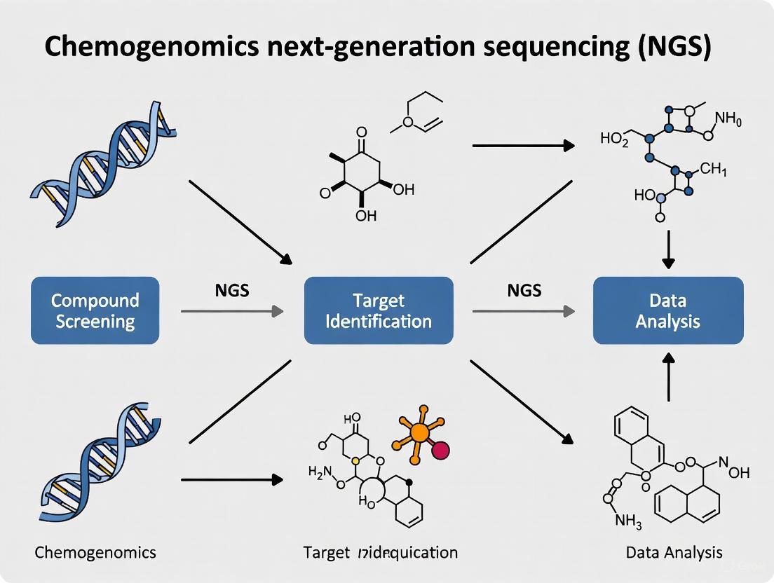
A Comprehensive Guide to Planning a Successful Chemogenomics NGS Experiment in 2025
This guide provides researchers, scientists, and drug development professionals with a comprehensive framework for planning and executing a robust chemogenomics Next-Generation Sequencing (NGS) experiment.
A Comprehensive Guide to Planning a Successful Chemogenomics NGS Experiment in 2025
Abstract
This guide provides researchers, scientists, and drug development professionals with a comprehensive framework for planning and executing a robust chemogenomics Next-Generation Sequencing (NGS) experiment. Covering the journey from foundational principles to advanced validation, it explores the integration of chemical and genomic data to uncover drug-target interactions. The article delivers actionable insights into experimental design, tackles common troubleshooting scenarios, and outlines rigorous methods for data analysis and cross-study comparison, ultimately empowering the development of targeted therapies and the advancement of precision medicine.
Laying the Groundwork: Core Principles of Chemogenomics and NGS
Chemogenomics is a foundational discipline in modern drug discovery that operates at the intersection of chemical biology and functional genomics. This field employs systematic approaches to investigate the interactions between chemical compounds and biological systems on a genome-wide scale, with the ultimate goal of defining relationships between chemical structures and their effects on genomic function. The core premise of chemogenomics lies in its ability to bridge two vast domains: chemical space—the theoretical space representing all possible organic compounds—and genomic function—the complete set of functional elements within a genome.
Next-generation sequencing (NGS) has revolutionized chemogenomics by providing the technological means to comprehensively assess how small molecules modulate biological systems. Unlike traditional methods that examined compound effects on single targets, NGS-enabled chemogenomics allows for the unbiased, genome-wide monitoring of transcriptional responses, mutational consequences, and epigenetic modifications induced by chemical perturbations [1] [2]. This paradigm shift has transformed drug discovery from a target-centric approach to a systems-level investigation, enabling researchers to deconvolve complex mechanisms of action, identify novel therapeutic targets, and predict off-target effects with unprecedented resolution.
The integration of NGS technologies has positioned chemogenomics as an essential framework for addressing fundamental challenges in pharmaceutical research, including polypharmacology, drug resistance, and patient stratification. By quantitatively linking chemical properties to genomic responses, chemogenomics provides the conceptual and methodological foundation for precision medicine approaches that tailor therapeutic interventions to individual genetic profiles [3].
Foundational NGS Technologies for Chemogenomics
Core Sequencing Principles and Platform Selection
Next-generation sequencing (NGS) technologies operate on the principle of massive parallel sequencing, enabling the simultaneous analysis of millions to billions of DNA fragments in a single experiment [1]. This represents a fundamental shift from traditional Sanger sequencing, which processes only one DNA fragment per reaction. The key technological advancement lies in this parallelism, which has led to a dramatic reduction in both cost (approximately 96% decrease per genome) and time required for comprehensive genomic analysis [1].
The NGS workflow comprises three principal stages: (1) library preparation, where DNA or RNA is fragmented and platform-specific adapters are ligated; (2) sequencing, where millions of cluster-amplified fragments are sequenced simultaneously using sequencing-by-synthesis chemistry; and (3) data analysis, where raw signals are converted to sequence reads and interpreted through sophisticated bioinformatics pipelines [1]. For chemogenomics applications, the choice of NGS platform and methodology depends on the specific experimental questions being addressed.
Table 1: NGS Method Selection for Chemogenomics Applications
| Research Question | Recommended NGS Method | Key Information Gained | Typical Coverage |
|---|---|---|---|
| Mechanism of Action Studies | RNA-Seq | Genome-wide transcriptional changes, pathway modulation | 20-50 million reads/sample |
| Resistance Mutations | Whole Genome Sequencing (WGS) | Comprehensive variant identification across coding/non-coding regions | 30-50x |
| Epigenetic Modifications | ChIP-Seq | Transcription factor binding, histone modifications | Varies by target |
| Off-Target Effects | Whole Exome Sequencing (WES) | Coding region variants across entire exome | 100x |
| Cellular Heterogeneity | Single-Cell RNA-Seq | Transcriptional profiles at single-cell resolution | 50,000 reads/cell |
Essential Bioinformatics Tools
The interpretation of NGS data in chemogenomics relies on a sophisticated bioinformatics ecosystem comprising both open-source and commercial solutions. These tools transform raw sequencing data into biologically meaningful insights about compound-genome interactions.
Open-source platforms provide transparent, modifiable pipelines for specialized analyses. The DRAGEN-GATK pipeline offers best practices for germline and somatic variant discovery, crucial for identifying mutation patterns induced by chemical treatments [4]. Tools like Strelka2 enable accurate detection of single nucleotide variants and small indels in compound-treated versus control samples [4]. For analyzing repetitive elements often involved in drug response, ExpansionHunter provides specialized genotyping of repeat expansions, while SpliceAI utilizes deep learning to identify compound-induced alternative splicing events [4].
Commercial solutions such as Geneious Prime offer integrated environments that streamline NGS data analysis through user-friendly interfaces, while QIAGEN Digital Insights provides curated knowledge bases linking genetic variants to functional consequences [5] [6]. These platforms are particularly valuable in regulated environments where reproducibility and standardized workflows are essential.
For quantitative morphological phenotyping often correlated with genomic data in chemogenomics, R and Python packages enable sophisticated image analysis and data integration, facilitating the connection between compound-induced phenotypic changes and their genomic correlates [7].
Experimental Design and Workflows
Strategic Framework for Chemogenomics Studies
A well-designed chemogenomics experiment requires careful consideration of multiple factors to ensure biologically relevant and statistically robust results. The experimental framework begins with precise definition of the chemical and biological systems under investigation, including compound properties (concentration, solubility, stability), model systems (cell lines, organoids, in vivo models), and treatment conditions (duration, replicates) [3].
Central to this framework is the selection of appropriate controls, which typically include untreated controls, vehicle controls (to account for solvent effects), and reference compounds with known mechanisms of action. These controls are essential for distinguishing compound-specific effects from background biological variation. Experimental replication should be planned with statistical power in mind, with most chemogenomics studies requiring at least three biological replicates per condition to ensure reproducibility [8].
The timing of sample collection represents another critical consideration, as it should capture both primary responses (direct compound-target interactions) and secondary responses (downstream adaptive changes). For time-course experiments, sample collection at multiple time points (e.g., 2h, 8h, 24h) enables differentiation of immediate from delayed transcriptional responses [9].
Quality Control and Validation
Implementing rigorous quality control measures throughout the experimental workflow is essential for generating reliable chemogenomics data. The Next-Generation Sequencing Quality Initiative (NGS QI) provides comprehensive frameworks for establishing quality management systems in NGS workflows [8]. Key recommendations include the use of the hg38 genome build as reference, implementation of standardized file formats, strict version control for analytical pipelines, and verification of data integrity through file hashing [3].
For library preparation, quality assessment should include evaluation of nucleic acid integrity (RIN > 8 for RNA studies), fragment size distribution, adapter contamination, and library concentration. During sequencing, key performance indicators include cluster density, Q-score distributions (aiming for Q30 ≥ 80%), and base call quality across sequencing cycles [1] [8].
Bioinformatics quality control encompasses verification of sequencing depth and coverage uniformity, assessment of alignment metrics, and evaluation of sample identity through genetically inferred markers. Pipeline validation should utilize standard truth sets such as Genome in a Bottle (GIAB) for germline variant calling and SEQC2 for somatic variant calling, supplemented with in-house datasets for filtering recurrent artifacts [3].
Data Analysis Pipelines and Computational Methods
Bioinformatics Framework for Chemogenomics
The analysis of NGS data in chemogenomics follows a structured pipeline that transforms raw sequencing data into biological insights about compound-genome interactions. This multi-stage process requires specialized tools and computational approaches at each step to ensure accurate interpretation of results.
Primary analysis begins with converting raw sequencing signals (BCL files) to sequence reads (FASTQ format) with corresponding quality scores (Phred scores) [1]. This demultiplexing and base calling step includes initial quality assessment using tools like FastQC to identify potential issues with sequence quality, adapter contamination, or GC bias [9].
Secondary analysis involves aligning sequence reads to a reference genome (e.g., GRCh38) using aligners such as BWA or STAR, generating BAM files that map reads to their genomic positions [3] [9]. Variant calling then identifies differences between the sample and reference genome, employing specialized tools for different variant types: GATK or Strelka2 for single nucleotide variants (SNVs) and small insertions/deletions (indels); Paragraph for structural variants; and ExpansionHunter for repeat expansions [4]. The output is a VCF file containing all identified genetic variants.
Tertiary analysis represents the most critical stage for chemogenomics interpretation, where variants are annotated with functional information from databases such as dbSNP, gnomAD, and ClinVar [1]. This annotation facilitates prioritization of variants based on population frequency, functional impact (e.g., missense, nonsense), and known disease associations. Subsequent pathway analysis connects compound-induced genetic changes to biological processes, molecular functions, and cellular components, ultimately enabling mechanism of action prediction and target identification [3].
Advanced Analytical Approaches
Beyond standard variant analysis, chemogenomics leverages several specialized computational approaches to extract maximal biological insight from NGS data. Graph-based pipeline architectures provide flexible frameworks for integrating diverse analytical tools, automatically compiling specialized tool combinations based on specific analysis requirements [10]. This approach enhances both extensibility and maintainability of bioinformatics workflows, which is particularly valuable in the rapidly evolving chemogenomics landscape.
For cancer chemogenomics, additional analyses include assessment of microsatellite instability (MSI) to identify DNA mismatch repair defects, evaluation of tumor mutational burden (TMB) to predict immunotherapy response, and quantification of homologous recombination deficiency (HRD) to guide PARP inhibitor therapy [3]. These specialized analyses require customized computational methods and interpretation frameworks.
Artificial intelligence approaches are increasingly being integrated into chemogenomics pipelines, with deep learning models like PrimateAI helping classify the pathogenicity of missense mutations identified in compound-treated samples [4] [2]. Large language models trained on biological sequences show promise for generating novel protein designs and predicting compound-protein interactions, potentially expanding the scope of chemogenomics from discovery to design [2].
The Scientist's Toolkit: Essential Research Reagents and Materials
Successful implementation of chemogenomics studies requires access to specialized reagents, computational resources, and experimental materials. The following table comprehensively details the essential components of a chemogenomics research toolkit.
Table 2: Essential Research Reagent Solutions for Chemogenomics
| Category | Specific Item/Kit | Function in Chemogenomics Workflow |
|---|---|---|
| Nucleic Acid Extraction | High-quality DNA/RNA extraction kits | Isolation of intact genetic material from compound-treated cells/tissues |
| Library Preparation | Illumina Nextera, KAPA HyperPrep | Fragmentation, adapter ligation, and amplification for sequencing |
| Target Enrichment | Illumina TruSeq, IDT xGen | Hybrid-capture or amplicon-based targeting of specific genomic regions |
| Sequencing | Illumina NovaSeq, Ion Torrent S5 | High-throughput sequencing of prepared libraries |
| Quality Control | Agilent Bioanalyzer, Qubit Fluorometer | Assessment of nucleic acid quality, fragment size, and concentration |
| Bioinformatics Tools | GATK, Strelka2, DESeq2 | Variant calling, differential expression analysis |
| Reference Databases | gnomAD, dbSNP, ClinVar, OMIM | Annotation and interpretation of identified genetic variants |
| Cell Culture Models | Immortalized cell lines, primary cells, organoids | Biological systems for compound treatment and genomic analysis |
| Compound Libraries | FDA-approved drugs, targeted inhibitor collections | Chemical perturbations for genomic functional studies |
Chemogenomics represents a powerful integrative framework that systematically connects chemical space to genomic function, with next-generation sequencing technologies serving as the primary enabling methodology. This approach has transformed early drug discovery by providing comprehensive, unbiased insights into compound mechanisms of action, off-target effects, and resistance patterns. The structured workflows, analytical pipelines, and specialized tools outlined in this technical guide provide researchers with a robust foundation for designing and implementing chemogenomics studies that can accelerate therapeutic development and advance precision medicine initiatives.
As NGS technologies continue to evolve, with improvements in long-read sequencing, single-cell applications, and real-time data acquisition, the resolution and scope of chemogenomics will expand correspondingly [9]. The integration of artificial intelligence and machine learning approaches will further enhance our ability to extract meaningful patterns from complex chemogenomics datasets, potentially enabling predictive modeling of compound efficacy and toxicity based on genomic features [2]. Through the continued refinement of these interdisciplinary approaches, chemogenomics will remain at the forefront of efforts to rationally connect chemical structures to biological function, ultimately enabling more effective and targeted therapeutic interventions.
The evolution of Next-Generation Sequencing (NGS) has fundamentally transformed chemogenomics research, enabling the systematic screening of chemical compounds against genomic targets at an unprecedented scale. This technological progression from first-generation Sanger sequencing to today's massively parallel platforms has provided researchers with powerful tools to understand complex compound-genome interactions, accelerating drug discovery and therapeutic development. The ability to sequence millions of DNA fragments simultaneously has created new paradigms for target identification, mechanism of action studies, and toxicity profiling, making NGS an indispensable technology in modern pharmaceutical research. This technical guide examines the evolution of sequencing technologies, their current specifications, and their specific applications in chemogenomics research workflows, providing a framework for selecting appropriate platforms for drug discovery applications.
Historical Progression of Sequencing Technologies
The journey of DNA sequencing spans nearly five decades, marked by three distinct generations of technological innovation that have progressively increased throughput while dramatically reducing costs.
First-Generation Sequencing: The Sanger Era
The sequencing revolution began in 1977 with Frederick Sanger's chain-termination method, a breakthrough that first made reading DNA possible [11]. This approach, also known as dideoxy sequencing, utilizes modified nucleotides (dideoxynucleoside triphosphates or ddNTPs) that terminate DNA strand elongation when incorporated by DNA polymerase [12] [13]. The resulting DNA fragments of varying lengths are separated by capillary electrophoresis, with fluorescent detection identifying the terminal base at each position [13]. Sanger sequencing produces long, accurate reads (500-1000 bases) with exceptionally high per-base accuracy (Q50, or 99.999%) [13]. This method served as the cornerstone of genomics for nearly three decades and was used to complete the Human Genome Project in 2003, though this endeavor required 13 years and approximately $3 billion [11] [14]. Despite its accuracy, the fundamental limitation of Sanger sequencing is its linear, one-sequence-at-a-time approach, which restricts throughput and maintains high costs per base [13].
Second-Generation Sequencing: The NGS Revolution
The mid-2000s marked a paradigm shift with the introduction of massively parallel sequencing platforms, collectively termed Next-Generation Sequencing [11]. The first commercial NGS system, Roche/454, launched in 2005 and utilized pyrosequencing technology [12]. This was quickly followed by Illumina's Sequencing-by-Synthesis (SBS) platform in 2006-2007 and Applied Biosystems' SOLiD system [11]. These second-generation technologies shared a common principle: parallel sequencing of millions of DNA fragments immobilized on surfaces or beads, delivering a massive increase in throughput while reducing costs from approximately $10,000 per megabase to mere cents [11]. This "massively parallel" approach transformed genetics into a high-speed, industrial operation, making large-scale genomic projects financially and practically feasible for the first time [14]. Illumina's SBS technology eventually came to dominate the market, at times holding approximately 80% market share due to its accuracy and throughput [11].
Third-Generation Sequencing: Long-Read Technologies
The 2010s witnessed the emergence of third-generation sequencing platforms that addressed a critical limitation of second-generation NGS: short read lengths [11]. Pacific Biosciences (PacBio) pioneered this transition in 2011 with their Single Molecule Real-Time (SMRT) sequencing, which observes individual DNA polymerase molecules in real time as they incorporate fluorescent nucleotides [11]. Oxford Nanopore Technologies (ONT) introduced an alternative approach using protein nanopores that detect electrical signal changes as DNA strands pass through [11]. These technologies produce much longer reads (thousands to tens of thousands of bases), enabling resolution of complex genomic regions, structural variant detection, and full-length isoform sequencing [11]. While initial error rates were higher than short-read NGS, these have improved significantly through technological refinements - PacBio's HiFi reads now achieve >99.9% accuracy, while ONT's newer chemistries reach ~99% single-read accuracy [11].
Figure 1: Evolution of DNA Sequencing Technologies from Sanger to Modern Platforms
Comparative Analysis of Modern NGS Platforms
Key Platform Specifications and Performance Metrics
The contemporary NGS landscape features diverse platforms with distinct performance characteristics, enabling researchers to select instruments optimized for specific applications and scales.
Table 1: Comparative Specifications of Major NGS Platforms (2025)
| Platform | Technology | Read Length | Throughput per Run | Accuracy | Key Applications in Chemogenomics |
|---|---|---|---|---|---|
| Illumina NovaSeq X | Sequencing-by-Synthesis (SBS) | 50-300 bp (short) | Up to 16 Tb [11] | >99.9% [14] | Large-scale variant screening, transcriptomic profiling, population studies |
| Illumina NextSeq 1000/2000 | Sequencing-by-Synthesis (SBS) | 50-300 bp (short) | 10-360 Gb [15] | >99.9% [15] | Targeted gene panels, small whole-genome sequencing, RNA-seq |
| PacBio Revio | HiFi SMRT (long-read) | 10-25 kb [11] | 180-360 Gb [11] | >99.9% (HiFi) [11] | Structural variant detection, haplotype phasing, complex region analysis |
| Oxford Nanopore | Nanopore sensing | Up to 4+ Mb (ultra-long) [16] | Up to 100s of Gb (PromethION) | ~99% (simplex) [11] | Real-time sequencing, direct RNA sequencing, epigenetic modification detection |
| Element Biosciences AVITI | avidity sequencing | 75-300 bp (short) | 10 Gb - 1.5 Tb | Q40+ [17] | Multiplexed screening, single-cell analysis, spatial applications |
| Ultima Genomics UG 100 | Sequencing-by-binding | 50-300 bp (short) | Up to 10-12B reads [17] | High | Large-scale population studies, high-throughput compound screening |
Technical Differentiation: Short-Read vs. Long-Read Technologies
The choice between short-read and long-read technologies represents a fundamental strategic decision in experimental design, with each approach offering distinct advantages for chemogenomics applications.
Short-Read Platforms (Illumina, Element, Ultima): Short-read sequencing excels at detecting single nucleotide variants (SNVs) and small indels with high accuracy and cost-effectiveness [14]. The massive throughput of modern short-read platforms makes them ideal for applications requiring deep sequencing, such as identifying rare mutations in heterogeneous cell populations after compound treatment or conducting genome-wide association studies (GWAS) to identify genetic determinants of drug response [14]. The main limitation of short reads is their difficulty in resolving complex genomic regions, including repetitive elements, structural variants, and highly homologous gene families [14].
Long-Read Platforms (PacBio, Oxford Nanopore): Long-read technologies overcome the limitations of short reads by spanning complex genomic regions in single contiguous sequences [11]. This capability is particularly valuable in chemogenomics for characterizing structural variants induced by genotoxic compounds, resolving complex rearrangements in cancer models, and phasing haplotypes to understand compound metabolism differences [11]. Recent accuracy improvements, particularly PacBio's HiFi reads and ONT's duplex sequencing, have made long-read technologies suitable for variant detection applications previously reserved for short-read platforms [11]. Additionally, Oxford Nanopore's direct RNA sequencing and real-time capabilities offer unique opportunities for studying transcriptional responses to compounds without reverse transcription or PCR amplification biases [11].
Table 2: NGS Platform Selection Guide for Chemogenomics Applications
| Research Application | Recommended Platform Type | Key Technical Considerations | Optimal Coverage/Depth |
|---|---|---|---|
| Targeted Gene Panels | Short-read (Illumina NextSeq, Element AVITI) | High multiplexing capability, cost-effectiveness for focused studies | 500-1000x for rare variant detection |
| Whole Genome Sequencing | Short-read (Illumina NovaSeq) for large cohorts; Long-read for complex regions | Balance between breadth and resolution of structural variants | 30-60x for human genomes |
| Transcriptomics (Bulk RNA-seq) | Short-read (Illumina NextSeq/NovaSeq) | Accurate quantification across dynamic range | 20-50 million reads/sample |
| Single-Cell RNA-seq | Short-read (Illumina NextSeq 1000/2000) | High sensitivity for low-input samples | 50,000-100,000 reads/cell |
| Epigenetics (Methylation) | Long-read (PacBio, Oxford Nanopore) | Single-molecule resolution of epigenetic modifications | 30x for comprehensive profiling |
| Structural Variant Detection | Long-read (PacBio Revio, Oxford Nanopore) | Spanning complex rearrangements with long reads | 20-30x for variant discovery |
NGS Workflow and Experimental Design for Chemogenomics
Core NGS Workflow Components
The NGS workflow consists of three fundamental stages that convert biological samples into interpretable genetic data, each requiring careful optimization for chemogenomics applications.
Figure 2: Core NGS Experimental Workflow with Key Decision Points
Library Preparation: The initial stage involves converting input DNA or RNA into sequencing-ready libraries through fragmentation, size selection, and adapter ligation [18]. For chemogenomics applications, specific considerations include preserving strand specificity in RNA-seq to determine transcript directionality, implementing unique molecular identifiers (UMIs) to control for PCR duplicates in rare variant detection, and selecting appropriate fragmentation methods to avoid bias in chromatin accessibility studies [15].
Sequencing and Imaging: This stage involves the actual determination of nucleotide sequences using platform-specific biochemistry and detection methods [18]. For Illumina platforms, this utilizes sequencing-by-synthesis with reversible dye-terminators, while Pacific Biosciences employs real-time observation of polymerase activity, and Oxford Nanopore measures electrical signal changes as DNA passes through protein nanopores [11] [16]. Key parameters to optimize include read length, read configuration (single-end vs. paired-end), and sequencing depth appropriate for the specific chemogenomics application [15].
Data Analysis: The final stage transforms raw sequencing data into biological insights through computational pipelines [18]. This includes quality control, read alignment to reference genomes, variant identification, and functional annotation [19]. For chemogenomics, specialized analyses include differential expression testing for compound-treated vs. control samples, variant allele frequency calculations in resistance studies, and pathway enrichment analysis to identify biological processes affected by compound treatment [19].
Essential Research Reagent Solutions
Table 3: Essential Research Reagents for NGS Workflows in Chemogenomics
| Reagent Category | Specific Examples | Function in NGS Workflow | Chemogenomics Considerations |
|---|---|---|---|
| Library Prep Kits | Illumina DNA Prep, KAPA HyperPrep, Nextera XT | Fragment DNA/RNA, add adapters, amplify libraries | Compatibility with low-input samples from limited cell numbers; preservation of strand information |
| Enzymes | DNA/RNA polymerases, ligases, transposases | Catalyze key biochemical reactions in library prep | High fidelity enzymes for accurate representation; tagmentase for chromatin accessibility mapping (ATAC-seq) |
| Barcodes/Adapters | Illumina TruSeq, IDT for Illumina, Dual Indexing | Unique sample identification for multiplexing | Sufficient complexity to avoid index hopping in large screens; unique molecular identifiers (UMIs) for quantitative applications |
| Target Enrichment | IDT xGen Panels, Twist Human Core Exome | Capture specific genomic regions of interest | Custom panels for drug target genes; comprehensive coverage of pharmacogenomic variants |
| Quality Control | Agilent Bioanalyzer, Qubit, qPCR | Quantify and qualify input DNA/RNA and final libraries | Sensitivity to detect degradation in clinical samples; accurate quantification for pooling libraries |
| Cleanup Beads | SPRIselect, AMPure XP | Size selection and purification | Tight size selection to remove adapter dimers; optimization for fragment retention |
Advanced Applications in Chemogenomics and Drug Discovery
Transcriptomic Profiling for Mechanism of Action Studies
NGS-based RNA sequencing has become a cornerstone for elucidating mechanisms of action (MOA) for novel compounds in drug discovery [19]. Bulk RNA-seq can quantify transcriptome-wide expression changes in response to compound treatment, revealing affected pathways and biological processes [15]. Single-cell RNA-seq (scRNA-seq) technologies extend this capability to resolve cellular heterogeneity in response to compounds, identifying rare resistant subpopulations or characterizing distinct cellular responses within complex tissues [19] [15]. Spatial transcriptomics further integrates morphological context with gene expression profiling, enabling researchers to map compound effects within tissue architecture - particularly valuable in oncology and toxicology studies [15].
Epigenetic Modifications in Compound Response
Epigenetic profiling using NGS provides critical insights into how compounds alter gene regulation without changing DNA sequence [15]. Key applications include ChIP-seq for mapping transcription factor binding and histone modifications, ATAC-seq for assessing chromatin accessibility, and bisulfite sequencing or enzymatic methylation detection for DNA methylation patterns [15]. In chemogenomics, these approaches can identify epigenetic mechanisms of drug resistance, characterize compounds that target epigenetic modifiers, and understand how chemical exposures induce persistent changes in gene regulation [16]. Long-read technologies from PacBio and Oxford Nanopore offer the unique advantage of detecting epigenetic modifications natively alongside sequence information [11].
Variant Detection for Pharmacogenomics and Resistance
Targeted sequencing panels focused on pharmacogenes enable comprehensive profiling of genetic variants that influence drug metabolism, efficacy, and adverse reactions [15]. These panels typically cover genes involved in drug absorption, distribution, metabolism, and excretion (ADME), as well as drug targets and immune genes relevant to therapeutic response [14]. In cancer research, deep sequencing of tumors pre- and post-treatment enables identification of resistance mechanisms and guidance of subsequent treatment strategies [15]. Liquid biopsy approaches using cell-free DNA sequencing provide a non-invasive method for monitoring treatment response and emerging resistance mutations [15] [14].
Future Directions and Emerging Technologies
The NGS landscape continues to evolve rapidly, with several emerging trends particularly relevant to chemogenomics research. The convergence of sequencing with artificial intelligence is creating new opportunities for predictive modeling of compound-genome interactions, with AI algorithms increasingly used for variant calling, expression quantification, and even predicting compound sensitivity based on genomic features [19]. Multi-omics integration represents another frontier, combining genomic, transcriptomic, epigenomic, and proteomic data to build comprehensive models of compound action [19]. Spatial multi-omics approaches are extending this integration to tissue context, simultaneously mapping multiple molecular modalities within morphological structures [17]. Emerging sequencing technologies, such as Roche's SBX (Sequencing by Expansion) technology announced in 2025, promise further reductions in cost and improvements in data quality [17]. Additionally, the continued maturation of long-read sequencing is gradually eliminating the traditional trade-offs between read length and accuracy, enabling more comprehensive genomic characterization in chemogenomics studies [11].
For researchers planning chemogenomics experiments, the expanding NGS toolkit offers unprecedented capability to connect chemical compounds with genomic consequences. Strategic platform selection based on experimental goals, sample types, and analytical requirements will continue to be essential for maximizing insights while efficiently utilizing resources. As sequencing technologies advance further toward the "$100 genome" and beyond, NGS will become increasingly integral to the entire drug discovery and development pipeline, from target identification through clinical application.
Next-generation sequencing (NGS) has revolutionized genomics research, providing unparalleled capabilities for analyzing DNA and RNA molecules in a high-throughput and cost-effective manner [16]. For researchers in chemogenomics and drug development, selecting the appropriate sequencing platform is a critical strategic decision that directly impacts experimental design, data quality, and research outcomes. NGS technology has evolved into three principal categories—benchtop, production-scale, and specialized systems—each with distinct performance characteristics, applications, and operational considerations [18]. This technical guide provides a structured framework for evaluating these platform categories, with a specific focus on their application within chemogenomics research, where understanding compound-genome interactions is paramount.
The evolution from first-generation sequencing to today's diverse NGS landscape represents one of the most transformative advancements in modern biology [20]. While the Human Genome Project required over a decade and nearly $3 billion using Sanger sequencing, modern NGS platforms can sequence entire human genomes in a single day for a fraction of the cost [18]. This dramatic improvement in speed and cost-efficiency has made large-scale genomic studies accessible to individual research laboratories, opening new frontiers in personalized medicine, drug discovery, and functional genomics [21].
Understanding NGS Platform Categories
Category 1: Benchtop Sequencers
Benchtop sequencers are characterized by their compact footprint, operational simplicity, and flexibility, making them ideal for individual laboratories focused on small to medium-scale projects [22] [18]. These systems represent the workhorse instrumentation for targeted studies, method development, and validation workflows commonly encountered in chemogenomics research. Their relatively lower initial investment and rapid turnaround times enable research groups to maintain sequencing capabilities in-house without requiring dedicated core facility support.
Key Applications in Chemogenomics:
- Targeted gene sequencing for validating compound-induced genetic changes
- Small whole-genome sequencing (microbes, viruses) for antimicrobial discovery
- Transcriptome sequencing for profiling gene expression responses to compounds
- 16S metagenomic sequencing for studying compound effects on microbiomes
- miRNA and small RNA analysis for epigenetic and regulatory studies
Table 1: Comparative Specifications of Major Benchtop Sequencing Platforms
| Specification | iSeq 100 | MiSeq | NextSeq 1000/2000 |
|---|---|---|---|
| Max Output | 1.2-1.6 Gb | 0.9-1.65 Gb | 120-540 Gb |
| Run Time | 9-19 hours | 4-55 hours | 11-48 hours |
| Max Reads | 4-25 million | 1-50 million | 400 million - 1.8 billion |
| Read Length | 1x36-2x150 bp | 1x36-2x300 bp | 2x150-2x300 bp |
| Key Chemogenomics Applications | Targeted sequencing, small RNA-seq | Amplicon sequencing, small genome sequencing | Single-cell profiling, exome sequencing, transcriptomics |
Category 2: Production-Scale Sequencers
Production-scale sequencers represent the high-throughput end of the NGS continuum, designed for large-scale genomic projects that generate terabytes of data per run [22] [18]. These systems are typically deployed in core facilities or dedicated sequencing centers supporting institutional or multi-investigator programs. For chemogenomics applications, these platforms enable comprehensive whole-genome sequencing of multiple cell lines, population-scale pharmacogenomic studies, and large-scale compound screening across diverse genetic backgrounds.
Key Applications in Chemogenomics:
- Large whole-genome sequencing (human, plant, animal) for population pharmacogenomics
- Exome and large panel sequencing for variant discovery in compound screening
- Single-cell profiling (scRNA-Seq, scDNA-Seq) for heterogeneous compound responses
- Metagenomic profiling for microbiome-therapeutic interaction studies
- Cell-free sequencing and liquid biopsy analysis for pharmacodynamic monitoring
Table 2: Comparative Specifications of Production-Scale Sequencing Platforms
| Specification | NovaSeq 6000 | NovaSeq X Plus | Ion GeneStudio S5 |
|---|---|---|---|
| Max Output | 3-6 Tb | 8-16 Tb | 15-50 Gb |
| Run Time | 13-44 hours | 17-48 hours | 2.5-24 hours |
| Max Reads | 10-20 billion | 26-52 billion | 30-130 million |
| Read Length | 2x50-2x250 bp | 2x150 bp | 200-600 bp |
| Key Chemogenomics Applications | Population sequencing, multi-omics studies | Biobank sequencing, large cohort studies | Targeted resequencing, rapid screening |
Category 3: Specialized Sequencing Systems
Specialized sequencing systems address specific technical challenges that cannot be adequately resolved with conventional short-read platforms [18] [11]. These include long-read technologies that overcome limitations in complex region sequencing, structural variant detection, and haplotype phasing. For chemogenomics research, these platforms provide crucial insights into the structural genomic context of compound responses, epigenetic modifications, and direct RNA sequencing without reverse transcription artifacts.
Key Applications in Chemogenomics:
- De novo genome assembly for non-model organisms used in compound screening
- Full-length transcript sequencing for isoform-specific drug responses
- Epigenetic modification detection for compound-induced chromatin changes
- Complex structural variation analysis for pharmacogenetic traits
- Real-time sequencing for rapid diagnostic applications
Table 3: Comparative Specifications of Specialized Sequencing Platforms
| Platform | Technology | Read Length | Accuracy | Key Advantage |
|---|---|---|---|---|
| PacBio Revio | HiFi Circular Consensus Sequencing | 10-25 kb | >99.9% (Q30) | High accuracy long reads |
| Oxford Nanopore PromethION | Nanopore electrical sensing | 10-100+ kb | ~99% (Q20) with duplex | Ultra-long reads, real-time |
| PacBio Onso | Sequencing by binding | 100-200 bp | >99.9% (Q30) | High-accuracy short reads |
NGS Workflow and Experimental Methodology
The fundamental NGS workflow comprises three major stages: template preparation, sequencing and imaging, and data analysis [18]. Understanding these steps is essential for designing robust chemogenomics experiments and properly interpreting resulting data.
Template Preparation and Library Construction
Library preparation converts biological samples into sequencing-compatible formats through a series of molecular biology steps. The quality of this initial process profoundly impacts final data integrity.
Detailed Protocol: Standard DNA Library Preparation
- Nucleic Acid Extraction: Isolate high-quality DNA/RNA using validated extraction methods. Quality control via fluorometry and fragment analysis is critical.
- Fragmentation: Shear DNA to appropriate fragment sizes (200-800 bp) using acoustic shearing, enzymatic fragmentation, or nebulization.
- End Repair and A-tailing: Convert fragmented DNA to blunt-ended fragments using polymerase and kinase activities, then add single A-overhangs.
- Adapter Ligation: Ligate platform-specific adapters containing sequencing priming sites and sample barcodes for multiplexing.
- Size Selection: Purify ligated fragments using magnetic bead-based cleanups or gel electrophoresis to optimize insert size distribution.
- Library Amplification: Enrich adapter-ligated fragments via limited-cycle PCR (typically 4-15 cycles) using high-fidelity polymerases.
- Final Quality Control: Quantify libraries via qPCR and assess size distribution using capillary electrophoresis.
Sequencing and Imaging
During sequencing, prepared libraries are loaded onto platforms where the actual base detection occurs through different biochemical principles [18] [16]:
- Sequencing by Synthesis (Illumina): Uses fluorescently-labeled reversible terminator nucleotides detected by imaging after each incorporation cycle [16].
- Semiconductor Sequencing (Ion Torrent): Detects hydrogen ions released during nucleotide incorporation via pH-sensitive semiconductors [18].
- Single Molecule Real-Time Sequencing (PacBio): Observes fluorescent nucleotide incorporation in real-time using zero-mode waveguides [11].
- Nanopore Sequencing (Oxford Nanopore): Measures electrical signal changes as DNA molecules pass through protein nanopores [11].
Data Analysis
The massive datasets generated by NGS require sophisticated bioinformatics pipelines for meaningful interpretation [18]:
- Base Calling: Convert raw signal data (images or electrical traces) into nucleotide sequences with associated quality scores.
- Quality Control: Assess read quality using tools like FastQC and perform adapter trimming and quality filtering.
- Alignment/Mapping: Map reads to reference genomes using aligners like BWA, Bowtie2, or minimap2 (for long reads).
- Variant Calling: Identify genetic variations using callers such as GATK, FreeBayes, or DeepVariant.
- Downstream Analysis: Perform application-specific analyses (differential expression, variant annotation, pathway enrichment).
Platform Selection Framework for Chemogenomics
Selecting the optimal NGS platform requires careful consideration of multiple technical and practical factors aligned with specific research objectives.
Application-Driven Selection Guide
Table 4: Platform Recommendations for Common Chemogenomics Applications
| Research Application | Recommended Platform Category | Optimal Read Length | Throughput Requirements | Key Considerations |
|---|---|---|---|---|
| Targeted Gene Panels | Benchtop | Short (75-150 bp) | Low-Moderate (1-50 Gb) | Cost-effectiveness, rapid turnaround |
| Whole Transcriptome | Benchtop/Production | Short (75-150 bp) | Moderate-High (10-100 Gb) | Detection of low-expression genes |
| Whole Genome Sequencing | Production-Scale | Short (150-250 bp) | Very High (100 Gb-3 Tb) | Coverage uniformity, variant detection |
| Single-Cell Multi-omics | Benchtop/Production | Short (50-150 bp) | Moderate (10-100 Gb) | Cell throughput, molecular recovery |
| Metagenomic Profiling | Production/Specialized | Long reads (>10 kb) | High (50-500 Gb) | Species resolution, assembly quality |
| Structural Variant Detection | Specialized | Long reads (>10 kb) | Moderate-High (20-200 Gb) | Spanning complex regions |
| Epigenetic Profiling | Benchtop/Production | Short (50-150 bp) | Low-Moderate (5-50 Gb) | Antibody specificity, resolution |
Technical Considerations for Platform Selection
- Throughput Requirements: Estimate required sequencing depth based on application (e.g., 30x for WGS, 10-50 million reads per sample for RNA-seq).
- Read Length Considerations: Short reads (50-300 bp) suffice for most applications, while long reads (>10 kb) excel in resolving complex genomic regions.
- Error Profiles: Different technologies exhibit distinct error patterns (substitution vs. indel errors) that impact downstream analysis.
- Multiplexing Capabilities: Consider sample batching options to optimize run efficiency and cost management.
- Operational Factors: Include instrument footprint, personnel expertise, and computational infrastructure requirements.
Essential Research Reagent Solutions for Chemogenomics NGS
Successful implementation of NGS workflows in chemogenomics research requires careful selection of reagents and consumables optimized for specific platforms and applications.
Table 5: Essential Research Reagent Solutions for Chemogenomics NGS
| Reagent Category | Specific Examples | Function | Application Notes |
|---|---|---|---|
| Library Prep Kits | Illumina DNA Prep, KAPA HyperPrep, NEBNext Ultra II | Fragment end repair, adapter ligation, library amplification | Select based on input material, application, and platform compatibility |
| Target Enrichment | Illumina Nextera Flex, Twist Target Enrichment, IDT xGen | Selective capture of genomic regions of interest | Critical for focused panels; consider coverage uniformity |
| Amplification Reagents | KAPA HiFi HotStart, Q5 High-Fidelity DNA Polymerase | Library amplification with minimal bias | High-fidelity enzymes essential for accurate variant detection |
| Quality Control | Agilent Bioanalyzer, Fragment Analyzer, Qubit assays | Assess nucleic acid quality, quantity, and fragment size | Critical for troubleshooting and optimizing success rates |
| Sample Barcoding | IDT for Illumina, TruSeq DNA/RNA UD Indexes | Sample multiplexing for cost-efficient sequencing | Enable sample pooling and demultiplexing in downstream analysis |
| Targeted RNA Sequencing | Illumina Stranded mRNA Prep, Takara SMARTer | RNA enrichment, cDNA synthesis, library construction | Maintain strand information for transcript orientation |
| Single-Cell Solutions | 10x Genomics Chromium, BD Rhapsody | Single-cell partitioning, barcoding, and library prep | Enable cellular heterogeneity studies in compound responses |
| Long-Read Technologies | PacBio SMRTbell, Oxford Nanopore Ligation | Library prep for long-read sequencing | Optimized for large fragment retention and structural variant detection |
Future Directions and Emerging Technologies
The NGS landscape continues to evolve with significant implications for chemogenomics research. Several emerging trends are positioned to further transform the field:
Enhanced Long-Read Technologies: Recent advancements in accuracy for both PacBio (HiFi) and Oxford Nanopore (duplex sequencing) technologies are making long-read sequencing increasingly viable for routine applications [11]. For chemogenomics, this enables comprehensive characterization of complex genomic regions impacted by compound treatments.
Multi-Omic Integration: New platforms and chemistries are enabling simultaneous capture of multiple molecular features from the same sample. The PacBio SPRQ chemistry, for example, combines DNA sequence and chromatin accessibility information from individual molecules [11].
Ultra-High Throughput Systems: Production-scale systems like the NovaSeq X series can output up to 16 terabases of data in a single run, dramatically reducing per-genome sequencing costs and enabling unprecedented scale in chemogenomics studies [21].
Portable Sequencing: The miniaturization of sequencing technology through platforms like Oxford Nanopore's MinION brings sequencing capabilities directly to the point of need, enabling real-time applications in field-deployable chemogenomics studies [11].
The global NGS market is projected to grow at a compound annual growth rate (CAGR) of 17.5% from 2025-2033, reaching $16.57 billion by 2033, reflecting the continued expansion and adoption of these technologies across research and clinical applications [21].
The strategic selection of NGS platform categories—benchtop, production-scale, and specialized systems—represents a critical decision point in designing effective chemogenomics research studies. Each category offers distinct advantages that align with specific research objectives, scale requirements, and technical considerations. Benchtop systems provide accessibility and flexibility for targeted studies, production-scale instruments deliver unprecedented throughput for population-level investigations, and specialized platforms overcome specific technical challenges associated with genomic complexity.
As sequencing technologies continue to evolve toward higher throughput, longer reads, and integrated multi-omic capabilities, chemogenomics researchers are positioned to extract increasingly sophisticated insights from their compound screening and mechanistic studies. By aligning platform capabilities with specific research questions through the framework presented in this guide, scientists can optimize their experimental approaches to advance drug discovery and development through genomic science.
Next-generation sequencing (NGS) is a high-throughput technology that enables millions of DNA fragments to be sequenced in parallel, revolutionizing genomic research and precision medicine [14] [23]. For researchers in chemogenomics, understanding the NGS workflow is fundamental to designing experiments that can uncover the complex interactions between chemical compounds and biological systems. This guide provides an in-depth technical overview of the core NGS steps, from nucleic acid extraction to data analysis, framed within the context of planning a robust chemogenomics experiment.
The Core NGS Workflow
The standard NGS workflow consists of four consecutive steps that transform a biological sample into interpretable genetic data. The following diagram illustrates this fundamental process and the key actions at each stage.
Step 1: Nucleic Acid Extraction
The NGS workflow begins with the isolation of genetic material. The required quantity and quality of the extracted nucleic acids are critical for success and depend on the specific NGS application [24] [18]. For chemogenomics studies, this could involve extracting DNA from cell lines or model organisms treated with chemical compounds to study genotoxic effects, or extracting RNA to profile gene expression changes in response to drug treatments.
After extraction, a quality control (QC) step is essential. UV spectrophotometry can assess purity, while fluorometric methods provide accurate nucleic acid quantitation [24]. High-quality input material is paramount, as any degradation or contamination can introduce biases that compromise downstream results.
Step 2: Library Preparation
Library preparation is the process of converting the extracted genomic DNA or cDNA sample into a format compatible with the sequencing instrument [24]. This complex process involves fragmenting the DNA, repairing the fragment ends, and ligating specialized adapter sequences [25] [18]. These adapters are critical as they enable the fragments to bind to the sequencer's flow cell and provide primer binding sites for amplification and sequencing [18].
For experiments involving multiple samples, unique molecular barcodes (indexes) are incorporated into the adapters, allowing samples to be pooled and sequenced simultaneously in a process known as multiplexing [23] [18]. This is particularly cost-effective for chemogenomics screens that test hundreds of chemical compounds. A key consideration is avoiding excessive PCR amplification during library prep, as this can reduce library complexity and introduce duplicate sequences that must be later removed bioinformatically [26] [25].
Step 3: Sequencing
During the sequencing step, the prepared library is loaded onto a sequencing platform, and the nucleotides of each fragment are read. The most common chemistry, used by Illumina platforms, is Sequencing by Synthesis (SBS) [24] [18]. This process involves the repeated addition of fluorescently labeled, reversible-terminator nucleotides. As each nucleotide is incorporated into the growing DNA strand, a camera captures its specific fluorescent signal, and the terminator is cleaved to allow the next cycle to begin [24] [18]. This cycle generates millions to billions of short reads simultaneously.
Two critical parameters to define for any experiment are read length (the length of each DNA fragment read by the sequencer) and depth or coverage (the average number of reads that align to a specific genomic base) [24]. The required coverage varies significantly by application, as detailed in the table below.
Step 4: Data Analysis and Interpretation
The raw data generated by the sequencer consists of short sequence reads and their corresponding quality scores [23]. Making sense of this data requires a multi-step bioinformatics pipeline, often starting with FASTQ files that store the sequence and its quality information for each read [26] [27].
A standard bioinformatics pipeline involves the following key steps, visualized in the diagram below:
- Quality Control and Adapter Trimming: Raw sequences are processed to remove low-quality bases and trim any remaining adapter sequences [26] [18].
- Alignment/Mapping: The cleaned reads are aligned to a reference genome to determine their genomic origin [26] [27].
- Post-Alignment Processing: This includes the removal of PCR duplicates to prevent false positive variant calls and base quality score recalibration to improve the accuracy of the base calls [26].
- Variant Calling: Specialized algorithms compare the aligned sequences to the reference genome to identify variations, such as single nucleotide polymorphisms (SNPs), insertions/deletions (indels), and larger structural variants [26] [27].
- Annotation and Interpretation: Identified variants are annotated with information from genomic databases, providing insights into their potential functional impact, population frequency, and clinical significance [26] [27]. For chemogenomics, this is where the link between a chemical treatment and specific genetic mutations or expression changes can be established.
Quantitative Considerations for Experimental Design
A well-designed NGS experiment requires careful planning of key parameters. The table below summarizes recommended sequencing coverages for common NGS applications relevant to chemogenomics research.
Table 1: Recommended Sequencing Coverage/Reads by NGS Application
| NGS Type | Application | Recommended Coverage (x) or Reads | Key Rationale |
|---|---|---|---|
| Whole Genome Sequencing (WGS) | Heterozygous SNVs [28] | 33x | Ensures high probability of detecting alleles present in half the cells. |
| Insertion/Deletion Mutations (INDELs) [28] | 60x | Higher depth needed to confidently align and call small insertions/deletions. | |
| Copy Number Variation (CNV) [28] | 1-8x | Lower depth can be sufficient for detecting large-scale copy number changes. | |
| Whole Exome Sequencing | Single Nucleotide Variants (SNVs) [28] | 100x | Compensates for uneven capture efficiency across exons while remaining cost-effective vs. WGS. |
| RNA Sequencing | Differential expression profiling [28] | 10-25 million reads | Provides sufficient data for robust statistical comparison of transcript levels between samples. |
| Alternative splicing, Allele specific expression [28] | 50-100 million reads | Higher read depth is required to resolve and quantify different transcript isoforms. |
The Researcher's Toolkit: Key Reagents and Materials
Successful execution of the NGS workflow depends on a suite of specialized reagents and materials.
Table 2: Essential Research Reagent Solutions for the NGS Workflow
| Item | Function |
|---|---|
| Nucleic Acid Extraction Kits | Isolate high-quality DNA/RNA from various sample types (e.g., tissue, cells, biofluids) [24]. |
| Library Preparation Kits | Contain enzymes and buffers for DNA fragmentation, end-repair, A-tailing, and adapter ligation [25] [28]. |
| Platform-Specific Flow Cells | The glass surface where library fragments bind and are amplified into clusters prior to sequencing [18]. |
| Sequencing Reagent Kits | Provide the nucleotides, enzymes, and buffers required for the sequencing-by-synthesis chemistry [24] [18]. |
| Multiplexing Barcodes/Adapters | Unique DNA sequences ligated to samples to allow pooling and subsequent computational deconvolution [18]. |
A thorough understanding of the NGS workflow—from nucleic acid extraction to data analysis—is a prerequisite for designing and executing a successful chemogenomics research project. Each step, governed by specific biochemical and computational principles, contributes to the quality and reliability of the final data. By carefully considering experimental goals, required coverage, and the appropriate bioinformatics pipeline, researchers can leverage the power of NGS to unravel the complex mechanisms of chemical-genetic interactions and accelerate drug discovery.
Chemogenomics represents a powerful, systematic approach in modern drug discovery that investigates the interaction between chemical compounds and biological systems on a genomic scale. The integration of Next-Generation Sequencing (NGS) technologies has fundamentally transformed this field, enabling researchers to decode complex molecular relationships at unprecedented speed and resolution. NGS provides high-throughput, cost-effective sequencing solutions that generate massive datasets characterizing the nucleotide-level information of DNA and RNA molecules, forming the essential data backbone for chemogenomic analyses [29]. This technical guide examines three critical applications—drug repositioning, target deconvolution, and mechanism of action (MoA) studies—within the framework of chemogenomics NGS experimental research, providing methodological details and practical protocols for implementation.
The convergence of artificial intelligence (AI) with NGS technologies has further accelerated drug discovery paradigms. AI-driven platforms can now compress traditional discovery timelines dramatically; for instance, some AI-designed drug candidates have reached Phase I trials in approximately two years, compared to the typical five-year timeline for conventional approaches [30]. More than 90% of small molecule discovery pipelines at leading pharmaceutical companies are now AI-assisted, demonstrating the fundamental shift toward computational-augmented methodologies [31]. This whitepaper provides researchers with the technical frameworks and experimental protocols necessary to leverage these advanced technologies in chemogenomics research, with a specific focus on practical implementation within drug development workflows.
Drug Repositioning via Chemogenomics
Conceptual Framework and Strategic Value
Drug repositioning (also referred to as drug repurposing) is a methodological strategy for identifying new therapeutic applications for existing drugs or drug candidates beyond their original medical indication. This approach offers significant advantages over traditional de novo drug discovery, including reduced development timelines, lower costs, and minimized risk profiles since the safety and pharmacokinetic properties of the compounds are already established [32]. The integration of chemogenomics data, particularly through NGS methodologies, has dramatically enhanced systematic drug repositioning efforts by providing comprehensive molecular profiles of both drugs and disease states.
The fundamental premise of drug repositioning through chemogenomics rests on analyzing the relationships between chemical structures, genomic features, and biological outcomes. By examining how drug-induced genomic signatures correlate with disease-associated genomic patterns, researchers can identify potential new therapeutic indications computationally before embarking on costly clinical validation [32]. This approach maximizes the therapeutic value of existing compounds and can rapidly address pressing medical needs, as demonstrated during the COVID-19 pandemic when computational models successfully predicted gene expression changes induced by novel chemicals for rapid therapeutic repurposing [33].
Key Methodological Approaches
Table 1: Computational Approaches for Drug Repositioning
| Method Category | Key Technologies | Primary Data Sources | Strengths |
|---|---|---|---|
| In Silico-Based Computational Approaches | Machine learning, deep learning, semantic inference | Chemical-genomic interaction databases, drug-target interaction maps | High efficiency, ability to screen thousands of compounds rapidly [32] |
| Activity-Based Experimental Approaches | High-throughput screening, phenotypic screening | Cell-based assays, transcriptomic profiles, proteomic data | Direct biological validation, captures complex system responses [32] |
| Target-Based Screening | Binding affinity prediction, molecular docking | Protein structures, binding site databases, structural genomics | Direct mechanism insight, rational design capabilities [32] |
| Knowledge-Graph Repurposing | Network analysis, graph machine learning | Biomedical literature, multi-omics data, clinical databases | Integrates diverse evidence types, discovers novel relationships [30] |
Experimental Protocol: NGS-Enhanced Drug Repositioning
Objective: Systematically identify novel therapeutic indications for approved drugs through transcriptomic signature matching.
Step 1: Sample Preparation and Sequencing
- Treat relevant cell lines with compounds of interest at multiple concentrations (typically spanning 3-5 logs) and time points (e.g., 6h, 24h, 48h)
- Include appropriate vehicle controls and replicate samples (minimum n=3 biological replicates)
- Extract total RNA using standardized kits (e.g., Qiagen RNeasy) with DNase treatment
- Prepare RNA-seq libraries using poly-A selection or ribosomal RNA depletion protocols
- Sequence libraries on appropriate NGS platform (Illumina NextSeq 500 or similar) to minimum depth of 30 million paired-end reads per sample [29]
Step 2: Bioinformatics Processing
- Perform quality control on raw sequencing data using FastQC or similar tool
- Align reads to reference genome (GRCh38 recommended) using splice-aware aligners (STAR or HISAT2)
- Generate gene-level count matrices using featureCounts or HTSeq-count
- Conduct differential expression analysis using DESeq2 or edgeR packages in R [29]
Step 3: Signature Generation and Matching
- Calculate differentially expressed genes for each treatment condition versus controls
- Generate gene signature profiles using rank-based methods (e.g., Connectivity Map approach)
- Compare drug-induced signatures against disease-associated gene expression profiles from public repositories (e.g., GEO, TCGA)
- Apply statistical frameworks (e.g., Kolmogorov-Smirnov tests) to identify signature reversals indicating therapeutic potential [32]
Step 4: Experimental Validation
- Prioritize top candidate drug-disease pairs based on statistical significance and biological plausibility
- Validate predictions in disease-relevant cellular models and animal systems
- Proceed to clinical evaluation for most promising repositioning opportunities
Diagram 1: Drug Repositioning Workflow via Transcriptomic Profiling
Target Deconvolution Strategies
Principles and Significance
Target deconvolution refers to the systematic process of identifying the direct molecular targets and associated mechanisms through which bioactive small molecules exert their phenotypic effects. This methodology is particularly crucial in phenotypic drug discovery approaches, where compounds are initially identified based on their ability to induce desired cellular changes without prior knowledge of their specific molecular targets [34]. The integration of NGS technologies with chemoproteomic approaches has significantly enhanced target deconvolution capabilities, enabling researchers to more rapidly and accurately elucidate mechanisms underlying promising phenotypic hits.
The strategic importance of target deconvolution lies in its ability to bridge the critical gap between initial phenotypic screening and subsequent rational drug optimization. By identifying a compound's direct molecular targets and off-target interactions, researchers can make informed decisions about candidate prioritization, guide structure-activity relationship (SAR) campaigns to improve selectivity, predict potential toxicity liabilities, and identify biomarkers for clinical development [34]. Furthermore, comprehensive target deconvolution can reveal novel biology by identifying previously unknown protein functions or signaling pathways relevant to disease processes.
Experimental Approaches for Target Identification
Table 2: Comparative Analysis of Target Deconvolution Methods
| Method | Principle | Resolution | Throughput | Key Limitations |
|---|---|---|---|---|
| Affinity-Based Pull-Down | Compound immobilization and target capture | Protein-level | Medium | Requires high-affinity probe, may miss transient interactions [34] |
| Photoaffinity Labeling (PAL) | Photoactivated covalent crosslinking to targets | Amino acid-level | Medium | Potential steric interference from photoprobes [34] |
| Activity-Based Protein Profiling (ABPP) | Detection of enzymatic activity changes | Functional residue-level | High | Limited to enzyme families with defined probes [34] |
| Stability-Based Profiling (CETSA, TPP) | Thermal stability shifts upon binding | Protein-level | High | Challenging for low-abundance proteins [34] |
| Genomic Approaches (CRISPR) | Gene essentiality/modification screens | Gene-level | High | Indirect identification, functional validation required [33] |
Experimental Protocol: Integrated NGS-Chemoproteomics for Target Deconvolution
Objective: Identify molecular targets of a phenotypic screening hit using affinity purification coupled with multi-omics validation.
Step 1: Probe Design and Validation
- Design chemical probes by incorporating biotin or other affinity handles without disrupting bioactivity
- Validate probe functionality through phenotypic assays comparing original compound and probe
- Determine appropriate probe concentration ranges through dose-response experiments
- Prepare control probes with scrambled or inactive chemistry for specificity assessment [34]
Step 2: Target Capture and Preparation
- Incubate functionalized probes with cell lysates or living cells (typically 1-10 μM concentration)
- For photoaffinity labeling approaches: irradiate samples with UV light (typically 300-365 nm) to induce crosslinking
- Capture probe-bound complexes using streptavidin beads or appropriate affinity matrix
- Perform rigorous wash steps to remove non-specific binders (typically high-salt and detergent washes)
- On-bead digest captured proteins using trypsin or Lys-C for mass spectrometry analysis [34]
Step 3: Multi-Omics Target Identification
- Analyze captured proteins by liquid chromatography-tandem mass spectrometry (LC-MS/MS)
- Process proteomics data using standard search engines (MaxQuant, Proteome Discoverer)
- In parallel, conduct CRISPR-based genetic screens to identify genes whose manipulation modifies compound sensitivity
- Integrate proteomic and genetic datasets to generate high-confidence target candidates [33]
Step 4: Functional Validation
- Apply orthogonal binding assays (SPR, ITC) to confirm direct compound-target interactions
- Use cellular assays (knockdown, knockout, or dominant-negative approaches) to validate functional relevance
- Employ structural biology approaches (crystallography, Cryo-EM) where feasible to characterize binding mode
Diagram 2: Integrated Target Deconvolution Workflow
Mechanism of Action (MoA) Studies
Comprehensive MoA Elucidation Frameworks
Mechanism of Action (MoA) studies aim to comprehensively characterize the full sequence of biological events through which a therapeutic compound produces its pharmacological effects, from initial target engagement through downstream pathway modulation and ultimate phenotypic outcome. While target deconvolution identifies the primary molecular interactions, MoA studies encompass the broader functional consequences across multiple biological layers, including gene expression, protein signaling, metabolic reprogramming, and cellular phenotype alterations [33]. The integration of multi-omics NGS technologies has revolutionized MoA elucidation by enabling systematic, unbiased profiling of drug effects across these diverse molecular dimensions.
Modern MoA frameworks leverage advanced AI platforms to integrate heterogeneous data types and extract biologically meaningful patterns. For instance, sophisticated phenotypic screening platforms like PhenAID combine high-content imaging data with transcriptomic and proteomic profiling to identify characteristic morphological and molecular signatures associated with specific mechanisms of action [33]. These integrated approaches can distinguish between subtly different MoA classes even within the same target pathway, providing crucial insights for drug optimization and combination therapy design.
Multi-Omics Approaches for MoA Characterization
Table 3: NGS Technologies for MoA Elucidation
| Omics Layer | NGS Application | MoA Insights Provided | Experimental Considerations |
|---|---|---|---|
| Genomics | Whole genome sequencing (WGS), targeted panels | Identification of genetic biomarkers of response/resistance | Minimum 30x coverage for WGS; tumor-normal pairs for somatic calling [29] |
| Transcriptomics | RNA-seq, single-cell RNA-seq | Pathway activation signatures, cell state transitions | 20-50 million reads per sample; strand-specific protocols recommended [29] |
| Epigenomics | ChIP-seq, ATAC-seq, methylation sequencing | Regulatory element usage, chromatin accessibility changes | Antibody quality critical for ChIP-seq; cell number requirements for ATAC-seq [33] |
| Functional Genomics | CRISPR screens, Perturb-seq | Gene essentiality, genetic interactions | Adeplicate library coverage (500x); appropriate controls for screen normalization [33] |
Experimental Protocol: Multi-Modal MoA Elucidation
Objective: Comprehensively characterize the mechanism of action for a compound with known efficacy but unknown downstream consequences.
Step 1: Experimental Design and Sample Preparation
- Treat disease-relevant models (cell lines, organoids, or animal models) with compound across multiple concentrations and time points
- Include appropriate controls (vehicle, tool compounds with known MoA)
- Process samples for parallel multi-omics analyses:
- RNA sequencing (bulk and single-cell)
- Assay for Transposase-Accessible Chromatin (ATAC-seq)
- Proteomic and phosphoproteomic profiling (mass spectrometry)
- High-content imaging (Cell Painting or similar) [33]
Step 2: NGS Library Preparation and Sequencing
- For RNA-seq: Prepare libraries using poly-A selection with unique dual indexes for multiplexing
- For ATAC-seq: Follow optimized protocols for tagmentation and library amplification
- For CRISPR screens: Prepare sequencing libraries from genomic DNA with sufficient coverage
- Sequence libraries on appropriate platforms (Illumina NextSeq or NovaSeq) with recommended read depths [29]
Step 3: Bioinformatics Analysis and Integration
- Process each data type through standardized pipelines:
- RNA-seq: Alignment, quantification, differential expression, pathway analysis
- ATAC-seq: Peak calling, differential accessibility, motif analysis
- CRISPR screens: Read count normalization, gene ranking, hit identification
- Apply multi-omics integration algorithms (MOFA+, mixOmics) to identify coordinated changes
- Use AI-based platforms to compare molecular signatures with reference databases of known MoAs [33]
Step 4: Systems-Level MoA Modeling
- Construct network models integrating all significantly altered molecular entities
- Prioritize key pathways and nodes based on statistical significance and biological coherence
- Generate testable hypotheses about causal relationships in the MoA
- Design and execute functional experiments to validate predicted network relationships
Diagram 3: Multi-Omics MoA Elucidation Workflow
The Scientist's Toolkit: Essential Research Reagents and Platforms
Table 4: Key Research Reagent Solutions for Chemogenomics Studies
| Reagent/Platform | Provider Examples | Primary Function | Application Context |
|---|---|---|---|
| SureSelect Target Enrichment | Agilent Technologies | Hybridization-based capture of genomic regions of interest | Targeted sequencing for focused investigations [29] |
| PhenAID Platform | Ardigen | AI-powered analysis of high-content phenotypic screening data | MoA studies, phenotypic screening [33] |
| TargetScout Service | Momentum Bio | Affinity-based pull-down and target identification | Target deconvolution for phenotypic hits [34] |
| PhotoTargetScout | OmicScouts | Photoaffinity labeling for target identification | Target deconvolution, especially membrane proteins [34] |
| CysScout Platform | Momentum Bio | Proteome-wide profiling of reactive cysteine residues | Covalent ligand discovery, target deconvolution [34] |
| SideScout Service | Momentum Bio | Label-free target deconvolution via stability shifts | Native condition target identification [34] |
| eProtein Discovery System | Nuclera | Automated protein expression and purification | Target production for functional studies [35] |
| MO:BOT Platform | mo:re | Automated 3D cell culture and organoid handling | Biologically relevant assay systems [35] |
| MapDiff Framework | AstraZeneca/University of Sheffield | Inverse protein folding for biologic drug design | Protein-based therapeutic engineering [31] |
| Edge Set Attention | AstraZeneca/University of Cambridge | Graph-based molecular property prediction | Small molecule optimization [31] |
The integration of chemogenomics with NGS technologies has created a powerful paradigm for modern drug discovery, enabling systematic approaches to drug repositioning, target deconvolution, and mechanism of action studies. The methodologies outlined in this technical guide provide researchers with comprehensive frameworks for designing and implementing robust experiments in these critical application areas. As AI and automation continue to transform the pharmaceutical landscape—with over 75 AI-derived molecules reaching clinical stages by the end of 2024—the strategic importance of these chemogenomics applications will only intensify [30].
Looking forward, several emerging technologies promise to further enhance these approaches. The ongoing development of more sophisticated multi-omics integration platforms, combined with advanced AI architectures like graph neural networks and foundation models, will enable even more comprehensive and predictive compound characterization [31]. Additionally, the increasing availability of automated benchside technologies—from liquid handlers to automated protein expression systems—will help bridge the gap between computational predictions and experimental validation, creating more efficient closed-loop design-make-test-analyze cycles [35]. By adopting the structured protocols and methodologies presented in this whitepaper, research scientists can position themselves at the forefront of this rapidly evolving field, leveraging chemogenomics NGS approaches to accelerate the development of novel therapeutic interventions.
Designing Your Experiment: Methodologies and Practical Applications
Next-generation sequencing (NGS) has revolutionized genomic research, offering multiple approaches for analyzing genetic material. For chemogenomics research—which explores the complex interactions between chemical compounds and biological systems—selecting the appropriate NGS method is critical for generating meaningful data. This technical guide provides an in-depth comparison of three core NGS approaches: whole genome sequencing, targeted sequencing, and RNA sequencing. We examine the technical specifications, experimental considerations, and applications of each method within the context of chemogenomics research, enabling scientists and drug development professionals to make informed decisions for their experimental designs.
Chemogenomics employs systematic approaches to discover how small molecules affect biological systems through their interactions with macromolecular targets. NGS technologies provide powerful tools for understanding these interactions at the genetic and transcriptomic levels, facilitating drug target identification, mechanism of action studies, and toxicology assessments [36]. The fundamental advantage of NGS over traditional sequencing methods lies in its massive parallelism, enabling simultaneous sequencing of millions to billions of DNA fragments [1] [16]. This high-throughput capability has led to a dramatic 96% decrease in cost-per-genome while exponentially increasing sequencing speed [1].
For chemogenomics research, NGS applications span from identifying novel drug targets to understanding off-target effects of compounds and stratifying patient populations for clinical trials [36]. The choice of NGS approach directly impacts the scope, resolution, and cost of experiments, making selection critical for generating biologically relevant and statistically powerful data.
Comparative Analysis of NGS Approaches
Technical Specifications and Applications
The table below summarizes the key characteristics, strengths, and limitations of the three primary NGS approaches relevant to chemogenomics research:
Table 1: Comparison of NGS Approaches for Chemogenomics Research
| Parameter | Whole Genome Sequencing (WGS) | Targeted Sequencing | RNA Sequencing |
|---|---|---|---|
| Sequencing Target | Complete genome including coding, non-coding, and regulatory regions [1] | Pre-defined set of genes or regions of interest [36] | Complete transcriptome or targeted RNA transcripts [36] [37] |
| Primary Applications in Chemogenomics | Novel target discovery, comprehensive variant profiling, structural variant identification [15] | Target validation, pathway-focused screening, clinical biomarker development [36] | Mechanism of action studies, biomarker discovery, toxicology assessment, transcriptomic point of departure (tPOD) calculation [36] [38] |
| Key Advantages | Unbiased discovery, detection of novel variants, comprehensive coverage [1] | High sensitivity for target genes, cost-effective, suitable for high-throughput screening [36] [38] | Functional insight into genomic variants, detects splicing events and expression changes [39] |
| Key Limitations | Higher cost, complex data analysis, may miss low-abundance transcripts [36] | Limited to pre-defined targets, blind to novel discoveries outside panel [36] | Does not directly sequence DNA variants, requires specialized library prep [39] |
| Typical Coverage Depth | 30-50x for human genomes [1] | 500-1000x for enhanced sensitivity [36] | 10-50 million reads per sample for bulk RNA-seq [39] |
| Best Suited For | Discovery-phase research, identifying novel genetic associations [36] | Validation studies, clinical applications, large-scale compound screening [36] | Functional interpretation of variants, understanding compound-induced transcriptional changes [39] |
Decision Framework for NGS Approach Selection
The following workflow diagram outlines a systematic approach for selecting the most appropriate NGS method based on research objectives and practical considerations:
Diagram 1: NGS Approach Selection Workflow
Experimental Protocols and Methodologies
Core NGS Workflow
While each NGS approach has unique considerations, they share a common foundational workflow:
Table 2: Core NGS Workflow Stages
| Stage | Key Steps | Considerations for Chemogenomics |
|---|---|---|
| Library Preparation | Fragmentation, adapter ligation, amplification, target enrichment (for targeted approaches) [1] | Compound treatment conditions should be optimized before RNA/DNA extraction to ensure relevant biological responses |
| Sequencing | Cluster generation, sequencing-by-synthesis, base calling [1] [16] | Sequencing depth must be determined based on application; targeted approaches require less depth per sample |
| Data Analysis | Primary analysis (base calling), secondary analysis (alignment, variant calling), tertiary analysis (annotation, interpretation) [1] | Appropriate controls are essential for distinguishing compound-specific effects from background variability |
Protocol for Targeted Gene Expression Profiling in Compound Screening
Targeted RNA sequencing approaches are particularly valuable for medium-to-high throughput compound screening in chemogenomics. The following protocol outlines a standardized workflow for targeted transcriptomic analysis:
Sample Preparation and Library Generation
- Cell Treatment and RNA Extraction: Plate cells in appropriate multi-well format and treat with compound libraries for predetermined exposure times. Extract total RNA using silica membrane-based columns, ensuring RNA Integrity Number (RIN) > 8.0 for optimal results [36].
- Library Preparation with Targeted Panels: Convert RNA to cDNA using reverse transcriptase. Amplify target genes using predesigned primer pairs for relevant pathways (e.g., stress response, apoptosis, metabolic activation). For more comprehensive targeted approaches, use hybrid-capture protocols with biotinylated RNA probes to enrich for genes of interest [1].
- Sequencing: Pool barcoded libraries in equimolar ratios and sequence on appropriate platform (e.g., Illumina NextSeq 1000/2000) to achieve minimum coverage of 5 million reads per sample and >500x coverage for target genes [36] [15].
Data Analysis and Interpretation
- Alignment and Quantification: Align sequencing reads to reference genome using Spliced Transcripts Alignment to a Reference (STAR) aligner. Generate count matrices for target genes using featureCounts or similar tools [39].
- Differential Expression Analysis: Normalize count data using DESeq2 median-of-ratios method. Identify significantly differentially expressed genes using appropriate statistical models with false discovery rate (FDR) correction [39].
- Pathway Enrichment and Signature Mapping: Conduct gene set enrichment analysis (GSEA) to identify affected pathways. Compare expression signatures to reference databases (e.g., LINCS L1000) to identify compounds with similar mechanisms of action [36].
Protocol for Whole Transcriptome Analysis for Mechanism of Action Studies
For comprehensive mechanism of action studies, whole transcriptome sequencing provides unbiased discovery capability:
Sample Preparation and Sequencing
- RNA Extraction and Quality Control: Extract RNA from compound-treated and vehicle-control cells using guanidinium thiocyanate-phenol-chloroform extraction. Assess RNA quality using Bioanalyzer (RIN > 8.0) and quantify using fluorometric methods [39].
- Library Preparation: Deplete ribosomal RNA using ribodepletion protocols to retain both coding and non-coding RNA species. Use unique molecular identifiers (UMIs) to correct for amplification bias and improve quantification accuracy [37]. Prepare stranded RNA-seq libraries using kits such as Illumina's TruSeq Stranded Total RNA Library Prep Kit.
- Sequencing Parameters: Sequence libraries on appropriate platform (e.g., Illumina NovaSeq X) to achieve minimum of 20-50 million reads per sample with 150 bp paired-end reads for comprehensive transcriptome coverage [37].
Bioinformatic Analysis
- Transcriptome Alignment and Quantification: Align reads to reference genome using STAR aligner with parameters optimized for splice junction discovery. Quantify gene-level counts using featureCounts and transcript-level expression using alignment-free methods like Salmon [39].
- Alternative Splicing and Novel Transcript Discovery: Use specialized tools (e.g., rMATS, StringTie) to identify compound-induced alternative splicing events and novel transcript isoforms [39] [37].
- Multi-optic Integration: Integrate transcriptomic data with complementary data types (e.g., proteomics, epigenomics) to build comprehensive models of compound mechanism of action [19].
The following diagram illustrates the complete experimental workflow for chemogenomics NGS studies:
Diagram 2: Chemogenomics NGS Experimental Workflow
Essential Research Reagent Solutions
Successful implementation of NGS approaches in chemogenomics requires careful selection of reagents and materials. The following table outlines essential research reagent solutions:
Table 3: Essential Research Reagents for Chemogenomics NGS
| Reagent Category | Specific Examples | Function in NGS Workflow |
|---|---|---|
| RNA Stabilization Reagents | RNAlater, TRIzol, Qiazol | Preserve RNA integrity immediately after compound treatment by inhibiting RNases [39] |
| Library Preparation Kits | Illumina TruSeq Stranded Total RNA, BioSpyder TempO-Seq, QIAseq Targeted RNA Panels | Convert RNA to sequencing-ready libraries with minimal bias [36] [38] |
| Target Enrichment Systems | IDT xGen Lockdown Probes, Twist Human Core Exome, BioSpyder S1500+ sentinel gene set | Enrich for specific genes or regions of interest in targeted approaches [36] [38] |
| Quality Control Assays | Agilent Bioanalyzer RNA Nano, Qubit dsDNA HS Assay, KAPA Library Quantification | Assess RNA/DNA quality and quantity, and accurately quantify final libraries [39] |
| NGS Multiplexing Reagents | IDT for Illumina UD Indexes, TruSeq DNA/RNA UD Indexes | Enable sample multiplexing by adding unique barcodes to each library [1] |
Selecting the appropriate NGS approach is fundamental to successful chemogenomics research. Whole genome sequencing offers comprehensive discovery power for identifying novel drug targets and genetic variants. Targeted sequencing provides cost-effective, sensitive analysis for validation studies and high-throughput compound screening. RNA sequencing delivers functional insights into compound mechanisms and transcriptional responses. The choice between these approaches should be guided by research objectives, scale of study, and available resources. As NGS technologies continue to evolve, integrating multiple approaches through multi-omics strategies will further enhance our understanding of compound-biology interactions, accelerating drug discovery and development.
Chemogenomic profiling represents a powerful framework for understanding the genome-wide cellular response to small molecules, directly linking drug discovery to target identification [40]. At its core, this approach uses systematic genetic perturbations to unravel mechanisms of drug action (MoA) in a direct and unbiased manner [40]. The budding yeast Saccharomyces cerevisiae serves as a fundamental model eukaryotic organism for these studies due to its genetic tractability and conservation of essential eukaryotic cellular biochemistry [41]. Chemogenomic screens provide two primary advantages: they enable direct identification of drug target candidates and reveal genes required for drug resistance, offering a comprehensive view of how cells respond to chemical perturbation [40].
Two complementary assay formats form the foundation of yeast chemogenomics: Haploinsufficiency Profiling (HIP) and Homozygous Profiling (HOP). HIP exploits drug-induced haploinsufficiency, a phenomenon where heterozygous deletion strains for essential genes exhibit heightened sensitivity when the deleted gene encodes the drug target or a component of the target pathway [41] [42]. In contrast, HOP assesses the non-essential genome by screening homozygous deletion strains to identify genes that buffer the drug target pathway or participate in resistance mechanisms such as drug transport, detoxification, and metabolism [41] [42]. Together, these approaches generate distinctive fitness signatures—patterns of sensitivity across mutant collections—that serve as molecular fingerprints for mechanism of action identification [41] [40].
Core Principles and Theoretical Framework
The Genetic Basis of HIP/HOP Assays
The theoretical foundation of HIP/HOP profiling rests on the concept of chemical-genetic interactions, where the combined effect of genetic perturbation and chemical inhibition produces a synergistic fitness defect [41]. In a typical HIP assay, when a heterozygous deletion strain (missing one copy of an essential gene) is exposed to a compound targeting the protein product of that same gene, the combined effect of reduced gene dosage and chemical inhibition produces a pronounced slow-growth phenotype relative to other strains in the pool [41] [42]. This occurs because the 50% reduction in protein levels from haploinsufficiency compounds with drug-mediated inhibition of the remaining protein [41].
HOP profiling operates on a different principle, identifying non-essential genes whose complete deletion enhances drug sensitivity. These genes typically encode proteins that function in pathways that buffer the drug target or in general stress response mechanisms [41] [42]. For example, strains lacking both copies of DNA repair genes typically display hypersensitivity to DNA-damaging compounds [41]. The combined HIP/HOP profile thus delivers a systems-level view of drug response, revealing primary targets through HIP and contextual pathway relationships through HOP [40].
Fitness Signatures as Mechanistic Fingerprints
The pattern of fitness defects across the entire collection of deletion strains creates a fitness signature characteristic of the drug's mechanism of action [41]. Comparative analysis has revealed that the cellular response to small molecules is remarkably limited and structured. One large-scale study analyzing over 35 million gene-drug interactions across more than 6,000 unique chemogenomic profiles identified that these responses can be categorized into approximately 45 major chemogenomic signatures [40]. The majority of these signatures (66.7%) were conserved across independently generated datasets, confirming their biological relevance as fundamental systems-level response programs [40].
These conserved signatures enable mechanism prediction by profile similarity, where unknown compounds can be matched to established mechanisms based on the correlation of their fitness signatures [40]. This guilt-by-association approach has proven particularly powerful for classifying novel compounds and identifying common off-target effects that might otherwise go unnoticed in conventional drug screening [41].
Experimental Design and Methodologies
Strain Collections and Pooled Screening Approaches
Traditional genome-wide HIP/HOP assays utilize comprehensive collections of ~6,000 barcoded yeast deletion strains [41]. However, recent work has demonstrated that simplified assays comprising only 89 carefully selected diagnostic deletion strains can provide substantial mechanistic insights while dramatically reducing experimental complexity [41]. These "signature strains" were identified through systematic analysis of large-scale chemogenomic data and respond specifically to particular mechanisms of action while showing minimal response to unrelated drugs [41].
The pooled screening approach forms the methodological backbone of efficient HIP/HOP profiling. In this format, all deletion strains are grown together in a single culture exposed to the test compound, with each strain identifiable by unique DNA barcodes integrated during strain construction [41] [43]. This pooled design enables parallel processing of thousands of strains under identical conditions, eliminating well-to-well variability and substantially reducing reagent requirements compared to arrayed formats [41].
Experimental Workflow
The following diagram illustrates the complete workflow for a pooled chemogenomic HIP/HOP assay, from strain preparation through data analysis:
Specialized Applications: Drug Synergy Screening
HIP/HOP methodology has been extended to investigate drug-drug interactions through systematic combination screening. This approach involves screening drug pairs in a checkerboard dose-response matrix while monitoring fitness effects across the mutant collection [43]. The resulting data identifies combination-specific sensitive strains that reveal genetic pathways underlying synergistic interactions [43].
In practice, drug combinations are screened in a 6×6 dose matrix with concentrations based on predetermined inhibitory concentrations (IC values: IC₀, IC₂, IC₅, IC₁₀, IC₂₀, IC₅₀) [43]. Interaction metrics such as the Bliss synergy score (ε) are calculated to quantify departures from expected additive effects [43]. This approach has revealed that synergistic drug pairs often produce unique chemogenomic profiles distinct from those of individual compounds, suggesting novel mechanisms emerge in combination treatments [43].
Data Generation and Next-Generation Sequencing
Barcode Sequencing and NGS Platform Selection
The conversion of population dynamics to quantitative fitness data relies on sequencing the unique molecular barcodes embedded in each deletion strain [41] [42]. Early implementations used microarray hybridization for barcode quantification, but next-generation sequencing (NGS) has largely superseded this approach due to its superior dynamic range and precision [42] [43].
Illumina sequencing platforms have emerged as the predominant technology for HIP/HOP barcode sequencing due to their high accuracy and throughput for short-read applications [16]. The sequencing-by-synthesis chemistry employed by Illumina instruments provides the Phred quality scores (Q>30 indicating <0.1% error rate) necessary for confident barcode identification and quantification [16] [44]. The resulting data consists of millions of short reads mapping to the unique barcode sequences, enabling digital quantification of each strain's relative abundance in the pool [41].
NGS Data Analysis Workflow
The bioinformatic processing of HIP/HOP sequencing data follows a structured pipeline with distinct analysis stages:
Table 1: Stages of NGS Data Analysis for HIP/HOP Profiling
| Analysis Stage | Key Steps | Output Files |
|---|---|---|
| Primary Analysis | Base calling, quality assessment, demultiplexing | FASTQ files |
| Secondary Analysis | Read cleanup, barcode alignment, strain quantification | Processed count tables |
| Tertiary Analysis | Fitness score calculation, signature generation, mechanistic interpretation | Fitness defect profiles |
Primary analysis begins with base calling and conversion of raw sequencing data (BCL files) to FASTQ format, followed by demultiplexing to assign reads to specific samples based on their index sequences [44]. Quality control metrics including Phred quality scores, cluster density, and phasing/prephasing percentages are assessed to ensure sequencing success [44].
Secondary analysis involves computational extraction and quantification of strain-specific barcodes. Read cleanup removes low-quality sequences and adapter contamination, typically using tools like FastQC [44]. The unique molecular barcodes are then mapped to their corresponding yeast strains using reference databases, generating count tables that reflect each strain's relative abundance under treatment conditions [41] [44].
Data Analysis and Fitness Signature Interpretation
Calculating Fitness Defect Scores
The core quantitative metric in HIP/HOP analysis is the fitness defect (FD) score, which represents the relative growth of each deletion strain compared to a reference condition (usually DMSO control) [40]. Computational methods vary between research groups, but generally follow this process:
For each strain, the relative abundance is calculated as the log₂ ratio of its frequency in the control condition versus the compound treatment [40]. These log ratios are then converted to robust z-scores by subtracting the median log ratio of all strains and dividing by the median absolute deviation (MAD) across the entire profile [41] [40]. This normalization accounts for experimental variability and enables comparison across different screens.
The resulting fitness signature represents a genome-wide pattern of chemical-genetic interactions, with negative z-scores indicating hypersensitivity (fitness defect) and positive scores indicating resistance [40]. HIP hits (likely target candidates) typically show the most extreme negative scores in heterozygous profiles, while HOP hits (pathway buffering genes) appear as sensitive homozygous deletions [41].
Profile Comparison and Signature Matching
Mechanism identification relies on comparing the fitness signature of unknown compounds to reference profiles of compounds with established mechanisms [40]. The high reproducibility of chemogenomic profiles enables confident mechanism assignment, with strong correlations between independent datasets for compounds sharing molecular targets [40].
The following diagram illustrates the conceptual relationship between fitness signatures and mechanism of action:
Large-scale comparisons have demonstrated that fitness signatures cluster strongly by mechanism rather than chemical structure, confirming their biological relevance [40]. For example, different microtubule inhibitors produce highly correlated profiles despite diverse chemical structures, while structurally similar compounds with different mechanisms show distinct signatures [40].
Practical Implementation Considerations
Research Reagent Solutions
Successful implementation of HIP/HOP profiling requires specific biological and computational resources. The following table outlines essential materials and their functions:
Table 2: Essential Research Reagents and Resources for HIP/HOP Profiling
| Resource Type | Specific Examples | Function/Purpose |
|---|---|---|
| Strain Collections | Euroscarf deletion collections | Provides barcoded yeast strains for pooled screens |
| Reference Data | Hoepfner lab portal (hiphop.fmi.ch) | Reference fitness signatures for mechanism assignment |
| Sequencing Platforms | Illumina NextSeq, NovaSeq | High-throughput barcode sequencing |
| Analysis Tools | FastQC, BWA, Bowtie, SAMtools | Quality control, alignment, and file processing |
| Visualization Software | Integrative Genomic Viewer (IGV) | Visualization of sequencing alignments and variants |
Experimental Design Strategies
Researchers can choose between multiple screening approaches based on their specific goals and resources:
- Genome-wide profiling using the complete collection of ~6,000 deletion strains provides comprehensive coverage but requires specialized robotics or pooled screening with NGS readout [41].
- Focused signature strain assays comprising 89 diagnostic strains offer a simplified alternative for rapid mechanism elucidation and off-target effect identification [41].
- Combination screening extends HIP/HOP to drug pairs, enabling systematic synergy detection and mechanism discovery [43].
Concentration selection represents a critical parameter, with most successful implementations using sub-lethal inhibitory doses (typically IC₁₀-IC₂₀) that reveal hypersensitivity patterns without causing overwhelming fitness defects [41] [43]. Timepoint selection should capture multiple doublings to ensure quantitative differences emerge between strains, with sampling typically occurring after 12-20 generations [40].
Technical Validation and Reproducibility
Rigorous quality control measures ensure robust fitness signature generation. These include:
- Biological replication to account for stochastic variability
- Control compounds with established mechanisms to validate assay performance
- Incorporation of control strains with known sensitivity patterns
- Monitoring of pool representation to ensure uniform strain recovery
Comparative studies have demonstrated excellent reproducibility between independent laboratories, with strong correlations for reference compounds despite differences in specific protocols [40]. This reproducibility underscores the robustness of fitness signatures as reliable indicators of mechanism of action.
HIP/HOP chemogenomic profiling represents a mature experimental framework for comprehensive mechanism of action elucidation. The integration of pooled mutant screening with NGS-based readout provides a powerful, scalable approach to understanding small molecule bioactivity. Fitness signatures emerge as conserved, class-specific response patterns that enable confident mechanism prediction and off-target effect identification. As screening methodologies evolve toward simplified strain sets and expanded application to drug combinations, chemogenomic profiling continues to offer unparalleled insights into the cellular response to chemical perturbation.
Next-generation sequencing (NGS) has revolutionized genomic research, transforming our approach to understanding biological systems and accelerating drug discovery [25]. The successful application of NGS in chemogenomics—where chemical and genomic data are integrated to understand drug-target interactions—heavily depends on the initial conversion of genetic material into a format compatible with sequencing platforms [45]. This library preparation process serves as the critical foundation for all subsequent data generation and interpretation, directly influencing data quality, accuracy, and experimental outcomes [46]. For researchers planning chemogenomics experiments, mastering library preparation is not merely a technical requirement but a strategic necessity to ensure that the resulting data can reliably inform on compound mechanisms, toxicity profiles, and therapeutic potential.
The core steps of fragmentation, adapter ligation, and amplification collectively transform raw nucleic acids into sequence-ready libraries [25] [47]. The strategic choices made during this process significantly impact the uniformity of coverage, detection of genuine genetic variants, and accuracy of transcript quantification—all essential parameters in chemogenomics research where discerning subtle compound-induced genomic changes is paramount [45]. This guide provides an in-depth technical examination of these critical steps, with specific consideration for chemogenomics applications where sample integrity and data reliability directly influence drug development decisions.
Core Steps in NGS Library Preparation
Fragmentation and Size Selection
The initial fragmentation step involves breaking DNA or RNA into appropriately sized fragments for sequencing. This is a critical parameter as fragment size directly impacts data quality and application suitability [25]. The choice of fragmentation method influences coverage uniformity and potential introduction of biases—a significant concern in chemogenomics where compound-induced effects must be distinguished from technical artifacts [47].
Table 1: Comparison of DNA Fragmentation Methods
| Method Type | Specific Techniques | Typical Fragment Size Range | Key Advantages | Limitations & Biases |
|---|---|---|---|---|
| Physical | Acoustic shearing (Covaris) [25]; Nebulization; Hydrodynamic shearing [45] | 100 bp - 20 kbp [25] | Reproducible, unbiased fragmentation [47]; Suits GC-rich regions [45] | Requires specialized equipment [47]; More sample handling [25] |
| Enzymatic | DNase I; Fragmentase (non-specific endonuclease cocktails) [25]; Optimized enzyme mixes [47] | 100 - 1000 bp | Quick, easy protocol [47]; No special equipment needed | Potential for sequence-specific bias [45]; Higher artifactual indel rates vs. physical methods [25] |
| Transposase-Based | Nextera tagmentation (Illumina) [25] | 200 - 500 bp | Fastest method; Simultaneously fragments and tags DNA [25]; Minimal sample handling | Higher sequence bias [45]; Less uniform coverage |
Following fragmentation, size selection purifies the fragments to achieve a narrow size distribution and removes unwanted artifacts like adapter dimers [25]. This step is crucial for optimizing cluster generation on the flow cell and ensuring uniform sequencing performance [25]. Common approaches include:
- Magnetic bead-based cleanup efficiently removes primers, enzymes, and salts while selecting for a specific size range. It is suitable for most high-input samples but may struggle with efficient adapter dimer removal when sample input is limiting [25].
- Agarose gel purification offers higher size resolution and is particularly valuable for challenging applications such as small RNA sequencing (where the target is only 20-30 bases larger than adapter dimers) or creating large-insert libraries for de novo genome assembly [25].
- Advanced column-based purification methods provide automation-compatible alternatives to gel extraction [45].
The optimal insert size is determined by both the sequencing platform's limitations and the specific application [25]. For example, exome sequencing typically uses ~250 bp inserts as a compromise to match the average exon size, while basic RNA-seq gene expression analysis may use single-end 100 bp reads [25].
End Repair and Adapter Ligation
Once nucleic acids are fragmented and sized, the resulting fragments must be converted into a universal format compatible with the sequencing platform through end repair and adapter ligation.
The end repair process converts the heterogeneous ends produced by fragmentation into blunt-ended, 5'-phosphorylated fragments ready for adapter ligation [45]. This is typically achieved using a mixture of three enzymes: T4 DNA Polymerase (which possesses both 5'→3' polymerase and 3'→5' exonuclease activities to fill in or chew back ends), Klenow Fragment (which helps create blunt ends), and T4 Polynucleotide Kinase (which phosphorylates the 5' ends) [25] [45].
Following end repair, an A-tailing step adds a single adenosine base to the 3' ends of the blunt fragments using Taq polymerase or Klenow Fragment (exo-) [25]. This creates a complementary overhang for ligation with thymine-tailed sequencing adapters, significantly improving ligation efficiency and reducing the formation of adapter dimers through incompatible end ligation [47].
Adapter ligation covalently attaches platform-specific oligonucleotide adapters to both ends of the A-tailed DNA fragments using T4 DNA ligase [45]. These adapters serve multiple essential functions:
- Flow cell attachment: Enable fragments to bind to the flow cell surface for cluster generation [47].
- Sequencing primer binding: Provide complementary sequences for the initiation of sequencing by synthesis [45].
- Sample multiplexing: Incorporate barcodes (indexes) that allow pooling of multiple libraries [47].
- Unique Molecular Identifiers (UMIs): Short random nucleotide sequences that tag individual molecules before amplification, enabling bioinformatic correction of PCR errors and duplicates for more accurate variant calling [47] [48].
The adapter-to-insert ratio is critical in the ligation reaction, with approximately 10:1 molar ratio typically optimal. Excess adapter can lead to problematic adapter-dimer formation that consumes sequencing capacity [25].
Library Amplification
Amplification is an optional but frequently employed step that increases the quantity of the adapter-ligated library to achieve sufficient concentration for cluster generation on the sequencer [47]. The necessity and extent of amplification depend on the initial input material and the specific application.
PCR-based amplification using high-fidelity DNA polymerases is the standard method [45]. The number of amplification cycles should be minimized (typically 4-10 cycles) to preserve library complexity and minimize the introduction of biases or duplicate reads [46]. Amplification biases are particularly problematic for GC-rich regions, which may be underrepresented in the final library [45]. The choice of polymerase can significantly impact these biases, with modern high-fidelity enzymes designed to maintain uniform coverage across regions of varying GC content [25].
PCR-free library preparation is possible when sufficient high-quality DNA is available (typically >100 ng) [47]. This approach completely avoids amplification-related biases and is ideal for detecting genuine genetic variants in applications like whole-genome sequencing [47]. However, for most chemogenomics applications where sample material may be limited (e.g., patient-derived samples, single-cell analyses, or low-input compound-treated cells), some degree of amplification is generally necessary.
The recent incorporation of Unique Molecular Identifiers (UMIs) has significantly improved the ability to account for amplification artifacts [47] [48]. UMIs are short random nucleotide sequences added to each molecule before amplification, providing each original fragment with a unique barcode. During bioinformatic analysis, reads sharing the same UMI are identified as PCR duplicates originating from the same original molecule, enabling more accurate quantification and variant calling [48].
Table 2: Amplification Strategies and Their Applications
| Strategy | Typical Input Requirements | Key Advantages | Limitations | Best Suited Applications |
|---|---|---|---|---|
| PCR-Amplified | Low-input (pg-ng) [25] | Enables sequencing from limited samples [46]; Adds indexes for multiplexing [47] | Potential for sequence-specific bias [45]; PCR duplicates [46] | Clinical samples; Single-cell RNA-seq; ChIP-seq; FFPE material |
| PCR-Free | High-input (>100 ng) [47] | No amplification bias; Uniform coverage [47] | Requires abundant DNA; Limited for multiplexing | High-quality genomic DNA; WGS for variant detection |
| UMI-Enhanced | Varies by protocol | Bioinformatic error correction [48]; Accurate molecule counting; Reduces false positives [48] | Additional library prep steps; Shorter initial inserts | Low-frequency variant detection; ctDNA analysis; Quantitative RNA-seq |
Application-Optimized Strategies for Chemogenomics
In chemogenomics research, library preparation must be tailored to the specific experimental question, whether profiling compound-induced transcriptomic changes, identifying drug-binding regions, or detecting mutation-driven resistance.
RNA Sequencing for Transcriptomic Profiling
RNA library preparation involves additional unique steps to convert RNA into a sequenceable DNA library. The process typically includes: (1) RNA fragmentation (often using heated divalent metal cations), (2) reverse transcription to cDNA, (3) second-strand synthesis, and (4) standard library preparation with end repair, A-tailing, and adapter ligation [25]. For gene expression analysis in compound-treated cells, maintaining strand specificity is crucial to accurately identify antisense transcripts and overlapping genes [45].
The quantity and quality of input RNA are critical considerations. While standard protocols require microgram quantities, single-cell RNA-seq methods have been successfully demonstrated with picogram inputs [25]. Efficient removal of ribosomal RNA (rRNA)—which constitutes >80% of total RNA—is essential to prevent it from dominating sequencing reads [45]. Poly(A) selection captures messenger RNA by targeting its polyadenylated tail, while ribosomal depletion uses probes to remove rRNA, enabling sequencing of non-polyadenylated transcripts.
DNA Sequencing for Variant Detection and Epigenomics
For DNA-based chemogenomics applications, the library preparation strategy depends on the genomic features of interest:
- Whole-genome sequencing requires high-complexity libraries with minimal biases to ensure uniform coverage across the entire genome [46]. Physical fragmentation methods are often preferred for their uniformity, particularly for GC-rich regions like promoter areas that may be affected by compound treatment [45].
- Targeted sequencing (including exome sequencing) focuses on specific genomic regions through hybrid capture or amplicon-based approaches [47]. These methods require careful optimization of fragmentation size—typically 200-350 bp—to maximize capture efficiency [47].
- ChIP-seq and epigenomic applications introduce additional considerations, as these protocols start with already fragmented DNA (through sonication or enzymatic digestion) derived from immunoprecipitated chromatin [25]. Library complexity is particularly important for these applications, as low complexity can lead to inaccurate peak calling and false positives in drug-target interaction studies.
Quality Control and Data Analysis Integration
Rigorous quality control is essential throughout the library preparation process to ensure sequencing success and data reliability. Key QC metrics include:
- Library quantification using fluorometric methods (Qubit) or qPCR to ensure adequate concentration for sequencing [45].
- Size distribution analysis via Bioanalyzer or TapeStation to verify expected fragment size and detect adapter dimer contamination [25].
- Quality metrics such as the Phred quality score (Q-score), with Q>30 representing <0.1% base call error rate considered acceptable for most applications [44].
Statistical guidelines for QC of functional genomics NGS files have been developed using thousands of reference files from projects like ENCODE [49]. These guidelines emphasize that quality thresholds are often condition-specific and that multiple features should be considered collectively rather than in isolation [49].
Following sequencing, primary analysis assesses raw data quality, while secondary analysis involves alignment to a reference genome and variant calling [44]. The quality of the initial library preparation directly impacts these downstream analyses, with poor library quality leading to alignment artifacts, inaccurate variant calls, and erroneous conclusions in chemogenomics experiments [49].
Essential Research Reagent Solutions
Table 3: Key Research Reagents for NGS Library Preparation
| Reagent Category | Specific Examples | Function in Workflow | Key Considerations for Chemogenomics |
|---|---|---|---|
| Fragmentation Enzymes | Fragmentase (NEB); TN5 Transposase (Illumina) [25] | Cuts DNA into appropriately sized fragments | Enzymatic methods quicker but may introduce sequence bias vs. physical methods [47] |
| End-Repair Mix | T4 DNA Polymerase; Klenow Fragment; T4 PNK [25] [45] | Creates blunt-ended, 5'-phosphorylated fragments | Critical for efficient adapter ligation; affects overall library yield [45] |
| Adapter Oligos | Illumina TruSeq Adapters; IDT xGen UDI Adapters [47] [48] | Provides flow cell binding sites and barcodes | Barcodes enable sample multiplexing; UMIs improve variant detection [48] |
| Ligation Enzymes | T4 DNA Ligase [45] | Covalently attaches adapters to fragments | Optimal adapter:insert ratio ~10:1 to minimize dimer formation [25] |
| High-Fidelity Polymerases | KAPA HiFi; Q5 Hot Start [45] | Amplifies library while minimizing errors | Reduces amplification bias in low-input compound-treated samples [46] |
| Size Selection Beads | SPRIselect beads (Beckman Coulter) | Purifies fragments by size | Critical for removing adapter dimers; affects insert size distribution [25] |
Library preparation represents the critical foundation of any successful chemogenomics NGS experiment. The strategic choices made during fragmentation, adapter ligation, and amplification directly influence data quality, variant detection sensitivity, and ultimately, the reliability of biological conclusions about compound-mode-of-action. As sequencing technologies continue to evolve toward single-cell, spatial, and multi-omic applications, library preparation methods will similarly advance to meet these new challenges. For researchers in drug discovery and development, maintaining expertise in these fundamental techniques—while staying informed of emerging methodologies—ensures that NGS remains a powerful tool for elucidating the complex interactions between chemical compounds and biological systems.
Next-generation sequencing (NGS) has revolutionized genomics research by enabling the massively parallel sequencing of millions to billions of DNA fragments simultaneously [14] [1]. For chemogenomics researchers investigating the complex interactions between small molecules and biological systems, a precise understanding of three critical NGS specifications—data output, read length, and quality scores—is fundamental to experimental success. These technical parameters directly influence the detection of compound-induced transcriptional changes, identification of resistance mechanisms, and characterization of genomic alterations following chemical treatment [16].
The selection of appropriate NGS specifications represents a crucial balancing act in experimental design, requiring researchers to optimize for specific chemogenomics applications while managing practical constraints of cost, time, and computational resources [50] [18]. This technical guide provides an in-depth examination of these core specifications, with tailored recommendations for designing robust chemogenomics studies that generate biologically meaningful and reproducible results.
Core NGS Specifications: Definitions and Importance
Data Output: The Foundation of Sequencing Scale
Data output, typically measured in gigabases (Gb) or terabases (Tb), represents the total amount of sequence data generated per sequencing run [18]. This specification determines the scale and depth of a chemogenomics experiment, influencing everything from the number of samples that can be multiplexed to the statistical power for detecting rare transcriptional events following compound treatment.
The required data output depends heavily on the specific application. Targeted sequencing of candidate resistance genes or pathway components may require only megabases to gigabases of data, while whole transcriptome analyses of compound-treated cells typically demand hundreds of gigabases to adequately capture expression changes across all genes [18]. For comprehensive chemogenomics profiling, sufficient data output ensures adequate sequencing coverage—the average number of reads representing a given nucleotide in the genome—which directly impacts variant detection sensitivity and quantitative accuracy in expression studies [51].
Modern NGS platforms offer a wide range of data output capabilities, from benchtop sequencers generating <100 Gb per run to production-scale instruments capable of producing >10 Tb [18]. The massive parallelism of NGS technology has driven extraordinary cost reductions, decreasing genome sequencing costs by approximately 96% compared to traditional Sanger methods [1].
Read Length: Determining Genomic Context
Read length specifies the number of consecutive base pairs sequenced from each DNA or RNA fragment [51]. This parameter profoundly influences the ability to accurately map sequences to reference genomes, distinguish between homologous genes, identify complex splice variants in transcriptomic studies, and characterize structural rearrangements induced by genotoxic compounds.
Most NGS applications in chemogenomics utilize either short-read (50-300 bp) or long-read (1,000-100,000+ bp) technologies, each with distinct advantages. Short-read platforms (e.g., Illumina) provide high accuracy at lower cost and are ideal for quantifying gene expression, detecting single nucleotide variants, and performing targeted sequencing [14] [11]. Long-read technologies (e.g., PacBio, Oxford Nanopore) enable direct sequencing of entire transcripts without assembly, complete haplotype phasing for understanding compound metabolism, and characterization of complex genomic regions that are challenging for short reads [11] [16].
The choice between single-read and paired-end sequencing strategies further influences the informational content derived from a given read length. In paired-end sequencing, DNA fragments are sequenced from both ends, effectively doubling the data per fragment and providing structural information about the insert that enables more accurate alignment and detection of genomic rearrangements relevant to compound safety profiling [51].
Quality Scores: Ensuring Data Reliability
Quality scores (Q scores) represent the probability that an individual base has been called incorrectly by the sequencing instrument [52]. These per-base metrics provide essential quality assurance for downstream analysis and interpretation, particularly when identifying rare variants or subtle expression changes in chemogenomics experiments.
The Phred-based quality score is calculated as Q = -10log₁₀(e), where 'e' is the estimated probability of an incorrect base call [52] [53]. This logarithmic scale means that each 10-point increase in Q score corresponds to a 10-fold decrease in error probability. In practice, Q30 has emerged as the benchmark for high-quality data across most NGS applications, representing a 1 in 1,000 error rate (99.9% accuracy) [52]. For clinical or diagnostic applications in compound safety assessment, even higher thresholds (Q35-Q40) may be required to ensure detection of low-frequency variants.
Quality scores typically decrease toward the 3' end of reads due to cumulative effects of sequencing chemistry, making quality trimming an essential preprocessing step [53]. Monitoring quality metrics throughout the NGS workflow—from initial library preparation to final data analysis—ensures that unreliable data does not compromise the interpretation of compound-induced genomic changes.
Table 1: Interpretation of Sequencing Quality Scores
| Quality Score | Probability of Incorrect Base Call | Base Call Accuracy | Typical Use Cases |
|---|---|---|---|
| Q20 | 1 in 100 | 99% | Acceptable for some quantitative applications |
| Q30 | 1 in 1,000 | 99.9% | Standard benchmark for high-quality data [52] |
| Q40 | 1 in 10,000 | 99.99% | Required for detecting low-frequency variants |
Platform Comparisons and Technical Specifications
The NGS landscape in 2025 features diverse technologies from multiple manufacturers, each offering distinct combinations of data output, read length, and quality characteristics [11] [18]. Understanding these platform-specific capabilities enables informed selection for chemogenomics applications.
Illumina's sequencing-by-synthesis (SBS) technology remains the dominant short-read platform, providing high accuracy (typically >80% bases ≥Q30) and flexible output ranging from 0.3-16,000 Gb across different instruments [16] [18]. Recent advancements in Illumina chemistry have increased read lengths while maintaining high quality scores, making these platforms suitable for transcriptome profiling, variant discovery, and targeted sequencing in chemogenomics screening.
Pacific Biosciences (PacBio) offers single-molecule real-time (SMRT) sequencing that generates long reads (average 10,000-25,000 bp) with high fidelity through circular consensus sequencing (CCS) [11] [16]. The HiFi read technology produces reads with Q30-Q40 accuracy (99.9-99.99%) while maintaining long read lengths, enabling complete transcript isoform characterization and structural variant detection in compound-treated cells.
Oxford Nanopore Technologies (ONT) sequences single DNA or RNA molecules by measuring electrical current changes as nucleic acids pass through protein nanopores [11] [16]. Recent chemistry improvements with the Q20+ and duplex sequencing kits have significantly improved accuracy, with simplex reads achieving ~Q20 (99%) and duplex reads exceeding Q30 (>99.9%) [11]. The platform's ability to directly sequence RNA and detect epigenetic modifications provides unique advantages for studying compound-induced epigenetic changes.
Table 2: Comparison of Modern NGS Platforms (2025)
| Platform/Technology | Typical Read Length | Maximum Data Output | Accuracy/Quality Scores | Best Suited Chemogenomics Applications |
|---|---|---|---|---|
| Illumina SBS (Short-read) | 50-300 bp [14] [16] | Up to 16 Tb per run [18] | >80% bases ≥Q30 [52] [18] | Gene expression profiling, variant discovery, targeted sequencing |
| PacBio HiFi (Long-read) | 10,000-25,000 bp [11] [16] | ~1.3 Tb (Revio system) | Q30-Q40 (99.9-99.99%) [11] | Full-length isoform sequencing, structural variant detection, haplotype phasing |
| Oxford Nanopore (Long-read) | 10,000-30,000+ bp [16] | Limited primarily by run time | Simplex: ~Q20 (99%)Duplex: >Q30 (99.9%) [11] | Direct RNA sequencing, epigenetic modification detection, rapid screening |
Experimental Design for Chemogenomics Applications
The following diagram illustrates the complete NGS workflow for chemogenomics studies, highlighting key decision points for specification selection:
Application-Specific Recommendations
Different chemogenomics applications demand distinct combinations of NGS specifications. The following table provides detailed recommendations for common experimental scenarios:
Table 3: NGS Specification Guidelines for Chemogenomics Applications
| Application | Recommended Read Length | Minimum Coverage/Data per Sample | Quality Threshold | Rationale |
|---|---|---|---|---|
| Whole Transcriptome Analysis | 2×75 bp to 2×150 bp [51] | 20-50 million reads [18] | Q30 [52] | Longer reads improve alignment across splice junctions; sufficient depth detects low-abundance transcripts |
| Targeted Gene Panels | 2×100 bp to 2×150 bp [51] | 500× coverage for variant calling | Q30 [52] | Enables deep sequencing of candidate genes; high coverage detects rare resistance mutations |
| Whole Genome Sequencing | 2×150 bp [51] | 30× coverage for human genomes | Q30 [52] | Balanced approach for comprehensive variant detection while managing data volume |
| Single-Cell RNA-seq | 2×50 bp to 2×75 bp [51] | 50,000 reads per cell | Q30 [52] | Shorter reads sufficient for digital gene expression; high quality ensures accurate cell type identification |
| Metagenomics/Taxonomic Profiling | 2×150 bp to 2×250 bp [51] | 10-20 million reads per sample | Q30 [52] | Longer reads improve taxonomic resolution; high quality enables species-level discrimination |
Quality Control Protocols
Robust quality control protocols are essential throughout the NGS workflow to ensure data reliability. The following steps should be implemented:
Pre-sequencing QC: Assess nucleic acid quality using appropriate metrics (RIN >8 for RNA, A260/A280 ~1.8 for DNA) [53]. Verify library concentration and size distribution using fluorometric methods or capillary electrophoresis.
In-run QC: Monitor sequencing metrics in real-time when possible, including cluster density (optimal range varies by platform), phasing/prephasing rates (<0.5% ideal), and intensity signals [53].
Post-sequencing QC: Process raw data through quality assessment tools like FastQC to evaluate per-base quality, GC content, adapter contamination, and duplication rates [53]. Trim low-quality bases and adapter sequences using tools such as CutAdapt or Trimmomatic.
For long-read technologies, employ specialized QC tools like NanoPlot for Oxford Nanopore data to assess read length distribution and quality metrics specific to these platforms [53].
The Chemogenomics Researcher's Toolkit
Successful implementation of NGS in chemogenomics requires both wet-lab and computational resources. The following table outlines essential components:
Table 4: Essential Research Reagent Solutions for NGS in Chemogenomics
| Reagent/Tool | Function | Application Notes |
|---|---|---|
| Library Prep Kits | Convert nucleic acids to sequencing-ready libraries | Select kit compatibility with input material (e.g., degraded RNA, FFPE DNA) [1] |
| Hybridization Capture Reagents | Enrich specific genomic regions | Essential for targeted sequencing; critical for focusing on candidate genes [1] |
| Barcoding/Oligos | Multiplex samples | Enable pooling of multiple compound treatments; reduce per-sample cost [18] |
| Quality Control Kits | Assess nucleic acid and library quality | Implement at multiple workflow stages to prevent downstream failures [53] |
| Trimming Tools (e.g., CutAdapt) | Remove adapter sequences and low-quality bases | Critical preprocessing step before alignment [53] |
| Alignment Software (e.g., BWA, STAR) | Map reads to reference genomes | Choice depends on read length and application [50] |
| Variant Callers (e.g., GATK) | Identify genetic variants | Optimize parameters for detection of compound-induced mutations [1] |
The strategic selection of NGS specifications—data output, read length, and quality scores—forms the foundation of robust chemogenomics research. These technical parameters directly influence experimental costs, analytical capabilities, and ultimately, the biological insights gained from compound-genome interaction studies. As sequencing technologies continue to evolve, with both short-read and long-read platforms achieving increasingly higher quality scores and longer read lengths, chemogenomics researchers have unprecedented opportunities to explore the complex relationships between chemical compounds and biological systems at nucleotide resolution.
By aligning platform capabilities with specific research questions through the guidelines presented in this technical guide, researchers can design NGS experiments that maximize informational content while maintaining practical constraints, accelerating the discovery of novel therapeutic compounds and their mechanisms of action.
Leveraging AI and Machine Learning for Predictive Drug-Target Interaction Modeling
In the contemporary drug discovery pipeline, the accurate prediction of drug-target interactions (DTIs) has emerged as a critical bottleneck whose resolution can dramatically accelerate development timelines and reduce astronomical costs. The traditional drug discovery paradigm faces formidable challenges characterized by lengthy development cycles (often exceeding 12 years) and prohibitive costs (frequently surpassing $2.5 billion per approved drug), with clinical trial success rates plummeting to a mere 8.1% [54]. Within this challenging landscape, artificial intelligence (AI) and machine learning (ML) have been extensively incorporated into various phases of drug discovery to effectively extract molecular structural features, perform in-depth analysis of drug-target interactions, and systematically model the complex relationships among drugs, targets, and diseases [54].
The prediction of DTIs represents a fundamental step in the initial stages of drug development, facilitating the identification of new therapeutic agents, optimization of existing ones, and assessment of interaction potential for various molecules targeting specific diseases [55]. The pharmacological principle of drug-target specificity refers to the ability of a drug to selectively bind to its intended target while minimizing interactions with other targets, though some drugs exhibit poly-pharmacology by interacting with multiple target sites, which has led to the development of promising drug repositioning strategies [55]. Understanding the intensity of binding between a drug and its target protein provides crucial information about desired therapeutics, target specificity, residence time, and delayed drug resistance, making its prediction an essential task in modern pharmaceutical research and development [55].
AI and ML Fundamentals for DTI Prediction
Machine Learning Paradigms in Drug Discovery
Machine learning employs algorithmic frameworks to analyze high-dimensional datasets, identify latent patterns, and construct predictive models through iterative optimization processes [54]. Within the context of DTI prediction, ML has evolved into four principal paradigms, each with distinct strengths and applications:
Supervised Learning: Utilizes labeled datasets for classification tasks via algorithms like support vector machines (SVMs) and for regression tasks using methods such as support vector regression (SVR) and random forests (RFs) [54]. This approach requires comprehensive datasets with known drug-target interactions to train models that can then predict interactions for novel compounds.
Unsupervised Learning: Identifies latent data structures through clustering and dimensionality reduction techniques such as principal component analysis and K-means clustering to reveal underlying pharmacological patterns and streamline chemical descriptor analysis [54]. T-distributed stochastic neighbor embedding (t-SNE) serves as a nonlinear visualization tool, effectively mapping high-dimensional molecular features into low-dimensional spaces to facilitate the interpretation of chemical similarity and class separation [54].
Semi-Supervised Learning: Boosts drug-target interaction prediction by leveraging a small set of labeled data alongside a large pool of unlabeled data, enhancing prediction reliability through model collaboration and simulated data generation [54]. This approach is particularly valuable given the scarcity of comprehensively labeled DTI datasets.
Reinforcement Learning: Optimizes molecular design via Markov decision processes, where agents iteratively refine policies to generate inhibitors and balance pharmacokinetic properties through reward-driven strategies [54]. This method has shown promise in de novo drug design where compounds are generated to satisfy multiple optimality criteria.
Deep Learning Architectures for DTI Prediction
Deep learning models have demonstrated remarkable success in DTI prediction due to their capacity to automatically learn relevant features from raw data and capture complex, non-linear relationships between drugs and targets. A comprehensive review analyzed over 180 deep learning methods for DTI and drug-target affinity (DTA) prediction published between 2016 and 2025, categorizing them based on their input representations [55]:
Table 1: Deep Learning Model Categories for DTI/DTA Prediction
| Category | Description | Key Architectures | Advantages |
|---|---|---|---|
| Sequence-Based | Utilizes protein sequences and compound SMILES strings | CNNs, RNNs, Transformers | No need for 3D structure data; works with abundant sequence data |
| Structure-Based | Leverages 3D structural information of proteins and compounds | 3D CNNs, Spatial GNNs | Captures spatial complementarity; models precise atomic interactions |
| Sequence-Structure Hybrid | Combines sequence and structural information | Multimodal networks, Attention mechanisms | Leverages both information types; robust to missing structural data |
| Utility-Network-Based | Incorporates heterogeneous biological networks | Graph Neural Networks | Integrates diverse relationship types; captures biological context |
| Complex-Based | Focuses on protein-ligand complex representations | Geometric deep learning | Models binding interfaces directly; high interpretability potential |
Advanced frameworks like Hetero-KGraphDTI combine graph neural networks with knowledge integration, constructing heterogeneous graphs that incorporate multiple data types including chemical structures, protein sequences, and interaction networks, while also integrating prior biological knowledge from sources like Gene Ontology (GO) and DrugBank [56]. These models have achieved state-of-the-art performance, with some reporting an average AUC of 0.98 and AUPR of 0.89 on benchmark datasets [56].
Data Representations and Feature Engineering for DTI Prediction
Molecular Representations for Drugs and Targets
The proper representation of drugs and targets constitutes a crucial aspect of DTI prediction, directly influencing the model's ability to extract meaningful patterns and relationships. Existing methods employ diverse representations for drugs and proteins, sometimes representing both in the same way or using complementary representations that optimize for different aspects of the prediction task [55].
For drug compounds, the most common representations include:
- SMILES (Simplified Molecular Input Line Entry System): A string-based notation describing molecular structure using ASCII characters, widely adopted for its compactness and compatibility with natural language processing models [55] [57].
- Molecular Graphs: Represent atoms as nodes and bonds as edges, effectively capturing structural topology and enabling the application of graph neural networks [55] [56].
- Molecular Fingerprints: Binary or count-based vectors representing the presence or absence of specific substructures or chemical features, facilitating similarity calculations and machine learning applications [57].
- 3D Structural Representations: Cartesian coordinates or internal coordinates (distances, angles, dihedrals) that capture spatial arrangements critical for binding interactions [55].
For protein targets, common representations include:
- Amino Acid Sequences: String representations of protein primary structure, compatible with various sequence modeling approaches [55].
- Evolutionary Information: Position-Specific Scoring Matrices (PSSMs) and multiple sequence alignments that capture evolutionary constraints and functional residues [55].
- Protein Graphs: Represent amino acids as nodes with edges based on spatial proximity or sequence distance, enabling graph-based learning [56].
- 3D Structural Representations: Atomic-level coordinates from experimental methods (X-ray crystallography, cryo-EM) or computational predictions (AlphaFold2), capturing structural motifs and binding pocket geometries [55].
Feature Engineering and Integration Strategies
Effective feature engineering transforms raw molecular representations into informative features that enhance model performance. Key strategies include:
- Descriptor Calculation: Deriving physicochemical properties such as molecular weight, logP, polar surface area, charge distributions, and topological indices that influence binding interactions [57].
- Interaction Fingerprints: Encoding specific interaction types (hydrogen bonds, hydrophobic contacts, ionic interactions) between drugs and targets based on structural complexes [58].
- Multi-Scale Representations: Integrating features at different biological scales, from atomic-level interactions to pathway-level consequences, to provide comprehensive context for prediction [56].
- Knowledge-Based Features: Incorporating domain knowledge from biomedical ontologies, databases, and literature to infuse biological plausibility into the feature set [56].
Experimental Design and Methodological Protocols
Benchmark Datasets and Evaluation Metrics
Robust evaluation of DTI prediction models requires standardized datasets and appropriate metrics. The most frequently used resources in the field include:
Table 2: Key Benchmark Datasets for DTI/DTA Prediction
| Dataset | Description | Interaction Types | Size Characteristics | Common Use Cases |
|---|---|---|---|---|
| Davis | Kinase-targeting drug interactions | Binding affinities (Kd values) | 68 drugs, 442 kinases | DTA prediction benchmark |
| KIBA | Kinase inhibitor bioactivity | KIBA scores integrating multiple sources | 2,111 drugs, 229 kinases | Large-scale DTA prediction |
| BindingDB | Measured binding affinities | Kd, Ki, IC50 values | 1,500+ targets, 800,000+ data points | Experimental validation |
| Human | Drug-target interactions in humans | Binary interactions | ~5,000 interactions | DTI classification tasks |
| C.elegans | Drug-target interactions in C. elegans | Binary interactions | ~3,000 interactions | Cross-organism generalization |
| DrugBank | Comprehensive drug-target database | Diverse interaction types | 14,000+ drug-target interactions | Knowledge-integrated models |
The standard evaluation metrics for DTI prediction include:
- Area Under the Receiver Operating Characteristic Curve (AUC-ROC): Measures the model's ability to distinguish between interacting and non-interacting pairs across all classification thresholds [55] [56].
- Area Under the Precision-Recall Curve (AUPR): Particularly informative for imbalanced datasets where non-interactions vastly outnumber interactions [56].
- Precision, Recall, and F1-Score: Provide threshold-specific performance measures for classification tasks [55].
- Root Mean Square Error (RMSE) and Mean Absolute Error (MAE): Used for continuous binding affinity prediction tasks [55].
Critical Considerations for Negative Sampling
A fundamental challenge in DTI prediction stems from the positive-unlabeled (PU) learning nature of the problem, where missing interactions in databases do not necessarily represent true negatives. To address this, sophisticated negative sampling frameworks incorporate multiple strategies [56]:
- Random Sampling: Selecting random drug-target pairs not present in positive datasets as negatives, though this may introduce false negatives.
- Bioinformatics-Informed Sampling: Leverating protein family information and chemical similarity to select unlikely interactions as more reliable negatives.
- Experimental Validation: Incorporating negative results from high-throughput screening campaigns when available.
- Triplet-Based Sampling: Constructing triplets of (anchor, positive, negative) for improved embedding learning in metric-based approaches.
Chemogenomic NGS Experimental Design for AI Model Training
The integration of next-generation sequencing (NGS) technologies within chemogenomics studies provides valuable data for enhancing DTI prediction models. A well-designed chemogenomic NGS experiment should consider several key aspects [59]:
Figure 1: Chemogenomic NGS Experimental Workflow for AI-Driven DTI Prediction
Hypothesis and Objective Definition: The experimental design must begin with a clear hypothesis about the relationship between chemical perturbations and genomic responses, explicitly considering how the data will train or validate DTI prediction models [59]. Key questions include whether the study aims for target identification, assessment of expression patterns in response to treatment, dose-response characterization, drug combination effects, biomarker discovery, or mode-of-action studies [59].
Model System Selection: The choice of cell lines or model systems should reflect the biological context in which the predicted DTIs are expected to operate, ensuring they adequately represent the human pathophysiology or target biology [59]. Considerations include whether the system is suitable for screening the desired drug effects and where variation is expected to enable separation of variability from genuine drug-induced effects [59].
Sample Size and Replication Strategy: Statistical power significantly impacts the reliability of results for model training [59]. For cell-based chemogenomic studies, 4-8 biological replicates per sample group are typically recommended to account for natural variation, with technical replicates assessing technical variation introduced during library preparation and sequencing [59].
Experimental Conditions and Controls: The experimental setup should include appropriate treatment conditions, time points, and controls to capture dynamic drug responses and control for non-specific effects [59]. Critical considerations include:
- Time Points: Drug effects on gene expression vary temporally, requiring multiple time points to capture primary versus secondary effects [59].
- Batch Effects: Large-scale studies should implement designs that minimize and enable computational correction of batch effects through randomized processing orders or balanced block designs [59].
- Spike-In Controls: Artificial RNA spike-ins (e.g., SIRVs) enable measurement of assay performance, normalization between samples, and quality control for large-scale experiments [59].
Wet Lab Workflow Optimization: The library preparation method should align with study objectives, with 3'-Seq approaches (e.g., QuantSeq) benefiting large-scale drug screens for gene expression analysis, while whole transcriptome approaches are necessary for isoform, fusion, or non-coding RNA characterization [59].
Advanced AI Methodologies and Implementation Protocols
Graph Neural Network Framework for DTI Prediction
Graph neural networks (GNNs) have emerged as powerful frameworks for DTI prediction by naturally representing the relational structure between drugs, targets, and their interactions. The Hetero-KGraphDTI framework exemplifies this approach with three key components [56]:
Graph Construction: Creating a heterogeneous graph that integrates multiple data types including chemical structures, protein sequences, and interaction networks, with data-driven learning of graph structure and edge weights based on similarity and relevance features [56].
Graph Representation Learning: Implementing a graph convolutional encoder that learns low-dimensional embeddings of drugs and targets through multi-layer message passing schemes that aggregate information from different edge and node types, often enhanced with attention mechanisms to weight the importance of different edges [56].
Knowledge Integration: Incorporating prior biological knowledge from knowledge graphs like Gene Ontology (GO) and DrugBank through regularization frameworks that encourage learned embeddings to align with established ontological and pharmacological relationships [56].
Figure 2: Advanced GNN Framework for DTI Prediction
Implementation Protocol for Deep Learning-Based DTI Prediction
A standardized protocol for implementing deep learning-based DTI prediction includes the following key steps:
Step 1: Data Collection and Curation
- Gather drug-related data (structures, properties, similarities) from sources like PubChem, DrugBank, and ChEMBL
- Collect target information (sequences, structures, functions) from UniProt, PDB, and KEGG
- Acquire known DTIs from dedicated databases (BindingDB, Davis, KIBA)
- Implement rigorous data cleaning and standardization procedures
Step 2: Input Representation and Feature Engineering
- Select appropriate molecular representations based on data availability and prediction task
- Generate comprehensive feature sets including molecular descriptors, interaction fingerprints, and network-based features
- Perform feature selection and dimensionality reduction if necessary
- Split data into training, validation, and test sets using appropriate strategies (scaffold split, time split)
Step 3: Model Selection and Architecture Design
- Choose model architecture based on data types and prediction goals
- Design multimodal architectures for integrating diverse data types
- Implement attention mechanisms for interpretability
- Establish appropriate loss functions and evaluation metrics
Step 4: Model Training and Optimization
- Employ cross-validation strategies for robust performance estimation
- Implement regularization techniques to prevent overfitting
- Utilize transfer learning when limited DTI data is available
- Perform hyperparameter optimization using grid search, random search, or Bayesian optimization
Step 5: Model Validation and Interpretation
- Evaluate model performance on held-out test sets
- Conduct external validation with completely independent datasets
- Perform ablation studies to assess contribution of different model components
- Interpret model predictions using attention weights, saliency maps, or SHAP values
Table 3: Essential Research Reagent Solutions for DTI-Focused Studies
| Category | Specific Resources | Function in DTI Research | Key Features |
|---|---|---|---|
| Chemical Databases | PubChem, DrugBank, ZINC15, ChEMBL | Source compound structures, properties, bioactivities | Annotated compounds; drug-likeness filters; substructure search |
| Target Databases | UniProt, PDB, KEGG, Reactome | Provide protein sequences, structures, pathway context | Functional annotations; structural data; pathway mappings |
| Interaction Databases | BindingDB, Davis, KIBA, DrugBank | Offer known DTIs for training and validation | Binding affinity values; interaction types; target classes |
| Cheminformatics Tools | RDKit, Open Babel, CDK | Process chemical structures; calculate descriptors | Molecular representation conversion; fingerprint generation |
| Bioinformatics Tools | BLAST, HMMER, PSI-BLAST | Analyze protein sequences and relationships | Sequence similarity; domain identification; family classification |
| NGS Library Prep | QuantSeq, LUTHOR, SMARTer | Prepare RNA-seq libraries from drug-treated samples | 3'-end counting; whole transcriptome; low input compatibility |
| AI/ML Frameworks | PyTorch, TensorFlow, DeepChem | Implement and train DTI prediction models | GNN support; pretrained models; chemistry-specific layers |
| Analysis Platforms | CACTI, Pipeline Pilot, KNIME | Integrate and analyze multi-modal drug discovery data | Workflow automation; visualization; data integration |
Challenges and Future Directions
Despite significant advances, the field of AI-driven DTI prediction continues to face several unresolved challenges that present opportunities for future research and development:
Data Quality and Availability: The lack of large-scale, high-quality, standardized DTI datasets remains a fundamental limitation [55] [56]. Future efforts should focus on community-driven data curation, standardization of reporting formats, and development of novel experimental techniques that can efficiently generate reliable negative interaction data.
Model Interpretability and Explainability: The "black box" nature of many deep learning models hampers their adoption in critical drug discovery decisions [55] [56]. Research should prioritize the development of inherently interpretable models and advanced explanation techniques that provide mechanistic insights into predicted interactions, potentially linking them to specific molecular substructures and protein motifs.
Generalization and Transfer Learning: Models that can accurately predict interactions for novel drug scaffolds or understudied protein targets remain elusive [55]. Promising directions include few-shot learning approaches, transfer learning from related prediction tasks, and incorporation of protein language models pretrained on universal sequence corpora.
Integration of Multi-Scale Data: Effectively integrating diverse data types across biological scales—from atomic interactions to cellular phenotypes—represents both a challenge and opportunity [56] [30]. Future frameworks should develop more sophisticated methods for cross-modal representation learning and multi-scale modeling.
Experimental Validation and Closed-Loop Optimization: Bridging the gap between computational prediction and experimental validation is crucial for building trust in AI models [30]. Research should focus on developing active learning frameworks that strategically select experiments for model improvement and closed-loop systems that iteratively refine predictions based on experimental feedback.
As AI-driven DTI prediction continues to evolve, the integration of these methodologies into chemogenomic NGS experimental design will become increasingly seamless, enabling more efficient drug discovery pipelines and ultimately contributing to the development of safer, more effective therapeutics.
Integrating multi-omics data represents a paradigm shift in biological research, moving beyond single-layer analysis to a holistic understanding of complex systems. Multi-omics involves the combined analysis of different "omics" layers—such as the genome, epigenome, transcriptome, and proteome—to provide a more accurate and comprehensive understanding of the molecular mechanisms underpinning biology and disease [60]. This approach is particularly powerful in chemogenomics, where it enables the linking of chemical compounds to their molecular targets and functional effects across multiple biological layers.
The fundamental premise of multi-omics integration rests on the interconnected nature of biological information flow. Genomics investigates the structure and function of genomes, including variations like single nucleotide variants and copy number variations. Epigenomics focuses on modifications of DNA or DNA-associated proteins that regulate gene activity without altering the DNA sequence itself. Transcriptomics analyzes RNA transcripts to understand gene expression patterns, serving as a bridge between genotype and phenotype. Proteomics examines the protein products themselves, providing a "snapshot" of the functional molecules executing cellular processes [60]. When these layers are studied in isolation, researchers can only color in part of the picture, but by integrating them, a more complete portrait of human biology and disease emerges.
In the context of chemogenomics Next-Generation Sequencing (NGS) experiments, multi-omics integration provides unprecedented opportunities to understand how chemical compounds influence biological systems across multiple molecular layers simultaneously. This approach can identify novel drug targets, elucidate mechanisms of drug action and resistance, and discover predictive biomarkers for treatment response [61]. The integration of heterogeneous datasets allows researchers to acquire additional insights and generate novel hypotheses about biological systems, ultimately accelerating drug discovery and development [62].
Methodologies for Multi-Omics Data Integration
Conceptual Framework for Integration
The integration of transcriptomic, proteomic, and epigenomic data can be approached through multiple computational frameworks, each with distinct advantages for specific research questions in chemogenomics. Sequential integration follows the central dogma of biology, connecting epigenetic modifications to transcript abundance and subsequently to protein expression. In contrast, parallel integration analyzes all omics layers simultaneously to identify overarching patterns and relationships that might be missed in sequential analysis [63]. The choice of integration strategy depends on the biological question, data characteristics, and desired outcomes.
More sophisticated approaches include network-based integration, which constructs molecular interaction networks that span multiple omics layers, and model-based integration, which uses statistical models to relate different data types. The integration of epigenomics and transcriptomics can tie gene regulation to gene expression, revealing patterns in the data and helping to decipher complex pathways and disease mechanisms [60]. Similarly, combining transcriptomics and proteomics provides insights into how gene expression affects protein function and phenotype, potentially revealing post-transcriptional regulatory mechanisms [60].
Technical Approaches and Tools
Several computational methods and tools have been developed specifically for multi-omics data integration, each with unique capabilities and applications:
Table 1: Multi-Omics Data Integration Tools and Methods
| Tool/Method | Approach | Data Types Supported | Key Features | Applications |
|---|---|---|---|---|
| MixOmics [62] | Multivariate statistics | Multiple omics | DIABLO framework for supervised integration | Biomarker identification, disease subtyping |
| MiBiOmics [62] | Network-based + ordination | Up to 3 omics datasets | Interactive interface, WGCNA, multilayer networks | Exploratory analysis, biomarker discovery |
| Pathway Tools [64] | Pathway visualization | Transcriptomics, proteomics, metabolomics | Cellular Overview with multiple visual channels | Metabolic pathway analysis, data visualization |
| Multi-WGCNA [62] | Correlation networks | Multiple omics | Dimensionality reduction through module detection | Cross-omics association detection |
| MOFA [63] | Factor analysis | Multiple omics | Identifies latent factors across data types | Disease heterogeneity, signature discovery |
These tools address the critical challenge of dimensionality in multi-omics data, where the number of features vastly exceeds the number of samples. Methods like Weighted Gene Correlation Network Analysis reduce dimensionality by grouping highly correlated features into modules, which can then be correlated across omics layers and with external parameters such as drug response [62]. This approach increases statistical power for detecting robust associations between omics layers.
For chemogenomics applications, integration can be enhanced through multivariate statistical tools including Procrustes analysis and multiple co-inertia analysis, which visualize the main axes of covariance and extract multi-omics features driving this covariance [62]. These methods help identify how the distribution of multi-omics sets can be compared and integrated to reveal complex relationships between chemical compounds and their multi-layered molecular effects.
Experimental Design and Workflows
Planning a Chemogenomics NGS Experiment
Proper experimental design is crucial for generating high-quality multi-omics data that can be effectively integrated. When planning a chemogenomics NGS experiment, several key considerations must be addressed. Sample preparation should ensure that the same biological samples or closely matched samples are used for all omics measurements to enable meaningful integration [63]. For cell line studies, this typically means dividing the same cell pellet for different omics analyses. For clinical samples, careful matching of samples from the same patient is essential.
Experimental workflow design must account for the specific requirements of each omics technology. For transcriptomics, RNA sequencing protocols must preserve RNA quality and minimize degradation. For epigenomics, methods such as ChIP-seq or ATAC-seq require specific crosslinking and fragmentation steps. For proteomics, sample preparation must compatible with mass spectrometry analysis, often requiring protein extraction, digestion, and purification. The workflow should be designed to minimize technical variation and batch effects across all omics platforms.
A critical consideration is temporal design—whether to collect samples at a single time point or multiple time points after compound treatment. Time-series designs can capture dynamic responses across omics layers, revealing the sequence of molecular events following compound exposure. Additionally, dose-response designs with multiple compound concentrations can help distinguish primary from secondary effects and identify concentration-dependent responses across molecular layers.
NGS Technology Selection
Selecting appropriate NGS technologies is fundamental to successful multi-omics chemogenomics studies. Second-generation sequencing platforms like Illumina provide high accuracy and are well-suited for transcriptomics and epigenomics applications requiring precise quantification [16]. Third-generation technologies such as Pacific Biosciences and Oxford Nanopore offer long-read capabilities that can resolve complex genomic regions and detect structural variations relevant to chemogenomics [16].
Table 2: NGS Technology Options for Multi-Omics Chemogenomics
| Technology | Read Length | Applications | Advantages | Limitations |
|---|---|---|---|---|
| Illumina [16] | 36-300 bp | RNA-seq, ChIP-seq, Methylation sequencing | High accuracy, low cost | Short reads, GC bias |
| PacBio SMRT [16] | 10,000-25,000 bp | Full-length transcriptomics, epigenetic modification detection | Long reads, direct detection | Higher cost, lower throughput |
| Oxford Nanopore [16] | 10,000-30,000 bp | Direct RNA sequencing, epigenetic modifications | Real-time sequencing, long reads | Higher error rate (~15%) |
| Ion Torrent [16] | 200-400 bp | Targeted sequencing, transcriptomics | Fast turnaround, semiconductor detection | Homopolymer errors |
For transcriptomics in chemogenomics studies, RNA sequencing provides comprehensive profiling of coding and non-coding RNAs, alternative splicing, and novel transcripts. For epigenomics, ChIP-seq identifies transcription factor binding sites and histone modifications, while ATAC-seq maps chromatin accessibility and bisulfite sequencing detects DNA methylation patterns. The integration of these data types with drug response profiles enables the identification of epigenetic mechanisms influencing compound sensitivity.
Data Analysis and Interpretation
Analytical Workflows
The analysis of integrated multi-omics data follows a structured workflow that begins with quality control and preprocessing of individual omics datasets. For transcriptomics data, this includes adapter trimming, read alignment, quantification, and normalization. For epigenomics data, processing involves peak calling for ChIP-seq or ATAC-seq, and methylation percentage calculation for bisulfite sequencing. For proteomics data, analysis includes spectrum identification, quantification, and normalization.
Following individual data processing, the integration workflow proceeds through several key steps:
Data transformation and scaling: Different omics data types have varying dynamic ranges and distributions, requiring appropriate transformation (e.g., log transformation for RNA-seq data) and scaling to make them comparable.
Feature selection: Identifying the most informative features from each omics layer reduces dimensionality and computational complexity. This can include filtering lowly expressed genes, variable peaks in epigenomics data, or detected proteins.
Integration method application: Applying the selected integration method (e.g., multivariate, network-based, or concatenation-based) to identify cross-omics patterns.
Visualization and interpretation: Using visualization tools to explore integrated patterns and relate them to biological and chemical contexts.
The Cellular Overview in Pathway Tools exemplifies an advanced visualization approach, enabling simultaneous visualization of up to four types of omics data on organism-scale metabolic network diagrams [64]. Different omics datasets are displayed using different "visual channels"—for example, transcriptomics data as reaction arrow colors, proteomics data as arrow thickness, and metabolomics data as metabolite node colors [64]. This approach facilitates the interpretation of complex multi-omics data in a biologically meaningful context.
Overcoming Analytical Challenges
Several significant challenges arise in multi-omics data integration that require specialized approaches:
Data heterogeneity stems from the different scales, distributions, and noise characteristics of various omics data types. Addressing this requires appropriate normalization methods tailored to each data type, such as variance stabilizing transformation for count-based data (RNA-seq) and quantile normalization for continuous data (proteomics).
Missing data is common in multi-omics datasets, particularly for proteomics where not all proteins are detected in every sample. Imputation methods must be carefully selected based on the missing data mechanism (missing at random vs. missing not at random), with methods like k-nearest neighbors or matrix factorization often employed.
Batch effects can introduce technical variation that confounds biological signals. Combat, Remove Unwanted Variation (RUV), and other batch correction methods should be applied within each omics data type before integration, with careful validation to ensure biological signals are preserved.
High dimensionality with small sample sizes is a common challenge in multi-omics studies. Regularization methods, dimensionality reduction techniques, and feature selection approaches are essential to avoid overfitting and identify robust signals.
Machine learning approaches are increasingly used for multi-omics integration, but require careful consideration of potential pitfalls including data shift, under-specification, overfitting, and data leakage [60]. Proper validation strategies, such as nested cross-validation and independent validation cohorts, are essential to ensure generalizable results.
Applications in Chemogenomics and Drug Discovery
Biomarker Discovery and Drug Response Prediction
Multi-omics integration has proven particularly powerful for identifying biomarkers that predict response to chemical compounds and targeted therapies. In a chemogenomic study of acute myeloid leukemia, the integration of targeted NGS with ex vivo drug sensitivity and resistance profiling enabled the identification of patient-specific treatment options [61]. This approach combined mutation data with functional drug response profiles, allowing researchers to prioritize compounds based on both genomic alterations and actual sensitivity patterns.
The integration of proteomics data with genomic and transcriptomic data has been shown to improve the prioritization of driver genes in cancer. In colorectal cancer, integration revealed that the chromosome 20q amplicon was associated with the largest global changes at both mRNA and protein levels, helping identify potential candidates including HNF4A, TOMM34, and SRC [63]. Similarly, integrating metabolomics and transcriptomics revealed molecular perturbations underlying prostate cancer, with the metabolite sphingosine demonstrating high specificity and sensitivity for distinguishing prostate cancer from benign prostatic hyperplasia [63].
These applications demonstrate how multi-omics integration moves beyond single-omics approaches by connecting different molecular layers to provide a more comprehensive understanding of drug mechanisms and resistance. By capturing the complex interactions between genomics, epigenomics, transcriptomics, and proteomics, researchers can develop more accurate predictive models of drug response and identify novel biomarker combinations.
Biological Network Analysis
Network-based approaches provide a powerful framework for interpreting multi-omics data in a systems biology context. Correlation networks identify coordinated changes across omics layers, while functional networks incorporate prior knowledge about molecular interactions. The multi-WGCNA approach implements a novel methodology for detecting robust links between omics layers by correlating module eigenvectors from different omics-specific networks [62]. This dimensionality reduction strategy increases statistical power for detecting significant associations between omics layers.
Hive plots offer an effective visualization for representing multi-omics networks, with each axis representing a different omics layer and modules ordered according to their association with parameters of interest [62]. These visualizations help researchers identify groups of features from different omics types that are collectively associated with drug response or other phenotypes of interest.
For chemogenomics applications, these network approaches can reveal how chemical compounds perturb biological systems across multiple molecular layers, identifying both primary targets and secondary effects. This systems-level understanding is crucial for developing effective therapeutic strategies and anticipating potential resistance mechanisms or off-target effects.
Key Research Reagents
Successful multi-omics studies require carefully selected reagents and resources tailored to each omics layer:
Table 3: Essential Research Reagents for Multi-Omics Chemogenomics
| Reagent/Resource | Function | Application Notes |
|---|---|---|
| Poly-A Selection Beads | mRNA enrichment for transcriptomics | Critical for RNA-seq library prep; preserve RNA integrity |
| Protein A/G Magnetic Beads | Antibody enrichment for epigenomics | Essential for ChIP-seq; antibody quality is critical |
| Trypsin/Lys-C Mix | Protein digestion for proteomics | Enables mass spectrometry analysis; digestion efficiency affects coverage |
| Bisulfite Conversion Kit | DNA methylation analysis | Converts unmethylated cytosines to uracils; conversion efficiency crucial |
| Crosslinking Reagents | Protein-DNA fixation for epigenomics | Formaldehyde is standard; crosslinking time optimization needed |
| Barcoded Adapters | Sample multiplexing for NGS | Enable pooling of samples; unique dual indexing reduces index hopping |
| Mass Spectrometry Standards | Quantification calibration for proteomics | Isotope-labeled standards enable precise quantification |
Public data repositories provide essential resources for multi-omics studies, offering reference datasets, normal samples for comparison, and validation cohorts:
- The Cancer Genome Atlas houses one of the largest collections of multi-omics data for more than 33 cancer types, including RNA-seq, DNA-seq, miRNA-seq, SNV, CNV, DNA methylation, and RPPA data [63].
- Clinical Proteomic Tumor Analysis Consortium provides proteomics data corresponding to TCGA cohorts, enabling integrated analyses [63].
- Cancer Cell Line Encyclopedia contains gene expression, copy number, and sequencing data from 947 human cancer cell lines, along with pharmacological profiles for 24 anticancer drugs [63].
- International Cancer Genomics Consortium coordinates large-scale genomic studies from 76 cancer projects, particularly useful for mutation analysis [63].
- Omics Discovery Index provides a unified framework to access datasets from 11 different repositories, facilitating discovery of relevant multi-omics data [63].
These resources enable researchers to contextualize their findings within larger datasets, validate discoveries in independent cohorts, and generate hypotheses for functional validation.
Visualizations and Workflows
Multi-Omics Integration Conceptual Framework
Chemogenomics Multi-Omics Experimental Workflow
The integration of transcriptomics, proteomics, and epigenomics data represents a transformative approach for chemogenomics research, enabling a systems-level understanding of how chemical compounds modulate biological systems. By simultaneously analyzing multiple molecular layers, researchers can overcome the limitations of single-omics approaches and capture the complex interactions that underlie drug response, resistance mechanisms, and off-target effects.
Successful multi-omics integration requires careful experimental design, appropriate computational methods, and sophisticated visualization tools. The rapidly evolving landscape of NGS technologies, mass spectrometry, and computational approaches continues to enhance our ability to generate and integrate multi-omics data. As these methods mature and become more accessible, they will increasingly enable the development of more effective, personalized therapeutic strategies based on a comprehensive understanding of biological systems in health and disease.
For chemogenomics specifically, multi-omics integration provides a powerful framework for linking chemical structures to their complex biological effects, accelerating drug discovery, and enabling more precise targeting of therapeutic interventions. By embracing these integrated approaches, researchers can unlock new insights into the molecular mechanisms of drug action and develop more effective strategies for combating disease.
Navigating Challenges: Troubleshooting and Optimizing Your NGS Workflow
In the intricate pipeline of a chemogenomics NGS experiment, the steps of sample input, fragmentation, and ligation are foundational. Errors introduced during these initial phases do not merely compromise immediate data quality; they propagate through the entire research workflow, potentially leading to erroneous conclusions about compound-target interactions and the functional annotation of chemical libraries. Robust library preparation is therefore not a preliminary step but the core of a reliable chemogenomics study. This guide details the common pitfalls in these three critical areas, providing diagnostic strategies and proven solutions to fortify your sequencing foundation, ensure the integrity of your data, and ultimately, support the development of robust, reproducible structure-activity relationships.
Sample Input and Quality: The Foundation of Your Experiment
The quality and quantity of the nucleic acid material used to create a sequencing library are the first and most critical variables determining success. In chemogenomics, where samples may be derived from compound-treated cell cultures or complex microbial communities, input quality is paramount.
Common Pitfalls and Failure Signals
- Degraded Nucleic Acids: DNA or RNA that is fragmented, nicked, or degraded due to improper extraction, excessive freeze-thaw cycles, or harsh treatment results in low library complexity and poor yield [65]. The electropherogram will show a smear rather than a distinct peak.
- Sample Contaminants: Residual substances from extraction or laboratory handling, such as phenol, EDTA, salts, guanidine, or polysaccharides, can inhibit enzymatic reactions (e.g., ligases, polymerases) downstream [65].
- Inaccurate Quantification: Relying solely on UV absorbance methods (e.g., NanoDrop) can overestimate usable material because it measures all nucleic acids, including non-template background and contaminants [65]. This leads to a skewed adapter-to-insert ratio during ligation.
Diagnostic and Corrective Strategies
- Quality Control (QC): Always use a combination of spectrophotometric and fluorometric methods. Assess purity via 260/280 and 260/230 ratios (target ~1.8 for both) and confirm concentration with a fluorescence-based assay (e.g., Qubit) that is specific for double-stranded DNA or RNA [65].
- Visualization: Use a fragment analyzer (e.g., BioAnalyzer, TapeStation) to visualize the integrity of your input material. A sharp, high-molecular-weight band indicates good quality, while a smear indicates degradation.
- Remediation: Re-purify the sample using clean columns or bead-based cleanups if contaminants are suspected. Ensure wash buffers are fresh and used in the correct volumes [65].
Table 1: Troubleshooting Sample Input and Quality Issues
| Pitfall | Observed Failure Signal | Root Cause | Corrective Action |
|---|---|---|---|
| Degraded Input | Low yield; smear on electropherogram; low complexity [65] | Improper storage/extraction; nuclease contamination | Re-extract; minimize freeze-thaw cycles; use fresh, high-quality samples |
| Chemical Contamination | Enzyme inhibition; failed ligation/amplification [65] | Residual phenol, salts, or ethanol from extraction | Re-purify input; ensure proper washing during extraction; check buffer freshness |
| Inaccurate Quantification | Skewed adapter-dimer peaks; low library yield [65] | Overestimation by UV absorbance | Use fluorometric quantification (Qubit); validate with qPCR for amplifiability |
Fragmentation and Ligation: Crafting the Library
This phase converts the purified nucleic acids into a format compatible with the sequencing platform. Errors here directly impact library structure, complexity, and the efficiency of downstream sequencing.
Fragmentation Pitfalls
- Inaccurate Shearing: Over-shearing produces fragments that are too short, potentially compromising target regions, while under-shearing results in excessive size heterogeneity and fragments outside the optimal size range for clustering on the flow cell [65].
- Shearing Bias: Regions with high GC-content or secondary structures may not fragment uniformly, leading to coverage biases that can misinterpret the abundance of specific genomic regions in a chemogenomics screen [65].
Ligation Pitfalls
- Inefficient Ligation: Poor ligase performance due to suboptimal buffer conditions, inactive enzyme, or inappropriate reaction temperature leads to a high proportion of unligated fragments and catastrophically low yield [65].
- Adapter-to-Insert Molar Imbalance: An excess of adapters promotes the formation of adapter dimers (a sharp peak at ~70-90 bp on an electropherogram), which compete for sequencing space and reagents. Too few adapters result in low ligation efficiency [65].
- Chimera Formation: During ligation or amplification, disparate DNA fragments can inappropriately recombine, creating artificial chimeric sequences. These are a significant source of false positives, especially in applications looking for structural variations like gene fusions [66].
Diagnostic and Corrective Strategies
Rigorous QC after library construction is non-negotiable. An electropherogram is your primary diagnostic tool.
- Addressing Adapter Dimers: A prominent peak at ~70-90 bp indicates adapter-dimer contamination. Titrate the adapter-to-insert molar ratio and use a dual-size selection bead cleanup to remove these short, unproductive fragments [65].
- Optimizing Fragmentation: For enzymatic fragmentation, optimize enzyme concentration and incubation time. For sonication, optimize the duty cycle. Always run a sample on a fragment analyzer after fragmentation to verify the size distribution before proceeding to ligation [65].
- Improving Ligation: Ensure fresh, high-activity ligase and buffer. Accurately quantify the fragmented DNA to calculate the correct adapter concentration. Maintain the optimal ligation temperature and avoid heated lid interference that can evaporate the reaction [65].
Table 2: Troubleshooting Fragmentation and Ligation
| Pitfall | Observed Failure Signal | Root Cause | Corrective Action |
|---|---|---|---|
| Inaccurate Shearing | Fragments too short/heterogeneous; biased coverage [65] | Over-/under-fragmentation; biased methods | Optimize fragmentation parameters; verify size distribution post-shearing |
| Inefficient Ligation | High unligated product; low final yield [65] | Suboptimal conditions; inactive enzyme; wrong ratio | Titrate adapter:insert ratio; use fresh enzyme/buffer; control temperature |
| Adapter-Dimer Formation | Sharp peak at ~70-90 bp on BioAnalyzer [65] | Excess adapters; inefficient cleanup | Optimize adapter concentration; implement rigorous size-selective cleanup |
A Strategic Workflow for Diagnosis and Prevention
A systematic approach to troubleshooting can rapidly isolate the root cause of a preparation failure. The following workflow outlines a logical diagnostic pathway.
The Scientist's Toolkit: Key Reagent Solutions
Selecting the right reagents and kits is a critical step in preventing the pitfalls described above. The following table outlines key solutions that address common failure points.
Table 3: Research Reagent Solutions for Robust NGS Library Prep
| Reagent / Kit | Primary Function | Key Benefit for Pitfall Prevention |
|---|---|---|
| Fluorometric Quantification Kits (e.g., Qubit dsDNA HS/BR Assay) | Accurate quantification of double-stranded DNA [65] | Prevents inaccurate input quantification and subsequent molar ratio errors in ligation. |
| Bead-Based Cleanup Kits (e.g., SPRIselect) | Size-selective purification and cleanup of nucleic acids [65] | Effectively removes adapter dimers and other unwanted short fragments; minimizes sample loss. |
| High-Fidelity Polymerase Mixes | PCR amplification of libraries with high accuracy [66] | Reduces base misincorporation errors, crucial for detecting rare variants in chemogenomics screens. |
| Low-Error Library Prep Kits (e.g., Twist Library Preparation EF Kit 2.0) | Integrated enzymatic fragmentation and library construction [66] | Minimizes chimera formation and reduces sequencing errors via optimized enzymes and buffers. |
| Automated Library Prep Systems (e.g., with ExpressPlex Kit) | Automated, hands-free library preparation [67] | Dramatically reduces human error (pipetting, sample mix-ups) and improves reproducibility across batches. |
In chemogenomics, where the goal is to accurately link chemical perturbations to genomic outcomes, the integrity of the initial sequencing data is non-negotiable. The pitfalls in sample input, fragmentation, and ligation are significant, but they are predictable and manageable. By implementing a rigorous QC regimen, understanding the failure signals, and utilizing the diagnostic workflow and reagent solutions outlined in this guide, researchers can transform their library preparation from a source of variability into a pillar of reliability. This disciplined approach ensures that the downstream data and subsequent conclusions about drug-gene interactions are built upon a solid and trustworthy foundation.
Low next-generation sequencing (NGS) library yield is a critical bottleneck that can compromise the success of chemogenomics experiments, which rely on detecting subtle, compound-induced genomic changes. A failing library directly undermines statistical power and can obscure the very biological signals researchers seek to discover. This guide provides a systematic framework for diagnosing and resolving the principal causes of low yield, from pervasive contaminants to subtle quantification errors.
Decoding Low Yield: A Systematic Diagnostic Workflow
Before attempting to rectify low yield, a systematic diagnostic approach is essential. The following workflow, synthesized from experimental findings, guides you through the most probable failure points, enabling targeted remediation.
The diagram below outlines a logical troubleshooting pathway to diagnose the root cause of low library yield.
Major Causes and Experimental Remediation
Contaminants and Their Impact
Contaminating nucleic acids compete with your target DNA during library preparation, depleting reagents and sequestering sequencing capacity. Their sources are diverse and often unexpected.
- Reagent-Derived Contaminants ("Kitome"): A primary source of contamination is the DNA extraction and library prep reagents themselves. These contaminants form a distinct background microbiota, or "kitome," which varies significantly between reagent brands and even between manufacturing lots of the same brand [68]. In metagenomic studies, these can lead to false positives and reduce the effective yield for your target sequences [68].
- Laboratory and Environmental Contaminants: Contamination can also originate from the laboratory environment, including collection tubes, laboratory surfaces, air, and the experimenters themselves. Common microbial contaminants include Mycoplasma, Bradyrhizobium, Pseudomonas, and Streptomyces [69].
- Adapter Dimers and PCR By-products: The most common failure point is the presence of adapter dimers and other short-fragment by-products [70]. These artifacts are preferentially amplified during PCR and can dominate the final library, drastically reducing the concentration of useful fragments. If these short fragments constitute >3% of the total library, the library is often considered a failure [70]. "Bubble products" or heteroduplexes, formed during PCR over-amplification, also appear as high molecular weight aberrations and reduce yield [71].
Experimental Protocol for Contamination Mitigation:
- Run Extraction Blanks: Include negative controls (e.g., molecular-grade water) in every DNA extraction batch to profile the background "kitome" [68].
- Profile Contaminants: Sequence these blanks alongside your samples. Use bioinformatics tools like Decontam [68] or SourceTracker [69] to identify and computationally remove contaminant sequences from your dataset.
- Re-purify Libraries: If electropherograms show significant adapter dimer peaks, re-purify the library using bead-based cleanups, carefully optimizing the bead-to-sample ratio to exclude short fragments [70].
Suboptimal Library Preparation
The library construction process itself is a major source of yield loss.
- PCR Over-amplification and Under-amplification: Excessive PCR cycles lead to "overcycling," exhausting reaction components and forming aberrant "bubble products" [71]. This results in smeared electropherograms and reduces library complexity. Conversely, "undercycling" produces yields too low for accurate quantification or sequencing [71].
- Enzymatic and Polymerase Errors: The choice of enzymes during library prep can introduce biases and errors. Target-enrichment PCR has been shown to increase the overall substitution error rate by approximately six-fold compared to non-enriched workflows [72]. Different polymerases also exhibit distinct error profiles [72].
- Fragmentation and Size Selection Inefficiency: Inconsistent or suboptimal fragmentation leads to broad size distributions, reducing the molar concentration of fragments within the ideal size range for sequencing [70]. Inaccurate size selection during cleanup allows unwanted fragments (either too short or too long) to persist, diluting the final pool of usable library molecules.
Experimental Protocol for Library Prep Optimization:
- Determine Optimal PCR Cycles: Use a qPCR assay during library generation to determine the minimal number of cycles required to achieve sufficient yield, thus avoiding overcycling [71].
- Validate Size Distribution: Always analyze the final library using a microfluidic capillary electrophoresis system (e.g., Bioanalyzer, Fragment Analyzer, or TapeStation) to confirm a tight, specific size distribution centered around the desired insert size (e.g., 300–600 bp for PE150) [70] [73].
Quantification and Normalization Errors
Inaccurate quantification is a pervasive and often overlooked cause of apparent low yield and poor sequencing performance.
- Fluorometric vs. Spectrophotometric Errors: Spectrophotometers (e.g., Nanodrop) are highly susceptible to contaminants like salts, proteins, and RNA, leading to overestimation of DNA concentration. Fluorometric methods (e.g., Qubit dsDNA HS Assay) are more specific for double-stranded DNA and are therefore the recommended standard [70].
- Ignoring Molarity vs. Mass Concentration: Library pooling for sequencing requires equimolar representation. Using mass concentration (ng/µL) alone without correcting for average fragment size leads to severe imbalances in read depth. The formula for conversion is essential: Library molarity (nM) = Mass (ng/µL) / (0.66 × Average fragment size (bp)) [73].
- qPCR for Accurate Quantification: Fluorometry measures all dsDNA, including non-ligated fragments and adapter dimers. Quantitative PCR (qPCR) using primers targeting the adapter sequences specifically quantifies only amplifiable, fully-functional library molecules, providing the most accurate data for normalization [71].
Experimental Protocol for Accurate Quantification:
- Quantify with Qubit: Use the Qubit dsDNA HS Assay for an initial, specific concentration measurement [70].
- Determine Fragment Size: Run the library on a Bioanalyzer or Fragment Analyzer to determine the average library size [73].
- Calculate Molarity: Convert the Qubit concentration to nM using the formula above [73].
- Normalize for Pooling: For the most even read distribution, manually normalize all libraries to the same molarity (e.g., 2-4 nM) before volumetric pooling. Use intermediate dilutions to ensure pipetting volumes are never less than 2 µL to minimize concentration errors [73].
The Scientist's Toolkit: Essential Reagent Solutions
Table 1: Key research reagents and their functions in optimizing NGS library yield.
| Reagent / Kit | Function | Technical Note |
|---|---|---|
| DNA Extraction Kits | Isolates genomic DNA from biological samples. | Different brands (e.g., Q, M, R, Z) have distinct contaminant profiles ("kitomes"); profile each lot [68]. |
| PCR-free Library Prep Kits | Constructs sequencing libraries without PCR amplification. | Reduces PCR-induced biases and errors, improving coverage uniformity [74]. |
| High-Fidelity Polymerases | Amplifies library fragments with low error rates. | Enzymes like Q5 and KAPA have distinct error profiles; choice impacts variant calling sensitivity [72]. |
| Bead-based Cleanup Kits | Purifies and size-selects DNA fragments. | The bead-to-sample ratio is critical for removing adapter dimers and tightening size distribution [70]. |
| dsDNA HS Assay (Qubit) | Precisely quantifies double-stranded DNA. | More accurate than spectrophotometry; essential for initial mass concentration measurement [70]. |
| qPCR Library Quantification Kit | Quantifies amplifiable library molecules. | Gold standard for pooling; only measures fragments with functional adapters [71]. |
| Microfluidic QC Kits | Analyzes library size distribution and profile. | Bioanalyzer/Fragment Analyzer traces reveal adapter dimers, smearing, and inaccurate sizing [70] [73]. |
| Decontam | Bioinformatics tool for contaminant identification. | Uses statistical classification to remove contaminant sequences based on negative controls [68]. |
Future Directions: AI and Multiomics in NGS Quality Control
The future of NGS troubleshooting lies in deeper integration of advanced computational methods. Artificial Intelligence (AI) and machine learning models are being deployed to enhance the accuracy of primary data itself. For instance, tools like Google's DeepVariant use deep learning to identify genetic variants with greater accuracy than traditional methods, effectively suppressing substitution error rates to between 10⁻⁵ and 10⁻⁴, a 10-100 fold improvement [19] [72] [75]. Furthermore, the shift towards multiomics—integrating genomic, transcriptomic, and epigenomic data from the same sample—demands even more rigorous QC protocols. Cloud-based platforms are enabling the scalable computation required for these complex analyses and the development of AI models that can predict library success based on QC parameters [19] [75].
Table 2: Quantitative data on NGS errors and detection capabilities, derived from experimental studies.
| Parameter | Experimental Finding | Method / Context |
|---|---|---|
| Substitution Error Rate | Can be computationally suppressed to 10⁻⁵ to 10⁻⁴ [72]. | Deep sequencing data analysis with in silico error suppression. |
| PCR Impact on Error Rate | Target-enrichment PCR increases overall error rate by ~6-fold [72]. | Comparison of hybridization-capture vs. whole-genome sequencing datasets. |
| Low-Frequency Variant Detection | >70% of hotspot variants can be detected at 0.1% to 0.01% allele frequency [72]. | Deep sequencing with in silico error suppression. |
| Adapter Dimer Threshold | Libraries may be rejected if short fragments exceed >3% of total distribution [70]. | Bioanalyzer electropherogram QC. |
| Library Concentration | Minimum ≥ 2 ng/μL is typically required for sequencing platforms [70]. | Fluorometric quantification (e.g., Qubit). |
Addressing Amplification Artifacts and High Duplication Rates
In the context of chemogenomics, where next-generation sequencing (NGS) is used to understand cellular responses to chemical compounds, addressing amplification artifacts is crucial for data integrity. Amplification artifacts, particularly PCR duplicates, can significantly skew variant calling and quantitative measurements, leading to inaccurate interpretations of drug-gene interactions. These artifacts arise during library preparation when multiple sequencing reads originate from a single original DNA or RNA molecule, artificially inflating coverage in specific regions and potentially generating false positive variant calls [76] [77]. In chemogenomics experiments, where detecting subtle changes in gene expression or rare mutations is essential for understanding compound mechanisms, controlling these artifacts becomes paramount for reliable results.
Understanding PCR Duplicates and Their Impact
How PCR Duplicates Arise
PCR duplicates originate during the library preparation phase of NGS workflows. The process begins with random fragmentation of genomic DNA, followed by adapter ligation and PCR amplification to generate sufficient material for sequencing [78]. When multiple copies of the same original molecule hybridize to different clusters on a flowcell, they generate identical reads that are identified as PCR duplicates [78]. The rate of duplication is directly influenced by the amount of starting material and the number of PCR cycles performed, with lower input materials and higher cycle counts leading to significantly increased duplication rates [77] [78].
The random assignment of molecules to clusters during sequencing means that some molecules will inevitably be represented multiple times. As one analysis demonstrates, starting with 1e9 unique molecules and performing 12 PCR cycles (4,096 copies of each molecule) can result in duplication rates as high as 15% [78]. This problem is exacerbated in applications with limited starting material, which is common in chemogenomics experiments where samples may be precious or limited.
Consequences for Chemogenomics Research
In chemogenomics, PCR artifacts can substantially impact data interpretation and experimental conclusions:
- False Variant Calls: Polymerase errors during amplification can create sequence changes not present in the original sample, generating false positive variant calls that may be misinterpreted as compound-induced mutations [77].
- Quantification Bias: Non-uniform amplification of different targets (PCR bias) distorts expression level measurements in gene expression studies, potentially leading to incorrect conclusions about a compound's effect on cellular pathways [77] [79].
- Reduced Statistical Power: The removal of duplicate reads effectively reduces sequencing depth, diminishing the ability to detect rare variants or subtle expression changes that are critical for understanding compound mechanisms [76].
- Wasted Sequencing Resources: High duplication rates mean a significant portion of sequencing capacity is wasted on redundant information rather than generating unique data, increasing costs without benefiting data quality [76].
Diagram Title: Formation of PCR Duplicates in NGS Workflow
Molecular Barcoding: A Solution for Accurate Mutation Detection
Principles of Molecular Barcoding
Molecular barcoding (also known as Unique Molecular Identifiers - UMIs) provides a powerful solution to distinguish true biological variants from amplification artifacts. This approach involves incorporating a unique random oligonucleotide sequence into each original molecule during library preparation, creating a molecular "tag" that identifies all amplification products derived from that single molecule [77]. Unlike sample barcodes used for multiplexing, molecular barcodes are unique to individual molecules and enable bioinformatic correction of PCR artifacts and errors.
When molecular barcodes are implemented, sequences sharing the same barcode are recognized as technical replicates (PCR duplicates) originating from a single molecule, while sequences with different barcodes represent unique molecules regardless of their sequence similarity [77]. This allows for precise identification of true variants present in the original sample, as a mutation must be observed across multiple independent molecules (with different barcodes) to be considered real, while mutations appearing only in multiple reads with the same barcode can be dismissed as polymerase errors [77].
Implementation in High Multiplex PCR
Recent advancements have enabled the implementation of molecular barcoding in high multiplex PCR protocols, which is particularly relevant for targeted chemogenomics panels. The key challenge in high multiplexing is avoiding barcode resampling and suppressing primer dimer formation [77]. An effective protocol involves:
- Primer Design: Incorporating a molecular barcode region (random 6-12mer) between the 5' universal sequence and 3' target-specific sequence in one of the two primers for each amplicon [77].
- Physical Separation: Pooling barcoded primers and non-barcoded primers separately to reduce primer dimer formation [77].
- Stepwise Amplification: First annealing and extending barcoded primers on target DNA, then removing unused barcoded primers through size selection purification before adding non-barcoded primers [77].
- Controlled Amplification: Performing limited PCR amplification followed by universal PCR to add platform-specific adapters [77].
This approach combines the benefits of high multiplex PCR (analyzing large regions with low input requirements) with the accuracy of molecular barcodes (excellent reproducibility and ability to detect mutations as low as 1% with minimal false positives) [77].
Diagram Title: Molecular Barcoding Workflow for NGS
Experimental Protocols for Duplicate Reduction
Wet-Lab Optimization Strategies
Reducing duplication rates begins with optimized laboratory protocols. Several evidence-based strategies can significantly minimize the introduction of amplification artifacts:
- Input Material Optimization: Use adequate DNA/RNA input amounts to avoid excessive PCR amplification. For example, starting with 15.6 ng of DNA and performing only 6 PCR cycles can achieve duplication rates as low as 4% [78].
- PCR Cycle Minimization: Limit the number of PCR cycles during library amplification. Each additional cycle exponentially increases duplication rates, with 6 cycles being ideal and 12 cycles leading to ~15% duplication [78].
- Library Complexity Preservation: Use properly calibrated thermocyclers and high-quality reagents to ensure uniform amplification across targets [76].
- Fragment Size Control: Minimize variance in fragment size, as smaller fragments amplify more efficiently and become over-represented [78].
- Molecular Barcode Implementation: Incorporate molecular barcodes into primer design for high multiplex PCR applications to enable bioinformatic artifact removal [77].
Duplicate Assessment and Quality Control
Robust quality control measures are essential for identifying problematic duplication levels before proceeding with downstream analysis:
- dupRadar Analysis: For RNA-Seq experiments, use the dupRadar Bioconductor package to assess duplication rates relative to gene expression levels [79]. This tool distinguishes natural duplication in highly expressed genes from technical artifacts in low-expression genes.
- Global Duplication Metrics: Tools like Picard MarkDuplicates or samtools rmdup provide overall duplication rates, which should be monitored across samples [79] [78].
- Coverage Uniformity Assessment: Evaluate Fold-80 base penalty, where values closer to 1.0 indicate more uniform coverage and efficient target capture [76].
Table 1: Quantitative Impact of Experimental Conditions on Duplication Rates
| Condition | Starting Material | PCR Cycles | Expected Duplication Rate | Key Implications |
|---|---|---|---|---|
| Ideal | 15.6 ng DNA (~7e10 molecules) | 6 | ~4% | Sufficient for most applications |
| Moderate | ~9e9 molecules | 9 | ~1.7% | May affect rare variant detection |
| Problematic | 1e9 molecules | 12 | ~15% | Significant data loss after deduplication |
| Severe | Very low input (<1e9 molecules) | >12 | >20% | Requires molecular barcoding for reliability |
Data derived from Poisson distribution modeling of molecule-to-bead ratios in NGS [78]
Bioinformatic Processing of Duplicate Reads
Deduplication Tools and Methods
After sequencing, bioinformatic processing is essential for identifying and handling duplicate reads. Several established tools are available for this purpose:
- Picard MarkDuplicates: Widely used in genomic applications, this tool identifies reads with identical external coordinates and marks them as duplicates [79] [78].
- samtools rmdup: Removes PCR duplicates from alignment files, though marking rather than removal is generally recommended for RNA-Seq data [79] [78].
- bamUtil dedup: An alternative tool for duplicate marking that integrates well with various analysis pipelines [79].
- biobambam: Provides deduplication functionality with various algorithm options [79].
For data generated with molecular barcodes, specialized bioinformatic pipelines are required that group reads by their molecular barcode before variant calling, ensuring that mutations are supported by multiple independent molecules rather than PCR copies of a single molecule [77].
Special Considerations for RNA-Seq Data
Duplicate read handling differs significantly between DNA and RNA sequencing applications. In RNA-Seq, complete removal of duplicate reads is generally not recommended because highly expressed genes naturally generate many duplicate reads due to transcriptional over-sampling [79]. The dupRadar package addresses this by modeling the relationship between duplication rate and gene expression level, helping distinguish technical artifacts from biological duplication [79].
Simulation studies demonstrate that PCR artifacts can significantly impact differential expression analysis, introducing both false positives (124 genes) and false negatives (720 genes) when comparing datasets with good quality versus those with simulated PCR problems [79]. This highlights the importance of proper duplicate assessment rather than blanket removal in RNA-Seq experiments.
Table 2: Comparison of Deduplication Approaches for Different NGS Applications
| Application | Recommended Approach | Key Tools | Special Considerations |
|---|---|---|---|
| Whole Genome Sequencing (DNA) | Remove duplicates after marking | Picard MarkDuplicates, samtools rmdup | Essential for accurate variant calling |
| Hybridization Capture Target Enrichment | Remove duplicates | Picard MarkDuplicates | Improves confidence in variant detection |
| PCR Amplicon Sequencing (DNA) | Use molecular barcodes with specialized pipelines | Custom UMI processing tools | Requires barcode-aware variant calling |
| RNA-Seq Expression Analysis | Assess, do not automatically remove | dupRadar | Natural duplication occurs in highly expressed genes |
| Single-Cell RNA-Seq | Mandatory molecular barcoding | UMI-tools | Critical due to extremely low starting material |
Based on recommendations from [76], [77], and [79]
Diagram Title: Decision Framework for Duplicate Read Classification
The Scientist's Toolkit: Essential Research Reagents and Materials
Table 3: Research Reagent Solutions for Addressing Amplification Artifacts
| Reagent/Material | Function | Implementation Considerations |
|---|---|---|
| Molecular Barcode-Embedded Primers | Unique identification of original molecules | Random 6-12mer barcodes positioned between universal and target-specific sequence [77] |
| High-Quality, Well-Designed Probes | Improve capture specificity and uniformity | Reduces Fold-80 base penalty and improves on-target rates [76] |
| Optimized Library Preparation Kits | Minimize GC bias and duplication artifacts | Select kits with demonstrated low bias; optimize PCR cycles [76] |
| Size Selection Magnetic Beads | Remove primer dimers and unused barcoded primers | Critical after initial barcoded primer extension to prevent barcode resampling [77] |
| Validated Reference Materials | Assess duplication rates and assay performance | Enables accurate quantification of technical artifacts [77] |
| High-Fidelity DNA Polymerases | Reduce polymerase errors during amplification | Minimizes introduction of sequence artifacts mistaken for true variants [77] |
| dupRadar Bioconductor Package | RNA-Seq specific duplicate assessment | Models duplication rate as function of gene expression [79] |
In the specialized field of chemogenomics, where the goal is to identify the complex interactions between chemical compounds and biological systems, the integrity of genomic data is paramount. Next-Generation Sequencing (NGS) has become a foundational technology in this pursuit, enabling researchers to generate vast amounts of genetic data to understand drug mechanisms and discover new therapeutic targets [14]. However, the sophistication of the analytical pipeline is meaningless if the fundamental data integrity is compromised. Two of the most pervasive and damaging threats to this integrity are gene name errors and batch effects. The "garbage in, garbage out" (GIGO) principle is acutely relevant here; the quality of your input data directly determines the reliability of your chemogenomics conclusions [80]. This guide provides a detailed technical framework for identifying, preventing, and mitigating these issues, ensuring that your research findings are both robust and reproducible.
The Gene Name Error Problem: More Than an Inconvenience
Origins and Impact of Gene Name Conversion
Gene name errors most notoriously occur through automatic data type conversions in spreadsheet software like Microsoft Excel. When processing large lists of gene identifiers, Excel's default settings can misinterpret certain gene symbols as dates or floating-point numbers, irreversibly altering them. For example, the gene SEPT2 (Septin 2) is converted to 2-Sep, and MARCH1 is converted to 1-Mar [81]. Similarly, alphanumeric identifiers like the RIKEN identifier 2310009E13 are converted into floating-point notation (2.31E+13) [82].
A systematic scan of leading genomics journals revealed that this is not a minor issue; approximately one-fifth of papers with supplementary Excel gene lists contained these errors [81]. In a field like chemogenomics, where accurately linking a compound's effect to specific genes is the core objective, such errors can lead to misidentified targets, invalidated research findings, and significant economic losses.
Quantitative Scope of the Problem
Table 1: Common Gene Symbols Prone to Conversion Errors
| Gene Symbol | Erroneous Excel Conversion | Gene Name |
|---|---|---|
| SEPT2 | 2-Sep | Septin 2 |
| MARCH1 | 1-Mar | Membrane Associated Ring-CH-Type Finger 1 |
| DEC1 | 1-DEC | Deleted In Esophageal Cancer 1 |
| SEPT1 | 1-Sep | Septin 1 |
| MARC1 | 1-Mar | Mitochondrial Amidoxime Reducing Component 1 |
| MARC2 | 2-Mar | Mitochondrial Amidoxime Reducing Component 2 |
Protocols for Preventing and Correcting Gene Name Errors
Preventing gene name errors requires a multi-layered approach, combining technical workarounds with rigorous laboratory practice.
Data Handling and Software Workflows: The most effective prevention is to avoid using spreadsheet software for gene lists altogether. Instead, use plain text formats (e.g.,
.tsv,.csv) for data storage and perform data manipulation using programming languages like R or Python. When Excel is unavoidable, implement these workarounds:- Pre-formatting: Before pasting data, pre-format the entire spreadsheet column as "Text" [82].
- Import Wizard: When opening a text file, use Excel's Text Import Wizard and explicitly set the column containing gene symbols to the "Text" format [82].
- Apostrophe Prefix: Place an apostrophe (') before the gene symbol (e.g., 'MARCH1). This forces Excel to treat the entry as text, though the apostrophe may not be visible in the cell [82].
Validation and Quality Control Protocol: Implement a mandatory QC step before data analysis.
- Script-Based Screening: Use automated scripts to scan gene lists for known error patterns (e.g., dates, floating-point numbers). Publicly available scripts can be adapted for this purpose [81] [82].
- Manual Spot-Check: Sort the column of gene symbols alphabetically. Erroneous conversions, which appear as dates or numbers, will typically cluster at the top of the list, making them easier to identify [81].
Adherence to Nomenclature Standards: Ensure the use of official, standardized gene symbols from the HUGO Gene Nomenclature Committee (HGNC) [83]. This minimizes ambiguity and facilitates correct data integration from public resources. Standardized symbols contain only uppercase Latin letters and Arabic numerals, and avoid the use of Greek letters or the letter "G" for gene [83] [84].
The Pervasive Challenge of Batch Effects
Batch effects are technical sources of variation introduced during the experimental workflow that are unrelated to the biological variables of interest. In chemogenomics, where detecting subtle gene expression changes in response to compounds is critical, batch effects can obscure true signals or, worse, create false ones.
The sources are numerous and can occur at any stage:
- Sample Preparation: Differences in reagents, technician skill, or sample storage time [85] [86].
- Sequencing Run: Variations between sequencing machines, flow cells, or library preparation kits, and even the date on which a sample was run [85] [86].
- Study Design: A confounded design where batch is correlated with a biological variable. For instance, if all control samples are sequenced in one batch and all treated samples in another, it becomes impossible to distinguish technical from biological variation [85].
The impact is profound. Batch effects can drastically reduce statistical power, making true discoveries harder to find. In severe cases, they can lead to completely irreproducible results and retracted papers [85]. One analysis found that in a large genomic dataset, only 17% of variability was due to biological differences, while 32% was attributable to the sequencing date alone [86].
Protocols for Mitigating Batch Effects
A multi-stage approach is essential to combat batch effects, beginning long before sequencing.
Experimental Design Protocol: Prevention at the design stage is the most effective strategy.
- Randomization: Never process all samples from one experimental group together. Randomize the order of sample processing and sequencing across all groups to ensure batch effects are distributed evenly and not confounded with your variable of interest (e.g., drug treatment) [86].
- Balancing: If full randomization is impossible, ensure each batch contains a balanced number of samples from each biological group.
- Replication: Include technical replicates across different batches to statistically quantify and account for batch variability.
Laboratory and Data Generation Protocol: Standardize workflows to minimize technical variation.
- Reagent Batching: Use the same lot of reagents (e.g., enzymes, kits) for an entire study whenever possible [80].
- Calibration and SOPs: Ensure all equipment is properly calibrated and that all personnel follow detailed, standardized operating procedures (SOPs) [80].
- Metadata Tracking: Meticulously record all potential batch variables, including sample preparation date, technician ID, sequencing machine ID, and reagent lot numbers. This metadata is essential for later statistical correction [80].
Bioinformatic Correction Protocol: Despite best efforts, some batch effects will remain and must be corrected computationally.
- Batch Effect Diagnostics: Before correction, use exploratory data analysis to visualize batch effects. Tools like Principal Component Analysis (PCA) can reveal whether samples cluster more strongly by batch than by biological group.
- Batch Effect Correction Algorithms (BECAs): Use established algorithms like ComBat (from the
svaR package), ARSyN, or limma'sremoveBatchEffectfunction to statistically remove batch variation [85]. The choice of algorithm depends on the data type and study design. It is critical to avoid over-correction, which can remove biological signal [85].
The Scientist's Toolkit: Essential Reagents and Materials
Table 2: Key Research Reagent Solutions for a Robust NGS Workflow
| Item | Function in NGS Experiment | Considerations for Avoiding Errors |
|---|---|---|
| NGS Library Prep Kits | Converts extracted nucleic acids into a sequence-ready library by fragmenting DNA/RNA and adding platform-specific adapters. | Use a single lot number for an entire study to minimize batch effects introduced by kit variability [80]. |
| Quality Control Assays | (e.g., Bioanalyzer, Qubit). Assesses the quantity, quality, and size distribution of nucleic acids before and after library prep. | Critical for identifying failed samples early. Standardized QC thresholds prevent low-quality data from entering the pipeline [80]. |
| Universal Human Reference RNA | Serves as a positive control in transcriptomics experiments. | Running this control in every batch allows for monitoring of technical performance and batch effect magnitude across runs [80]. |
| Automated Liquid Handlers | Robots for performing precise, high-volume liquid transfers in library preparation. | Reduces human error and variability between technicians, a common source of batch effects [80]. |
| Laboratory Information Management System (LIMS) | Software for tracking samples and associated metadata from collection through sequencing. | Essential for accurately linking batch variables (reagent lots, dates) to samples for later statistical correction [80]. |
In chemogenomics and modern drug development, the path from a genetic observation to a validated therapeutic target is fraught with potential for missteps. Gene name errors and batch effects represent two of the most systematic, yet avoidable, threats to data integrity. By integrating the preventative protocols, computational corrections, and rigorous standardization outlined in this guide, researchers can build a foundation of data quality that supports robust, reproducible, and impactful scientific discovery. The extra diligence required at the planning and validation stages is not a burden, but a necessary investment in the credibility of your research.
In the field of chemogenomics, next-generation sequencing (NGS) technologies enable the comprehensive assessment of how chemical compounds affect biological systems through genome-wide expression profiling and mutation detection. The integrity of these analyses is fundamentally dependent on the quality of the raw sequencing data. Quality control (QC) and read trimming are not merely optional preprocessing steps but essential components of a robust chemogenomics research pipeline [53]. Sequencing technologies, while powerful, are imperfect and introduce various artifacts including incorrect nucleotide calls, adapter contamination, and sequence-specific biases [87]. These technical errors can profoundly impact downstream analyses such as differential expression calling, variant identification, and pathway enrichment analysis—cornerstones of chemogenomics research aimed at understanding drug-gene interactions [49].
Failure to implement rigorous QC can lead to false positives in variant calling (critical when assessing mutation-dependent drug resistance) or inaccurate quantification of gene expression (essential for understanding drug mechanism of action). Statistical guidelines derived from large-scale NGS analyses confirm that systematic quality control significantly improves the clustering of disease and control samples, thereby enhancing the reliability of biological conclusions [49]. This technical guide provides comprehensive methodologies for implementing FastQC and Trimmomatic within a chemogenomics research context, ensuring that sequencing data meets the stringent quality standards required for meaningful chemogenomic discovery.
Understanding FASTQ Format and Quality Metrics
The FASTQ File Structure
NGS raw data is typically delivered in FASTQ format, which contains both nucleotide sequences and quality information for each read. Each sequencing read is represented by four lines [88] [87]:
- Line 1 (Header): Begins with
@followed by a sequence identifier and metadata (instrument, flowcell, coordinates). - Line 2 (Sequence): The actual nucleotide sequence (A, T, G, C, with N representing unknown bases).
- Line 3 (Separator): Begins with
+and may optionally contain the same identifier as line 1. - Line 4 (Quality Scores): String of ASCII characters representing the quality score for each base in line 2.
Phred Quality Scores
The quality scores in the FASTQ file are encoded using the Phred scoring system, which predicts the probability of an incorrect base call [88]. The score is calculated as:
Q = -10 × log₁₀(P)
where P is the estimated probability that a base was called incorrectly. These numerical scores are then converted to single ASCII characters for storage. Most modern Illumina data uses Phred+33 encoding, where the quality score is offset by 33 in the ASCII table [87]. For example, a quality score of 40 (which indicates a 1 in 10,000 error probability) is represented by the character 'I' in Phred+33 encoding.
Table 1: Interpretation of Phred Quality Scores
| Phred Quality Score | Probability of Incorrect Base Call | Base Call Accuracy | Typical Interpretation |
|---|---|---|---|
| 10 | 1 in 10 | 90% | Poor |
| 20 | 1 in 100 | 99% | Moderate |
| 30 | 1 in 1,000 | 99.9% | Good |
| 40 | 1 in 10,000 | 99.99% | High |
Quality Assessment with FastQC
FastQC is a Java-based tool that provides a comprehensive quality assessment of high-throughput sequencing data by analyzing multiple quality metrics from BAM, SAM, or FASTQ files [89]. It offers both a command-line interface and a graphical user interface, making it suitable for both automated pipelines and interactive exploration.
Key FastQC Modules and Interpretation
FastQC evaluates data across multiple modules, each focusing on a specific aspect of data quality. Understanding these modules is crucial for correct interpretation:
- Per Base Sequence Quality: Shows the distribution of quality scores at each position across all reads. Quality typically decreases toward the 3' end of reads in Illumina data [53]. Scores below Q20 (often marked in red) indicate problematic positions that may require trimming [90].
- Per Base Sequence Content: Examines the proportion of each nucleotide (A, T, G, C) at each position. In a random library, this should be roughly equal across all positions. Significant deviations may indicate contamination or overrepresented sequences [90].
- Adapter Content: Quantifies the proportion of sequences containing adapter sequences. High adapter content indicates the need for adapter trimming [89].
- Overrepresented Sequences: Identifies sequences that occur more frequently than expected. These often represent adapter sequences, contaminants, or highly expressed biological sequences [90].
It is important to note that not all failed metrics necessarily indicate poor data quality. For example, "Per base sequence content" often fails for RNA-seq data due to non-random priming, and "Per sequence GC content" may show abnormalities for specific organisms [90]. The context of the experiment should guide the interpretation.
Running FastQC
The basic command for running FastQC is:
For example, to analyze a file using 12 threads and send output to a specific directory:
FastQC generates HTML reports that provide visualizations for each quality metric, along with pass/warn/fail status indicators [89]. For paired-end data, both files should be analyzed separately, and results should be compared between the forward and reverse reads.
Read Trimming and Filtering with Trimmomatic
Trimmomatic is a flexible, Java-based tool for trimming and filtering Illumina NGS data. It can process both single-end and paired-end data and offers a wide range of trimming options, including adapter removal, quality-based trimming, and length filtering [91]. Its ability to handle multiple trimming steps in a single pass makes it efficient for preprocessing large datasets.
Core Trimmomatic Functions
Trimmomatic provides several trimming functions that can be combined in a single run:
- Adapter Clipping (
ILLUMINACLIP): Removes adapter sequences and other Illumina-specific artifacts using a reference adapter file [92]. - Sliding Window Trimming (
SLIDINGWINDOW): Scans reads with a sliding window and cuts once the average quality in the window falls below a specified threshold [91]. - Leading and Trailing Base Removal (
LEADING,TRAILING): Removes bases from the start or end of reads that fall below a specified quality threshold [90]. - Minimum Length Filtering (
MINLEN): Discards reads that fall below a specified length after trimming [93].
Trimmomatic Workflow and Parameters
The workflow for Trimmomatic involves specifying input files, output files, and the ordered set of trimming operations to be performed. For paired-end data, Trimmomatic produces four output files: paired forward, unpaired forward, paired reverse, and unpaired reverse reads [91].
Table 2: Essential Trimmomatic Parameters and Their Functions
| Parameter | Function | Typical Values | Usage Context |
|---|---|---|---|
ILLUMINACLIP |
Removes adapter sequences | TruSeq3-PE.fa:2:30:10 |
Standard Illumina adapter removal |
SLIDINGWINDOW |
Quality-based trimming using sliding window | 4:20 |
Removes regions with average Q<20 in 4bp window |
LEADING |
Removes low-quality bases from read start | 3 or 15 |
Removes bases below Q3 or Q15 from 5' end |
TRAILING |
Removes low-quality bases from read end | 3 or 15 |
Removes bases below Q3 or Q15 from 3' end |
MINLEN |
Discards reads shorter than specified length | 36 or 50 |
Ensures minimum read length for downstream analysis |
Running Trimmomatic: Basic Commands
For paired-end data:
For single-end data:
The parameters following ILLUMINACLIP specify: adapter_file:seed_mismatches:palindrome_clip_threshold:simple_clip_threshold [92].
Integrated Quality Control Workflow for Chemogenomics
A robust QC pipeline for chemogenomics research involves sequential application of FastQC and Trimmomatic, followed by verification of the improvements.
Workflow Execution
The diagram above illustrates the comprehensive quality control workflow:
- Initial Quality Assessment: Run FastQC on raw FASTQ files to establish a baseline quality profile and identify specific issues requiring correction [92].
- Results Interpretation: Analyze FastQC reports to determine appropriate trimming parameters. Key issues to address include adapter contamination, low-quality regions, and overrepresented sequences [90].
- Trimming Execution: Execute Trimmomatic with parameters tailored to address the identified issues. For chemogenomics applications where variant calling may be important, more stringent quality thresholds may be justified [49].
- Post-Trimming Quality Verification: Run FastQC again on the trimmed files to verify improvement in quality metrics and ensure all issues have been adequately addressed [92].
- Report Generation: Use MultiQC to aggregate results from multiple FastQC reports (before and after trimming) into a single, comprehensive report, facilitating comparison and documentation [92].
Batch Processing with MultiQC
When processing multiple samples, the number of FastQC reports can become overwhelming. MultiQC solves this problem by automatically scanning directories and consolidating all reports into a single interactive HTML report [92]. This is particularly valuable in chemogenomics studies that often involve multiple treatment conditions and replicates.
To run MultiQC:
This command will generate a comprehensive report containing all FastQC results from the current directory and its subdirectories.
The Scientist's Toolkit: Essential Research Reagent Solutions
Table 3: Key Tools and Resources for NGS Quality Control
| Tool/Resource | Function | Application Context |
|---|---|---|
| FastQC | Comprehensive quality assessment of raw sequencing data | Initial QC and post-trimming verification; identifies adapter contamination, quality issues, sequence biases [89] |
| Trimmomatic | Read trimming and filtering | Adapter removal, quality-based trimming, and length filtering; essential for data cleaning [91] |
| MultiQC | Aggregate multiple QC reports into a single report | Essential for studies with multiple samples; simplifies comparison and reporting [92] |
| Adapter Sequence Files | Reference sequences for adapter contamination | Required for Trimmomatic's ILLUMINACLIP function; platform-specific (e.g., TruSeq3-PE.fa) [91] |
| Reference Genomes | Organism-specific reference sequences | Downstream alignment and analysis; quality influences mapping statistics used as QC metric [49] |
Implementing rigorous quality control using FastQC and Trimmomatic establishes a foundation for reliable chemogenomics research. By ensuring that only high-quality, artifact-free sequences proceed to downstream analysis, researchers minimize technical artifacts that could compromise the identification of compound-gene interactions, resistance mechanisms, or novel therapeutic targets. The systematic approach outlined in this guide—assess, trim, verify, and document—provides a standardized framework that enhances reproducibility, a critical concern in preclinical drug development. In an era where NGS technologies are increasingly applied to personalized medicine and drug discovery, robust quality control practices ensure that biological signals accurately reflect compound effects rather than technical artifacts, ultimately leading to more reliable scientific conclusions and therapeutic insights.
In the field of chemogenomics, Next-Generation Sequencing (NGS) has become an indispensable tool for unraveling the complex interactions between chemical compounds and biological systems. The reliability of these discoveries, however, hinges on a foundational principle: reproducibility. For chemogenomics research aimed at drug development, ensuring that NGS experiments can be consistently replicated is not merely a best practice but a critical determinant of success, influencing everything from target identification to the validation of compound mechanisms. This technical guide explores how standardized protocols and automation serve as the core pillars for achieving robust, reproducible NGS data within a chemogenomics framework.
The Reproducibility Challenge in NGS
Genomic reproducibility is defined as the ability of bioinformatics tools to maintain consistent results across technical replicates—samples derived from the same biological source but processed through different library preparations and sequencing runs [94]. In the context of chemogenomics, where experiments often screen numerous compounds against cellular models, a failure in reproducibility can lead to misinterpretation of a compound's effect, ultimately derailing development pipelines.
The challenges to reproducibility are multifaceted and can infiltrate every stage of the NGS workflow:
- Pre-sequencing Variability: Technical variability arises from differences in sample handling, library preparation, and the sequencing platforms themselves. Inefficient library preparation can over- or under-represent specific genomic regions, while manual, labor-intensive protocols are prone to human error and contamination [95] [96].
- Bioinformatics Variability: The computational analysis of NGS data can also introduce inconsistency. Bioinformatics tools may exhibit algorithmic biases or stochastic variations, leading to different outcomes even when the same underlying data is analyzed [94]. For instance, some read alignment tools can produce variable results depending on the order of the reads [94].
Standardized Protocols: The Foundation for Reliability
Standardization is the first critical step to mitigating these variables. Implementing rigorous, detailed protocols ensures that every experiment and analysis is performed consistently, both within a single lab and across collaborative efforts.
Experimental Protocol: Key Steps for Standardization
A robust, standardized NGS workflow for chemogenomics should encompass the following methodologies:
- Sample Preparation and QC: Begin with high-quality, high-quantity DNA or RNA extracted from your chemogenomic model (e.g., compound-treated cell lines). Quality and quantity must be rigorously assessed using standardized methods [18].
- Library Preparation: Convert the nucleic acids into a sequence-ready format.
- Fragmentation: Use a consistent method (enzymatic or mechanical) to shear DNA into fragments of a defined size (e.g., 150–250 bp). Protocols should specify exact reagents, concentrations, and incubation times [97].
- Adapter Ligation: Ligate short, known DNA sequences (adapters) to both ends of the fragments. These allow for binding to the flow cell and contain unique barcodes to enable multiplexing—pooling multiple samples in a single run [18].
- Target Enrichment (if applicable): For targeted sequencing panels, use automated, hybridization-based enrichment to ensure uniform coverage across all genes of interest, minimizing manual variability [97].
- Quality Control and Sequencing: Perform stringent QC after library preparation and utilize consistent sequencing platforms and configurations (e.g., read length, output) to minimize platform-induced bias [18].
Bioinformatics Protocol: Standardizing Data Analysis
The computational pipeline must be as standardized as the wet-lab process.
- Quality Control (QC): Raw sequencing reads must undergo initial QC to assess quality, remove low-quality bases, and trim adapter sequences [18].
- Alignment/Mapping: Cleaned reads should be aligned to a reference genome using a specified alignment tool and version, with all parameters documented [18] [98].
- Variant Calling: Use standardized algorithms to identify genetic variations between the sequenced sample and the reference genome. The specific tool and its parameters should be fixed within a study [18].
- Validation of Bioinformatics Pipelines: Before applying an NGS-based test in a research setting, the entire bioinformatics pipeline must be validated. This establishes the sensitivity, specificity, and limitations of the assay for detecting sequence variations [99].
The Role of Automation in Enhancing Reproducibility
While standardization sets the rules, automation ensures they are followed with minimal deviation. Automating the NGS workflow, particularly the pre-analytical steps, directly addresses the major sources of technical variability.
Quantitative Benefits of Automation
Integrating automation into the NGS workflow yields significant, measurable improvements in quality and efficiency, as demonstrated by the following data compiled from industry studies:
Table 1: Measured Impact of Automation on NGS Workflow Metrics
| Metric | Manual Process | Automated Process | Benefit | Source |
|---|---|---|---|---|
| Hands-on Time for 96 samples | ~12 hours | ~4 hours | ~66% Reduction | [97] |
| User-to-User Variability | High (dependent on skill & fatigue) | Minimal | Eliminates pipetting technique differences | [95] [100] |
| Contamination Risk | Higher (open system, tip use) | Lower (closed system, non-contact dispensing) | Reduces sample cross-contamination | [95] [101] |
| Coefficient of Variation in % On-Target Reads | Higher (e.g., ~15%) | Lower (e.g., ~5%) | ~3x Improvement in reproducibility | [97] |
| Sample Throughput | Limited by manual speed | High (parallel processing) | Enables large-scale studies | [95] |
How Automation Improves Workflow Reproducibility
The following diagram illustrates how automation integrates into a chemogenomics NGS workflow to enforce standardization and enhance reproducibility at critical points.
The Scientist's Toolkit: Essential Research Reagent Solutions
Selecting the right tools is paramount for a reproducible chemogenomics NGS workflow. The following table details key reagent and automation solutions and their functions.
Table 2: Essential Reagents and Tools for a Reproducible NGS Workflow
| Category | Item | Function in Workflow |
|---|---|---|
| Library Preparation | KAPA Library Prep Kits [100] | Provides optimized, ready-to-use reagents for efficient and consistent conversion of DNA/RNA into sequencing libraries. |
| Target Enrichment | SureSeq myPanel Custom Panels [97] | Hybridization-based panels designed to enrich for specific genes of interest, ensuring high coverage for variant detection. |
| Liquid Handling | I.DOT Liquid Handler [95] [101] | A non-contact dispenser that accurately handles volumes as low as 8 nL, minimizing reagent use and cross-contamination during library prep. |
| Liquid Handling | Agilent Bravo Platform [97] | An automated liquid handling platform configured to run complex library prep and hybridization protocols with high precision. |
| Workflow Automation | AVENIO Edge System [100] | A fully-automated IVD liquid handler that performs end-to-end library preparation, target enrichment, and quantification. |
| Sample Clean-up | G.PURE NGS Clean-Up Device [95] [101] | An automated device that performs magnetic bead-based clean-up and size selection of libraries, replacing manual and variable steps. |
For researchers and drug development professionals in chemogenomics, achieving reproducibility is not an abstract goal but a practical necessity. The integration of meticulously standardized protocols with precision automation creates a robust framework that minimizes technical variability from the initial sample preparation through to final data analysis. By adopting these practices, scientists can ensure that their NGS data is reliable, interpretable, and capable of driving meaningful breakthroughs in drug discovery and development.
Ensuring Rigor: Data Validation and Comparative Analysis Frameworks
In the field of chemogenomics, where high-throughput sequencing technologies are employed to understand the genome-wide cellular response to small molecules, the reliability of findings hinges on the reproducibility and concordance of datasets. Reproducibility—the ability to obtain consistent results using the same data and computational procedures—serves as a fundamental checkpoint for validating scientific discoveries in this domain [102]. The integration of next-generation sequencing (NGS) into chemogenomic profiling has introduced new dimensions of complexity, making rigorous benchmarking of datasets not merely beneficial but essential for distinguishing true biological signals from technical artifacts [40].
The challenge is particularly acute in chemogenomic fitness profiling, where experiments aim to directly identify drug target candidates and genes required for drug resistance through the detection of chemical-genetic interactions. As the field expands to include more complex mammalian systems using CRISPR-based screening approaches, establishing the scale, scope, and reproducibility of foundational datasets becomes critical for meaningful scientific advancement [40]. This technical guide provides a comprehensive framework for assessing reproducibility and concordance in chemogenomic datasets, with practical methodologies designed to be integrated into the planning stages of chemogenomics NGS experiments.
Theoretical Foundations of Reproducibility Assessment
Defining Reproducibility in Genomic Studies
In genomics, reproducibility manifests at multiple levels. Methods reproducibility refers to the ability to precisely execute experimental and computational procedures with the same data and tools to yield identical results [102]. A more nuanced concept, genomic reproducibility, measures the capacity to obtain consistent outcomes from bioinformatics tools when applied to genomic data derived from different library preparations and sequencing runs, while maintaining fixed experimental protocols [102]. This distinction is particularly relevant for chemogenomic studies, where technical variability can arise from both experimental and computational sources.
The theoretical framework for reproducibility assessment often centers on the concordance correlation coefficient (CCC), a statistical index developed by Lin specifically designed to evaluate reproducibility [103]. Unlike standard correlation coefficients that merely measure association, the CCC assesses the degree to which pairs of observations fall on the 45-degree line through the origin, thereby capturing both precision and accuracy in its measurement of reproducibility [103]. This property makes it particularly suitable for benchmarking chemogenomic datasets, where both the strength of relationship and agreement between measurements are of interest.
Understanding potential sources of variability is prerequisite to designing effective reproducibility assessments. In chemogenomic NGS experiments, variability can emerge from both experimental and computational phases:
- Pre-sequencing technical variability: Differences in sample handling, library preparation techniques, and reagent lots can introduce batch effects [102] [40].
- Sequencing platform differences: Variations between sequencing platforms (Illumina, PacBio, Nanopore) and even between individual flow cells can affect results [102] [16].
- Bioinformatic processing: Algorithmic biases in read alignment, variant calling, and other analytical steps can introduce both deterministic and stochastic variations [102].
- Data normalization strategies: Differences in how raw sequencing data are normalized and batch-corrected can significantly impact final results [40].
Table 1: Categories of Reproducibility in Chemogenomic Studies
| Reproducibility Category | Definition | Assessment Approach |
|---|---|---|
| Methods Reproducibility | Ability to obtain identical results using same data and analytical procedures | Re-running identical computational pipelines on same datasets |
| Genomic Reproducibility | Consistency of results across technical replicates (different library preps, sequencing runs) | Concordance analysis between technical replicates |
| Cross-laboratory Reproducibility | Agreement between results generated in different research environments | Inter-laboratory comparisons using standardized protocols |
| Algorithmic Reproducibility | Consistency of results from different bioinformatics tools addressing same question | Benchmarking multiple tools against validated reference sets |
Quantitative Framework for Concordance Assessment
Statistical Measures for Reproducibility
A robust quantitative framework is essential for objective assessment of reproducibility in chemogenomic datasets. The concordance correlation coefficient (CCC) serves as a primary statistical measure, specifically designed to evaluate how closely paired observations adhere to the 45-degree line through the origin, thus providing a more appropriate assessment of reproducibility than traditional correlation coefficients [103]. The CCC combines measures of both precision and accuracy to determine how well the relationship between two datasets matches the perfect agreement line.
For chemogenomic fitness profiles, which typically report fitness defect (FD) scores or similar metrics across thousands of genes, the robust z-score approach facilitates meaningful comparisons between datasets. In this method, the log₂ ratios of strain abundances in treatment versus control conditions are transformed by subtracting the median of all log₂ ratios and dividing by the median absolute deviation (MAD) of all log₂ ratios in that screen [40]. This normalization strategy enables comparative analysis despite differences in absolute scale between experimental platforms.
Experimental Design for Reproducibility Studies
Proper experimental design is crucial for meaningful reproducibility assessment. The comparison between the HaploInsufficiency Profiling (HIP) and HOmozygous Profiling (HOP) datasets generated by academic laboratories (HIPLAB) and the Novartis Institute of Biomedical Research (NIBR) offers a paradigm for rigorous reproducibility study design [40]. Key elements include:
- Independent experimental pipelines: Each dataset should be generated using distinct but standardized laboratory protocols.
- Overlapping compound sets: Inclusion of common reference compounds enables direct comparison.
- Technical replicates: Multiple replicates for each condition quantify technical variability.
- Standardized reference materials: Use of common cell lines, compounds, and controls across studies.
Table 2: Key Quantitative Metrics for Assessing Chemogenomic Reproducibility
| Metric | Calculation | Interpretation | Application Context |
|---|---|---|---|
| Concordance Correlation Coefficient | ρc = 1 - E[(Y₁ - Y₂)²] / (σ₁² + σ₂² + (μ₁ - μ₂)²) | Measures deviation from 45° line; ranges from -1 to 1 | Overall reproducibility assessment between technical replicates or datasets [103] |
| Fitness Defect Score Concordance | Percentage of gene-compound interactions with same direction and significance | Proportion of hits replicated across datasets | Validation of specific chemical-genetic interactions [40] |
| Signature Reproducibility Rate | Percentage of identified biological signatures replicated across studies | Measures robustness of systems-level responses | Assessment of conserved cellular response pathways [40] |
| Intra-class Correlation Coefficient | ICC = σ²between / (σ²between + σ²within) | Proportion of total variance due to between-dataset differences | Variance component analysis in multi-laboratory studies |
Case Study: Reproducibility in Yeast Chemogenomic Datasets
Experimental Protocols and Methodologies
A landmark assessment of chemogenomic reproducibility was demonstrated through the comparison of two large-scale yeast chemogenomic datasets: one generated by an academic laboratory (HIPLAB) and another by the Novartis Institute of Biomedical Research (NIBR) [40]. Despite substantial differences in experimental and analytical pipelines, both studies employed the core HaploInsufficiency Profiling (HIP) and Homozygous Profiling (HOP) platform using barcoded heterozygous and homozygous yeast knockout collections.
The HIPLAB protocol utilized the following methodology:
- Pools of heterozygous and homozygous strains were grown competitively in a single pool
- Cells were collected based on actual doubling time rather than fixed time points
- Samples were processed robotically for both HIP and HOP assays
- Raw data was normalized separately for strain-specific uptags and downtags
- For each array, tags were removed if they didn't pass compound and control background thresholds
- Relative strain abundance was quantified as log₂(median control signal / compound treatment signal)
- Final FD scores were expressed as robust z-scores [40]
The NIBR protocol differed in several key aspects:
- Samples were collected at fixed time points serving as proxies for cell doublings
- The pool contained approximately 300 fewer detectable homozygous deletion strains corresponding to slow-growing deletions
- Arrays were normalized by "study id" but weren't corrected for batch effects
- Tags that performed poorly based on correlation values were removed
- The inverse log₂ ratio was used with average (rather than median) intensities
- Final gene-wise z-scores were normalized using quantile estimates [40]
Results and Interpretation
Despite the methodological differences, the comparative analysis revealed excellent agreement between chemogenomic profiles for established compounds and significant correlations between entirely novel compounds [40]. The study demonstrated that:
- 66.7% of the 45 major cellular response signatures previously identified in the HIPLAB dataset were also present in the NIBR dataset
- The majority (81%) of robust chemogenomic responses showed enrichment for Gene Ontology biological processes
- Gene signatures associated with these responses enabled inference of chemical diversity and structure
- Screen-to-screen reproducibility was high within replicates and between compounds with similar mechanisms of action
This case study provides compelling evidence that chemogenomic fitness profiling produces robust, biologically relevant results capable of transcending laboratory-specific protocols and analytical pipelines. The findings underscore the importance of assessing reproducibility through direct dataset comparison rather than relying solely within-dataset quality metrics.
Diagram 1: Experimental workflow for chemogenomic reproducibility assessment showing the parallel protocols and convergence for comparative analysis.
Computational Tools for Reproducibility Analysis
Bioinformatics Pipelines and Platforms
The critical role of bioinformatics tools in ensuring genomic reproducibility cannot be overstated. These tools can both remove unwanted technical variation and introduce algorithmic biases that affect reproducibility [102]. For chemogenomic NGS data analysis, several computational approaches have been developed:
RegTools is a computationally efficient, open-source software package specifically designed to integrate somatic variants from genomic data with splice junctions from transcriptomic data to identify variants that may cause aberrant splicing [104]. Its modular architecture includes:
- Variants module: Annotates genomic variant calls for potential splicing relevance based on position relative to exon edges
- Junctions module: Analyzes aligned RNA-seq data to extract and annotate splice junctions
- Cis-splice-effects module: Associates variants and junctions to identify potential splice-associated variants [104]
Performance tests demonstrate that RegTools can process approximately 1,500,000 variants and a corresponding RNA-seq BAM file of ~83 million reads in just 8 minutes, with run time increasing approximately linearly with increasing data volume [104].
OmniGenBench represents a more recent development—a modular benchmarking platform designed to unify data, model, benchmarking, and interpretability layers across genomic foundation models [105]. This platform enables standardized, one-command evaluation of any genomic foundation model across five benchmark suites, with seamless integration of over 31 open-source models [105].
Best Practices for Computational Reproducibility
To maximize reproducibility in chemogenomic computational analyses, researchers should adopt the following best practices:
- Containerization: Use Docker or Singularity containers to encapsulate complete software environments
- Workflow management: Implement reproducible analytical pipelines using workflow managers like Nextflow or Snakemake
- Version control: Maintain all code and analytical scripts in version control systems (e.g., Git)
- Parameter documentation: Systematically record all software parameters and thresholds used in analyses
- Provenance tracking: Implement systems to track data provenance throughout analytical pipelines
Table 3: Essential Research Reagents and Computational Tools for Chemogenomic Reproducibility
| Category | Specific Tool/Reagent | Function in Reproducibility Assessment | Implementation Considerations |
|---|---|---|---|
| Statistical Packages | Lin's Concordance Correlation Coefficient | Quantifies agreement between datasets | Available in most statistical software; requires normalized data [103] |
| Bioinformatics Tools | RegTools | Identifies splice-associated variants from integrated genomic/transcriptomic data | Efficient processing of large datasets; modular architecture [104] |
| Benchmarking Platforms | OmniGenBench | Standardized evaluation of genomic foundation models | Supports 31+ models; community-extensible features [105] |
| Reference Materials | Genome in a Bottle (GIAB) consortium standards | Provides benchmark datasets with reference samples | Enables platform-agnostic performance assessment [102] |
| Quality Control Tools | FastQC, MultiQC | Standardized quality metrics for sequencing data | Critical for identifying technical biases early in analysis |
Implementation Framework for Research Planning
Integrating Reproducibility Assessment into Experimental Design
When planning a chemogenomics NGS experiment, reproducibility assessment should be incorporated as a fundamental component rather than an afterthought. The following strategic framework ensures robust reproducibility by design:
- Technical replication structure: Include a minimum of three technical replicates for each experimental condition to enable variance component analysis and reproducibility quantification.
- Reference compound panel: Incorporate a standardized set of reference compounds with known mechanisms of action to serve as internal controls for cross-dataset comparability [40].
- Sample randomization: Implement strategic randomization of sample processing to avoid confounding batch effects with biological signals.
- Blinded analysis: Incorporate periods of blinded analysis where feasible to minimize unconscious bias in data interpretation.
Diagram 2: Integrated workflow for reproducible chemogenomic research planning showing key stages and iterative improvement.
Quality Thresholds and Success Criteria
Establishing clear quality thresholds prior to experimentation is essential for objective reproducibility assessment. Based on empirical evidence from comparative chemogenomic studies, the following benchmarks represent minimum standards for demonstrating adequate reproducibility:
- Concordance correlation: CCC values ≥0.7 for technical replicates of the same compound treatment [103] [40]
- Signature reproducibility: ≥65% overlap in significantly enriched biological pathways between independent datasets [40]
- Fitness profile concordance: ≥70% agreement in direction and significance of top chemical-genetic interactions (p<0.01) [40]
- Cross-platform validation: Significant detection (p<0.05) of expected positive controls across different sequencing platforms
For researchers utilizing targeted NGS approaches, recent evidence demonstrates that reproducibility remains very high even between independent external service providers, provided sufficient read depth is maintained [106]. However, whole genome sequencing approaches may show greater inter-laboratory variation, necessitating more stringent quality thresholds and larger sample sizes for adequate power [106].
Benchmarking chemogenomic datasets for reproducibility and concordance is not merely a quality control exercise but a fundamental requirement for generating biologically meaningful and translatable findings. The methodologies and frameworks presented in this technical guide provide researchers with practical approaches for integrating robust reproducibility assessment throughout their experimental workflow—from initial design to final interpretation.
As the field progresses toward more complex mammalian systems and increasingly sophisticated multi-omics integrations, the principles of reproducibility-centered design will become even more critical. Emerging technologies such as genomic foundation models [105] and long-read sequencing platforms [16] offer exciting new opportunities for discovery while introducing novel reproducibility challenges that will require continuous refinement of assessment methodologies.
By adopting the standardized approaches outlined in this guide—including appropriate statistical measures, computational best practices, and experimental design principles—researchers can significantly enhance the reliability, interpretability, and translational potential of their chemogenomic studies, ultimately accelerating the discovery of novel therapeutic targets and mechanisms of drug action.
Drug discovery is a cornerstone of medical advancement, yet it remains a process plagued by high costs, extended timelines, and high failure rates. The development of a new drug—from initial research to market—typically requires approximately $2.3 billion and spans 10–15 years, with a success rate that fell to 6.3% by 2022 [107]. Accurately predicting Drug-Target Interactions (DTI) is a pivotal component of the discovery phase, vital for mitigating the risk of clinical trial failures and enabling more focused, efficient resource utilization [107] [108].
Computational methods for DTI prediction have emerged as powerful tools to preliminarily screen thousands of compounds, drastically reducing the reliance on labor-intensive experimental validations [107]. These in silico approaches can be broadly categorized into three main paradigms: ligand-based, docking-based (structure-based), and chemogenomic methods [108] [109]. This review provides an in-depth technical guide to these core methodologies, framing them within the context of planning a chemogenomics Next-Generation Sequencing (NGS) experiment. We present a structured comparison of quantitative data, detailed experimental protocols, and essential research toolkits to inform researchers and drug development professionals.
Core Methodological Frameworks
Ligand-Based Virtual Screening
Ligand-based virtual screening (LBVS) methods operate on the principle that structurally similar compounds are likely to exhibit similar biological activities [110] [109]. These approaches do not require 3D structural information of the target protein, instead relying on the analysis of known active ligand molecules.
Theoretical Basis: The foundational assumption is the "similarity principle" or "neighborhood behavior" [111]. If a candidate drug molecule is sufficiently similar to a known active ligand for a specific target, it is predicted to also interact with that target. This principle allows for the creation of quantitative structure-activity relationship (QSAR) models, which establish mathematical correlations between molecular descriptors and bioactivity [107].
Experimental Protocols:
- Data Collection: Compile a set of known active compounds (positive controls) and, if possible, inactive or decoy compounds (negative controls) for the target of interest from databases like ChEMBL.
- Molecular Representation:
- Encode the chemical structures of all compounds, typically using Simplified Molecular-Input Line-Entry System (SMILES) strings or molecular graphs.
- Generate molecular fingerprints or descriptors. Common fingerprints include Extended-Connectivity Fingerprints (ECFPs) and Molecular ACCess System (MACCS) keys, which convert molecular structures into bit vectors representing the presence or absence of specific substructures [111].
- Similarity Calculation: Compute the similarity between candidate molecules and the known active set. Common metrics include the Tanimoto coefficient, Dice index, or Cosine similarity applied to the fingerprint vectors.
- Model Building & Prediction:
- For similarity searches, rank candidate compounds based on their similarity to the nearest active ligand or the centroid of the active set.
- For QSAR models, use machine learning (e.g., Random Forest, Support Vector Machines) to train a classifier or regressor that predicts bioactivity from molecular descriptors [110] [111].
Advantages and Limitations:
- Advantages: Does not require the target's 3D structure; fast and computationally efficient for screening large compound libraries [108] [109].
- Disadvantages: Predictive power is limited by the quantity and diversity of known ligands; cannot explore truly novel chemical spaces beyond the known actives ("scaffold hopping" can be challenging) [107].
Docking-Based (Structure-Based) Virtual Screening
Structure-based virtual screening (SBVS), primarily molecular docking, leverages the three-dimensional structure of the target protein to simulate and evaluate the binding mode and affinity of small molecules [110] [107].
Theoretical Basis: Docking algorithms position (or "dock") a small molecule ligand into the binding site of a target protein and score the stability of the resulting complex based on an energy-based scoring function. The underlying principle is that the binding affinity is correlated with the complementarity of the ligand and the protein binding site in terms of shape, electrostatics, and hydrophobicity [112].
Experimental Protocols:
- Protein Structure Preparation: Obtain the 3D structure of the target protein from the Protein Data Bank (PDB) or generate a homology model if an experimental structure is unavailable. Preprocess the structure by adding hydrogen atoms, assigning protonation states, and optimizing side-chain conformations.
- Binding Site Definition: Identify the binding site of interest, often known from literature or inferred from the co-crystallized ligand.
- Ligand Preparation: Generate 3D conformations for each compound in the screening library, ensuring correct tautomeric and stereoisomeric states.
- Molecular Docking Simulation: Execute the docking algorithm (e.g., AutoDock Vina, GOLD, Glide) to sample possible binding poses for each ligand within the defined binding site.
- Scoring and Ranking: Use the scoring function to evaluate and rank the generated poses based on predicted binding affinity. The top-ranked compounds are selected for further experimental validation.
Advantages and Limitations:
- Advantages: Provides atomic-level insights into binding modes; can handle novel targets if the structure is known; potential for scaffold hopping [112].
- Disadvantages: Highly dependent on the availability and accuracy of the protein 3D structure; computationally intensive; accuracy of scoring functions can be variable, leading to false positives/negatives [107] [108].
Chemogenomic Methods
Chemogenomic methods represent a holistic framework that integrates chemical information of drugs and genomic/proteomic information of targets to predict interactions [108] [113]. These methods have been significantly advanced by machine learning and deep learning.
Theoretical Basis: This approach frames DTI prediction as a link prediction problem within a heterogeneous network or a supervised learning task on a paired feature space. It assumes that interactions can be learned from the complex, non-linear relationships between the features of drugs and targets [108] [112].
Experimental Protocols:
- Feature Extraction:
- Drug Features: Extract features from molecular structure, such as fingerprints, molecular graphs, or SMILES strings [113] [111].
- Target Features: Extract features from protein sequences or structures. Common descriptors include:
- Evolutionary Information: Position-Specific Scoring Matrix (PSSM) [114] [111].
- Sequence-Based Features: Amino Acid Composition (AAC), Dipeptide Composition, and auto-cross covariance (ACC) transformations [111].
- Structure-Based Features: Protein contact maps or graphs derived from 3D structures [107].
- Network & Functional Features: Protein-Protein Interaction (PPI) network embeddings (e.g., using Node2vec) or pathway membership information (e.g., using GloVe on gene sets) [113].
- Data Integration and Representation: Create a unified feature representation for each drug-target pair, often by concatenating their respective feature vectors or by constructing a bipartite graph.
- Model Training: Train a machine learning or deep learning model on known drug-target pairs. Popular architectures include:
- Similarity-Based & Matrix Factorization: KronRLS, which uses Kronecker product with kernelized similarity matrices [110] [107].
- Feature-Based Classical ML: Models like SVM (e.g., MPSM-DTI combining Morgan fingerprint and PSSM features [114]) and Random Forest.
- Deep Learning: Models like DeepDTA (using CNNs on SMILES and protein sequences) [112], MONN (incorporating non-covalent interactions) [112], and DTIAM (a self-supervised pre-training framework for drugs and targets) [112].
- Prediction and Validation: Use the trained model to predict interactions for novel drug-target pairs, followed by rigorous cross-validation and experimental testing.
Advantages and Limitations:
- Advantages: Can predict interactions for new drugs and targets (addressing the cold-start problem with appropriate designs); integrates heterogeneous data; high predictive performance with sufficient data [108] [112].
- Disadvantages: Requires large datasets for training; model interpretability can be low; performance depends heavily on the quality of the input features [108].
Table 1: Comparison of Core Computational Approaches for DTI Prediction
| Feature | Ligand-Based | Docking-Based | Chemogenomic |
|---|---|---|---|
| Required Input | Known active ligands | 3D protein structure | Drug and target features (sequence, structure, network) |
| Theoretical Basis | Chemical similarity principle | Molecular complementarity and force fields | Machine learning on paired feature spaces |
| Handles Novel Targets | No | Yes | Yes |
| Handles Novel Scaffolds | Limited | Yes | Yes |
| Computational Cost | Low | High | Medium to High |
| Key Advantage | Speed, no structure needed | Provides binding mode | High accuracy, handles cold-start |
| Key Limitation | Limited by known ligands | Structure-dependent | Data hunger, "black box" models |
Visualizing Methodological Workflows
The following diagram illustrates the logical workflow and data flow for the three primary computational approaches to DTI prediction.
Figure 1: Workflow of Key DTI Prediction Methods. This diagram outlines the parallel pathways for ligand-based (red), docking-based (blue), and chemogenomic (green) approaches, from their respective required inputs to their final prediction outputs.
The Scientist's Toolkit: Research Reagent Solutions
Successful implementation of DTI prediction methods relies on a suite of computational tools and data resources. The following table details essential components of the modern computational scientist's toolkit.
Table 2: Essential Research Reagents and Resources for DTI Prediction
| Category | Resource/Solution | Function | Example Use Case |
|---|---|---|---|
| Bioactivity Databases | ChEMBL, BindingDB | Source of known drug-target interactions and binding affinity data (Kd, Ki, IC50) for model training and validation. | Curating a gold-standard dataset of known actives and negatives for a specific target family. |
| Protein Data | PDB, AlphaFold DB | Provides 3D protein structures for structure-based screening or for generating structure-based target features. | Obtaining a reliable 3D model of a target protein for molecular docking simulations. |
| Compound Libraries | ZINC, PubChem | Large repositories of purchasable or synthetically accessible compounds for virtual screening. | Sourcing a diverse set of candidate molecules for a high-throughput virtual screening campaign. |
| Cheminformatics Tools | RDKit, Open Babel | Software libraries for manipulating chemical structures, calculating molecular descriptors, and generating fingerprints. | Converting SMILES strings to molecular graphs and calculating ECFP4 fingerprints for a compound set. |
| Protein Feature Tools | PSI-BLAST, HMMER | Generate position-specific scoring matrices (PSSM) for protein sequences, capturing evolutionary conservation. | Creating evolutionarily informed feature vectors for input into a machine learning model. |
| Machine Learning Frameworks | TensorFlow, PyTorch, scikit-learn | Platforms for building, training, and evaluating classical ML and deep learning models for DTI prediction. | Implementing a custom deep learning architecture like a Graph Neural Network for DTI. |
| Specialized DTI Tools | DeepDTA, DTIAM, KronRLS | Pre-developed algorithms and models specifically designed for DTI/DTA prediction tasks. | Rapidly benchmarking a new method or generating baseline predictions for a dataset. |
Integration with Chemogenomics NGS Experiments
The planning of a chemogenomics NGS experiment is intrinsically linked to the computational approaches discussed. NGS technologies can generate massive genomic, transcriptomic, and epigenomic datasets that provide a rich source of features for chemogenomic DTI models [113].
Data Synergy for Feature Enhancement:
- Target Characterization: Transcriptomic data (RNA-seq) from diseased vs. healthy tissues can identify differentially expressed genes, which can be used to prioritize potential drug targets. Furthermore, gene expression profiles from genetic perturbations (e.g., knockdowns) can serve as highly informative target features, as demonstrated in studies using the LINCS L1000 dataset [113].
- MoA Elucidation: Drug-induced transcriptomic changes can serve as a functional readout, helping to distinguish between activation and inhibition mechanisms—a key challenge in DTI prediction that models like DTIAM aim to address [112]. Patterns in gene expression can be used to cluster drugs with similar mechanisms of action, providing a complementary view to structure-based predictions.
- Addressing Data Sparsity: The "cold-start" problem for new targets or drugs can be mitigated by using NGS-derived features. For a new target with no known ligands, its expression profile across cell lines or its embedding in a co-expression network can provide a feature vector for prediction, even in the absence of traditional similarity measures [113].
A Forward-Looking Workflow: A modern, integrated research plan would involve:
- Using NGS to deeply characterize the disease state and identify potential targets.
- Representing these targets using multi-view features (sequence, PPI network, expression profile).
- Screening a virtual compound library against these targets using a robust chemogenomic model trained on large-scale bioactivity data.
- Validating top predictions in vitro, and using subsequent NGS-based profiling of the hits to confirm and refine the understanding of their Mechanism of Action (MoA).
This closed-loop methodology, combining high-throughput sequencing with advanced in silico prediction, represents the future of efficient and insightful drug discovery.
Computational methods for DTI prediction—ligand-based, docking-based, and chemogenomic—offer powerful and complementary strategies for accelerating drug discovery. Ligand-based methods provide a fast initial filter, docking offers structural insights, and chemogenomic approaches deliver powerful, generalizable predictive models by integrating heterogeneous data. The choice of method depends critically on the available data and the specific question at hand.
The integration of these computational approaches with modern chemogenomics NGS experiments creates a synergistic cycle of discovery. NGS data provides the functional genomic context to prioritize targets and enrich feature sets for machine learning models, which in turn can efficiently prioritize compounds for experimental testing. As both computational power and biological datasets continue to grow, this integrated pipeline will become increasingly central to the development of novel therapeutics, helping to overcome the high costs and long timelines that have traditionally constrained the field.
In the era of data-driven drug discovery, public data repositories have become indispensable for validating findings from chemogenomics Next-Generation Sequencing (NGS) experiments. These repositories provide systematically organized information on drugs, targets, and their interactions, enabling researchers to contextualize their experimental results within existing biological and chemical knowledge. The Kyoto Encyclopedia of Genes and Genomes (KEGG), DrugBank, and ChEMBL represent three cornerstone resources that, when utilized in concert, provide complementary data for robust validation of chemogenomic hypotheses. KEGG offers a systems biology perspective with pathway-level integration, DrugBank provides detailed drug and target information with a clinical focus, while ChEMBL contributes extensive bioactivity data from high-throughput screening efforts [115] [116] [117].
The integration of these resources is particularly valuable for chemogenomics research, which systematically studies the interactions between small molecules and biological targets on a genomic scale. By leveraging these repositories, researchers can validate potential drug-target interactions (DTIs) identified through NGS approaches, assess the biological relevance of their findings through pathway enrichment, and prioritize candidates for further experimental investigation. This guide provides a comprehensive technical framework for utilizing these repositories specifically within the context of validating chemogenomics NGS experiments, complete with detailed methodologies, quantitative comparisons, and visualization approaches [116] [118] [119].
Resource Fundamentals and Comparative Analysis
KEGG (Kyoto Encyclopedia of Genes and Genomes)
KEGG is a database resource for understanding high-level functions and utilities of the biological system from molecular-level information. For chemogenomics validation, the most relevant components include KEGG DRUG, KEGG PATHWAY, and KEGG ORTHOLOGY. KEGG DRUG is a comprehensive drug information resource for approved drugs in Japan, USA, and Europe, unified based on the chemical structure and/or chemical component of active ingredients. Each entry is identified by a D number and includes annotations covering therapeutic targets, drug metabolism, and molecular interaction networks. As of late 2025, KEGG DRUG contained 12,731 entries, with 7,180 having identified targets, including 5,742 targeting human gene products [115] [120].
The KEGG PATHWAY database provides graphical representations of cellular and organismal processes, enabling researchers to map drug targets onto biological pathways and understand their systemic effects. KEGG also provides specialized tools for analysis, including KEGG Mapper for pathway mapping and BlastKOALA for functional annotation of sequencing data. This pathway-centric approach is particularly valuable for interpreting NGS data in a biological context and identifying potential polypharmacological effects or adverse reaction mechanisms [115] [119].
DrugBank
DrugBank is a comprehensive database containing detailed drug data with extensive drug-target information. It combines chemical, pharmacological, pharmaceutical, and molecular biological information in a single resource. As referenced in recent studies, DrugBank contains thousands of drug entries including FDA-approved small molecule drugs, biotech (protein/peptide) drugs, nutraceuticals, and experimental compounds. These are linked to thousands of non-redundant protein sequences, providing a rich resource for drug-target validation [116] [119].
A key strength of DrugBank for validation purposes is its focus on clinically relevant information, including drug metabolism, pharmacokinetics, and drug-drug interactions. This clinical context is essential when transitioning from basic chemogenomics discoveries to potential therapeutic applications. DrugBank also provides information on drug formulations, indications, and contraindications, enabling researchers to assess the clinical feasibility of drug repurposing opportunities identified through NGS experiments [116] [117].
ChEMBL
ChEMBL is a large-scale bioactivity database containing binding, functional, and ADMET (Absorption, Distribution, Metabolism, Excretion, Toxicity) information for drug-like molecules. Maintained by the European Bioinformatics Institute, ChEMBL incorporates data from scientific literature and high-throughput screening campaigns, providing standardized bioactivity measurements across thousands of targets and millions of compounds. This quantitative bioactivity data is invaluable for dose-response validation of potential interactions identified in chemogenomics studies [116] [117].
A distinctive feature of ChEMBL is its extensive coverage of structure-activity relationships (SAR), which can help researchers understand how chemical modifications affect target engagement. For validation purposes, this enables not only confirmation of whether a compound interacts with a target, but also provides context for the strength and specificity of that interaction relative to known active compounds [117].
Table 1: Comparative Analysis of Key Public Data Repositories for Chemogenomics Validation
| Repository | Primary Focus | Key Data Types | Unique Features | Statistics |
|---|---|---|---|---|
| KEGG | Systems biology | Pathways, drugs, targets, diseases | Pathway-based integration, KEGG Mapper tools | 12,731 drug entries; 7,180 with targets; 5,742 targeting human proteins [120] |
| DrugBank | Clinical drug information | Drug profiles, target data, interactions | Clinical focus, drug metadata, regulatory status | 7,685 drug entries (as of 2014); 4,282 non-redundant proteins [119] |
| ChEMBL | Bioactivity data | Bioassays, compound screening, SAR | Quantitative bioactivity, SAR data, HTS results | Millions of bioactivity data points from thousands of targets [116] [117] |
Experimental Protocols for Repository-Driven Validation
Protocol 1: KEGG Pathway Enrichment Analysis for Target Validation
This protocol validates potential drug targets identified through chemogenomics NGS experiments by determining their enrichment in biologically relevant pathways.
Materials and Reagents:
- Input Data: List of gene/protein targets from NGS analysis
- Software: KEGG Mapper Search & Color Pathway tool
- Reference: KEGG pathway database
Procedure:
- Data Preparation: Compile a list of UniProt or Entrez gene identifiers for targets of interest identified in your NGS experiment.
- KEGG Mapper Submission: Access the KEGG Mapper Search tool (available on the KEGG website) and submit your identifier list.
- Pathway Mapping: Select the option to map identifiers to KEGG reference pathways. The tool will return a list of pathways enriched with your targets.
- Statistical Analysis: Calculate enrichment statistics using a hypergeometric test or Fisher's exact test to determine if your target set is significantly associated with specific pathways compared to a background set (e.g., all human genes).
- Visualization: Use the Color Pathway tool to create a graphical representation of your targets within significantly enriched pathways.
- Interpretation: Analyze the biological context of enriched pathways to assess the therapeutic potential or mechanism of action for your drug candidates [119].
Validation Metrics:
- Enrichment Score: Calculate -log(p-value) for each significantly enriched pathway.
- Impact Factor: Consider the number of your targets in a pathway relative to the pathway's total gene count.
- Therapeutic Relevance: Assess whether enriched pathways are clinically relevant to the disease under investigation.
Protocol 2: Multi-Database Concordance Analysis for Drug-Target Interaction Validation
This protocol triangulates evidence for putative drug-target interactions (DTIs) discovered in chemogenomics experiments across multiple repositories to establish confidence.
Materials and Reagents:
- Input Data: Putative DTIs from chemogenomics analysis
- Software: Custom scripts or tools for database API queries
- Reference: KEGG DRUG, DrugBank, ChEMBL databases
Procedure:
- Data Extraction: For each putative DTI, query all three repositories programmatically using their respective APIs or through manual search interfaces.
- Evidence Collection: From KEGG DRUG, extract known therapeutic targets for the drug. From DrugBank, gather drug-target interaction data with supporting evidence types. From ChEMBL, retrieve bioactivity data (IC50, Ki, Kd values) for the drug-target pair.
- Concordance Scoring: Develop a scoring system that weights evidence from different sources:
- Direct Evidence: Experimental confirmation in any repository (highest weight)
- Indirect Evidence: Interactions with similar drugs or similar targets (medium weight)
- Predicted Evidence: Computational predictions only (lowest weight)
- Specificity Assessment: Check each repository for off-target interactions to assess the specificity of the drug.
- Clinical Contextualization: Use DrugBank to determine if the drug or similar compounds have been investigated or approved for related indications [116] [117] [119].
Validation Metrics:
- Concordance Score: Percentage of repositories containing supporting evidence (e.g., 3/3 = high confidence).
- Bioactivity Strength: For ChEMBL data, compare potency values to known effective drugs.
- Specificity Ratio: Number of primary targets versus off-targets across repositories.
Protocol 3: Chemogenomic Profile Matching for Drug Repurposing
This protocol validates drug repurposing hypotheses by comparing chemogenomic profiles across repositories to identify established drugs with similar target engagement patterns.
Materials and Reagents:
- Input Data: Target engagement profile from chemogenomics screening
- Software: Similarity calculation algorithms (Tanimoto, Jaccard)
- Reference: KEGG DRUG, DrugBank, ChEMBL
Procedure:
- Profile Generation: Create a binary or quantitative vector representing your drug's interaction profile across a standardized target panel.
- Reference Database Construction: Extract interaction profiles for established drugs from KEGG DRUG (therapeutic categories), DrugBank (target lists), and ChEMBL (bioactivity patterns).
- Similarity Calculation: Compute similarity scores between your drug's profile and reference drugs using appropriate metrics:
- Structural Similarity: Tanimoto coefficients based on chemical structure
- Target Profile Similarity: Jaccard similarity based on shared targets
- Pathway Similarity: Overlap in affected biological pathways from KEGG
- Ranking and Prioritization: Sort reference drugs by similarity scores and identify top candidates for repurposing.
- Mechanistic Validation: For high-similarity matches, analyze shared pathways and biological processes in KEGG to propose mechanisms of action [116] [118] [119].
Validation Metrics:
- Similarity Score: Quantitative measure of profile alignment (0-1 scale).
- Therapeutic Category Enrichment: Statistical significance of overlap with specific drug classes.
- Pathway Coherence: Degree to which shared targets participate in common biological pathways.
Data Integration and Visualization Frameworks
Integrated Chemogenomics Validation Workflow
The true power of repository-driven validation emerges from the strategic integration of multiple data sources. The following workflow diagram illustrates a systematic approach to validating chemogenomics NGS findings using KEGG, DrugBank, and ChEMBL in concert:
This integrated approach ensures that potential drug-target interactions are evaluated from multiple perspectives: biological relevance (through KEGG pathway analysis), clinical translatability (through DrugBank profiling), and molecular potency (through ChEMBL bioactivity data). The triangulation of evidence across these complementary sources significantly increases confidence in validation outcomes and helps prioritize the most promising candidates for further development [116] [117] [119].
Repository Integration Schema for Drug-Target Validation
The following diagram details the specific data types and relationships leveraged from each repository during the validation process, providing a technical blueprint for implementation:
This integration schema highlights how each repository contributes distinct but complementary data types to the validation process. KEGG provides the systems biology context, DrugBank contributes clinical and pharmacological insights, and ChEMBL delivers quantitative molecular-level bioactivity data. The convergence of evidence from these orthogonal sources enables robust, multi-dimensional validation of chemogenomics findings [115] [116] [117].
Table 2: Research Reagent Solutions for Repository-Driven Validation
| Tool/Resource | Function in Validation | Application Context | Access Method |
|---|---|---|---|
| KEGG Mapper | Pathway mapping and visualization | Placing targets in biological context | Web tool or API |
| BlastKOALA | Functional annotation of sequences | Characterizing novel targets from NGS | Web tool |
| KEGG DRUG API | Programmatic access to drug data | Automated querying of drug information | RESTful API |
| DrugBank API | Access to drug-target data | Retrieving clinical drug information | API (requires registration) |
| ChEMBL Web Services | Bioactivity data retrieval | Obtaining quantitative binding data | RESTful API |
| Cytoscape with KEGGscape | Network visualization and analysis | Integrating multi-repository data | Desktop application |
| RDKit or OpenBabel | Chemical similarity calculations | Comparing drug structures | Python library |
| Custom SQL Queries | Cross-repository data integration | Merging datasets from multiple sources | Local database |
Quantitative Data Integration and Analysis
Statistical Framework for Multi-Repository Validation
Effective validation requires a statistical framework to quantify confidence levels based on evidence from multiple repositories. The following table provides a scoring system that can be adapted for specific research contexts:
Table 3: Evidence Weighting System for Multi-Repository Validation
| Evidence Type | Repository Source | Weight | Example |
|---|---|---|---|
| Direct Experimental | ChEMBL (binding assays) | 1.0 | Ki < 100 nM in direct binding assay |
| Therapeutic Annotation | KEGG DRUG (approved targets) | 0.9 | Listed as primary therapeutic target |
| Clinical Drug Data | DrugBank (approved drugs) | 0.8 | FDA-approved interaction |
| Pathway Evidence | KEGG PATHWAY (pathway membership) | 0.7 | Target in disease-relevant pathway |
| Computational Prediction | Any repository (predicted only) | 0.3 | In silico prediction without experimental support |
This framework enables researchers to calculate a cumulative validation score for each putative drug-target interaction, with higher scores indicating stronger supporting evidence. A threshold can be established (e.g., 2.0) for considering an interaction validated. This quantitative approach brings rigor to the validation process and enables systematic prioritization of interactions for follow-up studies [116] [118] [119].
The strategic integration of KEGG, DrugBank, and ChEMBL provides a powerful framework for validating chemogenomics NGS findings. Each repository offers unique strengths—KEGG for biological context, DrugBank for clinical relevance, and ChEMBL for quantitative bioactivity—that when combined, enable robust multi-dimensional validation. The protocols and frameworks presented in this guide offer researchers structured approaches to leverage these public resources efficiently, accelerating the translation of chemogenomics discoveries into validated therapeutic hypotheses. As these repositories continue to grow and evolve, they will remain indispensable resources for bridging the gap between high-throughput genomic data and biologically meaningful therapeutic insights.
Statistical Frameworks for Robust Chemogenomic Signature Identification
Integrating large-scale chemical perturbation with genomic characterization represents a powerful strategy for understanding disease mechanisms and identifying novel therapeutic targets. Chemogenomics systematically explores interactions between chemical compounds and biological systems, with next-generation sequencing (NGS) enabling comprehensive molecular profiling. The identification of robust, reproducible chemogenomic signatures requires rigorous statistical frameworks to distinguish true biological signals from technical artifacts and biological noise. This guide outlines the core statistical methodologies and experimental design principles essential for planning a chemogenomics NGS experiment, focusing on analytical approaches that ensure identification of biologically and therapeutically relevant signatures.
Core Statistical Frameworks
Robust chemogenomic analysis employs multiple statistical paradigms to manage high-dimensional data complexity. The table below summarizes the primary frameworks and their applications.
Table 1: Key Statistical Frameworks for Chemogenomic Signature Identification
| Framework | Primary Function | Key Strengths | Common Algorithms/Implementations |
|---|---|---|---|
| Differential Expression Analysis | Identifies genes/proteins significantly altered by chemical treatment. | Well-established, intuitive biological interpretation, handles multiple conditions. | DESeq2, limma-voom, EdgeR. |
| Dimensionality Reduction | Visualizes high-dimensional data and identifies latent patterns. | Reveals sample relationships, batch effects, and underlying structure. | PCA, t-SNE, UMAP. |
| Machine Learning & Classification | Builds predictive models from chemogenomic features. | Handles complex non-linear relationships, high predictive accuracy. | Random Forest, SVM, XGBoost, Neural Networks. |
| Network & Pathway Analysis | Interprets signatures in the context of biological systems. | Provides mechanistic insights, identifies key regulatory pathways. | GSEA, SPIA, WGCNA. |
| Frequentist vs. Bayesian Methods | Quantifies evidence for signature robustness and effect sizes. | Bayesian methods provide probabilistic interpretations and incorporate prior knowledge. | MCMC sampling, Bayesian hierarchical models. |
Handling Technical and Biological Variability
A critical challenge in chemogenomics is distinguishing true compound-induced signals from confounding noise. Batch effects, introduced during sample processing across different sequencing runs or dates, can be a major source of false positives. Statistical correction is essential, with methods like ComBat (empirical Bayes framework) or including batch as a covariate in linear models proving highly effective [121].
For single-cell resolution studies, which capture cell-to-cell heterogeneity, emerging technologies like single-cell DNA–RNA sequencing (SDR-seq) demonstrate the importance of robust bioinformatics. SDR-seq simultaneously profiles hundreds of genomic DNA loci and RNA transcripts in thousands of single cells, enabling the direct linking of genotypes (e.g., mutations) to transcriptional phenotypes within the same cell [122]. Analyzing such data requires specialized statistical models that account for technical artifacts like allelic dropout (ADO) and can confidently determine variant zygosity at single-cell resolution.
Multi-Omic Data Integration and Validation
True chemogenomic signatures often manifest across multiple molecular layers. Statistical frameworks for multi-omic data integration are crucial for a systems-level understanding. Methods range as follows:
- Joint Dimensionality Reduction: Applying techniques like Multi-Omic Factor Analysis (MOFA) to identify common sources of variation across DNA, RNA, and protein data sets.
- Similarity Network Fusion (SNF): Constructing patient- or sample-similarity networks for each data type and then fusing them into a single network to identify consensus subtypes.
- Regularized Canonical Correlation Analysis (rCCA): Identifying relationships between two sets of variables, such as genomic alterations and drug response profiles.
A cornerstone of robustness is experimental validation. Signatures identified from discovery cohorts must be validated in independent sample sets. Furthermore, functional validation using in vitro or in vivo models is ultimately required to confirm the biological and therapeutic relevance of a chemogenomic signature.
Experimental Design and Methodologies
The statistical power and reliability of a chemogenomics study are fundamentally determined at the design stage.
Core NGS Methodologies in Chemogenomics
Choosing the appropriate NGS assay is the first critical step. The table below compares key methodologies.
Table 2: Core NGS Methodologies for Chemogenomic Experiments
| Methodology | Measured Features | Applications in Chemogenomics | Considerations |
|---|---|---|---|
| RNA Sequencing (RNA-Seq) | Transcript abundance (coding and non-coding RNA). | Signature identification via differential expression, pathway analysis, biomarker discovery. | Requires careful normalization; bulk vs. single-cell resolution. |
| Single-Cell DNA-RNA Seq (SDR-Seq) | Targeted genomic DNA loci and RNA transcripts in the same cell [122]. | Directly links genetic variants (coding/non-coding) to gene expression changes in pooled screens. | High sensitivity required to overcome allelic dropout; scalable to hundreds of targets. |
| RNA Hybrid-Capture Sequencing | Fusion transcripts, splice variants, expressed mutations. | Highly sensitive detection of known and novel oncogenic fusions (e.g., NTRK) in response to treatment [123]. | Ideal for FFPE samples; high sensitivity in real-world clinical settings. |
| Whole Genome Sequencing (WGS) | Comprehensive variant detection (SNVs, indels, CNVs, structural variants). | Identifying baseline genomic features that predict or modulate compound sensitivity. | Higher cost; more complex data analysis; greater storage needs. |
Workflow for a Typical Chemogenomic NGS Experiment
The following diagram illustrates the integrated workflow from experimental setup to signature identification, highlighting key decision points.
Detailed Experimental Protocol: SDR-Seq for Functional Phenotyping
The SDR-seq protocol is a powerful example of a method enabling high-resolution chemogenomic analysis [122]. The detailed workflow is as follows:
- Cell Preparation and Fixation: Generate a single-cell suspension from the model system (e.g., cell lines, primary cells). Fix and permeabilize cells. Glyoxal is recommended over paraformaldehyde (PFA) as it does not cross-link nucleic acids, resulting in more sensitive RNA readout.
- In Situ Reverse Transcription (RT): Perform RT inside the fixed cells using custom poly(dT) primers. These primers add a Unique Molecular Identifier (UMI), a sample barcode, and a capture sequence to each cDNA molecule.
- Droplet-Based Partitioning and Lysis: Load the cells onto a microfluidic platform (e.g., Mission Bio Tapestri) to generate the first emulsion. Subsequently, lyse the cells within the droplets using proteinase K to release gDNA and cDNA.
- Multiplexed Targeted PCR: Inside a second droplet, perform a multiplexed PCR using:
- A pool of reverse primers for the intended gDNA and RNA targets.
- Forward primers with a capture sequence overhang.
- A barcoding bead containing cell barcode oligonucleotides with a complementary overhang. This step simultaneously amplifies hundreds of gDNA and RNA targets while labeling all amplicons from a single cell with the same cell barcode.
- Library Preparation and Sequencing: Break the emulsions and generate separate, optimized NGS libraries for gDNA and RNA amplicons using distinct overhangs on the reverse primers. Sequence the libraries.
- Bioinformatic Analysis: Demultiplex reads based on sample and cell barcodes. Align gDNA reads to a reference genome to call variants and determine zygosity. Align RNA reads, count UMIs per gene to generate expression counts, and perform downstream statistical analysis to link genotypes to transcriptional phenotypes.
The Scientist's Toolkit
Successful execution of a chemogenomics experiment relies on a suite of essential reagents, technologies, and computational tools.
Table 3: Essential Research Reagent Solutions for Chemogenomic NGS
| Tool / Reagent | Function | Application Notes |
|---|---|---|
| Fixed Cell Suspension | Preserves cellular material for combined DNA-RNA analysis. | Glyoxal fixation is preferred for SDR-seq due to reduced nucleic acid cross-linking [122]. |
| Poly(dT) Primers with UMI & Barcode | Initiates cDNA synthesis and uniquely tags RNA molecules for single-cell resolution. | Critical for tracking individual transcripts and controlling for amplification bias. |
| Multiplex PCR Primer Panels | Simultaneously amplifies hundreds of targeted genomic DNA and RNA loci. | Panel design is crucial for coverage and specificity; scalable up to 480 targets [122]. |
| Barcoding Beads | Provides a unique cell barcode to all amplicons originating from a single cell. | Enables pooling of thousands of cells in a single run and subsequent computational deconvolution. |
| Hybrid-Capture RNA Probes | Enriches sequencing libraries for specific RNA targets of interest (e.g., fusion transcripts). | Provides high sensitivity for detecting low-abundance oncogenic fusions in real-world samples [123]. |
| CRISPR-based Perturbation Tools | Enables precise genome editing for functional validation of chemogenomic hits. | Used to introduce or correct variants to confirm their causal role in compound response. |
Visualization of Analytical Pathways
The process of moving from raw NGS data to a robust chemogenomic signature involves a structured analytical pathway, which integrates the statistical frameworks and validation steps detailed in previous sections.
The field of chemogenomics has progressively shifted from a single-target, single-compound paradigm to a comprehensive approach that systematically investigates the interactions between small molecules and biological systems. This evolution has been significantly accelerated by next-generation sequencing (NGS) technologies, which provide unprecedented capabilities for profiling cellular responses to chemical perturbations at scale. Chemogenomics is defined as the emerging research field aimed at systematically studying the biological effect of a wide array of small molecular-weight ligands on a wide array of macromolecular targets [124]. The core data structure in chemogenomics is a two-dimensional matrix where targets/genes are represented as columns and compounds as rows, with values typically representing binding constants or functional effects [124].
The integration of cross-platform and cross-study comparison methodologies has become essential for robust biological interpretation, as these approaches mitigate technical variability while preserving biologically significant signals. The fundamental challenge lies in the fact that even profiles of the same cell type under identical conditions can vary substantially across different datasets due to platform-specific effects, protocol differences, and other non-biological factors [125]. This technical review examines the methodologies, computational frameworks, and practical implementation strategies for effective cross-study analysis within the context of chemogenomics NGS experiment planning.
Cross-Study Normalization Methods for Data Harmonization
Foundational Principles and Methodological Approaches
Cross-study normalization, also termed harmonization or cross-platform normalization, refers to transformations that translate multiple datasets to a comparable state by adjusting values to a similar scale and distribution while conserving biologically significant differences [125]. The underlying assumption is that the real gene expression distribution remains similar across conditions and datasets, allowing technical artifacts to be identified and corrected. Several established methods have demonstrated efficacy in cross-study normalization, each with distinct strengths and operational characteristics.
Table 1: Comparison of Cross-Study Normalization Methods
| Method | Algorithmic Approach | Strengths | Optimal Use Cases |
|---|---|---|---|
| Cross-Platform Normalization (XPN) | Model-based procedure using nested loops | Superior reduction of experimental differences | Treatment groups of equal size |
| Distance Weighted Discrimination (DWD) | Maximum margin classification | Robust with different treatment group sizes | Datasets with imbalanced experimental designs |
| Empirical Bayes (EB) | Bayesian framework with empirical priors | Balanced performance across scenarios | General-purpose normalization; batch correction |
| Cross-Study Cross-Species Normalization (CSN) | Novel method addressing biological conservation | Preserves biological differences across species | Cross-species comparisons with maintained biological signals |
The performance evaluation of these methods requires specialized metrics that assess both technical correction efficacy and biological signal preservation. A robust evaluation framework tests whether normalization methods correct only technical differences or inadvertently eliminate biological differences of interest [125]. This is particularly crucial in chemogenomics applications, where preserving compound-induced phenotypic signatures is essential for accurate target identification and mechanism of action studies.
Implementation Protocols for Normalization Methods
Empirical Bayes (EB) Method Implementation: The EB method, implemented through the ComBat function in the SVA package, requires an expression matrix and a batch vector as primary inputs. The expression matrix merges all datasets, while the batch vector indicates sample provenance. Critical preprocessing steps include removing genes not expressed in any samples prior to EB application, with these genes subsequently reattached with their original zero values to the output [125]. This approach maintains data integrity while effectively addressing batch effects.
Cross-Platform Normalization (XPN) Workflow: XPN employs a structured model-based approach with default parameters that generally perform well across diverse datasets. The methodology operates through nested loops that systematically normalize across datasets, effectively reducing platform-specific biases while maintaining biological signals. Implementation requires dataset pairing and careful parameter selection based on data characteristics.
Application in Cross-Species Contexts: When applying normalization methods to datasets from different species, the process must be restricted to one-to-one orthologous genes between species. Ortholog lists can be obtained from resources like Ensembl Genes using the BioMart data mining tool [125]. This constrained approach ensures that comparisons remain biologically meaningful across evolutionary distances.
Cross-Species Comparative Analysis Frameworks
Evolutionary Considerations in Sequence Comparison
Cross-species sequence comparisons represent a powerful approach for identifying functional genomic elements, as functional sequences typically evolve at slower rates than non-functional sequences [126]. The biological question being addressed determines the appropriate evolutionary distance for comparison and the alignment method employed. Three strategic distance categories provide complementary insights:
Distant Related Species (∼450 million years): Comparisons between evolutionarily distant species such as humans and pufferfish primarily reveal coding sequences as conserved elements, as protein-coding regions are tightly constrained to retain function [126]. This approach significantly improves the ability to classify conserved elements into coding versus non-coding sequences.
Moderately Related Species (∼40-80 million years): Comparisons between species such as humans with mice, or different Drosophila species, reveal conservation in both coding sequences and a substantial number of non-coding sequences [126]. Many conserved non-coding elements identified at this distance have been functionally characterized as transcriptional regulatory elements.
Closely Related Species (∼5-10 million years): Comparisons between closely related species such as humans with chimpanzees identify sequences that have changed recently in evolutionary history, potentially responsible for species-specific traits [126]. This approach helps pinpoint genomic elements underlying unique biological characteristics.
Orthology Determination and Synteny Analysis
Accurate cross-species comparisons require careful distinction between orthologous and paralogous sequences. Orthologs are genes in different species derived from the same ancestral gene in the last common ancestor, typically retaining similar functions, while paralogs arise from gene duplication events and often diverge functionally [126]. Comparative analyses based on paralogs reveal fewer evolutionarily conserved sequences simply because these sequences have been diverging for longer periods.
Conserved synteny, where orthologs of genes syntenic in one species are located on a single chromosome in another species, provides valuable structural context for cross-species comparisons [126]. This organizational conservation has been observed between organisms as evolutionarily distant as humans and pufferfish, though conserved long-range sequence organization typically diminishes with increasing evolutionary distance.
Diagram 1: Cross-species comparative analysis framework. The workflow progresses from question definition through evolutionary distance selection to functional element identification, with normalization as a critical intermediate step.
Chemogenomic Experimental Design and Data Generation
Core Components of Chemogenomic Screens
Chemogenomic approaches to drug discovery rely on three fundamental components, each requiring rigorous experimental implementation [124]:
Compound Library: Collections of small molecules that can be designed for maximum chemical diversity or focused on specific chemical spaces. Key considerations include molecular complexity, scaffold representation, and physicochemical properties aligned with drug-likeness criteria [127].
Biological System: Libraries of different cell types, which may include well-defined mutants (e.g., yeast deletion strains), cancer cell lines, or other genetically defined cellular models. For yeast, three primary mutant library types are utilized: heterozygous deletions, homozygous deletions, and overexpression libraries [127].
Reliable Readout: High-throughput measurement systems capturing phenotypic effects, such as viability, growth rate, gene/protein expression, or specific functional assays. NGS-based transcriptomic profiling has become increasingly central to comprehensive response characterization.
Screening Methodologies and Detection Systems
Chemogenomic screens employ two fundamental experimental designs, each with distinct advantages and implementation requirements:
Non-competitive Array Screens: In this approach, individual mutant strains or cell lines are arrayed separately, typically in multi-well plates, with each well receiving a single compound treatment. This design enables direct measurement of phenotypic effects for each strain-compound combination without competition between mutants. The method provides high-quality data for individual interactions but requires substantial resources for large-scale implementations [127].
Competitive Mutant Pool Screens: This methodology involves pooling numerous genetically distinct cell populations, exposing the pool to compound treatments, and quantifying strain abundance before and after treatment through DNA barcode sequencing. The relative depletion or enrichment of specific mutants indicates gene-compound interactions [127]. This approach offers significantly higher throughput but may miss subtle phenotypic effects.
Table 2: Essential Research Reagents for Chemogenomic NGS Studies
| Reagent Category | Specific Examples | Function in Experimental Workflow |
|---|---|---|
| Genetic Perturbation Libraries | Yeast deletion collections (heterozygous/homozygous), CRISPR guide RNA libraries | Introduce systematic genetic variations for compound sensitivity profiling |
| Compound Libraries | Diversity-oriented synthesis (DOS) libraries, targeted chemotypes | Provide chemical probes to perturb biological systems and identify target interactions |
| Sequencing Adapters | Illumina-compatible adapters, barcoded index primers | Enable NGS library preparation and multiplexing of multiple samples in single runs |
| NGS Library Prep Kits | RNA-seq kits, DNA barcode sequencing kits | Facilitate conversion of biological samples into sequencing-ready libraries |
| Normalization Tools | XPN, DWD, EB, CSN algorithms | Computational methods for cross-study and cross-platform data harmonization |
Computational Infrastructure and Data Analysis Workflows
NGS Data Generation and Preprocessing
Next-generation sequencing has revolutionized genomics by enabling simultaneous sequencing of millions of DNA fragments, making large-scale DNA and RNA sequencing dramatically faster and more affordable than traditional methods [14]. The standard NGS workflow encompasses several critical stages:
Library Preparation: DNA or RNA samples are fragmented to appropriate sizes, and platform-specific adapter sequences are ligated to fragment ends. These adapters facilitate binding to sequencing surfaces and serve as priming sites for amplification and sequencing [14]. For barcode-based competitive screens, unique molecular identifiers are incorporated at this stage.
Cluster Generation: For platforms like Illumina's Sequencing by Synthesis, the library is loaded onto a flow cell where fragments bind to complementary adapter oligos and are amplified into millions of identical clusters through bridge amplification, creating detectable signal centers [14].
Sequencing and Base Calling: The sequencing instrument performs cyclic nucleotide addition with fluorescently-labeled nucleotides, capturing images after each incorporation event. Advanced base-calling algorithms translate image data into sequence reads while assigning quality scores to individual bases [14].
Read Alignment and Quantification: Sequence reads are aligned to reference genomes using specialized tools (e.g., HISAT2), followed by gene-level quantification with programs like FeatureCounts [125]. For chemogenomic applications, differential abundance analysis of barcodes or transcriptional profiling provides insights into compound mechanisms.
Data Integration and Multi-Omics Approaches
The integration of genomic data with complementary omics layers significantly enhances biological insight in chemogenomic studies. Multi-omics approaches combine genomics with transcriptomics, proteomics, metabolomics, and epigenomics to provide comprehensive views of biological systems [19]. This integrative strategy links genetic information with molecular function and phenotypic outcomes, offering particular power for understanding complex drug responses and resistance mechanisms.
Artificial intelligence and machine learning algorithms have become indispensable for analyzing complex chemogenomic datasets. Applications include variant calling with tools like DeepVariant, which utilizes deep learning to identify genetic variants with superior accuracy, and predictive modeling of compound sensitivity based on multi-omics features [19]. These approaches uncover patterns that might be missed by traditional statistical methods.
Diagram 2: Computational workflow for chemogenomic NGS data analysis. The pipeline progresses from raw data through normalization to predictive modeling, with cloud and high-performance computing platforms supporting computationally intensive steps.
Implementation Considerations for Robust Study Design
Experimental Planning for Cross-Study Compatibility
Effective cross-study comparisons require proactive experimental design decisions that facilitate future data integration. Several strategic considerations enhance interoperability across studies and platforms:
Platform Selection and Standardization: While technological diversity inevitably introduces variability, establishing standard operating procedures for sample processing, library preparation, and quality control metrics significantly reduces technical noise. When feasible, consistent platform selection across related studies simplifies downstream harmonization.
Reference Material Integration: Incorporating common reference samples across multiple studies or batches provides valuable anchors for normalization. These references enable direct measurement of technical variability and facilitate more accurate alignment of data distributions across experiments.
Metadata Annotation and Documentation: Comprehensive experimental metadata capturing critical parameters (platform details, protocol versions, processing dates, personnel) is essential for effective batch effect modeling and correction. Standardized metadata schemas promote consistency and machine-readability.
Quality Assessment and Validation Strategies
Rigorous quality assessment protocols are essential for successful cross-study analysis, particularly in chemogenomic contexts where subtle compound-induced phenotypes must be distinguished from technical artifacts:
Pre-normalization Quality Metrics: Evaluation of data distributions, outlier samples, batch-specific clustering patterns, and principal component analysis projections before normalization provides baseline assessment of data quality and technical variability sources.
Post-normalization Validation: Assessment of normalization efficacy includes verification that technical artifacts are reduced while biological signals of interest are preserved. Positive control genes with expected expression patterns and negative control genes with stable expression provide benchmarks for normalization performance [125].
Biological Validation: Independent experimental validation of key findings using orthogonal methodologies (e.g., functional assays, targeted proteomics) confirms that computational harmonization has maintained biological fidelity rather than introducing analytical artifacts.
Future Directions and Emerging Methodologies
The landscape of cross-platform and cross-study analysis continues to evolve with technological advancements and methodological innovations. Several emerging trends promise to enhance the scope and resolution of chemogenomic studies:
Single-Cell and Spatial Profiling Integration: Single-cell genomics reveals cellular heterogeneity within populations, while spatial transcriptomics maps gene expression within tissue architecture [19]. Incorporating these technologies into chemogenomic frameworks will enable characterization of compound effects at cellular resolution within complex tissues.
CRISPR-Based Functional Genomics: CRISPR screening technologies are transforming functional genomics by enabling precise gene editing and systematic interrogation of gene function [19]. Integration of CRISPR screens with compound profiling provides powerful approaches for identifying genetic modifiers of drug sensitivity and resistance mechanisms.
Advanced Normalization for Complex Designs: Continued development of specialized normalization methods addresses increasingly complex experimental designs, including cross-species comparisons and multi-omics integration. The recently proposed CSN method represents progress toward dedicated cross-study, cross-species normalization that specifically addresses the challenge of preserving biological differences while reducing technical variability [125].
Cloud-Native Computational Frameworks: Cloud computing platforms provide scalable infrastructure for storing and analyzing massive chemogenomic datasets, offering global collaboration capabilities and democratizing access to advanced computational resources without substantial infrastructure investments [19]. These platforms increasingly incorporate specialized tools for cross-study analysis and visualization.
In conclusion, effective cross-platform and cross-study comparison methodologies have become essential components of robust chemogenomics research programs. The integration of careful experimental design, appropriate normalization strategies, and computational frameworks that preserve biological signals while mitigating technical artifacts will continue to drive insights into compound-mode-of-action and target identification, ultimately accelerating therapeutic development.
Correlating Fitness Signatures with Biological Processes and Mechanisms of Action
Chemogenomics is a powerful field that integrates drug discovery with target identification by systematically analyzing the genome-wide cellular response to small molecules [40]. At the heart of this approach lies the concept of fitness signatures—quantitative profiles that measure how genetic perturbations (such as gene deletions or knockdowns) affect cellular survival or growth in the presence of chemical compounds. These signatures provide an unbiased, direct method for identifying not only potential drug targets but also genes involved in drug resistance pathways and broader biological processes affected by compound treatment [40].
The advent of next-generation sequencing (NGS) has revolutionized chemogenomic studies by enabling highly parallel, quantitative readouts of fitness signatures. Modern NGS platforms function as universal molecular readout devices, capable of processing millions of data points simultaneously and reducing the cost of genomic analysis from billions of dollars to under $1,000 per genome [14] [11]. This technological leap has transformed chemogenomics from a specialized, low-throughput methodology to a scalable approach that can comprehensively map the complex interactions between small molecules and biological systems, providing critical insights for drug development and functional genomics.
Technological Foundations of NGS in Chemogenomics
Evolution of Sequencing Technologies
The development of DNA sequencing has progressed through distinct generations, each offering improved capabilities for chemogenomic applications:
Table: Generations of DNA Sequencing Technologies
| Generation | Key Technology | Read Length | Key Applications in Chemogenomics |
|---|---|---|---|
| First Generation | Sanger Sequencing | 500-1000 bp | Limited targeted validation |
| Second Generation (NGS) | Illumina SBS, Pyrosequencing | 50-600 bp | High-throughput fitness signature profiling |
| Third Generation | PacBio SMRT, Oxford Nanopore | 1000s to millions of bp | Complex structural variation analysis |
Next-generation sequencing (NGS) technologies employ a massively parallel approach, allowing millions of DNA fragments to be sequenced simultaneously [14]. The core process involves: (1) library preparation where DNA is fragmented and adapters are ligated, (2) cluster generation where fragments are amplified on a flow cell, (3) sequencing by synthesis using fluorescently-labeled nucleotides, and (4) data analysis where specialized algorithms assemble sequences and quantify abundances [14]. This workflow enables the precise quantification of strain abundances in pooled chemogenomic screens, forming the basis for fitness signature calculation.
Platform Selection for Chemogenomic Applications
As of 2025, researchers can select from numerous sequencing platforms with distinct characteristics suited to different chemogenomic applications. For large-scale fitness profiling requiring high accuracy and throughput, short-read platforms like Illumina's NovaSeq X series (outputting up to 16 terabases per run) remain the gold standard [11]. For analyzing complex genomic regions or structural variations that may confound short-read approaches, long-read technologies such as Pacific Biosciences' HiFi sequencing (providing >99.9% accuracy with 10-25 kb reads) or Oxford Nanopore's duplex sequencing (achieving Q30 accuracy with ultra-long reads) offer complementary capabilities [11].
Experimental Design for Chemogenomic Profiling
Core Methodologies for Fitness Signature Acquisition
Robust chemogenomic screening employs two complementary approaches to comprehensively map drug-gene interactions:
Haploinsufficiency Profiling (HIP) exploits drug-induced haploinsufficiency, a phenomenon where heterozygous strains deleted for one copy of essential genes show increased sensitivity when the drug targets that gene product [40]. In practice, a pool of approximately 1,100 barcoded heterozygous yeast deletion strains is grown competitively in the presence of a compound, and relative fitness is quantified through NGS-based barcode sequencing.
Homozygous Profiling (HOP) simultaneously assays approximately 4,800 nonessential homozygous deletion strains to identify genes involved in the drug target's biological pathway and those required for drug resistance [40]. The combined HIP/HOP approach, often called HIPHOP profiling, provides a comprehensive genome-wide view of the cellular response to chemical perturbation, directly identifying chemical-genetic interactions beyond mere correlative inference.
Experimental Workflow and Quality Control
The complete workflow for chemogenomic fitness signature acquisition involves multiple critical stages that must be carefully controlled to ensure data quality and reproducibility:
Diagram: Chemogenomic Fitness Signature Workflow. Critical quality control checkpoints and replication strategies ensure data robustness.
Key quality considerations include: (1) strain pool validation to ensure equal representation before screening, (2) appropriate controls including untreated samples and multiple time points, (3) replication strategies incorporating both technical and biological replicates, and (4) sequencing depth optimization to ensure sufficient coverage for robust quantification [40]. Studies comparing major datasets (HIPLAB and NIBR) have demonstrated that while experimental protocols may differ (e.g., in sample collection timing and normalization approaches), consistent application of rigorous quality controls yields highly reproducible fitness signatures across independent laboratories [40].
Quantitative Analysis of Fitness Signatures
Data Processing and Fitness Score Calculation
The transformation of raw NGS data into quantitative fitness signatures requires specialized computational pipelines. Although specific implementations vary between research groups, the fundamental principles remain consistent:
In the HIPLAB processing pipeline, raw sequencing data is normalized separately for strain-specific uptags and downtags, and independently for heterozygous essential and homozygous nonessential strains [40]. Logged raw average intensities are normalized across all arrays using variations of median polish with batch effect correction. For each strain, the "best tag" (with the lowest robust coefficient of variation across control microarrays) is selected for final analysis.
Relative strain abundance is quantified for each strain as the log₂ of the median signal in control conditions divided by the signal from compound treatment [40]. The final Fitness Defect (FD) score is expressed as a robust z-score: the median of the log₂ ratios for all strains in a screen is subtracted from the log₂ ratio of a specific strain and divided by the Median Absolute Deviation (MAD) of all log₂ ratios. This normalization approach facilitates cross-experiment comparison and identifies statistically significant fitness defects.
Comparative Analysis Methods
Large-scale comparisons of chemogenomic datasets have established robust analytical frameworks for fitness signature interpretation. Research comparing over 35 million gene-drug interactions across 6,000+ chemogenomic profiles revealed that despite differences in experimental and analytical pipelines, independent datasets show strong concordance in chemogenomic response signatures [40].
Table: Quantitative Methods for Fitness Signature Analysis
| Method | Application | Key Outputs |
|---|---|---|
| Cross-Correlation Analysis | Assessing profile similarity between compounds | Correlation coefficients, similarity networks |
| Gene Set Enrichment Analysis | Linking signatures to biological processes | Enriched GO terms, pathway mappings |
| Clustering Algorithms | Identifying signature classes | Signature groups, conserved responses |
| Matrix Factorization | Dimensionality reduction | Core response modules, signature basis vectors |
Comparative analysis of the HIPLAB and NIBR datasets demonstrated that approximately 66% of major cellular response signatures identified in one dataset were conserved in the other, supporting their biological relevance as conserved systems-level small molecule response systems [40]. This high degree of concordance despite methodological differences underscores the robustness of properly controlled chemogenomic approaches.
Correlating Signatures with Biological Processes
Pathway Mapping and Functional Annotation
The biological interpretation of fitness signatures requires systematic mapping of signature genes to established pathways and functional categories. Gene Ontology (GO) enrichment analysis provides a standardized framework for this mapping, identifying biological processes, molecular functions, and cellular components that are statistically overrepresented in fitness signature gene sets [40].
In practice, pathway analysis tools such as Ingenuity Pathway Analysis (IPA) and PANTHER Gene Ontology classification are applied to differentially sensitive gene sets identified through HIP/HOP profiling [40]. These tools use Fisher's Exact Test with multiple comparison corrections (e.g., Bonferroni correction) to identify biological themes that connect genes within a fitness signature. For example, chemogenomic studies have revealed signatures enriched for processes including DNA damage repair, protein folding, mitochondrial function, and vesicular transport, providing immediate hypotheses about compound mechanisms.
Cross-Species Translation and Validation
The utility of fitness signatures extends beyond single-organism studies through cross-species comparative approaches. Transcriptional analysis of exercise response in both rats and humans demonstrated conserved pathways related to muscle oxygenation, vascularization, and mitochondrial function [128]. Similarly, chemogenomic signatures conserved between model organisms and human cell systems provide particularly compelling evidence for fundamental biological response mechanisms.
Cross-species comparison frameworks involve: (1) ortholog mapping to identify conserved genes across species, (2) signature alignment to detect conserved response patterns, and (3) functional validation to confirm conserved mechanisms. This approach is especially valuable for translating findings from tractable model organisms like yeast to more complex mammalian systems, bridging the gap between basic discovery and therapeutic development [128] [40].
Mechanisms of Action Elucidation
Target Identification and Validation
A primary application of chemogenomic fitness signatures is the elucidation of compound mechanisms of action (MoA). The HIP assay specifically identifies drug targets through the concept of drug-induced haploinsufficiency: when a compound targets an essential gene product, heterozygous deletion of that gene creates increased sensitivity, directly implicating the target [40].
The connection between fitness signatures and MoA elucidation can be visualized as a multi-stage inference pipeline:
Diagram: From Fitness Signatures to Mechanism of Action. Multiple evidence streams converge to generate and validate MoA hypotheses.
Comparative analysis of large-scale datasets has revealed that the cellular response to small molecules is surprisingly limited, with one study identifying just 45 major chemogenomic signatures that capture most response variation [40]. This constrained response landscape facilitates MoA prediction for novel compounds through signature matching to compounds with established mechanisms.
Resistance Mechanism Discovery
Complementary to target identification, fitness signatures reveal resistance mechanisms through the HOP assay. Genes whose deletion confers resistance to a compound often function in: (1) target pathway components that modulate target activity, (2) drug import/export systems that affect intracellular concentrations, or (3) compensatory pathways that bypass the target's essential function [40].
Systematic analysis of resistance signatures across compound classes has revealed conserved resistance modules that recur across multiple compounds sharing common targets or mechanisms. These resistance signatures provide insights into potential clinical resistance mechanisms that may emerge during therapeutic use, enabling proactive design of combination therapies or compounds less susceptible to these resistance pathways.
The Scientist's Toolkit: Research Reagent Solutions
Table: Essential Research Reagents for Chemogenomic Fitness Studies
| Reagent/Category | Function | Examples/Specifications |
|---|---|---|
| Barcoded Knockout Collections | Comprehensive mutant libraries for screening | Yeast knockout collection (~6,000 strains), Human CRISPR libraries |
| NGS Library Prep Kits | Preparation of sequencing libraries | Illumina Stranded TruSeq mRNA Library Prep Kit |
| Cell Culture Media | Support growth of reference strains | Rich media (YPD), minimal media, defined growth conditions |
| Compound Libraries | Small molecules for screening | Known bioactives, diversity-oriented synthesis collections |
| DNA Extraction Kits | Isolation of high-quality genomic DNA | Column-based purification, magnetic bead-based systems |
| Quantitation Assays | Precise quantification of nucleic acids | Fluorometric methods (Qubit), spectrophotometry |
| Normalization Controls | Reference standards for data normalization | Spike-in controls, barcode standards |
Successful chemogenomic profiling depends on carefully selected research reagents and systematic quality control. For model organisms like yeast, the barcoded heterozygous and homozygous deletion collections provide the foundational resource for HIPHOP profiling [40]. For mammalian systems, CRISPR-based knockout or knockdown libraries enable similar comprehensive fitness profiling. The selection of appropriate NGS library preparation kits is critical, with considerations for insert size, multiplexing capacity, and compatibility with downstream analysis pipelines. Experimental protocols must include appropriate controls, including untreated samples, vehicle controls for compound solvents, and reference compounds with established mechanisms to validate system performance [40].
Advanced Applications and Future Directions
Integration with Multi-Omic Data
Contemporary chemogenomics increasingly integrates fitness signatures with complementary data modalities to create comprehensive models of drug action. Transcriptomic profiling of compound-treated cells can reveal expression changes that complement fitness signatures, while proteomic approaches can directly measure protein abundance and post-translational modifications [129]. For example, studies of aerobic exercise have demonstrated how transcriptional signatures of mitochondrial biogenesis (e.g., upregulation of MDH1, ATP5MC1, ATP5IB, ATP5F1A) correlate with functional adaptations [129].
Advanced integration methods include: (1) multi-optic factor analysis to identify latent variables connecting fitness signatures to other data types, (2) network modeling to reconstruct drug-affected regulatory networks, and (3) machine learning approaches to predict compound properties from integrated signatures. These integrated models provide more nuanced insights into mechanism than any single data type alone.
Artificial Intelligence in Chemogenomics
The expanding scale of chemogenomic data has enabled artificial intelligence approaches to extract complex patterns beyond conventional statistical methods. Protein language models trained on diverse sequence data can generate novel CRISPR-Cas effectors with optimized properties [2]. Similarly, deep learning models applied to chemogenomic fitness signatures can identify subtle response patterns that predict compound efficacy, toxicity, or novel mechanisms.
Recent demonstrations include AI-designed gene editors like OpenCRISPR-1, which exhibits comparable or improved activity and specificity relative to natural Cas9 despite being 400 mutations distant in sequence space [2]. This AI-driven approach represents a paradigm shift from mining natural diversity to generating optimized molecular tools, with significant implications for future chemogenomic studies.
Chemogenomic fitness signatures, enabled by next-generation sequencing technologies, provide a powerful framework for connecting small molecule compounds to their biological processes and mechanisms of action. The robust quantitative nature of these signatures, demonstrated through concordance across independent large-scale datasets, offers an unbiased approach to drug target identification, resistance mechanism discovery, and systems-level analysis of cellular response. As sequencing technologies continue to advance in throughput and accuracy, and as analytical methods become increasingly sophisticated through AI integration, chemogenomic approaches will continue to expand their impact on basic research and therapeutic development. The systematic framework outlined in this guide provides researchers with both the theoretical foundation and practical methodologies to design, execute, and interpret chemogenomic studies that reliably connect chemical perturbations to biological outcomes.
Conclusion
A well-planned chemogenomics NGS experiment is a powerful engine for discovery in drug development. Success hinges on a solid grasp of foundational concepts, a meticulously designed methodology, proactive troubleshooting, and rigorous validation against public and internal benchmarks. As the field advances, the integration of long-read sequencing, AI-driven analysis, and multi-omics data will further refine our understanding of drug-target interactions. Embracing these evolving technologies and collaborative frameworks will be crucial for unlocking novel therapeutics and solidifying the role of chemogenomics in personalized clinical applications.
References
- 1. Next Generation Sequence Testing: Essential 2025 Guide [https://lifebit.ai/blog/next-generation-sequence-testing/]
- 2. Design of highly functional genome editors by modelling ... [https://www.nature.com/articles/s41586-025-09298-z]
- 3. Recommendations for bioinformatics in clinical practice [https://genomemedicine.biomedcentral.com/articles/10.1186/s13073-025-01543-4]
- 4. Open-Source Bioinformatics Tools | For NGS Data [https://www.illumina.com/science/genomics-research/open-source-bioinformatics.html]
- 5. Geneious | Bioinformatics Software for Sequence Data Analysis [https://www.geneious.com/]
- 6. QIAGEN Digital Insights: Bioinformatics Software [https://digitalinsights.qiagen.com/]
- 7. Systematic data analysis pipeline for quantitative ... [https://www.sciencedirect.com/science/article/pii/S2001037024002460]
- 8. The Next-Generation Sequencing Quality Initiative and ... [https://wwwnc.cdc.gov/eid/article/31/13/24-1175_article]
- 9. 🟥 Next-Generation Sequencing (NGS) Experimental ... [https://www.linkedin.com/pulse/next-generation-sequencing-ngs-experimental-methods-huang-md-phd-uyrme]
- 10. A graph-based approach for designing extensible pipelines [https://pmc.ncbi.nlm.nih.gov/articles/PMC3496580/]
- 11. A Primer on NGS Technologies and Their Specs (2025) [https://blog.latch.bio/p/a-primer-on-ngs-technologies-and]
- 12. Next-Generation Sequencing (NGS): Platforms and ... [https://pmc.ncbi.nlm.nih.gov/articles/PMC11001031/]
- 13. Next-Generation Sequencers vs. Sanger ... [https://www.labx.com/resources/next-generation-sequencers-vs-sanger-sequencers-key-differences/5597]
- 14. Next-generation sequencing: 2025 Revolution [https://lifebit.ai/blog/next-generation-sequencing/]
- 15. Applications of NGS [https://www.illumina.com/science/technology/next-generation-sequencing/beginners/applications.html]
- 16. Next-Generation Sequencing Technology: Current Trends ... [https://pmc.ncbi.nlm.nih.gov/articles/PMC10376292/]
- 17. The Best of NGS: Instrument Companies to Watch in 2025 [https://www.genengnews.com/topics/omics/the-best-of-ngs-instrument-companies-to-watch-in-2025/]
- 18. Next Generation Sequencing Platform: Ultimate Guide 2025 [https://lifebit.ai/blog/next-generation-sequencing-platform/]
- 19. 2025 and Beyond: The Future of Genomic Data Analysis ... [https://blog.crownbio.com/2025-and-beyond-the-future-of-genomic-data-analysis-and-innovations-in-genomics-services]
- 20. Next-Generation Sequencing Technologies and their ... [https://pmc.ncbi.nlm.nih.gov/articles/PMC5857210/]
- 21. United States Next Generation Sequencing (NGS) Market ... [https://crisprmedicinenews.com/press-release-service/card/united-states-next-generation-sequencing-ngs-market-research-2025-2033-personalized-medicine-res/]
- 22. Sequencing Platforms | Illumina NGS platforms [https://emea.illumina.com/systems/sequencing-platforms.html?langsel=/se/]
- 23. A New Look at Next-Generation Sequencing [https://compassbioinfo.com/blog/next-generation-sequencing/]
- 24. NGS Workflow Steps - Next-Generation Sequencing [https://www.illumina.com/science/technology/next-generation-sequencing/beginners/ngs-workflow.html]
- 25. Library construction for next-generation sequencing [https://pmc.ncbi.nlm.nih.gov/articles/PMC4351865/]
- 26. Bioinformatics: Standard NGS data analysis pipeline [https://www.celemics.com/resources/blogs/bioinformatics-standard-ngs-data-analysis-pipeline/]
- 27. Next-Generation Sequencing Bioinformatics Pipelines [https://myadlm.org/cln/articles/2020/march/next-generation-sequencing-bioinformatics-pipelines]
- 28. Next Generation Sequencing (NGS) – Experimental Design [https://info.abmgood.com/next-generation-sequencing-ngs-experimental-design]
- 29. A clinician's guide to bioinformatics for next-generation ... [https://pmc.ncbi.nlm.nih.gov/articles/PMC9870988/]
- 30. Leading AI-Driven Drug Discovery Platforms [https://www.sciencedirect.com/science/article/abs/pii/S0031699725075118]
- 31. AI Drug Discovery: MapDiff & Edge Set Attention ... [https://www.astrazeneca.com/content/astraz/what-science-can-do/topics/data-science-ai/mapdiff-edge-set-attention-ai-protein-design-breakthroughs.html]
- 32. Recent advances in drug repositioning and rediscovery for ... [https://pubmed.ncbi.nlm.nih.gov/40613877/]
- 33. The Future of Drug Discovery: Integrating Phenotypic Data ... [https://ardigen.com/the-future-of-drug-discovery-integrating-phenotypic-data-with-omics-and-ai/]
- 34. The Deconvolution Revolution: Strategies for Target ... [https://momentum.bio/content/the-deconvolution-revolution-strategies-for-target-identification/]
- 35. Inside ELRIG's Drug Discovery 2025 [https://www.drugtargetreview.com/article/190237/inside-elrigs-drug-discovery-2025/]
- 36. Whole Transcriptome vs. Targeted Gene Expression Profiling [https://missionbio.com/company/blog/comparison-whole-transcriptome-vs-targeted-gene-expression-profiling/]
- 37. Total RNA Sequencing Breakthroughs Transform ... [https://broadclinicallabs.org/total-rna-sequencing-breakthroughs-transform-research-accessibility/]
- 38. Comparison of whole transcriptome and targeted RNA ... [https://www.sciencedirect.com/science/article/abs/pii/S027323002500128X]
- 39. RNA sequencing provides functional insights and ... [https://pmc.ncbi.nlm.nih.gov/articles/PMC12619422/]
- 40. comparison of large-scale chemogenomic fitness signatures [https://bmcgenomics.biomedcentral.com/articles/10.1186/s12864-022-08395-x]
- 41. A simplified and easy‐to‐use HIP provides insights into... HOP assay [https://pmc.ncbi.nlm.nih.gov/articles/PMC10087691/]
- 42. The HipHop Chemical Genomics Lab-Research Overview [http://chemogenomics.pharmacy.ubc.ca/GGCN_Lab/hiphop.php]
- 43. Systematic Prediction of Antifungal Drug Synergy by ... [https://www.frontiersin.org/journals/fungal-biology/articles/10.3389/ffunb.2021.683414/full]
- 44. NGS Data Analysis for Illumina Platform—Overview and ... [https://www.thermofisher.com/us/en/home/life-science/cloning/cloning-learning-center/invitrogen-school-of-molecular-biology/next-generation-sequencing/ngs-data-analysis-illumina.html]
- 45. NGS library preparation [https://www.qiagen.com/us/knowledge-and-support/knowledge-hub/bench-guide/ngs/dna-sequencing/library-preparation]
- 46. Sample Preparation for NGS – A Comprehensive Guide [https://frontlinegenomics.com/sample-preparation-for-ngs-a-comprehensive-guide/]
- 47. Ligation based library preparation| IDT [https://www.idtdna.com/pages/technology/next-generation-sequencing/library-preparation/ligation-based-library-prep]
- 48. Adapter Ligation technology | Flexibility for many study ... [https://www.illumina.com/techniques/sequencing/ngs-library-prep/ligation.html]
- 49. Statistical guidelines for quality control of next-generation ... [https://pmc.ncbi.nlm.nih.gov/articles/PMC8408346/]
- 50. The Complete Guide to NGS Data Types and Formats [https://ngs101.com/the-complete-guide-to-ngs-data-types-and-formats-from-raw-reads-to-analysis-ready-files/]
- 51. Sequencing Read Length | How to calculate NGS ... [https://www.illumina.com/science/technology/next-generation-sequencing/plan-experiments/read-length.html]
- 52. Sequencing Quality Scores [https://www.illumina.com/science/technology/next-generation-sequencing/plan-experiments/quality-scores.html]
- 53. How-to: NGS Quality Control [https://frontlinegenomics.com/how-to-ngs-quality-control/]
- 54. Progress of AI-Driven Drug–Target Interaction Prediction and ... [https://pmc.ncbi.nlm.nih.gov/articles/PMC12563295/]
- 55. Drug-target interaction/affinity prediction: Deep learning ... [https://www.sciencedirect.com/science/article/abs/pii/S0010482525007899]
- 56. Enhancing drug-target interaction prediction with graph ... [https://pmc.ncbi.nlm.nih.gov/articles/PMC12583218/]
- 57. How Cheminformatics is Used in Drug Discovery in 2025? [https://neovarsity.org/blogs/how-cheminformatics-is-used-in-drug-discovery]
- 58. Advanced machine learning for innovative drug discovery [https://jcheminf.biomedcentral.com/articles/10.1186/s13321-025-01061-w]
- 59. RNA-Seq Experimental Design Guide for Drug Discovery [https://www.lexogen.com/blog/planning-for-success-a-strategic-design-guide-for-rna-seq-experiments-in-drug-discovery/]
- 60. A guide to multi-omics [https://frontlinegenomics.com/a-guide-to-multi-omics/]
- 61. A chemogenomic approach to identify personalized ... [https://www.nature.com/articles/s41408-020-0330-5]
- 62. MiBiOmics: an interactive web application for multi-omics data ... [https://bmcbioinformatics.biomedcentral.com/articles/10.1186/s12859-020-03921-8]
- 63. Multi-omics Data Integration, Interpretation, and Its Application [https://pmc.ncbi.nlm.nih.gov/articles/PMC7003173/]
- 64. Visual analysis of multi-omics data [https://www.frontiersin.org/journals/bioinformatics/articles/10.3389/fbinf.2024.1395981/full]
- 65. How to Troubleshoot Sequencing Preparation Errors (NGS ... [https://www.cd-genomics.com/resource-sequencing-preparation-troubleshooting.html]
- 66. Enzymatic Library Prep For NGS [https://www.twistbioscience.com/blog/science/improve-sequencing-quality-low-error-ngs-library-preparation]
- 67. Avoid Costly Errors in NGS Sample Handling and Library ... [https://seqwell.com/avoid-costly-errors-in-ngs-sample-handling-and-library-prep/]
- 68. Deciphering the impact of contaminating microbiota in DNA ... [https://pmc.ncbi.nlm.nih.gov/articles/PMC12502690/]
- 69. bacterial, viral, and computational contamination in whole ... [https://www.nature.com/articles/s41598-022-13269-z]
- 70. Rescuing Your Failed Libraries: A Guide to Ugly ... - Yeasen [https://www.yeasenbio.com/blogs/ngs/rescuing-your-failed-libraries-a-guide-to-ugly-electropherograms-and-what-they-mean]
- 71. Library Preparation Quality Control and Quantification [https://www.lexogen.com/blog/rna-lexicon-library-preparation-quality-control-and-quantification/]
- 72. Analysis of error profiles in deep next-generation sequencing ... [https://genomebiology.biomedcentral.com/articles/10.1186/s13059-019-1659-6]
- 73. Best practices for manually normalizing library concentrations [https://knowledge.illumina.com/library-preparation/general/library-preparation-general-reference_material-list/000001252]
- 74. Library Preparation and Sequencing Platform Introduce Bias ... [https://pmc.ncbi.nlm.nih.gov/articles/PMC9045301/]
- 75. 2025 Trends: NGS [https://www.genengnews.com/topics/genome-editing/2025-trends-ngs/]
- 76. Making the Most of Your NGS Data: Understanding Metrics for Target-enriched NGS [https://sequencing.roche.com/us/en/article-listing/metrics-for-target-enriched-ngs.html]
- 77. Reducing amplification artifacts in high multiplex amplicon sequencing by using molecular barcodes | BMC Genomics | Full Text [https://bmcgenomics.biomedcentral.com/articles/10.1186/s12864-015-1806-8]
- 78. How PCR duplicates arise in next-generation sequencing [https://www.cureffi.org/2012/12/11/how-pcr-duplicates-arise-in-next-generation-sequencing/]
- 79. dupRadar: a Bioconductor package for the assessment of PCR artifacts in RNA-Seq data | BMC Bioinformatics | Full Text [https://bmcbioinformatics.biomedcentral.com/articles/10.1186/s12859-016-1276-2]
- 80. Garbage In, Garbage Out: Dealing with Data Errors ... [https://blog.omni-inc.com/blog/garbage-in-garbage-out-dealing-with-data-errors-bioinformatics]
- 81. Gene name errors are widespread in the scientific literature [https://genomebiology.biomedcentral.com/articles/10.1186/s13059-016-1044-7]
- 82. Mistaken Identifiers: Gene name errors can be introduced ... [https://pmc.ncbi.nlm.nih.gov/articles/PMC459209/]
- 83. Guidelines for Human Gene Nomenclature - PMC [https://pmc.ncbi.nlm.nih.gov/articles/PMC7494048/]
- 84. Gene nomenclature [https://en.wikipedia.org/wiki/Gene_nomenclature]
- 85. Assessing and mitigating batch effects in large-scale omics ... [https://genomebiology.biomedcentral.com/articles/10.1186/s13059-024-03401-9]
- 86. Beware The Bane of Batch Effects [https://bitesizebio.com/20998/beware-the-bane-of-batch-effects/]
- 87. Sequence analysis / Quality Control / Hands-on [https://training.galaxyproject.org/training-material/topics/sequence-analysis/tutorials/quality-control/tutorial.html]
- 88. A Beginner's Guide to Genomic Data Analysis: Quality ... [https://medium.com/@manabeel.vet/a-beginners-guide-to-genomic-data-analysis-quality-control-and-data-preprocessing-using-fastqc-9761c0ef83dd]
- 89. FastQC A Quality Control tool for High Throughput ... [https://www.bioinformatics.babraham.ac.uk/projects/fastqc/]
- 90. Data Preprocess exercise - 22126 [https://teaching.healthtech.dtu.dk/22126/index.php/Data_Preprocess_exercise]
- 91. Trimming and Filtering - Data Carpentry [https://datacarpentry.github.io/wrangling-genomics/03-trimming.html]
- 92. Pre-processing and QC - NGS Analysis - NYU [https://learn.gencore.bio.nyu.edu/de-novo-genome-assembly/pre-processing-and-qc/]
- 93. Improving quality of raw fastq data with Trimmomatic and ... [https://medium.com/@shilparaopradeep/improving-quality-of-raw-fastq-data-with-trimmomatic-and-removebadtiles-66ab309c7691]
- 94. Genomic reproducibility in the bioinformatics era - PMC [https://pmc.ncbi.nlm.nih.gov/articles/PMC11312195/]
- 95. Integrating Automated Sample Prep into Workflows [https://dispendix.com/blog/streamlining-ngs-automation-integrating-automated-sample-prep-into-workflows]
- 96. From Bench to Breakthrough: Streamlining NGS Assay ... [https://dispendix.com/blog/ngs-assay-development-challenges]
- 97. Automated hybridization-based NGS library preparation [https://www.ogt.com/us/resources/ngs-resources-and-support/ngs-resources/improving-experimental-reproducibility-through-automated-hybridization-based-ngs-library-preparation/]
- 98. Challenges in the Setup of Large-scale Next-Generation ... [https://www.sciencedirect.com/science/article/pii/S2001037017300776]
- 99. Challenges of Bringing Next Generation Sequencing ... [https://pmc.ncbi.nlm.nih.gov/articles/PMC3625389/]
- 100. Automated Methods for NGS Sample Prep [https://sequencing.roche.com/us/en/content/support-for-non-roche-liquid-handlers.html]
- 101. How Automation Optimizes Your NGS Workflow [https://dispendix.com/blog/from-sample-to-sequence-how-automation-optimizes-your-ngs-workflow]
- 102. Genomic reproducibility in the bioinformatics era [https://genomebiology.biomedcentral.com/articles/10.1186/s13059-024-03343-2]
- 103. A concordance correlation coefficient to evaluate reproducibility [https://pubmed.ncbi.nlm.nih.gov/2720055/]
- 104. Integrated analysis of genomic and transcriptomic data for ... [https://www.nature.com/articles/s41467-023-37266-6]
- 105. [2505.14402] OmniGenBench: A Modular Platform for Reproducible ... [https://arxiv.org/abs/2505.14402]
- 106. Reproducibility of next-generation-sequencing-based ... [https://pmc.ncbi.nlm.nih.gov/articles/PMC10562171/]
- 107. In silico methods for drug-target interaction prediction - PMC [https://pmc.ncbi.nlm.nih.gov/articles/PMC12570315/]
- 108. Chemogenomic Approaches for Revealing Drug Target ... [https://pmc.ncbi.nlm.nih.gov/articles/PMC8844939/]
- 109. Current computational methods for predicting protein ... [https://www.sciencedirect.com/science/article/pii/S2001037019301059]
- 110. A review of deep learning methods for ligand drug virtual... based [https://pmc.ncbi.nlm.nih.gov/articles/PMC11330120/]
- 111. Drug–target interaction prediction based on protein ... [https://www.nature.com/articles/s41598-023-30026-y]
- 112. DTIAM: a unified framework for predicting drug-target ... [https://www.nature.com/articles/s41467-025-57828-0]
- 113. Comparison of Target Features for Predicting Drug ... [https://pmc.ncbi.nlm.nih.gov/articles/PMC6723794/]
- 114. MPSM- DTI : prediction of drug–target interaction via machine learning... [https://pubs.rsc.org/en/content/articlelanding/2022/dd/d1dd00011j]
- 115. KEGG: Kyoto Encyclopedia of Genes and Genomes [https://www.genome.jp/kegg/]
- 116. A comparative chemogenic analysis for predicting Drug ... [https://www.nature.com/articles/s41598-020-63842-7]
- 117. Advancing computer-aided drug discovery (CADD) by big ... [https://pmc.ncbi.nlm.nih.gov/articles/PMC7572559/]
- 118. Efficient multi-task chemogenomics for drug specificity ... [https://pmc.ncbi.nlm.nih.gov/articles/PMC6171913/]
- 119. Gene Ontology and KEGG Pathway Enrichment Analysis of ... [https://pmc.ncbi.nlm.nih.gov/articles/PMC4423955/]
- 120. KEGG DRUG Database [https://www.genome.jp/kegg/drug/]
- 121. PFMG2025–integrating genomic medicine into the national ... [https://pmc.ncbi.nlm.nih.gov/articles/PMC11910791/]
- 122. Functional phenotyping of genomic variants using joint ... [https://www.nature.com/articles/s41592-025-02805-0]
- 123. RNA hybrid-capture next-generation sequencing has high ... [https://www.frontiersin.org/journals/genetics/articles/10.3389/fgene.2025.1550706/full]
- 124. Chemogenomic approaches to rational drug design - PMC [https://pmc.ncbi.nlm.nih.gov/articles/PMC1978269/]
- 125. Comparison and development of cross-study normalization ... [https://pmc.ncbi.nlm.nih.gov/articles/PMC11386461/]
- 126. Cross-Species Sequence Comparisons: A Review of ... [https://pmc.ncbi.nlm.nih.gov/articles/PMC430969/]
- 127. Chemogenomics and biotechnology [https://www.sciencedirect.com/science/article/abs/pii/S0167779908000590]
- 128. Comparative Analysis of Skeletal Muscle Transcriptional ... [https://pmc.ncbi.nlm.nih.gov/articles/PMC7649134/]
- 129. Transcriptional Signatures of Aerobic Exercise-Induced ... [https://www.mdpi.com/2411-5142/10/3/281]